Embed Size (px)
Citation preview
Distributed neural system for emotional intelligencerevealed by lesion mappingAron K. Barbey,1,2,3,4,5,6 Roberto Colom,7 and Jordan Grafman8
1Decision Neuroscience Laboratory, 2Beckman Institute for Advanced Science and Technology, 3Department of Internal Medicine, 4Department of
Psychology, 5Department of Speech and Hearing Science, 6Neuroscience Program, University of Illinois at Urbana-Champaign, Champaign, IL
61820, USA, 7Universidad Autonoma de Madrid, Fundacion CIEN/Fundacion Reina Sofıa, Madrid, Spain, and 8Rehabilitation Institute of
Chicago, Chicago, IL 60611, USA
Cognitive neuroscience has made considerable progress in understanding the neural architecture of human intelligence, identifying a broadly distributednetwork of frontal and parietal regions that support goal-directed, intelligent behavior. However, the contributions of this network to social and emo-tional aspects of intellectual function remain to be well characterized. Here we investigated the neural basis of emotional intelligence in 152 patientswith focal brain injuries using voxel-based lesion-symptom mapping. Latent variable modeling was applied to obtain measures of emotional intelligence,general intelligence and personality from the Mayer, Salovey, Caruso Emotional Intelligence Test (MSCEIT), the Wechsler Adult Intelligence Scale andthe Neuroticism-Extroversion-Openness Inventory, respectively. Regression analyses revealed that latent scores for measures of general intelligence andpersonality reliably predicted latent scores for emotional intelligence. Lesion mapping results further indicated that these convergent processes dependon a shared network of frontal, temporal and parietal brain regions. The results support an integrative framework for understanding the architecture ofexecutive, social and emotional processes and make specific recommendations for the interpretation and application of the MSCEIT to the study ofemotional intelligence in health and disease.
Keywords: emotional intelligence; general intelligence; voxel-based lesion-symptom mapping
INTRODUCTION
Accumulating evidence supports an integrative understanding of the
human mind, uncovering the cognitive and emotional foundations of
intelligent behavior, and their joint contributions to perception,
memory, language and thought (for reviews, see Blakemore et al.,
2004; Ochsner, 2004; Barrett et al., 2007; Lieberman, 2007; Adolphs,
2010). Parallel developments in cognitive neuroscience have motivated
new perspectives about the information processing architecture of the
mind, breaking away from the classic view that each identifiable func-
tion is localized to a single cortical area [reviewed in (Miller, 2000;
Miller and Cohen, 2001; Miller and Phelps, 2010)]. One postulate of
this approach is that each cortical region has more than one function,
and that functions of distinct areas might overlap with one another to
support a coordinated architecture for cognitive and emotional
processes.
According to this emergent view, most neural computations should
not be thought of as implemented by an individual area, but rather by
the interaction and collaboration among multiple areas. Specific brain
regions are thought to belong to several intersecting networks based on
their structural topology and functional connectivity (Passingham
et al., 2002). Therefore, the impact of a brain region on behavior de-
pends on its structural and functional connectivity as a member of a
broader information processing network. Advances in network theory
have shown that regions characterized by a high degree of functional
connectivity are important in regulating the flow and integration of
information among areas (Guimera and Amaral, 2005; Guimera et al.,
2007; Sporns et al., 2007). A recent computational study indicates, for
example, that the dorsolateral prefrontal cortex (BA 46) in the ma-
caque links multiple functional clusters, supporting an integrative
architecture for the coordination of multiple brain systems (Sporns
et al., 2007). These developments suggest that connectivity information
will be particularly important in understanding a region’s role in the
coordination of cognitive and emotional processes.
Together with cognitive intelligence, emotional and social intelli-
gence form important components of general intelligence. One of
the major differences between the two is that the former is thought
to relate primarily to higher order mental processes like reasoning,
while the latter focuses more on perceiving, immediate processing
and applying emotional and social content, information and know-
ledge. It has also been suggested that another fundamental difference
between the two may be that cognitive intelligence depends primarily
on the prefrontal cortex, whereas emotional and social intelligence is
more limbically tactical for immediate behavior suited more for survi-
val and adaptation (Goleman, 1995; Bar-On et al., 2000; Stein and
Book, 2011). However, thus far these theories are supported more by
supposition than by empirical findings.
One of the primary purposes of this study is to investigate whether
the neural architecture of emotional and social intelligence is inte-
grated with cognitive intelligence or instead depends on distinct
brain systems. Of the neuropsychological patient studies that have
examined the neural bases of general intelligence (Basso et al., 1973;
Black, 1976; Eslinger and Damasio 1985; Shallice and Burgess, 1991;
Bechara et al., 1994; Duncan et al., 1995; Burgess and Shallice, 1996;
Isingrini and Vazou, 1997; Parkin and Java, 1999; Blair and Cipolotti,
2000; Kane and Engle, 2002; Bugg et al., 2006; Glascher et al., 2009,
2010; Roca et al., 2010; Barbey et al. 2012), social cognition (Kertesz
et al., 1979; Naeser et al., 1982; Alexander et al., 1989; Damasio, 1992;
Kertesz et al., 1993; Caplan et al., 1996; Bates et al., 2003; Dronkers
et al., 2004; Caplan et al., 2007; Tyler and Marslen-Wilson, 2008) and
emotion (Bechara et al. 1994; Devinsky et al., 1995; Rowe et al.,
Received 5 April 2012; Accepted 4 November 2012
We are grateful to S. Bonifant, B. Cheon, C. Ngo, A. Greathouse, V. Raymont, K. Reding and G. Tasick for their
invaluable help with the testing of participants and organization of this study.
This work was supported by funding from the US National Institute of Neurological Disorders and Stroke intramural
research program and a project grant from the United States Army Medical Research and Material Command administered
by the Henry M. Jackson Foundation (Vietnam Head Injury Study Phase III: a 30-year post-injury follow-up study, grant
number DAMD17-01-1-0675). R. Colom was supported by grant PSI2010-20364 from Ministerio de Ciencia e Innovacion
[Ministry of Science and Innovation, Spain] and CEMU-2012-004 [Universidad Autonoma de Madrid].
Correspondence should be addressed to Aron K. Barbey, Decision Neuroscience Lab, Beckman Institute for
Advanced Science and Technology, University of Illinois, Champaign, IL 61820. E-mail: [email protected],
doi:10.1093/scan/nss124 SCAN (2012) 1of 8
� The Author (2012). Publishedby Oxford University Press. For Permissions, please email: [email protected]
Social Cognitive and Affective Neuroscience Advance Access published December 6, 2012 by guest on D
ecember 9, 2012
http://scan.oxfordjournals.org/D
ownloaded from

2001; Stuss et al., 2001), all share one or more of the following fea-
tures: diffuse (rather than focal) brain lesions, lack of comparison
subjects carefully matched for pre- and post-injury performance meas-
ures and exclusive use of cognitive or emotional measures. As a con-
sequence, there has been no comprehensive evaluation of these
processes in a relatively large sample of patients with focal brain dam-
age and across a broad range of tasks and stimulus material. The ab-
sence of such data represents a substantial gap in understanding the
cognitive and neural architecture of human intelligence.
A central goal of the present study is to characterize the neural basis
of emotional intelligence in a large sample of patients with focal brain
injuries (n¼ 152), examining task performance on a comprehensive
battery of tests designed to measure: (i) emotional intelligence (Mayer,
Salovey, Caruso Emotional Intelligence Test; MSCEIT); (ii) general
intelligence [(fluid ability, crystallized ability, working memory and
processing speed; Wechsler Adult Intelligence Scale, third edition
(WAIS-III)] and (iii) personality (extraversion, agreeableness, con-
scientiousness, neuroticism and openness to experience; NEO-PI-R).
We investigated the neural substrates of each domain, examining the
(i) selectivity of cortical networks for specific domains and (ii) the
degree to which these systems engage common networks. We applied
confirmatory factor analysis to obtain latent scores of each domain,
followed by voxel-based lesion-symptom mapping.
MATERIALS AND METHODS
Participant data
Participants were drawn from the Phase 3 Vietnam Head Injury Study
(VHIS) registry, which includes American male veterans who suffered
brain damage from penetrating head injuries in the Vietnam War
(n¼ 152). All subjects gave informed written consent. Phase 3 testing
occurred between April 2003 and November 2006. Demographic and
background data for the VHIS are reported in Supplementary Table 1
[see also (Koenigs et al., 2009, 2001; Barbey et al., 2011, 2012)]. No
effects on test performance were observed in the VHIS sample on the
basis of demographic variables (e.g., age, years of education, lesion
size).
Lesion analysis
Computed tomography (CT) data were acquired during the Phase 3
testing period. The axial CT scans were acquired without contrast in
helical mode on a GE Electric Medical Systems Light Speed Plus CT
scanner at the Bethesda Naval Hospital. Structural neuroimaging data
were reconstructed with an in-plane voxel size of 0.4� 0.4 mm, an
overlapping slice thickness of 2.5 mm and a 1 mm slice interval.
Lesion location and volume from CT images were determined using
the interactive Analysis of Brain Lesions (ABLe) software implemented
in MEDx, version 3.44 (Medical Numerics) (Makale et al., 2002;
Solomon et al., 2007). Lesion volume was calculated by manually tra-
cing the lesion in all relevant slices of the CT image in native space, and
then summing the trace areas and multiplying by slice thickness.
Manual tracing was performed by a trained psychiatrist with clinical
experience of reading CT scans. The lesion tracing was then reviewed
by an observer who was blind to the results of the clinical evaluation
and neuropsychological testing enabling a consensus decision to be
reached regarding the limits of each lesion. The CT image of each
individual’s brain was normalized to a CT template brain image in
Montreal Neurological Institute (MNI) space. The spatial normaliza-
tion was performed with the automated image registration (AIR)
algorithm (Woods et al., 1993), using a 12-parameter affine fit. Note
that both the patient’s brain and the CT template brain are first
skull-stripped to maximize the efficacy of the AIR registration from
native space to MNI space. In addition, voxels inside the traced lesion
were not included in the spatial normalization procedure. For each
subject, a lesion mask image in MNI space was saved for voxel-based
lesion-symptom mapping (Bates et al., 2003). The lesion overlap map
for the entire VHIS patient sample is illustrated in Supplementary
Figure 2.
Neuropsychological tests
We administered the MSCEIT (Mayer et al., 2008), the WAIS-III
(Wechsler, 1997) and the NEO-PI-R (Costa and McCrae, 1997) to
investigate the common and specific neural substrates underlying emo-
tional intelligence, psychometric intelligence and basic personality
traits. Supplementary Table 2 summarizes the employed measures
[for further detail concerning their standardization, reliability and val-
idity, see (Wechsler, 1997; Mayer et al., 2008)].
Confirmatory factor analysis
The measurement model for psychometric and emotional intelligence
was tested (Supplementary Figure 1). Emotional intelligence was mea-
sured by the full MSCEIT inventory and psychometric intelligence was
defined by the WAIS-III, which includes measures of fluid/perceptual
ability, crystallized/verbal ability, working memory and processing
speed.
This five-factor model produced appropriate fit indices: �2¼ 344.9,
degrees of freedom (DF)¼ 199, �2/DF¼ 1.73, RMSEA¼ 0.07. All cor-
relations among factors are statistically significant (P < 0.000).
Nevertheless, the highest correlation was found for crystallized/verbal
and emotional intelligence (r¼ 0.78).
Computation of scores of interest
Using the imputation function of the Analysis of moment structures
(AMOS) program (Arbuckle, 2006), we obtained latent scores for the
five factors depicted in Figure 1. The four latent scores derived from
the WAIS-III subtests, along with the scores obtained from the NEO
big five scales, were submitted to a stepwise regression analysis for
predicting the latent score derived from the MSCEIT battery. Results
showed that crystallized/verbal intelligence, processing speed and con-
scientiousness significantly contributed to the prediction of emotional
intelligence (adjusted R2¼ 0.81, P < 0.000). Emotional intelligence un-
predicted defined a residual score reflecting its specific variance. This
residual score supported an investigation of the neural basis of emo-
tional intelligence while removing the variance shared with verbal in-
telligence, processing speed and conscientiousness.
Voxel-based lesion-symptom mapping
These scores were correlated to regional gray and white matter deter-
mined by voxel-based lesion-symptom mapping (Bates et al., 2003).
This method compares, for every voxel, scores from patients with a
lesion at that voxel contrasted against those without a lesion at that
voxel (applying a false discovery rate correction of q < 0.05). Unlike
functional neuroimaging studies, which rely on the metabolic demands
of gray matter and provide a correlational association between brain
regions and cognitive processes, voxel-based lesion-symptom mapping
can identify regions playing a causal role in human intelligence by
mapping where damage can interfere with performance.
RESULTS
Emotional intelligence
Figure 1 depicts a summary of lesion mapping results keeping correl-
ation values among fluid/perceptual intelligence, crystallized/verbal in-
telligence, working memory, processing speed and emotional
intelligence, as obtained from the latent variable analysis shown in
2 of 8 SCAN (2012) A.K.Barbey et al.
by guest on Decem
ber 9, 2012http://scan.oxfordjournals.org/
Dow
nloaded from

Supplementary Figure 1. Results for emotional intelligence removing
its variance shared with the other factors are reported below.
Impairments in emotional intelligence were associated with selective
damage to a social cognitive network [reviewed in (Saxe, 2006)]. This
network comprised the extrastriate body area within left posterior
temporal cortex, which is associated with perceiving the form of
other human bodies; left posterior superior temporal sulcus, which
is involved in interpreting the motions of a human body in terms of
goals; left temporo-parietal junction, which supports the uniquely
human ability to reason about the contents of mental states and left
orbitofrontal cortex, which is known to support emotional empathy
and triadic relations between two minds and an object, supporting
shared attention and collaborative goals (Figure 1).
This network additionally engaged major white matter fiber tracts,
including the superior longitudinal/arcuate fasciculus, which connects
temporal, parietal and inferior frontal areas; the superior fronto-
occipital fasciculus connecting dorsolateral prefrontal cortex and the
frontal pole with the superior parietal cortex and the uncinate fascic-
ulus, which connects anterior temporal cortex and amygdala with
orbitofrontal and frontopolar regions (Figure 1). The necessity of the
social cognitive network for emotional intelligence supports the inte-
gration of emotional and social processes at the neural level (Ochsner
and Lieberman, 2001; Ochsner, 2004).
As expected, the neural system for emotional intelligence shared
anatomical substrates with networks observed for psychometric
intelligence. We focus on the independent predictors of emotional
intelligence, as noted above, namely, verbal comprehension/crystallized
intelligence (�¼ 0.796, P < 0.000) and processing speed (�¼ 0.175,
P < 0.000). Impaired performance on measures of verbal
comprehension was associated with selective damage to a left hemi-
sphere perisylvian language network [reviewed in (Hickok and Poeppel
2007); Figure 2; regions highlighted in yellow]. This network is dis-
tributed throughout association areas in the left perisylvian cortex,
recruiting a ventral pathway that maps sound to meaning (language
comprehension) and a dorsal pathway that maps sound to action (lan-
guage production). The ventral pathway comprised anterior middle
temporal gyrus, posterior middle temporal gyrus and middle posterior
superior temporal sulcus. These regions are known to support the
process of mapping sensory or phonological representations onto lex-
ical/conceptual representations in language comprehension (Hickok
and Poeppel, 2007). The dorsal pathway engaged the anterior and
posterior insula and an area at the parietal–temporal boundary,
which are known to contribute to mapping sensory or phonological
representations onto articulatory motor representations in language
production (Hickok and Poeppel, 2007). Impaired performance on
measures of verbal comprehension was also associated with damage
to white matter fiber tracts that are widely implicated in language
processing, including the arcuate fasciculus, which connects temporal,
parietal and inferior frontal areas, and the uncinate fasciculus, which
connects anterior temporal cortex and amygdala with orbitofrontal
and frontopolar regions (Figure 2; regions highlighted in yellow).
The anatomical extent of this network is consistent with the notion
that crystallized intelligence is a complex process that derives from the
coordinated activity of several brain regions. Critically, this network
shared anatomical substrates with emotional intelligence (Figure 2;
regions highlighted in green), engaging both the dorsal and ventral
perisylvian language systems and indicating that emotional intelligence
relies on neural systems for crystallized intelligence.
Fig. 1 Summary of lesion mapping and structural equation modeling results (n¼ 152). The statistical map is thresholded at 5% false discovery rate. In each map of the cortical surface, the left hemisphere ison the reader’s left.
Emotional intelligence SCAN (2012) 3 of 8
by guest on Decem
ber 9, 2012http://scan.oxfordjournals.org/
Dow
nloaded from
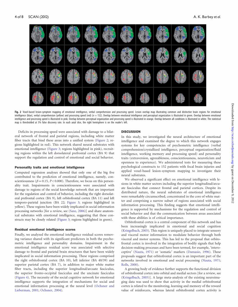
Deficits in processing speed were associated with damage to a bilat-
eral network of frontal and parietal regions, including white matter
fiber tracts that bind these areas into a unified system (Figure 2; re-
gions highlighted in red). This network shared neural substrates with
emotional intelligence (Figure 3; regions highlighted in pink), recruit-
ing regions within the left dorsolateral prefrontal cortex (BA 9) that
support the regulation and control of emotional and social behavior.
Personality traits and emotional intelligence
Computed regression analyses showed that only one of the big five
contributed to the prediction of emotional intelligence, namely, con-
scientiousness (�¼ 0.15, P < 0.000). Therefore, we focus on this person-
ality trait. Impairments in conscientiousness were associated with
damage to regions of the social knowledge network that are important
for the regulation and control of behavior, including the right dorsolat-
eral prefrontal cortex (BA 9), left orbitofrontal cortex (BA 11) and left
temporo-parietal junction (BA 22; Figure 3; regions highlighted in
yellow). These regions have been widely implicated in social information
processing networks [for a review, see (Saxe, 2006)] and share anatom-
ical substrates with emotional intelligence, suggesting that these con-
structs may be closely related (Figure 3; regions highlighted in green).
Residual emotional intelligence scores
Finally, we analyzed the emotional intelligence residual scores remov-
ing variance shared with its significant predictors in both the psycho-
metric intelligence and personality domains. Impairment in the
emotional intelligence residual score was associated with selective
damage to frontal and parietal brain structures that have been widely
implicated in social information processing. These regions comprised
the right orbitofrontal cortex (BA 10), left inferior (BA 40/39) and
superior parietal cortex (BA 7), in addition to major white matter
fiber tracts, including the superior longitudinal/arcuate fasciculus,
the superior fronto-occipital fasciculus and the uncinate fasciculus
(Figure 4). The necessity of the social cognitive network for emotional
intelligence supports the integration of mechanisms for social and
emotional information processing at the neural level (Ochsner and
Lieberman, 2001; Ochsner, 2004).
DISCUSSION
In this study, we investigated the neural architecture of emotional
intelligence and examined the degree to which this network engages
systems for key competencies of psychometric intelligence (verbal
comprehension/crystallized intelligence, perceptual organization/fluid
intelligence, working memory and processing speed) and personality
traits (extraversion, agreeableness, conscientiousness, neuroticism and
openness to experience). We administered tests for measuring these
psychological constructs to 152 patients with focal brain injuries and
applied voxel-based lesion-symptom mapping to investigate their
neural substrates.
We observed a significant effect on emotional intelligence with le-
sions in white matter sectors including the superior longitudinal/arcu-
ate fasciculus that connect frontal and parietal cortices. Despite its
distributed nature, the neural substrates of emotional intelligence
were remarkably circumscribed, concentrated in the core of white mat-
ter and comprising a narrow subset of regions associated with social
information processing. This finding suggests that emotional intelli-
gence is supported by mechanisms for the regulation and control of
social behavior and that the communication between areas associated
with these abilities is of critical importance.
Orbitofrontal cortex is a central component of this network and has
been increasingly implicated in emotional and social cognition
(Kringelbach, 2005). This region is uniquely placed to integrate sensory
and visceral motor information to modulate behavior through both
visceral and motor systems. This has led to the proposal that orbito-
frontal cortex is involved in the integration of bodily signals that help
decision-making processes and have been termed, for example, ‘intero-
ceptive’ (Nauta, 1971) or ‘somatic’ markers (Damasio, 1996). These
proposals suggest that orbitofrontal cortex is an important part of the
networks involved in emotional and social processing (Nauta, 1971;
Rolls, 2000).
A growing body of evidence further supports the functional division
of orbitofrontal cortex into orbital and medial sectors [for a review, see
(Kringelbach, 2005)]. A large meta-analysis of the existing neuroima-
ging data was used to show that activity in the medial orbitofrontal
cortex is related to the monitoring, learning and memory of the reward
value of reinforcers, whereas lateral orbitofrontal cortex activity is
Fig. 2 Voxel-based lesion-symptom mapping of emotional intelligence, verbal comprehension and processing speed. Lesion overlap map illustrating common and distinctive brain regions for emotionalintelligence (blue), verbal comprehension (yellow) and processing speed (red) (n¼ 152). Overlap between emotional intelligence and perceptual organization is illustrated in green. Overlap between emotionalintelligence and processing speed is illustrated in pink. Overlap between perceptual organization and processing speed is illustrated in orange. Overlap between all conditions is illustrated in white. The statisticalmap is thresholded at 5% false discovery rate. In each axial slice, the right hemisphere is on the reader’s left.
4 of 8 SCAN (2012) A.K.Barbey et al.
by guest on Decem
ber 9, 2012http://scan.oxfordjournals.org/
Dow
nloaded from

related to the evaluation of punishers, which can lead to a change in
ongoing behavior (Kringelbach and Rolls, 2004). Further studies have
since confirmed the role of the medial orbitofrontal cortex in moni-
toring affective properties in olfaction (Anderson et al., 2003; Rolls
et al., 2003), gustation (Small et al., 2003) and for somatosensory
(Rolls et al., 2003) and multimodal stimuli (de Araujo et al., 2003).
These findings support the observed role of medial orbitofrontal cortex
in emotional intelligence, indicating that this region is computationally
necessary for monitoring affective properties of social and
environmental stimuli. The involvement of lateral orbitofrontal
cortex in the evaluation of punishers (Kringelbach and Rolls, 2004)
elucidates the role of this region in executive function, suggesting that
it is necessary for evaluative processes in goal-directed behavior and
decision making (Barbey et al., 2009). The findings reported here in-
dicate that the orbitofrontal cortex plays a central role in the coord-
ination of social and affective systems, providing a nexus for sensory
integration and contributing to a fronto-parietal network for
goal-directed behavioral control.
Fig. 4 Voxel-based lesion-symptom mapping of latent (a) and residual (b) emotional intelligence scores (n¼ 152). The statistical map is thresholded at 5% false discovery rate. In each map of the corticalsurface, the left hemisphere is on the reader’s left.
Fig. 3 Voxel-based lesion-symptom mapping of emotional intelligence and conscientiousness. Lesion overlap map illustrating common and distinctive brain regions for emotional intelligence (blue) andconscientiousness (yellow) (n¼ 152). Overlap between emotional intelligence and conscientiousness is illustrated in green. The statistical map is thresholded at 5% false discovery rate. In each axial slice, theright hemisphere is on the reader’s left.
Emotional intelligence SCAN (2012) 5 of 8
by guest on Decem
ber 9, 2012http://scan.oxfordjournals.org/
Dow
nloaded from

The neural system for emotional intelligence shared anatomical sub-
strates with specific facets for psychometric intelligence, engaging peri-
sylvian language areas (Figure 2; highlighted in green) and regions
within the left anterior cingulate cortex (BA 32) and superior longitu-
dinal fasciculus also implicated in processing speed (Figure 2; high-
lighted in pink). These findings are consistent with the observed
pattern of correlations between these factors (Figure 1) and further
suggest that emotional intelligence depends on key competencies for
social information processing and psychometric intelligence. In add-
ition, we found that emotional intelligence engaged brain regions
implicated in conscientiousness, recruiting areas within the left orbito-
frontal cortex (BA 10), anterior insula (BA 13) and inferior temporal
cortex (BA 37) (Figure 3; highlighted in green). This result further
indicates conscientiousness, or the degree of organization, persistence,
control and motivation in goal-directed behavior, is a central feature of
emotional intelligence.
Finally, we investigated the neural systems underlying emotional
intelligence residual scores removing variance shared with its signifi-
cant predictors in both the psychometric intelligence and personality
domains. When compared to the neural system observed for emotional
intelligence at the latent variable level, we see that the residual factor
engages common and distinctive brain regions (Figures 4 and 5).
Common regions include right dorsolateral PFC, left posterior super-
ior temporal sulcus and left temporo-parietal junction (Figure 5; high-
lighted in green), while distinct areas reflect engagement of perisylvian
language areas for emotional intelligence at the latent variable level
(Figure 5; highlighted in blue) and recruitment of the right precentral
gyrus and left superior parietal cortex for the residual emotional intel-
ligence factor (Figure 5; highlighted in yellow).
CONCLUSION
Historically, cognitive and emotional processes have been viewed as
separate constructs. Research in the past two decades, however, has
increasingly shown that such a view may be limited and that, if we are
to understand how complex behaviors are carried out in the brain, an
understanding of their interactions is indispensable. The present study
provides neuropsychological patient data to suggest that emotional
and psychometric intelligence recruit shared neural systems for the
integration of cognitive, social and affective processes. Although
many behaviors might be reasonably well characterized in terms of
cognitive–social interactions such that cognitive and social processes
are partly separable, often true integration of cognitive and social
processes takes place, blurring the distinction between these domains
(Barbey et al., 2009). We propose that one fruitful way to refine our
understanding of their integration will involve a more quantitative
analysis of structural and functional brain connectivity, with particular
emphasis on the involvement of the observed social knowledge
network.
The findings of the present study elucidate the neural foundations of
emotional intelligence and provide evidence for the role of the social
knowledge network in the coordination of cognitive, social, and affec-
tive processes. Damage to this network may produce impairments in
emotional intelligence, which can have an ill effect on one’s ability to
effectively cope with daily demands. In particular, damage to this
system may produce impairments in one’s ability to: (i) be aware of
and express oneself; (ii) function interpersonally; (iii) manage and
control emotions; (iv) generate positive affect required in achieving
personal goals and (v) cope with the immediate situation, make deci-
sions and solve problems of a personal and interpersonal nature.
The data presented in this article are consistent with the view that
behavior is a product of the orchestration of many brain regions and
that the aggregate function of these areas supports emotional and cog-
nitive processes. As the functional neuroanatomy of this network is
further elucidated, we should try to understand the temporal dynamics
that underlie its functions, and the structural and functional changes
that occur during development. On a timescale of milliseconds, neu-
roimaging techniques such as magnetoencephalography (MEG) could
be used to elucidate the precise role of this system in cognitive, social
and emotional processes. The directionality and timing of neural ac-
tivity between regions of the social knowledge network is not clear but
could potentially be addressed with MEG by using sensitive measures
such as Granger Causality (Granger and Hatanaka, 1969).
On a longer, developmental timescale, it would be interesting to
investigate the role of this network in learning and, in particular, in
Fig. 5 Voxel-based lesion-symptom mapping of emotional intelligence (latent) and emotional intelligence (residual). Lesion overlap map illustrating common and distinctive brain regions for emotionalintelligence latent (blue) and emotional intelligence residual (yellow) (n¼ 152). Overlap between these factors is illustrated in green. The statistical map is thresholded at 5% false discovery rate. In each axialslice, the right hemisphere is on the reader’s left.
6 of 8 SCAN (2012) A.K.Barbey et al.
by guest on Decem
ber 9, 2012http://scan.oxfordjournals.org/
Dow
nloaded from

the consolidation of learning. One hypothesis is that the rate of im-
provement in learning depends on emotional and motivational influ-
ences (Cattell and Cattell, 1987) and therefore on changes in functional
activity in structures known to be involved in social and emotional
processes, such as the orbitofrontal cortex. This approach will be par-
ticularly important in understanding goal-directed social behavior and
cases of learning that depend on temporal and occipital brain struc-
tures that are initially set up through top–down interactions with
frontal and parietal regions.
From a clinical perspective, understanding cognitive and emotional
deficits in patients with brain damage may facilitate the design of ap-
propriate assessment tools and rehabilitation strategies, with potential
improvement in patients’ cognitive abilities and daily living. Our find-
ings identify specific tests of the MSCEIT and WAIS that may be tar-
geted in clinical investigations to assess the functioning of the social
knowledge network, particularly, emotional intelligence tests of the
MSCEIT and crystallized intelligence measures of the WAIS. These
findings support predictions about the nature and significance of cog-
nitive impairments that may result from damage to specific brain net-
works (Figure 1).
Many neurological disorders and mental illnesses are characterized
by profound deficits in emotional and cognitive behaviors, including
epilepsy, Alzheimer’s disease, autism and schizophrenia. Outstanding
questions concerning these and many other debilitating conditions
center on advancing our knowledge of how emotional and cognitive
processes interact in both normal and abnormal circumstances.
Understanding the neural mechanisms underlying these conditions
will ultimately require a broader assessment that examines the func-
tional organization of emotional and cognitive systems, and their
interactive role in high-level processes. The reported finding contribute
to this emerging research program by elucidating the role of the social
knowledge network in the coordination of cognitive, social and affect-
ive processes, demonstrating that this system provides an integrative
neural architecture for key competencies of human intelligence.
SUPPLEMENTARY DATA
Supplementary data are available at SCAN online.
Conflict of InterestNone declared.
REFERENCES
Adolphs, R. (2010). Conceptual challenges and directions for social neuroscience. Neuron,
65, 752–67.
Alexander, M.P., Hiltbrunner, B., Fischer, R.S. (1989). Distributed anatomy of transcortical
sensory aphasia. Archives of Neurology, 46, 885–92.
Anderson, A.K., Christoff, K., Stappen, I., et al. (2003). Dissociated neural representations
of intensity and valence in human olfaction. Nature Neuroscience, 6, 196–202.
Arbuckle, J. (2006). Amos (Version 7.0) [Computer Program]. Chicago: SPSS.
Bar-On, R., Brown, J.M., Kirkcaldy, B.D., Thome, E.P. (2000). Emotional expression and
implications for occupational stress; an application of the emotional quotient inventory
(EQ-i). Personality and Individual Differences, 28, 1107–18.
Barbey, A.K., Colom, R., Solomon, J., Krueger, F., Forbes, C., Grafman, J. (2012). An
integrative architecture for general intelligence and executive function revealed by
lesion mapping. Brain, 135, 1154–64.
Barbey, A.K., Koenigs, M., Grafman, J. (2011). Orbitofrontal contributions to human
working memory. Cerebral Cortex, 21, 789–95.
Barbey, A.K., Krueger, F., Grafman, J. (2009). An evolutionarily adaptive neural architec-
ture for social reasoning. Trends in Neurosciences, 32, 603–10.
Barrett, L.F., Mesquita, B., Ochsner, K.N., Gross, J.J. (2007). The experience of emotion.
Annual Review of Psychology, 58, 373–403.
Basso, A., De Renzi, E., Faglioni, P., Scotti, G., Spinnler, H. (1973). Neuropsychological
evidence for the existence of cerebral areas critical to the performance of intelligence
tasks. Brain, 96, 715–28.
Bates, E., Wilson, S.M., Saygin, A.P., et al. (2003). Voxel-based lesion-symptom mapping.
Nature Neuroscience, 6, 448–50.
Bechara, A., Damasio, A.R., Damasio, H., Anderson, S.W. (1994). Insensitivity to future
consequences following damage to human prefrontal cortex. Cognition, 50, 7–15.
Black, F.W. (1976). Cognitive deficits in patients with unilateral war-related frontal lobe
lesions. Journal of Clinical Psychology, 32, 366–72.
Blair, R.J., Cipolotti, L. (2000). Impaired social response reversal. A case of ‘acquired
sociopathy’. Brain, 123(Pt 6), 1122–41.
Blakemore, S.J., Winston, J., Frith, U. (2004). Social cognitive neuroscience: where are we
heading? Trends in Cognitive Sciences, 8, 216–22.
Bugg, J.M., Zook, N.A., DeLosh, E.L., Davalos, D.B., Davis, H.P. (2006). Age differences in
fluid intelligence: contributions of general slowing and frontal decline. Brain and
Cognition, 62, 9–16.
Burgess, P.W., Shallice, T. (1996). Response suppression, initiation and strategy use fol-
lowing frontal lobe lesions. Neuropsychologia, 34, 263–72.
Caplan, D., Hildebrandt, N., Makris, N. (1996). Location of lesions in stroke patients with
deficits in syntactic processing in sentence comprehension. Brain, 119(Pt 3), 933–49.
Caplan, D., Waters, G., Kennedy, D., et al. (2007). A study of syntactic processing in
aphasia II: neurological aspects. Brain and Language, 101, 151–77.
Cattell, R.B., Cattell, R.B. (1987). Intelligence: Its Structure, Growth, and Action. Amsterdam:
Elsevier Science Publications Co.
Costa, P.T. Jr, McCrae, R.R. (1997). Stability and change in personality assessment: the
revised NEO Personality Inventory in the year 2000. Journal of Personality Assessment,
68, 86–94.
Damasio, A.R. (1992). Aphasia. The New England Journal of Medicine, 326, 531–9.
Damasio, A.R. (1996). The somatic marker hypothesis and the possible functions of the
prefrontal cortex. Philosophical transactions of the Royal Society of London Series B,
Biological Sciences, 351, 1413–20.
de Araujo, I.E., Rolls, E.T., Kringelbach, M.L., McGlone, F., Phillips, N. (2003).
Taste-olfactory convergence, and the representation of the pleasantness of flavour, in
the human brain. The European Journal of Neuroscience, 18, 2059–68.
Devinsky, O., Morrell, M.J., Vogt, B.A. (1995). Contributions of anterior cingulate cortex
to behaviour. Brain, 118(Pt 1), 279–306.
Dronkers, N.F., Wilkins, D.P., Van Valin, R.D. Jr, Redfern., B.B., Jaeger, J.J. (2004). Lesion
analysis of the brain areas involved in language comprehension. Cognition, 92, 145–77.
Duncan, J., Burgess, P., Emslie, H. (1995). Fluid intelligence after frontal lobe lesions.
Neuropsychologia, 33, 261–8.
Eslinger, P.J., Damasio, A.R. (1985). Severe disturbance of higher cognition after bilateral
frontal lobe ablation: patient EVR. Neurology, 35, 1731–41.
Glascher, J., Rudrauf, D., Colom, R., et al. (2010). Distributed neural system for general
intelligence revealed by lesion mapping. Proceedings of the National Academy of Sciences
of the United States of America, 107, 4705–9.
Glascher, J., Tranel, D., Paul, L.K., et al. (2009). Lesion mapping of cognitive abilities linked
to intelligence. Neuron, 61, 681–91.
Goleman, D. (1995). Emotional Intelligence. New York: Bantam Books.
Granger, C.W.J., Hatanaka, H. (1969). Analyse Spectrale des Saeries Temporelles en
Aeconomie. Paris: Dunod.
Guimera, R., Amaral, L.A.N. (2005). Functional cartography of complex metabolic net-
works. Nature, 433, 895–900.
Guimera, R., Sales-Pardo, M., Amaral, L.A. (2007). Classes of complex networks defined by
role-to-role connectivity profiles. Nature Physics, 3, 63–9.
Hickok, G., Poeppel, D. (2007). The cortical organization of speech processing. Nature
Reviews Neuroscience, 8, 393–402.
Isingrini, M., Vazou, F. (1997). Relation between fluid intelligence and frontal lobe func-
tioning in older adults. International Journal of Aging & Human Development, 45,
99–109.
Kane, M.J., Engle, R.W. (2002). The role of prefrontal cortex in working-memory capacity,
executive attention, and general fluid intelligence: an individual-differences perspective.
Psychonomic Bulletin & Review, 9, 637–71.
Kertesz, A., Harlock, W., Coates, R. (1979). Computer tomographic localization, lesion
size, and prognosis in aphasia and nonverbal impairment. Brain and Language, 8, 34–50.
Kertesz, A., Lau, W.K., Polk, M. (1993). The structural determinants of recovery in
Wernicke’s aphasia. Brain and Language, 44, 153–64.
Koenigs, M., Acheson, D.J., Barbey, A.K., Solomon, J., Postle, B.R., Grafman, J. (2011).
Areas of left perisylvian cortex mediate auditory-verbal short-term memory.
Neuropsychologia, 49, 3612–9.
Koenigs, M., Barbey, A.K., Postle, B.R., Grafman, J. (2009). Superior parietal cortex is
critical for the manipulation of information in working memory. Journal of
Neuroscience, 29, 14980–6.
Kringelbach, M.L. (2005). The human orbitofrontal cortex: linking reward to hedonic
experience. Nature Reviews Neuroscience, 6, 691–702.
Kringelbach, M.L., Rolls, E.T. (2004). The functional neuroanatomy of the human orbito-
frontal cortex: evidence from neuroimaging and neuropsychology. Progress in
Neurobiology, 72, 341–72.
Lieberman, M.D. (2007). Social cognitive neuroscience: a review of core processes. Annual
Review of Psychology, 58, 259–89.
Makale, M., Solomon, J., Patronas, N.J., Danek, A., Butman, J.A., Grafman, J. (2002).
Quantification of brain lesions using interactive automated software. Behavior
Research Methods Instruments & Computers, 34, 6–18.
Emotional intelligence SCAN (2012) 7 of 8
by guest on Decem
ber 9, 2012http://scan.oxfordjournals.org/
Dow
nloaded from
Mayer, J.D., Salovey, P., Caruso, D.R. (2008). Emotional intelligence: new ability or eclectic
traits? The American Psychologist, 63, 503–17.
Miller, E.K. (2000). The prefrontal cortex and cognitive control. Nature Reviews
Neuroscience, 1, 59–65.
Miller, E.K., Cohen, J.D. (2001). An integrative theory of prefrontal cortex function.
Annual Review of Neuroscience, 24, 167–202.
Miller, E.K., Phelps, E.A. (2010). Current opinion in neurobiology�cognitive neuroscience
2010. Current Opinion in Neurobiology, 20, 141–2.
Naeser, M.A., Alexander, M.P., Helm-Estabrooks, N., Levine, H.L., Laughlin, S.A.,
Geschwind, N. (1982). Aphasia with predominantly subcortical lesion sites: description
of three capsular/putaminal aphasia syndromes. Archives of Neurology, 39, 2–14.
Nauta, W.J. (1971). The problem of the frontal lobe: a reinterpretation. Journal of
Psychiatric Research, 8, 167–87.
Ochsner, K.N. (2004). Current directions in social cognitive neuroscience. Current Opinion
in Neurobiology, 14, 254–8.
Ochsner, K.N., Lieberman, M.D. (2001). The emergence of social cognitive neuroscience.
The American Psychologist, 56, 717–34.
Parkin, A.J., Java, R.I. (1999). Deterioration of frontal lobe function in normal aging:
influences of fluid intelligence versus perceptual speed. Neuropsychology, 13, 539–45.
Passingham, R.E., Stephan, K.E., Kotter, R. (2002). The anatomical basis of functional
localization in the cortex. Nature Reviews Neuroscience, 3, 606–16.
Roca, M., Parr, A., Thompson, R., et al. (2010). Executive function and fluid intelligence
after frontal lobe lesions. Brain, 133, 234–47.
Rolls, E.T. (2000). Precis of the brain and emotion. Behavioral and Brain Sciences, 23,
177–91, discussion 192–233.
Rolls, E.T., O’Doherty, J., Kringelbach, M.L., Francis, S., Bowtell, R., McGlone, F. (2003).
Representations of pleasant and painful touch in the human orbitofrontal and cingulate
cortices. Cerebral Cortex, 13, 308–17.
Rowe, A.D., Bullock, P.R., Polkey, C.E., Morris, R.G. (2001). “Theory of mind” impair-
ments and their relationship to executive functioning following frontal lobe excisions.
Brain, 124, 600–16.
Saxe, R. (2006). Uniquely human social. Cognition, Current Opinion in Neurobiology, 16, 235–9.
Shallice, T., Burgess, P.W. (1991). Deficits in strategy application following frontal lobe
damage in man. Brain, 114(Pt 2), 727–41.
Small, D.M., Gregory, M.D., Mak, Y.E., Gitelman, D., Mesulam, M.M., Parrish, T. (2003).
Dissociation of neural representation of intensity and affective valuation in human
gustation. Neuron, 39, 701–11.
Solomon, J., Raymont, V., Braun, A., Butman, J.A., Grafman, J. (2007). User-friendly
software for the analysis of brain lesions (ABLe). Computer Methods and Programs in
Biomedicine, 86, 245–54.
Sporns, O., Honey, C.J., Kotter, R. (2007). Identification and classification of hubs in brain
networks. PloS One, 2, e1049.
Stein, S., Book, H.E. (2011). The EQ Edge Emotional Intelligence and Your Success 3rd edn.
Mississauga, Ontario: Jossey-Bass, p 1 online resource (xiv, 354 p.) ill.
Stuss, D.T., Gallup, G.G. Jr, Alexander, M.P. (2001). The frontal lobes are necessary for
‘theory of mind’. Brain, 124, 279–86.
Tyler, L.K., Marslen-Wilson, W. (2008). Fronto-temporal brain systems supporting spoken
language comprehension. Philosophical Transactions of the Royal Society of London Series
B, Biological Sciences, 363, 1037–54.
Wechsler, D. (1997). Wechsler adult intelligence test administration and scoring manual.
San Antonio, TX: The Psychology Corporation.
Woods, R.P., Mazziotta, J.C., Cherry, S.R. (1993). MRI-PET registration with automated
algorithm. Journal of Computer Assisted Tomography, 17, 536–46.
8 of 8 SCAN (2012) A.K.Barbey et al.
by guest on Decem
ber 9, 2012http://scan.oxfordjournals.org/
Dow
nloaded from