Embed Size (px)
Citation preview

Cellular Microbiology (2005)
7
(6), 811–823 doi:10.1111/j.1462-5822.2005.00513.x
© 2005 Blackwell Publishing Ltd
Blackwell Science, LtdOxford, UKCMICellular Microbiology 1462-5814Blackwell Publishing Ltd, 20057
6811823
Original Article
Array analysis of oral epithelial cellsM. Handfield et al.
Received 6 December, 2004; revised 10 January, 2005; accepted 13January, 2005. *For correspondence. E-mail [email protected]; Tel. (
+
352) 846 0763; Fax (
+
352) 392 2361.
Distinct transcriptional profiles characterize oral epithelium-microbiota interactions
Martin Handfield,
1
* Jeffrey J. Mans,
1
Gaolin Zheng,
3
M. Cecilia Lopez,
2
Song Mao,
1
Ann Progulske-Fox,
1
Giri Narasimhan,
3
Henry V. Baker
2
and Richard J. Lamont
1
1
Department of Oral Biology, College of Dentistry, University of Florida, Gainesville, FL 32610-0424, USA.
2
Department of Molecular Genetics and Microbiology and Department of Surgery, College of Medicine, University of Florida, Gainesville, FL 32610-0424, USA.
3
BioRG, School of Computer Science, Florida International University, FL 33199, USA.
Summary
Transcriptional profiling, bioinformatics, statisticaland ontology tools were used to uncover and dissectgenes and pathways of human gingival epithelial cellsthat are modulated upon interaction with the peri-odontal pathogens
Actinobacillus actinomycetem-comitans
and
Porphyromonas gingivalis
. Consistentwith their biological and clinical differences, the com-mon core transcriptional response of epithelial cellsto both organisms was very limited, and organism-specific responses predominated. A large number ofdifferentially regulated genes linked to the P53 apop-totic network were found with both organisms, whichwas consistent with the pro-apoptotic phenotypeobserved with
A. actinomycetemcomitans
and anti-apoptotic phenotype of
P. gingivalis.
Furthermore,with
A. actinomycetemcomitans
, the induction ofapoptosis did not appear to be Fas- or TNF
aaaa
-mediated. Linkage of specific bacterial componentsto host pathways and networks provided additionalinsight into the pathogenic process. Comparison ofthe transcriptional responses of epithelial cells chal-lenged with parental
P. gingivalis
or with a mutant of
P. gingivalis
deficient in production of major fimbriae,which are required for optimal invasion, showedmajor expression differences that reverberatedthroughout the host cell transcriptome. In contrast,gene ORF859 in
A. actinomycetemcomitans,
whichmay play a role in intracellular homeostasis, had a
more subtle effect on the transcriptome. These stud-ies help unravel the complex and dynamic interac-tions between host epithelial cells and endogenousbacteria that can cause opportunistic infections.
Introduction
The human microbiota comprises a complex ecosystemcharacterized by the simultaneous presence of a largenumber of ‘normal’ colonizers, associated with health andthriving in a dynamic environment. Because health is themost common state of a host, it has been speculated thatthe autochthonous flora has coevolved with its host tointeract in a balanced state that is beneficial to both thehost and the microbiota (Galan and Zhou, 2000). Thereare an appreciable number of benefits to the host that theindigenous microbiota are thought to provide, includingthe synthesis of vitamins (B complex and K), the preven-tion of infection by pathogens (by direct competition forniches or by immune cross-reactivity), and impacting thenormal development of the immune system (Hooper andGordon, 2001). Furthermore, there is an increasing real-ization that complex societies of indigenous microbes caninfluence human physiology and development. For exam-ple, in the GI tract the Gram-negative anaerobe
Bacteroi-des thetaiotaomicron
can modulate expression of ilealepithelia cell genes involved in nutrient adsorption,mucosal barrier fortification, xenobiotic metabolism,angiogenesis, and maturation (Hooper
et al
., 2001).Because host and microbiota interactions are dynamic,disease may arise at the mucosal surface of a susceptiblehost when a perturbation occurs in the epithelial environ-ment, for example, when the host becomes immunocom-promised, or as a result of the unintended (in anevolutionary sense) consequences of bacterial activity(Galan and Zhou, 2000).
In the oral cavity, periodontal infections that affect andultimately destroy the tissues supporting the teeth areamong the most common diseases of humans. Accordingto the 2000 Surgeons General’s Report on Oral Health(http://www.nidcr.nih.gov/AboutNIDCR/SurgeonGeneral/),these conditions afflict 14% of adults aged 45–54 yearsand 23% of those aged 65–74 years. Furthermore, anepidemiological association is emerging between peri-odontal infections and serious systemic conditions suchas coronary artery disease and preterm delivery of low

812
M. Handfield
et al.
© 2005 Blackwell Publishing Ltd,
Cellular Microbiology
,
7
, 811–823
birth weight infants (Scannapieco and Genco, 1999). Theaetiology of oral infectious diseases is complex andinvolves consortia of bacteria thriving in biofilms andexploiting immunological susceptibilities in the host.Despite the multifactorial nature of these diseases, thereis a consistent relationship between the Gram-negativecapnophile
Actinobacillus actinomycetemcomitans
andlocalized aggressive periodontitis (LAP) (Slots and Genco,1984; Zambon, 1985; Offenbacher, 1996; Meyer andFives-Taylor, 1997; Haffajee and Socransky, 1999), andbetween the Gram-negative anaerobe
Porphyromonasgingivalis
and severe, chronic manifestations of the dis-ease (Slots and Genco, 1984; Slots
et al
., 1986; Haffajeeand Socransky, 1999).
The initial interface between the host and potentiallyperiodontopathic organisms, such as
P. gingivalis
and
A.actinomycetemcomitans
, is the epithelial layer that linesthe subgingival crevice. Epithelial cells are both a physicalbarrier to infection and a component of a network thatefficiently signals microbial intrusion to the immune cellsto insure effective mobilization of the innate and specificdefence mechanisms (Kagnoff and Eckmann, 1997). Both
A. actinomycetemcomitans
and
P. gingivalis
are capableof invading gingival epithelial cells (GEC) and can remainviable intracellularly. Furthermore, epithelial cells maintainviability following intracellular penetration by either
P. gin-givalis
or
A. actinomycetemcomitans
(Kato
et al
., 2000;Nakhjiri
et al
., 2001; Takayama
et al
., 2003). However, theentry mechanisms employed by these invasive organismsare distinct.
Actinobacillus actinomycetemcomitans
entersepithelial cells through a dynamic multistep processwhereupon the organisms are first constrained in an intra-cellular vacuole from which they subsequently escape andspread cell-to-cell with the aid of microtubules (Meyer
et al
., 1996; 1999). Within epithelial cells
A. actinomycet-emcomitans
upregulates a distinct set of genes that facil-itate adaptation to the intracellular environment (Cao
et al
., 2004; Richardson
et al
., 2005). Among these genesis ORF859 encoding a conserved protein of unknownfunction. In the case of
P. gingivalis,
the major fimbriae(comprised of the FimA protein) bind to integrins on thesurfaces of GEC and stimulate integrin-dependent signal-ling to effect invasion through both microfilament andmicrotubule remodelling (Yilmaz
et al
., 2002; 2003).
Por-phyromonas gingivalis
also impacts the MAP-kinase path-way and causes transient increases in intracellular Ca
2
+
concentrations (Watanabe
et al
., 2001; Belton
et al
.,2004), Both of these signal transduction pathways canconverge on nuclear transcription factors and modulategene expression. Indeed,
P. gingivalis
has been shown toaffect expression of individual genes in epithelial cellsincluding those encoding IL-8 and Bcl-2 (Darveau
et al
.,1998; Nakhjiri
et al
., 2001).Transcriptional profiling using microarrays provides a
means to monitor epithelial cell responses to invadingmicroorganisms on a global scale (Cummings and Rel-man, 2000; Kellam, 2000; 2001; Ichikawa
et al
., 2000;Manger and Relman, 2000; Kagnoff and Eckmann, 2001;Kato-Maeda
et al
., 2001; Yowe
et al
., 2001; Lory andIchikawa, 2002; Sepulveda
et al
., 2002). Results fromsuch studies suggest that the encounter between hostand microbiota may involve a finely tuned set of interac-tions whereby both cell types adapt and coexist with eachother. Consequently, the regulation of normal processessuch as cell division or apoptosis may be key to maintain-ing a balanced long-standing intracellular state wherebyboth cell types inflict a minimal degree of harm on eachother. In support of this concept, epithelial cells recoveredfrom the oral cavity show high levels of intracellular
P.gingivalis
and
A. actinomycetemcomitans
(Christersson,1987a, b; 1993; Rudney
et al
. 2001). Hence an intracel-lular location may be a natural component of the lifestyleof these oral organisms. In this study, we have utilizedhuman microarrays to determine the transcriptionalresponse of human immortalized gingival keratinocytes(HIGK) to co-culture with
P. gingivalis
or
A. actinomycet-emcomitans
. Moreover, we have extended these studiesto investigate the transcriptional responses of epithelialcells that are manipulated by the major fimbriae (FimA) of
P. gingivalis
and the intracellulary upregulated ORF859 of
A. actinomycetemcomitans
.
Results and discussion
General considerations
To investigate early events in oral infection by
P. gingivalis
and
A. actinomycetemcomitans
we analysed differentialgene expression in HIGK using the Affymetrix HG U133-A oligonucleotide arrays that contain over 22 000 differentprobe sets. In addition, isogenic mutant strains were uti-lized to assess the roles of specific bacterial proteins inmodulation of the host cell transcriptome. Host cell apop-tosis, a major pathway impacted by
P. gingivalis
and
A.actinomycetemcomitans
, was validated by phenotypicassays.
Association of
A. actinomycetemcomitans
and
P. gingivalis
with epithelial cells
Porphyromonas gingivalis
and
A. actinomycetemcomi-tans
demonstrate differing efficiencies of binding to, andinternalization within, human immortalized gingival kerat-inocytes. In order to compare epithelial cell transcrip-tional profiles in response to an equivalent challenge ofthe two organisms, we first compared adhesion and inva-sion at multiplicity of infections (MOIs) predicted to resultin the same number of epithelial cell-associated bacteria

Array analysis of oral epithelial cells
813
© 2005 Blackwell Publishing Ltd,
Cellular Microbiology
,
7
, 811–823
for each species. As shown in Table 1, at an MOI of 100for
P. gingivalis
and 3000 for
A. actinomycetemcomitans
,the numbers of bacteria associated with the epithelialcells were of the same order of magnitude. These MOIswere then used in subsequent experiments. In contrast,the levels of invasion were significantly different;
P. gingi-valis
being a considerably more efficient invasive micro-organism as compared to
A. actinomycetemcomitans
.Notably, HIGK cells behaved similarly to primary GEC inco-cultures with
A. actinomycetemcomitans
and
P. gingi-valis
, with regard to both adhesion and invasion. Furtherconfirmation of the relevance of the HIGK cell model wasprovided by the finding that the gene for IL-1 beta wasupregulated in HIGK cells in co-cultures with both perio-pathogens (not shown). This is consistent with reportsdocumenting increased expression of this pro-inflamma-tory cytokine in primary GEC (Sandros
et al
., 2000;Sfakianakis
et al
., 2001).
Gene expression in GEC regulated by
A. actinomycetemcomitans
and
P. gingivalis
To characterize epithelial cell responses to
A. actino-mycetemcomitans
and
P. gingivalis,
and to assess theextent to which host responses may depend on the chal-lenging organism, we used human microarrays to moni-tor relative abundance of HIGK cell transcripts followingco-culture with
A. actinomycetemcomitans
or
P. gingiva-lis
. Array-to-array comparisons were carried out usingunsupervised and supervised methods to assess therelatedness of the specimens (arrays) under investiga-tion using the Cluster and TreeView Software (Eisen
et al
., 1998). The significance level used in identifyinggenes that were differentially expressed was
P
<
0.001.Hierarchical clustering was first used to perform anunsupervised analysis. Visual representation of theunsupervised cluster analysis of
P. gingivalis
-infected,
A.actinomycetemcomitans
-infected, and uninfected cellswas performed using TreeView software. The resultingdendrogram revealed that the array chips from eachinfection state clustered together (not shown). Thus,each infection state elicited a specific and distincttranscriptome in HIGK cells. This was also an indication
of the quality and consistency of the hybridizationprocedure.
Supervised analyses were next performed to identifygene expression differences between the
P. gingivalis
-infected or
A. actinomycetemcomitans
-infected as com-pared to uninfected HIGK cells, at a significance level of
P
<
0.001. To test the predictive validity of the probe setsidentified at this level of significance, a ‘leave-one-out’cross-validation was performed with four different predic-tion models (linear discriminant, 1KNN, 3KNN and near-est centroid). This validation step addressed the ability ofprobe sets to distinguish between the different classes(i.e. infection states). Briefly, this analysis determined ifthe classifier performed better than one would expect bychance alone. In the present study there were threeclasses; on average one would expect to correctly classifythe arrays by chance alone 33% of the time. Using thegene expression classifier, the arrays were correctly clas-sified 92% of the time. Thus, the gene expression differ-ences significant at
P
<
0.001 can be used to distinguishbetween the strains and their miss-classification rate of8% is much lower than the miss-classification rate of 67%expected by chance. Figure 1 shows the K-means clus-tering patterns of probe sets where the expression pat-terns were significantly different between the treatmentclasses. Several interesting clusters of genes are high-lighted by blocks one through six in Fig. 1. For instance,block 1 (182 probe sets) represents genes that aredownregulated in
A. actinomycetemcomitans
-infectedcells, but not modulated in
P. gingivalis
-infected orcontrol uninfected cells. Conversely, block 5 (252 probesets) represents genes that are upregulated in
A.actinomycetemcomitans
-infected cells, but not modulatedin
P. gingivalis
-infected or control uninfected cells. Thesetwo clusters of genes may be characteristic of cellularinteractions specifically associated with
A. actinomycet-emcomitans
. Similarly,
P. gingivalis
elicited a transcrip-tional response in HIGK cells that is specific to thisorganism (block 2, upregulated; and block 4, down-regulated). Overall, transcriptional response appeared tobe diametrically opposed between the two organisms withonly a small number of genes (41 probe sets) up- ordownregulated by both species (blocks 3 and 6). These
Table 1.
Microbial-epithelial cell interaction characteristics with human primary (GEC) and transformed (HIGK) gingival cells.
Epithelial cells Microorganism MOI
a
Total interaction
b
(cfu/cell) % of Interacting bacteria that invade
c
GEC A. actinomycetemcomitans 3000:1 40 ± 4 <0.02HIGK A. actinomycetemcomitans 3000:1 35 ± 25 <0.05GEC P. gingivalis 100:1 14 ± 8 25HIGK P. gingivalis 100:1 18 ± 4 24
a. Multiplicity of infection (bacteria: epithelial cell).b. Total count of adhering and invading organisms after co-culture and cell lysis at 60 min. Data are reported as mean value from two independentassays in triplicate ± the standard deviation.c. Calculated from intracellular cfu counts after antibiotic treatment.

814 M. Handfield et al.
© 2005 Blackwell Publishing Ltd, Cellular Microbiology, 7, 811–823
common genes may be involved in a general host cellresponse to infection that may be universal for oral Gram-negative organisms or possibly even for bacterial stimula-tion in general. However, possibly more importantly, thedata also suggest that individual organisms may haveevolved to modulate a finite number of pathways that arecharacteristics of the genus. Moreover, host cells appearto be able to distinguish between infecting organisms andtailor transcriptional responses accordingly.
Ontology analysis
In order to mine the array data for biologically relevantinformation, an ontology analysis based on relatedness toknown metabolic pathways was performed. The ontologyanalysis was performed at P < 0.005 (Table 2) against the354 different biological processes that have been identi-fied thus far in the human GO syntax ontology database(http://obo.sourceforge.net/). Sixteen gene ontology path-ways, including molecular functions, cellular components,and biological processes were identified as representingthe canonical response to both organisms. Moreover, 21additional gene ontology pathways were specifically foundamong the genes that responded to exposure to P. gingi-valis. Similarly, a specific response for A. actinomycetem-comitans-infected HIGK cells resulted in modulation of 49pathways. Those pathways with relevance to documentedhost-pathogen interactions are presented in Table 2.
Of particular interest to host-pathogen interactions inthe oral cavity, one of the over-represented pathways wasthe apoptosis pathway. Porphyromonas gingivalis and A.actinomycetemcomitans have been shown to alter cytok-ine expression and modulate apoptosis in various celltypes. The induction of apoptosis in immune cells of theoral cavity is thought to have a significant immunomodu-latory (immunosuppressive) effect and contribute to thepathogenesis of A. actinomycetemcomitans in periodontaldiseases (Lally et al., 1989a,b; Ebersole et al., 1990;Spitznagel et al., 1995; Korostoff et al., 1998; Kato et al.,2000; Demuth et al., 2003). Furthermore, A. actinomyce-temcomitans can also induce apoptosis in a leukotoxin-independent manner in oral epithelial cells, periodontalligament cells, and gingival fibroblasts (Kato et al., 2000;Belibasakis et al., 2002; Teng and Hu, 2003). A recentreport suggests that the effector molecule associated withA. actinomycetemcomitans apoptosis in human GEC is aCagE homologue, which encodes a component of a puta-tive type IV secretion system (Teng and Hu, 2003). Por-phyromonas gingivalis, by contrast, suppresses apoptosisin primary cultures of GEC. Porphyromonas gingivalis-induced suppression of apoptosis is correlated with acti-vation of Bcl-2 at the transcriptional level and inhibition ofcytochrome c release from the mitochondria (Nakhjiriet al., 2001; Yilmaz et al., 2004).
Fig. 1. Different patterns of gene expression of oral epithelial HIGK cells upon co-culture with A. actinomycetemcomitans or P. gingivalis. Hierarchical clustering of variance-normalized gene expression data from uninfected human HIGK cells and from cells in co-culture with either organism for 2 h before RNA isolation and purification. Expres-sion and variation filters were applied to the data set before clustering. Probe sets giving hybridization signal intensity at or below back-ground levels on all arrays tested were eliminated from further anal-ysis. The resulting data set was culled by ranking on the coefficient of variation and eliminating the bottom half of the data set to remove probe sets whose expression did not vary between the treatment regimens. The gene expression observations were variance normal-ized to a mean of 0 and a standard deviation of 1, and this normalized data set was subjected to hierarchical cluster analysis with average linkage clustering of the nodes. The variation in gene expression for a given gene is expressed as distance from the mean observation for that gene. Each expression data point represents the ratio of the fluorescence intensity of the cRNA from A. actinomycetemcomitans-infected (columns Aa VT 1169) or P. gingivalis-infected HIGK cells (columns Pg 33277) to the fluorescence intensity of the cRNA from mock-infected HIGK cells (columns control R1–R5). The scale adja-cent to the dendrogram relates to Pearson’s correlation coefficient. Highlighted blocks are described in the text.
0
-1.0
1.0
1
2
43
5
6
Aa VT1169
Aa VT1169
Aa VT1169
Aa VT1169
Pg 33277
Pg 33277
Pg 33277
Pg 33277
CTRL R8
CTRL R2
CTRL R1
CTRL R4
CTRL R3
<–2.0 >2.0–1.0 1.00
S.D. From Mean

Array analysis of oral epithelial cells 815
© 2005 Blackwell Publishing Ltd, Cellular Microbiology, 7, 811–823
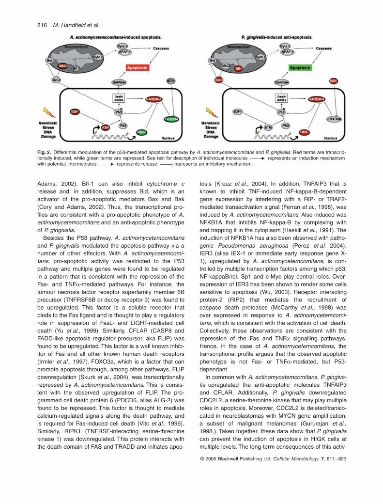
The ontology analysis presented in Table 2a revealedthat a total of 55 distinct apoptosis-associated geneswere modulated upon A. actinomycetemcomitans or P.gingivalis co-cultures of HIGK cells at a significance ofP < 0.005. Of these, eight were modulated in both organ-isms, 31 were modulated only in A. actinomycetemcomi-tans, and 17 were differentially transcribed in P. gingivalisonly. Interestingly, a large number of differentially regu-lated genes linked to the P53 network were found in bothorganisms. The P53 protein is a tumour suppressor genethat is positioned at a major node of a network that isinvolved in cell division and apoptosis. There are threemajor types of stress that modulate P53: aberrant growthsignals, DNA damage and physicochemical stress. Theapoptosis induction by P53 can be mediated either bystimulation of Bax and Fas antigen expression, or byrepression of Bcl-2 expression. A summary of the majorapoptotic effector molecules impacted by the organisms
is presented in Fig. 2. Actinobacillus actinomycetemcom-itans activated the pro-apoptotic molecules BBC3,GADD45A, E2F1 and ATM and repressed cMYC. Por-phyromonas gingivalis activated cMYC and SGK, both ofwhich are anti-apoptotic and play a role in cell survivaland proliferation. cMYC can repress transcription of thepro-apoptotic GADD45A, while SGK phosphorylates andnegatively regulates the transcription factor FOXO3A thatcan participate in apoptosis, in part through theGADD45a protein (Brunet et al., 2001; Tran et al., 2002;Barsyte-Lovejoy et al., 2004). SGKs are related to Akt, aserine/threonine kinase that plays a crucial role in pro-moting cell survival and has been shown to be activatedby P. gingivalis in primary GEC (Yilmaz et al., 2004).Most of the activity of P. gingivalis, however, revolvedaround the mitochondrial pathway, with upregulation ofBcl-2 and Bfl1. Bcl-2 inhibits release of cytochrome cfrom the mitochondria and can inhibit P53 (Cory and
Table 2. Gene ontologya analysis of the transcriptome of HIGK cells infected with P. gingivalis or A. actinomycetemcomitans.
(a) Pathways common to P. gingivalis- and A. actinomycetemcomitans-infected HIGK cells.
(b) Pathways specific to P. gingivalis-infected HIGK cells.
(c) Pathways specific to A. actinomycetemcomitans-infected HIGK cells.
GO ID TermP. gingivalis-infected(total change)b P-value
A. actinomycetemcomitans-infected (total change)b P-value
45449 Regulation of transcription 49 0.0017 118 016021 Integral to membrane 39 0.0025 96 05215 Transporter activity 17 0.0034 55 0.0036
12501 Programmed cell death 25 0 39 0.003117017 MAP kinase phosphatase activity 4 0.0001 6 0
GO ID Term Change P-value
7275 Development 64 0.00319653 Morphogenesis 47 0.00448283 Cell proliferation 42 0.00099888 Histogenesis 10 0.00048544 Epidermal differentiation 7 0.0004
16265 Death 25 0.000174 Regulation of cell cycle 22 0.0009
5125 Cytokine activity 12 0.002945073 Regulation of chemokine biosynthesis 2 0.00348138 Protein tyrosine/serine/threonine phosphatase activity 4 0.00485149 Interleukin-1 receptor binding 3 0.001
GO ID Term Change P-value
8152 Metabolism 330 0.000530528 Transcription regulator activity 93 04888 Transmembrane receptor activity 26 0.0049581 Detection of external stimulus 6 0.00064930 G-protein coupled receptor activity 5 0
15268 Alpha-type channel activity 5 0.00025216 Ion channel activity 4 0.00025261 Cation channel activity 3 0.0025
43066 Negative regulation of apoptosis 14 0.00183773 Heat shock protein activity 8 0.0015
16337 Cell–cell adhesion 3 0.00363786 Actin lateral binding 2 0.0036
a. Only pathways with documented relevance to host-pathogen interactions are presented.b. The total change represents the total number of under- and over-represented genes in a particular pathway.
816 M. Handfield et al.
© 2005 Blackwell Publishing Ltd, Cellular Microbiology, 7, 811–823
Adams, 2002). Bfl-1 can also inhibit cytochrome crelease and, in addition, suppresses Bid, which is anactivator of the pro-apoptotic mediators Bax and Bak(Cory and Adams, 2002). Thus, the transcriptional pro-files are consistent with a pro-apoptotic phenotype of A.actinomycetemcomitans and an anti-apoptotic phenotypeof P. gingivalis.
Besides the P53 pathway, A. actinomycetemcomitansand P. gingivalis modulated the apoptosis pathway via anumber of other effectors. With A. actinomycetemcomi-tans, pro-apoptotic activity was restricted to the P53pathway and multiple genes were found to be regulatedin a pattern that is consistent with the repression of theFas- and TNFa-mediated pathways. For instance, thetumour necrosis factor receptor superfamily member 6Bprecursor (TNFRSF6B or decoy receptor 3) was found tobe upregulated. This factor is a soluble receptor thatbinds to the Fas ligand and is thought to play a regulatoryrole in suppression of FasL- and LIGHT-mediated celldeath (Yu et al., 1999). Similarly, CFLAR (CASP8 andFADD-like apoptosis regulator precursor, aka FLIP) wasfound to be upregulated. This factor is a well known inhib-itor of Fas and all other known human death receptors(Irmler et al., 1997). FOXO3a, which is a factor that canpromote apoptosis through, among other pathways, FLIPdownregulation (Skurk et al., 2004), was transcriptionallyrepressed by A. actinomycetemcomitans. This is consis-tent with the observed upregulation of FLIP. The pro-grammed cell death protein 6 (PDCD6, alias ALG-2) wasfound to be repressed. This factor is thought to mediatecalcium-regulated signals along the death pathway, andis required for Fas-induced cell death (Vito et al., 1996).Similarly, RIPK1 (TNFRSF-interacting serine-threoninekinase 1) was downregulated. This protein interacts withthe death domain of FAS and TRADD and initiates apop-
tosis (Kreuz et al., 2004). In addition, TNFAIP3 that isknown to inhibit TNF-induced NF-kappa-B-dependentgene expression by interfering with a RIP- or TRAF2-mediated transactivation signal (Ferran et al., 1998), wasinduced by A. actinomycetemcomitans. Also induced wasNFKB1A that inhibits NF-kappa-B by complexing withand trapping it in the cytoplasm (Haskill et al., 1991). Theinduction of NFKB1A has also been observed with patho-genic Pseudomonas aeruginosa (Perez et al. 2004).IER3 (alias IEX-1 or immediate early response gene X-1), upregulated by A. actinomycetemcomitans, is con-trolled by multiple transcription factors among which p53,NF-kappaB/rel, Sp1 and c-Myc play central roles. Over-expression of IER3 has been shown to render some cellssensitive to apoptosis (Wu, 2003). Receptor interactingprotein-2 (RIP2) that mediates the recruitment ofcaspase death proteases (McCarthy et al., 1998) wasover expressed in response to A. actinomycetemcomi-tans, which is consistent with the activation of cell death.Collectively, these observations are consistent with therepression of the Fas and TNFa signalling pathways.Hence, in the case of A. actinomycetemcomitans, thetranscriptional profile argues that the observed apoptoticphenotype is not Fas- or TNFa-mediated, but P53-dependant.
In common with A. actinomycetemcomitans, P. gingiva-lis upregulated the anti-apoptotic molecules TNFAIP3and CFLAR. Additionally, P. gingivalis downregulatedCDC2L2, a serine-theronine kinase that may play multipleroles in apoptosis. Moreover, CDC2L2 is deleted/translo-cated in neuroblastomas with MYCN gene amplification,a subset of malignant melanomas (Gururajan et al.,1998.). Taken together, these data show that P. gingivaliscan prevent the induction of apoptosis in HIGK cells atmultiple levels. The long-term consequences of this activ-
Fig. 2. Differential modulation of the p53-mediated apoptosis pathway by A. actinomycetemcomitans and P. gingivalis. Red terms are transcrip-tionally induced, while green terms are repressed. See text for description of individual molecules. represents an induction mechanism with potential intermediates; represents release; represents an inhibitory mechanism.

Array analysis of oral epithelial cells 817
© 2005 Blackwell Publishing Ltd, Cellular Microbiology, 7, 811–823
ity for normal physiologic function of epithelial cells remainto be established.
Apoptosis in GEC modulated by A. actinomycetemcomitans or P. gingivalis
The apoptotic responses of HIGK cells at the transcrip-tional level revealed by array analyses were verified by aphenotypic assay for apoptosis. As shown in Fig 3, A.actinomycetemcomitans-induced apoptosis in HIGK cellswhereas P. gingivalis did not stimulate apoptotic activity.Furthermore, P. gingivalis cells were capable of inhibitingcamptothecin-induced apoptosis in HIGK cells. Theseresults both corroborate other reports in the literature withdifferent epithelial cells (Kato et al., 2000; Nakhjiri et al.,2001; Teng and Hu, 2003; Yilmaz et al., 2004) and showthat the mRNA expression levels correlate with pheno-typic properties, at least with regard to some genesinvolved with apoptosis.
Gene expression in response to isogenic mutants
A bacterial mutant analysis was combined with host tran-scriptional profiling to assess the role of specific bacterialproducts or phenotypes on the epithelial cell geneexpression programs. Actinobacillus actinomycetemcom-itans ORF859 (PEDANT database) was initially found tobe induced in vivo in infected humans using IVIAT (Hand-field et al., 2000). The product of this gene was furthershown to be induced in plaque from infected patients(Handfield et al., 2000; 2002) and in various cell lines,including HIGK cells (Richardson et al., 2005), and is a
potential marker for active disease in LAP patients (Caoet al., 2004). A bioinformatic analysis of this gene productdid not reveal a predicted function, although the geneproduct is highly conserved across genera (Cao et al.,2004). As shown in Fig. 4A, a supervised hierarchicalclustering analysis showed that several genes were dif-ferentially regulated by the ORF859 mutant strain JMS04in comparison to the parental strain. The ontology analy-sis presented in Table 3 further revealed that the mostsignificant and numerous variations (P < 0.001) wereassociated with intermediate metabolism functions, sig-nal transduction and cytokine activity. Interestingly, IL-27was found to be induced by wild-type A. actinomycetem-comitans, but not by the JMS04 mutant (not shown).IL-27 is closely related to IL-12 in both sequence andstructure (Artis et al., 2004), and has been shown topromote Th1 cell-mediated immune responses (Hunteret al., 2004). Together, this suggests that the product ofORF859 may be related to the intracellular adaptationand homeostasis of A. actinomycetemcomitans, a pro-cess that does not impact large numbers of host cellpathways. However, upregulation of IL-27 will stimulatehost cell-mediated immunity and hence the ORF859gene product may contribute to the inflammatory proper-ties of the organism.
The P. gingivalis mutant (YPF1) tested is deficient inproduction of the major fimbrial protein, FimA, a multifunc-tional adhesin (Lamont and Jenkinson, 1998). FimA medi-ates attachment of P. gingivalis to GEC through engagingan integrin receptor on the host cell surface (Yilmaz et al.,2002). Fimbrial-integrin interaction results in assembly ofintegrin focal adhesion complexes, and the initiation ofsignalling pathways that induce remodelling of cytoskele-tal architecture that allows entry of the organism (Yilmazet al., 2003). The YPF1 mutant is thus significantlyimpaired in invasion and in cytoskeletal remodelling activ-ity (Yilmaz et al., 2002; 2003). In contrast to the profilesobtained with the A. actinomycetemcomitans mutant, theP. gingivalis mutant strain YPF1 had a transcriptional pat-tern strikingly divergent from the parental strain. As shownin Fig. 4B and Table 3, and consistent with the phenotypicproperties of the mutant, a large proportion of genesrelated to the cytoskeleton and to membrane and receptoractivity were underrepresented in the transcriptional pro-file of YPF1-infected cells. For example, YPF1 failed toupregulate: actin binding LIM protein 1, which may play ageneral role in bridging the actin-based cytoskeleton withan array of potential LIM protein-binding partners; filaminB, beta (actin binding protein 278) which connects cellmembrane constituents to the actin cytoskeleton; andcoronin 2A another actin binding protein (Roof et al.,1997; de Hostos, 1999; Feng and Walsh, 2004). Addition-ally, YPF1 did not upregulate beta 3, 4 and 6 integrin,along with alpha V, 3 and 4 integrin, and CD47, an
Fig. 3. Apoptotic responses of HIGK cells to A. actinomycetemcom-itans or P. gingivalis by ELISA of cytoplasmic histone-associated DNA fragments. Control (C) represents HIGK under normal culture condi-tions. Actinobacillus actinomycetemcomitans (Aa) was incubated with HIGK cells at an MOI of 3000 for 4 h. Porphyromonas gingivalis parental (Pg) or mutant (YPF1) strains were incubated with HIGK at an MOI 100 for 20 h. Camptothecin (CAM) was incubated with HIGK for 4 h. For inhibition of camptothecin-induced apoptosis, HIGK cells were incubated with P. gingivalis strains for 16 h followed by camp-tothecin for 4 h. Error bars represent standard deviation, n = 3. The asterisk denotes statistically different from control P < 0.005.
0
0.2
0.4
0.6
0.8
1
1.2
1.4
1.6
1.8
Cyt
opla
smic
His
tone
-ass
ocia
ted
DN
A fr
agm
ents
(O
D40
5)
*
*
C CAM Aa Pg YPF1 Pg YPF1+
CAM

818 M. Handfield et al.
© 2005 Blackwell Publishing Ltd, Cellular Microbiology, 7, 811–823
integrin-associated signal transducer. YPF1 also demon-strated a significant inability to impinge on cell cycle andcell proliferation pathways indicating that a successfulinvasion event may be necessary for P. gingivalis tomanipulate these pathways. The apoptosis ontology path-way was not differentially influenced by YPF1, indicating
that the fimbriae deficient mutant strain should be capableof inhibiting apoptosis in HIGK cells to the same extent asthe parental strain. This was confirmed by the phenotypicapoptosis assay (Fig. 3) that showed YPF1 could antago-nize chemically induced apoptosis to the same extent asthe parental strain.
Fig. 4. Comparison of transcriptional profile of HIGK cells following co-culture with isogenic A. actinomycetemcomitans (A) or P. gingivalis (B) mutant strains. Hierarchical clustering of variance-normalized gene expression data. The expression pattern of the cRNAs analysed by microarray is represented as a supervised K-means analysis of the variance-normalized data set of differentially expressed genes with the algorithm Cluster and displayed with TreeView. Each row represents an individual cRNA element spotted on the array, and each column represents the expression states of cRNAs for the challenge condition indicated. Each expression data point represents the ratio of the fluorescence intensity of the cRNA from A. actinomycetemcomitans-infected [columns Aa VT1169 (parent) and Aa JMSO4 (mutant)]; or P. gingivalis-infected cells [columns Pg 33277 (parent) and Pg YPF1 (mutant)] to the fluorescence intensity of the cRNA from mock-treated uninfected cells (columns CTRL). The cluster is subdivided into three groups consisting of genes that were repressed (green), genes that were induced (red), and genes whose expression did not change (black). Leave-one-out cross validation studies were used to establish the ability of probe sets significant at the P < 0.001 level of significance to predict the class label of the specimen left out of the analysis.
A B
CTRL R8
CTRL R9
CTRL R4
CTRL R3
CTRL R2
CTRL R1
AaVT1169
AaVT1169
AaVT1169
AaVT1169
AaJMSO4
AaJMSO4
AaJMSO4
AaJMSO4
CTRL R3
CTRL R2
CTRL R4
CTRL R1
Pg33277
Pg33277
Pg33277
Pg33277
PgYPF1
PgYPF1
PgYPF1
PgYPF1
CTRL R7
<–2.0 >2.0–1.0 1.00
S.D. From Mean

Array analysis of oral epithelial cells 819
© 2005 Blackwell Publishing Ltd, Cellular Microbiology, 7, 811–823
Conclusions
The transcriptional profiling presented herein begins toprovide insights into both the intricate biological phenom-ena occurring during host-pathogen interactions and thedistinct pathophysiology of A. actinomycetemcomitansand P. gingivalis. A characteristic clinical outcome is asso-ciated with infection with either organism. Actinobacillusactinomycetemcomitans-associated disease involves anacute tissue destruction in the absence of overt inflamma-tion, whereas P. gingivalis-associated disease is chronicand involves inflammatory tissue destruction. Moreover,the mechanism of intracellular invasion of both organismsis distinct. Consistent with these biological and clinicaldifferences, the common core transcriptional response ofepithelial cells to these organisms is very limited, andorganism-specific responses predominate. Interestingly,this contrasts with disease models in other cell types. Forexample, infection of dendritic cells with Escherichia coli,Candida albicans, or the influenza virus resulted in asubstantial shared core response along with a pathogen-specific pattern of gene expression (Huang et al., 2001).Thus oral epithelial cells, that encounter an array ofmicrobes with varying degrees of pathogenicity, maydirect a measured response that is tailored to thepathogenic potential of the infecting organism. Theseresponses can then influence disease progression. Forexample induction of apoptosis in epithelial cells by A.actinomycetemcomitans could contribute to immunologi-cally silent tissue destruction. Inhibition of apoptosis byintracellular P. gingivalis, in contrast, could contribute tobacterial persistence and chronic, slowly progressing tis-sue destruction. Linkage of specific bacterial components
to host pathways and networks provides additional insightinto the pathogenic process. The loss of P. gingivalis fim-briae retards adherence and invasion and the conse-quences of this reverberate throughout the transcriptome.Genes such as ORF859 in A. actinomycetemcomitansthat appear to be involved in intracellular homeostasishave a more subtle effect on the transcriptome. Suchpatterns of gene expression changes in response toisogenic mutants may provide a means to evaluate thebiological function of as yet undefined bacteria products.
Experimental procedures
Bacterial strains
Actinobacillus actinomycetemcomitans strain VT1169 is analidixic-acid and rifampin-resistant clone derived from the clini-cal strain SUNY 465 (Mintz et al., 2002). JMS04 is an isogenicmutant for ORF859 constructed in VT1169, and obtained byinsertional inactivation with a spectinomycin cassette (Cao et al.,2004). Actinobacillus actinomycetemcomitans strains weregrown in Trypticase Soy Broth supplemented with 0.6% yeastextract (TSB-YE) in a humidified, 10% CO2 atmosphere, at 37∞C.Porphyromonas gingivalis strains ATCC 33277 and its fimbriaedeficient mutant YPF1 (Yilmaz et al., 2002), were cultured anaer-obically for 24 h at 37∞C in trypticase soy broth supplementedwith yeast extract (1 mg ml-1), haemin (5 mg ml-1), and menadi-one (1 mg ml-1).
Eukaryotic cell lines
Human immortalized gingival keratinocytes cells (human HPV-immortalized gingival keratinocyte, or HIGK) were originally gen-erated by transfection of primary GEC with E6/E7 from HPV (Odaet al., 1996;). Human immortalized gingival keratinocytes cells
Table 3. Gene ontologya analysis for HIGK cells infected with A. actinomycetemcomitans or P. gingivalis mutants.
GO ID Under Over Change P-value Term
(a) JMS04b
6082 14 1 15 0.0001 Organic acid metabolism9451 7 0 7 0 RNA modification5625 9 2 11 0.0001 Soluble fraction5125 5 4 9 0.0014 Cytokine activity6950 15 7 22 0.0022 Response to stress3754 7 0 7 0.0025 Chaperone activity7165 8 8 16 0.009 Signal transduction6983 2 0 2 0.0002 Response to ER-overload
(b) YPF1b
8283 112 26 138 0 Cell proliferation7049 94 16 110 0 Cell cycle166 87 29 116 0 Nucleotide binding
5856 64 13 77 0 Cytoskeleton4872 31 21 52 0.0006 Receptor activity6811 6 10 16 0.0002 Ion transport
16020 104 76 180 0 Membrane6974 24 6 30 0.0001 Response to DNA damage stimulus7186 15 10 25 0.0087 G-protein coupled receptor protein signalling pathway
19207 12 1 13 0.0014 Kinase regulator activity
a. Only pathways with documented relevance to host-pathogen interactions are presented.b. Comparison to infected with parental strain.

820 M. Handfield et al.
© 2005 Blackwell Publishing Ltd, Cellular Microbiology, 7, 811–823
are capable of normal keratin synthesis and exhibit degree ofdifferentiation similar to parent normal cells (Oda et al., 1996).Human immortalized gingival keratinocytes cells were culturedunder 5% CO2 in keratinocyte serum-free medium (K-SFM,Gibco/Invitrogen, Carlsbad, CA) supplemented with: 0.05 mMcalcium chloride, 200 mM L-glutamine (Gibco/Invitrogen, Carls-bad, CA). Primary cultures of GEC were generated as describedpreviously (Oda and Watson, 1990; Lamont et al., 1995). Briefly,healthy gingival tissue was collected from patients undergoingsurgery for removal of impacted third molars and following Insti-tutional Review Board Guidelines. Basal epithelial cells wereseparated and cultured in keratinocyte growth medium (KGM;Cambrix, East Rutherford, NJ), at 37∞C in 5% CO2. Gingivalepithelial cells were used at passage four.
Microbial/host cell co-culture
Bacteria were harvested and washed by centrifugation, andresuspended in antibiotic-free K-SFM media. Human immortal-ized gingival keratinocytes cells (105) were washed three timeswith phosphate-buffered saline (PBS) and incubated with bacte-ria at an MOI of 100 for P. gingivalis and 3000 for A. actinomyce-temcomitans. After 2 h at 37∞C in 5% CO2, the cells were washedthree times with PBS and lysed with Trizol (Invitrogen, Carlsbad,CA) before RNA extraction. In parallel, total numbers of bacteriaassociated with the HIGK cells, both external and internal, after2 h incubation and washing, were determined by lysis and platecounts (Meyer et al., 1996). In addition, levels of A. actinomyce-temcomitans and P. gingivalis invasion were measured by antibi-otic protection assays as previously described (Lamont et al.,1995; Meyer et al., 1996). Co-cultures were carried out inquadruplicate.
RNA isolation, cRNA synthesis and chip hybridization
Total RNA was extracted, DNAse-treated, purified and quantifiedaccording to standard methods (Qiagen and Affymetrix). cRNAsynthesis was performed with 10 mg of total cellular RNA, basedon the Affymetrix protocol. Double-stranded cDNA was synthe-sized according to a standardized protocol (SuperScript double-stranded cDNA synthesis kit; Invitrogen, Carlsbad, CA). cRNAwas transcribed in vitro, incorporating biotinylated nucleotides byusing a BioArray high-yield RNA transcript labeling kit (T7) (EnzoLife Sciences, Farmingdale, NY), and hybridized onto the humanHG U133-A oligonucleotide arrays (Affymetrix). Each samplewas studied in parallel, and the samples were not pooled. Themicroarrays were hybridized for 16 h at 45∞C, stained withphycoerythrin-conjugated streptavidin and washed according tothe Affymetrix protocol (EukGE-WS2v4) using an Affymetrixfluidics station, and scanned with an Affymetrix scanner.
Microarray data analysis and expression filter
Probe sets that were flagged as absent on all arrays analysed inthis study by the Affymetrix GCOS software (with default settings)were removed from the data sets and were not included in theanalyses. The signal intensity measurements as detected reflectthe degree of hybridization of synthesized cRNA to the probesets on the microarray chip. These probe sets represent genesor DNA sequences within genes. Some genes are represented
by more than one probe set on a given microarray, and henceprobe sets are not uniquely correlated to genes. However, forease of discussion, we use the terms ‘probe sets’ and ‘genes’interchangeably (Feezor et al., 2003).
Variation filter, normalization, and cluster analysis
The signal intensities of the probe sets remaining after applyingthe expression filter were ranked according to the coefficient ofvariation, and 50% of the data set with the greatest coefficient ofvariation were then normalized to a mean of 0 and a standarddeviation of 1. K-means clustering and hierarchical cluster anal-yses were performed with the variance-normalized data set andviewed with the algorithms in the software packages Cluster andTreeView developed by Eisen et al. (1998; Feezor et al., 2003).
Supervised learning, discrimination analysis, and cross validation
The hybridization signal intensities of the genes passing the initialexpression filter were analysed (in part) with BRB Array Tools3.01 (developed by Dr Richard Simon and Amy Peng Lam,National Cancer Institute, Bethesda, MD) to identify genes differ-entially expressed among the treatment classes: uninfected cells,cells infected with A. actinomycetemcomitans or mutant strainJMS04, or cells infected with P. gingivalis or mutant strain YPF1(P < 0.001). The ability of gene identification to predict treatmentclass was assessed by a ‘leave-one-out’ cross-validation usingeach of four methods of class prediction: nearest-neighbour pre-diction, three-nearest-neighbours prediction, linear discriminantanalysis, and nearest-centroid analysis (Feezor et al., 2003).
Ontology analysis
The procedure delineated in Zheng and colleagues (2005) wasfollowed to perform the ontology analysis. Briefly, sets of genesdifferentially expressed under experimental conditions were fedinto the GoMiner software and P-values were computed for eachGO term using the Fisher exact test (Zeeberg et al., 2003). TheGene Ontology (GO) database organizes genes into hierarchicalcategories based on biological process, molecular function andsubcellular location. GoMiner helps to identify all the GO-termsor categories that have been particularly enriched or depleted inthe set of significantly differentiated genes (Zeeberg et al., 2003)
Assessment of HIGK cell apoptosis
To detect fragmentation of DNA in apoptotic epithelial cells, his-tone associated DNA fragments were examined in a cell deathdetection ELISA kit (Roche, Indianapolis, IN). Human immor-talized gingival keratinocytes cytoplasmic extracts were addedto wells of ELISA plates coated with monoclonal antibodiesagainst histones. The presence of histone-associated DNAfragments was then detected in a sandwich ELISA using anti-DNA peroxidase-conjugated antibodies, with 2,2¢-azino-di-[3-ethylbenzthiazoline-sulfonate] substrate. Absorbance wasmeasured at 405 nm and background at 490 nm. As a positivecontrol for apoptosis, HIGK cells were incubated with camptoth-ecin (2 mg ml-1) for 4 h.

Array analysis of oral epithelial cells 821
© 2005 Blackwell Publishing Ltd, Cellular Microbiology, 7, 811–823
Acknowledgements
This work was supported in part by NIDCR Grants DE13523(M.H.), DE11111 and DE14955 (R.J.L.), T32 Training GrantDE07200 (J.M.) and DE13545 (A.P.F.). The research presentedhere has complied with all relevant federal guidelines and insti-tutional policies regarding the use of human subjects. We thankDr Dolphine Oda (University of Washington) for kindly providingHIGK cells; and Drs K. Mintz and P. Fives-Taylor for kindly pro-viding strain VT1169. Analyses were performed using BRB ArrayTools developed by Dr Richard Simon and Amy Peng Lam. Wethank Renata Salas Collazo and Paolo Rodrigues for their tech-nical assistance.
References
Artis, D., Villarino, A., Silverman, M., He, W., Thornton, E.M.,Mu, S., et al. (2004) The IL-27 receptor (WSX-1) is aninhibitor of innate and adaptive elements of type 2 immu-nity. J Immunol 173: 5626–5634.
Barsyte-Lovejoy, D., Mao, D.Y., and Penn, L.Z. (2004) c-Mycrepresses the proximal promoters of GADD45a andGADD153 by a post-RNA polymerase II recruitment mech-anism. Oncogene 23: 3481–3486.
Belibasakis, G., Johansson, A., Wang, Y., Claesson, R.,Chen, C., Asikainen, S., and Kalfas, S. (2002) Inhibitedproliferation of human periodontal ligament cells and gin-gival fibroblasts by Actinobacillus actinomycetemcomitans:involvement of the cytolethal distending toxin. Eur J OralSci 110: 366–373.
Belton, C.M., Goodwin, P.C., Fatherazi, S., Schubert, M.M.,Lamont, R.J., and Izutsu, K.T. (2004) Calcium oscillationsin gingival epithelial cells infected with Porphyromonas gin-givalis. Microbes Infect 6: 440–447.
Brunet, A., Park, J., Tran, H., Hu, L.S., Hemmings, B.A., andGreenberg, M.E. (2001) Protein kinase SGK mediates survivalsignals by phosphorylating the forkhead transcription factorFKHRL1 (FOXO3a). Mol Cell Biol 21: 952–965.
Cao, S., Progulske-Fox, A., Hillman, J.D., and Handfield, M.(2004) IVIAT Screening of the entire Actinobacillus actino-mycetemcomitans HK1651 genome for in vivo inducedgenes. FEMS Microbiol Lett 237: 97–103.
Christersson, L.A. (1993) Actinobacillus actinomycetem-comitans and localized juvenile periodontitis. Clinical,microbiologic and histologic studies. Swed Dent J suppl 90:1–46.
Christersson, L.A., Wikesjo, U.M., Albini, B., Zambon, J.J.,and Genco, R.J. (1987a) Tissue localization of Actinoba-cillus actinomycetemcomitans in human periodontitis. II.Correlation between immunofluorescence and culturetechniques. J Periodontol 58: 540–545.
Christersson, L.A., Albini, B., Zambon, J.J., Wikesjo, U.M.,and Genco, R.J. (1987b) Tissue localization of Actinoba-cillus actinomycetemcomitans in human periodontitis. I.Light, immunofluorescence and electron microscopic stud-ies. J Periodontol 58: 529–539.
Cory, S., and Adams, J.M. (2002) The Bcl2 family: regulatorsof the cellular life-or-death switch. Nat Rev Cancer 2: 647–656.
Cummings, C.A., and Relman, D.A. (2000) Using DNAmicroarrays to study host-microbe interactions. EmergInfect Dis 6: 513–525.
Darveau, R., Belton, C.M., Reife, R., and Lamont, R.J. (1998)
Local chemokine paralysis: a novel pathogenic mechanismfor Porphyromonas gingivalis. Infect Immun 66: 1660–1665.
Demuth, D.R., James, D., Kowashi, Y., and Kato, S. (2003)Interaction of Actinobacillus actinomycetemcomitans outermembrane vesicles with HL60 cells does not require leu-kotoxin. Cell Microbiol 5: 111–121.
Ebersole, J.L., Kraig, E., Bauman, G., Spitznagel, J.K., andKolodrubetz, D. (1990) Molecular approaches to leukotoxinas a virulence component in Actinobacillus actinomycet-emcomitans. Arch Oral Biol 35: 69S–78S.
Eisen, M.B., Spellman, P.T., Brown, P.O., and Botstein, D.(1998) Cluster analysis and display of genome-wideexpression patterns. Proc Natl Acad Sci USA 95: 14863–14868.
Feezor, R.J., Oberholzer, C., Baker, H.V., Novick, D.,Rubinstein, M., Moldawer, L.L., et al. (2003) Molecularcharacterization of the acute inflammatory response toinfections with gram-negative versus gram-positive bacte-ria. Infect Immun 71: 5803–5813.
Feng, Y., and Walsh, C.A. (2004) The many faces of filamin:a versatile molecular scaffold for cell motility and signalling.Nat Cell Biol 6: 1034–1038.
Ferran, C., Stroka, D.M., Badrichani, A.Z., Cooper, J.T.,Wrighton, C.J., Soares, M., et al. (1998) A20 inhibits NF-kappaB activation in endothelial cells without sensitizingto tumor necrosis factor-mediated apoptosis. Blood 91:2249–2258.
Galan, J.E., and Zhou, D. (2000) Striking a balance: modu-lation of the actin cytoskeleton by Salmonella. Proc NatlAcad Sci USA 97: 8754–8761.
Gururajan, R., Lahti, J.M., Grenet, J., Easton, J., Gruber, I.,Ambros, P.F., and Kidd, V.J. (1998) Duplication of agenomic region containing the Cdc2L1-2 and MMP21-22genes on human chromosome 1p36.3 and their linkage toD1Z2. Genome Res 8: 929–939.
Haffajee, A.D., and Socransky, S.S. (1999) Microbial etiolog-ical agents of destructive periodontal diseases. Periodontol2000: 78–111.
Handfield, M., Brady, J., Progulske-Fox, A., and Hillman, J.D.(2000) IVIAT: a novel method to select for bacterial genesinduced specifically in human infections. Trends Microbiol8: 336–339.
Handfield, M., Seifert, T., and Hillman, J.D. (2002) In vivoexpression of bacterial genes during human infections.Meth Mol Med 71: 225–242.
Haskill, S., Beg, A.A., Tompkins, S.M., Morris, J.S., Yurochko,A.D., Sampson-Johannes, A., et al. (1991) Characteriza-tion of an immediate-early gene induced in adherent mono-cytes that encodes I kappa B-like activity. Cell 65: 1281–1289.
Hooper, L.V., and Gordon, J.I. (2001) Commensal host-bacterial relationships in the gut. Science 292: 1115–1118.
Hooper, L.V., Wong, M.H., Thelin, A., Hansson, L., Falk,P.G., and Gordon, J.I. (2001) Molecular analysis of com-mensal host-microbial relationships in the intestine. Sci-ence 291: 881–884.
de Hostos, E.L. (1999) The coronin family of actin-associatedproteins. Trends Cell Biol 9: 345–350.
Huang, Q., Liu, D., Majewski, P., Schulte, L.C., Korn, J.M.,Young, R.A., et al. (2001) The plasticity of dendritic cellresponses to pathogens and their components. Science294: 870–875.
Hunter, C.A., Villarino, A., Artis, D., and Scott, P. (2004) The

822 M. Handfield et al.
© 2005 Blackwell Publishing Ltd, Cellular Microbiology, 7, 811–823
role of IL-27 in the development of T-cell responses duringparasitic infections. Immunol Rev 202: 106–114.
Ichikawa, J.K., Norris, A., Bangera, M.G., Geiss, G.K., van,T., Wout, A.B., et al. (2000) Interaction of Pseudomonasaeruginosa with epithelial cells: identification of differen-tially regulated genes by expression microarray analysis ofhuman cDNAs. Proc Natl Acad Sci USA 97: 9659–9664.
Irmler, M., Thome, M., Hahne, M., Schneider, P., Hofmann,K., Steiner, V., et al. (1997) Inhibition of death receptorsignals by cellular FLIP. Nature 388: 190–195.
Kagnoff, M.F., and Eckmann, L. (1997) Epithelial cells assensors for microbial infection. J Clin Invest 100: 6–10.
Kagnoff, M.F. and Eckmann, L. (2001) Analysis of hostresponses to microbial infection using gene expressionprofiling. Curr Opin Microbiol 4: 246–250.
Kato, S., Nakashima, K., Inoue, M., Tomioka, J., Nonaka, K.,Nishihara, T., and Kowashi, Y. (2000) Human epithelial celldeath caused by Actinobacillus actinomycetemcomitansinfection. J Med Microbiol 49: 739–745.
Kato-Maeda, M., Gao, Q., and Small, P.M. (2001) Microarrayanalysis of pathogens and their interaction with hosts. CellMicrobiol 3: 713–719.
Kellam, P. (2000) Host-pathogen studies in the post-genomicera. Genome Biol 1: 1009.1–1009.4.
Kellam, P. (2001) Post-genomic virology: the impact of bio-informatics, microarrays and proteomics on investigatinghost and pathogen interacions. Rev Med Virol 11: 313–329.
Korostoff, J., Wang, J.F., Kieba, I., Miller, M., Shenker, B.J.,and Lally, E.T. (1998) Actinobacillus actinomycetemcomi-tans leukotoxin induces apoptosis in HL-60 cells. InfectImmun 66: 4474–4483.
Kreuz, S., Siegmund, D., Rumpf, J.J., Samel, D., Leverkus,M., Janssen, O., et al. (2004) NFkappaB activation by Fasis mediated through FADD, caspase-8, and RIP and isinhibited by FLIP. J Cell Biol 166: 369–380.
Lally, E.T., Kieba, I.R., Demuth, D.R., Rosenbloom, J.,Golub, E.E., Taichman, N.S., and Gibson, C.W. (1989a)Identification and expression of the Actinobacillus actino-mycetemcomitans leukotoxin gene. Biochem Biophys ResCommun 159: 256–262.
Lally, E.T., Golub, E.E., Kieba, I.R., Taichman, N.S.,Rosenbloom, J., Rosenbloom, J.C., et al. (1989b) Analysisof the Actinobacillus actinomycetemcomitans leukotoxingene. Delineation of unique features and comparison tohomologous toxins. J Biol Chem 264: 15451–15456.
Lamont, R.J., and Jenkinson, H.F. (1998) Life below the gumline: pathogenic mechanisms of Porphyromonas gingivalis.Microbiol Mol Biol Rev 62: 1244–1263.
Lamont, R.J., Chan, A., Belton, C.M., Izutsu, K.T., Vasel, D.,and Weinberg, A. (1995) Porphyromonas gingivalis inva-sion of gingival epithelial cells. Infect Immun 63: 3878–3885.
Lory, S., and Ichikawa, J.K. (2002) Pseudomonas–epithelialcell interactions dissected with DNA microarrays. Chest121: 36S–39S.
McCarthy, J.V., Ni, J., and Dixit, V.M. (1998) RIP2 is a novelNF-kappaB-activating and cell death-inducing kinase. JBiol Chem 273: 16968–16975.
Manger, I.D., and Relman, D.A. (2000) How the host ‘sees’pathogens: global gene expression responses to infection.Curr Opin Immunol 12: 215–218.
Meyer, D.H., and Fives-Taylor, P.M. (1997) The role of Acti-nobacillus actinomycetemcomitans in the pathogenesis ofperiodontal disease. Trends Microbiol 5: 224–228.
Meyer, D.H., Lippmann, J.E., and Fives-Taylor, P.M. (1996)Invasion of epithelial cells by Actinobacillus actinomycet-emcomitans: a dynamic, multistep process. Infect Immun64: 2988–2997.
Meyer, D.H., Rose, J.E., Lippmann, J.E., and Fives-Taylor,P.M. (1999) Microtubules are associated with intracellularmovement and spread of the periodontopathogen Actino-bacillus actinomycetemcomitans. Infect Immun 67: 6518–6525.
Mintz, K.P., Brissette, C., and Fives-Taylor, P.M. (2002) Arecombinase A-deficient strain of Actinobacillus actino-mycetemcomitans constructed by insertional mutagenesisusing a mobilizable plasmid. FEMS Microbiol Lett 206: 87–92.
Nakhjiri, S.F., Park, Y., Yilmaz, O., Chung, W.O., Watanabe,K., El-Sabaeny, A., et al. (2001) Inhibition of epithelial cellapoptosis by Porphyromonas gingivalis. FEMS MicrobiolLett 200: 145–149.
Oda, D., and Watson, E. (1990) Human oral epithelial cellculture. Improved conditions for reproducible culture inserum-free medium. In Vitro Cell Dev Biol Anim 26: 589–595.
Oda, D., Bigler, L., Lee, P., and Blanton, R. (1996) HPVimmortalization of human oral epithelial cells: a model forcarcinogenesis. Exp Cell Res 226: 164–169.
Offenbacher, S. (1996) Periodontal diseases: pathogenesis.Ann Periodontol 1: 821–878.
Perez, A., and Davis, P.B. (2004) Gene profile changes afterPseudomonas aeruginosa exposure in immortalizedairway epithelial cells. J Struct Funct Genomics 5: 179–194.
Richardson, J., Craighead, J., Cao, S.L., and Handfield, M.(2005) Concurrence between the gene expression patternof Actinobacillus actinomycetemcomitans in periodontitisand epithelial cells. J Med Microbiol (in press).
Roof, D.J., Hayes, A., Adamian, M., Chishti, A.H., and Li, T.(1997) Molecular characterization of abLIM, a novel actin-binding and double zinc finger protein. J Cell Biol 138: 575–588.
Rudney, J.D., Chen, R., and Sedgewick, G.J. (2001) Intrac-ellular Actinobacillus actinomycetemcomitans and Porphy-romonas gingivalis in buccal epithelial cells collected fromhuman subjects. Infect Immun 69: 2700–2707.
Sandros, J., Karlsson, C., Lappin, D.F., Madianos, P.N.,Kinane, D.F., and Papapanou, P.N. (2000) Cytokineresponses of oral epithelial cells to Porphyromonas gingi-valis infection. J Dent Res 79: 1808–1814.
Scannapieco, F.A., and Genco, R.J. (1999) Association ofperiodontal infections with atherosclerotic and pulmonarydiseases. J Periodontal Res 34: 340–345.
Sepulveda, A.R., Tao, H., Carloni, E., Sepulveda, J., Graham,D.Y., and Peterson, L.E. (2002) Screening of gene expres-sion profiles in gastric epithelial cells induced by Helico-bacter pylori using microarray analysis. Aliment PharmacolTher 16: 145–157.
Sfakianakis, A., Barr, C.E., and Kreutzer, D.L. (2001) Actino-bacillus actinomycetemcomitans-induced expression of IL-1alpha and IL-1beta in human gingival epithelial cells: rolein IL-8 expression. Eur J Oral Sci 109: 393–401.
Skurk, C., Maatz, H., Kim, H.S., Yang, J., Abid, M.R., Aird,W.C., and Walsh, K. (2004) The Akt-regulated forkheadtranscription factor FOXO3a controls endothelial cell viabil-ity through modulation of the caspase-8 inhibitor FLIP. JBiol Chem 279: 1513–1525.

Array analysis of oral epithelial cells 823
© 2005 Blackwell Publishing Ltd, Cellular Microbiology, 7, 811–823
Slots, J., and Genco, R.J. (1984) Black-pigmented Bacteroi-des species, Capnocytophaga species, and Actinobacillusactinomycetemcomitans in human periodontal disease: vir-ulence factors in colonization, survival, and tissue destruc-tion. J Dent Res 63: 412–421.
Slots, J., Bragd, L., Wikstrom, M., and Dahlen, G. (1986) Theoccurrence of Actinobacillus actinomycetemcomitans,Bacteroides gingivalis and Bacteroides intermedius indestructive periodontal disease in adults. J Clin Periodontol13: 570–577.
Spitznagel, J., Jr, Kraig, E., and Kolodrubetz, D. (1995) Theregulation of leukotoxin production in Actinobacillus actino-mycetemcomitans strain JP2. Adv Dent Res 9: 48–54.
Takayama, A., Satoh, A., Ngai, T., Nishimura, T., Ikawa, K.,Matsuyama, T., et al. (2003) Augmentation of Actinobacil-lus actinomycetemcomitans invasion of human oral epithe-lial cells and up-regulation of interleukin-8 production bysaliva CD14. Infect Immun 71: 5598–5604.
Teng, Y.T., and Hu, W. (2003) Expression cloning of aperiodontitis-associated apoptotic effector, cagE homo-logue. Actinobacillus actinomycetemcomitans. BiochemBiophys Res Commun 303: 1086–1094.
Tran, H., Brunet, A., Grenier, J.M., Datta, S.R., Fornace, A.J.,Jr, DiStefano, P.S., et al. (2002) DNA repair pathway stim-ulated by the forkhead transcription factor FOXO3a throughthe Gadd45 protein. Science 296: 530–534.
Vito, P., Lacana, E., and D’Adamio, L. (1996) Interfering withapoptosis: Ca(2+)-binding protein ALG-2 and Alzheimer’sdisease gene ALG-3. Science 271: 521–525.
Watanabe, K., Yilmaz, O., Nakhjiri, S.F., Belton, C.M., andLamont, R.J. (2001) Association of mitogen activated pro-tein kinase pathways with gingival epithelial cell responsesto Porphyromonas gingivalis. Infect Immun 69: 6731–6737.
Wu, M.X. (2003) Roles of the stress-induced gene IEX-1 inregulation of cell death and oncogenesis. Apoptosis 8: 11–18.
Yilmaz, O., Watanabe, K., and Lamont, R.J. (2002)Involvement of integrins in fimbriae-mediated binding andinvasion by Porphyromonas gingivalis. Cell Microbiol 4:305–314.
Yilmaz, O., Young, P.A., Lamont, R.J., and Kenny, G.E.(2003) Gingival epithelial cell signalling and cytoskeletalresponses to Porphyromonas gingivalis invasion. Microbiol149: 2417–2426.
Yilmaz, O., Jungas, T., Verbeke, P., and Ojcius, D.M. (2004)Activation of the phosphatidylinositol 3-kinase/Akt pathwaycontributes to survival of primary epithelial cells infected withthe periodontal pathogen Porphyromonas gingivalis. InfectImmun 72: 3743–3751.
Yowe, D., Cook, W.J., and Gutierrez-Ramos, J.C. (2001)Microarrays for studying the host transcriptional response tomicrobial infection and for the identification of host drug tar-gets. Microbes Infect 3: 813–821.
Yu, K.Y., Kwon, B., Ni, J., Zhai, Y., Ebner, R., and Kwon, B.S.(1999) A newly identified member of tumor necrosis factorreceptor superfamily (TR6) suppresses LIGHT-mediatedapoptosis. J Biol Chem 274: 13733–13736.
Zambon, J.J. (1985) Actinobacillus actinomycetemcomitansin human periodontal disease. J Clin Periodontol 12: 1–20.
Zeeberg, B.R., Feng, W., Wang, G., Wang, M.D., Fojo, A.T.,Sunshine, M., et al. (2003) GoMiner: A resource for biologicalinterpretation of genomic and proteomic data. Genome Biol 4:R28.
Zheng, G., George, E.O., and Narasimhan, G. (2005) Neuralnetwork classifiers and gene selection methods formicroarray data on human lung adenocarcinoma. In Meth-ods of Microarray Data Analysis IV. Springer (in press).


![[VI]. Post-Transcriptional Processing and Post-Transcriptional Control of Gene Expression](https://img.dokumen.tips/doc/110x75/56815a87550346895dc7f921/vi-post-transcriptional-processing-and-post-transcriptional-control-of-gene.jpg)