Embed Size (px)
Citation preview

HKMJ Vol 7 No 3 September 2001 291
MEDICAL PRACTICE
Key words:Achilles tendon;Foot ulcer/surgery;Leg ulcer/surgery;Sural nerve;Surgical flaps
�� !
��
�� L�� !"
�� L�� !"
�� !"#$
�� !"#
Distally based sural neurocutaneousflaps for ankle and heel ulcers
This study assessed the use of sural neurocutaneous flaps to repairchronic ulcers in difficult-to-cover areas around the ankle and heel.Follow-up of the 14 patients included in this study ranged from 6months to 3 years after their operation. Total flap loss occurred intwo patients, both of whom had rheumatoid arthritis complicated byvasculitis. Partial flap loss occurred in three patients; all were heelulcers. Additional skin grafting procedures were required to covertheir ulcers. A lateral malleolus ulcer in a patient with rheumatoidarthritis recurred after 1 year and had to be covered with a freeparascapular flap. The sural neurocutaneous flap is thus a reliablemeans of resurfacing ulcers in the ankle and heel region. It requiresno sacrifice of major peripheral vessels and may be a useful alternativefor patients with poor peripheral pulses. Its use in the presence ofvasculitis, however, needs further refinement.
�� !"#$%&'()*+,-./0123456789:;<=
�� !"#$NQ�� !"#$"#%&'()*+,-./0123
�� !"#$%&'()*+,-./!0123456789%:;
�� !"#$%&'()*+,-./01")2345�6789:
�� !"#$%&'()*+,!-./0123456789:!;
�� !"#$%&'()*+,-./01234536789:;<
�� !"#$%&'()*+,-./0"123456789:;<
�� !"#$%& '"()�*+,-.�/012345$671
�� !"#$%&'()*+,-
○ ○ ○ ○ ○ ○ ○ ○ ○ ○ ○ ○ ○ ○ ○ ○ ○ ○ ○ ○ ○ ○ ○ ○ ○ ○ ○ ○ ○ ○ ○ ○ ○ ○ ○ ○ ○ ○ ○ ○
KH Mak ��
HKMJ 2001;7:291-5
�� !"#$%&'()*+,
Department of Orthopaedics andTraumatology, Kwong Wah Hospital, 25Waterloo Road, Kowloon, Hong KongKH Mak, FRACS, FHKAM (Orthopaedics)
Correspondence to: Dr KH Mak
Introduction
Soft tissue defects (ulcers) around the ankle and heel are usually found inareas with poor or marginal circulation. These defects are thus difficult toresurface because there is a lack of well-nourished soft tissue from whichto take a skin graft. In addition, the recipient area demands that a durablesoft tissue cover be used. Inappropriate treatment of these ulcers can leadto chronic inflammation, infection, and amputation.
Various procedures are available to repair soft tissue defects of thelower leg. The medial plantar neurovascular flap, for example, has beencommonly used to repair weight-bearing areas, especially near the backof the heel. The abductor hallucis muscle flap with a pedicle from theposterior tibial artery has been used to cover ulcers on the dorsum of thefoot or the malleolus, whereas the extensor digitorum muscle flap feedingonto the dorsalis pedis has been used to resurface ulcers on the lateralborder of the foot. Free tissue transfer from procedures such as the groinflap, parascapular flap, and free myocutaneous flap are necessary when

292 HKMJ Vol 7 No 3 September 2001
Mak
there is a lack of local tissue available for transfer.These procedures are, however, often complicated inpatients without peripheral pulses, and they requirethe sacrifice of a major source of blood supply to thefoot. As these patients usually have arteriosclerosis andpoor peripheral circulation, the need to preserve anyperfusing vessel in the lower leg is paramount.
First described by Ponten in 1981,1 the fasciocutane-ous flap is very useful in the repair of soft tissue defectsof the lower leg. The circulation is believed to dependon the suprafascial vascular network in the leg. Theobjective of this study was to assess the use of the suralneurocutaneous flap—a fasciocutaneous flap that israised along the course of the sural nerve—to resurfacechronic ulcers in difficult-to-cover areas of the ankleand heel.
Subjects and methods
Between April 1997 and December 1999, 14 patients(6 men and 8 women) had their ankle and heel ulcerscovered with distally pedicled sural neurocutaneousflaps. The patients aged from 23 to 92 years (mean,66.3 years) [Table]. Five of the ulcers were locatedover the Achilles tendon. Three ulcers were located onthe medial malleoli, two on the lateral malleoli, two onthe lower shin, and one on the posterior heel. The ulceron the posterior heel was around a discharging sinusof osteomyelitic fibula. Four of the patients had dia-betes mellitus. In all but two patients, either the poster-ior tibial and/or dorsalis pedis pulses of the ankle wereabsent. The absence of peripheral pulses made the choiceof a medial plantar neurocutaneous flap unsuitable.
The sural neurovascular flap is a fasciocutaneousflap that is raised along the course of the sural nerve.
The nerve emerges between the two heads of the gastro-cnemius muscle in the middle of the popliteal fossaand travels to the lateral aspect of the heel between thelateral malleolus and the anterior margin of the Achillestendon. Its blood supply depends on a constant suralartery that accompanies the nerve along its veryproximal course. Distally, it depends on perforatorscoming from the peroneal artery. The most distal setof perforators leave the peroneal artery at about5 cm above the lateral malleolus.
Briefly, the island flap was raised from proximalto distal after division of the sural nerve along its course(Fig 1). A 2-cm wide fascial pedicle was kept with theflap and dissected distally to the pivot point, whichwas about 5 cm above the lateral malleolus. Skin onboth sides of the pedicle was dissected from the fascialpedicle, with care taken to include the sural nerve andthe short saphenous vein. The viability of the flap waschecked on release of the tourniquet before insetting.The island flap was then rotated to cover up the defect
Table. Characteristics of 14 patients with ankle and heel ulcers covered with sural neurocutaneous flaps
Patient Sex Age (years) Location of ulcer Ulcer size (cm x cm) Palpable pedal pulse
1 M 67 Heel, TA* tendon 3 x 4 No2 F 56 Heel, TA tendon 4 x 2 Dorsalis pedis only3 M 60 Heel, TA tendon 4 x 4 No4 F 69 Heel, TA tendon 4 x 2 No5 F 23 Heel, TA tendon 5 x 5 Dorsalis pedis, posterior tibial
6 F 81 Lateral malleolus 6 x 7 No7 F 68 Lateral malleolus 10 x 8 No8 M 65 Medial malleolus 11 x 8 Dorsalis pedis, posterior tibial9 M 41 Medial malleolus 8 x 8 Dorsalis pedis only10 F 92 Medial malleolus 6 x 8 No11 M 80 Plantar heel 6 x 4 No12 F 76 Medial heel 4 x 4 No13 F 90 Plantar heel, decubitus sore 4 x 4 No14 M 61 Osteomyelitis, fibular involvement 5 x 3 No
* Tendo-achilles† Split-thickness skin graft
Fig 1. Sural neurocutaneous flap raised on its fascialpedicle and perforators from the peroneal vessels

HKMJ Vol 7 No 3 September 2001 293
Sural neurocutaneous flaps for ankle and heel ulcers
and a subcutaneous tunnel was created for the passageof the pedicle. Any twisting, kinking, or pressure onthe pedicle was avoided during its transfer or tunnellingof the flap. The donor site was either closed primarilyor covered with a split-thickness skin graft. The footand ankle were then immobilised temporarily forapproximately 2 weeks in a position that could relievetension in the fascial pedicle, as well as pressure in thesubcutaneous tunnel.
Results
Follow-up of the 14 patients in this study ranged from6 months to 3 years after the operation. Nine of thefourteen flaps performed were successful, with goodcoverage of the ulcer obtained (Figs 2 and 3). Therewas no significant donor site morbidity. The largestflaps measured more than 10 cm x 8 cm in size andwere in the malleolar region. There were three venousulcers located on the medial malleoli and were associ-ated with varicose veins and incompetent perforators.
All of these ulcers were covered successfully and with-out recurrence. Defects over the Achilles tendon tendedto be smaller in size and excellent coverage was achieved,without affecting the integrity of the tendon. A totalflap loss occurred in a 90-year-old woman. This patienthad rheumatoid arthritis complicated by vasculitis.Coverage of her decubitus heel ulcer was attempted,but inflammation and oedema occurred in the sur-rounding skin and subcutaneous tissue, as well asaround the subcutaneous tunnel made for the fascialpedicle, which possibly explained the failure of theprocedure. She had a below-knee amputation due tothe uncontrolled infection. Use of the flap to cover aninfected and exposed Achilles tendon in a youngwoman also failed because of uncontrolled infection.The patient had been taking steroids for vasculitis. Theexposed Achilles tendon was subsequently coveredsuccessfully with a pedicled medial plantar flap.
In addition to the above, three partial flap lossesoccurred, all in the heel region. The presence of
Previous surgery Systemic illness Complications
SSG† twice Pulmonary tuberculosis; rheumatoid, vasculitis, Total loss, medial plantar flapinfected, necrotic TA
SSG twiceSSG thrice Rheumatoid, vasculitis Partial loss, recurred in 1 year
Venous ulcerVenous ulcer
Partial lossPartial loss
Rheumatoid, vasculitis Total loss, below-knee amputation
Fig 2. Sural neurovascular flap to repair a lateralmalleolar ulcer
Fig 3. Sural neurovascular flap to repair an ulcer overthe Achilles tendon
294 HKMJ Vol 7 No 3 September 2001
Mak
wrinkles in the skin flap during the insetting mayaccount for some of the partial losses. Partial lossdefects were subsequently covered by direct suturingor with a split-thickness skin grafts.
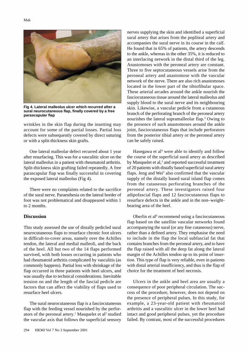
One lateral malleolar defect recurred about 1 yearafter resurfacing. This was for a vasculitic ulcer on thelateral malleolus in a patient with rheumatoid arthritis.Split-thickness skin grafting failed repeatedly. A freeparascapular flap was finally successful in coveringthe exposed lateral malleolus (Fig 4).
There were no complaints related to the sacrificeof the sural nerve. Paraesthesia on the lateral border offoot was not problematical and disappeared within 1to 2 months.
Discussion
This study assessed the use of distally pedicled suralneurocutaneous flaps to resurface chronic foot ulcersin difficult-to-cover areas, namely over the Achillestendon, the lateral and medial malleoli, and the backof the heel. All but two of the 14 flaps performedsurvived, with both losses occurring in patients whohad rheumatoid arthritis complicated by vasculitis (ascommonly happens). Partial loss with shrinkage of theflap occurred in three patients with heel ulcers, andwas usually due to technical considerations. Inevitabletension on and the length of the fascial pedicle arefactors that can affect the viability of flaps used toresurface heel ulcers.
The sural neurocutaneous flap is a fasciocutaneousflap with the feeding vessel nourished by the perfor-ators of the peroneal artery.1 Masquelet et al2 studiedthe vascular axis that follows the superficial sensory
nerves supplying the skin and identified a superficialsural artery that arises from the popliteal artery andaccompanies the sural nerve in its course in the calf.He found that in 65% of patients, the artery descendsto the ankle, whereas in the other 35%, it is reduced toan interlacing network in the distal third of the leg.Anastomoses with the peroneal artery are constant.Three to five septocutaneous vessels arise from theperoneal artery and anastomose with the vascularnetwork of the nerve. There are also rich anastomoseslocated in the lower part of the tibiofibular space.These arterial arcades around the ankle nourish thefasciocutaneous tissue around the lateral malleolus andsupply blood to the sural nerve and its neighbouringskin. Likewise, a vascular pedicle from a cutaneousbranch of the perforating branch of the peroneal arterynourishes the lateral supramalleolar flap.3 Owing tothe presence of such anastomoses around the anklejoint, fasciocutaneous flaps that include perforatorsfrom the posterior tibial artery or the peroneal arterycan be safely raised.
Hasegawa et al4 were able to identify and followthe course of the superficial sural artery as describedby Masquelet et al,3 and reported successful treatmentof 20 patients with distally based superficial sural arteryflaps. Jeng and Wei5 also confirmed that the vascularsupply of the distally based sural island flap comesfrom the cutaneous perforating branches of theperoneal artery. These investigators raised fouradipofascial flaps and 12 fasciocutaneous flaps toresurface defects in the ankle and in the non–weight-bearing area of the heel.
Oberlin et al6 recommend using a fasciocutaneousflap based on the satellite vascular networks foundaccompanying the sural (or any fine cutaneous) nerve,rather than a defined artery. They emphasise the needto include in the flap the local subfascial fat thatcontains branches from the peroneal artery, and to havethe flap raised with all the deep fat along the lateralmargin of the Achilles tendon up to its point of inser-tion. This type of flap is very reliable, even in patientswith distal arterial insufficiency, and thus is the flap ofchoice for the treatment of heel necrosis.
Ulcers in the ankle and heel area are usually aconsequence of poor peripheral circulation. The suc-cess of the procedure, however, does not depend onthe presence of peripheral pulses. In this study, forexample, a 23-year-old patient with rheumatoidarthritis and a vasculitic ulcer in the lower heel hadintact and good peripheral pulses, yet the procedurefailed. By contrast, most of the successful procedures
Fig 4. Lateral malleolus ulcer which recurred after asural neurocutaneous flap, finally covered by a freeparascapular flap

HKMJ Vol 7 No 3 September 2001 295
Sural neurocutaneous flaps for ankle and heel ulcers
were in older patients who had absence of either theposterior tibial and/or dorsalis pedis pulses of the ankle.Thus, the sural neurocutaneous flap healed all ulcersaround the Achilles tendon insertions and the lateralmalleoli, irrespective of the presence of palpableperipheral pulses. It appears that the circulation to theseextremities might have been through the perforators,thus bypassing the major peripheral vessels. Thepatency of perforators in peripheral vascular diseaseis clearly essential for the success of sural neuro-cutaneous and other neurocutaneous flaps. Vasculitisper se causes the ulcer or is a complication of the ulcerand affects the circulation in the perforators, and hencethe success of the procedure.
Neurocutaneous flaps other than the sural nerve flaphave been described. These use the saphenous nerve,the superficial peroneal nerve,2 and the cutaneousbranches of the radial and ulnar nerves.7 They toowill surely evolve as important sources of soft tissuefor the resurfacing of local skin defects without theneed to sacrifice any major vessels.
Conclusions
The distally based sural neurocutaneous flap withfascial pedicle, including the perforators of the peronealartery around the ankle region, is a reliable sourceof soft tissue to cover defects in the ankle and heel,without the sacrifice of a major blood vessel. Thisprocedure involves a single operation without the needfor microsurgical anastomosis. Its success does not
depend on the presence of a good peripheral pulse andmay prove useful for patients who have peripheralvascular insufficiency. The morbidity associated withthe loss of the sural nerve is minimal. A long pediclecan be designed to allow the flap to be transferred asfar as the instep area. For defects in the weight-bearingarea of the sole or heel, however, the medial plantarneurocutaneous flap gives more durable and sensateskin cover. For larger defects in the ankle or foot, or tofill up a defect due to osteomyelitis, free tissue transfersuch as the myocutaneous flap should be considered,as this brings in a new source of circulation.
References
1. Ponten B. The fasciocutaneous flap: its use in soft tissuedefects of the lower leg. Br J Plast Surg 1981;34:215-20.
2. Masquelet AC, Romana MC, Wolf G. Skin island flapssupplied by the vascular axis of the sensitive superficialnerves: anatomic study and clinical experience in the leg. PlastReconstr Surg 1992;89:1115-21.
3. Masquelet AC, Beveridge J, Romana C, Gerber C. The lateralsupramalleolar flap. Plast Reconstr Surg 1988;81:74-81.
4. Hasegawa M, Torii S, Katoh H, Esaki S. The distally basedsuperficial sural artery flap. Plast Reconstr Surg 1994;93:1012-20.
5. Jeng SF, Wei FC. Distally based sural island flap for footand ankle reconstruction. Plast Reconstr Surg 1997;99:744-50.
6. Oberlin C, Azoulay B, Bhatia A. The posterolateral malleolarflap of the ankle: a distally based sural neurocutaneous flap - areport of 14 cases. Plast Reconstr Surg 1995;96:400-5.
7. Bertelli JA, Khoury Z. Neurocutaneous island flaps in thehand: anatomical basis and preliminary results. Br J Plast Surg1992;45:586-90.