Embed Size (px)
Citation preview

Distal Radius FracturesLee W Hash, MD
Affinity Orthopedics and Sports Medicine
The Problem of Distal Radius Fractures
• Common injury: >450,000/yr. in USA• High potential for functional impairment
and frequent complications

Introduction• Distal radius fractures occur through the
distal metaphysis of the radius• May involve articular surface• Frequently involving the ulnar styloid• Most often result from a fall on the
outstretched hand. – forced extension of the carpus, – impact loading of the distal radius.
• Associated injuries may accompany distal radius fractures.

Introduction
• Classified by:– presence or absence of
intra-articular involvement,– degree of comminution,– dorsal vs. volar
displacement,– involvement of the distal
radioulnar joint.

Diagnosis: History and Physical Findings
• History of fall on outstretched hand or trauma • A visible deformity of the wrist is usually noted,
with the hand most commonly displaced in the dorsal direction.
• Movement of the hand and wrist are painful. • Adequate and accurate assessment of the
neurovascular status of the hand is imperative, before any treatment is carried out.

Diagnosis: Diagnostic Tests and Examination
• General physical exam of the patient, including an evaluation of the injured joint, and a joint above and below
• Radiographs of the injured wrist• Radiographs of other areas, if symptoms
warrant.• CT scan of the distal radius in selected
instances.

Treatment Goals• Preserve hand and wrist function
• Realign normal osseous anatomy•• Promote bony healing
• Avoid complications
• Allow early finger and elbow ROM

Osseous Anatomy
• Distal radius – 80% of axial load– Scaphoid fossa– Lunate fossa– Sigmoid notch – DRUJ
• Distal ulna

Anatomy • scaphoid and lunate
fossa– Ridge normally exists
between these two• sigmoid notch: second
important articularsurface
• triangular fibrocartilagecomplex(TFCC):distal edge of radius to base of ulnar styloid

Radiology
• Radial inclination = 22°• Radial length = 12mm
– ulnar neutral• Palmar tilt = 11-14°• Scapho-lunate angle =
47° +/- 15°

Measurement of Radial Length and InclinationInclination = 23 degrees

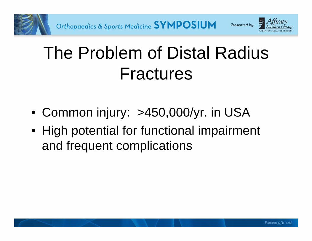
1: Line connecting dorsal and volar tip of lunate
2: Line perpendicular to lunate
3: Line along axis of scaphoid
Scapholunate angle measured between lines 2 and 3
(normal 47 ± 15 degrees)

• Intra-articular fxs with multiple fragments
• centrally impacted fragments• DRUJ incongruity
• 19 consecutive fx, CT had better sensitivity for intraarticular frag
• management change in 5 pts
Computed TomographyIndications
Cole et al: J Hand Surg, 1997

Classification of Distal Radius Fractures
• Ideal system should describe:– Type of injury– Severity– Evaluation– Treatment– Prognosis
Common Classifications
• Gartland/Werley• Frykman• Weber (AO/ASIF)• Melone• Column theory• Fernandez (mechanism)

Assessment of X-rays
• Assess involvement of dorsal or volar rim– Is comminution mainly volar or dorsal?– is one of four cortices intact?
• Look for “die-punch” lesions of the scaphoid or lunate fossa.
• Assess amount of shortening• Look for DRUJ involvement

Dorsal angulation and comminution

Volar subluxation of carpus with fracture fragment

Treatment Choice
• Depends on assessment of fracture stability
• Indicators of instability are:– Shortening– Comminution– Reversal of normal volar angulation– Articular involvement
Options for Treatment• Casting
– Long arm vs short arm– Sugar-tong splint
• External Fixation– Joint-spanning– Non bridging
• Percutaneous pinning• Internal Fixation
– Dorsal plating– Volar plating– Combined dorsal/volar plating– focal (fracture specific) plating

Indications for Closed Treatment
• Low-energy fracture• Low-demand patient• Medical co-morbidities• Minimal displacement- acceptable
alignment• Match treatment to demands of the
patient

Closed Treatment of Distal Radial Fractures
• Depends on obtaining and then maintaining an acceptable reduction.
• Immobilization: – long arm (cast or sugar-tong for high
demand)– short arm adequate for elderly patients
• Frequent follow-up necessary in order to diagnose redisplacement.

Technique of Closed Reduction• Anesthesia
– Hematoma block– Intravenous sedation– Bier block
• Traction: finger traps and weights• Reduction Maneuver (dorsally angulated fracture):
– hyperextension of the distal fragment, – Maintain weighted traction and reduce the distal to
the proximal fragment with pressure applied to the distal radius.
• Apply well-molded “sugar-tong” splint or cast, with wrist in neutral to slight flexion.
• Avoid Extreme Positions!

Acceptable Reduction Criteria
• dorsal angulation < 10 degrees • > 15 degrees of inclination• Articular step-off < 2mm• < 5 mm shortening compared to opposite wrist.• DRUJ congruent

After-treatment• Watch for median nerve symptoms
– parasthesias common but should diminish over few hours
– If persist release pressure on cast, take wrist out of flexion
– Acute carpal tunnel: symptoms progress; CTR required
• Follow-up x-rays needed in 1-2 weeks to evaluate reduction.
• Change to short-arm cast after 2-3 weeks, continue until fracture healing.

Management of Redisplacement
• Repeat reduction and casting – high rate of failure
• Repeat reduction and percutaneouspinning
• External Fixation• ORIF

Indications for Surgical Treatment
• High-energy injury• Open injury• Secondary loss of reduction• Articular comminution, step-off, or gap• Metaphyseal comminution or bone loss• Loss of volar buttress with displacement• DRUJ incongruity

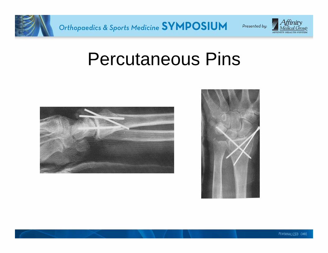
Percutaneous Pinning-Methods
• variety described• most common radial styloid pinning +
dorsal-ulnar corner of radius pinning• supplemental immobilization with cast,
splint• in conjunction with external fixation
(Augmented external fixation)

Percutaneous Pins
Percutaneous Pins

Percutaneous Pinning
• 2 radial styloid pins - Mah and Atkinson, J Hand Surg 1992– excellent anatomic 82%– good-excellent functional results 100%
• radial styloid with dorsal - prospective study, 30 pts (Clancey JBJS 1984)
– excellent anatomic results in 90%

Percutaneous Pinning-Kapandji
• intrafocal pinning through fracture site
• buttress against displacement
• good results in literature• -Greatting & Bishop, OCNA
1993
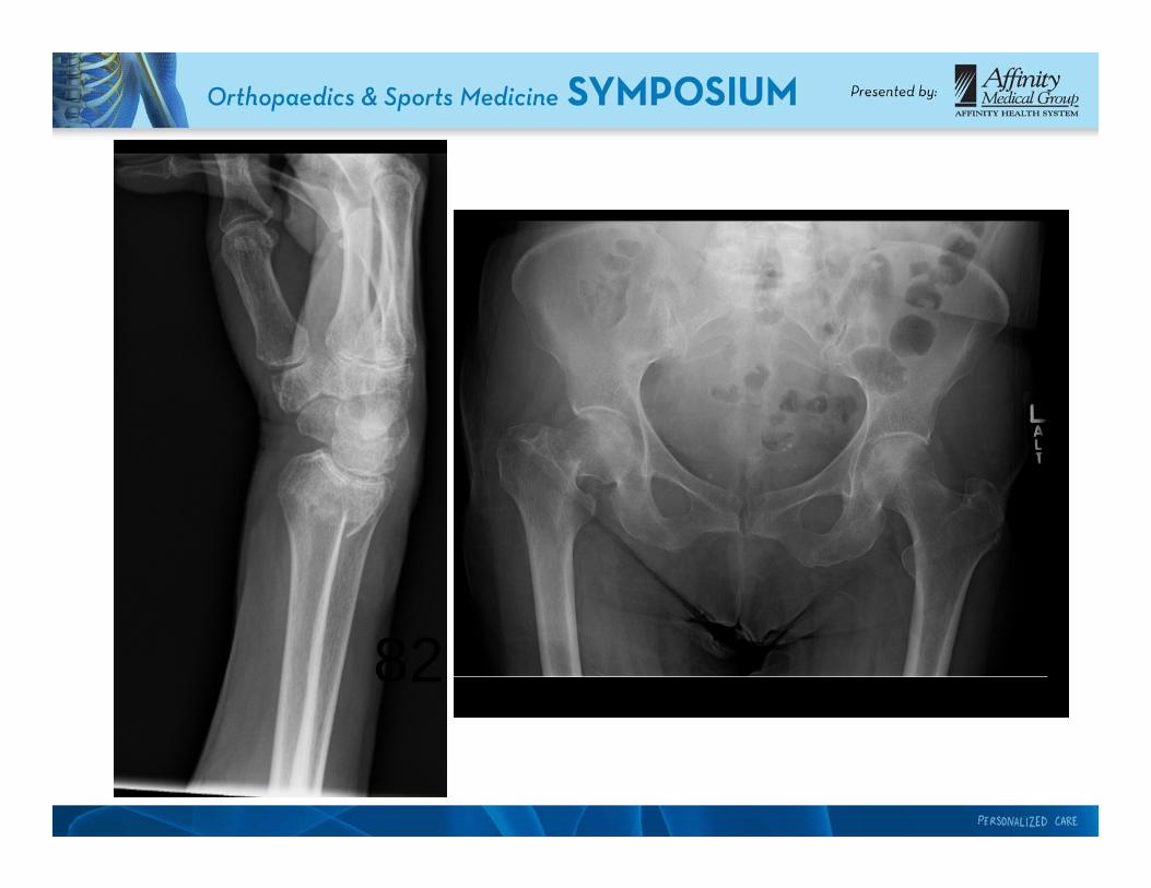
82 yo Female


After Pin Removal

Internal Fixation of Distal Radius Fractures
• Useful for elevation of depressed articularfragments
• required if articular fragments can not be adequately reduced with percutaneousmethods
• Dorsal and/or volar approaches both used.
Selection of Approach
• Based on location of comminution.• Dorsal approach for dorsally angulated
fractures.• Volar approach for volar rim fractures• Radial styloid approach for buttressing of styloid• Combined approaches needed for high-energy
fractures with significant axial impaction.

-
WHICH APPROACH?WHICH APPROACH?
DORSALDORSAL3rd DC –EPL
(extensile)1-2nd DC

Classical Henry approach Extended carpal tunnel approach
VOLARVOLAR

Distal Radius-volar barton• 64 yo M, MVA, contralateral tibial shaft Fx


-less tendon irritation than dorsal
- Indirect reduction -better tolerated than Ex fix
Volar Plating for Dorsal Fractures

Fixed angle locked screws

Courtesy J. Orbay, MD


50 yo Female

Volar Locking Plate

81 yo Female


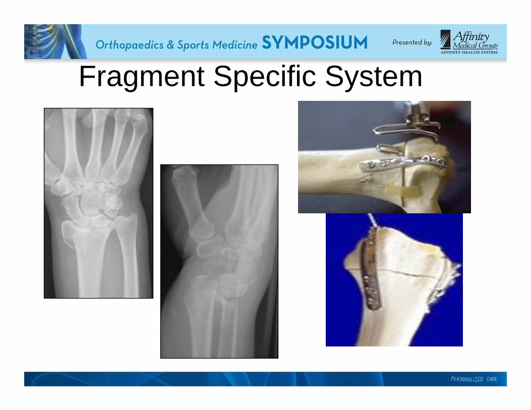
Fragment Specific System

Radial and Ulnar Columns
-Pin plates
-90-90 plating technique

Focal Plating
Radial Styloid FragmentDorsal ulnar fragment
70 – 90 degrees apart

Advanced TechniquesArthroscopic-Assisted
• reduce articular incongruities• also diagnose associated soft tissue
lesions• minimally invasive

Arthroscopic-Assisted
Culp and Osterman, OCNA 26(4) 1995

Malunion of Distal Radius Fractures
• Changes load-bearing patterns on the distal radius and load sharing between the radius and ulna.
• Can lead to arthrosis.


Conclusions• Identify the fracture• R/O need for immediate tx• Immobilization- well padded splint• Ice and elevation to control edema• Ortho referral

Conclusions• Treatment goals – restore function
– Patient specificNonoperative and operative treatmentRecovery takes months