Embed Size (px)
Citation preview

The Rockefeller University Press $30.00J. Exp. Med. Vol. 208 No. 12 2393-2401www.jem.org/cgi/doi/10.1084/jem.20110525
2393
Brief Definit ive Report
During inflammation, leukocytes extravasating from the blood into the surrounding tissue have to overcome the endothelial barrier of the blood vessel wall. Capture and arrest of leuko-cytes to the luminal surface of the endothelium at sites of emigration requires the interplay of several adhesion and signaling molecules, such as selectins, chemokines, and integrin ligands (Ley et al., 2007). This is then followed by the actual transmigration step, a process called dia-pedesis (Vestweber, 2007; Muller, 2009).
Several adhesion molecules at endothelial cell contacts, such as platelet endothelial cell ad-hesion molecule 1 (Muller et al., 1993) and CD99 (Schenkel et al., 2002; Bixel et al., 2004), and some at endothelial tight junctions, such as the junctional adhesion molecules (Martìn-Padura et al., 1998; Nourshargh et al., 2006) and
endothelial cell–specific adhesion molecule (Wegmann et al., 2006) are known to support leukocyte diapedesis. The fact that they are located at the interface between endothelial cells is a strong indication that they participate in the migration of leukocytes through the junctional pathway (Muller, 2001; Nourshargh et al., 2010). However, in vitro studies have shown that several of these adhesion molecules, particularly PECAM-1, junctional adhesion mole-cule A, and CD99 are also found surrounding leukocytes that migrate through the body of an endothelial cell on a transcellular route (Carman
CORRESPONDENCE Dietmar Vestweber: [email protected]
Abbreviations used: ES, embry-onic stem; FKBP, FK 506 binding protein; IRES, internal ribosome entry site; KI, knock-in; MLC, myosin light chain; RMCE, recombination-mediated cassette exchange; VEGF, vascu-lar endothelial growth factor; VE-PTP, vascular endothelial protein tyrosine phosphatase.
Dissociation of VE-PTP from VE-cadherin is required for leukocyte extravasation and for VEGF-induced vascular permeability in vivo
Andre Broermann,1 Mark Winderlich,1 Helena Block,1,2 Maike Frye,1 Jan Rossaint,1,2 Alexander Zarbock,1,2 Giuseppe Cagna,1 Ruth Linnepe,1 Dörte Schulte,1 Astrid Fee Nottebaum,1 and Dietmar Vestweber1
1Max-Planck-Institute for Molecular Biomedicine, D-48149 Münster, Germany2Department of Anesthesiology and Critical Care Medicine, University of Münster, D-48149 Münster, Germany
We have recently shown that vascular endothelial protein tyrosine phosphatase (VE-PTP), an endothelial membrane protein, associates with VE-cadherin and is required for optimal VE-cadherin function and endothelial cell contact integrity. The dissociation of VE-PTP from VE-cadherin is triggered by vascular endothelial growth factor (VEGF) and by the binding of leukocytes to endothelial cells in vitro, suggesting that this dissociation is a prerequisite for the destabilization of endothelial cell contacts. Here, we show that VE-cadherin/VE-PTP dissociation also occurs in vivo in response to LPS stimulation of the lung or systemic VEGF stimulation. To show that this dissociation is indeed necessary in vivo for leukocyte extravasation and VEGF-induced vascular permeability, we generated knock-in mice expressing the fusion proteins VE-cadherin-FK 506 binding protein and VE-PTP-FRB* under the control of the endogenous VE-cadherin promoter, thus replacing endogenous VE-cadherin. The additional domains in both fusion proteins allow the heterodimeric com-plex to be stabilized by a chemical compound (rapalog). We found that intravenous appli-cation of the rapalog strongly inhibited VEGF-induced (skin) and LPS-induced (lung) vascular permeability and inhibited neutrophil extravasation in the IL-1 inflamed cremas-ter and the LPS-inflamed lung. We conclude that the dissociation of VE-PTP from VE-cadherin is indeed required in vivo for the opening of endothelial cell contacts during induction of vascular permeability and leukocyte extravasation.
© 2011 Broermann et al. This article is distributed under the terms of an Attribu-tion–Noncommercial–Share Alike–No Mirror Sites license for the first six months after the publication date (see http://www.rupress.org/terms). After six months it is available under a Creative Commons License (Attribution–Noncommercial–Share Alike 3.0 Unported license, as described at http://creativecommons.org/licenses/by-nc-sa/3.0/).
The
Journ
al o
f Exp
erim
enta
l M
edic
ine
on January 15, 2019jem.rupress.org Downloaded from http://doi.org/10.1084/jem.20110525Published Online: 24 October, 2011 | Supp Info:
2394 VE-PTP/VE-cadherin control endothelial junctions | Broermann et al.
To this end, we have fused additional protein domains (FKBP and FRB*) to the C termini of VE-cadherin and VE-PTP, which contain binding sites for a small molecule called rapalog, allowing for stabilization of VE-cadherin/VE-PTP heterodimers. DNA constructs for both fusion proteins were knocked into the VE-cadherin locus of gene-targeted mice, thereby replacing endogenous VE-cadherin. We found that LPS-triggered leukocyte recruitment into the lung, as well as i.v. injected VEGF, both stimulate the dissociation of VE-PTP from VE-cadherin in vivo. Administering rapalog into the circulation prevented this dissociation and strongly reduced VEGF- and LPS-induced vascular permeability, IL-1-induced recruitment of neutrophils in the cremaster tissue, and LPS-induced infiltration of neutrophils into lungs. Thus, dissociation of VE-PTP from VE-cadherin is indeed necessary in vivo for the opening of endothelial cell contacts in both processes.
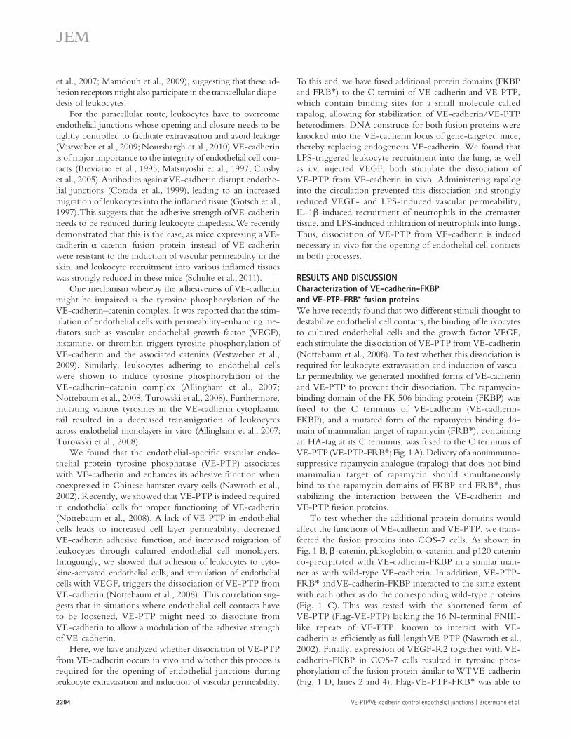
RESULTS AND DISCUSSIONCharacterization of VE-cadherin-FKBP and VE-PTP-FRB* fusion proteinsWe have recently found that two different stimuli thought to destabilize endothelial cell contacts, the binding of leukocytes to cultured endothelial cells and the growth factor VEGF, each stimulate the dissociation of VE-PTP from VE-cadherin (Nottebaum et al., 2008). To test whether this dissociation is required for leukocyte extravasation and induction of vascu-lar permeability, we generated modified forms of VE-cadherin and VE-PTP to prevent their dissociation. The rapamycin-binding domain of the FK 506 binding protein (FKBP) was fused to the C terminus of VE-cadherin (VE-cadherin-FKBP), and a mutated form of the rapamycin binding do-main of mammalian target of rapamycin (FRB*), containing an HA-tag at its C terminus, was fused to the C terminus of VE-PTP (VE-PTP-FRB*; Fig. 1 A). Delivery of a nonimmuno-suppressive rapamycin analogue (rapalog) that does not bind mammalian target of rapamycin should simultaneously bind to the rapamycin domains of FKBP and FRB*, thus stabilizing the interaction between the VE-cadherin and VE-PTP fusion proteins.
To test whether the additional protein domains would affect the functions of VE-cadherin and VE-PTP, we trans-fected the fusion proteins into COS-7 cells. As shown in Fig. 1 B, -catenin, plakoglobin, -catenin, and p120 catenin co-precipitated with VE-cadherin-FKBP in a similar man-ner as with wild-type VE-cadherin. In addition, VE-PTP-FRB* and VE-cadherin-FKBP interacted to the same extent with each other as do the corresponding wild-type proteins (Fig. 1 C). This was tested with the shortened form of VE-PTP (Flag-VE-PTP) lacking the 16 N-terminal FNIII-like repeats of VE-PTP, known to interact with VE- cadherin as efficiently as full-length VE-PTP (Nawroth et al., 2002). Finally, expression of VEGF-R2 together with VE-cadherin-FKBP in COS-7 cells resulted in tyrosine phos-phorylation of the fusion protein similar to WT VE-cadherin (Fig. 1 D, lanes 2 and 4). Flag-VE-PTP-FRB* was able to
et al., 2007; Mamdouh et al., 2009), suggesting that these ad-hesion receptors might also participate in the transcellular diape-desis of leukocytes.
For the paracellular route, leukocytes have to overcome endothelial junctions whose opening and closure needs to be tightly controlled to facilitate extravasation and avoid leakage (Vestweber et al., 2009; Nourshargh et al., 2010). VE-cadherin is of major importance to the integrity of endothelial cell con-tacts (Breviario et al., 1995; Matsuyoshi et al., 1997; Crosby et al., 2005). Antibodies against VE-cadherin disrupt endothe-lial junctions (Corada et al., 1999), leading to an increased migration of leukocytes into the inflamed tissue (Gotsch et al., 1997). This suggests that the adhesive strength of VE-cadherin needs to be reduced during leukocyte diapedesis. We recently demonstrated that this is the case, as mice expressing a VE- cadherin--catenin fusion protein instead of VE-cadherin were resistant to the induction of vascular permeability in the skin, and leukocyte recruitment into various inflamed tissues was strongly reduced in these mice (Schulte et al., 2011).
One mechanism whereby the adhesiveness of VE-cadherin might be impaired is the tyrosine phosphorylation of the VE-cadherin–catenin complex. It was reported that the stim-ulation of endothelial cells with permeability-enhancing me-diators such as vascular endothelial growth factor (VEGF), histamine, or thrombin triggers tyrosine phosphorylation of VE-cadherin and the associated catenins (Vestweber et al., 2009). Similarly, leukocytes adhering to endothelial cells were shown to induce tyrosine phosphorylation of the VE-cadherin–catenin complex (Allingham et al., 2007; Nottebaum et al., 2008; Turowski et al., 2008). Furthermore, mutating various tyrosines in the VE-cadherin cytoplasmic tail resulted in a decreased transmigration of leukocytes across endothelial monolayers in vitro (Allingham et al., 2007; Turowski et al., 2008).
We found that the endothelial-specific vascular endo-thelial protein tyrosine phosphatase (VE-PTP) associates with VE-cadherin and enhances its adhesive function when coexpressed in Chinese hamster ovary cells (Nawroth et al., 2002). Recently, we showed that VE-PTP is indeed required in endothelial cells for proper functioning of VE-cadherin (Nottebaum et al., 2008). A lack of VE-PTP in endothelial cells leads to increased cell layer permeability, decreased VE-cadherin adhesive function, and increased migration of leukocytes through cultured endothelial cell monolayers. Intriguingly, we showed that adhesion of leukocytes to cyto-kine-activated endothelial cells, and stimulation of endothelial cells with VEGF, triggers the dissociation of VE-PTP from VE-cadherin (Nottebaum et al., 2008). This correlation sug-gests that in situations where endothelial cell contacts have to be loosened, VE-PTP might need to dissociate from VE-cadherin to allow a modulation of the adhesive strength of VE-cadherin.
Here, we have analyzed whether dissociation of VE-PTP from VE-cadherin occurs in vivo and whether this process is required for the opening of endothelial junctions during leukocyte extravasation and induction of vascular permeability.

JEM Vol. 208, No. 12 2395
Br ief Definit ive Repor t
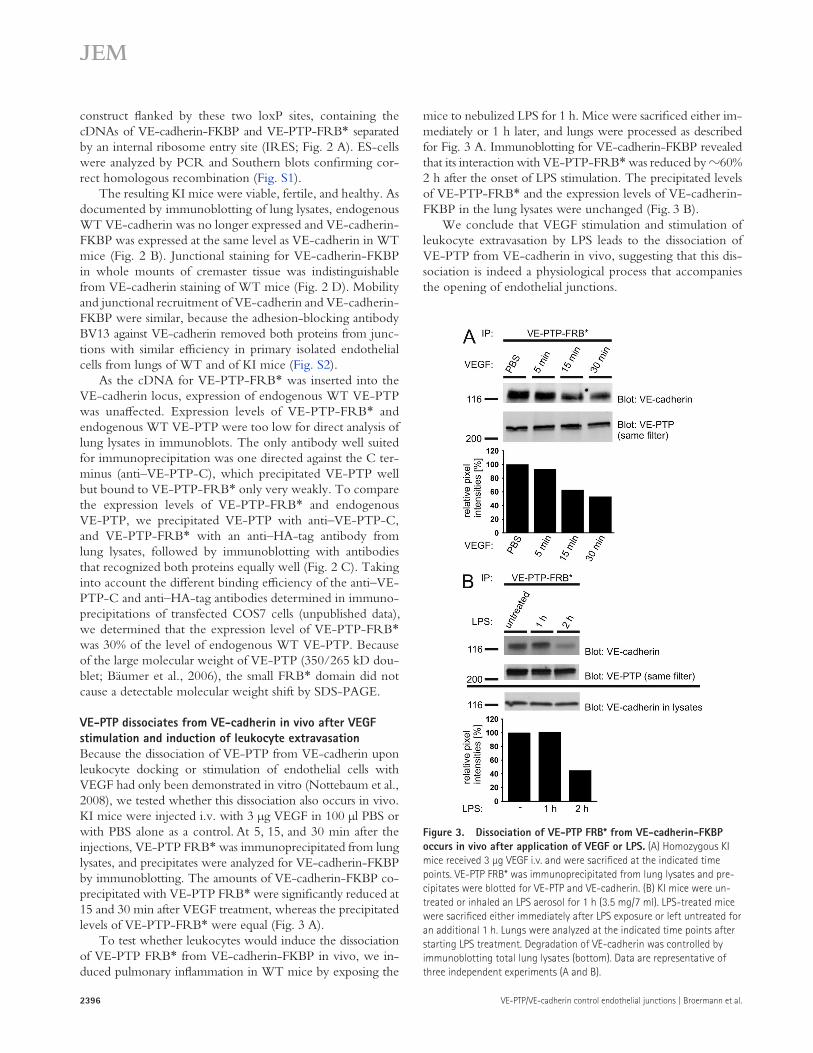
Generation of transgenic mice expressing VE-cadherin-FKBP and VE-PTP-FRB* under the endogenous VE-cadherin promoterTo test whether the dissociation of VE-PTP from VE-cadherin is a prerequisite for the destabilization of endothelial cell contacts in vivo, we generated knock-in (KI) mice express-ing VE-cadherin-FKBP and full-length VE-PTP-FRB* specifically under the endothelial specific VE-cadherin pro-moter. To this end, the ATG-containing exon 2 of VE-cadherin was flanked in mouse embryonic stem (ES) cells with two incompatible loxP sites (Fig. 2 A). Based on recombination-mediated cassette exchange (RMCE), these two loxP sites al-lowed exon 2 to be replaced by any cDNA construct that was also flanked by such a pair of incompatible loxP sites upon co-transfection with the recombinase Cre. We designed a KI
reverse this phosphorylation of VE-cadherin-FKBP with similar efficiency as WT Flag-VE-PTP dephosphorylated WT VE-cadherin (Fig. 1 D, lanes 1 and 3). Collectively, we conclude that the additional FKBP and FRB* domains do not affect the molecular interactions of VE-cadherin and VE-PTP.
Figure 1. VE-cadherin-FKPB and Flag-VE-PTP-FRB* show the same characteristics as WT proteins. (A) Schematic illustration of VE-cadherin-FKBP and VE-PTP-FRB* transmembrane domain (TM) and HA-tag (HA). (B) VE-cadherin or VE-cadherin-FKBP were expressed in COS-7 cells. VE-cadherin was immunoprecipitated, and the indicated proteins were detected by immunoblotting. The horizontal line indicates different experiments. (C) The indicated proteins were expressed in COS-7 cells, and Flag-VE-PTP or Flag-VE-PTP-FRB* were immunoprecipitated, followed by analysis for Flag-VE-PTP and co-precipitated VE-cadherin-FKBP or VE-cadherin by immunoblotting. Expression of transfected proteins was con-trolled by immunoblots of total cell lysates. (D) VEGFR-2 was coexpressed with the indicated proteins in COS-7 cells. VE-cadherin was immuno-precipitated, followed by immunoblotting for phosphotyrosine and VE-cadherin. Data are representative for two (B), six (C), and three (D) independent experiments.
Figure 2. Generation of transgenic mice expressing VE- cadherin-FKBP and VE-PTP-FRB*. (A) Schematic illustration of the RMCE approach. The gene replacement cassette contains cDNAs for VE-cadherin-FKBP and full-length VE-PTP-FRB* separated by an IRES site, as well as a Hygromycin cassette, flanked by two FRT sites. The cassette is flanked by two incompatible loxP sites. Using RMCE, the ATG-containing exon 2 of VE-cadherin was replaced by the cDNA insertion cassette. Posi-tions of the primers for PCR-genotyping are indicated. (B) Lung lysates of either wild-type (+/+), heterozygous (+/KI), or homozygous mutant (KI/KI) mice were immunoblotted for VE-cadherin and as loading control for Tie-2. (C) Endogenous VE-PTP was immunoprecipitated with anti–VE-PTP-C and VE-PTP-FRB* was immunoprecipitated with anti-HA antibodies from lung lysates of homozygous KI (KI/KI) or WT (+/+) mice. Immunoprecipita-tions with goat IgG (CoIgG) were performed as a control. Precipitates were analyzed by immunoblotting with antibodies against the extracel-lular domain of VE-PTP. Note that KI mice showed only slightly increased overall amounts of VE-PTP compared with WT mice (left) because the anti–VE-PTP-C antibodies bind to VE-PTP-FRB* only very weakly. (D) Whole mount staining of venules in cremaster from WT mice (left) and KI mice (right) for VE-cadherin or VE-cadherin-FKBP, respectively. Bars, 20 µm. Data are representative for three (B and C) and two (D) independent experiments.
2396 VE-PTP/VE-cadherin control endothelial junctions | Broermann et al.
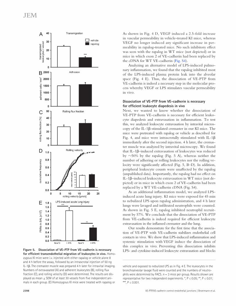
mice to nebulized LPS for 1 h. Mice were sacrificed either im-mediately or 1 h later, and lungs were processed as described for Fig. 3 A. Immunoblotting for VE-cadherin-FKBP revealed that its interaction with VE-PTP-FRB* was reduced by 60% 2 h after the onset of LPS stimulation. The precipitated levels of VE-PTP-FRB* and the expression levels of VE-cadherin-FKBP in the lung lysates were unchanged (Fig. 3 B).
We conclude that VEGF stimulation and stimulation of leukocyte extravasation by LPS leads to the dissociation of VE-PTP from VE-cadherin in vivo, suggesting that this dis-sociation is indeed a physiological process that accompanies the opening of endothelial junctions.
construct flanked by these two loxP sites, containing the cDNAs of VE-cadherin-FKBP and VE-PTP-FRB* separated by an internal ribosome entry site (IRES; Fig. 2 A). ES-cells were analyzed by PCR and Southern blots confirming cor-rect homologous recombination (Fig. S1).
The resulting KI mice were viable, fertile, and healthy. As documented by immunoblotting of lung lysates, endogenous WT VE-cadherin was no longer expressed and VE-cadherin-FKBP was expressed at the same level as VE-cadherin in WT mice (Fig. 2 B). Junctional staining for VE-cadherin-FKBP in whole mounts of cremaster tissue was indistinguishable from VE-cadherin staining of WT mice (Fig. 2 D). Mobility and junctional recruitment of VE-cadherin and VE-cadherin-FKBP were similar, because the adhesion-blocking antibody BV13 against VE-cadherin removed both proteins from junc-tions with similar efficiency in primary isolated endothelial cells from lungs of WT and of KI mice (Fig. S2).
As the cDNA for VE-PTP-FRB* was inserted into the VE-cadherin locus, expression of endogenous WT VE-PTP was unaffected. Expression levels of VE-PTP-FRB* and endogenous WT VE-PTP were too low for direct analysis of lung lysates in immunoblots. The only antibody well suited for immunoprecipitation was one directed against the C ter-minus (anti–VE-PTP-C), which precipitated VE-PTP well but bound to VE-PTP-FRB* only very weakly. To compare the expression levels of VE-PTP-FRB* and endogenous VE-PTP, we precipitated VE-PTP with anti–VE-PTP-C, and VE-PTP-FRB* with an anti–HA-tag antibody from lung lysates, followed by immunoblotting with antibodies that recognized both proteins equally well (Fig. 2 C). Taking into account the different binding efficiency of the anti–VE-PTP-C and anti–HA-tag antibodies determined in immuno-precipitations of transfected COS7 cells (unpublished data), we determined that the expression level of VE-PTP-FRB* was 30% of the level of endogenous WT VE-PTP. Because of the large molecular weight of VE-PTP (350/265 kD dou-blet; Bäumer et al., 2006), the small FRB* domain did not cause a detectable molecular weight shift by SDS-PAGE.
VE-PTP dissociates from VE-cadherin in vivo after VEGF stimulation and induction of leukocyte extravasationBecause the dissociation of VE-PTP from VE-cadherin upon leukocyte docking or stimulation of endothelial cells with VEGF had only been demonstrated in vitro (Nottebaum et al., 2008), we tested whether this dissociation also occurs in vivo. KI mice were injected i.v. with 3 µg VEGF in 100 µl PBS or with PBS alone as a control. At 5, 15, and 30 min after the injections, VE-PTP FRB* was immunoprecipitated from lung lysates, and precipitates were analyzed for VE-cadherin-FKBP by immunoblotting. The amounts of VE-cadherin-FKBP co-precipitated with VE-PTP FRB* were significantly reduced at 15 and 30 min after VEGF treatment, whereas the precipitated levels of VE-PTP-FRB* were equal (Fig. 3 A).
To test whether leukocytes would induce the dissociation of VE-PTP FRB* from VE-cadherin-FKBP in vivo, we in-duced pulmonary inflammation in WT mice by exposing the
Figure 3. Dissociation of VE-PTP FRB* from VE-cadherin-FKBP occurs in vivo after application of VEGF or LPS. (A) Homozygous KI mice received 3 µg VEGF i.v. and were sacrificed at the indicated time points. VE-PTP FRB* was immunoprecipitated from lung lysates and pre-cipitates were blotted for VE-PTP and VE-cadherin. (B) KI mice were un-treated or inhaled an LPS aerosol for 1 h (3.5 mg/7 ml). LPS-treated mice were sacrificed either immediately after LPS exposure or left untreated for an additional 1 h. Lungs were analyzed at the indicated time points after starting LPS treatment. Degradation of VE-cadherin was controlled by immunoblotting total lung lysates (bottom). Data are representative of three independent experiments (A and B).

JEM Vol. 208, No. 12 2397
Br ief Definit ive Repor t
but not of other VE-PTP substrates such as Tie-2, we treated the animals with VEGF and rapalog or vehicle, and immuno-precipitates of VE-cadherin or Tie-2 from lung lysates were analyzed for phospho-tyrosines in immunoblots. As shown in Fig. 4 (B and C), the rapalog decreased tyrosine phosphoryla-tion of VE-cadherin, -catenin and plakoglobin, but not of Tie-2. As an additional control, we tested whether the rapa-log or the expression of VE-cadherin-FKBP affected the dis-tribution of cortical actin or myosin light chain (MLC)-2 in primary endothelial cells isolated from lungs of the KI mice. As shown in Fig. S3, we found no such effects.
Because i.v. injection of VEGF did not induce plasma protein leakage into the alveolar space, probably because of a lack of effect on the alveolar epithelium, we used the Miles permeability assay in the skin. In this way, we tested whether preventing the dissociation of VE-PTP from VE-cadherin would impair the induction of vascular permeability by VEGF in vivo. We injected KI mice i.v. with 250 µg rapalog or vehicle 8 and 4 h before the assay. Subsequently, mice were i.v. injected with Evan’s blue dye and stimulated intradermally 10 min later with VEGF or PBS. Injected skin areas were ex-cised 30 min later, and the dye was extracted with formamide.
Dissociation of VE-PTP from VE-cadherin is required for the induction of vascular permeability by VEGFTo test whether the rapalog would indeed block the VEGF-induced dissociation of VE-cadherin-FKBP and VE-PTP-FRB* in vivo, we injected KI mice i.v. with 250 µg rapalog or vehicle 8 and 4 h before the assay. Subsequently, VEGF or PBS was administered i.v., and 30 min later, lung lysates were immunoprecipitated for VE-PTP-FRB*. Immunoprecipi-tates were first blotted for VE-cadherin-FKBP, and the same filters were subsequently reanalyzed for VE-PTP-FRB*. In addition, lung lysate aliquots were set aside and analyzed directly by immunoblotting for VE-cadherin-FKBP, to con-trol for degradation. As shown in Fig. 4 A, the amount of VE-cadherin-FKBP (standardized to the amount of precipi-tated VE-PTP-FRB*) that was co-precipitated was reduced by 31% upon VEGF stimulation in the presence of vehicle, whereas VEGF no longer reduced the co-precipitation effi-ciency if the mice had been treated with the rapalog. Thus, the rapalog does inhibit VEGF-induced dissociation of VE-PTP-FRB* from VE-cadherin-FKBP in vivo.
To assess whether the rapalog affects tyrosine phosphory-lation of components of the VE-cadherin-catenin complex,
Figure 4. Dissociation of VE-PTP from VE-cadherin is necessary for VEGF-induced permeability in vivo. (A) Homozygous KI mice were in-jected i.v. either with vehicle alone (left) or with rapalog (right) 8 and 4 h before the assay. Mice were then i.v. injected with either PBS or with VEGF (as indicated), and 30 min later lung lysates were immunoprecipitated for VE-PTP-FRB*. Precipitated material was analyzed by immunoblotting for VE-cadherin-FKBP and VE-PTP expression. To control for degradation, aliquots of total lung lysates were set aside and analyzed by immunoblotting for VE-cadherin-FKBP (bottom). The amount of immunoprecipitated VE-PTP-FRB and co-precipitated VE-cadherin-FKBP detected from VEGF-treated mice is shown as percentage of the amount precipitated from PBS-treated mice (numbers below). Graphs in inserts give co-precipitation efficiency. Note that the rapalog blocked dissociation of VE-cadherin from VE-PTP. (B) Homozygous KI mice were treated with rapalog or vehicle and subsequently with VEGF as in A. Lung lysates were immunoprecipitated for VE-cadherin and precipitates were immunoblotted first for phosphotyrosine, and then for VE-cadherin and plakoglobin (-catenin). (C) Lung lysates from KI mice were immunoprecipitated for Tie-2 and immunoblots were analyzed for phospho-tyrosine and subsequently for Tie-2. (D) Homozygous KI mice were injected i.v. with rapalog (black bars) or vehicle alone (white bars) 8 and 4 h before the assay. At the start of the assay, Evan’s blue was i.v. injected, followed by intradermal injection of VEGF or PBS 10 min later. After 30 min, mice were sacrificed and the dye was extracted from skin samples and quantified. n = 5 mice per group. Data are representative of two (A–C) or three (D) independent experiments. (E) Homozygous KI mice were treated with rapalog or vehicle as in D, followed by exposing the mice to nebulized LPS for 45 min. 4 h later, lungs were lavaged and protein content was measured. n = 3 mice per group. Results shown are representative of two indepen-dent experiments. *, P ≤ 0.05; **, P ≤ 0.01.
2398 VE-PTP/VE-cadherin control endothelial junctions | Broermann et al.
As shown in Fig. 4 D, VEGF induced a 2.5-fold increase in vascular permeability in vehicle-treated KI mice, whereas VEGF no longer induced any significant increase in per-meability in rapalog-treated mice. No such inhibitory effect was seen with the rapalog in WT mice (not depicted) or in mice in which exon 2 of VE-cadherin had been replaced by the cDNA for WT VE-cadherin (Fig. S4).
Analyzing an alternative model of LPS-induced pulmo-nary inflammation, we found that the rapalog inhibited most of the LPS-induced plasma protein leak into the alveolar space (Fig. 4 E). Thus, the dissociation of VE-PTP from VE-cadherin is indeed a necessary step in the molecular pro-cess whereby VEGF or LPS stimulates vascular permeability in vivo.
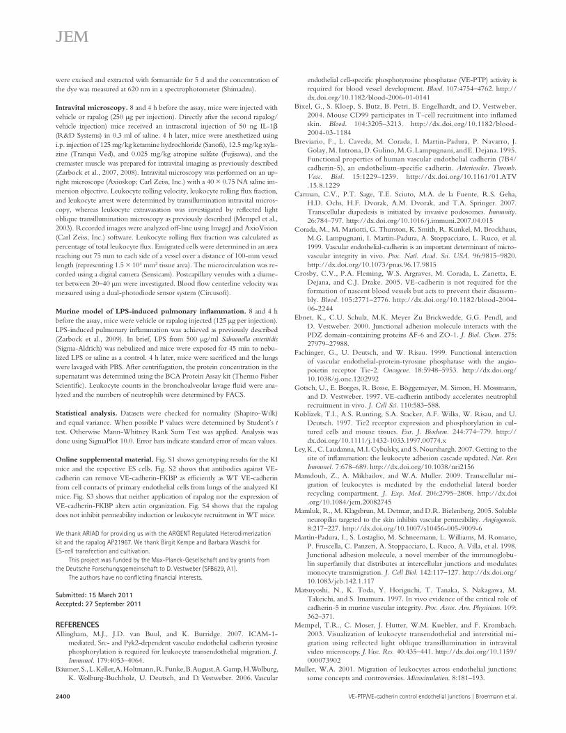
Dissociation of VE-PTP from VE-cadherin is necessary for efficient leukocyte diapedesis in vivoNext, we wanted to know whether the dissociation of VE-PTP from VE-cadherin is necessary for efficient leuko-cyte diapedesis and extravasation in inflammation. To test this, we analyzed leukocyte extravasation by intravital micros-copy of the IL-1-stimulated cremaster in our KI mice. The mice were pretreated with rapalog or vehicle as described for Fig. 4, and mice were intrascrotally stimulated with IL-1 immediately after the second injection. 4 h later, the cremas-ter muscle was analyzed by intravital microscopy. We found that IL-1–induced extravasation of leukocytes was reduced by 50% by the rapalog (Fig. 5 A), whereas neither the number of adhering or rolling leukocytes nor the rolling ve-locity were significantly affected (Fig. 5, B–D). In addition, peripheral leukocyte counts were unaffected by the rapalog (unpublished data). Importantly, the rapalog had no effect on IL-1–induced leukocyte extravasation in WT mice (not de-picted) or in mice in which exon 2 of VE-cadherin had been replaced by a WT VE-cadherin cDNA (Fig. S4).
As an additional inflammation model, we analyzed LPS-induced acute lung injury. KI mice were exposed for 45 min to nebulized LPS upon rapalog administration, and 4 h later lungs were lavaged and infiltrated neutrophils were counted. As shown in Fig. 5 E, rapalog inhibited neutrophil recruit-ment by 57%. We conclude that the dissociation of VE-PTP from VE-cadherin is indeed required for efficient leukocyte extravasation in the inflamed cremaster and the lung.
Our results demonstrate for the first time that the associa-tion of VE-PTP with VE-cadherin stabilizes endothelial cell contacts in vivo. We show that LPS-induced inflammation and systemic stimulation with VEGF induce the dissociation of this complex in vivo. Preventing this dissociation inhibits LPS- and cytokine-induced leukocyte extravasation and blocks Figure 5. Dissociation of VE-PTP from VE-cadherin is necessary
for efficient transendothelial migration of leukocytes in vivo. Homo-zygous KI mice were i.v. injected with either rapalog or vehicle alone 8 and 4 h before the assay, followed by an intrascrotal injection of 50 ng IL-1. The cremaster muscle was prepared 4 h later for intravital imaging. Numbers of extravasated (A) and adherent leukocytes (B), rolling flux fraction (C), and rolling velocity (D) were determined. The results are dis-played as mean ± SEM of at least 30 vessels from five independent ani-mals in each group. (E) Homozygous KI mice were treated with rapalog or
vehicle and exposed to nebulized LPS as in Fig. 4 E. The leukocytes in the bronchoalveolar lavage fluid were counted and the numbers of neutro-phils were determined by FACS. n = 3 mice per group. Results shown are representative for two independent experiments. *, P ≤ 0.05; **, P ≤ 0.01; ***, P ≤ 0.001.

JEM Vol. 208, No. 12 2399
Br ief Definit ive Repor t
recombination of mouse ES cells using the targeting vector U2-HR that contained a 6.9-kb long arm 5 of exon 2 and the LoxP/Lox2272-flanked exon 2 of VE-cadherin, followed by an FRT-flanked neo cassette and a 1.5-kb short arm. Neomycin-resistant clones were screened by PCR and Southern blot analysis. For RMCE, positive clones were co-transfected with the afore-mentioned RMCE exchange vector and a Cre recombinase expression plas-mid. Hygromycin-resistant colonies were screened by PCR and confirmed by Southern blotting. Positive ES cell clones were injected into blastocysts of C57BL/6 mice to generate chimeras, which were mated with C57BL/6 mice. The generated mice were intercrossed to generate homozygous VE-cadherin FKBP/VE-PTP FRB* mice. For controls mice were gener-ated by inserting a VE-cadherin cDNA in the same way into the VE-cadherin locus. Genotyping was performed using primers RMCEfor (5-GAAGAGCTTTCGGGCTGGAATGACC-3) and RMCErev (5-GGA-TGATATGGTAGCAGGTGTTGGGG-3), generating a 520-bp PCR prod-uct in WT mice and a 565-bp PCR product in KI mice. Homozygous KI mice developed normally and were viable.
All animal experiments were approved by the Animal Care and Use Com-mittees of the University of Münster (Germany) and the local government. Animals were kept in a barrier facility under special pathogen-free conditions.
Immunoprecipitation and immunoblotting. For co-immunoprecipita-tions cells were lysed in lysis buffer containing (20 mM Imidazole, pH 6.8, 100 mM NaCl, 2 mM CaCl2, 1% Triton X-100, 0.04% NaN3, and 1X Complete EDTA-free protease inhibitor cocktail [Roche]). For detection of phosphotyrosine after immunoprecipitation, cells were lysed in lysis buffer containing 20 mM Tris-HCl, pH 7.4, 150 mM NaCl, 2 mM CaCl2, 1 mM Na3VO4, 1% Triton X-100, 0.04% NaN3, and 1X Complete EDTA-free. For co-immunoprecipitations from mouse lung lysates, lungs were homoge-nized with an Ultra Turrax (IKA-Werke) in three parts of PBS containing 1 mM CaCl2 and 2X Complete EDTA-free protease inhibitor cocktail. Sub-sequently, one part of 4X lysis buffer (PBS, 4% NP-40, 4 mM dithiothreitol [DTT], and 1 mM CaCl2) was added and incubated over night at 4°C. Lysates were centrifuged at 4°C for 30 min or 1 h for lung lysates at 20,000 g, aliquots were set aside for direct blot analysis, and aliquots for immunopre-cipitation were incubated for 2 h at 4°C with protein A–Sepharose loaded with the respective antibodies. Immunocomplexes were washed five times with lysis buffer and analyzed by SDS-PAGE. In some cases, mice received an i.v. injection of 3 µg VEGF165 (PeproTech) in 100 µl PBS, or were ex-posed for 1 h to nebulized LPS from Sigma-Aldrich (3.5 mg in 7 ml 0.9% NaCl) before preparation of the lung lysates. Control mice received PBS only. Total cell lysates, organ lysates, or immunoprecipitates were separated by SDS-PAGE and transferred to nitrocellulose (Schleicher & Schuell) by wet blotting. Blots were analyzed as previously described (Ebnet et al., 2000). In some cases blots were quantified using the luminescent image analyzer LAS-4000 (Fuji Film) and the software MultiGauge V3.2 (Fuji Film). For detection of phosphotyrosine, milk powder in the blocking buffer was replaced by 2% BSA, and 200 µM Na3VO4 was added.
Immunofluorescence staining. Primary murine endothelial cells isolated from lungs were grown to confluency on laminin-coated transwell filters. Cells were fixed using 2% PFA/PBS. Primary antibodies were detected using Alexa Fluor 488– or Alexa Fluor 633–coupled secondary antibodies (Invitro-gen). Fluorescence signals were detected using a confocal laser scanning micro-scope (LSM 780 inverted microscope; Carl Zeiss, Inc.). Cremaster whole mount stainings were done as previously described (Schulte et al., 2011).
In vivo permeability assay. Mice were injected i.v. with rapalog (250 µg per injection) or with vehicle 8 and 4 h before the assay. Modified Miles assay for VEGF165-stimulated (PeproTech) vascular permeability in the skin was described previously (Mamluk et al., 2005). For each assay, 3 to 5 8–12-wk-old KI and 3 to 5 sex-matched wild-type KI mice were used. In brief, Evan’s blue dye (Sigma-Aldrich) was injected into the tail vein (100 µl of a 1% solution in PBS), and 10 min later, 50 µl PBS or 100 ng mouse VEGF165 in 50 µl PBS was intradermally injected into the shaved back skin. 30 min later, skin areas
VEGF- and LPS-induced vascular permeability. Thus, we show here that VE-PTP is indeed crucial for the pathophysio-logical regulation of endothelial cell contacts in vivo.
This is the first in vivo evidence demonstrating that tyrosine phosphorylation of components of the VE-cadherin–catenin complex, or of proteins associated with this complex and within reach of VE-cadherin–associated VE-PTP, is an essential step during the opening of endothelial junctions in both processes. This establishes VE-PTP as an attractive target for the treatment of pathological vascular permeability and inflammation.
MATERIALS AND METHODSCell culture and reagents. COS-7 cells were cultured as described previ-ously (Fachinger et al., 1999). Transient transfections were performed using the GeneJammer transfection reagent (Stratagene Europe) according to the manu-facturer’s instructions. Cells were analyzed 24 h after transfection. Rapalog (AP21967) was provided by ARIAD Pharmaceuticals, Inc. Lyophilized rapalog was diluted in ethanol to prepare a 62.5 mg/ml stock solution. Mice were in-jected with freshly prepared rapalog using 4% of the rapalog stock solution, 10% PEG-400 (Sigma-Aldrich) in 1.7% Tween-20 (Merck) in dH2O. Primary endothelial cells from lungs of WT or KI mice were isolated and cultured as previously described (Schulte et al., 2011).
Antibodies. The following antibodies were used: pAb VE42 and mAb 11D4.1 against mouse VE-cadherin (Gotsch et al., 1997); mAb BV-13 against mouse VE-cadherin (Abcam); mAb against -catenin, mAb against plakoglobin, mAb against -catenin and mAb against p120-catenin (all from BD); mAb 4G10 against phosphotyrosine (Millipore); pAb PTP 1–8 against the extracellular fibronectin type III-like domains 1–8 of VE-PTP; pAb VE-PTP-C against VE-PTP (Nawroth et al., 2002); mAb 3G1 against Tie-2 (Koblizek et al., 1997); mAb and pAb against Flag-tag (Sigma-Aldrich); pAb against HA-tag (Abcam); pAb against MLC-2 (Cell Signaling Technology); and Phalloidin-Alexa Fluor 568 (Invitrogen).
Constructing fusion proteins. VE-cadherin-FKBP and Flag-VE-PTP-FRB* (a shortened form of VE-PTP containing an extracellular, N-terminal Flag-tag and the 17th FNIII-like repeat) were generated by PCR amplification of VE-cadherin from pCDNA3-VE-cadherin (Nottebaum et al., 2008) and Flag-VE-PTP were generated by PCR amplification of pFlagCMV1-VE-PTP (Fachinger et al., 1999) using primers containing an XbaI site. For VE-cadherin amplification, VE-cad-XbaI-S (5-GTATCTAGAACTGCAGTGGAGAGA-GCC-3) and VE-cad-XbaI-AS (5-GCGTCTAGAGATGATGAGTTCC-TCCTG-3) were used. For Flag-VE-PTP amplification, VE-PTP-XbaI-S (5-CGTCTAGAGAGCTCGTTTAGTGAACC-3) and PTP-XbaI-AS (5-CGTCTAGAATGTCTCGAGTAGATTGC-3) were used. VE-cadherin and Flag-VE-PTP were cloned via the XbaI sites into pC4EN-F1 and pC4-RHE (ARIAD Pharmaceuticals, Inc). VE-cadherin-FKBP was also inserted into pCDNA3-VE-cadherin via EcoNI, and HindII/EcoRV and Flag- VE-PTP-FRB* was inserted into pGeneV5HisC-VE-PTP (Nottebaum et al., 2008) via ClaI and MfeI/BstBI.
Generation of KI mice. VE-cadherin-FKBP and full-length VE-PTP-FRB* were expressed in mice by replacing endogenous VE-cadherin by cDNAs for the two fusion proteins, using RMCE. To this end, the two cDNAs, separated by an IRES site were cloned into a cassette exchange vector, by inserting VE-cadherin-FKBP into the MCS A and full-length VE-PTP-FRB* into MCS B of the pIRES vector (Takara Bio Inc.). For subsequent ES cell transfection, the two cDNAs flanking the IRES site were inserted into the cassette exchange vector U5-3, containing a polyA transcriptional stop cassette and a hygromycin cassette flanked with FRT sites. The com-plete insertion cassette was flanked by the two incompatible recombination sites LoxP and Lox2272. For RMCE, exon 2 of VE-cadherin containing the start codon was flanked with a LoxP and a Lox2272 site by homologous
2400 VE-PTP/VE-cadherin control endothelial junctions | Broermann et al.
endothelial cell-specific phosphotyrosine phosphatase (VE-PTP) activity is required for blood vessel development. Blood. 107:4754–4762. http://dx.doi.org/10.1182/blood-2006-01-0141
Bixel, G., S. Kloep, S. Butz, B. Petri, B. Engelhardt, and D. Vestweber. 2004. Mouse CD99 participates in T-cell recruitment into inflamed skin. Blood. 104:3205–3213. http://dx.doi.org/10.1182/blood- 2004-03-1184
Breviario, F., L. Caveda, M. Corada, I. Martin-Padura, P. Navarro, J. Golay, M. Introna, D. Gulino, M.G. Lampugnani, and E. Dejana. 1995. Functional properties of human vascular endothelial cadherin (7B4/cadherin-5), an endothelium-specific cadherin. Arterioscler. Thromb. Vasc. Biol. 15:1229–1239. http://dx.doi.org/10.1161/01.ATV .15.8.1229
Carman, C.V., P.T. Sage, T.E. Sciuto, M.A. de la Fuente, R.S. Geha, H.D. Ochs, H.F. Dvorak, A.M. Dvorak, and T.A. Springer. 2007. Transcellular diapedesis is initiated by invasive podosomes. Immunity. 26:784–797. http://dx.doi.org/10.1016/j.immuni.2007.04.015
Corada, M., M. Mariotti, G. Thurston, K. Smith, R. Kunkel, M. Brockhaus, M.G. Lampugnani, I. Martin-Padura, A. Stoppacciaro, L. Ruco, et al. 1999. Vascular endothelial-cadherin is an important determinant of micro-vascular integrity in vivo. Proc. Natl. Acad. Sci. USA. 96:9815–9820. http://dx.doi.org/10.1073/pnas.96.17.9815
Crosby, C.V., P.A. Fleming, W.S. Argraves, M. Corada, L. Zanetta, E. Dejana, and C.J. Drake. 2005. VE-cadherin is not required for the formation of nascent blood vessels but acts to prevent their disassem-bly. Blood. 105:2771–2776. http://dx.doi.org/10.1182/blood-2004- 06-2244
Ebnet, K., C.U. Schulz, M.K. Meyer Zu Brickwedde, G.G. Pendl, and D. Vestweber. 2000. Junctional adhesion molecule interacts with the PDZ domain-containing proteins AF-6 and ZO-1. J. Biol. Chem. 275: 27979–27988.
Fachinger, G., U. Deutsch, and W. Risau. 1999. Functional interaction of vascular endothelial-protein-tyrosine phosphatase with the angio-poietin receptor Tie-2. Oncogene. 18:5948–5953. http://dx.doi.org/ 10.1038/sj.onc.1202992
Gotsch, U., E. Borges, R. Bosse, E. Böggemeyer, M. Simon, H. Mossmann, and D. Vestweber. 1997. VE-cadherin antibody accelerates neutrophil recruitment in vivo. J. Cell Sci. 110:583–588.
Koblizek, T.I., A.S. Runting, S.A. Stacker, A.F. Wilks, W. Risau, and U. Deutsch. 1997. Tie2 receptor expression and phosphorylation in cul-tured cells and mouse tissues. Eur. J. Biochem. 244:774–779. http://dx.doi.org/10.1111/j.1432-1033.1997.00774.x
Ley, K., C. Laudanna, M.I. Cybulsky, and S. Nourshargh. 2007. Getting to the site of inflammation: the leukocyte adhesion cascade updated. Nat. Rev. Immunol. 7:678–689. http://dx.doi.org/10.1038/nri2156
Mamdouh, Z., A. Mikhailov, and W.A. Muller. 2009. Transcellular mi-gration of leukocytes is mediated by the endothelial lateral border recycling compartment. J. Exp. Med. 206:2795–2808. http://dx.doi .org/10.1084/jem.20082745
Mamluk, R., M. Klagsbrun, M. Detmar, and D.R. Bielenberg. 2005. Soluble neuropilin targeted to the skin inhibits vascular permeability. Angiogenesis. 8:217–227. http://dx.doi.org/10.1007/s10456-005-9009-6
Martìn-Padura, I., S. Lostaglio, M. Schneemann, L. Williams, M. Romano, P. Fruscella, C. Panzeri, A. Stoppacciaro, L. Ruco, A. Villa, et al. 1998. Junctional adhesion molecule, a novel member of the immunoglobu-lin superfamily that distributes at intercellular junctions and modulates monocyte transmigration. J. Cell Biol. 142:117–127. http://dx.doi.org/ 10.1083/jcb.142.1.117
Matsuyoshi, N., K. Toda, Y. Horiguchi, T. Tanaka, S. Nakagawa, M. Takeichi, and S. Imamura. 1997. In vivo evidence of the critical role of cadherin-5 in murine vascular integrity. Proc. Assoc. Am. Physicians. 109: 362–371.
Mempel, T.R., C. Moser, J. Hutter, W.M. Kuebler, and F. Krombach. 2003. Visualization of leukocyte transendothelial and interstitial mi-gration using reflected light oblique transillumination in intravital video microscopy. J. Vasc. Res. 40:435–441. http://dx.doi.org/10.1159/ 000073902
Muller, W.A. 2001. Migration of leukocytes across endothelial junctions: some concepts and controversies. Microcirculation. 8:181–193.
were excised and extracted with formamide for 5 d and the concentration of the dye was measured at 620 nm in a spectrophotometer (Shimadzu).
Intravital microscopy. 8 and 4 h before the assay, mice were injected with vehicle or rapalog (250 µg per injection). Directly after the second rapalog/vehicle injection) mice received an intrascrotal injection of 50 ng IL-1 (R&D Systems) in 0.3 ml of saline. 4 h later, mice were anesthetized using i.p. injection of 125 mg/kg ketamine hydrochloride (Sanofi), 12.5 mg/kg xyla-zine (Tranqui Ved), and 0.025 mg/kg atropine sulfate (Fujisawa), and the cremaster muscle was prepared for intravital imaging as previously described (Zarbock et al., 2007, 2008). Intravital microscopy was performed on an up-right microscope (Axioskop; Carl Zeiss, Inc.) with a 40 × 0.75 NA saline im-mersion objective. Leukocyte rolling velocity, leukocyte rolling flux fraction, and leukocyte arrest were determined by transillumination intravital micros-copy, whereas leukocyte extravasation was investigated by reflected light oblique transillumination microscopy as previously described (Mempel et al., 2003). Recorded images were analyzed off-line using ImageJ and AxioVision (Carl Zeiss, Inc.) software. Leukocyte rolling flux fraction was calculated as percentage of total leukocyte flux. Emigrated cells were determined in an area reaching out 75 mm to each side of a vessel over a distance of 100-mm vessel length (representing 1.5 × 104 mm2 tissue area). The microcirculation was re-corded using a digital camera (Sensicam). Postcapillary venules with a diame-ter between 20–40 µm were investigated. Blood flow centerline velocity was measured using a dual-photodiode sensor system (Circusoft).
Murine model of LPS-induced pulmonary inflammation. 8 and 4 h before the assay, mice were vehicle or rapalog injected (125 µg per injection). LPS-induced pulmonary inflammation was achieved as previously described (Zarbock et al., 2009). In brief, LPS from 500 µg/ml Salmonella enteritidis (Sigma-Aldrich) was nebulized and mice were exposed for 45 min to nebu-lized LPS or saline as a control. 4 h later, mice were sacrificed and the lungs were lavaged with PBS. After centrifugation, the protein concentration in the supernatant was determined using the BCA Protein Assay kit (Thermo Fisher Scientific). Leukocyte counts in the bronchoalveolar lavage fluid were ana-lyzed and the numbers of neutrophils were determined by FACS.
Statistical analysis. Datasets were checked for normality (Shapiro-Wilk) and equal variance. When possible P values were determined by Student’s t test. Otherwise Mann-Whitney Rank Sum Test was applied. Analysis was done using SigmaPlot 10.0. Error bars indicate standard error of mean values.
Online supplemental material. Fig. S1 shows genotyping results for the KI mice and the respective ES cells. Fig. S2 shows that antibodies against VE- cadherin can remove VE-cadherin-FKBP as efficiently as WT VE-cadherin from cell contacts of primary endothelial cells from lungs of the analyzed KI mice. Fig. S3 shows that neither application of rapalog nor the expression of VE-cadherin-FKBP alters actin organization. Fig. S4 shows that the rapalog does not inhibit permeability induction or leukocyte recruitment in WT mice.
We thank ARIAD for providing us with the ARGENT Regulated Heterodimerization kit and the rapalog AP21967. We thank Birgit Kempe and Barbara Waschk for ES-cell transfection and cultivation.
This project was funded by the Max-Planck-Gesellschaft and by grants from the Deutsche Forschungsgemeinschaft to D. Vestweber (SFB629, A1).
The authors have no conflicting financial interests.
Submitted: 15 March 2011Accepted: 27 September 2011
REFERENCESAllingham, M.J., J.D. van Buul, and K. Burridge. 2007. ICAM-1-
mediated, Src- and Pyk2-dependent vascular endothelial cadherin tyrosine phosphorylation is required for leukocyte transendothelial migration. J. Immunol. 179:4053–4064.
Bäumer, S., L. Keller, A. Holtmann, R. Funke, B. August, A. Gamp, H. Wolburg, K. Wolburg-Buchholz, U. Deutsch, and D. Vestweber. 2006. Vascular
JEM Vol. 208, No. 12 2401
Br ief Definit ive Repor t
Muller, W.A. 2009. Mechanisms of transendothelial migration of leukocytes. Circ. Res. 105:223–230. http://dx.doi.org/10.1161/CIRCRESAHA.109 .200717
Muller, W.A., S.A. Weigl, X. Deng, and D.M. Phillips. 1993. PECAM-1 is required for transendothelial migration of leukocytes. J. Exp. Med. 178:449–460. http://dx.doi.org/10.1084/jem.178.2.449
Nawroth, R., G. Poell, A. Ranft, S. Kloep, U. Samulowitz, G. Fachinger, M. Golding, D.T. Shima, U. Deutsch, and D. Vestweber. 2002. VE-PTP and VE-cadherin ectodomains interact to facilitate regulation of phosphorylation and cell contacts. EMBO J. 21:4885–4895. http://dx.doi.org/10.1093/emboj/cdf497
Nottebaum, A.F., G. Cagna, M. Winderlich, A.C. Gamp, R. Linnepe, C. Polaschegg, K. Filippova, R. Lyck, B. Engelhardt, O. Kamenyeva, et al. 2008. VE-PTP maintains the endothelial barrier via plakoglobin and becomes dissociated from VE-cadherin by leu-kocytes and by VEGF. J. Exp. Med. 205:2929–2945. http://dx.doi .org/10.1084/jem.20080406
Nourshargh, S., F. Krombach, and E. Dejana. 2006. The role of JAM-A and PECAM-1 in modulating leukocyte infiltration in inflamed and isch-emic tissues. J. Leukoc. Biol. 80:714–718. http://dx.doi.org/10.1189/ jlb.1105645
Nourshargh, S., P.L. Hordijk, and M. Sixt. 2010. Breaching multiple barriers: leukocyte motility through venular walls and the interstitium. Nat. Rev. Mol. Cell Biol. 11:366–378. http://dx.doi.org/10.1038/nrm2889
Schenkel, A.R., Z. Mamdouh, X. Chen, R.M. Liebman, and W.A. Muller. 2002. CD99 plays a major role in the migration of monocytes through endothelial junctions. Nat. Immunol. 3:143–150. http://dx.doi .org/10.1038/ni749
Schulte, D., V. Küppers, N. Dartsch, A. Broermann, H. Li, A. Zarbock, O. Kamenyeva, F. Kiefer, A. Khandoga, S. Massberg, and D. Vestweber. 2011. Stabilizing the VE-cadherin-catenin complex blocks leukocyte extravasation and vascular permeability. EMBO J. In press.
Turowski, P., R. Martinelli, R. Crawford, D. Wateridge, A.P. Papageorgiou, M.G. Lampugnani, A.C. Gamp, D. Vestweber, P. Adamson, E. Dejana, and J. Greenwood. 2008. Phosphorylation of vascular endothelial cadherin controls lymphocyte emigration. J. Cell Sci. 121:29–37. http://dx.doi .org/10.1242/jcs.022681
Vestweber, D. 2007. Adhesion and signaling molecules controlling the trans-migration of leukocytes through endothelium. Immunol. Rev. 218:178–196. http://dx.doi.org/10.1111/j.1600-065X.2007.00533.x
Vestweber, D., M. Winderlich, G. Cagna, and A.F. Nottebaum. 2009. Cell adhesion dynamics at endothelial junctions: VE-cadherin as a major player. Trends Cell Biol. 19:8–15. http://dx.doi.org/10.1016/ j.tcb.2008.10.001
Wegmann, F., B. Petri, A.G. Khandoga, C. Moser, A. Khandoga, S. Volkery, H. Li, I. Nasdala, O. Brandau, R. Fässler, et al. 2006. ESAM supports neutrophil extravasation, activation of Rho, and VEGF- induced vascular permeability. J. Exp. Med. 203:1671–1677. http://dx.doi.org/10.1084/jem.20060565
Zarbock, A., C.A. Lowell, and K. Ley. 2007. Spleen tyrosine kinase Syk is necessary for E-selectin-induced alpha(L)beta(2) integrin-mediated roll-ing on intercellular adhesion molecule-1. Immunity. 26:773–783. http:// dx.doi.org/10.1016/j.immuni.2007.04.011
Zarbock, A., C.L. Abram, M. Hundt, A. Altman, C.A. Lowell, and K. Ley. 2008. PSGL-1 engagement by E-selectin signals through Src kinase Fgr and ITAM adapters DAP12 and FcR gamma to induce slow leuko-cyte rolling. J. Exp. Med. 205:2339–2347. http://dx.doi.org/10.1084/ jem.20072660
Zarbock, A., M.R. Distasi, E. Smith, J.M. Sanders, G. Kronke, B.L. Harry, S. von Vietinghoff, K. Buscher, J.L. Nadler, and K. Ley. 2009. Improved survival and reduced vascular permeability by eliminating or blocking 12/15-lipoxygenase in mouse models of acute lung in-jury (ALI). J. Immunol. 183:4715–4722. http://dx.doi.org/10.4049/ jimmunol.0802592