Embed Size (px)
Citation preview

Poster Abstracts
21
P-01 Release study of Levofloxacin Hemihydrate from Nanostructured Hetrolipid matrix for Ocular Delivery
Sachin R Verma, Abha Doshi.MET Institute Of Pharmacy, Bandra, Mumbai, India
E-mail : [email protected]
Key Words: Nanostructured hetrolipid matrix, dialysis bag method.
Introduction:
Eye drops are the preferred dosage form for treating bacterial conjunctivitis. The bioavailability of the drug when using eye drops is very low only about 1-5% eventually reaches the target tissue, while the remaining 95-99% drains out into nasal cavity or washes out in tears. Levofloxacin hemihydrate (LFX) is a broad spectrum third generation fluoroquinolone antibacterial agent for treatment of bacterial conjunctivitis. Nanostructured hetrolipid matrix (NLM) of LFX was prepared using combination of lipids for ocular delivery. The formulation was designed for slow release of drug in ocular tissue thus reducing the dosing frequency and pre-corneal release of the drug.
Objective :
The present study compares the in vitro release of LFX from nanostructured hetrolipid matrix (NLM) and solution to ascertain that NLM prolongs the residence time and so the release of the drug.
Preparation :
The NLM were prepared by solvent injection method followed by ultrasonication using Compritol, Cetostearyl alcohol and Gelucire in different combinations. Stearylamine as a cationic lipid and Poloxamer 188 as surfactant were used in formulation. Drug (LFX) was dissolved in iso propyl alcohol solution of lipid (organic phase). Organic phase was injected in aqueous phase (both the phases maintained at 80°C) under stirring followed by sonication. The formulations were stored in refrigerator till further use. In vitro release study :
The drug load was determined as entrapped and free drug by UV spectroscopy. The release of the drug LFX from NLM was compared with simple solution. In vitro release studies were performed using the dialysis bag method. It was modified to maintain a sink condition and achieve satisfactory reproducibility. The dissolution medium was freshly prepared phosphate buffer pH 7.4. The dialysis bag (Himedia, molecular
A
weight cut off 12000-14000) was previously soaked overnight in distilled water and was tied at both ends after filling 3 ml of sample into it. The dialysis bag was suspended in dissolution
0medium maintained at 37 ± 2 C. The dissolution medium was shaken at 50 rpm using mechanical shaker. Aliquot, each of 5 mL was withdrawn at fixed intervals and replaced with equal volume of fresh medium. The withdrawn aliquot was filtered using whatman filter paper. Drug content in withdrawn aliquots was determined by measuring the absorbance at 288 nm.
Result and discussion
Drug load determination in NLM showed that about 70% of drug was entrapped. The % cumulative release of formulations is shown in figure 1. LFX solution showed rapid drug release within 3 hours, whereas NLM showed sustained release for upto 24 hours. The common feature of NLM in these profiles was an initial burst release followed by a slower exponential release of the remaining drug for 24 hours. Formulation F4 showed better release compared to other formulations. The release data were fitted to various kinetic models in order to calculate the release constant and coefficient of determination
2(R ). Among the model tested, the drug release profiles of NLM formulations were best fitted in Higuchi Matrix model. The linearity of the plot indicated that the release process was diffusion controlled. Thus, the amount of drugs released was dependent on the matrix drug load. The release exponent (n) of formulations indicative of non-fickian drug release.

B
22
Poster Abstracts
Acknowledgement :
Special thanks to FDC Limited, Gattefosse, BASF and Indoamines for the gift samples.
References :
1. Hao J, Fang X, Zhou Y, Wang J, Guo F, Li F, Peng X;
Development and Optimization of Solid Lipid
Nanoparticles Formulation for Ophthalmic Delivery of
Chloramphenicol Using a Box- Behnken Design;
International journal of Nanomedicine; 2011; 6: 683-692
2. Urban-Morlan Z, Ganem-Rondero A, Melgoza- Contreras
L, Escobar-Chavez J, Nava- Arzaluz M, Quintanar -
Guerrero D; Preparation and Characterization of Solid
Lipid Nanoparticles containing Cyclosporine by the
Emulsification- Diffusion Method; International Journal Of
Nanomedicine; 2011; 5: 611-620.
3. Youshia J, Kamel A, Shamy A, Mansour S; Design of
Cationic Nanostructured Hetrolipids Matrices for Ocular
Delivery of Methazolamide; International Journal of
Nanomedicine; 2012;7 2483-2489.
Fig. 1: % cumulative release of LFX from A) Solution and formulations F1, F2 prepared using CSA and Gelucire B) Solution and formulations F3, F4, F5 prepared using Compritol and Gelucire
Conclusion :
Based on the in vitro release data it could be concluded that the LFX NLM showed prolonged release than the LFX solution, so a better alternative to eye drops for treatment of bacterial conjunctivitis. In vivo release studies can be carried out to check the usefulness of formulation.

Poster Abstracts
23
P-02 Dissolution and Solubility Enhancement of HPMC - based Solid Dispersions of Carbamazepine by Hot-Melt Extrusion and Spray Drying Technology
1* . 1Sharadchandra D. Javeer Purnima D. Amin1Department of Pharmaceutical Sciences and Technology,
Institute of Chemical Technology, N. P. Marg, Matunga (E), Mumbai 400019, IndiaE-mail : [email protected]
Key words: Carbamazepine (CBZ), hot melt extrusion (HME),
spray drying, hydroxypropyl methyl cellulose (HPMC).
Introduction:
Carbamazepine is an effective antiepileptic drug which is
characterized by a slow and irregular absorption into the [1]systemic circulation. Due to poor solubility, low bioavailability,
narrow therapeutic index and relatively high plasma
concentration variability, CBZ was selected as a drug
candidate for this study. Solubility enhancement of
carbamazepine using low viscosity grade HPMC by HME
technique has not been reported. In the present study CBZ
solid dispersion was prepared by hot melt extrusion and spray
drying technology using low viscosity grades of HPMC
(Methocel® E3 LV and Methocel® E5 LV).
Preparation, evaluation and characterization of SDs
prepared by HME and spray drying:
The SDs were prepared by varying drug loading from 10 to 50%
using HME and spray drying technique. The SDs were then
subjected to solubility and dissolution study in distilled water
with and without 1% SLS dissolution media. Characterization
of hot melt extruded and spray dried samples was done by
Fourier-transform infrared spectroscopy (FTIR), Differential
scanning calorimetry (DSC) and X-ray diffraction studies
(XRD).
Results and discussion:
Saturation solubility of neat CBZ was found to be 25.08 µg/ml in
distilled water and 1437.58 µg/ml in distilled water with 1 %
SLS. In vitro release study of neat drug showed only 32.14 %
drug release in distilled water and 61.37% in distilled water with
1% SLS after 60 minutes. The SDs prepared by HME and spray
drying showed increase in the dissolution rate and solubility of
drug with increase in the carrier polymer. The dissolution and
solubility of carbamazepine were found to be higher in SDs with
higher concentration of HPMC.
®Fig. 1: Polymer - Methocel E3
®Fig. 2: Polymer - Methocel E5
Figs. 1 and 2 show dissolution profiles of neat drug and SDs prepared by HME in distilled water as dissolution medium.

24
Poster Abstracts
®Fig. 3: Polymer - Methocel E3
®Fig 4: Polymer - Methocel E5
Figs. 3 & 4 show dissolution profiles of neat drug and SDs prepared by spray drying in distilled water as a dissolution medium.
Conclusion :
From the above study it was found that dissolution rate and solubility of CBZ can be enhanced by SDs containing
® ®Methocel E3 and Methocel E5 prepared by using HME and spray drying. The crystalline drug was converted in to the amorphous form through formation of solid dispersions by HME and spray drying techniques. HPMC increased wettability and dispersibility of the drug leading to alteration of its surface properties.
Reference:
1. Zerrouk N, Toscani S, Gines DJM, Chemtob C, Ceolin R, Dugue J. Interactions between carbamazepine and polyethylene glycol (PEG) 6000: characteristics of physical, solid dispersed and eutectic mixtures. Eur J Pharm Sci 2001;12:395-404.

Balaso D.Virpe, Shruti J. ParmarDepartment of Pharmaceutics, Bombay College of Pharmacy, Kalina, Santacruz (E), Mumbai-400098
E-mail : [email protected]
Keywords : Push-pull Osmotic tablets, Diltiazem HCl, Osmotic
agent.
Introduction:
Osmotic drug delivery system uses the principle of osmosis as
a driving force to release the drug from the system. It offers
various advantages over other systems such as zero order
drug release, drug release is independent of pH and other
physiological factors, drug delivery can be pulsed, delayed or
targeted, and drug release can be predictable, programmable
and reproducible. One of the promising osmotic pumps is push 1pull osmotic pump (PPOP) . It consists of a bilayered tablet
coated with a semi permeable membrane. Drug along with
osmotic agent is present in the pull or drug layer, whereas the
push layer consists chiefly of polymers and osmotic agent. The
drug compartment is connected to the outside environment via
a delivery orifice. After coming in contact with the aqueous
environment, the polymer in the push layer swells and pushes
the drug layer, thereby delivering the drug in the form of a fine 2,3dispersion or solution via the orifice .
Diltiazem , with its low oral
bioavailability, short half-life and multiple daily dosing is an
appropriate drug candidate for a formulation in an extended
release, once a day dosage form for osmotic drug delivery.
Objective:
The objective of the present study is to provide a once a day
oral osmotic controlled tablet with zero order rate of drug
delivery for a desired period of time. Diltiazem , with its low
oral bioavailability, short half-life and multiple daily dosing is an
appropriate drug candidate for a formulation in an extended
release, once a day dosage form.
Experimental methods:
Bilayer push-pull osmotic tablets of Diltiazem were
prepared by direct compression on a single punch tablet
machine (UNIMEK) by precompression of drug layer and final
compression after addition of the push layer using 10.5 mm
concave punches. The pull layer consisted of the drug
Diltiazem HCl, suitable hydrophilic polymers, Osmotic agent,
Diltiazem HCl is a
calcium channel blocker, used for the treatment of chronic
stable angina pectoris. HCl
HCl
HCl
lubricant and the push layer comprised of swelling polymer,
osmotic agent, lubricant and pigment. The prepared tablets ®were coated with semi permeable membrane (Opadry CA ),
using (GANSONS) coater. The prepared tablets were coated
with various percentages, 6%, 8% and 10% of the semi
permeable membrane and were drilled using a mechanical
driller. The prepared tablets were evaluated for hardness,
friability, uniformity of weight, content uniformity and drug
release profile. The dissolution was performed on USP II
Dissolution Apparatus (ELECTROLAB, India) at 37°C±0.5with
freshly prepared distilled water (900 ml) as a dissolution
medium. 5 ml of aliquots were withdrawn at specific intervals of
time and analysed for the release of drug by Ultra Violet
spectroscopy at ë 236 nm. In order to study the effect of pH max
and to assure a reliable performance of the developed
formulations independent of pH, release studies of the
optimized formulation were conducted in media of different pH
(SGF, pH 1.2 and SIF, pH 6.8) and pH change method. The
effect of agitational intensity of release media was also
determined by studying the release profile of the optimized
formulation at various rotational speeds viz. 50, 100 and 150
rpm.
Results and Discussion:
The percentage friability, uniformity of weight and drug content
performed for active ingredient was found to be within the 2limits. Hardness of the tablets was found to be 5-6 kg/cm .The
dissolution profile of optimized formulation showed that the
drug release profile followed zero order kinetics. The release of
the drug from the tablets was found to be dependent on the
percentage of the semi permeable membrane coating applied.
The drug release from the tablets was found to decrease with
the increase in the percentage of coating. The coating
percentages with 6%, 8%, and 10% were performed and the
release was found to be 96.69%, 93.224% and 86.132%
respectively for 24 hours. It was found that 6% coating with ®Opadry CA gave the optimum zero order release as shown in
fig.1. As shown in fig. 2 the release profile was found to be
similar in all the media demonstrating that the optimized
formulation showed pH-independent release. There was no
significant difference in the percent release under different
agitation rates.
Poster Abstracts
25
P-03 Evaluation and Release studies of Push Pull Osmotic Dosage form of Diltiazem HCl

26
Poster Abstracts
Fig.1 Dissolution profile of Diltiazem HCl
Fig.2 Effect of pH on drug release
Conclusion:
Thus, osmotic drug delivery system of Diltiazem HCl with zero order drug release and controlled drug delivery was designed, optimized and evaluated. The prepared formulation of push pull osmotic tablet of Diltiazem HCl would be of a higher therapeutic potential than the immediate release dosage form, which will allow the achievement of the control of the symptoms of hypertension. Such attributes can enhance patient compliance and convenience.
Acknowledgements:
The authors are thankful to Pellets Pharma Hyderabad, for providing the gift sample of Diltiazem HCl, Colorcon Asia Pvt.
®Ltd. for providing gift sample of Opadry CA .
References :
1. Vyas, S.P., et al., 2008. Controlled drug delivery: concept
and advances. Vallabh Prakashan, New Delhi. 1, 477-
501.
2. R.K.Verma, D.M. Krishna, S.Garg, 2002. Formulation
aspects in the development of osmotically controlled oral
drug delivery systems, J. Control. Rel. 79, 7–27.
3. S.L. Shamblin, H Wen, K Park. 2010. Oral controlled
release formulation design and drug delivery: Theory to
practice, John Wiley & Sons, Inc., 129-153.

Poster Abstracts
27
P-04 Nano Structured Lipid Carriers of an Anti Retroviral drug (Lopinavir) –Characterization, In Vitro and In Vivo Evaluation
Sampada Agnihotri, Rameshwar Kalel, Sachin Hole, Rita LalaPharmaceutics Department, Prin. K.M.Kundnani College of Pharmacy,
Rambhau Salgaonkar Marg, Cuffe Parade, Mumbai- 400005, India
Key words : NLCs, Entrapment Efficiency, Bioavailability
Introduction:
Nanostructured Lipid Carriers (NLCs), composed of a solid lipid matrix with a content of a liquid lipid, are a new generation of lipid nanoparticles. NLCs are considered a smarter generation of nanoparticles which possess improved properties of drug loading and stability of incorporated drug. Lopinavir was selected as the model drug to be incorporated into NLCs (LOP-NLC).
Methodology :
1) Solubility studies :A physical mixture of the drug ranging from 1-4 % (w/w) in
o different lipids was heated to 100 C and melts were observed for presence or absence of insoluble drug crystals.
2) Preparation of NLCs :
NLCs were prepared using h igh pressure homogenization. The organic phase was prepared by dissolving glyceryl monostearate and Labrafil M 1944 CS in methanol along with the drug. Aqueous phase was prepared by dissolving Transcutol P in distilled water. Both aqueous and organic phases were heated to 85°C, then aqueous phase was added to pre stirred organic phase. The mixture was homogenized using high-pressure homogenization.
3) Optimization of formulation variables :
Primary variables such as solid lipid, liquid lipid and emulsifier concentration be used in the formulation were optimized by Central Composite Rotatable Design-Response Surface Methodology (CCRD-RSM) by using computer simulation programming Design-Expert® 7.0
Evaluation of NLCs :
1) Drug Entrapment Efficiency (% EE) and Drug Loading (% DL):
Drug encapsulation efficiency and drug loading of the
prepared NLCs were determined by using UV-Visible Spectrophotometer (Shimadzu) with Vision pro software.
2) Particle size and zeta potential measurement : The average particle size, size distribution and zeta
potential of NLCs were determined by using Zetasizer Ver. 6.34 (Malvern Instruments Ltd) and Nanophox Particle Size Analysis Windox 5.
3) In vitro drug release studies :
The in vitro release studies of LOP from NLC were carried out by the bulk-equilibrium reverse dialysis technique in 0.1 N HCl (pH 1.2) and in phosphate buffer (pH 6.8). Results are given in the figure below.
4) Pharmacokinetic studies in rats :
Relative bioavailability of the formulation is being determined in rats. Drug concentration in plasma will be estimated by a validated HPLC method and compared with a pure drug suspension of LOP.
Result and Discussion:
with low surfactant and lipid concentrations small homogeneous particle size (159.5 nm) with high encapsulation efficiency (97.77 %).
LOP–NLC under the optimized conditions were of
Particle size (nm) (Y )1
Entrapment efficiency (%) (Y )2
Drug loading (%) (Y )3
165.729
95.45503
4.676286
159.5
97.77
4.46
3.75
2.43
4.49
PredictionObserved
valueBias (%)
Response

28
Poster Abstracts
The oral bioavailability of LOP is expected to improve due to the higher intestinal lymphatic uptake of LOP-NLC.
Conclusion:
The NLC prepared thus offer a potential approach to enhance the oral bioavailability of poorly water-soluble drug.
Acknowledgements :
This work was partly funded by University of Mumbai
References:
1) M. R. Aji Alex , A. J. Chacko Lopinavir loaded solid lipid nanoparticles (SLN) for intestinal lymphatic targeting, European Journal of Pharmaceutical Sciences, 42 (2011) 11–18.
2) Jeetendra Singh Negi, Pronobesh Chattopadhyay, Development of solid lipid nanoparticles (SLNs) of lopinavir using hot self nano-emulsification (SNE) technique, European Journal of Pharmaceutical Sciences, 23 (2012) 50-59.
a a,
Fig : In vitro drug release profile of lopinavir in phosphate buffer (pH 6.8)

Poster Abstracts
29
P-05 Dissolution Study of Multi-unit Particulate System (MUPS) Tablet for Once-a-day Dosage regimen
1 2 1Pooja Ramnathkar , Pankaj Mandpe Ganga Srinivasan1.Vivekanand Education Society's College of Pharmacy, Chembur East, Mumbai-400074
2.Micro Labs Limited, Chandivali, Andheri East, Mumbai 400072
Keywords: MUPS, Kinetic models, Anti-hypertensive
Introduction:
The recent technology in extended release formulation in form
of Multiunit Particulate System (MUPS) consisting of small
discrete units, typically of spherical particles has gained
immense importance due to reduced frequency of dosing,
reduction in drug blood level fluctuation, overall healthcare cost
reduction leading to many successful commercial [1]developments . An anti hypertensive agent belonging to BCS
class-I having high solubility and high membrane permeability
was selected for the study. Objective of the current study was
formulation development of extended release (ER) preparation
of the highly soluble drug for once-a-day dosage regimen using
compatible excipients. Owing to IPR issues drug will be
referred as drug 'X' and release retardant polymer as polymer
'Y'.
Methods:
Extended Release tablets of drug 'X' were formulated using
different approaches and technologies like bottom spray
coating, top spray granulation etc. Various trials were
conducted by varying extended release polymer
concentrations and compositions to control the drug release for
24 hrs. Composition, concentration and % of extended release
coating layer were varied using polymer 'Y' and HPMC as
release retardant polymer and channeling agent respectively.
Trials were conducted using different cushioning agents such
as PEG 6000, HPMC by over coating the ER coated drug
pellets to overcome to compression challenge which can
damage the integrity of the pellets during formation of MUPS
tablets.
Drug solubility was measured in different media:
Water, 0.1N HCl, Acetate Buffer pH 4.5, Phosphate Buffer pH
7.4. Dissolution studies were performed using Phosphate
buffer pH 6.8 as the release medium with USP Apparatus type
– II (Paddle) rotating at 50 rpm and UV VIS Spectrophotometric
method was employed for drug quantification in the dissolution
medium. Model dependent methods were used to investigate
the kinetics of drug release from the formulation; Plots of
cumulative % drug release vs time (zero order), log cumulative
of % drug remaining vs time (first order kinetic model), % drug
release vs square root of time (Higuchi model), cube root of %
drug remaining in matrix vs time (Hixson –Crowell cube root [2]law) were prepared
Results:
In solubility studies, it was observed that the drug 'X' was more
or less soluble in all buffer media, thus the drug does not
possess any solubility problem. (Fig 1). Out of the various
technologies used the most feasible for formulating extended
release multiunit particulate system was found to be fluid bed
processor, using bottom spray assembly. The optimised
concentration of polymer 'Y' and HPMC in composition of
extended release layer gave feasible results.(Figs 2 & 3).
Tablets compressed using HPMC as cushioning agent in
protective layering showed acceptable dissolution profile
compared with tablets without cushioning agent. Thus the
optimized batch achieved desired drug release profile. Tablets
compressed appeared smooth. Weight variation was within the
specified limit. Physical and chemical parameters of blend and
formulation were evaluated.
2Best linearity was found in Higuchi's equation plot (r =0.954) 2and Hixon-Crowell's cube root law (r =0.996)
Fig 1: Solubility profile of drug in buffer media

30
Poster Abstracts
Fig 2: Comprasion of dissolution profile ofOptimised batch & change in % of
ER layering on drug
3:Comprasion of dissolution profile of4Optimised batch & trial without use of
cushioning agent
Conclusion
The extended release MUPS tablets of anti hypertensive were prepared successfully using polymer 'Y' and HPMC as release retarding polymer and channeling agent in optimised % of ER layering to achieve desired dissolution profile, and release kinetics of this formulation related best to Hixson-Crowell's cube law indicating a change in surface area and diameter of tablets with progressive dissolution from the drug layered extended release coated pellets as function of time. Thus, this extended drug release system offers promising approach for reduction in frequent dosing regimen thereby increasing patient compliance.
Acknowledgements
We wish to express gratitude to Micro Labs Limited for this collaborative work.
References
1. Vyas S.P, Khar R. K, Controlled drug delivery: stConcept and advances. 1 Ed. Vallabh Prakashan,
Delhi:2002:156-189
2. Paulo Costa, Jose´ Manuel Sousa Lobo, Modeling
and comparison of dissolution profiles. European
J o u r n a l o f P h a F r m a c e u t i c a l
Sciences:13:(2001);123–133
Fig 4: Hixon Crowell cube root plot

Poster Abstracts
31
P-06 Development of In Vitro Release Method for Generic Parenteral Suspension Using USP Type IV Dissolution Apparatus
1 1 2 2 2 1Rajesh Jain , Smita Chavan , Suhas Yewale , Dhanaji Patil , Sudhakar More , Mala Menon1 Dept of Pharmaceutics, Bombay College of Pharmacy, Kalina, Santacruz (E), Mumbai 400098
2 Famy Care Ltd., Pharmez, Sanand, Ahmedabad 382213.E-mail : [email protected]
Keywords: USP Type IV apparatus, parenteral suspension, in vitro release
Introduction
In vitro dissolution testing is an important tool used for development and approval of generic dosage forms, and can be used to predict the in vivo performance of certain products. It has been successfully used for development and approval of generic solid oral dosage forms. Recently, the use of dissolution testing has been extended to suspensions-oral, ophthalmic as well as injectable.
During the approval process of generic products, the Division of Bioequivalence (DBE), in the Office of Generic Drugs, Center for Drug Evaluation and Research, US-FDA recommends that investigators conduct comparative dissolution testing using at least 12 dosage units each of test and reference products. For parenteral, implants, microparticles and suspensions, if there is no USP or FDA-recommended method then, as per DBE, a drug release test using USP 4 (Flow-Through Cell), or, if applicable, apparatus 2 (paddle) or any other appropriate method, for comparative evaluation is to be developed. In vitro drug dissolution data generated from dissolution testing experiments can be related to in vivo pharmacokinetic data by means of in vitro-in vivo correlations (IVIVC). The main objective of developing and evaluating an IVIVC is to establish the dissolution test as a surrogate for human bioequivalence studies. Analytical data from drug dissolution testing are sufficient in many cases to establish safety and efficacy of a drug product without in vivo tests, following minor formulation and manufacturing changes of approved product.
In the present study, a comparative and discriminating method for intramuscular depot suspension of steroid hormonal drug, indicated for conception control and treatment of endometriosis, was developed.
Methods A) Initial trials-The drug is practically insoluble in water and
hence requires a surfactant, SLS in dissolution medium. The initial in vitro release trials were carried out using USP apparatus 2 (paddle). The release was found to be non discriminatory as >85% drug release was observed in 15 min.
B) Further, a method was developed using USP type 4 flow though cell (Sotax CE 7). In initial trials, suspension samples were placed using A4D adaptor in dialysis
membrane (MWCO: 12-14 and 50 kDa) in the tablet cell of apparatus. But the drug permeated very slowly (< 5% in 24 hours) across the dialysis membrane. Hence another method was developed by sandwiching the suspension in between 1 mm glass beads kept in 22.6 mm suspension cell. The influence of different parameters like SLS conc. in dissolution media, flow rate, volume of dissolution media and filter types were optimized in order to develop a robust, reproducible and discriminating dissolution method.
C) Using the optimized dissolution method, various batches of reference and test products were evaluated and effect of drug particle size and processing parameters on dissolution profile were studied.
Results and Discussion
The optimized dissolution conditions with USP type IV apparatus were as follows.
Type: Closed loop method, Medium: 0.25% SLS in Water, 0Volume: 5 L, Flow rate: 8 ml/min, Temp.: 37±0.5 C, Filters:
Whatman GF/F (0.7µ) and Glass wool and Time Points: 0.5, 1, 2, 3, 4, 6, 8, 12, 16, 20, 24 Hrs.
In vitro release profile of different batches of reference product was found to be similar to each other (Fig 1 A). However In vitro release profile of test product (Batch 1) was found to be slower (fig 1 B) as compared to reference product & F2 value (Similarity Factor) of 29.6 only.
A

32
Poster Abstracts
B
Fig.1 Dissolution of different batches of Reference products and comparison with test product
Particle size analysis of test batch revealed the difference in particle size distribution (PSD) of reference and test product. The process of terminal sterilization of test product was responsible for aggregation of particles and increase in particle size. Hence manufacturing process was modified with the aim to match the PSD with reference product. The process was changed to aseptic manufacturing without terminal sterilization and also order of mixing of certain excipients was changed. As given in table 1, the test product developed with modified method (batches 2 & 3) had PSD similar to reference product. In vitro release data of test batches 2 and 3 (fig 2) was found to be similar to reference product with F2 value of 74.4 and 70.4 respectively.
Table 1: PSD of various batches
Product
Reference -1 (RLD-1)
Reference -2 (RLD-2)
Test Batch-1
Test Batch-2
Test Batch-3
PSD (d90)
28.3
27.1
51.7
27.0
25.3
Conclusion:
This study reveals the usefulness of USP type IV apparatus for developing effective dissolution methods for parenteral suspension, which can be used for in vivo surrogacy.
Acknowledgement:
Dissolution studies using USP type IV was sponsored by Famy Care Ltd.
Fig 2 Dissolution profile of Reference and test product (n=12)

33
Poster Abstracts
P07 -Studying the in vitro drug release behaviour of Carbamazepine cocrystals with different coformers using USP Dissolution Apparatus II
Jaywant N. Pawar, Harita R. Desai, Purnima D. AminDepartment of Pharmaceutical Sciences and Technology, Institute of Chemical Technology,
N.P.Marg, Matunga-400 019, Mumbai, India.E-mail : [email protected]
Keywords : Carbamazepine, cocrystals, solvent evaporation,
physical mixture
Introduction:
Cocrystallization as a part of crystal engineering of pharmaceutical drugs is currently being widely explored in an attempt to tailormake physicochemical properties of the drug like its stability, apparent solubility, dissolution rate etc. A cocrystal can be defined as a multicomponent molecular crystal i.e. a crystalline substance comprising of two or more chemically different molecular entities. The two entities comprise of a drug and a coformer or a cocrystal forming compound with a GRAS (Generally Regarded as Safe) status. Dicarboxylic acids, amides, etc containing functional groups capable of forming a synthon(association involving hydrogen bonding) with the corresponding hydrogen donor or acceptor present in the drug have been preferred as coformers.[1] Several techniques like Neat grinding, Solvent-assisted grinding, cooling crystallization, solvent evaporation etc have been studied to formulate cocrystals.
In the current study, Carbamazepine cocrystals were formulated using different coformers like Itaconic acid, Methyl p-Hydroxy benzoic acid and p-Amino benzoic acid.Solvent evaporation was adopted as the method of cocrystallization. A stoichiometric combination of 1:1 was kept constant and cocrystallization ability of the three coformers was studied. The drug release from the formulated cocrystals was studied using USP Dissolution Apparatus II (Paddle Type) using 1%Sodium lauryl sulphate(SLS) in distilled water as the dissolution medium (official medium for Carbamazepine). The drug release profile of the cocrystals was compared to that of a physical mixture of Carbamazepine with coformer and plain Carbamazepine.
Methods :
Formulation of Carbamazepine cocrystals Carbamazepine IP and the various coformers were weighed in a molar ratio of 1:1 and dissolved in 50 ml of acetone by stirring. A clear solution was obtained by continuous stirring of the dispersion. The solution was covered with an Aluminium foil and the solvent from the clear solution was allowed to be evaporated by piercing 5-6 fine holes in the foil. The entire process was carried out at room temperature with constant stirring. The process was continued till a solid cocrystalline product was obtained.
The product was dried in oven at 60°C for 5 minutes till all the traces of acetone were removed.The obtained product was evaluated for its drug release profile. A physical mixture of Carbamazepine and the coformers was prepared in a ratio of 1:1. Care was taken to prevent any reaction induction while manual mixing.
In Vitro Drug Release Testing using USP Dissolution Apparatus II :
The in vitro release of drug from the cocrystals was evaluated using USP Dissolution Apparatus II. An amount of cocrystalline powder equivalent to 100 mg Carbamazepine drug were weighed and filled in hard gelatin capsules size 000. The hard gelatin capsules were clamped in sinkers and added to 900 ml of 1% Sodium lauryl sulphate(SLS) in water. Dissolution was carried out at 75 rpm at 37±0.5°C. Aliquots of 10 ml were withdrawn at 5, 10, 15, 30, 45 and 60 minutes and sink conditions were maintained. The absorbance of the aliquots was measured after single dilution using Shimadzu 1650 PC UV Spectrophotometer at an absorption maxima of 287 nm using 1% SLS in water as blank. Dissolution testing of 100 mg of plain drug and physical mixture of Carbamazepine with the coformers was conducted in a similar manner.[1]
Results:
Cocrystals of Carbamazepine with p-Amino benzoic acid were found to show statistically significant(P<0.05) increase in drug release in 1% SLS in water. Cocrystals containing Itaconic acid and Methyl p-Hydroxy benzoic acid showed an increase in drug release that was found to be statistically not significant(P>0.05).[Fig 1(a-c)]

34
Poster Abstracts
Conclusion :p-Amino benzoic acid was found to give cocrystals with an enhanced drug release profile in 1% SLS in water. The increase was also found to be statistically significant. Cocrystals using Itaconic acid and Methyl p-Hydroxy benzoic acid were found to show statistically insignificant increase in drug release when measured in 1% SLS in water. The increase in drug release from cocrystals can be attributed to hydrogen bond formation. The slight increase in drug release observed for physical mixture of drug and coformer can be attributed to salt formation due to reaction between the coformer and Sodium lauryl sulphate present in medium.
Acknowledgements-
The authors are thankful to Bajaj Healthcare Ltd for providing Carbamazepine drug.
References:
1)Hickey MB, Petterson ML, Scoppettuolo LA, Morrisette SL, Vetter A, Guzman H et al. Performance comparison of a cocrystal of carbamazepine with marketed product. Eur J Pharm Biopharm 2007;67:112-119

Anuja A. Naik, Tanvee M. Thakur, Mala D. Menon, Hema A. NairDepartment of Pharmaceutics, Bombay College of Pharmacy, Kalina, Santacruz (E), Mumbai-400098
E-mail : [email protected]
Key words: Nanosizing, class IV, class II, dissolution, pharmacokinetics Introduction:
Poor dissolution leading to poor bioavailability has been a major concern for many drugs. Many formulation approaches have been used to overcome these drawbacks nanosizing being one of them.
The current therapies for infections include many anti-infective drugs which have proven efficacy but suffer from above
(1)mentioned drawbacks . In addition, some agents also exhibit poor permeability, variable and erratic absorption from GIT and food effects resulting in high dose requirement and subsequent toxicity. The aim of the present project was to evaluate the impact of nanosizing on the in vitro dissolution and in-vivo pharmacokinetic profile of the drugs in question. Two poorly soluble drugs- cefixime (class IV) and atovaquone (class II)
(2)nanosized by bottom-up and top-down techniques respectively were evaluated.
Experimental:
Formulation of nanocrystals : Nanocrystalline form of cefixime was prepared by bottom-up or precipitation technique using water as antisolvent and polyoxyethylene oleyl ether as stabilizer. Drug was dissolved in solvent (methanol) and added to water containing stabilizer. This was followed by evaporation of solvent and retrieval of nanocrystals by ultracentrifugation and vacuum drying. Nanocrystals of atovaquone were prepared by subjecting the drug to media milling in water in presence of HPMC and PEG as stabilizers for 11hrs followed by freeze drying.
Dissolution studies: Comparative dissolution studies for both the drugs and their nanocrystals were performed in various
O media at 37 C using USP type II apparatus at 100 rpm and 50 rpm for cefixime and atovaquone respectively. Aliquots withdrawn were filtered through a 0.025ìm membrane filter and drugs were estimated by corresponding HPLC method developed.
In-vivo pharmacokinetics : Both cefixime and atovaquone and their respective nanosized products were fed orally to male Wistar rats (CPCSEA-BCP 2012/20). Blood was withdrawn at fixed intervals from retro-orbital plexus and analysed for drug levels by HPLC. In case of cefixime dosed animals, the study
was repeated at 75% of the original dose for the nanosized product, whereas in case of atovaquone, a fasted fed state comparison was also included.
Results And Discussion :
Irrespective of the medium used for dissolution, nanocrystals of cefixime (mean size-250nm) revealed higher dissolution in all media in comparison to the original drug, though 0.1 N HCl was found to be the most discriminatory medium (fig 1a). In case of atovaquone complete dissolution was not achieved in any medium but the dissolution data showed statistically significant difference in the dissolution rate of nanocrystals (mean size-570nm) compared to untreated drug and innovator product (fig 1b).
Fig1: Dissolution profiles of
cefixime(a) and atovaquone(b)
35
Poster Abstracts
P08 - In-vitro dissolution and in-vivo pharmacokinetics of nanosized form of poorly soluble anti-infective drugs

36
Poster Abstracts
Studies using the nanosized cefixime at 75% of the original dose resulted in a profile superimposable with plasma profiles generated on dosing the animals with of the unprocessed drug at 36 mg/kg (fig 2a). A similar improvement in kinetic parameters was also observed for atovaquone. Additionally, fasted and fed variation in the rats was also nullified in case of dosing with nanocrystals (fig 2b).
Fig2: Pharmacokinetic profiles of
cefixime(a) and atovaquone(b)
Conclusion:
Increase in the surface area results in a marked increase in dissolution rate of poorly soluble drugs. This is translated into an improved pharmacokinetic profile for the drug. The improved availability of the drug can allow for dose reduction. Moreover, with molecules such as atovaquone, where a fatty meal can improve dissolution and therefore bioavailability, such food effects can be obliterated by nanosizing. In vitro dissolution can serve as a surrogate for predicting in vivo improvement in bioavailability for Class II and Class IV drugs as well.
Acknowledgements:
Authors are grateful to IPCA Laboratories Ltd and Glenmark Pvt. Ltd for gift samples of cefixime and atovaquone respectively.
References:
1. Huh AJ, Kwon YJ, Nanoantibiotics: A new paradigm for treating infectious diseases using nanomaterials in the antibiotics resistant era; J Control Release, 156(2011) 128–145
2. Merisko-Liversidge E, Liversidge GG, Cooper ER, Nanosizing: a formulation approach for poorly-water-soluble compounds; Eur J Pharm Sci, 18(2003) 113–120

37
Poster Abstracts
P-09 In vitro transcorneal permeation studies of in situ gelling systems of catalase
Hemali Savla, Shruti A. Hazare, Mala Menon.Bombay College of Pharmacy, Kalina, Santacruz (E), Mumbai 400 098
E-mail : [email protected]
Key Words: In situ gelling system, Permeation, Franz diffusion cells, Catalase, Cataract.
Introduction:
Cataract is a common ageing disorder, manifested as a progressive opacification of eye lens. Oxidation of lens protein is an early event in the development of cataract. In ageing, the levels and activities of antioxidant enzymes are known to decrease, even though oxidative stress continually increases; this imbalance is the major cause of ageing disorders, including cataract. In the present study, a novel stable, polymer-based mucoadhesive in situ gelling system of antioxidant enzyme, Catalase (CAT), for ocular administration has been developed. To act on H O the enzyme has to cross the corneal barrier to 2 2,
reach the site of action i.e. lenses. To improve the topical bioavailability of ophthalmic drugs the following main lines are in use (i) prolongation of the ocular time of residence of the medication (vehicle approach, mucoadhesives); (ii) increase of the drug penetration characteristics (prodrug approach); and (iii) enhancement of the corneal permeability. In the present study, prolongation of ocular residence by mucoadhesion and enhancement of the corneal permeability were combined to see the effect of catalase on cataract.
Objective:
To evaluate the in vitro permeation of CAT through rabbit cornea using Franz diffusion cells.
Experimental:
A) Preparation of in situ gelling systems of catalase:
In situ gelling systems of CAT based on temperature – dependent phase transition were developed using combination of polymers, such as Poloxamer 407 P (407), HPMC-E15LV and Glycerin. P (407) was selected due to its thermosensitive gelling properties; HPMC E15LV and Glycerin were incorporated to increase gel viscosity and to reduce the amount of Poloxamer.
B) In Vitro transcorneal permeation studies:
The in vitro permeation studies through rabbit cornea were carried out for plain CAT solution and for the in situ gelling systems (0.2 ml of 1mg/ml of CAT) using Franz diffusion cells.
Method: Male albino rabbits were sacrificed by injection of a lethal dose of ketamine into the marginal ear vein. The eyes
were removed and the cornea was carefully separated from other ocular tissues. The individual cornea was placed as membranes between donor and receptor chambers of diffusion cell which was maintained at 37±1°C. The donor chamber with the exterior surface of the cornea was filled with 0.2 ml of the plain CAT solution or in situ gelling systems.
Parameters: Permeation medium: Potassium Phosphate Buffer, pH 7.0; Temperature: 37±1°C; Aliquot Withdrawals: 0.2 ml periodically over a period of 6 hrs
The donor compartment was covered with paraffin film to prevent drying of in situ gelling system. The receptor fluid was constantly stirred with a small bar magnet. Aliquots of 0.2 ml were withdrawn from the sampling port of the receptor compartment at regular time intervals of 10 min, 20 min, 30min, 1, 2, 3, 4, 5, and 6 hrs. Aliquots were replaced with equal amount of fresh Potassium Phosphate Buffer, pH 7.0 after each withdrawal. Each withdrawn aliquot, without further dilution was analyzed for enzyme content and cumulative percent release of CAT enzyme was calculated.
Results:
The in situ gelling systems of CAT were developed successfully. In the transcorneal permeability studies, higher permeability coefficient, almost double, was observed for in situ gelling system containing 0.005 % of Benzalkonium chloride (BKC) (Batch-IG/IX/2B) as compared to Plain CAT solution, and in situ gelling systems containing combination of 0.005 % of BKC with 0.1 % Na-EDTA (Batch-IG/IX/2A) (Table 1 & Fig 1).
F ig. 1: In vitro Transcorneal Permeation Profile.

38
Poster Abstracts
Apparent permeability coefficient, P = AQ/( ?t.C .A.3600) app 0
2Where, A= Exposed corneal surface area (0.78cm ) and C = Initial permeate concentration calculated from the steady-0
state slopes of plots of amount of drug in receiving chamber (Q) v/s time (t)
Na-EDTA did not show any marked effect on permeation. This was may be due to presence of HPMC; as such macromolecular polymers would be more readily adsorbed to biological membranes than the individual drug molecule. The polymers adhering to the outer surface of the cornea may promote the retention time on cornea which results in enhanced permeability. Also, BKC (0.005-0.02%), effective in increasing the corneal permeability in vitro and in vivo of many drugs, is known to cause morphological changes in the epithelium. Reports indicate that BKC showed a statistically significant permeation enhancing effect only in the case of hydrophilic drugs. Hence enhanced permeability for the formulation was the observed; effect may be due to combined effect of HPMC and BKC.
Conclusion:
This study reveals that the CAT permeation through corneal membrane was enhanced in presence of benzalkonium chloride.
References:
1 Bhuyan KC, Bhuyan DK. Superoxide dismutase of the eye: relative functions of superoxide dismutase and catalase in protecting the ocular lens from oxidative damage. Biochim Biophys Acta., 1978; 542:28-38.
2 Spector A. Garner WH., Hydrogen peroxide and human cataract. Exp Eye Res., 1981; 33: 673 – 681.
3 Bhuyan KC, Bhuyan DK. Regulation of hydrogen peroxide in eye humors. Effect of 3-amino-1H-1,2,4-triazole on catalase and glutathione peroxidase of rabbit eye. Biochim Biophys Acta. 1977; 497:641-651.
4 Saettone MF, Chetoni P, Cerbai R, Mazzanti G, Braghiroli L. Evaluation of ocular permeation enhancers: In vitro effects on corneal transport of four â-blockers, and in vitro/in vivo toxic activity. Int J Pharm 1996; 142: 103-113
5 Wang S, Li D, Ito Y, Liu X, Zhang J, Wu C. An ocular drug delivery system containing zinc diethyldithiocarbamate and HPbetaCD inclusion complex--corneal permeability, anti-cataract effects and mechanism studies. J Pharm Pharmacol. 2004; 56:1251-1257.
39
Poster Abstracts
P-10 Formulation and release studies of press – coated tablets of an anti – hypertensive drug
Madhur S. Thole, Dr. Namita D. DesaiDepartment of Pharmaceutics, Bombay College of Pharmacy, Kalina, Santacruz (E), Mumbai 98
E-mail : [email protected]
Introduction:
Chronodelivery is a state of art to deliver drugs at selected time, chosen rate and at target site to mimic and restore biological rhythm and to treat symptoms occurring during specific day and night time and to minimize side effects. Chronotherapeutic approach is important in diseases like bronchial asthma, pain, cancer and cardiovascular diseases etc. that follow an
[1]established circadian rhythm . Several functions of the cardiovascular system such as heart rate, stroke volume, cardiac output and blood flow follow a circadian rhythm. In addition, blood pressure and heart rate in both normotensive and hypertensive patients are higher during early morning than any other time of the day due to a decrease in sympathetic
[2, 3]output occurring at night while asleep . Antihypertensive drugs can thus be delivered as a pulsatile system at these designated times to counteract the early morning surge in
[2, 3]hypertension . Hence, Verapamil Hydrochloride, a calcium channel blocker was selected as drug candidate for chronodelivery. Verapamil is predominantly absorbed from
[4]upper part of GI tract due to greater solubility at acidic pH and hence gastro-retention is a useful approach for sustaining the drug release. Press coating is a technology by which coat is
[1]compressed over a core tablet using tableting machine . Press coating is advantageous over film coating as there is no special requirement for coating equipment or solvents, shorter processing times and is economical.
Aim and objective:
To develop and evaluate press coated pulsatile tablets of Verapamil Hydrochloride for once daily dosing.
Method:
Development of press coated tablets for pulsatile release:
Press coating technique was used to prepare pulsatile release gastroretentive tablets of verapamil hydrochloride with various
®ratios and combinations of polymers such as Methocel , ® ® ® ® Kollidon SR, Eudragit , Compritol ATO888, Polyox in the
core and coat. The powder blends containing the drug and polymer were direct compressed as tablet within a tablet using a Single stroke tablet press (Unimek Machines, Model No. UM8). Sodium bicarbonate was used as a gas generating agent in the coat to obtain buoyancy and lactose was added as channelizing agent in the core.
Evaluation of the press coated tablets for in – vitro release
and other parameters:
The developed tablets were evaluated for in – vitro release
using USP Apparatus 2: 50 rpm, 900 mL, pH 1.2 simulated
gastric fluid without enzyme; using wire helix and also for other
parameters like dimensions, floating time, floating lag time,
mechanical strength, uniformity of weight, assay etc.
Ultraviolet spectroscopy at ë of 278 nm was developed as max
analytical method for routine analysis of Verapamil
hydrochloride and the linearity range was 20 – 70 ppm with
regression coefficient 0.9999.
Results:
Development of press coated tablets for pulsatile release
® ® ®Compritol ATO888, Polyox WSR and Methocel K100M were
chosen as polymers, alone and in combination in the core and
the coat considering the desirable outcomes of floating
behavior, pulsatile release and sustained release of the drug. ® ®Compritol ATO888 in combination with Methocel K100M in
the coat considerably reduced the floating lag time and also
retarded the drug release. The amount of sodium bicarbonate
was optimized to yield a floating lag time of less than 5 minutes.
The core comprised of Verapamil hydrochloride (120 mg) in
combination with Compritol ATO888 and/or lactose and the
core – coat ratio and composition was optimized to give desired
pulsatile and sustained release.
Evaluation of the press coated tablets for in – vitro release
and other parameters:
Optimized formulations showed a floating lag time of less than
5 minutes and a floating time of greater than 16 hours. The
floating lag time and the drug release was controlled solely by
core - coat composition as well as the density of the polymers.
All the formulations showed a lag phase of 5 – 6 hours, followed
by a sustained release for 24 hours. All the other tablet
parameters were within limits and hardness was optimized at 5 2– 6 kg/cm . The optimized formulation followed zero order
release kinetics as shown in Fig.1

40
Poster Abstracts
Conclusion :
Thus, Verapamil hydrochloride pulsatile tablets were developed successfully for once daily dosing using press coating technology and might prove to be economical over film
® ®coating technology. Compritol ATO888 and Methocel K100M gave the desired gastro-retention and pulsatile sustained release for Verapamil. The developed formulation is expected to deliver the drug in higher concentrations to the body when the need is greatest which may spur its therapeutic potential and mitigate side effects. It also elaborates the application of existing drug molecules in a different and more biologically efficient manner.
Acknowledgements:
We thank IPCA Laboratories and Emcure Pharmaceuticals Ltd ®for gift sample of the drug, Colorcon Asia Pvt. Ltd for Methocel
®and Polyox and Gattefosse India Pvt. Ltd for Compritol ® ATO888 as gift samples. We also thank Electrolab India Pvt.
Ltd for the gift samples of the sinkers for dissolution studies.
References:
1) S.Y. Lin, Y. Kawashima, Current status and approaches to developing press-coated Chronodelivery drug systems J Cont. Rel. 157, (2012), 331 – 353
2) S. Ohdo et al., Chronotherapeutic strategy: Rhythm monitoring, manipulation and disruption Adv. Drug Del. Rev., 62, (2010), 859 – 875
3) R.C. Hermida et al, Chronotherapy of hypertension: Administration-time-dependent effects of treatment on the circadian pattern of blood pressure, Adv. Drug Del. Rev, 59, (2007), 923 – 939
4) Vogelpoel et al, Bio-waiver Monographs for Immediate Release Solid Oral Dosage Forms based on Biopharmaceutics Classification System Literature Data: Verapamil Hydrochloride, Propranolol Hydrochloride, and Atenolol J. Pharm. Sci, 93 (8), (2004), 1945 – 1956

41
Poster Abstracts
P-11 Formulation and in vitro release of solid self microemulsifying formulation of Meloxicam
Vidhi J. Parekh, Namita D. DesaiDepartment of Pharmaceutics, Bombay College of Pharmacy, Kalina, Santacruz (E), Mumbai 98
E-mail : [email protected]
Key words: Meloxicam, s – SMEDDS, Syloid 244 FP, Syloid XDP 3150
Introduction:
Meloxicam (MLX) is a preferential COX II inhibitor used in treatment of rheumatoid arthritis. MLX is BCS class II drug showing poor and pH dependent solubility. It is a readily ionisable drug with high solubility in strong basic conditions and extremely low solubility in pH range 1 – 8. Self Microemulsifying Drug Delivery Systems is one of the methods to improve dissolution of poorly soluble drugs and hence their oral
[1]bioavailability . Solid SMEDDS combine the advantages of SMEDDS (i.e. enhanced solubility and bioavailability) with those of solid dosage forms (e.g. low production cost, convenience of process control, high stability and reproducibility, better patient compliance.)
Aim And Objectives:
To develop MLX loaded solid SMEDDS with an aim to improve solubility of Meloxicam.
Methods:
Development of SMEDDS of Meloxicam:
® ® ®Various lipids like Labrafac PG, Peceol , Labrafil 19944 CS, ® ®Labrafil 2125 CS, Lauroglycol FCC, oleic acid etc.,
® ®surfactants like Cremophor EL, Cremophor RH 40, Tween 20, Tween 80, Labrasol, Span 20 etc. and co-surfactants like Transcutol P (TCP), PEG 400 etc. were screened for maximum solubility of MLX. Pseudo-ternary phase diagrams were plotted with the selected excipients by using water titration method. MLX SMEDDS preconcentrates were optimized by response surface methodology using DESIGN EXPERT 7. Depending on globule size, maximum drug loading in 1g preconcentrate, Polydispersity Index (PDI) and emulsification time of drug loaded liquid preconcentrate, an optimum formulation was selected which was taken further for
[1,2]formulation of solid SMEDDS . The liquid SMEDDS were converted into solid SMEDDS (MLX s-SMEDDS) by adsorbing on various water insoluble carriers such as Aerosil 200 Pharma, Aeropearl 300 Pharma, Florite, Syloid 244 FP, and
[3]Syloid XDP 3150 .
Evaluation of the SMEDDS of Meloxicam for in – vitro release and other parameters
The developed MLX s-SMEDDS were evaluated for parameters like bulk density, tap density, angle of repose, total weight of MLX SMEDDS loaded powder corresponding to unit dose (7.5 mg), globule size on reconstitution with water (Z-Average) and release studies. Invitro release studies were performed on powder formulation equivalent to 7.5 mg MLX using USP type II paddle apparatus in 900 mL of pH 7.5
0 phosphate buffer USP at 37 C, 75 rpm for 1h. Aliquots were analyzed for MLX content by UV spectroscopy at ë of 362nm max
(Fig. 1).
Results:
Development of SMEDDS of Meloxicam:
MLX liquid SMEDDS preconcentrates were prepared by using ® ®Labrafil M1944 CS as lipid, Cremophor RH 40 and Tween 80
as surfactants and PEG 400 and Transcutol P as cosurfactants. A small quantity of stearylamine was added to increase solubility of MLX into SMEDDS mixture. Briefly, all the
0ingredients except MLX were weighed and warmed at 60 - 70 C with stirring until stearylamine was dissolved, MLX was then added with continuous stirring until a clear preconcentrate was obtained. Among all the carriers, Syloid 244 FP and Syloid XDP 3150 were chosen optimum for formulation of solid SMEDDS because both the grades of Syloid were better as compared to the other carriers with respect to properties like flow, adsorbing capacity, uniformity of mixing. Solid SMEDDS were formed by adsorption.
Evaluation of the SMEDDS of Meloxicam for in – vitro release and other parameters
The mentioned two formulations were subjected to in vitro release studies. As compared to pure drug, solid SMEDDS with both the carriers showed a faster release of drug (Fig 1). At the interval of 5 mins, solid SMEDDS with Syloid XDP 3150 showed a release of 66+1.5% MLX as compared to that with Syloid 244 FP which was found to be 75+0.82% MLX. But both were found to comply with USP limits as in both the cases release at the end of 30 minutes was more than 70%. The other quality control parameters were within limits.

42
Poster Abstracts
Conclusion:
We conclude that solubility and subsequently the oral bioavailability of Meloxicam can be improved by formulating it as solid SMEDDS. SMEDDS technology is versatile because it is economical and easily scalable with minimum processing steps. Since Meloxicam is a BCS class II drug, improving solubility, can also improve permeation rate which can lead to faster onset of action and also reduction in dose.
Acknowledgements:
We thank Venus Laboratories and Viraj Pharmaceuticals Ltd for gift sample of the drug, Gattefosse India Pvt. Ltd for
® ® ®Labrafac , Labrafil , Transcutol P; BASF India Ltd for ®Cremophor , PEG 400 as gift samples. We also thank Evonik
India Pvt. Ltd and Grace Davison Chemicals India Pvt. Ltd for providing us gift samples of Aerosil 200 Pharma, Aeropearl 300 Pharma and Syloid 244 FP, Syloid XDP 3150 respectively.
References
1. R. Neslihan Gursoy, Simon Benita. Biomedicine & Pharmacotherapy 2004; 58: 173–182.
2. Bo Tang, Gang Cheng. Drug Discovery Today 2008; 13:606-612.
3. Jannin, J. Musakhanian, D. Marchaud. Advanced Drug Delivery Reviews 2008; 60:734–746

P-12 Novel Ex vivo dissolution method for intramuscular in situ depot formulations
Shilpa Dawre and Padma V. DevarajanDepartment of Pharmaceutical Sciences and Technology, Institute of Chemical Technology (Deemed University), Elite Status and Center of Excellence - Government of Maharashtra,
Matunga, Mumbai-400019, India Email : [email protected]
Table1. t and t values of SMEDDS with polymer 50 90
43
Poster Abstracts
Key Words: Arteether (ART), in situ gel, ex vivo method
Introduction:
In vitro drug release studies are essential for the development and quality control of drug delivery systems. In vitro methods reported to assess drug release from in situ depot formulations are USP Flow through cell, Rotating dialysis cell, water shaker, centrifuge tubes etc. In the present study, we report a novel ex vivo method. The use of the rat extensor digitorum muscle to assess myotoxicity of in situ depot formulations is reported. In our study, we evaluate the ex vivo drug release in the extensor digitorum muscle model. As the rat muscle is small in size, we have selected the extensor digitorum muscle from gallus gallus domesticus. SMEDDS of arteether which exhibited in situ gelling to form an intramuscular depot was the formulation selected for the study.
Objective:
The objective of the present study was to evaluate this new ex vivo model (extensor digitorum muscle from gallus gallus domesticus) for drug release and to determine the drug release mechanism.
Experimental Method:
The SMEDDS of ART comprised of oils, surfactants, cosurfactants developed by mixing all phases which upon intramuscular administration formed an in situ gel at the site. Lipids and polymer were incorporated as release retardant. Gelling efficiency was checked in vitro by injecting 0.5 mL of SMEDDS in PBS buffer and ex vivo in the extensor digitorum muscle.Ex vivo Release:
3The extensor digitorum muscle (approximately 2 cm ) weighed 1.6 mg. SMEDDS 0.3mL equivalent to 10mg ART was injected into muscle by 21G needle to a depth of 0.5mL using a marked needle. The muscle was placed in a vessel of an organ bath with 50mL of PBS (pH 7.4) as a dissolution medium. The medium was maintained at 37°±0.5°C and air was bubbled at constant rate of 10 bubbles/sec to provide agitation. At 0.5, 1, 2, 4, 8, 12, 24, 48 and 72 h samples (1 mL) were withdrawn and replaced with fresh PBS to maintain sink conditions. The
samples were analyzed for arteether at 254 nm by UV spectrophotometry. Each dissolution study was carried out in triplicate. The effect of SMEDDS composition, release retardant type (polymer/lipid) and concentration, on drug release was evaluated. Release data were compared with the marketed formulation. The in vitro drug-release data were fitted to kinetic models such as zero order, first order, Higuchi equation and Korsmeyer–Peppas equation.
Results:
Formulations exhibited gelling in vitro was selected. Ex vivo gelling in extensor digitorum muscle was confirmed by taking sectioning of muscle after 5min of injecting SMEDDS. In comparison of solution, selected SMEDDS composition revealed good ex vivo gelling. Significant enhancement in t 50
and t values compared to marketed formulation observed. 90
Increase in concentration of release retardants played an important role on t and t of formulations. Drug release kinetic 50 90
stfollows 1 order kinetic from polymer while lipid containing SMEDDS exhibited zero order kinetic (Table 3). Compared to t of 8h with market formulation which is recommended for 90
once a day administration for 3 days, SMEDDS containing polymer/lipid shows t of 46h and 48h (Table 1 and 2). Inclusion 90
of lipid/polymer as release retardant influenced t and t , 50 90
whereas significant difference was not seen in lipid and polymer containing SMEDDS. Therefore, high t value 90
suggests feasibility of SMEDDS for one shot therapy compared to arteether three days intramuscular injection.

Table 2. t and t values of SMEDDS with lipid50 90
To investigate the drug-release kinetics, data were fitted to various kinetic models such as zero order, first order, Higuchi equation, and Korsmeyer–Peppas equation, and the
2coefficient of correlation (r ) for arteether from in situ depot is indicated in table 3.
Table 3. Models for release of arteether
SR.NO.
Models
2 r of SMEDDS
with polymer
2r of SMEDDS with lipid
1
2
3
4
Zero order kinetics
First order kinetics
Higuchi kinetic model
Koresmeyer-Peppas model
0.654
0.941
0.846
0.876
0.984
0.947
0.827
0.894
Conclusion:
Ex vivo model represents an innovative approach for dissolutuion study of intramuscular in situ depot. This method may be used to determine release profile of other in situ depot formulations. The in situ depot of ART can serve as a single shot therapy of arteether.
References:
1. International Journal of Pharmaceutics 157, 1997, 163–169
2. International Journal of Pharmaceutics 427, 2012, 284–
292
Acknowledgement:
Author wants to acknowledge University Grant Commission for scholarship, Micro orgo chem for ART.
44
Poster Abstracts

45
Poster Abstracts
P-13 In vitro release studies of in situ gelling systems of catalase with modified usp xxx dissolution testing apparatus
Smita N. Chavan, Shruti A. Hazare, Mala Menon.Bombay College of Pharmacy, Kalina, Santcruz (E), Mumbai 400 098
E-mail : [email protected]
Key Words: In situ gelling system, Dissolution, Dissolution testing apparatus.
Introduction:
In situ gel-forming systems can be described as low-viscosity solutions that undergo phase transition in the ocular cul-de-sac to form viscoelastic gels due to conformational changes of polymers in response to the physiological environment. For ocular in situ gelling sustained release delivery systems suitable dissolution methods are not available and can be performed using conventional dissolution apparatus. In the present study in situ gelling systems of catalase were prepared using Poloxamer P (407), HPMC-E15LV and Glycerin. The in-vitro release profile of the selected formulation was determined by using a modified USP XXX dissolution testing apparatus (Apparatus III and Apparatus VII).
Objective:
To perform and evaluate in vitro release studies of in situ gelling systems of catalase with modified USP XXX dissolution testing apparatus.
Experimental:
A)Preparation of in situ gelling systems of catalase:
In the present study, in situ gelling systems of CAT based on temperature – dependent phase transition were developed using combinations of polymers such as Poloxamer 407 P (407), HPMC-E15LV and Glycerin. P (407) was selected due to its thermosensitive gelling properties; HPMC E15LV and Glycerin were incorporated to increase gel viscosity and to reduce the amount of Poloxamer.
B) Development of modified USP XXX dissolution testing apparatus:
The apparatus was fabricated as described below:
Sample tube holder: Aluminium rack was fabricated to hold 12 tubes each of 18 mm in diameter. This particular rack was suspended in the water bath with the help of four rods from an aluminium frame (Figs. 1, 2).
Containers and sample holders:
15 ml glass test tubes of diameter 18 mm and length 17 mm were used as containers (A). 10 mm diameter glass tube, open at both the ends was used as holder for gel (B). This tube was inserted through rubber closure (C) and fitted on container (Fig. 3).
In Vitro release studies:
In vitro drug release studies of the developed in situ gels were carried out using modified USP XXX dissolution testing apparatus with following parameters: Dissolution medium: 5 ml of Potassium phosphate buffer, pH 7.0 in each container. Temperature: 37±1°C.Volume of aliquot withdrawn: 5 ml periodically over a period of 4 hrs (i.e. entire contents)
Method:
To one side of the sample holder (B), preformed gel at 37± 1°C (0.5 g) was filled and immersed in glass test tube (A) containing 5 ml of Potassium phosphate buffer, pH 7.0 such that entire gel length was dipped in the dissolution medium (Fig. 3). The speed of the metallic drive shaft was 31 cycles/min. At regular intervals (15 min, 30 min, 1, 2, 3 and 4 hrs) complete medium was withdrawn and replaced with fresh medium (Pre-warmed to 37±1°C). Each withdrawn aliquot was analyzed for units of catalase enzyme by activity assay. The percent amount of enzyme released at each time interval was calculated by enzyme assay method.
Result:
In the present study in situ gelling systems of catalase were developed successfully. The modification of the USP XXX dissolution testing apparatus was done successfully to perform in-vitro release profile of the selected formulation. The reciprocating movement of shaft simulates blinking action of eye and is responsible for complete release of drug by preventing formation of stagnant layer of hydrogel. With this apparatus use of low dissolution volume for testing of ophthalmic products is possible. Almost 90% of the drug was released from the optimized formulation over a period of 4 h in in vitro release studies. As shown in Fig. 4 the drug release exhibited near zero-order kinetics.

46
Poster Abstracts
Fig.1: Fabricated dissolution apparatus. Fig.2: Schematic diagram of in vitro dissolution apparatus.
Fig. 3: Container and Sample holder. Fig. 4: In vitro Release Profile of catalase
through in situ gel
Conclusion:
This study reveals the usefulness of modification of the USP XXX dissolution testing apparatus as effective dissolution equipment for design and optimization of in situ gelling systems.
References:
2 Shirolkar S., Sustained Release Ophthalmic
Formulations: Pharmaceutics. Mumbai, University of
Mumbai, 1989, PhD.
1 United States Pharmacopoeia / National Formulary
edition (XXX/NF-25), Rock Ville, United States
Pharmacopoeial Convention Inc,2007

Poster Abstracts
47
P-14 Discriminating Dissolution Method Development for Ellagic Acid Nanosuspension
Using Flow Through Cell System (USP IV apparatus)
Meenal Ghune, Amit Mirani, Sandip Gite, and Vandana Patravale*Department of Pharmaceutical Sciences and Technology,
Institute of Chemical Technology, N.P.Marg, Matunga, Mumbai 400019, India.E-mail : [email protected]
Key words : Ellagic acid nanosuspension, Flow through cell,
antioxidant drug
Introduction:
Ellagic acid is an Antiproliferative and antioxidant drug which
belongs to BCS Class IV. Ellagic acid has low bioavailability
which is mainly due to its poor solubility, hence to overcome this
problem it was formulated in nanosuspension based system.
Since ellagic acid and its formulation are not official in any
pharmacopoeia and also dissolution method recommendation
is not made by Food and Drug Administration, it becomes
important to develop a discriminating dissolution method to
support development and quality control of ellagic acid
formulation. There are several approaches available for
discrimination such as dialysis membrane, ultrafiltration,
volume challenge & rpm challenge in USP I & USP II, etc.
However, such approaches mainly fail to discriminate the
dissolution of drug belonging to BCS Class II & IV. Implication
of USP IV apparatus for dissolution testing will overcome all the
problems associated with conventional approach as it does
continuous extraction of the drug, simulating the absorption
into the systemic circulation, generating intermittent flow of
dissolution medium into the cell where the dosage form is
placed.
Hence the objective of the present work is to develop and
validate the discriminating dissolution method using USP
Apparatus IV for ellagic acid nanosuspension.
Materials:
Ellagic acid was purchased from Total Herb Solution Ltd,
Mumbai, Cellulose ester dialysis membrane from Spectrum
labs, Deionized water, etc.
Methods:
The development of USP IV dissolution method involves
selection of dissolution medium considering the solubility of
ellagic acid at different pH, determination of medium volume
sufficient to provide sink condition as well as to simulate in vivo
conditions and selection of flow rate which helps to provide
discriminatory profile. The developed method is further
validated with the objective of possible compendial adaptation
for nanosuspension in vitro release testing.
Results and discussion:
The dissolution method for ellagic acid nanosuspension using
USP Apparatus IV was developed using pH 6.8 phosphate
buffer as dissolution medium with flow rate of 16ml/min. The
developed dissolution method significantly reduced the test
duration and showed a good discriminatory release profile. The
accelerated conditions were used for method validation
(robustness and reproducibility). The robustness testing
results revealed that release from the ellagic acid
nanosuspension was not flow rate dependent, and was not
affected by minor variations in the method (such as cell
preparation technique, amount of microspheres, flow-through
cell size and size of glass beads). The developed method was
reproducible as changing the analyst did not affect the release
profile.
Conclusion:
This study showed the feasibility and discriminatory ability of
the USP apparatus IV method for in vitro release testing of
ellagic acid nanosuspension formulation. This work
establishes the suitability of the modified USP apparatus IV for
possible compendial adaptation for drug release testing of
ellagic acid nanosuspension.

48
Poster Abstracts
Acknowledgments :
The authors are grateful to University Grants Commission and
AICTE for the financial assistance provided for the research work.
References:
1. Jinno, J., Kamada, N., Miyake, M., Yamada, K., Mukai, T.,
Odomi, M., Toguchi, H., Liversidge, G.G., Higaki, K.,
Kimura, T., 2008. In vitro-in vivo correlation for wet-milled
tablet of poorly watersoluble cilostazol. Journal of
Controlled Release 130, 29–37.
2. Okumu, A., Dimaso, M., Lo ¨ benberg, R., 2008. Dynamic
dissolution testing to establish in vitro/in vivo correlations
for montelukast sodium, a poorly soluble drug.
Pharmaceutical Research 25, 2778– 2785.
3. Zhang, G.H., Vadinno, W.A., Yang, T.T., Cho, W.P.,
Chaudry, I.A., 1994. Evaluation of the ?ow-through cell
dissolution apparatus: effects of ?ow rate, glass beads and
tablet position on drug release from different type of tablets.
Drug Development and Industrial Pharmacy 20,
2063–2078.

49
Poster Abstracts
P-15 Comparative in vitro dissolution study of Atorvastatin calcium delayed release nanoparticles using USP I and USP IV dissolution apparatus
Sandip Gite, Amit Mirani and Vandana PatravaleDepartment of Pharmaceutical Sciences and Technology, Institute of Chemical Technology,
N.P.Marg, Matunga, Mumbai 400019, India.Email : [email protected]
Introduction:
The Biopharmaceutics Classi?cation System groups drugs into four classes ( ).Atorvastatin Calcium belongs to Class II (low solubility/high permeability), and its absorption in the GIT is limited by the dissolution rate. For Class II drugs, it is imperative to establish a signi?cant in vitro/in vivo correlation (IVIVC). Hence, an appropriate selection of dissolution apparatus and study conditions is essential in order to discriminate between products with potential bioavailability problems.
Flow-through cell system (USP Apparatus IV) is presented as an alternative dissolution apparatus to the conventional USP Apparatus 1 and 2 because of several advantages ( ). The USP Apparatus IV simulates the absorption into the systemic circulation, generating intermittent ?ow of dissolution medium into the dissolution cell where the dosage form is placed ( ). It is possible to use it as an open loop system that can work under sink conditions which facilitates the dissolution of poorly soluble drugs as well as by changing the dissolution medium within a range of physiological pH values throughout the test ( ). Despite the advantages of ?ow-through cell system over the USP Apparatus 1 and 2, information of the dissolution of Atorvastatin calcium delayed-release oral dosage forms using USP Apparatus IV is not available.
The objective of this study was to evaluate the dissolution characteristics of Atorvastatin calcium delayed-release nanoparticles (NP) under the hydrodynamic environment
1
2-3
4
5
generated by the ?ow-through cell system and to compare it with the results obtained with the USP Basket method.
Materials and methods`
Material:
Atorvastatin Calcium was gifted by Cadila Pharmaceuticals Ltd, Ahmedabad, India. Potassium dihydrogen phosphate and hydrochloric acid were purchased from S.D.Fine Chemicals (Mumbai, India). Milli Q water (Millipore, Bedford, MA, USA) was used for the preparation of buffer media. USP I basket dissolution rate test apparatus and USP IV flow through cell dissolution apparatus were used. USP IV method for atorvastatin delayed-release NPs was validated for various parameters such as; accuracy, precision and robustness.
“Results” and discussion:
USP IV gives better drug dissolution profile in comparison to USP I. A precision in the data is observed in case of USP IV even when 5 discriminating neutral analyst run the batches. Lack of precision is observed in case of conventional dissolution rate test apparatus. Furthermore, the media-change-over facility is faster, versatile and better in USP IV as against the conventional dissolution apparatus. The results obtained are reflected in the profile obtained, which illustrates that the time required for 80% release of the drug in the conventional dissolution is 90mins as opposed to 60mins in the USP IV apparatus.

50
Poster Abstracts
Figure Drug-Release profile for Atorvastatin Delayed Release NPs
Conclusion
Data obtained from ?ow-through cell system con?rms that the dissolution method proposed has a greater discriminating ability than the USP Basket method to identify signi?cant differences between rate and extent of dissolution of Atorvastatin calcium delayed-release nanoformulation.
Acknowledgments
The authors are grateful to University Grants Commission and AICTE for the financial assistance provided for the research work.
References:
1. Amidon, G.L., Lennerna ¨ s, H., Shah, V.P., Crison, J.R., 1995. A theoretical basis for a biopharmaceutic drug classi?cation: the correlation of in vitro drug product dissolution and in vivo bioavailability. Pharmaceutical Research 12, 413–420.
2. Chevalier, E., Viana, M., Artaud, A., Chomette, L., Haddouchi, S., Devidts, G., Chulia, D., 2009. Comparison of three dissolution apparatuses for testing calcium phosphate pellets used as ibuprofen delivery systems. AAPS PharmSciTech 10, 597–605.
3. Greco, K., Bergman, T.L., Bogner, R., 2011. Design and characterization of a laminar ?ow-through dissolution apparatus: comparison of hydrodynamic conditions to those of common dissolution techniques. Pharmaceutical Development and Technology 16, 75– 87
4. Shiko, G., Gladden, L.F., Sederman, A.J., Connolly, P.C., Butler, J.M., 2011. MRI studies of the hydrodynamics in a USP 4 dissolution testing cell. Journal of Pharmaceutical Sciences 100, 976–991
5. Qureshi, S.A., Caille ´ , G., Brien, R., Piccirilli, G., Yu, V., Mc Gilveray, I.J., 1994. Application of ?ow-through dissolution method for the evaluation of oral formulations of nifedipine. Drug Development and Industrial Pharmacy 20, 1869–1882

51
Poster Abstracts
P-16 Impact of cyclodextrin complexation on in vitro dissolution and in vivo therapeutic efficacy of an anti-diabetic agent.
1 1 1*Preethi Kulkarni , Dinesh Brahmane , Mangal Nagarsenker1 Department of pharmaceutics Bombay College of Pharmacy, Kalina, Mumbai-098.
Email : [email protected]
Introduction:
Pioglitazone (PE), a thiazolidinedione derivative, is a potent
PPAR ã agonist and a promising therapeutic agent for
management of Diabetes Mellitus type 2. According to
biopharmaceutical classification system, PE is a class II agent [1]characterized with low aqueous solubility (0.112 mg/ml) . Its
poor solubility and slow dissolution rate negatively impact its
oral bioavailability resulting in reduced therapeutic efficacy in
many cases. Hence studies were initiated to design inclusion
complexes of PE with â-cyclodextrin (BCD) and its hydrophilic
derivativesviz. hydroxylpropyl-â-cyclodextrin (HPBCD) and
sulfobutyl ether-â-cyclodextrin(SBEBCD) with the objective of
increasing aqueous solubility, improving dissolution rate and
consequent therapeutic efficacy as demonstrated in [3]hypoglycemic Wistar rat model .
Method:
Phase solubility studies in the presence of increasing
concentrations of host was undertaken for each cyclodextrin
(CD) type and apparent stability constants were computed.
Binary mixtures at PE : CD :: 1 : 1 molar ratio were prepared by
kneading method and simple physical mixing and
complexation was confirmed by employing FTIR, DSC & XRD
characterization techniques. Mode of inclusion and stability of
the binary mixtures were postulated by applying in silico
molecular dynamic simulation studies using Desmond v3.1 1software and evidenced by H NMR study in D O.2
In vitro dissolution study of PE powder, PE : CD :: 1 : 1 kneaded
mixtures & physical mixtures was performed in in USP type 2
paddle apparatus, using 900 ml 0.01N HCl-0.3MKCl buffer pH [2]2 at 37°C & 75 rpm and the dissolution profiles were
compared.
In vivo blood glucose lowering efficacy of PE in the kneaded
mixtures was evaluated in Alloxan induced hypoglycemic,
diabetic Wistar rat model.
Results:
Phase solubility study revealed the nature of graphs to be A L
type for each of the three CDs with apparent stability constants -1of 254.33, 737.48 & 5959.06 M for BCD, HPBCD and
SBEBCD respectively (figure 1 &2). Physical mixtures were
prepared by simple mixing of the two components with minimal
energy input while kneading was done using aqueous ethanol
as kneading solvent. Characterization of the binary mixtures
using DSC and XRD confirmed complexation with marked 1reduction in crystallinity of PE. H NMR study suggested that
phenoxypart of PE molecule was involved in interaction with
the inner hydroxyl groups of CDs. This finding was supported
by molecular dynamic modelling data which indicated equal
energy of binding of -3.3 to -3.5 Kcal/mol for 1:1 : PE : CD for all
three CDs.
Inclusion of PE into the hydrophilic carriers significantly
improved rate of dissolution of PE from 35% (PE powder) to 90-
95 % within 10 min and ~ 100 % within 60 min for all kneaded
binary mixtures (figure 3). This effect can be attributed to the
high degree of complexation and consequently improved
wettability and high solubility of carriers. The dissolution
enhancement effected by the CDs was in the order HPBCD >
SBEBCD = BCD.
Improved solubility and dissolution of the binary mixtures
reflected in enhanced pharmacodynamics property of PE
exhibiting better blood glucose lowering action vis-à-vis pure
PE powder. % reduction in blood glucose levels were
significantat 2 & 4 hours after oral administration (figure 4).
Conclusion:
PE was successfully complexed with all three cyclodextrins by
employing kneading method. Positive impact of complexation
on improved wettability, high degree of complexation and
consequent increase in solubility not only resulted in faster
dissolution but also enhanced therapeutic efficacy of PE.

52
Acknowledgement:
The authors wish to acknowledge Macleods Pharmaceuticals Ltd. for providing with gift sample of PE.
References
1. Derle D.V., Khangaonkar S., Sagar S., Pimpale R. Development of dissolution medium for pioglitazone, Indian drugs, 2005, 42(5); 313- 315.
Poster Abstracts
Tables and figures
2. US FDA, Center for drug evaluation and research, Dissolution methods for drug products.
3. Chaudhry, J., Ghosh, N.N., Roy, K., Chandra, R. (2007) Antihyperglycemic effect of a new thiazolidinedione analogue and its role in ameliorating oxidative stress in alloxan induced diabetes rats. Life sciences.80, pp. 1135-1142.

53
Poster Abstracts
P-17 Cyclodextrin complexation: Exploring avenues in solubility enhancement and pharmacodynamic behaviour of a poorly soluble drug.
1 1 1*Megha Marwah , Swathi Subramanian , Mangal Nagarsenker1Department of Pharmaceutics, Bombay College of Pharmacy, Kalina, Mumbai-098.
Email : [email protected]
Introduction :
Lovastatin (LVS), an HMG CoA reductase inhibitor, is a BCS class II prodrug used in the treatment of hypercholesterolemia. Poor solubility due to high lipophilicity (log P : 4.26) and octanol/water
high melting point (175°C)leads to lower dissolution and hence absorption rates, thereby causing an increase int [1].With a max
view to improve the solubility and hence bioavailability of LVS (which is < 5%) [2], cyclodextrin complexation of the drug employing beta cyclodextrin (BCD) and sulfobutyl ether beta cyclodextrin (SBEBCD) was carried out and their influence on the dissolution properties and pharmacodynamic behaviour of the drug was investigated.
Methods :
Formulation and characterisation of cyclodextrin solid dispersions: Phase solubility studies with LVS were performed using different concentrations ofcyclodextrins (CD) in distilled water. Four different methods, kneading, cogrinding, coevaporation and spray drying were employed to prepare solid dispersions of CD and LVS in 1:1 molar proportion. The complexes were characterised by IR, DSC, XRD and SEM. Evaluation of cyclodextrin solid dispersions for in vitro release and drug content: The release was profiled employing 500 ml of phosphate buffer pH 7.0 with 0.25% SLS as release medium, USP apparatus II, at 37±0.5°C at 50±2 rpm for 3 hours. The release profiles of solid dispersions prepared using both the CD by different methods were compared and release behaviour was described by dissolution efficiency at 15 minutes (DE ). Effect of ageing on dissolution and drug 15
content: The solid dispersions were subjected to stability studies under accelerated conditions, viz., 30°C/65%RH, 40°C/75% RH and desiccation for 3 months (as per ICH guidelines) and evaluated for in vitro release and drug content. Preparation and evaluation of tablets containing solid dispersion of drug: Directly compressible, fast dispersible tablets of BCD-LVS binary mixture were prepared and their in vitro release profiles compared with that of marketed tablets of drug. Pharmacodynamic evaluation in rats: The lipid lowering potential of solid dispersions was evaluated by inducing hyperlipidaemia in rats for a period of 2 weeks and measuringlevels of total cholesterol at the end of treatment period of 12 days.
Results :
Phase solubility studies revealed A type of plot with a slope L
less than unity. Hence, a stoichiometry of 1:1 was assumed. IR
confirmed interaction of LVS with CD. DSC and XRD indicated
partial to complete amorphisation of drug in the different binary
mixtures. The release medium containing surfactant served to
discriminate and delineate the effect of method of preparation
of solid dispersion on release behaviour of drug. The release
profiles of the binary mixtures were superior to that of plain drug
with a 3-3.5 fold increase in DE observed for the former. A 15
distinction was also made amongst methods, wherein, the
Coevaporation method (COEV) for SBEBCD-drug binary
mixture (~80%) (figure 1) and the Cogrinding method (CG) for
BCD-drug binary mixture (~90%) (figure2) respectively
resulted in greater release in 15 minutes in comparison with
plain drug powder (~25%) and other methods employed for the
same. The improvement in dissolution profiles could be
attributed to formation of soluble complexes, enhanced
wettability and increased surface area due to micronisation of
drug. Augmented release profiles for SBEBCD dispersions
subjected to accelerated stability were observed.This could be
conjectured to be due to dehydration at elevated temperatures.
The release profile of the fast dispersible tablets (~83%) was
also found to be superior to that of the marketed tablets (~45%)
(% release in 15 minutes) (figure 3). A statistically significant (p
<0.05) reduction of total cholesterol was noticed in animals
treated with BCD-drug binary mixture at the end of treatment
period of 12 days implying enhanced absorption of the drug
from the latter as opposed to that from the plain drug.
Conclusion:
Coevaporation method for SBEBCD –drug binary mixture and
cogrinding method for BCD-drug binary mixture resulted in
superior dissolution profiles vis-à-vis other methods used.
Improved dissolution profiles even on storage and from fast
dispersible tablets in contrast to marketed tablets validate the
utility of these multifaceted excipients in the arena of solubility
improvement of drugs. Encouraging results from
pharmacodynamic evaluation provide for proof of concept and
thus envisage augmented bioavailability for the latter.

54
Poster Abstracts
Fig.1: Dissolution profiles of SBEBCD drug binary
prepared by different methods
Fig.2: Dissolution profiles of BCD drug binary mixtures
mixtures prepared by different methods.
Fig.3: Dissolution profiles of tablets prepared
with BCD-drug solid dispersion.
Acknowledgements:
Authors are thankful to Cerestar (Cedar Rapid, IA), USA for gift
sample of BCD and Kurani (Phemis Labs, Artemis Biotech) for
gift sample of drug, to IIT for SEM and TIFR for XRD studies.
References:
1. Analytical Profiles of Drug Substances and Excipients, ed. Brittain H.G., Academic Press Inc.,1992, V-21, 277-305.
2. Nature Reviews Cancer, 2005,5, pgs. 930-942.

Poster Abstracts
55
P-18 Exploring different membranes for in-vitro evaluation of Gastro-Intestinal Muco-Adhesive Patch System (GIMAPS) of Duloxetine HCl
Saritha R. Shetty, Anil K. Thaker, Ram S. GaudDepartment of Pharmaceutics, SPP School of Pharmacy and Technology Management,
SVKM's NMIMS, Vile Parle (W), Mumbai-400056, Maharashtra, IndiaEmail : [email protected]
Keywords: Mucoadhesive, Gastrointestinal patch,
Duloxetine, Eudragit, Carbopol
Introduction:
Gastro-Intestinal Muco-Adhesive Patch System (GIMAPS) is
an oral drug delivery system for achieving greater levels of
absorption and stability in gastrointestinal system. It comprises
layers of thin, flexible multi-membranes like an impermeable
backing – water insoluble polymer; a drug reservoir; a rate
controlling membrane and an adhesive. These intestinal
patches provide protection from gastrointestinal degradative
processes combined with site specific delivery to absorptive
regions of the intestinal tract. This concept is purported to yield
high local concentrations of the drug in close proximity with
epithelial cell layer and hence transport across the barrier of the
intestinal membrane. However, there is dearth of suitable
discriminating in vitro tools to investigate the batch to batch
uniformity of such specialized products.
Objective:
The thrust of the present paper was to explore different
membranes to study the in vitro release profile of the developed
GIMAPS. Further, it also discusses the IVIVC for developed
GIMAPS for oral drug delivery.
Methods:
GIMAPS was developed using solvent evaporation and
mercury substrate method. Unidirectional release of drug from
patch system was measured in vitro using Franz diffusion cell
across three different membranes viz. dialysis membrane, egg
membrane, and porcine membrane.
In-vivo studies:
As per the protocol no. (CPCSEA/SPTM/P-94/2010) approved
by Institutional Animal Ethics Committee, X-ray studies were
carried out to study the gastric transit time Additionally,
pharmacokinetic studies were conducted in the healthy rabbits
(average weight 3-4kgs) (nCPSEA/SPTM/P95/2010). The
blood was withdrawn from marginal ear vein at predetermined
time intervals viz. 0.5hrs, 1, 2, 3, 4, 5, 6, 7, 8, 9, 10, 11, 12, 24,
36, 48, 60hrs. IVIVC was performed so that further in vitro data
can be extrapolated to in vivo performance.
Results and Discussion:
From the in-vitro study, it was observed that the egg membrane
gave erratic results whereas porcine membrane and dialysis
membrane showed relatively better permeability with almost
60% and 51% drug release respectively in 6 hours.
Figure 1: A. Comparative in vitro membrane permeation profile B. Pharmacokinetic profile of GIMAPS
A
B

56
Poster Abstracts
GIMAPS was found to be intact upto 5 hrs of time. The pharmacokinetic parameters of the DLX from optimized GIMAPS showed Tmax at 10 hours and Cmax was 563.38
2ng/ml (Figure 1B). IVIVC of GIMAPS showed correlation of R = 0.9758 and proportionality was clearly observed.
Conclusions:
The study revealed the significant role of the choice of membrane for in vitro evaluation of GIMAPS. The in-vitro and in-vivo data depicted potential level A IVIVC which was further influenced by the selection of the membrane for the permeation studies.
Acknowledgements:
Authors are thankful to Hetero drugs for providing the gift samples of drugs. Thanks to S. Zhaveri Pharmakem Pvt. Ltd., Lubrizol, Evonik and Colorcon Asia for providing the gift sample of excipients.
References:
1. K. Whitehead et al., Oral delivery of macromolecules using intestinal patches: application for insulin delivery, J. Control. Release, 98 (2004), pp. 37–45.
2. S.L. Tao et al., Bioadhesive poly (methyl methacrylate) microdevices for controlled drug delivery J. Control. Release, 88 (2003), pp. 215–228.
3. S.L. Tao et al., Synthesis of cytoadhesive poly (methylmethacrylate) for applications in targeted drug delivery, J. Biomed. Mater. Res. A, 67 (2003), pp. 369–375.

Poster Abstracts
57
P-19 Effect of dissolution apparatus on in vitro release profile of Rifampicinfrom polymeric nanoparticles prepared by two different methods
Priyanka S. Jahagirdar and Padma V. DevarajanDepartment of Pharmaceutical Sciences and Technology, Institute of Chemical Technology,
Elite Status and Centre of Excellence-Government of Maharashtra, Matunga, Mumbai-400019, IndiaEmail : [email protected]
Keywords : Rifampicin, nanoparticles, dialysis bag
Introduction :
Different methods are reported for preparation of nanoparticles which could significantly affect their properties. In the present study, Rifampicin Gantrez nanoparticles (RFM GTZ NPs) are prepared by two methods namely Emulsion Solvent Diffusion and Spray drying. In vitro dissolution of polymeric nanoparticles is a crucial parameter that establishes mechanism of drug release, batch to batch release profiles and predicts the in vivo drug release. Here we use dialysis bag method to study the release profile of RFM from polymeric nanoparticles using USP apparatus I and USP apparatus II.
Objectives :
The aim of the current study was to evaluate the in vitro release profile of RFM using dialysis bag method with special objectives of comparing(a) Release profile in USP apparatus I and II and (b) Effect of method of preparation of nanoparticles on release profile
Experimental Methods :
a) Formulation of nanoparticles
Emulsion Solvent Diffusion method (ESD) involved drop wise addition of organic phase comprising of RFM and GTZ AN 119 in methyl ethyl ketone and ethanol mixture to aqueous phase comprising of PVA and stabilizer with continuous stirring to form primary emulsion. The primary emulsion was diluted with water followed by addition of cross linking agent. The nanoparticles formed were centrifuged, suspended in water and subsequently freeze dried using trehalose as a cryoprotectant.
Spray dried RFM GTZ NPs were prepared using Nano spray dryer (Labultima). Briefly, a solution of RFM, GTZ and stabilizer was introduced as a mist of fine droplets by piezoelectric actuator followed by subsequent drying to give solid particles.
b) In vitro release
Spray dried and freeze dried RFM GTZ NPs (equivalent to 10 mg RFM in 2ml PBS) were loaded into pre-treated dialysis bags (Sigma, molecular weight cut-off 12-14 kDa,) and introduced into the basket of USP apparatus I or in the dissolution vessels of USP apparatus II (Electrolab, Mumbai, India). In case of Apparatus II, the dialysis bags were inserted inside the sinkers so as to prevent them from floating. Phosphate-buffered saline (900ml) containing 1% w/v ascorbic acid and 0.05% w/v sodium azide, at a pH of 7.4 was used as the dissolution medium. Aliquots (5 mL) were withdrawn at specific time intervals, and analyzed for RFM by UV spectroscopy at ëmax 475 nm. Percent cumulative drug release versus time profiles were plotted.
Results and Discussion :
Comparative release profiles of RFM from spray dried and freeze dried RFM GTZ NPs are shown in Figure 1a (USP Apparatus I) and Figure 1b (USP Apparatus II). It was observed that the method of preparation did not influence in vitro release in case of USP apparatus I (F2 value=54). However, in case of USP apparatus II, freeze dried RFM GTZ NPs showed significantly faster release compared to spray dried RFM GTZ NPs (F2 value=25.2).
(a)

58
Poster Abstracts
(b)
Figure 1: In vitro release profile of RFM from spray dried and freeze dried RFM GTZ NPs using (a) USP Apparatus I and (b) USP Apparatus II
In vitro release data for RFM was fitted to kinetic models such as zero order, first-order, Higuchi equation and Korsemeyer-Peppas equation and regression analysis was performed. The plots of percent cumulative drug release vs. square root of time were found to be linear with higher correlation coefficient value, and hence it was concluded that the release followed Higuchi square root model, and the mechanism of drug release from the nanoparticles was diffusion controlled. The t50% and t90% values are shown in Table I.
Table 1: t50% and t90% values of spray dried and freeze dried nanoparticles
t50% (h)
t90% (h)
4.05
11.43
4.66
10.84
3.04
9.5
1.9
8.09
USP Apparatus I USP Apparatus II
Spray dried RFM GTZ NPs Freeze dried RFM GTZ NPs
Conclusion :
Based on the results, we suggest USP Apparatus I as a suitable apparatus to predict the in vitro release profile of nanoparticles as in case of USP apparatus II, the unpredictable impact of dialysis bag with the sinker/paddle may vitiate the in vitro model.
References :
Acknowledgements :
Author wants to acknowledge UGC for fellowship, Maneesh Pharma for RFM gift sample, Ashland Inc. for Gantrez gift sample and Gangwal Chemicals for gift sample of Trehalose (Hayashibara Co. Ltd., Japan).
Date, P. V., Samad, A., & Devarajan, P. V. Freeze thaw: a simple approach for prediction of optimal cryoprotectant for freeze drying. AAPS PharmSciTech, 2010, 11(1), 304-313.
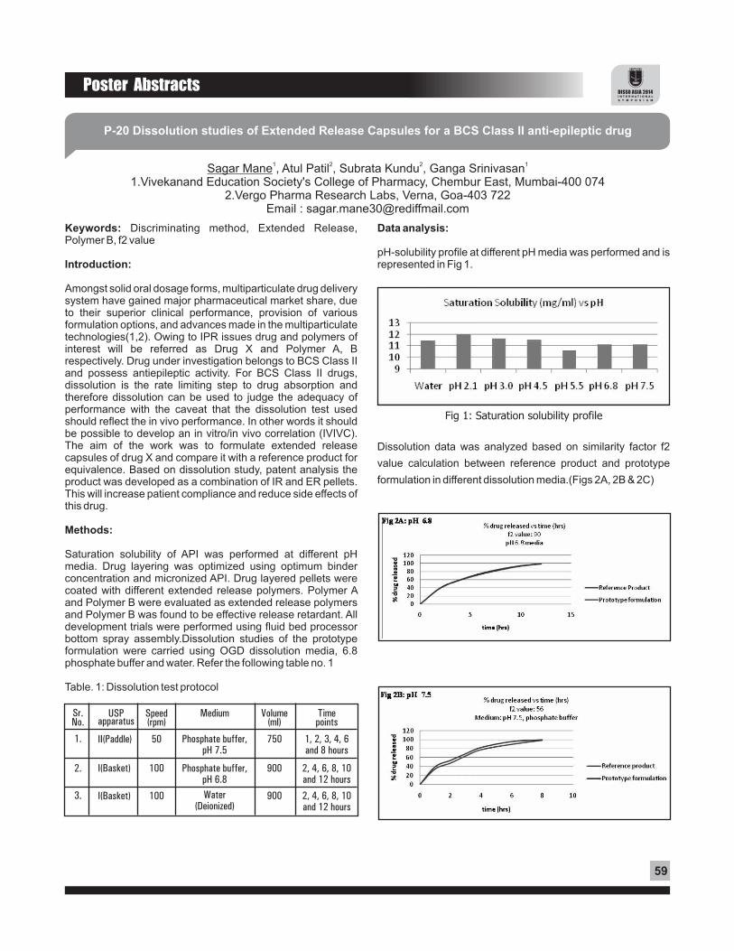
Poster Abstracts
59
P-20 Dissolution studies of Extended Release Capsules for a BCS Class II anti-epileptic drug
1 2 2 1Sagar Mane , Atul Patil , Subrata Kundu , Ganga Srinivasan1.Vivekanand Education Society's College of Pharmacy, Chembur East, Mumbai-400 074
2.Vergo Pharma Research Labs, Verna, Goa-403 722Email : [email protected]
Keywords: Discriminating method, Extended Release, Polymer B, f2 value
Introduction:
Amongst solid oral dosage forms, multiparticulate drug delivery system have gained major pharmaceutical market share, due to their superior clinical performance, provision of various formulation options, and advances made in the multiparticulate technologies(1,2). Owing to IPR issues drug and polymers of interest will be referred as Drug X and Polymer A, B respectively. Drug under investigation belongs to BCS Class II and possess antiepileptic activity. For BCS Class II drugs, dissolution is the rate limiting step to drug absorption and therefore dissolution can be used to judge the adequacy of performance with the caveat that the dissolution test used should reflect the in vivo performance. In other words it should be possible to develop an in vitro/in vivo correlation (IVIVC). The aim of the work was to formulate extended release capsules of drug X and compare it with a reference product for equivalence. Based on dissolution study, patent analysis the product was developed as a combination of IR and ER pellets. This will increase patient compliance and reduce side effects of this drug.
Methods:
Saturation solubility of API was performed at different pH media. Drug layering was optimized using optimum binder concentration and micronized API. Drug layered pellets were coated with different extended release polymers. Polymer A and Polymer B were evaluated as extended release polymers and Polymer B was found to be effective release retardant. All development trials were performed using fluid bed processor bottom spray assembly.Dissolution studies of the prototype formulation were carried using OGD dissolution media, 6.8 phosphate buffer and water. Refer the following table no. 1
Table. 1: Dissolution test protocol
Sr. No.
1.
2.
3.
USP apparatus
II(Paddle)
I(Basket)
I(Basket)
Speed (rpm)
50
100
100
Medium
Phosphate buffer, pH 7.5
Phosphate buffer, pH 6.8
Water (Deionized)
Volume (ml)
750
900
900
Time points
1, 2, 3, 4, 6 and 8 hours
2, 4, 6, 8, 10 and 12 hours
2, 4, 6, 8, 10 and 12 hours
Data analysis:
pH-solubility profile at different pH media was performed and is represented in Fig 1.
Fig 1: Saturation solubility profile
Dissolution data was analyzed based on similarity factor f2
value calculation between reference product and prototype
formulation in different dissolution media.(Figs 2A, 2B & 2C)

60
Poster Abstracts
Figs 2A, 2B & 2C: Dissolution profiles of reference product and prototype formulation
Results and conclusion:
From the saturation solubility data, it was concluded that drug exhibits nearly pH independent drug release profile. In order to develop discriminating dissolution medium, prototype formulation was subjected to dissolution test as per protocol described in table no 1. Based on f2 value calculation, it was
clear that the pH 6.8 media was more discriminating than the other media evaluated. This discriminating dissolution media was selected for dissolution testing of further developmental batches.Thus extended release capsules were prepared comprising Polymer B as release retarding polymer. Such system was expected to be helpful in reducing dosing frequency to once a day and thus increasing patient compliance.
References:
1) Rekhi, G.S. Advances in Solid Dose Oral Drug
Deliveries. ONdrugDelivery 2010; pp. 14-18.
2) Porter, S. C. Coating of Tablets and Multiparticulates.
In Aulton's Pharmaceutics: The Design and
Manufacture of Medicines, Fourth Edition; Aulton, M.
E.; Taylor, K. M.; Churchill Livingstone: New York,
2001; pp. 566-583.

Poster Abstracts
61
P-21 Fabrication and Evaluation of Apparatus for Simultaneous Dissolution and Permeation studies-A feasibility study
Ravi Sonkamble, Mala Menon.Bombay College of Pharmacy, Kalina, Santacruz (E), Mumbai 400 098
Email : [email protected]
Key Words: In vitro Dissolution-permeation, Permeation, Diltiazem, Ketoprofen, Hydrochlorothiazide
Introduction:
Oral administration is one of the most convenient and practical route of administration, from the point of view of both the patient and the manufacturer. Good oral bioavailability occurs when the drug has maximum solubility/dissolution in the g.i. fluids and good permeability through the intestinal mucosal membrane. The extent of absorption of drug in vivo thus, could be predicted based on permeability and solubility measurements. Stephan A. Motz has reported the simultaneous dissolution and permeation of dosage form by coupling Type IV dissolution apparatus with CaCO-2 permeation cell; such equipments would be useful in -screening of new API and innovative formulations to detect if any of the excipients interact with intestinal epithelia and alter the absorption of various drugs.
Objective:
In present research work, an attempt has been made to fabricate a simple Dissolution-Permeation Apparatus, wherein a modified permeation cell has been combined with the USP Type IV dissolution apparatus (Flow though cell), for simultaneous dissolution and permeation evaluation of oral drugs and formulations.
General Features Of The Apparatus:
The apparatus (Figs 1A & B) consists of: 1)Dissolution unit, and 2) Permeation unit. The Dissolution unit consists of the USP apparatus 4 (?ow through cell, Sotax, Germany) equipped with a 12 mm dissolution cell (C). The Permeation unit is subdivided into two compartments: Donor compartment (D) and Receiver compartment (R) with an intestinal membrane segment or other suitable membrane (M) mounted between these both compartments.
Fig 1A Schematic Diagram
Fig 1B Fabricated Apparatus
Dissolution-Permeation Apparatus(C =Flow through cell (12mm), A = Dissolution media reservoir,
B = Permeation media reservoir,D = Donor Compartment, R = Receiver compartment
M = Intestinal membrane segment)
Working Principle:
• A Pump P1 continuous circulates medium for dissolution (mimics gi fluids) from reservoir A to flow through cell C to donor compartment D.
• As the medium moves through D, based on permeability, dissolved drug will permeate through membrane (M) to the receptor compartment R, where there is a constant circulation of permeation medium by pump P2 from reservoir B.(mimics Blood compartment).
• With periodic sampling from A (dissolved drug) and B (permeated drug), we can calculate amount of drug dissolved and permeated and thereby simultaneous assessment of dissolution and permeation is possible.
Experimental :
In vitro Dissolution-Permeation studies:
The in vitro dissolution-permeation studies were carried out in triplicate, on 30 mg drugs belonging different BCS Class, viz Diltiazem (DLTZ)) (BCS Class 1), Ketoprofen (KTF) (BCS Class 2) and Hydrochlorothiazide (HTZ) (BCS Class 3) using the fabricated dissolution– permeation apparatus

62
Poster Abstracts
Medium and conditions:
Dissolution Medium :- 100 ml Kreb-Ringer Buffer solution pH 7.4; Permeation Medium :- 50 ml Kreb-Ringer Buffer solution pH 7.4; Temp :- 37±0.5°C; Glass beads :- 4 gm; Flow rate :- 4ml/min; Filters used :- GF-D and GF-F
Results :
Dissolution and permeation plots of drugs are depicted in Fig 2 (A-D)
Dissolution and Permeation plots :
It was observed that depending upon class of each drug, its dissolution and permeation curves varied (Fig 2); percent dissolution of the drugs in about 30 min was found to be 100%, 61% and 85% for DLTZ, KTF and HTZ respectively, which are in accordance to solubility characteristics. % permeability of the same drugs in about 120 min was found to be 3.4 %, 0.78% and 0.53% for DLTZ, KTF, and HTZ respectively which again are indicative of permeability characteristics.
Conclusion:
With these preliminary studies using only API it can be concluded that the fabricated instrument is simple, and capable of distinguishing simultaneously dissolution and permeation characteristics. However, further studies are necessary with different drugs as well as their formulations and further improvements in the instrument are also being explored.
References:
Stephan A. Motz, Michael Bur, Ulrich F. Schaefer, and Claus-Michael Lehr, Instrumented In Vitro Approaches to Assess Epithelial Permeability of Drugs from Pharmaceutical formulations, Drug absorption studies, Biotechnology: Pharmaceutical aspects 7 (2008) 430-455.
Fig 2 A Dltz Dissolution Plot
Fig 2 B Dltz Permeation Plot
Fig 2 C KTF & HTZ Dissolution plot
Fig 2 D KTF & HTZ Permeation Plot

Poster Abstracts
63
P-22 In-vitro Drug Release Studies of Once a Day Dosing Bilayer Tablet
1 2 2 1VaishnaviVaidya , Dhanraj Amin ,Arun Kumar ,Ganga Srinivasan1.Vivekanand Education Society's College of Pharmacy, Chembur East, Mumbai-400074
Ajanta Pharma Limited, Charkop, Kandivali (West), Mumbai 400067Email : [email protected]
Keywords:
Bilayer tablet, intermittent claudication, matrix system, kinetic models
Introduction:
Within the oral drug delivery market, modified release formulations create the largest demand. [1]Bi-layer tablet technology is one of the approach of designing modified drug delivery systems. In the present study an attempt has been made to develop a bi-layer tablet, in which aspirin and cilostazol layers have different release profiles. Cilostazol is cyclic AMP (cAMP)phosphodiesterase III (PDE III) inhibitor. It reduces the pain of intermittent claudication by widening the arteries, thereby improving the flow of blood and oxygen to the legs.Since the immediate release tablet of cilostazol is quickly disintegrated in the body when orally administered, a large amount of cilostazol is released within a short period of time yielding high concentration in blood, resulting in side effects such as headache, heavy feel in head. Formulation of cilostazol into sustained release layer reduces dosing frequency from twice a day to once a day and also helps to maintain constant concentration of drug in blood for prolonged period, thus reducing side effects. Aspirin, being antiplatelet agent is used synergistically with cilostazol in the treatment of intermittent claudication. Cilostazol and aspirin both belong to BCS class-II. These drugs have low solubility and high permeability drugs. Dissolution is often the rate limiting step for drug absorption. The challenges encountered in dissolution of BCS Class II drug are unpredictability of in vitro dissolution data to in-vivo, as they show pH dependent solubility, precipitation of drug and if these drugs are not completely released in the gastrointestinal tract, they will have a low bioavailability.
Methods:
Different formulation approaches like direct compression, wet granulation were tried. Various trials were taken by varying parameters like proportion of excipients, polymer concentration, while keeping the amount of drug constant.Owing to IPR issues polymers are referred to as Polymer “X” and Polymer “Y”. Swellable matrix type of system was developed using Polymer “X” and Polymer “Y” as hydrophilic release controlling polymers. Release profile of formulation was studied using method described in table 1. HPLC method of analysis was used to quantify drug release in dissolution tests.
Data analysis:
Model dependant methods were used to investigate the kinetics of drug release from the formulation. For model dependant drug release, the drug release parameters were fitted into mathematical equations like Higuchi's model, Hixoncrowell cube root model, zero order and first order models. Plots of cumulative % drug release vs time (zero order), log cumulative of % drug remaining vs time (first order kinetic model), % drug release vs square root of time (Higuchi model), cube root of % drug remaining in matrix vs time (Hixon –crowell cube root law) were made. [2]
Layer
Cilostazol
Aspirin
Type of dissolution apparatus
USP-2 paddle
USP-I Basket
Speed of rotation (RPM)
75
50
Dissolution medium
0.3% SLS in water
Acetate buffer pH 4.5
Volume of dissolution
medium (ml)
900
500
Time(hours)
14812
Amount of drug
dissolved
NMT 20%25% - 50%50% - 75%NLT 80%
NLT 75% in 45minutes
Acceptance Criteria
Table 1: Dissolution test parameters
Results:
Bilayer tablet was developed in which immediate release layer of aspirin and sustained release layer of cilostazol both were formulated by wet granulation technology. Initial trials of sustained release layer were taken using combination of Polymer “X” and Polymer “Y”. But later it was observed that Polymer “Y” alone could control the drug release. More than 80% of aspirin was released from aspirin layer within 45 minutes.
Drug release kinetics:
Best linearity was found in Hixon-Crowell's cube root law. From Figs 1 & 2, it is evident that use of polymer “Y” in optimized concentration achieved desired release profile.

64
Poster Abstracts
Figure 1:Hixon-Crowell's cube root plot for formulation with the combination of Polymer “X” and Polymer “Y”
Figure 2:Hixon-Crowell's cube root plot for formulation only with Polymer “Y”
Conclusion:
Modified release tablets of Cilostazol and aspirin were prepared successfully using Polymer “Y” to achieve desired dissolution profile. As the two layers have different release profiles, dissolution tests for these layers were performed separately in two different dissolution media. 0.3% SLS is used as dissolution medium because it aids solubilisation of poorly soluble cilostazol.Drug release kinetics of this formulation relates best to Hixon-Crowell's cube law indicating a change in surface area and diameter of tablets with progressive dissolution as function of time. This approach helps to reduce dosing frequency and thus increase patient compliance.
References:
1. Drug technologies. Advances in solid dose oral drug delivery. http://www.ondrugdelivery.com. (accessed feb 20, 2014)
2. Paulo Costa, Jose´ Manuel Sousa Lobo, Modeling and comparison of dissolution profiles. European Journal of Pharmaceutical Sciences:13:(2001);123–133.

Poster Abstracts
65
P-23 Investigation of Various Factors on Ion Exchange Resin Complexation with Fexofenadine Hydrochloride
Divya Suares, Arti HirayShobhaben Pratapbhai Patel School of Pharmacy & Technology Management,
SVKM's NMIMS, Vile Parle (W), Mumbai – 400056, Maharashtra, IndiaEmail: [email protected]
Keywords: Ion exchange resin, Fexofenadine hydrochloride,
Indion, Kyron, Orodispersible
Introduction:
Ion exchange resins are high molecular weight water insoluble 1, 2polymers. They are inert and are not absorbed by the body .
Use of ion exchange resin is one of the most economical
methods reported for masking bitter taste of various drugs.
Fexofenadine hydrochloride is a antihistaminic agent and is
highly active via oral administration. When a bitter drug is
complexed with an ion exchange resin in an orodispersible
tablet, the drug is not released in the saliva, but undergoes
decomplexation when it comes in contact with the gastric fluid
and releases the drug immediately in the stomach. This does
not affect the intrinsic bioavailability of the drug. Though
numerous pharmaceutical compositions for the oral
administration of fexofenadine hydrochloride have been
proposed, there still exists a need for commercially acceptable
cost-effective formulation for oral administration with good
patient convenience and acceptance, especially for pediatrics 3and geriatrics .
Objective:
The objective of this study was to mask the intense bitter taste
of Fexofenadine hydrochloride using weak cation exchange
resins and to formulate orodispersible tablet of taste masked
drug resin complex.
Methods:
Formulation of orodispersible tablets containing Fexofenadine
hydrochloride by using ion exchange resins viz. Indion 204,
Indion 234, Indion 414, Kyron T-114 and Kyron T-314 were
explored. The optimization of drug loading capacity of resin
was performed by determining the effect of various factors
such as drug: resin ratio, soaking time, stirring time,
temperature and pH.
Data analysis:
The drug resin complexation was confirmed by infra red spectroscopy. The evaluation parameters studied included taste masking, drug content and drug release. The optimized blends were formulated to tablets by direct compression technique. The tablets were evaluated for diameter and thickness measurement, hardness test, weight variation test, in vitro USP disintegration test, wetting time, test for content uniformity, assay, friability test and in vitro dissolution studies. In vitro dissolution studies were performed as per the dissolution guideline for fexofenadine hydrochloride given by CDER, in 0.001 N HCl at 37±0.5°C. The data obtained was kinetically analyzed and dissolution efficiency was computed.
Results:
The amount of drug released at salivary pH was found to be very less. Thus, the amount of drug released from the DRC at salivary pH indicates its insufficiency in imparting bitter taste in the mouth during administration. The drug release from drug resin complex at gastric pH was found to be more than 80% after 10 minutes. Thus, the amount of drug released from drug resin complex at gastric pH indicates decomplexation of DRC in presence of gastric fluids and availability of drug for absorption.
Figure 1: In-vitro dissolution profile of Orodispersible tablet.

66
Poster Abstracts
Conclusion :
Effective taste masking of Fexofenadine hydrochloride was achieved with selected ion exchange resin (i.e. Indion 234 and Kyron T-314). The resins used have an added advantage of fast-disintegrating property and direct compressible quality. Hence it can be concluded that, preparation of orodispersible tablets of taste masked Fexofenadine hydrochloride using direct compression technique, without the addition of superdisintegrants is a successful cost-effective method. They would be effective in providing fast onset of action without the need of water for swallowing.
Acknowledgement :
Authors wish to thank Sun Pharmaceuticals, Mumbai for the gift sample of Fexofenadine hydrochloride.
References :
1. Devi V, Krishna M. Advances in controlled and novel drug delivery. In: Jain NK, editor. 1st Ed.; CBS publishers and Distributors; 2001. p. 290–306.
2. Amina Faham, Dominique Marechal, Philippe Chenevier. Orodispersible tablets containing Fexofenadine. “U.S. Patent” 2004, No. 6,723,348.
3. Redkar MR, Gore SP, Devarajan PV. D-Zolv Taste masked mouth dissolving Tablets. Indian J Pharm Sci. 2002; 64(3): 291-292.

Poster Abstracts
67
P-24 Sustained release tablets of venlafaxine hydrochloride: A novel approach to decrease the frequency of dosing
1 1 2Swati Mittal , Kshitija R Khedekar , Heeshma C Shah 1 VES College of Pharmacy, HAMC Chembur, Mumbai.2Signet Chemical Corporation Pvt. Ltd. BKC Mumbai.
Key words: Sustained release, venlafaxine hydrochloride, ethyl cellulose, Metolose
Introduction:
Venlafaxine is a bicyclic phenyl ethylamine derivative, unique 1antidepressant used as first-line therapy . The successful
treatment of depression depends on the maintenance of effective drug concentration levels in the body for which a constant and uniform supply of drug is desired. The steady state half life of venlafaxine is 5 necessitating frequent administration, two or three times daily to maintain adequate
2 plasma levels. Because of its short half life and its high water solubility venlafaxine hydrochloride was chosen as suitable candidate for matrix tablet formulation that provides sustained release with desired therapeutic effect.
Methods:
Drug- excipients interaction studies:were assessed at 25ºC ± 2ºC/60% RH ± 5% RH and 40ºC ± 2ºC/75% RH ± 5% RH for 1 month to study the drug- excipients compatibility.
Preparation of sustained release venlafaxine hydrochloride matrix tablet:Matrix tablets were prepared by wet granulation method using
Rvarying concentrations of hydrophilic (HPMC (Metolose )) and hydrophobic polymer (ethyl cellulose) (table no.1)
3The optimized formula was obtained using a 2 factorial design.
In- vitro dissolution studies: Drug release rate of tablets was determined using USP apparatus II (paddle). The test was performed in 900ml distilled water maintained at 37 ± 0.5°C stirring at a speed of 50rpm. Aliquots of 10ml were collected at periodic intervals up to 24hr, and equivalent amount replaced to maintained sink conditions.
The drug content in withdrawn aliquots was analyzed at ? max
of 224nm.
Data Analysis:
Dissolution profile comparisons:In-vitro release profile of venlafaxine hydrochloride sustained release tablets was compared with drug release profile of marketed formulation VENLOR-XR capsules under similar conditions. Difference factor (f1) and similarity factor (f2) were
n ncalculated using the formula, F = {[Ó |R –T |]/[Ó R ]}×100 1 t=1 t t t=1 tn 2 -0.5and F = 50× log {[1+ (1/n) Ó ( R – T ) ] × 100},where, N is 2 t=1 t t
number of time points, Ri and Ti are dissolution of reference and test product at time I respectively. Values of f (<15) and f 1 2
(>50) indicate, that the curves can be considered similar and have similar drug release behavior (table no.5 and figure no.3).
Results And Discussion:
Ethyl cellulose and venlafaxine hydrochloride were used in the ratios of 1:1 and 1:3. At higher concentration, 90% drug release was observed at end of 6hr.The drug release was sustained due to hydrophobic nature of polymer. Metolose 90SH-100000SR was used in ratios of 1:1 and 1:4 with respect to venlafaxine hydrochloride. At higher concentration, 90% drug release was observed at end of 8.5hr. High polymer content results in a greater amount of gel being formed. This viscous nature of gel increases the diffusional path length of the drug, and its diffusion coefficient, thus retarding the drug release. Metolose and venlafaxine hydrochloride (90SH-100SR and Metolose 90SH-100000SR) were tried in conc.1: 2. Metolose could sustain the release for a period of 21 h. The dissolution profile of optimized formula was matched with marketed formulation (Table 2, Figs 1 & 2).
Table no.1: Developed formulations of venlafaxine hydrochloride tablets
Ingredients
VNL
EC7
90SH-100000SR
90SH-100SR & Metolose 90SH-100000SR
Avicel 101
Magnesium stearate
Aerosil
F1
84.87
34
2%
F2
84.87
59
2%
F3
84.87
34
2%
F4
84.87
41
2%
F5
84.87
84.87 254.6 - - -
- - 84.87 339.4 -
- - - - 169.74
57
1% 1% 1% 1% 1%
2%

68
Poster Abstracts
Fig .1: Release profile of sustained release tablets
of venlafaxine hydrochloride
Table no. 2: Comparison of dissolution profiles
2
4
6
8
10
24
sum
Time (hr)
30
51.84
62.73
75.12
80.63
102.57
402.89
Avg. Reference (mg)
29.38
44.05
59.9
66.83
76.00
99.31
375.47
Avg. Test (mg)
0.62
7.79
2.83
8.29
4.63
3.26
27.42
/R-T/
0.3844
60.684
8.0089
68.724
21.437
10.628
169.87
2/R-T/
Fig 2: Comparison of dissolution profiles of marketed formulation with developed formulation
Conclusion:
From the above study, it has been observed that both hydrophobic polymer ethyl cellulose and hydrophilic polymer Metolose were capable of retarding the release of water soluble drug venlafaxine hydrochloride. However, optimum release could be obtained by using combination of ethyl cellulose, 90SH-100SR and Metolose 90SH-100000SR.
Acknowledgments:
Aarti Industries Ltd., Mumbai and Signet Chemical
References:
1. Shanmugam S, Archana M and Vetrichelvan T: Formulation and Evaluation of Sustained Release Matrix Tablets of Venlafaxine Hydrochloride. Bio Med Rx 2013; 1(5): 506-510.
2. Makhija SN, Vavia PR: Once daily sustained release tablets of venlafaxine, a novel antidepressant. European Journal of Pharmaceutics and Biopharmaceutics 2002; 54: 9–15.
Corporation Pvt. Ltd. for providing gift samples.