Embed Size (px)
Citation preview

1
COMPARISON OF VISUAL OUTCOME AT ONE YEAR FOLLOWING
PHOTOREFRACTIVE KERATECTOMY (PRK) AND PHOTOREFRACTIVE
KERATECTOMY WITH MITOMYCIN C IN HIGH MYOPIA
Dissertation Submitted to The Tamil Nadu M.G.R. MEDICAL UNIVERSITY
CHENNAI, INDIA.
M.S. DEGREE EXAMINATION BRANCH – III, OPTHALMOLOGY
MARCH, 2010

2
ACKNOWLEDGEMENT
I would like to express my profound gratitude to our Director Dr. C.A. Nelson Jesudasan, M.S, D.O.M.S, FRCS (Glas& Edin).ICEH,
Joseph Eye Hospital, Trichy.
I am really indebted to Dr. Pragya Pamer, M.S., Professor for being
my guide in this study. Her correction moulded me in every step of this
study.
I would like to thank, Dr. C.M. Kalavathy, M.S., D.O., Professor and
HOD of Cornea, for her support and understanding in completing this
study.
I would like to thank Dr. M.D. Ramalingam, M.s., Professor for support
and understanding in completing this study.
I would like to thank Dr. Amjad Salman, M.s., Registrar who gave me
guidance and inspiration.
I extemd my thanks and gratitude Mr. R. Mohamed ismail for
preparing me the manuscript in this study.
I also thank Mr. B.E. Rajkumar, ,Librarian, Assistant Registrar, Mr. Daniel Prince, Assistant Librarian Mr. R. Venkatraman for their help
during the study.
I thank all the patients who participated in this study and for their co-
operation.
3
CONTENTS TABLE
S.NO CONTENTS PAGE NO.
1 Aim 1
2 Introduction 2 – 6
3 Review of literature 7 – 36
4 Materials and methods 37 – 40
5 Results 41 – 47
6 Discussion 48 – 51
8 Conclusion 52
9 Bibliography 53 – 55
10 Proforma 56 – 57

4
AIM

5
AIM
To compare visual outcome at one year following photorefractive
keratectomy (PRK) and photorefractive keratectomy with mitomycin C in high
myopia

6
INTRODUCTION

7
INTRODUCTION
Myopia / short sightedness is a refractive error in which the parallel
rays of light from infinity come to focus in front of the retina when the eye is at
rest, thus grossly reducing the vision. Myopia could be axial due to
elongation of anteroposterior diameter of the eyeball or curvature myopia due
to increase in radius of curvature of the cornea; and index myopia due to
change in refractive index of lens, cornea, aqueous and vitreous, thus
increasing the dioptric power of the eye.
Photorefractive keratectomy (PRK)
Consists of the application of energy ofthe ultraviolet range generated
by an argon fluoride (ArF) excimer laser to the anterior corneal stroma to
change its curvature and, thus, to correct a refractive error. The physical
process of remodeling the corneal stroma by ultraviolet (193 nm wavelength)
high-energy photons is known as photoablation.
History of the Procedure
During the 1980s, several applications of the 193-nm ArF excimer
laser were investigated, including its use on human corneas for the correction
of refractive errors. In 1988, Munnerlyn, Kroons, and Marshall reported an
algorithm relating diameter and depth of the ablation to the required dioptric
change.
8
McDonald performed the first excimer PRK for the correction of myopia
on a normally sighted human eye in the United States. That same year, the
Food and Drug Administration (FDA) organized a 3-phase trial, the PRK study
(which ended in 1996), to demonstrate the safety, predictability, and stability
of PRK for the treatment of myopia. At the end of this trial, 2 ophthalmic
companies, VISX and Summit, were allowed to manufacture excimer lasers
for widespread use in the United States. Since then, Nidek also has obtained
approval for the manufacture of excimer lasers in the United States, and
several hundred thousand patients have undergone this procedure
throughout the world. The first excimer lasers used to perform PRK in the late
1980s have been improved significantly in terms of size, efficiency, and
accuracy.
Pathophysiology
The mechanism of ablation of the excimer laser appears to be
photochemical in nature and is known as photochemical ablation or ablative
photodecomposition. This highly localized tissue interaction is based on the
fact that each photon produced by the ArF excimer laser has 6.4 eV of
energy, enough to break covalent bonds.
The efficacy and success of PRK depends largely on the type of laser
platform in use. Current fifth generation systems use a very rapidly repetitive
and extremely small spot delivery with automated tracking of the eye
movements to ensure precise treatment.

9
The intramolecular bonds of exposed organic macromolecules are
broken when a large number of high-energy 193-nm photons are absorbed in
a short time. The resulting fragments rapidly expand and are ejected from the
exposed surface at supersonic velocities. This mechanism explains why only
the irradiated organic materials are affected, whereas the adjacent areas are
not affected.
The return of corneal innervation up to 5 years after PRK was
measured. Corneal subbasal nerve density does not recover to near
preoperative densities until 2 years after PRK, as compared to 5 years after
laser in situ keratomileusis (LASIK).
Mitomycin C
Isolated from streptococcus calspitosus in 1958, mitomycin react with DNA in
ways similar to alkylating agents. It cross links DNA and inhibits its synthesis.
It is highly effective antimitotic agent used in treatment of carcinoma of
stomach and colon. The ocular indications for the mitomycin C are recurrent
pterygium and glaucoma filtering surgery.
Kulitoma and Mori (1976) reported favorably on the efficacy of mitomycin C
eye drops in recurrent pterygium.
10
Use of mitomycin C in PRK
They act by inhibiting activated keratocytes, probably by interfering
with DNA synthesis, which decreases cellular activity and reduces collagen
synthesis. Mitomycin inhibits fibroblst function by a dose dependent inhibition
of fibroblast proliferation.
Mujmudar et aladn Raviv et al reported mitomycin C efficacy in treating
significant pre existing corneal scarring
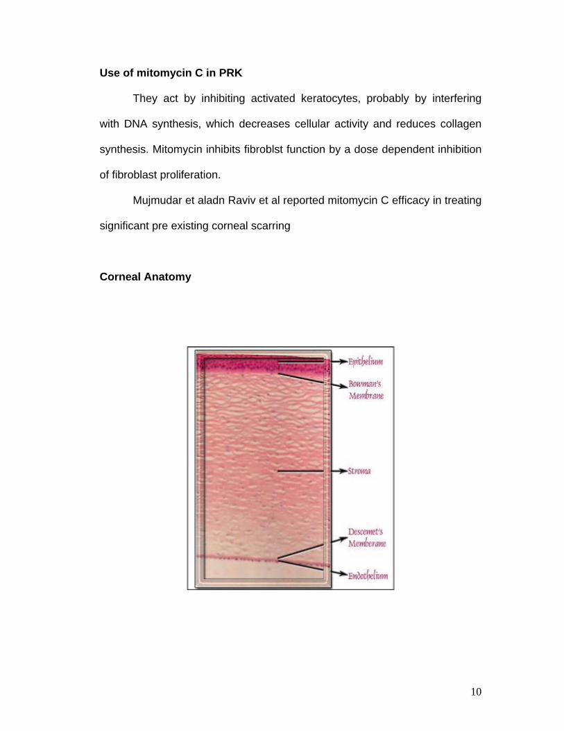
Corneal Anatomy
11
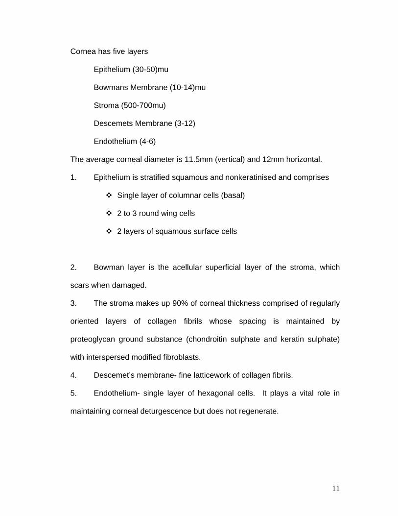
Cornea has five layers
Epithelium (30-50)mu
Bowmans Membrane (10-14)mu
Stroma (500-700mu)
Descemets Membrane (3-12)
Endothelium (4-6)
The average corneal diameter is 11.5mm (vertical) and 12mm horizontal.
1. Epithelium is stratified squamous and nonkeratinised and comprises
Single layer of columnar cells (basal)
2 to 3 round wing cells
2 layers of squamous surface cells
2. Bowman layer is the acellular superficial layer of the stroma, which
scars when damaged.
3. The stroma makes up 90% of corneal thickness comprised of regularly
oriented layers of collagen fibrils whose spacing is maintained by
proteoglycan ground substance (chondroitin sulphate and keratin sulphate)
with interspersed modified fibroblasts.
4. Descemet’s membrane- fine latticework of collagen fibrils.
5. Endothelium- single layer of hexagonal cells. It plays a vital role in
maintaining corneal deturgescence but does not regenerate.

12
REVIEW OF LITERATURE

13
REVIEW OF LITERATURE
Myopia
Short sightedness is a type of refractive error in which parallel rays of
light coming from infinity are focused in front of the retina when
accommodation is at rest.
Mechanism of production
1. Axial myopia results from increase in anteroposterior length of the
eyeball. It is the commonest.
2. Curvatural myopia occurs due to increased curvature of the cornea,
lens, or both.
3. Positional myopia- anterior placement of crystalline lens in the eye.

14
4. Index myopia- results increase in RI of crystalline lens associated with
nuclear sclerosis.
5. Myopia due to excessive accommodation occurs in patients with
spasm of accommodation.
Optics of Myopia
In myopes, a near object may be focused without any effort of
accommodation if it is situated at the punctum remotum. The image of an
object at infinity is made up of circles of diffusion formed by diverging beam.
The nodal point in myopes is further away from the retina and image formed
is larger in size. Accomodation is of little value to myopes for higher errors.
The amplitude of accommodation is small.
Classification of Myopia
a. Congenital Myopia
b. Simple Myopia
c. Pathological Myopia
d. Acquired Myopia
Congenital Myopia
Is present since birth
Diagnosed by the age of 2-3 years
Seen in premature children or in various birth defects such as Marfan’s
syndrome or Homocysteinuria

15
Is unilateral and manifests as anisometropia
Error is about 8 to 10 D associated with congenital squint
Associated with cataract, microphthalmos, aniridia, megalocornea and
congenital separation of retina
Simple Myopia
Simple or developmental myopia is the commonest type and considered as a
physiological error and not associated with any disease of the eye.
Etiology: It results from normal biological variations in development of the
eye which may or may not be genetically determined.
Axial Myopia: results from the increase in the anteroposterior length of the
eyeball. It is the commonest form.
Curvature Myopia: results from increase in the radius of curvature of the
cornea, lens or both. A variation of 1 mm of radius of curvature results in a
refractive change of 6 D.
Index Myopia: Here, changes in dioptric system may be due to change in
refractive index of lens, cornea, aqueous or vitreous. The dioptric power of
the eye is too strong for the axial length of the eye.
16
Clinical Symptoms:
Poor vision for distance (short sightedness) is the main symptom of
myopia.
Asthenopic symptoms occur in patients with small degree of myopia.
Eyestrain develops due to dissociation between convergence and
accommodation.
Fusion becomes weak and binocular vision is affected.
Patient starts suppressing one eye and suppressed eye deviates
outwards.
Decreased visual acuity in indoor activities.
Signs:
The eye of simple myopia is large and prominent. Chamber is deep
and pupil reaction is sluggish. Macula appears slightly nearer to disc. There
may be divergent squint.
Pathological / Progressive / Degenerative Myopia
It is a rapidly progressive error resulting in high myopia during
early adult life, usually associated with degenerative changes in
the eye.

17
It results from rapid axial growth of the eyeball outside the
normal biological variation of development.
It is definitely linked to heredity and general growth process.
Heredity factor: Progressive myopia is familial and more common in races
like the Chinese, Japanese, Arabs and Jews.
Role of general growth process: Factors like nutritional deficiency, debilitating
diseases, endocrinal disturbances and indifferent general health also
influence progress of myopia.
Clinical Picture
Defective vision
Night blindness
Muscae volitantes
Signs
Eyes are more prominent
Anterior Chamber is deep
Cornea appears large
Pupil slightly large and sluggish
Refractive error increases by as much as 4D yearly and usually
stabilizes at the age of 20.

18
Fundus Picture
o Optic disc appears large and pale
o Myopic crescent
o Atrophy of retinal pigment epithelium and choriocapillaries- tigroid
appearance due to visible prominent large choroidal vessels
o Atrophic patches at macula
o Foster Fuchs spot
o Cystoid degeneration
o Posterior staphyloma
o Posterior vitreous detatchment
o Contraction of visual fields
o ERG subnormal
Complications
Retinal detachment
Complicated cataract
Vitreous haemorrhage
Choroidal haemorrhage

19
Treatment
Non Surgical
Spectacle correction with concave lens
Contact lens
Surgical
Radial Keratotomy
Photo-Refractive-Keratectomy
LASIK
Intra corneal ring implantation for low myopia
Phakic IOLS
History
Refractive surgery has been used to correct myopia and
hypermetropia for more than 40 years. Conceptually, refractive corneal
surgery attempts to remove, add or modify the corneal stroma, so that the
radius of curvature of the anterior corneal interface is altered as desired.
Based on the fundamental principle that cornea contributes twothirds
of refracting power of the eye, Barraquer attempted to alter the tear film/
anterior cornea interface radius of curvature by adding or removing corneal
tissue.

20
Keratomileusis in situ- derived from the Greek word keras ( horn-
like=cornea) smileusis- carving. Keratomileusis in situ for myopia was the first
to develop in late 1940. The procedure involved raising a corneal flap and
removing tissue from residual stromal bed. Barraquer performed a free hand
lamellar dissection of the anterior half of the cornea with a keratome.
Subsequently, refractive cut was attempted with second pass of the knife to
remove stromal bed and cap was replaced with flattening of corneal curvature
thus reducing myopia. This procedure was abandoned due to many technical
difficulties.
The gateway to Keratophakia opened in 1961, which involved
steepening of central corneal curvature by placing a disc of tissue under the
lamellar cap derived from alloplastic stromal disc harvested from a donor
cornea.
This was seen a possible solution in aphakia, but with advent of IOL,
interest in keratophakia subsided.
Freeze myopic Keratomileusis
In an effort to overcome technical difficulties of manual cut, Barraquer
used contact lens lathe to sculpture the frozen lamellar cap. Barraquer
recognized that the cutting speed and the relation between FOP and diameter
of resection were factors directly affecting quality and depth of cut. His efforts
for more predictable and accurate cuts led to the development of applanator

21
lenses, suction rings of various diameters and various heights of
microkeratome tracks. This work constituted the basis of future
microkeratome evolution.
Disadvantages:
1. Learning curve was too steep with higher complication rate.
2. Cryolathe was too expensive and complex to maintain.
Epikeratoplasty
Epikeratophakia was introduced in1979 by Kaufmann and Werblin to
avoid use of cryolathe. They used preprocessed refractive lenticules. A
stromal disc was removed from the donor eye with microkeratome and was
frozen and lathed into concave or convex lenses and then lyophilized and
stored for later use. It was intended for use in myopia, hypermetropia and
keratoconus.
Complications
1. Persistent epithelial defect
2. Epithelial ingrowth
3. Melting, scarring
4. Epithelialisation of donor lenticule

22
Barraquer-Krumeich Swinger (BKS) technique
An improved microkeratome, a set of dyes and suction stand
microkeratome was used to perform a total lamellar cap. The cap was placed
epithelial side down on suction rings for microkeratome to perform second cut
on stromal aspect of cap. The sculptured lamellar disc was finally sutured
back to the bed. The nonfreezing technique has more advantage over freeze
techniques with rapid and comfortable recovery, preservation of fibroblasts
and epithelium. However, significant astigmatism could not be avoided.
Automated lamellar keratoplasty
Development of automated geared microtome by Ruiz in 1980
introduced Automated lamellar keratoplasty. Speed of the cut could be
controlled resulting in more even and consistent cuts.
The second cut was made on the bed. The depth of second cut was
adjusted by altering the height of suction ring and corneal cap was sutured
back.
Advantages
Rapid recovery
Efficacy in correcting high myopia
Ease of use

23
Disadvantages
High degree of irregular astigmatism
Photorefractive Keratectomy
It is a procedure of photoablation by Excimer laser which has been in
use for the treatment of myopia, hypermetropia and astigmatism.
Photorefractive keratectomy has gained maximum success in myopic
patients. Photorefractive keratectomy has become popular after Srinivasan,
Barren and Trokel in 1983 thought that excimer laser can be used to cut the
cornea.
Photorefractive keratectomy gives good results from –2 to –6 D of
myopia.
Indications
1. Superficial scar with myopia
2. Basement membrane dystrophy with myopia
3. myopia when unable to use a microkeratome because of high brow or
tight lid with narrow palpebral fissures
4. Glaucoma suspects
5. cornea thinner than 500 microns

24
Photorefractive keratectomy can be satisfactorily performed under
topical anesthesia. The center of the pupil is marked; the epithelium is
removed by mechanical debridement with a blunt hockey knife followed by
followed by ablation of corneal stroma with excimer laser. Reepithelialisation
occurs in 3 to 4 days. Improvements in the laser profile, small spot size,
flying spot technology and Gaussian curve in conjunction with mitomycin C
have made it possible to treat higher degrees of myopia. Epithelium can also
be removed using alcohol or by excimer laser.
Complications are Uncommon Which Include
1. Corneal haze and regression
2. Night glare and halos
3. Delayed epithelial healing
4. Corneal ulceration
5. Corneal infiltration
6. Central islands
7. Decentration of ablation zone
Lasik
Is a keratorefractive surgery that combines the precision of excimer
laser photoablation with advantages of an intrastromal procedure that
maintains the integrity of Bowmans layer and overlying epithelium. It was
25
introduced, designed and developed at University of Crete and Verdin
Oyannion Eye Institute of Crete in 1988.
Currently, this procedure is being considered the refractive surgery of
choice for myopia because of its definite advantages over PRK and RK.
LASIK can be used to correct upto –15D of myopia and upto –6D of
astigmatism depending on the excimer laser platform used.
Mel 80 excimer laser is approved by the FDA for correction of myopia
upto -7.00 D. It can be used for correcting errors upto -15.00D, but it is a “off
label” use.
Preoperative evaluation
Marguente Mc Donald MD of New Orleans, Louisiana performed the
first PRK and has been one of the key surgeons responsible for extensive
pioneering work in appropriate approach to PRK.
A thorough preoperative evaluation is of critical importance in
achieving a successful outcome following refractive surgery. During this
evaluation, the surgeon decides if the patient is or not a good candidate for
refractive surgery.
1. MEDICAL HISTORY: It should include history of any systemic disease
like diabetes, prior surgeries, connective tissue disorders, and any

26
immunocompromised states like HIV / AIDS, drug history - isotretinold,
somatripten, amiadarone, and hormone replacement therapy.
2. REALISTIC PATIENT EXPECTATION: The ophthalmologist should
explain in detail about the refractive results. Patients should
understand will not prevent possible future ocular problems like
cataract, glaucoma or retinal detachment. Patients should be told that
PRK is being done to decrease dependence on glasses and not to get
rid of glasses.
3. Patient ocular history, blepharitis, recurrent erosion, dry eyes, retinal
tears, detachment, use of glasses, stability of current refraction,
contact lens history- types of contact lens, duration of contact lens use.
Patient should discontinue soft contact lens for at least three days to
two weeks prior to surgery. After 40 years, patients who have
undergone refractive surgery will need presbyopic correction with
glasses and this should be explained to the patient.
4. Uncorrected visual acuity for distance and near should be measured to
determine the amount of correction to be performed. Full cycloplegic
refraction is mandatory. A final subjective refraction using the
autorefractometer, Wasca aberrometer values and cycloplegic

27
refraction should be done by the ophthalmologist and refined using
duochrome test.
Pupil size
Pupillary examination involves evaluating pupil size in bright room light
and dim illumination, any afferent papillary defect, various techniques to
measure the size of the pupil like light amplification pupillometer, infrared
pupillometer, Wasca aberrometer.
Large pupil may be one of the risk factors for postoperative glare and
halo after refractive surgery. Optical zone should be larger than pupil
diameter to prevent glare and haloes.
Ocular motility or confrontation fields
Asymptomatic tropia and phoria may develop symptoms after surgery
if change of refraction causes the motility status to break down. Orthoptic
evaluation should be done preoperatively. Confrontation fields should be
done in all patients.
Intraocular pressure
In patients with glaucoma, lasik elevates the IOP during the procedure,
aggravating optic nerve damage.
Topical corticosteroid use after PRK may cause elevation of IOP in
corticosteroid responders.

28
Slit lamp Examination
Complete slit lamp examination of eye, lids, cornea and anterior
segment should be performed to check for meibonitis, stye, blepharitis, tear
film stability, conjunctival scarring, pterygium, epithelial erosions, dystrophies,
keratoconus or any opacities.
Depth of anterior chamber and lens clarity should be assessed.
Detailed Fundus Examination
It is important to be certain that the posterior segment is normal.
Special attention is given to the optic nerve (glaucoma, optic nerve drusen),
peripheral retina (retinal tear, detachment, retinal holes, peripheral
degeneration, high myopia) are at increased risk for retinal detachment.
Corneal Topography
It should be done to rule out keratoconus, contact lens
warpage
Ultra sound Pachymetry
It is done for central corneal thickness.
Wavefront analysis
This is done by using wasca aberrometer..

29
Informed consent
After evaluation, the surgeon analyses all the information and
discusses the possibilities with the patient. Risks and benefits of various
medical and surgical alternatives are discussed.
Discussions with the patient should include
1) Expected uncorrected visual acuity
2) Risk of decreased BCVA, severe visual loss, glare haloes, dry eyes
3) Need of distant or reading glasses
4) Patient should be given informed consent documents and should sign
the form well before the surgery.
PROCEDURE
In this technique to correct myopia, a central optical zone of anterior
corneal stroma is photoablated using Excimer Laser to cause flattening of
central cornea.
Surgical steps are:-
1. ANAESTHESIA: PRK can be satisfactorily performed under topical
anaesthesia.
2. EPITHELIUM REMOVAL: Deepithelialisation methods include
mechanical debridement with blunt blade under topical anaesthesia.

30
Other deepithelialisation methods include alcohol and photoablative
deepithelialisation. Attempt should be made to deepithelialise 0.5mm
to 1mm larger area than the desired ablative zone. The time lapsed
from the removal of corneal epithelium to the application of laser
energy should be minimized to prevent extreme drying or wetting of
corneal surface. Always avoid leaving any residual islands of
epithelium.
3. ABLATION ZONE DIAMETER: small ablative zones are known to
cause symptomatic haloes while night driving. Increasing experience
in PRK has shown that the ideal diameter of ablation zone for myopia
is 6mm and for hypermetropia is 9mm.
4. FIXATION AND CENTRATION OF THE ABLATION ZONE: some
surgeons use hand held suction rings, while others promote the
method of self fixation by the patient during ablation. Fixation light on
the microscope should be co axial with surgeon’s and patient’s line of
vision.
5. Co-axiality of fixation light should be maintained with the laser light
also. It should always be explained to the patient that the clarity of
fixation target will decline during ablation, but will be visible and that he
should try to fix it. It is always better to patch the fellow eye to prevent
cross fixation. Laser beam should be aimed at the center of the pupil.

31
6. FLUENCE AND REPETITION RATE: Changes in fluence and
repetition rate affect not only the rate at which tissue is removed and
the operation time, but also the surface morphology of the ablated
corneal tissue.
7. SCANNING LASER BEAM: The laser device can be smaller and
cheaper if small diameter circular or narrow slit beam is used for
scanning the ablated area.
8. ASPHERIC ABLATIONS: This is a unique advantage of PRK.
Planned aspheric ablations are made in high myopia, which avoid
central islands, thereby decreasing postoperative spherical
aberrations. Astigmatism occurring due to naturally, post traumatic,
post infective and post surgical correction is done by ablating the
superficial cornea in a cylindrical fashion known as toric photoablation.
The laser is centered and focused according to the manufacturer’s
recommendations. Although the excimer laser beam, a 193nm, is invisible to
the human eye, a faint fluorescence of deep blue light is sometimes visible
during stromal ablation.
The sound of the laser firing is the main feedback signal to the surgeon
along with an alteration in light reflex as the stromal ablation progresses.

32
In order to attempt to decrease the chance of postoperative corneal
haze after PRK in high myopia, a soaked pledget usually 0.2mg/ml mitomycin
can be placed on the ablated surface for 30 seconds to two minutes at the
end of laser exposure. The cornea is then irrigated with balanced salt
solution to remove excess mitomycin to avoid damage to limbal stem cells.
Immediate post ablation methods:
After the procedure is completed, a single drop of antibiotic and NSAID
are placed in the eye after a bandage soft contact lens is inserted.
Subsequent postoperative care:
The patient should be followed closely until the epithelium is
completely healed, which usually occurs within 72 hrs. As long as the
bandagesoft contact lens is in place, patients are treated with topical broad
spectrum antibiotics and corticosteroids, usually 4 times a day.
Studies have shown that corticosteroids are effective in limiting haze
and myopic regression after PRK, particularly after higher myopic corrections
when used after bandage soft lens removal. Corticosteroid drops are tapered
over 3 to 4 months depending on the corneal haze and refractive outcome.
Artificial tears are used frequently.

33
EVOLVING TECHNOLOGY:
As the early broad beam Excimer laser systems improved and as
surgeon experience increased, PRK results improved markedly. The ablation
zone diameter enlarged because small ablation zones, originally selected to
limit depth of tissue removal, produced more haze and regression as well as
glare and haloes. The larger treatment diameter used today includes optical
zones and aspheric peripheral bald zones improve both optical quality and
refractive stability in myopia and hyperopic treatments.
Central island elevation has become less common with improvement in
beam quality and with development of scanning excimer lasers.
TRACKING DEVICES:
Two types of tracker technology exist:
a. Closed loop: High speed oscillating infrared beams scan across the
edge of fixed dilated pupil. The beams detect the abrupt change in
reflected light at the edge of the pupil. This signal then directs rapidly
responding mirrors to create a space-stabilised image and the laser
beam is located on the cornea on next image.
b. Open loop: Uses video to monitor the location of an infrared image of
the pupil and to shift the laser beam accordingly.

34
COMPLICATIONS:
1) OVERCORRECTION: of more than +1.0D at one year occurs in less than
5% of myopes. Myopic or hyperopic PRK undergoes regression for at
least 3 to 6 months. Refractive stability must be achieved before deciding
whether overcorrection requires retreatment.
2) UNDERCORRECTION: occurs much more frequently at higher degree of
myopia because of decreased predictability resulting from the greater
frequency and severity of regression. Regression is markedly increased
with optical zones of less than 6mm diameter.
3) CENTRAL ISLANDS: Kreuger RR, McDonnell PJ clinical analysis of
steep central islands after excimer laser PRK Arch Ophthalmol 1996
114.377-381 with computer assisted topographic analysis. Corneas show
a central region of higher corneal refractive power compared to adjacent
paracentral cornea. These are the causes of undercorrection, asphericity
and irregular astigmatism. Many theories have been put forward to explain
their formation. These include:
a) Shock wave formation and ejection of plume of gaseous and
particulate debris, which interfere with subsequent proper
delivery of laser.
b) Undesired optics of the laser or variation in beam homogeneity.
c) Differential hydration of the corneal tissue postoperatively.
d) Healing being non-uniform leads to greater epithelial
hyperplasia centrally.

35
4) DECENTRATION OF THE ABLATION ZONE: Usually occurs either due
to poor alignment with patient fixation or due to eye movements during
surgery. These patients experience degeneration of optical performance.
Diplopia, glare haloes and induced astigmatism with loss of BCVA are the
problems associated with decentration.
Layer decentrations may cause complaints of glare, haloes and
decreased visual acuity. The patient may experience more symptoms if the
decentration zone is more than 1.0mm. Patients with larger pupil may
experience symptoms with smaller amounts of decentration, because the
edge of the decentered ablation will more easily be perceived within the
patient’s visual axis. It can be prevented by proper stabilization of the
patient’s head and by alertness of the surgeon in stopping the ablation if the
patient begins to lose fixation.
5) OPTICAL ABERRATIONS: Some patients report optical aberrations
including glare, ghost images and haloes. These symptoms are more
prevalent after treatment with smaller ablation zones and after higher
attempted correction. These complaints seem to be exacerbated at night
and are more prevalent in young myopic patients with large pupillary
diameters due to an optical zone that is smaller than entrance pupil under
conditions of dim illumination. This problem can be treated with second
ablation by increasing diameter to 6mm.

36
6) CORNEAL HAZE: Wound healing patterns after PRK can be separated
into three groups.
i) Normal healers who have trace to 1+ haze and a refraction of 0 to
1.0D at 1 month.
ii) Inadequate healers who have no haze and a refraction >+1.0D
beyond the target correction at 1 month.
iii) Aggressive healers, whose have 1+ or greater haze that increases
in month 2 and 3 accompanied by regression of the correction.
iv) Subepithelial corneal haze typically appears several weeks after
PRK, peaks in intensity at 1 to 2 months and gradually disappears
during the following 6 to 12 months.
v) Late onset corneal haze that occurs several months or even 1 year
or more postoperatively after a prior period of relatively clear
cornea.
vi) Histologic studies in animals with cornea haze after PRK
demonstrate abnormal glycosaminoglycans and or nonlamellar
collagen deposited in anterior stroma as a consequence of
epithelial-stromal wound healing.
vii) Topical steroids are useful in resolving the level of haze as well as
any refractive regression due to haze. Excimer laser retreatment
may be required in cases where have persists beyond 6 months
and is associated with regression.

37
7) DELAYED EPITHELIAL HEALING: Keratoconjuctivitis sicca, topical anti-
inflammatory drugs, prophylactic antibiotic therapy and bigger debrided
area are the commonly known causes for delayed epithelial healing.
8) RECURRENT EPITHELIAL EROSIONS: There are known to occur if
epithelial defect made before ablation procedure is larger than the ablation
zone.
9) CORNEAL INFILTRATION: The sterile corneal infiltrates are usually focal
but may be multicentric. These appear days to weeks after surgery. It
they are central they may cause reduction in visual acuity.
10) CORNEAL ULCERATION: Patients getting bandage soft contact lens
after PRK are prone for corneal ulcers.
11) DECREASED CORNEAL SENSATIONS: patient with high myopia
undergoing larger and deeper ablations show reduced sensitivity more
than other.
12) CORTICOSTEROID INDUCED COMPLICATION: Raised IOP is thought
to be due to postoperative topical corticosteroids. Corticosteroid induced
glaucoma occurs in 1.5% to 3% of patients using fluoromethasone. 25%
of patient using dexamethasone. Other complications that have been
reported after PRK are corticosteroid induced induced herpes simple virus
keratitis, corticosteroid induced ptosis and corticosteroid induced cataract.
13) DRY EYE: condition occurs after PRK as a result of denervation with less
severity and denervation.

38
Research Review
Bedei et al (2009) reported in a prospective, consecutive,
observational study of 124 eyes of 62 patients were divided into two groups of
31 patients, 62 eyes each (Groups A and B). Only Group A was treated with
MMC 0.02%. The data of the two groups of eyes, related to the best-
corrected visual acuity (BCVA), to the difference of refraction pre- and post-
treatment, and to the corneal haze, were analyzed through combined
permutation tests by using the NPC Test software.
Bedei et al concluded the application of MMC 0.02% solution
immediately after PRK produced lower haze rates and had better
predictability and improved efficacy 1 year after treatment.
Gambato et al (2004) studied the role of topical mitomycin C in corneal
wound healing (CWH) after photorefractive keratectomy (PRK) in highly
myopic eyes .A prospective, double-masked, randomized clinical trial.
Seventy-two eyes of 36 patients affected by high (>7 diopters) myopia. At 1
year, corneal haze developed in 20% of corticosteroid-treated eyes, versus
0% of mitomycin C-treated eyes. At 12, 24, and 36 months, corneal confocal
microscopy showed activated keratocytes and extracellular matrix
significantly more evident in untreated eyes (Ps = 0.004, 0.024, and 0.046,
respectively).

39
Gambato et al concluded that topical intraoperative application of
0.02% mitomycin C can reduce haze formation in highly myopic eyes
undergoing PRK.
Hashemi et al (2003) studied the effect of prophylactic application of
mitomycin-C on haze formation in photorefractive keratectomy (PRK) for high
myopia. 54 eyes of 28 myopic patients were enrolled in this prospective
study. All eyes were operated by PRK followed by 0.02% mitomycin-C
application for two minutes and washed with 20 ml normal saline afterwards.
All eyes were examined thoroughly on the first 7 days and one month after
surgery; 48 eyes (88.9%) at 3 and 6 months postoperatively. Hanna grading
(in the scale of 0 to 4+) was used for assessment of corneal haze.
Hashemi et al (2003) concluded the efficacy of mitomycin-C in
preventing corneal haze after treatment of high myopia with PRK. This
method- PRK + mitomycin-C - can be considered an alternative treatment for
myopic patients whose corneal thickness is inadequate for laser in situ
keratomileusis (LASIK).
Carones et al (2002) studied the prophylactic use of mitomycin-C to
inhibit haze formation after excimer laser photorefractive keratectomy (PRK)
for medium and high myopia in eyes that were not good candidates for laser

40
in situ keratomileusis (LASIK). This prospective randomized masked study
comprised 60 consecutive eyes (60 patients). The inclusion criteria were a
spherical equivalent correction between -6.00 and -10.00 diopters (D) and
inadequate corneal thickness to allow a LASIK procedure with a residual
stromal thickness of more than 250 microns.
Carones et al concluded prophylactic use of a diluted mitomycin-C
0.02% solution applied intraoperatively in a single dose after PRK produced
lower corneal haze rates, better UCVA and BCVA results, and more accurate
refractive outcomes than those achieved in the control group.
Nassaralla et al (2007) studied the efficacy and safety of mitomycin C
(MMC) 0.02% in inhibiting haze formation after excimer laser photorefractive
keratectomy (PRK) for residual myopia following radial keratotomy (RK). A
prospective, nonrandomized, noncomparative interventional case series was
conducted of 22 eyes (14 patients) with residual myopia after RK.
Nassaralla et al concluded a single intraoperative application of MMC
0.02% for 2 minutes appears to be effective in preventing subepithelial haze
after PRK for residual myopia in patients with undercorrection or regression
following RK.

41
Srinivasan et al (2008) reported the efficacy and safety of prophylactic
mitomycin C (MMC) during photorefractive keratectomy (PRK) over LASIK
flaps for the treatment of residual refractive errors following LASIK. In this
single center, retrospective clinical study, 30 eyes of 33 patients (mean age
37.2 years) who had MMC (0.02%, 30 to 120 seconds) during PRK for the
treatment of residual refractive errors following myopic LASIK were evaluated.
Srinivasan et al concluded photorefractive keratectomy with
prophylactic MMC (0.02%) is a safe and effective option for treating myopic
regression following LASIK. A single intraoperative application of 0.02% MMC
for as few as 30 seconds was effective in preventing postoperative haze
formation.
Wallau and Campos (2008) studied 88 eyes of 44 patients with a
minimum estimated ablation depth of 50 microns were randomized to receive
PRK with MMC 0.002% for 1 minute in one eye and LASIK in the fellow eye
to compare photorefractive keratectomy (PRK) with prophylactic use of
mitomycin C (MMC) and LASIK in custom surgeries for myopic astigmatism.
He concluded photorefractive keratectomy with MMC appears to be more
effective than LASIK in custom surgery for moderate myopia. During 6-month
follow-up, no toxic effects of MMC were evident.

42
Shojaei A, Mohammad-Rabei H, Eslani M, Elahi B, Noorizadeh
F.reported long-term evaluation of complications and results of
photorefractive keratectomy in myopia: an 8-year follow-up. Cornea. 2009
Apr; 28 (3):304-10.
Shojaei et al (2009) evaluated 8-year results of photorefractive
keratectomy (PRK) for myopia in terms of safety, efficacy, stability, and late
complications. From 371 myopic eyes of 203 patients, who underwent PRK
using NIDEK EC-5000 excimer laser with 5.5- to 6-mm ablation zones in
Basir Eye Center, Tehran, Iran,
Shojaei et al concluded PRK seems to be a safe, efficient, and stable
surgical procedure, and if patients obtain a good result with the initial
treatment, then their results are relatively stable over time.

43
MATERIALS AND METHODS

44
PATIENTS AND METHODS
Patients with high myopia (-6D to –16D) undergoing PRK and PRK with
mitomycin C using the Mel80 Excimer system at the cornea clinic, Institute of
Ophthalmology, Joseph Eye Hospital, Trichy, between September 2008 and
September 2009.
Design
Prospective, comparative, randomized study
Inclusion criteria
Superficial scar with myopia
Basement membrane dystrophy with myopia
Myopia when unable to use a microkeratome because of high brow or
tight lid with narrow palpebral fissure
Cornea thinner than 500 microns
Glaucoma suspects
Exclusion criteria
Cataract
Immunocompromised patients (poor healing, increased risk of
infection)

45
Strabismus
Retinal detatchment
A standard protocol was used to collect and document all the details
regarding the cases included in the study.
Detailed information about history, complaints, occupation of the
patient was taken. This included type of visual problem, duration of
symptoms, duration of wearing glasses/ contact lens, frequency of changing
glasses, any prior corneal surgery, trauma, any prolonged use of topical
medications and any history of systemic disease.
A complete ocular examination was done for each patient which
included uncorrected visual acuity, best corrected visual acuity following
cycloplegic refraction, slit lamp examination, corneal topography, ultrasound
pachymetry, pupillary size, non contact tonometry, slit lamp biomicroscopy
with a +90D, indirect ophthalmoscopy and Wasca aberrometer.
Antibiotic drops (Ofloxacin/ Gatifloxacin) 4 times a day prescribed 1
day before surgery.
Patient was advised not to use deodorants, perfumes, flowers on the
day of surgery to avoid attenuation of laser energy.
Patient was asked to wash face with soap and water
Ofloxacin eye drops and NSAID eye drops applied 3 times at 15 min.
intervals

46
One drop of proparacaine applied 20 min. before scheduled surgery
Patient was positioned on the laser bed and one more drop of
proparacaine applied
Eyelid margins were prepared with betadine (0.5%)
Head was draped with towel and speculum applied
Deepithelialisation was done with blunt hockey knife under topical
anaesthesia
0.5 to 1.0 mm larger area than desired ablative zone was
deepithelialised
Hand held suction rings were used to fix during ablation
Fixation light on the microscope was coaxial with surgeon’s and
patient’s line of vision
The fellow eye was fixed to prevent cross fixation
Laser beam was aimed at the center of the pupil
In Mel80 excimer laser, the operating assistant (TOPASS) displays the
progress of laser ablation
After PRK, a soaked pledget with 0.2mg/ml of mitomycin was placed
on the ablated surface for 12 seconds at the end of laser procedure
The cornea is then irrigated with BSS to remove excess mitomycin to
avoid damage to limbal stem cells
After the procedure is completed, a drop of antibiotic and NSAID is
instilled and bandage contact lens is placed
Patient is followed closely until the epithelium is healed in 72 hrs

47
As long as the bandage contact lens is in place, patients are treated
with topical broad spectrum antibiotics and NSAIDs usually 4 times a
day
Patients are discharged after removal of the bandage contact lens,
after 3 days
The patient was asked to come for follow up on day one, day 3,
1month, 3 months, 6months, and 1 year, when a thorough slit lamp
examination was done and visual acuity and refraction recorded.

48
First Day Post Operative Picture
Three Months Post Operative Picture

49
Wasca Aberrometer
Mel 8O Excimer Laser

50
Corneal Pachymetry
Corneal Topography

51
RESULTS
52
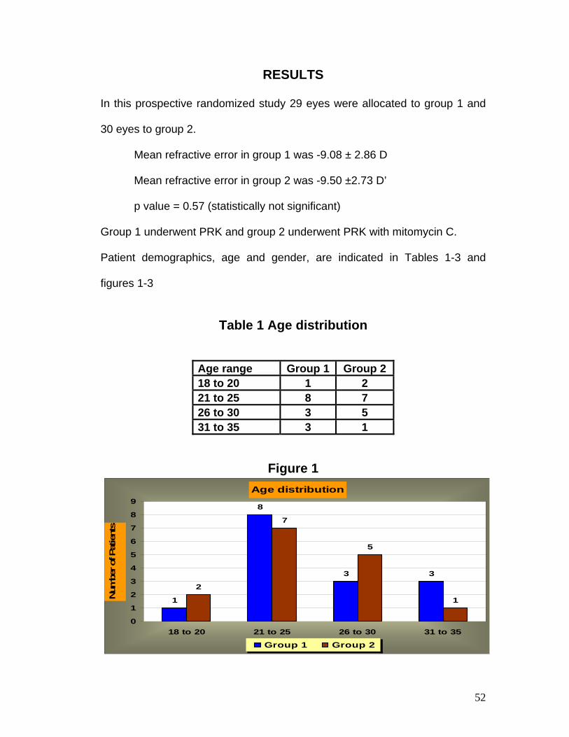
RESULTS
In this prospective randomized study 29 eyes were allocated to group 1 and
30 eyes to group 2.
Mean refractive error in group 1 was -9.08 ± 2.86 D
Mean refractive error in group 2 was -9.50 ±2.73 D’
p value = 0.57 (statistically not significant)
Group 1 underwent PRK and group 2 underwent PRK with mitomycin C.
Patient demographics, age and gender, are indicated in Tables 1-3 and
figures 1-3
Table 1 Age distribution
Age range Group 1 Group 2 18 to 20 1 2 21 to 25 8 7 26 to 30 3 5 31 to 35 3 1
Figure 1
Age distribution
1
8
3 3
2
7
5
1
0
1
2
3
4
5
6
7
8
9
18 to 20 21 to 25 26 to 30 31 to 35
Num
ber o
f Pat
ient
s
Group 1 Group 2

53
Table 2
Sex distribution in group 1
S.NO Sex No. of Patients
1 Male 7 2 Female 8
Figure 2
Sex distribution group 1
7
8
Male
Female

54
.
Table 3
Sex distribution group 2
S.NO Sex No. of Patients
1 Male 5 2 Female 10
Figure 3
Sex distribution group 2
5
10Male
Female

55
As shown below in Table-4,
After treatment, 15 eyes in each of the groups had UCVA of more than
6/9 (p value=0.018). 8 eyes of group 1 and 9 eyes of group 2 had UCVA of
6/12 to 6/9 (p value=0.042). 5 eyes of group 1 and 4 eyes of group 2 had
UCVA of 6/24 to 6/18 (p value=0.174). 1 eye of group 1 and 2 eyes of group 2
had UCVA of 6/60 to 6/36 (p value=0.357).
Table 4
Post treatment UCVA
Visual Acuity Post PRK Post PRK with MMC
Pvalue
6/60 to 6/36 1 2 0.357 6/24 to 6/18 5 4 0.174 6/12 to 6/9 8 9 0.042 >6/9 15 15 0.018
Figure 4
12
54
89
15 15
02468
10121416
Num
ber
of p
atie
nts
6/60 to 6/36 6/24 to 6/18 6/12 to 6/9 >6/9
Comparision of UCVA in Post PRK and Post PRK with MMC
Post PRK Post PRK with MMC

56
As shown in Table-5,
18 eyes in group 1and 25 eyes in group 2 had BCVA of more than 6/9
(p value=3.374), 8 eyes of group 1 and 4 eyes of group 2 had BCVA of 6/12
to 6/9 (p value=1.849), 3 eyes of group 1 and 1 eye of group 2 had BCVA of
6/24 to 6/18 (p value=1.147), None of the eyes of both groups had BCVA of
less than 6/36 .
Table 5 Post treatment BCVA
Visual acuity Group 1 Group 2 6/60 to 6/36 0 0 6/24 to 6/18 3 1 6/12 to 6/9 8 4 >6/9 18 25
Figure 5
0 0 3 1
84
18
25
0
5
10
15
20
25
Num
ber o
f pat
ient
s
6/60 to 6/36 6/24 to 6/18 6/12 to 6/9 >6/9
BCVA Post PRK
Post PRK BCVA Post PRK with MMC BCVA

57
As seen in Tables 4 & 5 and figures 4 & 5 , on comparing the visual
acuity, it was found that final visual outcome in EACH of the groups was not
statistically significant , p value>0.05.
Complications noted are as shown in the table 6 below
Table 6
Post Treatment Complications
Complications Group 1 Group 2 Myopic
Regression 10 10
Corneal Haze 12 11 Complicated
Cataract 2 0 Transient Glaucoma 0 2
Myopic Regression:
Mean myopic regression in Group 1 at the final follow-up of one year
was -2.175 D.
Mean myopic regression in Group 2 at the final follow-up of one year
was -3.125 D.
There was no significant statistical difference between the two groups for
myopic regression. p value=0.26.

58
Corneal Haze:
Corneal Haze was noted in 12 eyes in Group 1 and in 11 eyes in Group 2.
There was no significant statistical difference between the two groups for
corneal haze, p value=0.73.
Complicated Cataract:
Posterior subcapsular cataract was noted in 2 eyes in Group 1 for which
cataract surgery was advised.
Transient Glaucoma:
Transient Glaucoma was noted in 2 eyes in Group 2, which was controlled by
withholding steroids and instituting anti-glaucoma therapy.

59
DISCUSSION

60
Discussion
In this study, the mean spherical equivalent refraction in Group 1 was -
9.08 ±2.86 D and in group 2 was -9.505±2.73 D preoperatively.
At one year, out of 29 eyes in Group 1, in 15 eyes, an UCVA of 6/9 or
better was achieved. In the same group, in 18 eyes, BCVA of 6/9 or better
was achieved.
At one year, out of 30 eyes in group 2, in 15 eyes, an UCVA of 6/9 or
better was achieved. In the same group, in 25 eyes, BCVA of 6/9 or better
was achieved.
Corneal haze was noticed at first month follow-up in 12 eyes in group 1
and 11 eyes in group 2. It remained the same at one year. Myopic regression
was seen in 10 eyes in each of the groups.
In a similar study, Hashemi et al (2004) the mean spherical equivalent
refraction (SE) was -7.08 diopters (D) +/- 1.11 (SD) preoperatively. Six
months after surgery, 37 eyes (77.1%) achieved an uncorrected visual acuity
(UCVA) of 20/20 or better, all eyes had a UCVA of 20/40 or better and 45
(93.7%) eyes had an SE within +/- 1.00D. One month postoperatively, 2 eyes
(3.7%) had grade 0.5+ of haze, while at 3 and 6 months after surgery no
visited eye had haze at all. All eyes had a best corrected visual acuity (BCVA)
of 20/40 or better and there were no lost lines in BCVA by 6 months after
surgery. In spatial frequencies of 6 and 12 cycles per degree contrast

61
sensitivity had decreased immediately after PRK and it had increased 1.5
lines by the 6th postoperative month compared to the preoperative data.
In another study, Carones et al (2002), No toxic or side effects were
encountered postoperatively. No study group eye had a haze rate higher than
1 during the 6-month follow-up; 19 eyes (63%) in the control group did (P
=.01). At 6 months, the between-group difference in the refractive outcome
was statistically significant (P =.05), with 26 study group eyes (87%) and 14
control eyes (47%) within +/-0.50 D of the attempted correction. No study
group eye had a BCVA loss during the follow-up; 7 control eyes had lost 1 to
3 lines at 6 months (P =.0006).
Nassaralla et al (2007) described results at twelve months
postoperatively, 3 eyes showed grade 1 haze, and 2 eyes showed grade 0.5
haze. Twelve months postoperatively, 2 (9%) eyes had UCVA > or = 20/20.
No eye before and 17 (77%) eyes after treatment had UCVA > or = 20/40,
and no eye before and 9 eyes (40.9%) after treatment had UCVA > or =
20/25. Best spectacle-corrected visual acuity was > or = 20/40 in all (100%)
eyes and 21 (95%) eyes before and after treatment, respectively, and > or =
20/25 in 12 (54.5%) eyes before and after treatment. One (4.5%) eye lost 1
line of BSCVA. Mean spherical equivalent refraction achieved was -0.18
diopters (D) (range: -0.75 to +0.50 D) compared to -2.72 D (range: -1.50 to -
4.00 D) before treatment. Twelve months after treatment, 19 (85.5%) eyes
had a refractive outcome within +/- 0.50 D.

62
Wallau and Campos (2008) described Mean spherical equivalent
refraction error before surgery and mean ablation depth were -3.99+/-1.20
diopters (D) and 73.09+/-14.55 microm in LASIK eyes, and -3.85+/-1.12 D
and 70.7+/-14.07 microm in PRK with MMC eyes, respectively. Uncorrected
visual acuity was significantly better in PRK with MMC eyes 3 months (P=.04)
and 6 months (P=.01) after surgery. Best spectacle-corrected visual acuity
and spherical equivalent refraction did not differ significantly in the groups
during follow-up (P>.05). Significant haze was not observed in any PRK with
MMC eye. Mean higher order aberration was lower in PRK with MMC eyes
postoperatively compared with LASIK eyes (P=.01). Better contrast sensitivity
was observed in PRK with MMC eyes than LASIK eyes (P<.05). The
endothelial cell count did not differ significantly between groups (P=.65). In
terms of visual satisfaction, PRK with MMC eyes were better rated.
Shojaei et al (2009) described follow-up at 8 years after PRK, 69.64%,
44.44%, and 45.65% of the low, moderate, and high myopic groups were
within +/-0.5 D of emmetropia. Sixteen eyes (4.31% of original cases)
underwent retreatment mainly because of regression. Although a small
myopic shift occurred up to 8 years after surgery, changes in myopic
regression stabilized in all myopic groups within 24 months. Four eyes
(2.06%) lost 2 lines of best spectacle-corrected visual acuity (1 eye for
corneal haze and other 3 for problems not related to refractive surgery).
63
Corneal haze occurred in 11.34% especially in medium and high
myopic groups, but it cleared within 2 years in 68.2% of cases.
Contrary to most studies in literature, viz., Bedei et al,2009; Srinivasan
et al,2008; Wallau et al 2008;Hasan Hashemi,2004; gamboto et al,
2004;Carones et al,2002 and others, in this study it was found that addition of
Mitomycin C following PRK did not significantly decrease the incidence of the
complications, corneal haze and myopic regression, as indicated by the p
value.
However, most studies advocating the use of mitomycin C have used
it for longer periods on the cornea, viz, 30secs(Srinivasan et al 2008),
1minute ( Wallau et al 2008), 2minutes(Hasan Hashemi,2004).
A possible limitation in this study is that the 0.02% mitomycin C that
was used was applied on the cornea for 12 seconds, as suggested by .
It is possible that application for longer durations could yield better results.
Also, it remains to be seen if follow-up of the given cases for longer periods
could show a difference in the incidence of complications between the two
groups.

64
CONCLUSION

65
Conclusion
Following PRK and PRK with mitomycin C in high myopia, no
difference in the visual outcome was statistically discernable at one year.

66
BIBLIOGRAPHY

67
Bibliography
1. Srinivasan S, Drake A, Herzig S. Photorefractive keratectomy with 0.02%
mitomycin C for treatment of residual refractive errors after LASIK. J Refract
Surg. 2008 Apr; 24(4):326-36.Links
2. Nassaralla BA, McLeod SD, Nassaralla JJ Jr. Prophylactic mitomycin C to
inhibit corneal haze after photorefractive keratectomy for residual myopia
following radial keratotomy. J Refract Surg. 2007 Mar; 23(3):226-32.
3. Carones F, Vigo L, Scandola E, Vacchini L. Evaluation of the prophylactic
use of mitomycin-C to inhibit haze formation after photorefractive
keratectomy, J Cataract Refract Surg. 2002 Dec;28 (12):2088-95.
4. Hashemi H, Taheri SM, Fotouhi A, Kheiltash A. Evaluation of the
prophylactic use of mitomycin-C to inhibit haze formation after photorefractive
keratectomy in high myopia: a prospective clinical study. J Refract Surg.
2003 Jul-Aug; 19(4):438-42.
5. Gambato C, Ghirlando A, Moretto E, Busato F, Midena E., Mitomycin C
modulation of corneal wound healing after photorefractive keratectomy in
highly myopic eyes. BMC Ophthalmol. 2004 Sep 14; 4:12.
6. Bedei A, Marabotti A, Giannecchini I, Ferretti C, Montagnani M, Martinucci
C, Barabesi L.. Photorefractive keratectomy in high myopic defects with or
without intraoperative mitomycin C: 1-year results Br J Ophthalmol. 2009 Oct;
93(10):1313-8. E pub 2008 Feb 21.

68
7. Wallau AD and Campos M. Photorefractive keratectomy with mitomycin C
versus LASIK in custom surgeries for myopia: a bilateral prospective
randomized clinical trial. J Refract Surg. 2008 Apr; 24(4):326-36.
8. Shojaei A, Mohammad-Rabei H, Eslani M, Elahi B, Noorizadeh F. Long-
term evaluation of complications and results of photorefractive keratectomy in
myopia: an 8-year follow-up. Cornea. 2009 Apr; 28(3):304-10.
9. Dr. Rathinam Sivakumar DNB, Ocular Pharmacology and Therapeutics,
Aravind Eye Hospital edition.
10. Kunitoma N, Mori S: Studies on pterygium, 4 treatment of pterygim by
mitomycin C. Acta Soc Ophthalmol Jpn 67:601, 1953.
11. Majmudar PA, ForstotSL, Dennis RF et al: Topical mitomycin C for
subepithelial fibrosis after refractive corneal surgery, Ophthalmology 2000,
107(1) 89-94.
12. Raviv T Majmudar PA, Dennis RF et al: mitomycin C for post PRK corneal
haze J Cataract Refractive Surgery 2000, 26(8), 1105-1106.
13. Harold Stein MD.The Excimer- Fundamentals and Clinical Use 1997. 2nd
Ed.
14. Mc Donald M. Surgical Technique for RK. Highlights of Ophthalmology.
World Atlas Series of Ophthalmic Surgery Vol 1. 1993:111-120.
15. Carones F, Vigo L, Scandola E, et al. Evaluation of the prophylactic use
of mitomycin-C to inhibit haze formation after PRK. Journal of Cataract
Refractive Surgery. 2002; 28: 2088-2095.

69
16. Corbett MC, Prydall JI, Verma S, et al.An in vivo investigation of the
structures responsible for corneal haze after PRK and their effect on visual
function. Ophthalmology. 1996; 103: 1366-1380.
17. Krueger RR. Saedy NF, McDonnell PJ. Clinical analysis of steep central
islands after excimer laser PRK. Arch Ophthalmology. 1996; 114: 377-381.
18. Matta CA, Piebanga LW, Dietz MR, et al. Excimer retreatment for myopic
PRK failures. Six- to 18- month follow-up. Ophthalmology. 1996; 103:444-
451.
19. Wilson SE. Analysis of the keratocyte apoptosis, keratocyte proliferation,
and myofibroblast transformation responses after PRK and Laser in situ
keratomileusis. Trans Am Ophthalmol Soc. 2002; 100:411-433.
20. Pallikaris I Siganos D. Preoperative Evaluation LASIK 1998:23-128.

70
PROFORMA
71
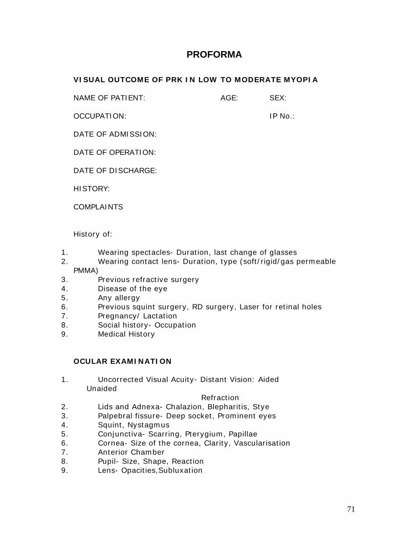
PROFORMA
VISUAL OUTCOME OF PRK IN LOW TO MODERATE MYOPIA NAME OF PATIENT: AGE: SEX: OCCUPATION: IP No.: DATE OF ADMISSION: DATE OF OPERATION: DATE OF DISCHARGE: HISTORY: COMPLAINTS History of:
1. Wearing spectacles- Duration, last change of glasses 2. Wearing contact lens- Duration, type (soft/rigid/gas permeable
PMMA) 3. Previous refractive surgery 4. Disease of the eye 5. Any allergy 6. Previous squint surgery, RD surgery, Laser for retinal holes 7. Pregnancy/ Lactation 8. Social history- Occupation 9. Medical History
OCULAR EXAMINATION
1. Uncorrected Visual Acuity- Distant Vision: Aided Unaided Refraction
2. Lids and Adnexa- Chalazion, Blepharitis, Stye 3. Palpebral fissure- Deep socket, Prominent eyes 4. Squint, Nystagmus 5. Conjunctiva- Scarring, Pterygium, Papillae 6. Cornea- Size of the cornea, Clarity, Vascularisation 7. Anterior Chamber 8. Pupil- Size, Shape, Reaction 9. Lens- Opacities,Subluxation

72
INVESTIGATIONS:
1. Slit lamp examination- including IOP measurement 2. Corneal Topography- irregular astigmatism 3. Ultrasound pachymetry 4. Indirect Ophthalmoscopy 5. Tear film Stability 6. WASCA aberrometry- 3rd, 4th aberrations pupil size in dark
VISION RE LE Distant vision- unaided
with pinhole Refraction - with correction AR BCVA Near vision- unaided With correction PROCEDURE POST OPERATIVE EVALUATION Day 0-visual acuity, refraction, slit lamp examination Day 1 Day 3 1 month 3 months 6 months 1 year Remarks