Embed Size (px)
Citation preview

DISSERTATION
Titel der Dissertation
Experimental data guided ligand docking at the benzodiazepine binding site of GABAA receptors
Verfasser
Mag. pharm. Lars Richter
angestrebter akademischer Grad
Doktor der Naturwissenschaften (Dr.rer.nat.)
Wien, 2012
Studienkennzahl lt. Studienblatt: A 091 449
Dissertationsgebiet lt. Studienblatt: Pharmazie
Betreuer: Prof. Gerhard F. Ecker


In liebevoller Erinnerung
an meinen Vater Emil, meine Schwester Michaela und meinen Bruder Kay


Aknowledgement
First and foremost I offer my sincerest gratitude to my supervisor, Prof. Dr. Gerhard Ecker. I had the great luck to work with such an extraordinary personality. Those who know Prof. Ecker know his passion and commitment to science and peak performance that is so impressive as infectious. Furthermore, I strongly admire his distinguished interpersonal competences, making him an excellent group leader, and mixed with his deep scientific knowledge, a valuable enrichment in the scientific community of computer-aided drug design. In total I can state that Prof. Ecker is an idol for me and other group members in many facets of life. Thank you, Gerhard.
I want to thank Dr. Margot Ernst for her kind introduction into the field of GABAA receptors. I learned a lot in the cooperation with such an intelligent and creative scientist. Let the nightly “Sternenhaufen” remind us of this very productive time.
I'm very proud that I can mention that I was also supervised by Prof. Dr. Werner Sieghart, one of the world top scientist, if not the top scientist at the GABAA receptor. I expected his deep knowledge in the GABAA receptor field but I was astonished about his curiosity and openness towards computational methodologies. I want to thank him for his trust and his unbelievable endurance that finally led to a publication in a top journal.
I was in the lucky position to cooperate with a further world top scientist at the GABAA field, Prof. Erwin Sigel. I want to thank him for the helpful critical discussions and his support in many fields that strongly influenced this thesis.
Further I want to thank Zdravko Varagic who performed the experimental testing of selected compounds working late often. All the best to this hard worker and great volleyball player.
I would like to thank all my friends and colleagues at Pharmacoinformatics Research Group. Thanks to Barbara Zdrazil, Dominik Kaiser, Rita Schwaha, Michael Demel, Khac-Minh Thai, Daniela Digles, Ishrat Jabeen, Nathan Poongavanam, René Weissensteiner, Andrea Schiesaro, Andreas Jurik, Freya Klepsch, Marta Pinto, Amir Seddik, Daria Tsareva, Rainer Dangl, Katharina Prokes, Christoph Waglechner for their wonderful company during my PhD thesis.
Furthermore I also want to address special thanks to my friends Christoph Kerschbaum and Markus Mayerhofer, which accompanied me in every life situation. I will never forget that.
My personal "hit" during this PhD-study was not found in virtual screening runs, instead I instantly met her in reality. I want to thank my "hit" Victoria Slubowski for all her love and support during my PhD-thesis.
Mein größter Dank gilt meiner Familie, die mich zu der Persönlichkeit formte, die ich heute bin. Ich möchte mich bei meinen Eltern, Tabea Mueller Richter und Emil Friedrich Richter, vor allem für ihre bedingungslose Liebe bedanken, aus der ich viel geschöpft habe und sicherlich noch viel schöpfen werde. Desweiteren möchte ich mich bei meiner Schwester Michaela und bei meinem Bruder Kay für all die schönen unvergesslichen Momente bedanken, die wir gemeinsam erlebt haben. Ich vermisse euch und Papa.


Abstract 3
Abstract English …………………………………………………………………… 3 Abstract Deutsch ……………………………………………………………………. 5
A. Background 7
I. Motivation …………………………………………………………………………… 9
II. Biological Background (Clayton et al., 2007, Current Medicinal Chemistry, 14, 2755-2775)……………… 12
III. Computational Background ………………………………………………………. 34
III.1 Homology Modeling ………………………………………………………..... 34 III.1.1 Homology modeling workflow ……………………………………………... 35 III.1.2 Homology modeling accuracy ……………………………………………… 36 III.2 Molecular Docking …………………………………………………………… 37
III.2.1 Posing………………………………………………………………………… 38 III.2.2 Scoring……………………………………………………………………….. 44 III.2.3 Molecular docking performance…………………………………………….. 48
IV. Aim of the study…………………………………………………………………… 61
B. Results and Discussion 65 I. Diazepam-bound GABAA receptor models identify new benzodiazepine site liands (Richter et al., 2012, Nature Chemical Biology, 8, 455-464 ) …… 66 II. A residue close to α1 loop F disrupts modulation of GABAA receptors by benzodiazepines while their binding is maintained (Bauer et al., 2010, Journal of Neurochemistry, 115, 1478-1485)……………….. 140
C. Concluding Discussion 149
Curriculum vitae 153
Curriculum vitae, English ………………………………………………………... 153 Curriculum vitae, German ………………………………………………………... 154
- 1 -

- 2 -

Benzodiazepines exert their anxiolytic, anticonvulsant, muscle relaxant, and sedative-hypnotic
properties by allosterically enhancing the action of GABA at GABAA receptors via their
benzodiazepine binding site. After 50 years of clinical use, the molecular basis of this
interaction still is not known as all attempts for structural resolution failed so far. In the
absence of a crystal structure, protein homology modeling and molecular dockings are the
only source for structure-based binding mode hypotheses for benzodiazepines. But two
obstacles make this undertaking extraordinary challenging. First, the homology models were
quite uncertain due to the low target-to-template sequence identity. And secondly, although
standard docking tools are capable of reproducing the correct binding mode, they regularly
fail to select this out of the produced pose list.
In this thesis, the obstacles were tackled in a two step approach. Primarily, the model
uncertainty was faced by an explorative step, where nine benzodiazepines were docked into
an array of α1β2γ2 GABAA homology models, considering flexible sidechains and keeping
the 100 best scored poses per ligand per model. Consequently in the selection step, exhaustive
implementations of various validation sources in an orchestrated and integrative manner were
necessary to filter the gigantic pose space. A key criterion in this filtering process was the
common binding mode (CBM) hypothesis, which assumes that diazepam and its close
structural analogues exhibit a common binding mode within the binding site. Therefore,
ligand poses were clustered according to their common scaffold, which led to three common
binding mode geometries, CBM I–III. Then, the integrative qualities of CBM came into effect
by incorporating experimental information from nine benzodiazepines for CBM I-III
validation. The evaluation clearly demonstrated that CBM I is convincingly supported by a
large variety of structural, computational and experimental evidence. The CBM I ligand-
receptor complex was then used in structure-based virtual screening runs and led to the
successful discovery of three novel, experimentally validated, benzodiazepine binding site
ligand classes. The structural models were also used to investigate mechanistic receptor
features and finally identified α1Y168 to be important for the mechanistic coupling from
benzodiazepine binding to GABAA channel modulation.
In a broader view, the gained structural models for α1β2γ2 GABAA receptors can be used for
modelling other GABAA receptor subtypes that will ultimately improve our understanding of
the structural determinants for subtype selectivity of GABAA receptors, leading to drugs
excluding subtypes known to be responsible for addicting. Finally, the underlying
computational workflow can stimulate other groups to integrate various information pieces in
binding mode elucidation of structurally unresolved membrane proteins.
- 3 -

- 4 -

Die anxiolytische, antikonvulsive, muskelrelaxierende und sedative-hypnotische Wirkung von
Benzodiazepinen wird durch allosterische Verstärkung der GABA Aktivität am GABAA
Rezeptor ausgeübt. Nach 50 Jahren in klinischer Verwendung ist die molekulare Basis für
diese Interaktion immer noch ungeklärt, da alle Maßnahmen zur strukturellen Aufklärung
scheiterten. In Abwesenheit einer Kristallstruktur sind Homology Modeling und Molekulares
Docking die einzigen Möglichkeiten zur Darstellung von Bindungshypothesen für
Benzodiazepine. Zwei Tatsachen erschweren diesen Weg aber deutlich: Erstens sind die
Homology Modelle ungenau da die Sequenzidentität zwischen Target und Template gering
ist. Und zweites, obwohl Molekulares Docking den richtigen Bindungsmodus generieren
kann, kann es ihn selten aus der Menge an produzierten Posen selektieren. In dieser
Dissertation wurden diese Probleme in zwei Schritten gelöst. Zuerst stellte man sich der
Modellungenauigkeit in einem explorativem Schritt, indemneun Benzodiazepine in einen
Array von α1β2γ2 GABAA Homologie Modellen gedockt wurden, unter Berücksichtigung
von flexiblen Seitenketten und Einbeziehung der besten 100 Posen pro Ligand pro Modell. Im
folgenden Selektionsschritt waren eine ausführliche Implementierung von verschiedenen
Validierungsquellen in einem orchestrierten und integrativen Verfahren notwendig, um den
sehr umfangreichen Poseraum zu filtern. Ein Schlüsselkriterium in diesem Prozess war die
common binding mode (CBM) Hypothese. Diese nimmt an, dass Diazepam und seine
strukturellen Analoga einen gemeinsamen Bindungsmodus in der Bindetasche haben.
Anschließendes Clustern der Posen führte zu drei common binding mode Cluster, CBM I-III.
Die nachfolgende Validierung der drei CBMs erfolgte durch Inkorporation von
experimentellen Informationen von neun Benzodiazepinen . Die Bewertung zeigte eindeutig,
dass CBM I durch eine Reihe von strukturellen, energetisch-rechnerischen und
experimentellen Befunden gestützt wird und somit als korrekter Bindungsmodus angesehen
wird. Der CBM I Ligand-Rezeptor Komplex wurde dann für Virtuelles Screening verwendet
und führte zur Entdeckung von drei neuen, experimentell bestätigten Substanzklassen, die an
der Benzodiazepine Bindetasche binden. Die Strukturmodelle wurden auch für die
Untersuchung von Rezeptor Mechanismen verwendet, die letztendlich die Bedeutung von
α1Y168 in der Transduktion von Benzodiazepine Bindung zu GABAA Kanal Modulation
zeigten. Die gewonnen Modelle für α1β2γ2 GABAA Rezeptoren können die Modellierung
anderer Subtypen verbessern und damit die Entwicklung von Arzneistoffen vorantreiben
welche nicht mehr mit Subtypen reagieren welche bekanntermaßen für die Abhängigkeit
verantwortlich sind. Schließlich kann der Workflow von Gruppen mit ähnlichen
Problemstellungen aufgegriffen werden.
- 5 -

- 6 -

A. Background I. Motivation ………………………………………… 9
II. Biological Background …………………………… 12
(Clayton et al., 2007, Curr Med Chem, 14, 2755-2775)
III. Computational Background …………………….. 34
III.1 Homology Modeling………………………………….. 34 III.1.1 Homology modeling workflow …………………………..…… 35 III.1.2 Homology modeling accuracy …………..……………………. 36
III.2 Molecular Docking …………………………………… 37
III.2.1 Posing …………………………………………………………. 37 III.2.1.1 Systematic Search ……………………………………………………… 39 III.2.1.2 Random Search ………………………………………………………… 40 III.2.1.3 Simulation Search ……………………………………………………... 41 III.2.1.4 Protein flexibility ……………………………………………………… 42 III.2.2 Scoring ………………………………………………………… 44 III.2.2.1 Force-field-based scoring ……………………………………………… 45 III.2.2.2 Empirical scoring functions …………………………………………… 45 III.2.2.3 Knowledge-based functions …………………………………………… 47 III.2.3 Molecular docking performance ………………………………. 48 III.2.3.1 Biding Mode Predition …………………………………………………. 49 III.2.3.2 Virtual Screening ……………………………………………………….. 50 III.2.3.3 Affinity Prediction Tool ………………………………………………… 54 III.2.3.4 Docking into Homology Models ……………………………………….. 54 IV. Aim of the study……………………………………. 61
- 7 -

- 8 -

I. Motivation
Benzodiazepines belong to the most widely prescribed drugs in current therapeutic use and
exert their anxiolytic, anticonvulsant, muscle relaxant, and sedative hypnotic properties by
allosterically enhancing the action of GABA at GABAA receptors1. In spite of being used
clinically for 50 years, their exact mode of interaction with the benzodiazepine binding site of
GABAA receptors still is not known.
All experimental attempts to structurally resolve the GABAA receptor has failed so far. In
particular the hydrophobic transmembrane domain of the protein aggravates the elsewhere
quite successful X-ray crystallography.
In the absence of the GABAA receptor structure, a bunch of experimental methodologies were
applied to gain knowledge of the spatial arrangement of ligands within the benzodiazepine
site of the GABAA-receptor. Over the years, methodologies like receptor mutation and ligand
labelling have contributed a lot to this understanding2,3. But also affinity data from ligand
binding assays which become manifest in structure-activity relationships gave valuable hints
about the orientation of ligands in the benzodiazepine binding site4. However, these findings
gave only a vague schematic idea of the benzodiazepine binding mode.
Suddenly new hope emerged in the year 2001 when a crystal structure from a functional and
structural homologue of the extracellular ligand binding domain of the GABAA receptor, the
ACh-binding protein (AChBP), became available5.
Shortly after, the first homology models based on this crystal structure (AChBP) were
generated and computational docking was used to generate structural hypotheses about the
binding mode of benzodiazepines in the benzodiazepine binding site3,6-9. Overall, however,
conclusions remained contradictory and unsatisfactory. Also after the resolution of other,
more distant structural homologues, the quality of the hypotheses did not improve. The low
success in the elucidation of the binding mode of 1,4-benzodiazepines with structure based
approaches were based mainly on two shortcomings:
1. High ambiguity in the GABAA receptor homology models due to the low sequence identity
to the templates of around 18%. Models that are built on such a low sequence identity are
considered speculative at best for structure based studies10 ("ambiguity problem").
- 9 -

2. Even if a correct model is available and molecular docking is able to generate the correct
binding mode, docking scoring functions are not competent to identify this orientation out
of other solutions11 ("evaluation problem").
The fact that 60% of today’s drug targets are membrane proteins12, make the described
constellation of poor structural target models in line with intensive research and accumulation
of experimental data, a quite frequent problem in the drug discovery field.
The ambiguity in the models ("ambiguity problem") could be faced by broadly sampling
protein and ligand flexibility during docking. This results in an enormous amount of possible
ligand-receptor complexes, but with a high likelihood of generating the correct binding mode
for benzodiazepines.
However, the great challenge of this thesis lies in the subsequent step, i.e. the selection of the
correctly generated binding mode out of an ocean of ligand-receptor complexes ("evaluation
problem"). Here a protocol has to be established that supports docking scoring functions in
the evaluation of ligand-receptor complexes. This protocol should implement various
knowledge sources (mutagenesis data, labeling data, structure-activity relationships) in an
orchestrated and integrative manner, leading to the selection of one single benzodiazepine
binding mode.
In a broader view the gained structural model of the benzodiazepine binding site will also
improve our understanding of the molecular determinants for subtype selectivity of
experimental ligands on GABAA receptors. This knowledge will aid the development of
subtype selective benzodiazepine binding site ligands, avoiding interactions with subtypes
known to be responsible for benzodiazepine addiction13.
But these models and the established methodology should also stimulate structure based
approaches beyond the GABAA receptor field. Together with the nicotinic acetylcholine
receptors (nAChR), glycine, and serotonin type3 receptors, the GABAA receptors belong to
the pharmacological important protein class of ligand gated ion channels. All members of this
class share a sequence identity of at least 30% to each other14. So the findings of this study
could be of interest for researches in this important field. After all, 10% of all currently
approved drug targets belong to the class of ligand gated ion channels15, highlighting once
again the pharmacological relevance of this protein class.
- 10 -

References:
1. Sieghart, W. Structure and pharmacology of gamma-aminobutyric acidA receptor subtypes. Pharmacol Rev 47, 181-234 (1995).
2. Sigel, E. Mapping of the benzodiazepine recognition site on GABA(A) receptors. Curr Top Med Chem 2, 833-9 (2002).
3. Clayton, T. et al. An updated unified pharmacophore model of the benzodiazepine binding site on gamma-aminobutyric acid(a) receptors: correlation with comparative models. Curr. Med. Chem. 14, 2755-2775 (2007).
4. Zhang, W., Koehler, K.F., Zhang, P. & Cook, J.M. Development of a comprehensive pharmacophore model for the benzodiazepine receptor. Drug. Des. Discov. 12, 193-248 (1995).
5. Brejc, K. et al. Crystal structure of an ACh-binding protein reveals the ligand-binding domain of nicotinic receptors. Nature 411, 269-276 (2001).
6. Mokrab, Y. et al. Exploring ligand recognition and ion flow in comparative models of the human GABA type A receptor. J. Mol. Graph. Model. 26, 760-774 (2007).
7. Ci, S., Ren, T. & Su, Z. Investigating the putative binding-mode of GABA and diazepam within GABA A receptor using molecular modeling. Protein J. 27, 71-78 (2008).
8. Berezhnoy, D., Gibbs, T.T. & Farb, D.H. Docking of 1,4-benzodiazepines in the alpha1/gamma2 GABA(A) receptor modulator site. Mol. Pharmacol. 76, 440-450 (2009).
9. Sancar, F., Ericksen, S.S., Kucken, A.M., Teissére, J.A. & Czajkowski, C. Structural determinants for high-affinity zolpidem binding to GABA-A receptors. Mol. Pharmacol. 71, 38-46 (2007).
10. Cavasotto, C.N. & Phatak, S.S. Homology modeling in drug discovery: current trends and applications. Drug Discov Today 14, 676-83 (2009).
11. Warren, G.L. et al. A critical assessment of docking programs and scoring functions. J. Med. Chem. 49, 5912-31 (2006).
12. Yildirim, M.A., Goh, K.I., Cusick, M.E., Barabasi, A.L. & Vidal, M. Drug-target network. Nat Biotechnol 25, 1119-26 (2007).
13. Tan, K.R., Rudolph, U. & Luscher, C. Hooked on benzodiazepines: GABAA receptor subtypes and addiction. Trends Neurosci 34, 188-97 (2011).
14. Sieghart, W. et al. Structure and subunit composition of GABA(A) receptors. Neurochem Int 34, 379-85 (1999).
15. Rask-Andersen, M., Almen, M.S. & Schioth, H.B. Trends in the exploitation of novel drug targets. Nat Rev Drug Discov 10, 579-90 (2011).
- 11 -

II. Biological Background
Contribution of the thesis author to this review:
The thesis author contributed to the computational section of this review (p. 2770 – 2774)
with the aim of positioning the ligand based pharmacophore model into homology models of
the benzodiazepine binding site of the α1β2γ2 GABAA receptor. The homology models were
provided by M. Ernst and W. Sieghart. The molecular docking of diazepam into the homology
models and the subsequent pose analysis was undertaken by the thesis author. Computational
tools were programmed that aided the pose selection towards a diazepam pose that fit also the
orientation and residue assignments of the ligand based pharmacophore model.
- 12 -

Current Medicinal Chemistry, 2007, 14, 2755-2775 2755
0929-8673/07 $50.00+.00 © 2007 Bentham Science Publishers Ltd.
An Updated Unified Pharmacophore Model of the Benzodiazepine Binding Site on -Aminobutyric Acida Receptors: Correlation with Comparative Models
T. Clayton1, J.L. Chen1, M. Ernst4, L. Richter4,5, B.A. Cromer2, C.J. Morton*,2, H. Ng2, C.C. Kaczorowski1,F.J. Helmstetter2, R. Furtmüller4, G. Ecker5, M.W. Parker3, W. Sieghart4 and J.M. Cook*,1
1Departments of Chemistry & Biochemistry and 2Psychology, University of Wisconsin-Milwaukee, Milwaukee, WI 53201; 3St. Vin-cent’s Institute of Medical Research, Victoria 3065, Australia; 4Division of Biochemistry & Molecular Biology, Center for Brain Re-search, Medical University Vienna, A-1090, Vienna, Austria; 5Department of Medicinal Chemistry, University Vienna, A-1090 Vi-enna, Austria
Abstract: A successful unified pharmacophore/receptor model which has guided the synthesis of subtype selective compounds is re-viewed in light of recent developments both in ligand synthesis and structural studies of the binding site itself. The evaluation of experi-mental data in combination with a comparative model of the GABAA receptor leads to an orientation of the pharmacophore model within the Bz BS. Results not only are important for the rational design of selective ligands, but also for the identification and evaluation of possible roles which specific residues may have within the benzodiazepine binding pocket.
Keywords: Aminobutyric acid(A) receptors, gated ion channels, GABA-A, computer-assisted analysis, benzodiazepine.
The GABAA receptor is the major inhibitory neurotransmitter receptor of the central nervous system (CNS) and the site of action of a variety of pharmacologically and clinically important drugs, such as benzodiazepines, barbiturates, neuroactive steroids, anes-thetics and convulsants [1]. It is now clear that these receptors regu-late the excitability of the brain, anxiety, muscle tone, circadian rhythms, sleep, vigilance, memory, and learning [1]. There are sev-eral disease states associated with the improper functioning of this protein, including anxiety, epilepsy [2], insomnia [3], depression and bipolar disorder [4, 5], schizophrenia [6], as well as mild cogni-tive impairment and Alzheimer's disease [7]. A role of GABAAreceptors in drug and alcohol abuse has also been reported [8-10].
GABAA receptors are composed of 5 subunits that form a cen-tral chloride channel and can belong to different subunit classes. A total of 19 subunits ( ) of the GABAAreceptor have been cloned and sequenced from the mammalian nervous system [11, 12]. All these polypeptides possess an ap-proximate molecular mass of ~ 50 kD and are structurally related. Each subunit consists of a large extracellular region, which contains several potential glycosylation sites and a characteristic “cys-loop” formed by a covalent bond between two conserved cysteines. This extracellular region is also important in contributing to the agonist GABA and modulatory benzodiazepine binding sites. The protein then traverses the lipid bilayer four times and has a large intracellu-lar loop located between transmembrane regions 3 and 4 (M3 and M4). This intracellular region contains possible phosphorylation sites necessary for regulation of the receptor. The homology within each subunit class is about 60 – 80 %, while the homology between subunit classes is about 30 – 40 %. In Fig. (1) the proposed topol-ogy of a single GABAA receptor subunit is shown. The pentameric structure of a ligand-gated ion channel is shown in Fig. (2) [13, 14].
The existence of multiple GABAA receptor subunits can give rise to a large number of different GABAA receptor subtypes [15]. The majority of GABAA receptors, however, is composed of 1 and 2 and 2 subunits. The presence of a subunit within a GABAAreceptor is necessary for the formation of a benzodiazepine binding site that is located at the interface of an and subunit. Whereas the classical benzodiazepines, such as diazepam or flunitrazepam, exhibit a high affinity for receptors composed of 1 2, 2 2,
3 2 or 5 2 subunits (diazepam sensitive (DS) receptors), as well as for their less intensively investigated analogues containing the 3 subunit, other benzodiazepine binding site ligands are also
*Address correspondence to this author at the Department of Chemistry, 3210 North Cramer Street, UW-Milwaukee, Chemistry Building Milwaukee, WI 53201, UW-Milwaukee, Milwaukee, WI 53211, USA; Tel: 414-229-5856; Fax: 414-229-5530; E-mail: [email protected]
able to interact with 4 2 or 6 2 receptors (diazepam insensi-tive (DI) receptors), or with receptors containing 1 subunits [1]. Receptors containing 1 or 3 subunits exhibit a quite low abun-dance in the brain [16-18] and their contribution to the “in vivo”effects of benzodiazepine binding site (BZ BS) ligands currently is unclear.
Fig. (1). Proposed topology of a GABAA receptor subunit. The extracellular domain begins with the N-terminus and M1-M4 represent the four trans-membrane domains. Figure reprinted with permission [13].
The concept of receptor multiplicity has been extremely valu-able, in that different receptor subtypes reside within anatomically distinct regions of the brain and are responsible for different physiological and pathological processes [15, 19, 20]. For example, the receptor subtype has a prominent role in seizure suscep-tibility and sedation [21-23], the 2 2 and possibly also the 3 2subtypes are involved in anxiety, whereas the subtype has a prominent role in memory and learning (Table 1) [15, 24-26]. These distinctions have thus become a motivation for the design of subtype selective ligands in order to elicit a single specific response [15, 27-34]. Differences observed in the action of such drugs may be due to subtype-selective affinity and absolute and/or relative subtype-selective efficacy [35].
Agonist binding to the receptor opens an intrinsic chloride ion channel, typically hyperpolarizing the cell membrane or at least opposing depolarization, thereby inhibiting neuronal transmission. Bz BS ligands are allosteric modulators, unable to induce channel openings themselves, but function to vary the frequency and not the channel opening times [37, 38]. Positive allosteric modulators at the benzodiazepine binding site (agonists) increase this frequency,
- 13 -

2756 Current Medicinal Chemistry, 2007, Vol. 14, No. 26 Clayton et al.
while negative allosteric modulators (inverse agonists) decrease the frequency. Currently it is not clear whether Bz BS ligands allosteri-cally modulate GABA affinity or channel gating. Recent studies [39] support the view that high-affinity classical benzodiazepines modulate GABAA receptors via allosteric coupling to channel gating [40, 41]. Further studies are needed to determine whether the mechanism of modulation varies in different receptor subtypes. Table 1. Action of Benzodiazepines at GABAA Receptor
Subtypes [36]
Subtype Associated Effect
Sedation, anterograde amnesia, Some anticonvulsant action, ataxia
Anxiolytic, hypnotic (EEG), some muscle relaxation
Some anxiolytic action, some anticonvulsant action, Maybe some muscle relaxation
Diazepam-insensitive site
Cognition, temporal and spatial memory (Maybe memory component of anxiety)
Diazepam-insensitive site
In recent years a unified pharmacophore/receptor model for agonists, antagonists and inverse agonists at the Bz BS was devel-oped, using the techniques of chemical synthesis, radioligand bind-ing and receptor mapping [42, 43]. The overlap of these different modulators within the Bz BS has been supported by experimental data [44-46]. Using this ligand-based pharmacophore/receptor model and our GABAA receptor models [47, 48], the ex-perimental data of recent and past years have been evaluated, and definite trends with regard to the orientation of the regions of the protein relative to the descriptors of the pharmacophore/receptor model have been identified and are presented in this work. The
need to define such an orientation has been established [49], since it permits inspection of ligand docking studies and the identification of possible roles specific residues may have within the Bz BS. These roles may then be explored in future studies involving cova-lent labeling, site-directed mutagenesis and structure-activity rela-tionships, all of which contribute to the rational design of subtype-specific modulators of the Bz BS of GABAA receptors.
Fig. (3). Relative locations of the descriptors and regions of the unified pharmacophore/ receptor model. The pyrazolo[3,4-c] quinolin-3-one CGS-9896 (dotted line), a diazadiindole (thin line), and diazepam (thick line) aligned within the unified pharmacophore/receptor model for the Bz BS. H1
and H2 represent hydrogen bond donor sites within the Bz BS while A2
represents a hydrogen bond acceptor site necessary for potent inverse ago-nist activity in vivo. L1, L2, L3 and LDi are four lipophilic regions and S1, S2,and S3 are regions of negative steric repulsion. LP = lone pair of electrons on the ligands.
Fig. (2). Longitudinal (A) and cross-sectional (B) schematic representations of a ligand-gated ion channel. The numbers 1-4 refer to the M1-M4 segments. The M2 segment contributes to the majority of the pore lining within the membrane lipid bilayer. Figures reprinted with permission [14].
- 14 -

Correlation with Comparative Models Current Medicinal Chemistry, 2007 Vol. 14, No. 26 2757
THE UNIFIED PHARMACOPHORE/RECEPTOR MODEL
More than 150 agonists, antagonists and inverse agonists at the Bz BS [42, 43] which encompassed 15 structural families were used for generating the unified pharmacophore/receptor model. Although the relative affinities, efficacies and functional effects displayed by various ligands from the same structural class at the diazepam-sensitive and diazepam-insensitive benzodiazepine binding sites were taken into account, the approximate locations of descriptors (hydrogen bond donor sites, hydrogen bond acceptor sites, lipo-philic regions, and regions of steric repulsion) were based primarily on in vitro binding affinities. Ligands from different structural classes were then superposed on each other to satisfy the same de-scriptors, resulting in the unified pharmacophore model.
Briefly, the pharmacophore/receptor model consists of two hy-drogen bond donating descriptors (H1 and H2), one hydrogen bond
accepting descriptor (A2) and one lipophilic descriptor (L1). In addi-tion to these descriptors, there are lipophilic regions of interaction (L2, L3 and LDi) as well as regions of negative steric repulsion (S1,S2 and S3). While occupation of L2 and/or L3 as well as interactions at H1, H2, and L1 are important for positive allosteric modulation, inverse agonists only require interactions with the H1, L1, and A2descriptors of the pharmacophore/receptor model for potent activity in vivo. [42, 50-53] The LDi descriptor is a region of lipophilic inter-action, for which the difference between the diazepam sensitive (DS) and the diazepam insensitive (DI) sub-pharmacophore models is most pronounced. Depicted in Fig. (3) are the relative locations of the different descriptors and regions of the model.
The structures of ligands frequently referred to in this paper are shown in Fig. (4).
The alignments of several Bz BS ligands within this model are shown in Fig. (5).
N
NO
Me
Cl –O
OF
N
NO
Me
N+Cl
Me
N
N
N
F
N
N
Cl
N
Cl
Me
N
N
O
NCO2Et
MeF N
N
O
NCO2Et
MeN
N+–N
N
NMe
Me
O
NMe2
NN
NN
Me
CF3
NH
N
CO2Me
NH
N
EtCO2MeMeO
MeO
NH
N
MOMCO2EtBnO
NH
N
MOMCO2EtO
NH
N
CO2t-Bu
NH
N
O-nPr
Pr-n-
NH
N
CO2Et
diazepam, 1agonist
flunitrazepam, 2Agonist
midazolam, 3agonist
triazolam, 4agonist
Ro15-1788, 5antagonist
Ro15-4513, 6inverse agonist
zolpidem, 7agonist
CL-218, 872, 8partial agonist
-CCM, 9inverse agonist
DMCM, 10inverse agonist
ZK-93423, 11agonist
6-PBC, 12partial agonist
-CCT, 13antagonist
3-PBC, 14Antagonist
-CCE, 15inverse agonist
N
N
N
H OMe
O
O
N
N
N
HOMe
O
O
XLi-093, 16antagonist at 5
Fig. (4). Structures of ligands and their modulation at the subtype.
- 15 -

2758 Current Medicinal Chemistry, 2007, Vol. 14, No. 26 Clayton et al.
Ro 15-1788, 5 Ro 15-1788 rotated 90º
Diazepam, 1 Diazepam rotated 90º
DMCM, 10 DMCM rotated 90º
zolpidem, 7 zolpidem rotated 90º
CL-218,872, 8 CL-218, 872 rotated 90º Fig. (5). Alignments of several Bz BS ligands within the pharmacophore model.
- 16 -

Correlation with Comparative Models Current Medicinal Chemistry, 2007 Vol. 14, No. 26 2759
INCLUDED VOLUME ANALYSIS OF LIGANDS BINDING TO RECEPTORS CONTAINING DIFFERENT ALPHA SUBUNITS
The benzodiazepine binding site of 2 GABAA receptors is strongly influenced by the type of subunit present in these recep-tors as indicated by the existence of ligands exhibiting certain selec-tivity for receptors containing the respective subunits [1, 20, 54]. If subtype selective ligands are then aligned within the pharma-cophore model according to the resulting alignment rules [55], their included volumes can be constructed and used to compare the to-pologies of benzodiazepine binding pockets of different receptor subtypes [55, 56]. Ligands employed in the included volume for each receptor subtype exhibited potent affinity (Ki 20 nM) at the respective receptor subtype. CL-218,872 5 (Ki = 57 nM at ) and zolpidem 4 (Ki = 26.7 nM at ) were added to the included vol-ume of the subtype since they are both -subtype selective ligands. The major differences with regard to volume and affinity are shown below and are important for interpreting experimental data.
The included volume requirements show some trends that are useful to explain subtype preferences of ligands and that have al-ready guided substance development: 1. The included volumes of the and subtypes are
similar both in size and topology. This is consistent with the similar affinity profiles these subtypes displayed for classi-cal benzodiazepine agonists, pyrazoloquinolinones and imi-dazobenzodiazepines (i-BZDs) [57-59]. The included vol-ume of the 1 subtype is slightly different from that of 2and 3 subtype as indicated by the selective affinities of zolpidem, CL-218,872, 6-substituted -carbolines (e.g. 6-methylbenzyl amino betacarboline) and pyridodiindoles for this subtype and the space needed for accommodating these structures (Fig. 6a,b) [59]. Results from both the SAR stud-ies and the included volume analysis imply that the and subtypes are very similar in shape, polarity and lipophil-
icity. 2. The included volumes of the and 4 or subtypes are
very different. Looking at Fig. (6d), it is evident that the in-cluded volume of the 6 subtype is significantly smaller
Fig. (6). Overlap between pairs of included volumes derived from receptor subtype selective ligands: a) and , b) and , c) and , d) and e) and . Yellow color indicates overlapping regions and each grid measures 4 Å in width and height. In order to provide the connection between this figure and other figures, f) shows diazepam and the descriptors of the unified pharmacophore model depicted in the included volume requirement of the subtype.
- 17 -

2760 Current Medicinal Chemistry, 2007, Vol. 14, No. 26 Clayton et al.
than that of the 1 subtype. Especially the LDi region is much larger in 1 receptors. Contributions to the LDi region are derived in a large measure from 6-substituted -carbolines and ring-A substituted pyrazoloquinolines, thus implying that occupation of the LDi region may be critical for ligand selectivity at the subtype. This is supported by the finding that the LDi region was also larger for the subtype when compared with the subtype
(Fig. 6e).3. L3 region is very small or non-existent for the and
DI subtypes. Based on the inability to bind 1,4-benzodiazepines, the lack of region L3 was believed to be responsible for the diazepam-insensitivity of these receptor subtypes (Fig. 6c, d) [42, 51]. With a few exceptions, -carbolines also do not bind to the receptor subtype, while i-BZDs and pyrazoloquinolinones (CGS series) rep-resent the primary ligands that bind to this subtype.
4. The L2 and LDi regions are slightly smaller for the ver-sus the subtype. The sequence of the subunit is most homologous to the subunit and it was determined that the pharmacological profiles of these DI sites toward classi-cal benzodiazepines are very similar [60]. Differences in the included volume of these DI sites are shown in Fig. (6c).
5. The L2 region contributes to and selectivity. It has been observed that ligands with and/or selectivity are generally i-BZDs and have a lipophilic C(8)-substituent that
occupies the L2 pocket. Based on ligands from various stud-ies, examination of data in Fig. (6) illustrates that the L2 re-gion is deeper and larger for the and subtypes, re-spectively, than for the corresponding or subtypes [42, 43, 51, 61]. This L2 region seems to account for the se-lective affinity of ligands at the subtype, as clearly illus-trated in Fig. (6e), whereas the same region may account for the selective efficacy observed at the subtype.
RECENT ALIGNMENT OF NON-CLASSICAL BZ BS LIGANDS SUPPORT THE UNIFIED PHARMA-COPHORE/RECEPTOR MODEL
Besides the major classes of Bz ligands used to define the model, several non-Bz ligands also fit within the pharma-cophore/receptor model very well. Examples include zopiclone (17)and its active enantiomer, the flavonoid 25 and the 8-chloropyrazolo[5,1-c][1,2,4]-benzotriazine 5-oxide analog 38, as illustrated in (Figs. 7 and 8) and (Figs. 11 and 13).
Zopiclone and its Active Enantiomer
Since the sedative-hypnotic zopiclone 17 was first reviewed in Drugs in 1986 [62], a much larger body of clinical data has become available to permit a more detailed comparison of the non-benzodiazepine zopiclone with classical benzodiazepines. Results have shown that, regardless of the duration of action, zopiclone was
Fig. (7). Structure of zopiclone 17, its alignment within the pharmacophore/receptor model and rotated 90 degrees.
Fig. (8). Alignment of zopiclone 17 (left) and its active enantiomer (right) in the included volume of the pharmacophore/receptor model for the 3 2 subtype.
N
N
N
O
N
O
ON
NMe
Cl
H
- 18 -

Correlation with Comparative Models Current Medicinal Chemistry, 2007 Vol. 14, No. 26 2761
generally at least as effective as benzodiazepines in the treatment of insomnia, although comparisons between zopiclone and flurazepam have produced inconsistent results. It was observed that zopiclone had a relatively low propensity to elicit residual clinical effects, such as difficulty in waking or reduced morning concentration. While tolerance to the effects of zopiclone was not noticed in short term clinical trials ( 4 weeks), the results from longer term studies were conflicting and, therefore, the potential for tolerance during long term administration of zopiclone was unclear. Rebound in-somnia to a level above that at baseline can occur after withdrawal of zopiclone. However, on the basis of data from short term studies, this does not appear to be common. Evaluation of prescriptions filled has indicated that zopiclone does not have a high dependence potential, at least in those who are not regular drug abusers/addicts. Zopiclone was well-tolerated in both the elderly and younger pa-tients with insomnia. A bitter after-taste was usually the most com-mon adverse event, but was relatively infrequent at 3.6 % in the largest available post-marketing study. Thus, zopiclone has now been firmly established as an effective and well-tolerated sleep agent [63].
The structure and alignment of zopiclone within the unified pharmacophore/receptor model is shown in Fig. (7). The centroid of the pyridine moiety of zopiclone overlapped with region L1 of the receptor, while the lone pair of electrons of the carbonyl oxygen (O) atom interacted with H1 of the receptor to form a hydrogen bond between the ligand and the receptor. A second hydrogen bond was formed between the amide carbonyl oxygen atom (LP) and H2of the receptor protein (Fig. 7).
However, a recent study by Sepracor [64] indicated that one of the enantiomers of zopiclone was much more active than zopiclone
itself. This may be the result of receptor subtype-selective efficacy or simply a pharmacokinetic effect, but the pharmacophore/receptor model developed here revealed that the active enantiomer fits better into the included volume of the subtype than zopiclone 17 itself (Fig. 8). This enantiomer has just been approved by the FDA for treatment of insomnia.
SH-053BZ Enantiomers
In pursuing this approach using BZ enantiomers, the behavioral activity of three newly-synthesized compounds [65], functionally selective for 2, 3 and 5-containing subtypes of GABAA recep-tors (SH-053-S-CH3 and SH-053-S-CH3-2’F), or essentially selec-tive for 5 subtypes (SH-053-R-CH3) were examined. Motor influ-ence was tested in the elevated plus maze, spontaneous locomotor activity and rotarod test, which are considered primarily predictive of the anxiolytic, sedative and ataxic influence of BZs, respectively. There was substantially diminished ataxic potential of BZ site ago-nists devoid of 1 subunit-mediated effects, with preserved anti-anxiety effects at 30 mg/kg of SH-053-S-CH3 and SH-053-S-CH3-2’F. However, all three ligands, dosed at 30 mg/kg, decreased spon-taneous locomotor activity, suggesting that sedation may be partly dependent on activity mediated by 5-containing GABAA receptors. Such an effect could not have been observed previously, because to date, all apparently non-sedating BZ receptor ligands, which are devoid of activity at 1-containing GABAA receptors, engendered essentially antagonist [66] or only partial agonist efficacy at 5-containing GABAA receptors [67]. Therefore, it cannot be ruled out that substantial efficacy at 5-containing GABAA receptors may contribute to sedative effects besides the effects on learning and memory processes [68, 69]. This is supported by two sets of data
N
N
N
O
O
18A1) A2) A3)
N
N
N
O
O
19B1) B2)
B3)
Fig. (9). Structures and conformations of SH-053-S-CH3 (A1-A3) and SH-053-R-CH3 (B1-B3). The molecular modeling was carried out as described in Zhang et al. 1995 and He et al. 2000. SH-053-S-CH3 fits to the pharmacophore within the included volume of the 2 subtype (A2); A3) is the same image rotated 90 degrees. It can be clearly seen that this conformer fits within the included volume. SH-053-R-CH3 fit to the pharmacophore in the included volume of the 2 subtype (B2); B3) is the same image rotated 90 degrees. The R-CH3 at the prochiral center C4 changes the conformation of the molecule causing the pendant 6-phenyl to stick outside the included volume, consequently this ligand is not efficacious at the 2 subtype. It simply does not interact strongly with
1, 2 or 3 subtypes.
- 19 -

2762 Current Medicinal Chemistry, 2007, Vol. 14, No. 26 Clayton et al.
which indicate the possibility of substantial motor influence via 5-GABAA receptor modulation: In the spinal cord, somatic and preganglionic motoneurons (lamina IX and lateral cell column) exhibited a moderate to strong staining for the 5 subunit, suggest-ing a possible influence of receptors containing these subunits in motor behavior [70]. In addition, the knock-in mice harboring the
5 subunit insensitive to diazepam are refractory to development of tolerance to the sedative effect of diazepam dosed subchronically. Such a tolerance development might have been caused by a down-regulation of receptors containing 5 subunits in the appropriate brain regions of wild-type mice [71]. These two sets of evidence indicate that the motor influence of 5-GABAA receptor modula-tion is not necessarily an indirect consequence of the established effects on learning and memory processes [68, 69]. Hence, it has been hypothesized [65] that locomotor activity changes induced by ligands possessing a substantial 5-efficacy may be, at least partly, contributed by modulation at GABAA receptors containing the 5subunit. It therefore could be of importance to avoid substantial agonist potentiation of 5-subunits by candidate anxioselective anxiolytics, if clinical sedation is to be avoided. Nevertheless, as a caveat to all studies examining sedation, it should be remembered that a decrease in automatically measured locomotor activity can be due to a variety of causes other than sedation, including the occur-rence of stereotyped behavior, motor impairments or pain [72]. To dissect these overlaps in activity und uncertainties, much is ex-pected from screening of even more selective BZ site ligands in the future.
Flavonoids
Flavonoids represent a class of non-Bz ligands that fit ex-tremely well within the unified pharmacophore/receptor model. These compounds are a class of natural products isolated from a variety of herbal plants and employed as tranquilizers in folk medi-cine. They exhibit a wide range of biological activity, such as anti-viral, anti-inflammatory, antithrombotic, antioxidant and estrogenic effects [73, 74]. Flavonoids also displayed potent anxiolytic effects and appeared devoid of myorelaxant, amnesic or sedative actions. Haberlein et al. has shown that the steric orientation of the substitu-ents which lie coplanar to the aromatic ring was crucial for ligand affinity to the Bz BS [75]. This was especially true for the C(5) and C(6) positions. Furthermore, the recent work of Huang et al. indi-cated that 6,3’-dinitroflavone 25 exhibited a Ki value of 12 nM at the Bz BS (Table 2) and was an extremely potent anxiolytic devoid of muscle relaxant effects. The 6-Br-3’-nitroflavone 26 also demon-strated increased binding affinity, however, the anxiolytic effect was lower than the dinitroanalog 25 [76].
Table 2. Affinities of a Series of Flavonoid Ligands for Benzodi-azepine Receptors [76]
O
O
R3'
R6 5
7
8
32
1'
4'
5'
6'
2'
Compound R6 R3’ Ki (nM)
20 F NO2 182
21 Cl NO2 8.0
22 Br Cl 17.0
23 Cl Br 23.0
24 Br Br 19.1
25 NO2 NO2 12.0
26 Br NO2 1.0
The flavonoids 21, 25 and 26 fit very well within the unified pharmacophore/receptor model. The alignment of 6,3’-dinitroflavone 25 within the subtype is shown in Fig. (11).The centroid of the phenyl moiety overlapped with the L1 region
Fig. (10). Concentration-effect curves for SH-053-S-CH3 and SH-053-R-CH3 on 1 3 2, 2 3 2, 3 3 2, and 5 3 2 GABAA receptors. Data points represent means SEM from at least 4 oocytes from 2 batches.
Fig. (11). Alignment of 25 within the included volume of the subtype.
- 20 -

Correlation with Comparative Models Current Medicinal Chemistry, 2007 Vol. 14, No. 26 2763
and the 3’-nitro group occupied region L2, while the lone pair of electrons of the carbonyl oxygen (O) atom interacted with H1 to form a hydrogen bond between the ligand and the receptor. A sec-ond hydrogen bond was formed between the oxygen lone pair and H2 of the receptor protein. This alignment essentially agrees with what has been discussed by Marder [77].
Recently, some flavone-related analogs were prepared (Fig. 12)[78] however, these flexible ligands with additional lipophilic groups did not bind to any GABAA receptor subtype. It is believed that more planar ligands are required for affinity to these subtypes, as the twist chair of the dihydropyran unit may have prevented the fit required for high affinity at the Bz BS. Together these data, combined with the work of others, indicate the phenyl ring, the carbonyl group and the double bond between C(2) and C(3) of the flavone are the key structural features that contribute to the binding affinity of flavonoids to the Bz BS.
O
O
O
O
RR
R = H, Et, COCH3 or acetylene
Fig. (12). A series of flavone-related analogs that was inactive at the Bz BS.
Analogs of 8-chloropyrazolo[5,1-c][1,2,4]-benzotriazine 5-oxide
It has been recognized that small structural modifications in the same chemical family could lead to ligands which display different intrinsic activity. Costanzo et al. reported that small structural modifications in analogs of 8-chloropyrazolo[5,1-c][1,2,4]-benzotriazine 5-oxide produced ligands that displayed different intrinsic activity (Table 3) [79]. When an aryl ester function occu-pied position-3, this class of ligands exhibited high affinity at the Bz BS (Table 3, Ki = 11 – 35 nM). It was proposed the methoxy group of ligand 38, which bound with an affinity of 1.0 nM, acted
as an electron donor group to enhance the stacking interactions between the phenyl ring of this ligand and the receptor protein. However, it may also be that the methoxy group enhanced lipo-philic interactions within the L1 region. Costanzo et al. proposed that net inductive and resonance effects as well as the electron do-nating properties ( = - 0.27) and suitable lipophilic features (- )of the ligand facilitated this interaction.
Alignment of 38 overlaid with diazepam is shown in Fig. (13),wherein the N(1) and N(4) functions hydrogen bonded to the H2and H1 donor receptor sites, respectively. It was felt that the 3-ester function fits into a limited dimension lipophilic pocket in the L1/ L2region. Moreover, the orientation of the lone pair of electrons of the carbonyl oxygen atom of the ester function reinforced the receptor binding by means of a 3-centered hydrogen bond (N(4)/ CO/ H1). This hydrogen bond, which is similar to that described for the C(3) ester substituent in -carbolines, is also strengthened by the interac-tion between the 5-oxide group and the nitrogen atom with H1.
Fig. (13). Alignment of ligand 38 (black) overlayed with diazepam (cyan) within the unified pharmacophore/ receptor model and rotated 90 degrees.
Dimer Affinity for the Binding Site
Some studies which involved monomeric i-BZDs indicated that there might be a limit on the size of 3’-imidazo substituents the receptor may accommodate [53]; However, studies that involved bivalent ligands suggested otherwise (Table 4) [80, 81]. It is thus possible that the spacer for i-BZDs dimers threaded the second portion of the bivalent ligand through and/or around the LDi/A2region that presented steric hindrance for the monomers.
Design of the bivalent ligand XLi-093 16 was based on the modeling of the selective monomer RY-80 52 in the phar-macophore model. The ability of this ligand to bind (Table 4) and
Table 3. Affinities of a Series of 8-Chloropyrazolo[5,1-c][1,2,4] benzotriazine 5-oxide Ligands at the Bz BS of GABAA
Receptors [79]
NN
N
NCO2RCl
O
Ligand R Ki (nM)
27 Ph 47.5
28 4-F-Ph 146.0
29 2-F-Ph 14.0
30 4-Cl-Ph 49.2
31 2-Cl-Ph 14.4
32 4-Me-Ph 51.9
33 2-Me-Ph 38.3
34 4-NO2-Ph 41.8
35 2-NO2-Ph 106.0
36 4-MeO-Ph 11.6
37 3-MeO-Ph 41.1
38 2-MeO-Ph 1.0
- 21 -

2764 Current Medicinal Chemistry, 2007, Vol. 14, No. 26 Clayton et al.
Table 4. Structures and Affinities at the x Subtype [53]. NR = not reported; ANT = antagonist; IA = inverse agonist; ND = not determined yet. Activity is Defined for Receptor Subtype Listed in Bold
N
N
NCO2R
MeO
F NH
N
R CO2t-Bu
(R = H )(R = TMS )
Ro15-1788, 5 (R = Et)-CCT, 13 (R = H)
WY-S-8, 39WY-B-14, 40
NH
N
t-BuO2C
HN
N
CO2t-Bu
WY-S-2, 41
NH
N
t-BuO2C
HN
N
CO2t-Bu
WY-S-6, 42
N
N
N
H O
O
O
N
N
N
HO
O
O
MeMe
XLi-093, 16
N
HN
HN
N
bis(7)-THA, 43
N
N
N
H
O
O
N
N
N
H
O
O
O OMeMe
XLi-210, 44
N
N
N
H
O
O
N
N
N
H
O
O
O
Ph PhDMH-III-96, 45
Ki (nM)
Ligand Activity
5, Ro15-1788 0.8 0.9 1.05 NR 0.6 148 ANT
13, -CCT 0.72 15 18.9 NR 110.8 5000 IA
39, WY-S-8 0.97 111 102 NR 208 1980 ND
40, WY-B-14 6.8 30 36 2000 108 1000 ND
41, WY-S-2 30 124 100 300 300 4000 ND
42, WY-S-6 120 1059 3942 NR 5000 5000 ND
16, XLi-093 1000 1000 858 1550 15 2000 ANT
43, bis(7)-THA NR NR NR NR NR NR ANT
44, XLi-210 231 661 2666 NR 5.4 54.2 ND
45, DMH-III-96 460 5000 NR NR 5000 5000 ND
- 22 -

Correlation with Comparative Models Current Medicinal Chemistry, 2007 Vol. 14, No. 26 2765
fit well within the pharmacophore (Fig. 14) requires the 3-carbon linker to be in a linear (versus folded) conformation. This linear conformation of XLi-093 has been confirmed both in the solid phase and in solution (Fig. 14) [82]. The J values calculated from the dihedral angles (J = 5.38) were in excellent agreement with those determined from the solution NMR spectrum (J = 6.39). Be-cause this bivalent ligand is the most -selective ligand reported, the enriched selectivity of this dimer and those similar to it, was presumably entropic in nature, as the loss of affinity at the other subtypes was profound [81, 82].
It has been shown via crystallographic and solution NMR stud-ies that modification of the aliphatic spacer to a –CH2OCH2– or a -(CH2)2O(CH2)2– group, provided the bivalent ligand with a folded conformation (Fig. 15) [82]. Modeling of this type of ligand (e.g.DMH-III-96 45, Fig. 16) within the pharmacophore model illus-trated the inability of bivalent ligands in the folded conformation to bind presumably because they are too hindered to access the Bz BS. Current data displaying these trends is shown in Table 5, while further studies are underway to evaluate the length the lipophilic spacers may contribute toward the selectivity of both the benzodi-azepine and -carboline bivalent ligands (e.g. XLi-210 44 and WY-S-6 42).
Success of XLi-093 and earlier in vitro data on WY-S-8 39 andWY-B-14 40 (Table 4) indicated the C(6)-substituent of -
carbolines lies in the LDi region of the pharmacophore model or in the extracellular domain of Bz BS. For this reason, the -CCT di-mer, WY-S-2 41 was designed and synthesized. Although the subtype selectivity was not amplified with this particular -carboline dimer, the ligand does bind. It was proposed that the two-carbon linker was not long enough and that crowding between the second -CCT unit and the receptor protein decreased the binding affinity at subtype, thereby negating potential selectivity [80]. Further evaluation of the -CCT bivalent ligand with a bis-acetylene linker (WY-S-6 42, Fig. 17) should shed light on this hypothesis.
Similar to XLi-093, the dimer bis-(7)-THA 43 of Wang et al.was determined to be a competitive antagonist at the Bz BS in both electrophysiological experiments and receptor binding assays (Ta-ble 4) [73]. Although additional experimental data is needed, in-spection of the alignment of XLi-093 (Fig. 14) and our receptor model (see below) indicated the aliphatic linker of these bivalent ligands would thread the second half of the dimer through the LDi/A2 regions and toward the solvent accessible space outside of the pocket.
Beta-Carbolines
The crystal structures and molecular mechanics simulations of several -carbolines has recently been reported [83]. In general,
Fig. (14). Alignments of XLi-093 16 (white) and Ro15-1788 5 (cyan) within the included volume of the subtype.
Fig. (15). Crystal structures of the linear XLi-093 16 (left) and the folded DMH-III-96 45 (right). Figures reprinted with permission [81, 82].
- 23 -

2766 Current Medicinal Chemistry, 2007, Vol. 14, No. 26 Clayton et al.
substitution at C(3) and C(6) had the greatest effect on affinity. Consistent with the interaction with the H1 descriptor, high affinity ligands were always associated with groups able to interact as hy-drogen bond acceptors at C(3). Furthermore, affinity was much
lower for constrained -carbolines which contained the carbonyl group in the anti conformation, in agreement with the proposed 3-centered hydrogen bond [42, 50, 52, 61] afforded by many -carbolines in the stable syn conformation with the H1 descriptor.
Table 5. The Molecular Composition and Stable Conformation of Various Bz BS Bivalent Ligands. ND = not determined yet
N
N
N
O
O
O
A
Ligand Monounit 1 Monounit 2 Spacer Conformation in solution Crystal structure
16, XLi-093 A A (CH2)3 linear linear
44, XLi -210 A A (CH2)5 linear ND
46, XLi -347 A A (CH2)2O(CH2)2 folded ND
47, XLi-374 A A CH2OCH2 folded ND
45, DMH-III-96 - - (CH2)2O(CH2)2 folded folded
Fig. (16). Alignment of a DMH-III-96 45 within the included volume of the subtype. It is apparent that the folded conformation prevented it from binding to this GABAA subtype.
Fig. (17). Alignment of WY-S-2 41 (left) and WY-S-6 42 (right) within the included volume of the subtype.
- 24 -
Correlation with Comparative Models Current Medicinal Chemistry, 2007 Vol. 14, No. 26 2767
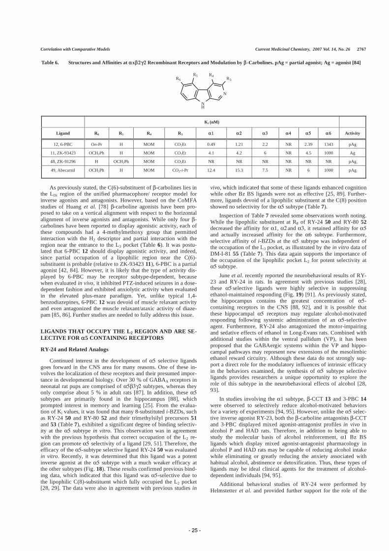
As previously stated, the C(6)-substituent of -carbolines lies in the LDi region of the unified pharmacophore/ receptor model for inverse agonists and antagonists. However, based on the CoMFA studies of Huang et al. [78] -carboline agonists have been pro-posed to take on a vertical alignment with respect to the horizontal alignment of inverse agonists and antagonists. While only four -carbolines have been reported to display agonistic activity, each of these compounds had a 4-methylmethoxy group that permitted interaction with the H2 descriptor and partial interaction with the region near the entrance to the L3 pocket (Table 6). It was postu-lated that 6-PBC 12 should display agonistic activity, and indeed, since partial occupation of a lipophilic region near the C(6)-substituent is probable (relative to ZK-93423 11), 6-PBC is a partial agonist [42, 84]. However, it is likely that the type of activity dis-played by 6-PBC may be receptor subtype-dependent, because when evaluated in vivo, it inhibited PTZ-induced seizures in a dose-dependent fashion and exhibited anxiolytic activity when evaluated in the elevated plus-maze paradigm. Yet, unlike typical 1,4-benzodiazepines, 6-PBC 12 was devoid of muscle relaxant activity and even antagonized the muscle relaxant/ataxic activity of diaze-pam [85, 86]. Further studies are needed to fully address this issue.
LIGANDS THAT OCCUPY THE L2 REGION AND ARE SE-LECTIVE FOR 5 CONTAINING RECEPTORS
RY-24 and Related Analogs
Continued interest in the development of selective ligands goes forward in the CNS area for many reasons. One of these in-volves the localization of these receptors and their presumed impor-tance in developmental biology. Over 30 % of GABAA receptors in neonatal rat pups are comprised of subtypes, whereas they only comprise about 5 % in adult rats [87]. In addition, these subtypes are primarily found in the hippocampus [88], which prompted interest in memory and learning [25]. From the evalua-tion of Ki values, it was found that many 8-substituted i-BZDs, such as RY-24 50 and RY-80 52 and their trimethylsilyl precursors 51and 53 (Table 7), exhibited a significant degree of binding selectiv-ity at the subtype in vitro. This observation was in agreement with the previous hypothesis that correct occupation of the L2 re-gion can promote selectivity of a ligand [29, 51]. Therefore, the efficacy of the -subtype selective ligand RY-24 50 was evaluated in vitro. Recently, it was determined that this ligand was a potent inverse agonist at the subtype with a much weaker efficacy at the other subtypes (Fig. 18). These results confirmed previous bind-ing data, which indicated that this ligand was -selective due to the lipophilic C(8)-substituent which fully occupied the L2 pocket [28, 29]. The data were also in agreement with previous studies in
vivo, which indicated that some of these ligands enhanced cognition while other Bz BS ligands were not as effective [25, 89]. Further-more, ligands devoid of a lipophilic substituent at the C(8) position showed no selectivity for the subtype (Table 7).
Inspection of Table 7 revealed some observations worth noting. While the lipophilic substituent at R8 of RY-24 50 and RY-80 52decreased the affinity for , and , it retained affinity for and actually increased affinity for the subtype. Furthermore, selective affinity of i-BZDs at the subtype was independent of the occupation of the L3 pocket, as illustrated by the in vitro data of DM-I-81 55 (Table 7). This data again supports the importance of the occupation of the lipophilic pocket L2 for potent selectivity at subtype.
June et al. recently reported the neurobehavioral results of RY-23 and RY-24 in rats. In agreement with previous studies [28], these selective ligands were highly selective in suppressing ethanol-maintained responding (Fig. 19) [91]. As previously stated, the hippocampus contains the greatest concentration of -containing receptors in the CNS [88, 92], and it is possible that these hippocampal receptors may regulate alcohol-motivated responding following systemic administration of an -selective agent. Furthermore, RY-24 also antagonized the motor-impairing and sedative effects of ethanol in Long-Evans rats. Combined with additional studies within the ventral pallidum (VP), it has been proposed that the GABAergic systems within the VP and hippo-campal pathways may represent new extensions of the mesolimbic ethanol reward circuitry. Although these data do not strongly sup-port a direct role for the modulatory influences of intrinsic efficacy in the behaviors examined, the synthesis of subtype selective ligands provides researchers a unique opportunity to explore the role of this subtype in the neurobehavioral effects of alcohol [28, 93].
In studies involving the subtype, -CCT 13 and 3-PBC 14were observed to selectively reduce alcohol-motivated behaviors for a variety of experiments [94, 95]. However, unlike the selec-tive inverse agonist RY-23, both the -carboline antagonists -CCT and 3-PBC displayed mixed agonist-antagonist profiles in vivo in alcohol P and HAD rats. Therefore, in addition to being able to study the molecular basis of alcohol reinforcement, Bz BS ligands which display mixed agonist-antagonist pharmacology in alcohol P and HAD rats may be capable of reducing alcohol intake while eliminating or greatly reducing the anxiety associated with habitual alcohol, abstinence or detoxification. Thus, these types of ligands may be ideal clinical agents for the treatment of alcohol-dependent individuals [94, 95].
Additional behavioral studies of RY-24 were performed by Helmstetter et al. and provided further support for the role of the
Table 6. Structures and Affinities at x Recombinant Receptors and Modulation by -Carbolines. pAg = partial agonist; Ag = agonist [84]
NH
N
R4 R3R6R5
Ki (nM)
Ligand R6 R5 R4 R3 Activity
12, 6-PBC On-Pr H MOM CO2Et 0.49 1.21 2.2 NR 2.39 1343 pAg
11, ZK-93423 OCH2Ph H MOM CO2Et 4.1 4.2 6 NR 4.5 1000 Ag
48, ZK-91296 H OCH2Ph MOM CO2Et NR NR NR NR NR NR pAg
49, Abecarnil OCH2Ph H MOM CO2-i-Pr 12.4 15.3 7.5 NR 6 1000 pAg
- 25 -

2768 Current Medicinal Chemistry, 2007, Vol. 14, No. 26 Clayton et al.
Table 7. Structures and Affinities of Some -Subtype Selective Ligands
N
N
N
R8
O Me
CO2R3
N
N
N
R8
O
O
56
N
N
NCO2Et
DM-I-81, 55
Ki (nM) Ligand R8 R3
50, RY-24 H t-Bu 26.9 26.3 18.7 0.4 5.1
51, RY-23 TMS t-Bu 197 143 255 2.61 58.6
52, RY-80 H Et 28.4 21.4 25.8 0.49 28.8
53, RY-79 TMS Et 121 142 198 5.0 114
54 H Et 1.2 2.0 1.1 0.4 > 300
55, DM-I-81 Ph NA > 2000 > 2000 > 2000 176 > 2000
56, PWZ-02990 Cl NA >300 >300 >300 38.8 >300
Fig. (18). Subtype efficacy of RY-24 50. Dose response curves for RY-24 in oocytes expressing different subunit combinations of GABAA receptors. Subtype combinations are indicated in legends. cRNA–injected Xenopus oocytes were held at –60 mV under two-electrode voltage clamp. Increasing concentrations of RY-24 were superfused together with a GABA concentration eliciting ~ 20% of the maximal current amplitude. RY-24 was pre-applied for 30 sec before the addition of GABA, which was co-applied with the drugs until a peak response was observed. Data were normalized for each curve assuming 100% for the response in the absence of RY-24. RY-24 was made up and diluted as stock solution in DMSO. Final DMSO concentrations perfusing the oocyte were 0.1%. Values are presented as mean ± SD of at least 4 oocytes from at least 2 batches.
hippocampus in anxiety and learning [25]. Moreover, the data sug-gested that Bz BSs within the hippocampus are important for the acquisition of fear conditioning. Although this subtype selective ligand has been shown to be an inverse agonist at the subtype [29, 96], this study suggested that RY-24 may act as an agonist at other alpha subtypes because larger doses of RY-24 were not as anxiogenic as the smaller doses and resulted in decreased learning. Consistent with the studies of Stephens et al. using knock-out mice [97] and the efficacy studies of Lüddens, June and Cook et al.[98] these findings support the concept that the pharmacology ob-served depends upon the dose, behavioral paradigm employed and subunit composition activated. Ligands such as RY-24 have proven to be valuable in the study of the biochemical and pharmacological
properties of GABAA receptors and have permitted insight into the role this protein plays in anxiety and learning.
QH-ii-066
Due to the pharmacological profile RY-24 exhibited in vivo, the development of additional subtype selective ligands was pur-sued. Thus, the 7-acetyleno analog of diazepam, QH-ii-066 57, was synthesized and was determined to also exhibit a binding and func-tional selectivity at the subtype over the subtype (Table 8)[27]. This was due to the full occupation of the L2 pocket, relative to diazepam (Fig. 20). To our knowledge, this was the first agonist ligand to display some selectivity from the 1,4-benzodiazepine
- 26 -

Correlation with Comparative Models Current Medicinal Chemistry, 2007 Vol. 14, No. 26 2769
family. Importantly, the 7-cyano congener 58 (Table 8) did not potently bind to recombinant receptors of the 5 subtype, which is in agreement with earlier work of Haefely and Fryer et al. on the SAR of 1,4-benzodiazepines [99, 100]. This cyano ligand also did not exhibit any subtype selectivity, re-emphasizing that occupation of the L2 region with lipophilic groups is important for 5 selectiv-ity as well as for high affinity. The selective efficacy of this QH-ii-066 ligand over the 1 subtype was demonstrated by reversing the convulsant actions of RY-24 50, an 5-selective inverse agonist, in NIH mice [87]. This ability was not observed at comparable doses for the 1-selective agonist zolpidem 7.
Fig. (20). Comparison of non-selective diazepam (black) with the 5-selective QH-ii-066 (cyan) when aligned within the unified pharmacophore/ receptor model. The acetylene group of QH-ii-066 increased the occupation of the L2 region relative to that of diazepam.
Furthermore, Lelas and Cook et al. have recently determined that although QH-ii-066 had similar affinity for the DS subtypes in rats, it displayed functional selectivity in vivo, with diazepam-like efficacy at the 5 subtype and partial efficacy at the 1 subtype [27]. The study also indicated that this 7-acetyleno substituted di-azepam analog exhibited less potency in protection against ECS-induced seizures relative to diazepam than against PTZ-induced seizures. Hence, the 1 subtype may play a more prominent role in ECS-induced seizures than in PTZ-induced seizures [27].
COMPARATIVE MODEL OF THE BENZODIAZEPINE BINDING SITE
Crystallization of GABAA receptors thus far has not been ac-complished, but the successful structure determination of the water-soluble acetylcholine binding protein (AChBP) [101] has generated much interest in the GABAA receptor community. Although this protein shares only ~ 18 % sequence homology with the extracellu-lar domain of the GABAA receptor [101], the structural resem-blance has been estimated to be relatively high (60 – 75 %) [48]. Several comparative modeling studies have used the AChBP struc-ture to derive models of the extracellular domain of GABAA recep-tors [47, 48]. Following the cryo-EM determination of the extracel-lular and transmembrane domain structure of the nACh receptors, these structures also were used as templates for modeling GABAAreceptors [102]. Sequence homology is so low however, that de-tailed features of the models are highly uncertain, and the proposed dockings of Bz BS ligands [103-105] have a qualitative character and do not sufficiently explain the observed differential effects of
Fig. (19). Suppression of alcohol-motivated responding by RY-24 50 and RY-23 51 [93, 96]. Left: Dose-response of IP RY-24 (0.0–3.5 mg/ kg) and vehicle on responding maintained by ethanol (10 % v/v) (top panel) and water (bottom panel) in male Long-Evans rats. Right: Dose-response of unilateral infusions of RY-23 (0.0-40 g) in the hippocampus on a concurrent fixed-ratio (FR4) schedule for ethanol (10 % v/v) (top panel) and saccharin-maintained (0.025 % v/v) (bottom panel). For both studies, *p 0.05 versus control condition values was determined using ANOVA and post hoc Newman-Keuls test. Each bar repre-sents the mean (± SEM) (n = 15 for RY-24 and n = 7 for RY-23). Figures reprinted with permission of the authors [93, 96].
- 27 -

2770 Current Medicinal Chemistry, 2007, Vol. 14, No. 26 Clayton et al.
ligands interacting with different receptor subtypes. The experimen-tal structure of the nAChR [106] has provided first data on how much the fold can vary between members of a family [106]. From this data the extent to which GABAA receptor subunits differ from each other in structure can be qualitatively extrapolated, but not predicted in detail.
As mentioned before, the majority of GABAA receptors are composed of , 2 and subunits. Each subunit has per conven-tion a “plus” and a “minus” side (Fig. 21). The subunit interfaces consequently consist of the plus and minus sides of neighboring subunits. The modulatory Bz BS is located at the subunit inter-face and is larger than, but homologous to the two agonist (GABA) binding sites, which are located at the subunit interfaces [49, 107, 108]. The absolute subunit configuration for the GABAA receptor appears to be , when viewed counter clockwise (from + to - , Fig. 21) [48, 109-111].
Fig. (21). Absolute subunit arrangement of the GABAA receptor when viewed from the synaptic cleft. The GABA binding sites are located at the subunit interfaces and the modulatory Bz binding site is located at the subunit interface. The part of the schematically drawn subunits marked by the + indicates loop C of the respective subunits.
While the 2 subunit is required for recognition and binding of benzodiazepines as well as many other substance classes that act via the Bz BS [112, 113], it is now clear that sequence variations between different and subunits determine subtype selectivity and efficacy of Bz BS ligands [20, 112-114]. The Bz BS has been proposed to consist of three segments provided by the side, the so-called “loops A, B and C” and by three segments of the , the so-called “loops D, E and F” [112, 113]. These segments were then confirmed by X-ray crystallographic and EM-structures of AChBP [101] and the nAChR [106] to form a groove-like pocket at
the interface boundary between subunits that appears to be con-served in the entire superfamily.
Even later than the nAChR structure, a series of AChBP crystal structures with co-crystallized ligands appeared [115]. These struc-tures revealed how ligand binding can alter the local conformation of the binding site. Particularly loop C has been found to be a highly mobile subdomain, additional more subtle changes are seen along the entire subunit boundary [115]. These findings are consis-tent with the hypothesis that many receptor conformations exist, that are separated by low energy barriers, and can be stabilized by different ligands. Unfortunately it cannot be decided a priori which of the experimental structures is the “best” template to model a particular receptor/ligand complex. Depending on template and alignment choice, model Bz pockets differ in total volume by as much as 40% and can vary by several Angstrom in the distances between key residues in the binding site loops.
Although changes in protein conformation may be minor, it has been demonstrated that they can profoundly affect the efficacy of Bz BS ligands [116]. Furthermore, efficacy can vary for the same ligand at different GABAA receptor subtypes [27, 31, 35]. As pro-teins are inherently dynamic and able to sample many conforma-tions, and the stabilization of the active state relative to the inactive state has been calculated to be less than 1 kcal/mol [117], it is im-possible to provide absolute assignments of specific side chains to specific descriptors for any particular conformation. This conforma-tional flexibility may imply that residues which satisfy certain pharmacophoric descriptors can vary, resulting in a “soft” orienta-tion of the pharmacophore in the receptor. Thus, a unified view of a pharmacophore model and a homology derived receptor model will assign large areas of lipophilic interaction to specific regions in the protein, but allow a flexible assignment of specific interactions such as H-bridges or - stacking.
RELATIVE ORIENTATION OF THE PHARMACOPHORE WITHIN THE COMPARATIVE MODEL
Prior to structure determination of the AChBP, we published a review which evaluated results of site-directed mutagenesis and provided insights as to where certain side chains in the Bz BS might be located relative to the pharmacophoric descriptors [118]. How-ever, with the knowledge gained from recent experimental data and with the aid of our GABAA receptor models, built by homology to lymnea AChBP [47], aplysia AChBP and the nAChR [102] an up-date of the orientation is provided here. We now propose an orien-tation which is favored by experimental evidence, allows some degree of conformational flexibility which can lead to variable as-
Table 8. Structures and Affinities of 1,4-Benzodiazepines
N
N
MeO
R7
Ki (nM) Ligand R7
1, diazepam Cl 14 20 15 11 > 3000
57, QH-ii-066 H 76.3 42.1 47.4 6.8 > 3000
58 C N 320 310 350 265 > 3000
- 28 -

Correlation with Comparative Models Current Medicinal Chemistry, 2007 Vol. 14, No. 26 2771
signments for H-bridge interactions, but is based on specific areas of lipophilic interactions that are determined by binding site geome-try.
Evidence from Covalently Reactive Ligands Allows to Position the L2 Lipophilic Pocket
Covalent labeling studies contributed significantly to the deter-mination of residues that are located within the Bz BS [104, 119-124]. Table 9. Ligands Used for Affinity Labeling Studies [104, 121-124]
N
N
NC
S
Me O
N
N
N O
O
O
N
S59 60
7-isothiocyano-1,4-benzodiazepine
Ligand Activity Site of Interaction
2, [3H]flunitrazepam Agonist H102
6, [3H]Ro15-4513 inverse agonist Y210
5960
Agonist part. Agonist
H101C (rat) H101C, G157C
V202, V211C (rat)
Photoincorporation studies with the agonist [3H]flunitrazepam identified H102 of the human sequence as the primary site of incorporation [123]. Although it is possible that flunitrazepam is coupling via the nitro group at the 7-position, the coupling group of flunitrazepam is currently not known. Studies with the inverse ago-nist [3H]Ro15-4513 indicated Y210 as the primary site of incor-poration [104]. Thus, the azido group at the 7-position of Ro15-4513 should be in close apposition to 1Y210, assuming no rear-rangement of the photo-activated intermediate. Further information comes from recent studies reporting the covalent coupling of 7-isothiocyano- derivatives of a 1,4-benzodiazepine, (substance 59
[122]) and of Ro15-1788 (substance 60 [124]) to GABAA receptors in which individual amino acid residues had been mutated to cys-teines. Primary site of reaction of both substances is the rat
1H101C mutant that is homologous to 1H102 of the human subunit. Thus, the 7-substituent both of 1,4-benzodiazepines and of imidazodiazepines appears to be in apposition to H102. The imidazobenzodiazepine 60 reacts with additional cysteines in posi-tions corresponding in the human sequence to positions 1V203C and 1V212C in the loop C stem, and with 1G158C in loop B (see alignment in Fig. 22). Thus, the 7-substituent of this compound, in agreement with the data from photolabeling H102 with flunitraze-pam and Y210 with Ro15-4513, is in apposition to loop A, the loop C base, and additionally loop B.
Thus, all the findings from covalently incorporated ligands given above can be reconciled by placing the 7-substituent, and thus the L2 descriptor of the unified pharmacophore model, between the loop C base near 1V203/ 1V212/ 1Y210 and loop A 1H102. The homology models reveal that this is not only topologically possible, but also provides the additional apposition to loop B that is sug-gested by the reaction of 60 with rat 1G157C. Thus, the L2 lipo-philic pocket is most likely formed by the base of loop C, together with parts of loop A.
THE A2 DESCRIPTOR AND THE LDI REGION
Experimental data yields some very convincing evidence for as-signing the A2 descriptor and the LDI region of lipophilic interac-tion next to A2 to protein segments close to residues A79 and T81 at loop D. Certain imidazobenzodiazepines, which occupy
the pharmacophore volume close to A2 (see Fig. 5), are more sensi-tive to A79 mutations than classical benzodiazepines or benzodi-azepines that lack a 3'-imidazo substituent. This was shown in a study where mutations of A79 and T81 had little effect on the binding of the agonists diazepam and flunitrazepam, but in a roughly size dependent fashion decreased the affinities of antago-nist Ro15-1788 and inverse agonists Ro15-4513 [125, 126]. In agreement with other studies, midazolam, which lacks a 3’ substitu-ent, behaved more closely to classical benzodiazepines than i-BZDs [44, 125, 127,128]. This trend is enhanced by rigid and bulky 3'-imidazo substituents, altogether suggesting that A79 and T81are located close to the region of the Bz BS surrounding the 3' sub-stituent of i-BZDs [125]. The localization of the 3'-imidazo sub-stituents in the unified pharmacophore model, see Fig. (5), thus positions the A2 and LDI descriptors near residues A79 and
Fig. (22). Alignment and homology model depiction of the so called “loops A-F” and flanking regions of the human sequences of the Bz recognition site in different subunits. The homology model is as seen from “outside” of the channel mouth, the membrane would be parallel to the lower edge of the image. Key residues are shown in stick representation: Loop A His102, loop B Tyr 160 and loop D Phe77.
- 29 -

2772 Current Medicinal Chemistry, 2007, Vol. 14, No. 26 Clayton et al.
T81. Further support for this localization comes from the obser-vation that DMCM also looses affinity upon mutagenesis in posi-tions A79 and T81, [125, 126] consistent with the alignment of this substance in the pharmacophore. Fig. (5) shows that changes to the protein near the A2 descriptor would in all likelihood not be tolerated by DMCM.
COMBINING HOMOLOGY MODEL AND EXPERIMEN-TAL EVIDENCE
If the two assignments discussed above are correct, they should be reflected in results of computational ligand docking to homology models of the binding site. Thus, computational docking of diaze-pam and flumazenil was performed in multiple different models based on different templates [101, 106, 115] and the best 100 dock-ing poses per model were collected in a database. A database query was then formulated to search among the (unrefined) docking poses for those with the following properties: The seven membered ring of diazepam or flumazenil is in the conformation that is assumed to
be the active one; [99] the 7-substituents are near residues H102/ V203/ and Y210/ V212 in order to be consistent
with the covalent labeling data; the 3’ ester group of flumazenil is near residues A79 and T81. Such a query yields docking poses that are consistent with all covalent labeling studies on one hand, and with the steric requirements that were found for 3’ substituted imidazobenzodiazepines [125] on the other hand. Such poses were indeed present in our database, and one representative diazepam pose is shown in Fig. (23): To be consistent with the presentation of the pharmacophore and its descriptors so far, the orientation of the benzodiazepine binding pocket had to be turned upside down and slightly tilted.
This and similar docking poses not only satisfy the two assign-ments for L2 and A2 that are discussed in the sections above, but at the same time position the remaining descriptors as follows: H1 can be satisfied by appropriate side chains near the tip of loop C, the LDI region is near the subunit interface, essentially between loop B (Y160) and the region spanned by the interface forming sheet involving M130, T142 and F77; A2 can be satisfied by H-bridge
Fig. (23). The main figure shows a diazepam docking pose in an 1 2 pocket matching the orientation proposed in the text. Diazepam is rendered space fill-ing, (turquoise: carbon, white: hydrogen, green: halogen, red: oxygen, blue: nitrogen), the protein as ribbon, and key amino acids in stick representation. The insert figure shows the empty pocket in the “upright” position to facilitate orientation in the main figure, where the model has been turned and tilted to bring diazepam approximately into the orientation depicted in the unified pharmacophore model, Fig. (3). In this particular docking pose, His102 and Thr206 could satisfy H2 and H1, respectively, and His 102, Val 212 and Tyr 210 would be part of the L2 hydrophobic pocket. This positions 7-substituted reactive ana-logues such as compound 59 into a position where the reactive groups find free access to His102, Tyr 210, Val 212, Val 203. It should be noted that due tohigh uncertainties in model backbone position and side chain rotamers, high accuracy docking that correctly reflects all ligand-protein interactions is not feasi-ble, this image is one representative of a big cluster of similar docking poses.
- 30 -

Correlation with Comparative Models Current Medicinal Chemistry, 2007 Vol. 14, No. 26 2773
accepting sites near Y160, A79, M130 and T142. If these proximity relations that are found in the docking poses are transferred to the unified pharmacophore model, the following pic-ture emerges:
Within the different poses that exhibit this orientation, there is still some degree of variation in structural details. There are several sources for this variability in the computationally derived struc-tures: template and alignment choice, model construction and re-
finement, as well as docking algorithms contribute to the variability of the poses that are found.
This orientation is also strongly supported by the observation that XLi-093, as aligned in Fig. (14) with the docked diazepam shown above, will be able to thread the portion of the molecule not inside the included volume towards the solvent accessible space through the subunit boundary.
Fig. (24). Location of important Bz BS residues relative to the pharmacophoric descriptors in two views that correspond to a 90 rotation about L1, with A2 in front, as determined by evaluation of experimental data and the comparative model of the GABAA receptor. The ligand shown is diazepam. Since inverse agonists stabilize protein conformations that vary from the conformations preferred by agonists, it seems plausible that more than one side chain could satisfy the same descriptor.
- 31 -

2774 Current Medicinal Chemistry, 2007, Vol. 14, No. 26 Clayton et al.
Agreement between this orientation of the pharmacophore model inside the structural model of the protein with single ligand dockings that have been proposed in the past [103-105] will have to be examined in detail. The overall picture appears to be pointing towards a satisfactory orientation of the pharmacophore model in the pocket, and for those instances where there could be a discrep-ancy, further experiments will clarify.
Refinement of the predicted protein-ligand complexes with the aid of the unified pharmacophore model appears to be a very prom-ising approach to arrive at 3D structures that are of sufficient accu-racy to be used for structure guided drug design. Studies that com-bine advanced protein modeling techniques with the classical pharmacophore approach are currently underway.
CONCLUSION
A unified pharmacophore model incorporating many substance classes that act at the DS and DI benzodiazepine binding sites of GABAA receptors has been updated to include new substance classes. Compound development guided by this pharmacophore model has led to new agents with interesting pharmacological pro-files, particularly enhanced preference for or containing GABAA receptor subtype. Based on the evaluation of experimental data and comparative models of the GABAA receptor, the location of several residues relative to the descriptors of the phar-macophore/receptor model has been proposed. Although no abso-lute assignments were made regarding which amino acids satisfy the pharmacophoric descriptors, experimental data strongly indi-cated definite trends with regard to how ligands of varying pharma-cological activity are oriented within the receptor. Because the uni-fied pharmacophore/receptor model accounts for the binding and activity profiles at the six GABAA receptor subtypes containing any one of the different alpha subunits, the proposed orientation should also be similar within the different models [110] of the various receptor subtypes. Information to be immediately gained from this proposed orientation can have far reaching benefits, not only for the rational design of selective ligands and the interpretation of ligand docking results, but also for the identification and evaluation of possible roles certain residues may have within the pocket. As structure determination of the GABAA receptor is eagerly awaited, it is hoped that this proposed orientation may be used by others to gain additional insight into the potential mechanisms underlying binding and modulation at the Bz site, all of which will lead to a better understanding of the structure and function of GABAA recep-tors.
ACKNOWLEDGEMENTS
We wish to thank Qi Huang for helpful discussions. We wish to acknowledge support by the Research Growth Initiative of the Uni-versity of Wisconsin-Milwaukee and the NIMH (MH 46851) as well as by grants of the Austrian Science Fund (P15165 and P16397).
REFERENCES
[1] Sieghart, W.; Ernst, M. Curr. Med. Chem. Central Nervous Syst. Agents 2005, 5, 217-242.
[2] Bowser, D. N.; Wagner, D. A.; Czajkowski, C.; Cromer, B. A.; Parker, M. W.; Wallace, R. H.; Harkin, L. A.; Mulley, J. C.; Marini, C.; Berkovic, S. F.; Williams, D. A.; Jones, M. V.; Petrou, S. Proc. Natl. Acad. Sci. USA 2002,99, 15170-15175.
[3] Bateson, A. N. Sleep Med. 2004, 5, S9-S15. [4] Otani, K.; Ujike, H.; Tanaka, Y.; Morita, Y.; Katsu, T.; Nomura, A.; Uchida,
N.; Hamamura, T.; Fujiwara, Y.; Kuroda, S. Neurosci. Lett. 2005, 381, 108-113.
[5] Dean, B.; Scarr, E.; McLeod, M. Brain Res. Mol. Brain Res. 2005, 138, 145-155.
[6] Guidotti, A.; Auta, J.; Davis, J. M.; Dong, E. B.; Grayson, D. R.; Veldic, M.; Zhang, X. Q.; Costa, E. Psychopharmacology 2005, 180, 191-205.
[7] Maubach, K. Drug Targets-CNS Neuro Disorders 2003, 2, 233-239.
[8] Barrett, A. C.; Negus, S. S.; Mello, N. K.; Caine, S. B. J. Pharmacol. Exp. Ther. 2005, 315, 858-871.
[9] Anthenelli, R.; Schuckit, M. A. Int. J. Addict. 1990/1991, 25, 81-94. [10] Schuckit, M. A. Alcohol Clin. Exp. Res. 1988, 12, 465-470. [11] Barnard, E. A.; Skolnick, P.; Olsen, R. W.; Möhler, H.; Sieghart, W.; Biggio,
G.; Braestrup, C.; Bateson, A. N.; Langer, S. Z. Pharmacol. Rev. 1998, 50,291-313.
[12] Simon, J.; Wakimoto, H.; Fujita, N.; Lalande, M.; Barnard, E. A. J. Biol. Chem. 2004, 279, 41422-41435.
[13] Burt, D. R.; Kamatchi, G. L. FASEB J. 1991, 5, 2916-2923. [14] Keramidas, A.; Moorhouse, A.; Schofield, P. C.; Barry, P. Prog. Biophys.
Mol. Biol. 2004, 86, 161-204. [15] Sieghart, W.; Sperk, G. Curr. Top. Med. Chem. 2002, 2, 795-816. [16] Mossier, B.; Togel, M.; Fuchs, K.; Sieghart, W. J. Biol. Chem. 1994, 269,
25777-25782. [17] Togel, M.; Mossier, B.; Fuchs, K.; Sieghart, W. J. Biol. Chem. 1994, 269,
12993-12998. [18] Pirker, S.; Schwarzer, C.; Wieselthaler, A.; Sieghart, W.; Sperk, G. Neuro-
science 2000, 101, 815-850. [19] Doble, A.; Martin, I. L. Trends Pharmacol. Sci. 1992, 13, 76-81. [20] Sieghart, W. Pharmacol. Rev. 1995, 47, 181-234. [21] Kralic, J. E.; Korpi, E. R.; O'Buckley, T. K.; Homanics, G. E.; Morrow, A. L.
J. Pharmacol. Exp. Ther. 2002, 302, 1037-1045. [22] Crestani, F.; Assandri, R.; Tauber, M.; Martin, J.; Rudolph, U. Neurophar-
macology 2002, 43, 679-684. [23] Rudolph, U.; Crestani, F.; Benke, D.; Brunig, I.; Benson, J. A.; Fritschy, J.
M.; Martin, J. R.; Bluethmann, H.; Möhler, H. Nature 1999, 401, 796-800. [24] Collinson, N.; Cothliff, R.; Rosahl, T.; Sur, C.; Kuenzi, F. M.; Howell, O.;
Seabrook, G.; Atack, J.; McKernan, R.; Dawson, G. R.; Whiting, P. Eur. J. Neurosci. 2000, 12, 171.
[25] Bailey, D. J.; Tetzlaff, J. E.; Cook, J. M.; He, X. H.; Helmstetter, F. J. Neu-robiol. Learn. Mem. 2002, 78, 1-10.
[26] DeLorey, T. M.; Lin, R. C.; McBrad, B.; He, X. H.; Cook, J. M.; Lameh, J.; Loew, G. H. Eur. J. Pharmacol. 2001, 426, 45-54.
[27] Lelas, S.; Rohrbach, K.; Glick, S. D.; Zeller, K.; Bourin, C.; Sieracki, K.; Bertekap, R.; Chen, J.; Cook, C. M.; Helmstetter, F. J.; Li, X. H.; Westphal, R.; Lindner, M.; Tertyshnikova, S.; Cook, J.; McElroy, J. F. Manuscript in preparation.
[28] Liu, R. Y.; Zhang, P. W.; McKernan, R. M.; Wafford, K.; Cook, J. M. Med. Chem. Res. 1995, 5, 700-709.
[29] Liu, R. Y.; Hu, R. J.; Zhang, P. W.; Skolnick, P.; Cook, J. M. J. Med. Chem. 1996, 39, 1928-1934.
[30] Griebel, G.; Perrault, G.; Simiand, J.; Cohen, C.; Granger, P.; Decobert, M.; Francon, D.; Avenet, P.; Depoortere, H.; Tan, S.; Oblin, A.; Schoemaker, H.; Evanno, Y.; Sevrin, M.; George, P.; Scatton, B. J. Pharmacol. Exp. Ther. 2001, 298, 753-768.
[31] Minier, F.; Sigel, E. Proc. Natl. Acad. Sci. USA 2004, 101, 7769-7774. [32] Wafford, K. A.; Macaulay, A. J.; Fradley, R.; O'Meara, G. F.; Reynolds, D.
S.; Rosahl, T. W. Biochem. Soc. Trans. 2004, 32, 553-556. [33] Casula, M. A.; Bromidge, F. A.; Pillai, G. V.; Wingrove, P. B.; Martin, K.;
Maubach, K.; Seabrook, G. R.; Whiting, P. J.; Hadingham, K. L. J. Neuro-chem. 2001, 77, 445-451.
[34] Wafford, K. A.; Whiting, P. J.; Kemp, J. A. Mol. Pharmacol. 1993, 43, 240-244.
[35] Atack, J. R. Expert Opin. Invest. Drugs 2005, 14, 601-618. [36] Möhler, H.; Rudolph, U.; McKernan, R. In ACNP, December 20, 2004.[37] Study, R. E. B. J.L. Proc. Natl. Acad. Sci. USA 1981, 78, 7180-7184. [38] Rogers, C.; Twyman, R. E.; MacDonald, R. L. J. Physiol. (London) 1994,
475, 69-82. [39] Kash, T. L.; Jenkins, A.; Kelley, J. C.; Trudell, J.; Harrison, N. L. Nature
2003, 421, 272-275. [40] Rüsch, D.; Forman, S. A. Anesthesiology 2005, 102, 783-792. [41] Baur, R.; Sigel, E. Mol. Pharmacol. 2005, 67, 1005-1008. [42] Zhang, W.; Koehler, K. F.; Zhang, P.; Cook, J. M. Drug Des. Discov. 1995,
12, 193-248. [43] Zhang, W.; Diaz-Arauzo, H.; Allen, M. S.; Koehler, K. F.; Cook, J. M.
Chemical and Computer Assisted Development of the Inclusive Pharma-cophore of Benzodiazepine Receptors, in Studies in Medicinal Chemistry, M.I. Choudhary, Editor. Harwood Academic Publishers: 1996; pp. 303.
[44] Sigel, E.; Schaerer, M. T.; Buhr, A.; Baur, R. Mol. Pharmacol. 1998, 54,1097-1105.
[45] Dunn, S. M. J.; Davies, M.; Muntoni, A. L.; Lambert, J. J. Mol. Pharmacol. 1999, 56, 768-774.
[46] Davies, M.; Bateson, A. N.; Dunn, S. M. J. J. Neurochem. 1998, 70, 2188-2194.
[47] Cromer, B.; Morton, C.; Parker, M. W. Trends Biochem. Sci. 2002, 27, 280-287.
[48] Ernst, M.; Brauchart, D.; Boresch, S.; Sieghart, W. Neuroscience 2003, 119,933-943.
[49] Sigel, E. Curr. Top. Med. Chem. 2002, 2, 833-840. [50] Allen, M. S.; Tan, Y. C.; Trudell, M. L.; Narayanan, K.; Schindler, L.; Mar-
tin, M. J.; Schultz, C. A.; Hagen, T. J.; Koehler, K. F.; Codding, P.; Skolnick, P.; Cook, J. J. Med. Chem. 1990, 33, 2343-2357.
[51] Zhang, P. W.; Zhang, W. J.; Liu, R. Y.; Harris, B.; Skolnick, P.; Cook, J. M. J. Med. Chem. 1995, 38, 1679-1688.
- 32 -

Correlation with Comparative Models Current Medicinal Chemistry, 2007 Vol. 14, No. 26 2775
[52] Allen, M. S.; Laloggia, A. J.; Dorn, L. J.; Martin, M. J.; Constantino, G.; Hagen, T. J.; Koehler, K. F.; Skolnick, P.; Cook, J. J. Med. Chem. 1992, 35,4001-4010.
[53] Wong, G.; Koehler, K. F.; Skolnick, P.; Gu, Z. Q.; Ananthan, S.; Schönhol-zer, P.; Hunkeler, W.; Zhang, W. J.; Cook, J. M. J. Med. Chem. 1993, 36,1820-1830.
[54] Hevers, W.; Luddens, H. Mol. Neurobiol. 1998, 18, 35-86. [55] He, X. B.; Huang, Q.; Ma, C. R.; Yu, S.; McKernan, R.; Cook, J. Drug Des.
Discov. 2000, 17, 131-171. [56] Huang, Q.; He, X. H.; Ma, C. R.; Liu, R. Y.; Yu, S.; Dayer, C. A.; Wenger,
G. R.; McKernan, R.; Cook, J. M. J. Med. Chem. 2000, 43, 71-95. [57] Hadingham, K. L.; Wingrove, P. B.; Wafford, K. A.; Bain, C.; Kemp, J. A.;
Palmer, K. J.; Wilson, A. W.; Wilcox, A. S.; Sikela, J. M.; Ragan, C. I.; Whiting, P. J. Mol. Pharmacol. 1993, 44, 1211-1218.
[58] Barnard, E. A. Trends Biochem. Sci. 1992, 17, 368-374. [59] Huang, Q.; Liu, R. Y.; Zhang, P. W.; He, X. H.; McKernan, R.; Gan, T.;
Bennett, D. W.; Cook, J. M. J. Med. Chem. 1998, 41, 4130-4142. [60] Wafford, K. A.; Thompson, S. A.; Thomas, D.; Sikela, J.; Wilcox, A. S.;
Whiting, P. J. Mol. Pharmacol. 1996, 50, 670-678. [61] Huang, Q.; Cox, E.; Gan, T.; Ma, C. R.; Bennett, D. A.; McKernan, R.;
Cook, J. Drug Des. Discov. 1999, 16, 55-76. [62] Drover, D. R. Clin. Pharmacokinet. 2004, 43, 227-238. [63] Noble, S.; Langtry, H. D.; Lamb, H. M. Drugs 1998, 55, 277-302. [64] Jerussic, T. 2006, unpublished results. [65] Savic, M. M.; Huang, S.; Furtmuller, R.; Clayton, T.; Huck, S.; Obradovic,
D. I.; Ugresic, N. D.; Sieghart, W.; Bokonjic, D. R.; Cook, J. M. Neuropsy-chopharmacology 2007.
[66] Dias, R.; Sheppard, W. F. A.; Fradley, R. L.; Garrett, E. M.; Stanley, J. L.; Tye, S. J.; Goodacre, S.; Lincoln, R. J.; Cook, S. M.; Conley, R.; Hallett, D.; Humphries, A. C.; Thompson, S. A.; Wafford, K. A.; Street, L. J.; Castro, J. L.; Whiting, P. J.; Rosahl, T. W.; Atack, J. R.; McKernan, R. M.; Dawson, G. R.; Reynolds, D. S. J. Neurosci. 2005, 25, 10682-10688.
[67] McKernan, R. M.; Rosahl, T. W.; Reynolds, D. S.; Sur, C.; Wafford, K. A.; Atack, J. R.; Farrar, S.; Myers, J.; Cook, G.; Ferris, P.; Garrett, L.; Bristow, L.; Marshall, G.; Macaulay, A.; Brown, N.; Howell, O.; Moore, K. W.; Car-ling, R. W.; Street, L. J.; Castro, J. L.; Ragan, C. I.; Dawson, G. R.; Whiting, P. J. Nat. Neurosci. 2000, 3, 587-592.
[68] Collinson, N.; Kuenzi, F. M.; Jarolimek, W.; Maubach, K. A.; Cothliff, R.; Sur, C.; Smith, A.; Otu, F. M.; Howell, O.; Atack, J. R.; McKernan, R. M.; Seabrook, G. R.; Dawson, G. R.; Whiting, P. J.; Rosahl, T. W. J. Neurosci. 2002, 22, 5572-5580.
[69] Crestani, F.; Keist, R.; Fritschi, J.; Benke, D.; Vogt, K. E.; Prut, L. Proc. Natl. Acad. Sci. USA 2002, 99, 8980-8985.
[70] Bohlhalter, S.; Weinmann, O.; Möhler, H.; Fritschy, J. M. J. Neurosci. 1996,16, 283-297.
[71] van Rijnsoever, C.; Tauber, M.; Choulli, M. K.; Keist, R.; Rudolph, U.; Möhler, H.; Fritschy, J. M.; Crestani, F. J. Neurosci. 2004, 24, 6785-6790.
[72] Porsolt, R. D.; McArthur, R. A.; Lenegre, A. Psychotropic Screening Proce-dures. Elsevier: 1993; pp. 23-51.
[73] Wang, Q.; Han, Y.; Xue, H. CNS Drug Rev. 1999, 5, 125-144. [74] Miksieck, R. Mol. Pharmacol. 1993, 44, 37-43. [75] Häberlein, H.; Tschiersch, K. P.; Schäfer, H. L. Pharmazie 1994, 49, 912-
922. [76] Huang, X. Q.; Liu, T.; Gu, J. D.; Luo, X. M.; Ji, R. Y.; Cao, Y.; Xue, H.;
Wong, J. T. F.; Wong, B. L.; Pei, G.; Jiang, H. L.; Chen, K. X. J. Med. Chem. 2001, 44, 1883-1891.
[77] Marder, M.; Estiu, G.; Blanch, L. B.; Viola, H.; Wasowski, C.; Medina, J. H.; Paladini, A. C. Bioorg. Med. Chem. 2001, 9, 323-335.
[78] Yu, J.; Cook, J. 1999, unpublished results. [79] Costanzo, A.; Guerrini, G.; Ciciani, G.; Bruni, F.; Costagli, C.; Selleri, S.;
Besnard, F.; Costa, B.; Martini, C.; Malmberg-Aiello, P. J. Med. Chem. 2002, 45, 5710-5720.
[80] Yin, W. Y.; Sarma, P.; Han, J.; Chen, J. L.; Cook, J. M. Tetrahedron Lett. 2005, 46, 6363-6368.
[81] Li, X. Y.; Cao, H.; Zhang, C. C.; Furtmueller, R.; Fuchs, K.; Huck, S.; Sieghart, W.; Deschamps, J.; Cook, J. M. J. Med. Chem. 2003, 46, 5567-5570.
[82] Han, D. M.; Forsterling, F. H.; Li, X. Y.; Deschamps, J. R.; Cao, H.; Cook, J. M. Bioorg. Med. Chem. Lett. 2004, 14, 1465-1469.
[83] Ferretti, V.; Gilli, P.; Borea, P. A. Acta Crystalogr. Sect. B Struct. Sci. 2004,60, 481-489.
[84] Cox, E. D.; Diaz-Arauzo, H.; Huang, Q.; Reddy, M. S.; Ma, C. R.; Harris, B.; McKernan, R.; Skolnick, P.; Cook, J. M. J. Med. Chem. 1998, 41, 2537-2552.
[85] Diaz-Arauzo, H.; Evoniuk, G.; Skolnick, P.; Cook, J. J. Med. Chem. 1991,34, 1754-1756.
[86] Diaz-Arauzo, H.; Koehler, K. F.; Hagen, T. J.; Cook, J. M. Life Sci. 1991, 49,207-216.
[87] Skolnick, P.; Hu, R. J.; Cook, C. M.; Hurt, S. D.; Trometer, J. D.; Lu, R. Y.; Huang, Q.; Cook, J. M. J. Pharmacol. Exp. Ther. 1997, 283, 488-493.
[88] Wisden, W.; Laurie, D. J.; Monyer, H.; Seeburg, P. H. J. Neurosci. 1992, 12,1040-1062.
[89] Harris, D. L.; DeLorey, T. M.; He, X. H.; Cook, J. M.; Loew, G. H. Eur. J. Pharmacol. 2000, 401, 271-287.
[90] Roth, B. 2006, unpublished results. [91] Kelly, M. D.; Smith, A.; Banks, G.; Wingrove, P.; Whiting, P. W.; Atack, J.;
Seabrook, G. R.; Maubach, K. A. Br. J. Pharmacol. 2002, 135, 248-256. [92] Turner, D.; Sapp, D.; Olsen, R. W. J. Pharmacol. Exp. Ther. 1991, 257,
1236-1242. [93] McKay, P. F.; Foster, K. L.; Mason, D.; Cummings, R.; Garcia, M.; Wil-
liams, L. S.; Grey, C.; McCane, S.; He, X. H.; Cook, J. M.; June, H. L. Psy-chopharmacology 2004, 172, 455-462.
[94] Harvey, S. C.; Foster, K. L.; McKay, P. F.; Carroll, M. R.; Seyoum, R.; Woods, J. E.; Grey, C.; Jones, C. M.; McCane, S.; Cummings, R.; Mason, D.; Ma, C. R.; Cook, J. M.; June, H. L. J. Neurosci. 2002, 22, 3765-3775.
[95] Rowlett, J. K.; Spealman, R. D.; Lelas, S.; Cook, J. M.; Yin, W. Y. Psycho-pharmacology 2003, 165, 209-215.
[96] June, H. L.; Harvey, S. C.; Foster, K. L.; McKay, P. F.; Cummings, R.; Garcia, M.; Mason, D.; Grey, C.; McCane, S.; Williams, L. S.; Johnson, T. B.; He, X. H.; Rock, S.; Cook, J. M. J. Neurosci. 2001, 21, 2166-2177.
[97] Stephens, D. N.; Pistovcakova, J.; Worthing, L.; Atack, J. R.; Dawson, G. R. Eur. J. Pharmacol. 2005, 526, 240-250.
[98] Lüddens, H.; June, H. L.; Cook, J. (In preparation). [99] Haefely, W.; Kyburz, E.; Gerecke, M.; and Mohler, H. Recent Advances in
the Molecular Pharmacology of Benzodiazepine Receptors and in the Struc-ture-Activity Relationships of their Agonists and Antagonists. Academic Press: 1985; pp. 165-322.
[100] Fryer, R. I. Comprehensive Medicinal Chemistry. Pergamon Press: 1989; pp. 539-566.
[101] Brejc, K.; van Dijk, W.; Klassen, R.; Schuurmans, M.; van Der Oost, J.; Smit, A. B.; Sixma, T. Nature 2001, 411, 269-276.
[102] Ernst, M.; Bruckner, S.; Boresch, S.; Sieghart, W. Mol. Pharmacol. 2005, 68,1291-1300.
[103] Kucken, A. M.; Wagner, D. A.; Ward, P. R.; Teissere, J. A.; Boileau, A. J.; Czajkowski, C. Mol. Pharmacol. 2000, 57, 932-939.
[104] Sawyer, G. W.; Chiara, D. C.; Olsen, R. W.; Cohen, J. B. J. Biol. Chem. 2002, 277, 50036-50045.
[105] Sancar, F.; Ericksen, S. S.; Kucken, A. M.; Teissere, J. A.; Czajkowski, C. Mol. Pharmacol. 2007, 71, 38-46.
[106] Unwin, N. J. Mol. Biol. 2005, 346, 967-89. [107] Sigel, E.; Buhr, A. Trends Pharmacol. Sci. 1997, 18, 425-429. [108] Baumann, S. W.; Baur, R.; Sigel, E. J. Biol. Chem. 2002, 277, 46020-46025. [109] Neish, C. S.; Martin, I. L.; Davies, M.; Henderson, R. M.; Edwardson, J. M.
Nanotechnol. 2003, 14, 864-872. [110] Chou, K. C. Biochem. Biophys. Res. Commun. 2004, 316, 636-642. [111] Trudell, J. R. Biochim. Biophys. Acta 2002, 1565, 91-96. [112] Buhr, A.; Baur, R.; Malherbe, P.; Sigel, E. Mol. Pharmacol. 1996, 49, 1080-
1084. [113] Sigel, E.; Baur, R.; Trube, G.; Möhler, H.; Malherbe, P. Neuron 1990, 5,
703-711. [114] Khom, S.; Baburin, I.; Timin, E. N.; Hohaus, A.; Sieghart, W.; Hering, S.
Mol. Pharmacol. 2006, 69, 640-649. [115] Hansen, S. B.; Sulzenbacher, G.; Huxford, T.; Marchot, P.; Taylor, P.;
Bourne, Y. EMBO J. 2005, 24, 3635-46. [116] Mihic, S. J.; Whiting, P.; Klein, R. L.; Wafford, K.; Harris, R. A. J. Biol.
Chem. 1994, 269, 32768-32773. [117] Downing, S.; Lee, Y. F.; Farb, D. H.; Gibbs, T. T. Br. J. Pharmacol. 2005,
145, 894-906. [118] He, X. H.; Zhang, C. C.; Cook, J. M. Med. Chem. Res. 2001, 10, 269-308. [119] Colquhoun, D. Br. J. Pharmacol. 1998, 125, 923-947. [120] Smith, G. B.; Olsen, R. W. Neuropharmacology 2000, 39, 55-64. [121] McKernan, R.; Farrar, S.; Collins, I.; Emms, F.; Asuni, A.; Quirk, K.;
Broughton, H. B. Mol. Pharmacol. 1998, 54, 33-43. [122] Berezhnoy, D.; Nyfeler, Y.; Gonthier, A.; Schwob, H.; Goeldner, M.; Sigel,
E. J. Biol. Chem. 2004, 279, 3160-3168. [123] Duncalfe, L.; Carpenter, M.; Smillie, L.; Martin, I. L.; Dunn, S. L. J. Biol.
Chem. 1996, 271, 9209-9214. [124] Tan, K. R.; Baur, R.; Gonthier, A.; Goeldner, M.; Sigel, E. Structure of the
benzodiazepine binding pocket in GABAA receptors: proximity-accelerated chemical reaction. In Abstract D32-527.7 SFN 2006.
[125] Kucken, A. M.; Teissere, J. A.; Seffinga-Clark, J.; Wagner, D. A.; Czajkowski, C. Mol. Pharmacol. 2003, 63, 289-296.
[126] Teissere, J. A.; Czajkowski, C. J. Neurosci. 2001, 21, 4977-4986. [127] Buhr, A.; Baur, R.; Sigel, E. J. Biol. Chem. 1997, 272, 11799-11804. [128] Ogris, W.; Poltl, A.; Hauer, B.; Ernst, M.; Oberto, A.; Wulff, P.; Hoger, H.;
Wisden, W.; Sieghart, W. Biochem. Pharmacol. 2004, 68, 1621-1629.
Received: May 08, 2007 Revised: August 09, 2007 Accepted: August 10, 2007
- 33 -
III. Computational Background
1 Homology Modeling
Introduction In the field of drug discovery, we aim to comprehend the three-dimensional structure of a
protein binding site as it uncovers the proteins’ interaction properties in molecular detail.
These detailed structural insights provide the basis for the rational design of future drugs.
Experimental procedures like X-ray and NMR spectroscopy lead to the structural resolution
of about 31000 non-homologous (<=95% sequence identity) protein structures until April
2012 (Ref1). However, this number sounds small in comparison to the approximately
15000000 protein sequences which are deposited in the UniProt knowledge database2.
Based on this abundance of sequence information and on the general observation that proteins
with similar sequence have similar structures the methodology of homology modeling
evolved. Homology modeling generates three dimensional models of a protein necessitating
only its amino-acid sequence3. In this process the target amino-acid sequence is first
compared against sequences of known structures. If a similar sequence of a known structure
(template) could have been found, the protein backbone of this template works as a structural
frame for the target sequence.
In the field of drug discovery, homology modeling for membrane proteins is of special
interest because of two facts. First, membrane proteins account for around 60% of todays'
approved drug targets4. And second, the structural resolution of membrane proteins is
difficult, especially for human membrane proteins. Of the about 31000 resolved non-
homologous proteins in the RCSB PDB1, only around 2000 are membrane proteins (GO5
ID:16020) and approximately 500 are humane membrane proteins.
- 34 -

1.1 Homology modeling workflow In general the homology modeling procedure can be divided into four steps as described in the
following6 (Fig. 1):
(i) Identification of evolutionarily related proteins with experimentally determined structures
(ii) Sequence alignment between target and template sequence
(iii) building the three dimensional model based on the alignment of template structure
(iv) validation/refinement/evaluation of the models
Figure 1 – The four main steps of comparative protein structure modeling: template selection, target-template alignment, model building and model quality evaluation. Figure adapted from Ref6.
- 35 -

1.2 Homology modeling accuracy As the name already implies, models are, by definition, an abstraction and thus contain errors.
It has been shown that there is a clear correlation between target-template sequence identity
and homology model accuracy7.
Table 1 - Correlation between target-template sequence identity and homology model accuracy. Data and Table from Ref7. sequence
identity root mean square
error for main-chain frequent type of errors
high-accuracy homology models
> 50% 1Å side-chain packing
medium-accuracy homology models
30 - 50 % 1.5Å (~90% of main-chain
atoms)
frequently side-chain packing errors,
mainchain distortions low-accuracy homology models
< 30% >1.5Å alignment error, sidechain packing errors, mainchain
distortions
In the field of drug discovery, the following rule for homology model accuracy emerged:
Models built with over 50% sequence identities are good enough for drug discovery purposes
(elucidation of binding mode, hit discovery). In case the sequence identities are between 30
and 50%, derived models can be used for estimation of target drugability, and for the design
of mutagenesis experiments. Finally, models that are built with a sequence identity between
15 and 30% are only suitable for the assignment of protein function and for compilation of
mutagenesis experiments8.
But next to the sequence identity also other features have to be considered in the estimation of
model accuracy. For example in GPCRs, low sequence identity within the family is partly
compensated by the common heptahelical transmembrane domain. This also holds true for the
extracellular domain of Cys-loop pentameric ligand gated ion channels9 (LGIC).
- 36 -

2 Molecular Docking Introduction In the past decades the number of structurally determined protein structures has increased
from one structure in 1972 to approximately 75 000 protein structures in 2012 (Ref1). This
trend clearly promotes the application of computational methods that can exploit knowledge
from structurally determined protein binding sites in hit identification and lead optimization10.
Such a methodology, docking of small molecules into protein binding sites was introduced in
1980 with the introduction of DOCK11. In principle, molecular docking aims to predict the
binding mode and the affinity of the docked molecules against a target protein. The whole
docking process can be subdivided into two steps, 1) posing and 2) scoring12. In the first step,
the posing step, the program samples the ligands' translational, rotational and conformational
degrees of freedom within the binding site. This results in a multitude of computationally
generated "ligand-receptor complexes", called poses. Then, in the second step, the scoring
step, the poses are evaluated by a "scoring function" which applies algorithms that
approximate the underlying enthalpic and entropic contributions to binding free energy.
Finally, the poses are ranked according to their scoring values, assuming the best ranked pose
to be correct and its scoring value representing the ligands' binding affinity.
Nowadays dozens of docking software are available (Fig. 2). They mainly differ in either
their posing algorithms or in their evaluative scoring functions. The next paragraphs should
give a structured overview about the most important algorithms and functions applied in
docking.
- 37 -

Figure 2 - Chart presentation for the most common docking software based on number of citations taken from ISI Web of Science (2005). Figure and data taken from Ref13.
2.1 Posing As already mentioned, posing algorithms aim to sample the ligands' translational (Tx, Ty, Tz),
rotational (Rx, Ry, Rz) and conformational (N) degrees of freedom within the constraints of
the binding site. During this sampling process a coarse and fast evaluation function omits
poses that show severe protein clashes.
In general three groups of search methods are used for the sampling purpose and thus are
implemented in docking software12:
• Systematic search methods 2.1.1
• Stochastic/ random search methods 2.1.2
• Simulation methods 2.1.3
These three methods are described in more detail in the following paragraphs.
- 38 -

2.1.1 Systematic search Systematic search algorithms aim to systematically sample all degrees of freedom during
docking (Fig. 3). In dependence of the desired sampling resolution, sampling parameters like
torsion angle increment (∆α = 10°, 30°, ... for sigma bonds) can be adjusted for ligand
flexibility. As for larger molecules (high number of rotatable bonds) one will inevitably face
the problem of combinatorial explosion14, certain adaptations have been developed. Here the
most prominent approach is the incremental ligand growth within the active site. In this
procedure the ligand is first separated into rigid (core fragment) and flexible parts (side
chains). Once the rigid cores are docked, the flexible parts are incrementally added15,16.
Incremental growth algorithms are implemented in popular software packages such as
DOCK11, FlexX15, Glide17 and Hammerhead18.
Next to incremental growth another popular method is the use of libraries of pre-generated
ligand conformations. Here, the ligands' internal degrees of freedom are sampled once and
then stored as a conformational library. In the subsequent step each conformation is then
rigidly translated and rotated within the pocket.
Figure 3 - Visual scheme for the systematic search approach. Here represented for ligand conformation sampling. In the scheme, the x-axis represents possible ligand conformations while the y-axis resemble the conformations' corresponding energy level. The aim of the search algorithms is the seek for the global minimum conformation. In the systematic algorithms the torsional angles of the ligand are systematically rotated by a pre-defined raster (∆α = 10°, 30°, ...) aiming to find one conformation near to the global energetic minimum.
- 39 -

2.1.2 Random search In total contrast to the systematic search algorithms, the stochastic search methods aim to
discover the global minimum configuration of an intermolecular complex by randomly
sampling the ligands' degrees of freedom within a binding site (Fig. 4). Monte Carlo19 and
genetic algorithms20 are here the most popular among the random search methods. The
advantageous progress of tabu search21 is that it takes into consideration already sampled
degrees of freedom. Stochastic search algorithms are often combined with energy
minimization algorithms that optimize the intermolecular complex towards the next
minimum. The very prominent docking programs, AutoDock22 and GOLD23 use stochastic
algorithms as search algorithms. The genetic algorithm in GOLD represents a pose as a
chromosome consisting of a set of genes. The genes correspond to the ligands' translational,
rotational and conformational degrees of freedom and are altered randomly. In the very
beginning the starting population is created by randomly generating thousands of
chromosomes (poses). During the docking procedure a fitness function then represses
improper genes during evolution.
Figure 4 - Visual scheme of a stochastic search. The x-axis resemble the pose space while the y-axis correspond to the energy level. Stochastic search algorithms aim to detect the global minimum by implementation of random algorithms. Variations of rotational, translational and conformational states are introduced in a random manner. The generated poses are then frequently taken as starting point for energy minimization tools that optimize the intermolecular complex to the nearest minimum.
- 40 -

2.1.3 Simulation methods The most popular method among the simulation methods is molecular dynamics. The
thorough sampling of the ligands' degrees of freedom is the key advantage of these
algorithms. On the other hand the drawback of molecular dynamics simulations is that they
often fail to rise above high-energy barriers24. So if the starting configuration is chosen
unfavorably (as shown in Fig. 5), one ends up only in local minima of the energy surface. One
approach to address this drawback is to start molecular dynamics calculations from different
ligand positions.
Another simulation method, energy minimization, is hardly used alone as a search algorithm
due to its limitation to reach only the nearest local minima. But it is often combined with
other search methods to optimize the geometry of an intermolecular complex. DOCK for
example conducts a minimization after each fragment addition. Glide, DOCK and AutoDock
all use simulation methods as a complementary tool in posing.
Figure 5 - Visual scheme of a molecular simulation run. Molecular simulation methods enable a high-resolution sampling but lack to overcome high-energy barriers. The starting orientation plays a critical role in the simulation output. If the starting orientation is trapped by a high-energy barrier (conf. 2) the molecule cannot reach the targeted global minimum. In another constellation (conf. 1) it is capable. One can circumvent this problem by taking multiple starting orientations, but will have to accept an increase of computational resources needed.
- 41 -

2.1.4 Protein flexibility The importance of protein flexibility during ligand binding is demonstrated by the structural
changes of holo and apo X-ray structures and between holo structures harboring different
ligands. The mutual adaption of ligand and receptor is termed "induced fit" phenomenon. The
necessity for sampling protein flexibility is even more important in case no experimental
structure is available and thus ambiguous homology models are used in the docking
procedure.
However, considering protein flexibility also opens a new order of complexity for posing
algorithms, as the proteins' degrees of freedom have to be additionally sampled. The search
algorithms for protein flexibility are less advanced than those designed for ligand flexibility.
Nonetheless one has to notify that great advances have been made in the recent years.
Several methods have emerged that should account for protein flexibility in the docking
process. In general, they can be grouped into four categories25 (a) soft docking, (b) rotamer
exploration, (c) molecular relaxation and (d) docking into multiple protein structures.
(a) Soft docking
The earliest and simplest attempt to account for protein flexibility was rigid receptor docking
with a softened scoring function. Softened scoring functions tolerate some clash between
ligand and receptor atoms thus resembling some receptor plasticity26,27. The big advantage of
soft docking is its computationally economy (only some parameters have to be lowered). But
on the other hand soft docking can only consider small conformational changes from the
receptor side.
(b) Rotamer exploration
In these methods, side-chain flexibility of the receptor is sampled during the docking
procedure while keeping the backbone fixed. One easy implementation was pioneered by
Leach et al.28 using a rotamer library resembling discrete site-chain flexibility. Newer more
sophisticated algorithms like those implemented in the software package GOLD23, offer in
addition to rotamer libraries also continuous side-chain flexibility during docking. However,
only ten amino acids at maximum can be treated flexible during docking with GOLD.
Interestingly, also some minor variations in the backbone can be sampled within GOLD by
enabling the "improper" option for a flexibilized amino acid. Next to these, further
methodologies emerged29.
- 42 -

(c) Molecular relaxation
This procedure can be divided into two steps. In the first step the ligand is docked into a rigid
receptor using a softened potential. In the subsequent second step a relaxation of the binding
site backbone and side-chain atoms is conducted. The relaxation or minimization is performed
by Molecular Dynamics or other methods30.
In comparison to rotamer exploration or soft docking, the molecular relaxation method is
computationally more expensive because next to side-chain conformational changes also
backbone flexibility has to be taken into account.
One prominent docking tool that uses this molecular relaxation algorithm is "Induced-Fit
Docking" within the Schrödinger Suite31. Here in a sophisticated manner GLIDE docking and
minimization protocols are combined to thoroughly sample protein space and induced fit
phenomena.
(d) Docking into multiple protein structures
The most frequent method for taking protein flexibility into account is to dock into an
ensemble of conformationally different protein structures. Knetgel et al.32 was one of the
pioneers in this field by constructing an averaged energy grid derived from experimentally-
determined protein structures using a weighting scheme. Claussen et al.33 implemented a fast
and economic search algorithm that calculates protein flexibility based on an internal analysis
of input structures. These input structures can consist of a set of molecular dynamics
snapshots or are a series of holo X-ray structures. The detected variability between the input
structures is then considered in the docking procedure in a combinatorial manner.
As the additional degrees of freedom also enhance the chance to generate false positives, the
knowledge of the user is decisive. Only those parts of the protein should be kept flexible that
are justifiable.
- 43 -

2.2 Scoring The output of the posing process is a list of possible ligand orientations within the binding
site, which is also referred to as pose list. In order to discriminate between artificially
generated and the correct ligand orientation, scoring functions for pose evaluation are applied.
These scoring functions are algorithmic approximations of the underlying biophysical and
biochemical events that contribute to the free energy release which occurs upon ligand
binding. While enthalpic contributions like H-bonding, van der Waals interactions, charge-
charge interactions, etc. can be modeled quite fast and accurate, entropic contributions like
solvation and reduced conformational flexibility in the bound-state, still remain inaccurate
and/or time-consuming.
However, the scoring function is used to rank the pose list, assuming the best scored pose to
be correct. Ideally, this pose represents the bioactive ligand orientation and its scoring value
correlate with the ligands' affinity.
In total, three different classes of scoring functions are implemented in todays' docking
programs12:
• Force-field based scoring functions 2.2.1
• Empirical scoring functions 2.2.2
• Knowledge-based scoring functions 2.2.3
and will be described in more detail in the following:
- 44 -

2.2.1 Force-field-based-scoring In principal force-field-based scoring functions can be viewed as the sum of two energies, the
1) ligand-receptor interaction energy (nonbonded energy) and the 2) internal ligand energy
(bonded energy). The calculation of the ligand-receptor interactions, again can be subdivided
into two terms, the van der Waals term represented by a Lennard-Jones potential and the
electrostatic term represented by a Coulombic formulation. On the other hand, internal ligand
energy sums up strain resulting from bond stretching/ bending/ torsional distortions. The
force-field parameters for energy calculation derive from AMBER34 (AutoDock35),
CHARMM36, or other packages For the representation of Hydrogen-bonding different terms
have evolved within different programs.
Initially the force-field parameter sets were determined to model enthalpic gas-phase
contributions and thus do not inherently consider solvation and entropic effects. The most
sophisticated method to account for solvents is to treat water molecules explicitly as
implemented in Free Energy Perturbation (FEP) and Thermodynamic Integration (TI)37.
However, these functions are computationally very expensive and would make virtual
screening of large libraries nearly impossible. Thus methods like generalized-Born/surface
area (GB/SA) models37 and the Poisson-Boltzmann/surface (PB/SA) models38 that have
implicit solvent models, are normally preferred. Next to these, even faster methods exist that
try to cover the entropic effects by the addition of a simple torsional entropy term as
implemented in G-Score39.
Well-known force-field-based scoring functions are: GOLD, AutoDock, DOCK, G-Score.
2.2.2 Empirical scoring functions Empirical scoring functions are based on experimentally determined ligand binding energies
and their structural X-ray information. These scoring functions are represented as a sum of
parameterized functions as exemplified in the FlexX scoring function15 (Fig. 6). Each
individual function covers a certain type of free energy contribution, such as an entropic term,
an H-bond term, an ionic interaction term, a hydrophobic term and an aromatic interaction
term. The coefficients for the individual functions derive from regression analysis using a
database of experimentally determined binding energies in connection with the corresponding
structural complexes.
- 45 -

Figure 6 - Structure of the FlexX Scoring function as a sum of parameterized functions. Each function accounts for a certain type of binding free energy contribution. The parameters (∆R, ∆α) derive from regression analysis of experimental data sets.
In the representation of hydrogen bonds a great diversity of algorithms exists among empirical
scoring functions. For example, in LUDI40, two hydrogen bonds (a neutral hydrogen bond and
an ionic hydrogen bond) are distinguished, whereas ChemScore41 does not have this
differentiation. In a similar manner the calculation of hydrophobic terms is manifold. In
LUDI, hydrophobic contributions are calculated on basis of the molecular surface area,
whereas ChemScore considers distances between hydrophobic atom pairs.
One disadvantage of empirical scoring functions is their great variability in their coefficients
depending on which dataset of experimentally binding energies was used to parameterize the
scoring function.
Terms for entropy and desolvation have been introduced into several empirical scoring
functions like ChemScore and FlexX Score. However, these terms can only approximate this
complex part in protein-ligand binding.
Well-known empirical scoring functions are: GlideScore17, PLP42, X-Score43, ChemScore41
and LUDI40.
- 46 -

2.2.3 Knowledge-based scoring functions As opposed to the other scoring functions, knowledge-based scoring functions are developed
to primarily reproduce experimentally determined ligand-receptor complexes rather than
binding energies. The basis for knowledge-based scoring functions is described by the inverse
Boltzmann relation (Fig.7).
Figure 7 - Inverse Boltzmann relation, where kB is the Boltzmann constant, T stands for the absolute temperature, ρ(r) is the pair density between protein-ligand atoms at a distance of r in the training set, and ρ*(r) is the density of protein-ligand atom pairs in a reference state with no interatomic interactions.
In analogy to empirical scoring functions, also knowledge-based scoring functions extract
their parameters from experimental structures. They use the interatomic distances in the
ligand-recptor complexes and transform them into distance-dependent interaction free
energies.
In principle, the scoring value is the sum of pairs of ligand atom – receptor atom interactions
which makes the calculation very fast- This is one of the big advantages of knowledge-based
scoring functions and predispose them for virtual screening issues.
Prominent representatives of these scoring functions are: POTENTIAL OF MEAN FORCE
(PMF)44 and DrugScore45.
- 47 -

2.3 Molecular Docking Performance Pharmaceutical companies have a deep interest in an objective and thorough performance
check for applications in drug discovery. This fact made the survey of Warren et al.46
especially interesting as it was driven by GlaxoSmithKline Pharmaceuticals to rigorously
evaluate molecular docking performance from a unbiased position. In their survey they
evaluated the performance of not less than 10 widely used docking programs across seven
different target types. More interestingly the ligand set for each target consisted of a large
number of experimentally determined affinities with closely related structures (Table 2),
mimicking corporate compound libraries.
For this reason the findings of Warren et al. constitute the core in the next paragraphs that
describe the docking performance in the light of three tasks in structure-based drug discovery
• binding mode prediction 2.3.1
• virtual screening 2.3.2
• potency prediction 2.3.3
In the study of Warren et al. for each of the 8 targets the corresponding ligand set was docked
with the following docking pograms: Dock411, DockIt47, FlexX15, Glide17, GOLD23, Flo, Fred,
LigFit, MOEdock and MVP. See Table 2 for the composition of the data set.
Table 2 – Protein and ligand data set details used in the survey of Warren et al.46. protein No. of
ligands No. of ligand classes
No. of cocrystals
Max affinity (nM)
Min affinity (nM)
Chk1 193 2 15 7 >10000 factorXa 218 4 10 <1 5000 Gyrase B 138 3 7 4 >10000 HCV polymerase
205 2 13 5.6 >10000
Met tRNA synthetase
144 2 31 1 >10000
E. coli PDF 199 3 2 1 >10000 Strep PDF 186 3 4 <2 >10000 PPARd 206 5 54 0.3 >10000
Finally the molecular docking performance against homology models will be reviewed
(2.3.4). Here the focus was set on two studies: The study of Kairys et al.48 which was mainly
concerned with the capability of molecular docking software packages to reproduce the
- 48 -

experimentally determined binding mode (2.3.4.a), and the study of Fan et al.49 that analyzed
the virtual screening performance of docking into homology models (2.3.4.b).
2.3.1 Binding Mode Prediction First, the capability of docking programs to reproduce the crystallographically determined
ligand-receptor complex will be described. The test set for this evaluation contains 136
ligands (representing 21 compound classes) for which cocrystals exist (Table 2). Ten docking
programs were used to redock these ligands into its native X-ray structure and the similarity
between the docking poses and the experimentally determined ligand bound orientations was
assessed by calculating the Root-Means-Square-Deviation (RMSD) of the ligands' heavy
atoms. Poses with a RMSD <= 2Å to the crystallographic orientation were considered as
good. And poses with a RMSD <= 4Å are considered as fairly good, roughly representing the
correct global orientation. Each docking program produced multiple docking poses for each of
the 136 ligands and rmsd values for all generated poses were calculated. The performance of
the 10 docking programs against the 8 targets is summarized in Figure 8.
Figure 8 - Representation of docking performance in binding mode prediction from the study of Warren et al.46. Plot of docking poses with rmsd value of <= 4Å generated with all ten docking programs, against the 21 compound classes used in the binding mode prediction survey. Only for three compound classes no pose was generated that had a rmsd of <= 2Å to the crystal structure. Figure taken from Ref46.
- 49 -
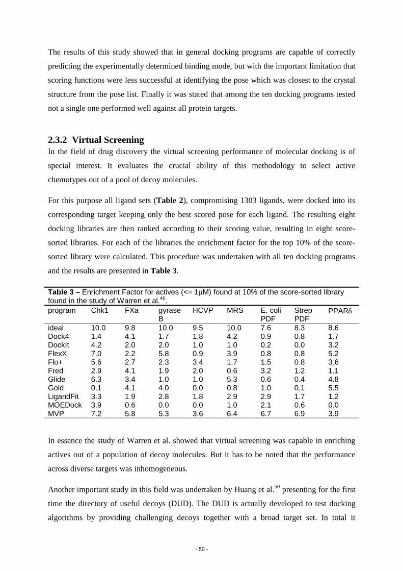
The results of this study showed that in general docking programs are capable of correctly
predicting the experimentally determined binding mode, but with the important limitation that
scoring functions were less successful at identifying the pose which was closest to the crystal
structure from the pose list. Finally it was stated that among the ten docking programs tested
not a single one performed well against all protein targets.
2.3.2 Virtual Screening In the field of drug discovery the virtual screening performance of molecular docking is of
special interest. It evaluates the crucial ability of this methodology to select active
chemotypes out of a pool of decoy molecules.
For this purpose all ligand sets (Table 2), compromising 1303 ligands, were docked into its
corresponding target keeping only the best scored pose for each ligand. The resulting eight
docking libraries are then ranked according to their scoring value, resulting in eight score-
sorted libraries. For each of the libraries the enrichment factor for the top 10% of the score-
sorted library were calculated. This procedure was undertaken with all ten docking programs
and the results are presented in Table 3.
Table 3 – Enrichment Factor for actives (<= 1µM) found at 10% of the score-sorted library found in the study of Warren et al.46. program
Chk1 FXa gyrase B
HCVP MRS E. coli PDF
Strep PDF
PPARδ
ideal 10.0 9.8 10.0 9.5 10.0 7.6 8.3 8.6 Dock4 1.4 4.1 1.7 1.8 4.2 0.9 0.8 1.7 DockIt 4.2 2.0 2.0 1.0 1.0 0.2 0.0 3.2 FlexX 7.0 2.2 5.8 0.9 3.9 0.8 0.8 5.2 Flo+ 5.6 2.7 2.3 3.4 1.7 1.5 0.8 3.6 Fred 2.9 4.1 1.9 2.0 0.6 3.2 1.2 1.1 Glide 6.3 3.4 1.0 1.0 5.3 0.6 0.4 4.8 Gold 0.1 4.1 4.0 0.0 0.8 1.0 0.1 5.5 LigandFit 3.3 1.9 2.8 1.8 2.9 2.9 1.7 1.2 MOEDock 3.9 0.6 0.0 0.0 1.0 2.1 0.6 0.0 MVP 7.2 5.8 5.3 3.6 6.4 6.7 6.9 3.9
In essence the study of Warren et al. showed that virtual screening was capable in enriching
actives out of a population of decoy molecules. But it has to be noted that the performance
across diverse targets was inhomogeneous.
Another important study in this field was undertaken by Huang et al.50 presenting for the first
time the directory of useful decoys (DUD). The DUD is actually developed to test docking
algorithms by providing challenging decoys together with a broad target set. In total it
- 50 -

contains 2950 known active ligands for 40 structurally determined targets. And for each
active ligand, 36 “decoys” with dissimilar structural topology but similar physicochemical
properties (e.g. number of H-bond donors, number of H-bond acceptors, molecular weight,
calculated logP, …) were selected summing up to 95316 decoy compounds (Table 4).
Ligands and decoys are then docked into their corresponding target using DOCK11 and
enrichment factors for actives were calculated for the top 1% and top 20% of the score-sorted
library, respectively (Table 4). The average enrichment for the top 1% and top 20% across all
40 proteins was 17.3 and 2.6, respectively.
- 51 -

Table 4 - DUD benchmark set consisting of 40 targets, 2950 unique ligands and 95316 decoys, described in Huang et al.50. Each set was docked into its corresponding target and EF1% and EF20% for actives was calculated. protein PDB-code resolution (A) no ligands no decoys EF 1% EF20% AR 1xq2 1.9 74 2630 33.5 3.8 ERagonist 1l2i 1.9 67 2361 19.2 4.5 ERantagonist 3ert 1.9 39 1399 12.7 1.3 GR 1m2z 2.5 78 2804 8.9 1.4 MR 2aa2 1.9 15 535 46.2 3.7 PPARg 1fm9 2.1 81 2910 0 0 PR 1sr7 1.9 27 967 0 2 RXRa 1mvc 1.9 20 708 24.8 2.2 CDK2 1ckp 2.1 50 1780 13.9 1.4 EGFr 1m17 2.6 416 14914 2.1 2.4 FGFr1 1agw 2.4 118 4216 0 0.2 HSP90 1uy6 1.9 24 861 8.6 2 P38 MAP 1kv2 2.8 234 8399 2.1 2.4 PDGFrb model n/a 157 5625 0 0.6 SRC 2src 1.5 162 5801 1.2 1.5 TK 1kim 2.1 22 785 54 5 VEGFr2 1vr2 2.4 74 2647 1.3 1.4 FXa 1f0r 2.7 142 5102 14.6 3.8 thrombin 1ba8 1.8 65 2294 13.7 2.9 trypsin 1bju 1.8 43 1545 22.5 2.6 ACE 1o86 2 49 1728 40.4 3.7 ADA 1stw 2 23 822 12.9 2.4 COMT 1h1d 2 12 430 0 3.3 PDE5 1xp0 1.8 51 1810 11.8 2.3 DHFR 3dfr 1.7 201 7150 21.7 3.5 GART 1c2t 2.1 21 753 42.4 3.3 AChE 1eve 2.5 105 3732 1.9 2 ALR2 1ah3 2.3 26 920 38.1 2.3 AmpC 1xgj 2 21 734 17.1 4.7 COX-1 1p4g 2.1 25 850 4 1.6 COX-2 1cx2 3 349 12491 20.1 3.3 GPB 1a8i 1.8 52 1851 22.8 4.1 HIVPR 1hpx 2 53 1888 3.7 2.2 HIVRT 1rt1 2.6 40 1439 5 3 HMGR 1hw8 2.1 35 1242 33.9 2.1 InhA 1p44 2.7 85 3043 0 0.3 NA 1a4g 2.2 49 1745 20.2 3.3 PARP 1efy 2.2 33 1178 6 3.6 PNP 1b8o 1.5 25 884 31.7 4.4 SAHH 1a7a 2.8 33 1159 78 5
- 52 -

An overviewof successfully experimentally validated hits derived from docking-based virtual
screening is presented in Kolb et al.51 for the time range 2007 to mid-2009 (see Table 5).
These results are in line with the good screening performance reported in the survey of
Warren et al.46 and Huang et al.50.
Table 5 - Docking predictions subsequently confirmed by experiments: 2007 to mid-2009. Data and table taken from Ref51
Target Docking program Lead inhibitor IC50 (μm) AdoMetDC Glide 12 AHAS DOCK 4/AutoDock 15.2 Aldose reductase N/A 0.53 CDC25 phosphatase FRED/Surflex/LigandFit 13 DNA gyrase DOCK 5 50 EphB4 DAIM-SEED-FFLD 1.5 FFAR Glide 3.6 Histamine H4 FlexX 95.8 Human pregnane X Surflex 0.049 MCH-R1 ICM 7.5 Pim-1 kinase Glide 0.091 PNP GOLD 18.9 PPAR-γ Glide/IFD 2.9 Tm0936 DOCK 3.5 105 m−1 S−1 (Kcat/Km) TRH-R1/TRH-R2 FlexX 0.29 β2-Adrenergic receptor DOCK 3.5.54 0.009 β-Lactamase DOCK 3.5.54 140 SHP2 DOCK 100 Al-2 quorum sensing DOCK 5 35 Anthrax edema factor HINT/AutoDock 1.7 hPRMT1 GOLD 12
- 53 -
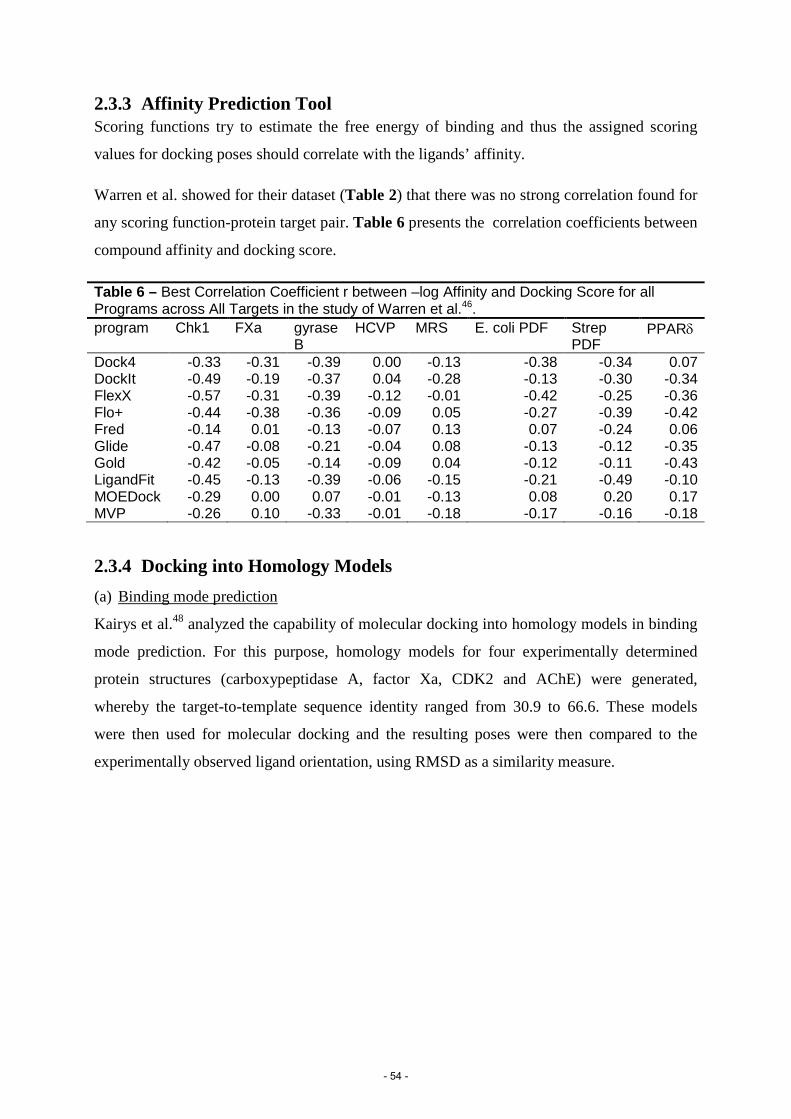
2.3.3 Affinity Prediction Tool Scoring functions try to estimate the free energy of binding and thus the assigned scoring
values for docking poses should correlate with the ligands’ affinity.
Warren et al. showed for their dataset (Table 2) that there was no strong correlation found for
any scoring function-protein target pair. Table 6 presents the correlation coefficients between
compound affinity and docking score.
Table 6 – Best Correlation Coefficient r between –log Affinity and Docking Score for all Programs across All Targets in the study of Warren et al.46. program Chk1 FXa gyrase
B HCVP MRS E. coli PDF Strep
PDF PPARδ
Dock4 -0.33 -0.31 -0.39 0.00 -0.13 -0.38 -0.34 0.07 DockIt -0.49 -0.19 -0.37 0.04 -0.28 -0.13 -0.30 -0.34 FlexX -0.57 -0.31 -0.39 -0.12 -0.01 -0.42 -0.25 -0.36 Flo+ -0.44 -0.38 -0.36 -0.09 0.05 -0.27 -0.39 -0.42 Fred -0.14 0.01 -0.13 -0.07 0.13 0.07 -0.24 0.06 Glide -0.47 -0.08 -0.21 -0.04 0.08 -0.13 -0.12 -0.35 Gold -0.42 -0.05 -0.14 -0.09 0.04 -0.12 -0.11 -0.43 LigandFit -0.45 -0.13 -0.39 -0.06 -0.15 -0.21 -0.49 -0.10 MOEDock -0.29 0.00 0.07 -0.01 -0.13 0.08 0.20 0.17 MVP -0.26 0.10 -0.33 -0.01 -0.18 -0.17 -0.16 -0.18
2.3.4 Docking into Homology Models (a) Binding mode prediction
Kairys et al.48 analyzed the capability of molecular docking into homology models in binding
mode prediction. For this purpose, homology models for four experimentally determined
protein structures (carboxypeptidase A, factor Xa, CDK2 and AChE) were generated,
whereby the target-to-template sequence identity ranged from 30.9 to 66.6. These models
were then used for molecular docking and the resulting poses were then compared to the
experimentally observed ligand orientation, using RMSD as a similarity measure.
- 54 -

Figure 9 – Representation of the findings of Kairys et al.48 in examining the performance of molecular docking into homology models in binding mode prediction. For the four experimentally determined ligand-target complexes (carboxypeptidase A, factor Xa, CDK2 and AChE), a total of 16 homology models was created. These models were based on 16 templates (Table 7) listed on the left hand side of the Figure. Bars show the percentage of ligand-target complexes with available crystal structures for which top scoring pose has a RMSD < 2Å (black) and < 4Å (black + gray). Figure edited from Ref48.
Table 7 - Templates and associated template-to-target sequence identity for proteins analyzed in Kairys et al.48. target template sequence identity carboxypeptidase A Procarboxypeptidase A 30.9 Carboxypeptidase T 31.8 Procarboxypeptidase A2 66.6 factor Xa Protein C 34.9 Factor VIIa 38.8 Thrombin 40.7 Factor Xa 100 CDK2 c-Abl 28.6 PKA 31.2 ERK2 37.0 CDK6 48.2 AChE para-nitrobenzyl esterase 31.4 Cholesterol Esterase 32.8 Triacylglycerol Lipase 32.8 Carboxylesterase 1 36.2 BuChE 53.3 These findings clearly demonstrate that molecular docking into homology models is capable
to regenerate the experimentally observed ligand-protein complex for proteins with a
sequence identity greater than 30%.
- 55 -

(b) Virtual screening The first exhaustive virtual screening performance comparison between experimentally
determined structures and homology models thereof was assessed by Fan et al.49. They used
the widely applied docking benchmark set of the “directory of useful decoys” (DUD) for
performance evaluation50.
In the study of Fan et al. 38 of the 40 subsets described in DUD were used to compare the
virtual screening performance using homology models or crystal structures. For each of the 38
targets, template structures were selected (1-12 per target) with varying sequence identity
(ranging from 20% to 99%), resulting in a total set of 222 templates and 222 homology
models (5 models per target on average). Finally DUD ligands and decoys (38 of the 40
subsets in Table 4) were docked with DOCK against the 38 X-ray structures and against the
222 homology models. Subsequently, enrichment factors expressed as logAUC were
calculated and the results were compared (Table 8).
- 56 -

Table 8 - Ligand enrichments found in Fan et al. for molecular docking into homology models and X-ray structures. Table taken from Fan et al.49.
protein target most enriching model best model by SI consensus
enrichment holo X-ray enrichment SI enrichment SI enrichment
ACE 44.1 50.2 54.8 37.9 48.9 40.6 ADA 26.5 40.3 84.8 38.8 41.1 22.7 COMT 20.1 0 20.1 0 0.0 27.6 PDE5 30.4 22.1 95.5 20.5 18.6 12.1 DHFR 27.9 34.6 37.3 13.1 20.3 18.9 GART 40.4 27.5 41.5 0.6 27.4 35.3 FXa 30.4 52.1 63.8 27.2 49.6 41.8 thrombin 30 43.4 39.5 36.9 42.4 29.4 Trypsin 78.8 38 78.8 38 32.3 29.3 AChE 38 35.7 92.7 17.3 29.1 38.5 ALR2 68.7 41.9 86.6 39.5 37.1 39.7 AmpC 76.6 46.6 76.6 37.2 40.3 47.4 COX-1 63.4 26.2 63.4 26.2 25.3 28.3 COX-2 64.4 13.3 64.7 8.2 12.4 40.8 GPB 41.8 8.9 80.7 6.5 6.8 17.1 HIVPR 49 30.5 96.9 4.8 23.7 11.9 HIVRT 97.7 12.9 98.9 0 12.9 25.8 HMGR 100 43.2 100 43.2 41.5 40.9 InhA 29 14.6 29 14.6 11.8 8.2 NA 28.8 51.2 34.2 20.4 42.6 47.6 PARP 46.4 9.2 86.8 6.4 6.3 8.2 PNP 36.9 33.5 93.1 19.3 23.9 49.1 SAHH 84.7 82.4 84.7 82.4 78.4 82.8 CDK2 27.6 13.5 94.9 10.1 11.7 11.3 EGFr 37.7 13.2 38.1 12.3 10.7 21.5 FGFr1 41.3 12.3 51 6.2 11.9 6.7 HSP90 62.4 18.7 70.4 14.5 11.5 24.6 P38 MAP 29.4 31.4 62.2 21.2 15.6 12.5 SRC 68.8 13.8 82 13.4 12.3 9.5 TK 20.7 13.4 40.4 3 2.9 63.5 AR 56 31.7 84.3 31.5 14.5 48.2 ERagonist 93.1 41 93.1 41 19.6 55.4 ERantagonist 21 19.2 93 9 14.7 23.2 GR 27.4 18.8 56.6 15.2 16.7 20.5 MR 56.8 30.5 56.8 30.5 29.5 57 PPARg 20.2 13.2 65 9.2 8.8 4.4 PR 53.5 35.9 53.5 35.9 11.8 23.2 RXRa 87 26.9 88.8 2 11.2 37.9 Enrichment, the ligand enrichment is represented by logAUC. SI, stands for the target to template sequence identity.
- 57 -

Taking only the most enriching model for each target, the averaged enrichment (logAUC) for
the holo X-ray structures was 30.6 which was only slightly higher than the average
enrichment (logAUC) of the most enriching homology models (28.7). However, this
difference was not statistically significant.
Subsequently they analyzed model properties (template resolution, binding-site sequence
identity, overall sequence identity, N-DOPE score) which could correlate with enrichment
performance, in the hope to identify parameters that can predict model performance. However
none of the parameters showed a strong correlation with model enrichment performance.
But on the other hand they noticed that homology models performed significantly better if
models were left out which derive from orthologous templates and template-model pairs
where the sequence identity is lower than 25%. This indicates that models below a sequence
identity of 25% show a significant weaker performance.
References:
1. Berman, H.M. et al. The Protein Data Bank. Nucleic Acids Res 28, 235-42 (2000). 2. UniProtConsortium. Reorganizing the protein space at the Universal Protein Resource (UniProt). Nucleic Acids Res 40, D71-5 (2012). 3. Sali, A. & Blundell, T.L. Comparative protein modelling by satisfaction of spatial restraints. J Mol Biol 234, 779-815 (1993). 4. Yildirim, M.A., Goh, K.I., Cusick, M.E., Barabasi, A.L. & Vidal, M. Drug-target network. Nat Biotechnol 25, 1119-26 (2007). 5. Ashburner, M. et al. Gene ontology: tool for the unification of biology. The Gene Ontology Consortium. Nat Genet 25, 25-9 (2000). 6. Bordoli, L. et al. Protein structure homology modeling using SWISS-MODEL workspace. Nat Protoc 4, 1-13 (2009). 7. Baker, D. & Sali, A. Protein structure prediction and structural genomics. Science 294, 93-6 (2001). 8. Hillisch, A., Pineda, L.F. & Hilgenfeld, R. Utility of homology models in the drug discovery process. Drug Discov Today 9, 659-69 (2004). 9. Cavasotto, C.N. & Phatak, S.S. Homology modeling in drug discovery: current trends and applications. Drug Discov Today 14, 676-83 (2009). 10. Bajorath, J. Integration of virtual and high-throughput screening. Nat Rev Drug Discov 1, 882-94 (2002). 11. Kuntz, I.D., Blaney, J.M., Oatley, S.J., Langridge, R. & Ferrin, T.E. A geometric approach to macromolecule-ligand interactions. J Mol Biol 161, 269-88 (1982). 12. Kitchen, D.B., Decornez, H., Furr, J.R. & Bajorath, J. Docking and scoring in virtual screening for drug discovery: methods and applications. Nat Rev Drug Discov 3, 935-49 (2004). 13. Sousa, S.F., Fernandes, P.A. & Ramos, M.J. Protein-ligand docking: current status and future challenges. Proteins 65, 15-26 (2006). 14. Leach, A.R. Molecular Modelling: Principles and Applications. Addison Wesley Longman Limited (1996).
- 58 -

15. Rarey, M., Kramer, B., Lengauer, T. & Klebe, G. A fast flexible docking method using an incremental construction algorithm. J Mol Biol 261, 470-89 (1996). 16. DesJarlais, R.L., Sheridan, R.P., Dixon, J.S., Kuntz, I.D. & Venkataraghavan, R. Docking flexible ligands to macromolecular receptors by molecular shape. J Med Chem 29, 2149-53 (1986). 17. Friesner, R.A. et al. Glide: a new approach for rapid, accurate docking and scoring. 1. Method and assessment of docking accuracy. J Med Chem 47, 1739-49 (2004). 18. Welch, W., Ruppert, J. & Jain, A.N. Hammerhead: fast, fully automated docking of flexible ligands to protein binding sites. Chem Biol 3, 449-62 (1996). 19. Hart, T.N. & Read, R.J. A multiple-start Monte Carlo docking method. Proteins 13, 206-22 (1992). 20. Oshiro, C.M., Kuntz, I.D. & Dixon, J.S. Flexible ligand docking using a genetic algorithm. J Comput Aided Mol Des 9, 113-30 (1995). 21. Westhead, D.R., Clark, D.E. & Murray, C.W. A comparison of heuristic search algorithms for molecular docking. J Comput Aided Mol Des 11, 209-28 (1997). 22. Goodsell, D.S., Lauble, H., Stout, C.D. & Olson, A.J. Automated docking in crystallography: analysis of the substrates of aconitase. Proteins 17, 1-10 (1993). 23. Jones, G., Willett, P. & Glen, R.C. Molecular recognition of receptor sites using a genetic algorithm with a description of desolvation. J Mol Biol 245, 43-53 (1995). 24. Brooijmans, N. & Kuntz, I.D. Molecular recognition and docking algorithms. Annu Rev Biophys Biomol Struct 32, 335-73 (2003). 25. Huang, S.Y. & Zou, X. Advances and challenges in protein-ligand docking. Int J Mol Sci 11, 3016-34 (2010). 26. Jiang, F. & Kim, S.H. "Soft docking": matching of molecular surface cubes. J Mol Biol 219, 79-102 (1991). 27. Ferrari, A.M., Wei, B.Q., Costantino, L. & Shoichet, B.K. Soft docking and multiple receptor conformations in virtual screening. J Med Chem 47, 5076-84 (2004). 28. Leach, A.R. Ligand docking to proteins with discrete side-chain flexibility. J Mol Biol 235, 345-56 (1994). 29. Desmet, J., Wilson, I.A., Joniau, M., De Maeyer, M. & Lasters, I. Computation of the binding of fully flexible peptides to proteins with flexible side chains. FASEB J 11, 164-72 (1997). 30. Davis, I.W. & Baker, D. RosettaLigand docking with full ligand and receptor flexibility. J Mol Biol 385, 381-92 (2009). 31. Sherman, W., Day, T., Jacobson, M.P., Friesner, R.A. & Farid, R. Novel procedure for modeling ligand/receptor induced fit effects. J Med Chem 49, 534-53 (2006). 32. Knegtel, R.M., Kuntz, I.D. & Oshiro, C.M. Molecular docking to ensembles of protein structures. J Mol Biol 266, 424-40 (1997). 33. Claussen, H., Buning, C., Rarey, M. & Lengauer, T. FlexE: efficient molecular docking considering protein structure variations. J Mol Biol 308, 377-95 (2001). 34. Ponder, J.W. & Case, D.A. Force fields for protein simulations. Adv Protein Chem 66, 27-85 (2003). 35. Morris, G.M. et al. Automated Docking Using a Lamarckian Genetic Algorithm and and Empirical Binding Free Energy Function. J Computational Chemistry 19, 1639-1662 (1998). 36. Brooks, B.R. et al. CHARMM - a programm for macromolecular energy minimization, and dynamics calculations. J Computational Chemistry 4, 187 (1983). 37. Wang, W., Donini, O., Reyes, C.M. & Kollman, P.A. Biomolecular simulations: recent developments in force fields, simulations of enzyme catalysis, protein-ligand, protein-protein, and protein-nucleic acid noncovalent interactions. Annu Rev Biophys Biomol Struct 30, 211-43 (2001). 38. Guimaraes, C.R. & Cardozo, M. MM-GB/SA rescoring of docking poses in structure-based lead optimization. J Chem Inf Model 48, 958-70 (2008). 39. Kramer, B., Rarey, M. & Lengauer, T. Evaluation of the FLEXX incremental construction algorithm for protein-ligand docking. Proteins 37, 228-41 (1999). 40. Bohm, H.J. LUDI: rule-based automatic design of new substituents for enzyme inhibitor leads. J Comput Aided Mol Des 6, 593-606 (1992). 41. Eldridge, M.D., Murray, C.W., Auton, T.R., Paolini, G.V. & Mee, R.P. Empirical scoring functions: I. The development of a fast empirical scoring function to estimate the binding affinity of ligands in receptor complexes. J Comput Aided Mol Des 11, 425-45 (1997).
- 59 -

42. Gehlhaar, D.K. et al. Molecular recognition of the inhibitor AG-1343 by HIV-1 protease: conformationally flexible docking by evolutionary programming. Chem Biol 2, 317-24 (1995). 43. Wang, R., Lai, L. & Wang, S. Further development and validation of empirical scoring functions for structure-based binding affinity prediction. J Comput Aided Mol Des 16, 11-26 (2002). 44. Muegge, I. & Martin, Y.C. A general and fast scoring function for protein-ligand interactions: a simplified potential approach. J Med Chem 42, 791-804 (1999). 45. Gohlke, H., Hendlich, M. & Klebe, G. Knowledge-based scoring function to predict protein-ligand interactions. J Mol Biol 295, 337-56 (2000). 46. Warren, G.L. et al. A critical assessment of docking programs and scoring functions. J Med Chem 49, 5912-31 (2006). 47. Blaney, J.M. & Dixon, J.S. DockIt, version 1.0. Metaphorics, LLC: Mission Viejo; CA; www.metaphorics.com/products/dockit.html. 48. Kairys, V., Fernandes, M.X. & Gilson, M.K. Screening drug-like compounds by docking to homology models: a systematic study. J Chem Inf Model 46, 365-79 (2006). 49. Fan, H. et al. Molecular docking screens using comparative models of proteins. J Chem Inf Model 49, 2512-27 (2009). 50. Huang, N., Shoichet, B.K. & Irwin, J.J. Benchmarking sets for molecular docking. J Med Chem 49, 6789-801 (2006). 51. Kolb, P., Ferreira, R.S., Irwin, J.J. & Shoichet, B.K. Docking and chemoinformatic screens for new ligands and targets. Curr Opin Biotechnol 20, 429-36 (2009).
- 60 -

IV. Aim of the study
1,4-benzodiazepines exert their anxiolytic, anticonvulsant, muscle relaxant, and sedative-
hypnotic properties by allosterically enhancing the action of GABA at GABAA receptors via
their benzodiazepine binding site1. After 50 years of clinical use, the molecular basis of this
interaction still is not known. The primary aim of this thesis is the elucidation of the binding
mode of 1,4-benzodiazepines in the benzodiazepine binding site of α1β2γ2 GABAA-receptors
with the computational means of homology modeling and molecular docking. The identified
binding mode should be in accordance with existing experimental data, and ultimately the
gained structural knowledge should predict novel ligand classes acting at the benzodiazepine
binding site. Furthermore the derived structural models will also improve the current
mechanistic understanding from modulator binding (at the benzodiazepine binding site) to
alterations in channel gating. This can be achieved by the analysis of intra-molecular protein-
protein interactions in proximity to the benzodiazepine binding site that may mediate GABAA
receptor modulation.
But let’s face the facts:
No crystal structures for GABAA receptors available
Target to template sequence identities of around 18% at the time the thesis was conducted
A highly flexible loop shaping the benzodiazepine binding site
And finally, major difficulties of scoring functions to rank the correctly computed ligand
orientation on the top position even in redocking experiments
Honestly spoken the aim to uncover the binding mode of 1,4-benzodiazepines in the
GABAA-receptor seems at least challenging. In principle there are two problems associated
with the development of accurate models of ligand bound benzodiazepine binding sites.
First, the homology models that could be built at the time of the study are quite uncertain as
the sequence identity between templates and target is only around 18%. Second, although
standard docking tools are capable of reproducing the correct binding mode, they normally
fail to rank these on top of the pose list.
In the workflow of the thesis these problems will be tackled in a two step approach. In the
first step, the explorative step, we want to face the high ambiguity in the input data by broadly
sampling the pose space. This will inevitably result in an enormous amount of poses, but
with a high likelihood of generating a plausible ligand-receptor complex.
Consequently, the aim of the subsequent step, the selection step, will be the selection of
the most plausible ligand-receptor complex(es) out of an ocean of poses. Exhaustive
implementation of various sources of information (experimental, computational, structural) in
- 61 -

an orchestrated and integrative manner will be the key factor for this evaluative process that
should finally narrow down the gigantic pose space to one single coherent solution.
Once the plausible binding mode has been identified the predictive power of the identified
ligand-receptor complex should be tested in the field of drug discovery by the conduction of
structure-based virtual screening runs.
In the following a short description of the steps is provided.
Step 1, exploration step. In this step, homology modeling and molecular docking will be
used in order to produce binding hypotheses for 1,4-benzodiazepines at the benzodiazepine
binding site. The sampling during the docking has to be broad as the input data is highly
ambiguous (low target-template sequence identity, flexible loop in binding site, low resolution
of templates).
In detail multiple homology models will be built deriving from different templates and
varying sequence alignments. Additionally, flexible side-chains will be used during the
docking procedure, once more adding degrees of freedom. And finally the first 100 poses per
ligand per model will be kept for further evaluation. This all will result in an enormous
amount of poses, but with a high likelihood of generating the correct ligand-receptor
complex.
Step 2, selection step. The aim of this step will be the selection of the most plausible
binding mode out of the thousands of docking poses, generated in the exploration step. A
large array of evaluation criteria (comparison with experimental data, docking scores,
structure activity relationships and others) will be used in this sensible selection step.
In this process the emphasis will be on the integration of existing experimental data as
binding mode quality criteria. Plenty information about the spatial arrangement between
ligand and binding site has already been accumulated in the last 50 years of research2,3. They
are encoded as labeling data, mutagenesis data or can be found as structure-activity
relationships. But as the experiments were compiled with various ligands we face a
challenging situation: The plethora of information is dispersed through either i) different
ligands within a compound class or ii) through ligands that belong to different classes.
In this thesis the common binding mode (CBM) hypothesis will be applied as an
adequate integrative platform that connects these seemingly dispersed informative pieces. The
CBM provides an assumption about the ligand alignment of ligands acting at the same
binding site. Hence the CBM can be used to incorporate information from ligands that derive
from a common ligand class (narrow definition) or beyond that, also implements information
that derive from other ligands classes (extended definition).
- 62 -

In the narrow definition, the CBM hypothesis is based on the general assumption that the
scaffold of ligands belonging to the same class (here: 1,4-benzodiazepines, Fig. 10a) bind in
the same mode in the binding site, thus providing a hypothesis for the mutual spatial
alignment of these ligands.
In the more extended view of the CBM hypothesis, which will be used as supplementary
validation, information from further ligand classes (e.g. imidazobenzodiazepines, Fig. 10b)
will be used in validating the binding mode of 1,4-benzodiazepines (Fig. 10a).
Figure 10 - Representation of the structures of 1,4-benzodiazepines (a),
imidazobenzodiazepines (b) and their mutual alignment in a widely accepted pharmacophore model (c). (a) The common scaffold of 1,4-benzodiazepines is depicted in black colour. For the most prominent representative of this ligand class (diazepam) additional structural elements are shown in grey colour. (b) The common scaffold of imidazobenzodiazepines is depicted in black colour. For the most prominent representative of this ligand class (flumazenil) additional structural elements are shown in grey colour. (c) Representation of the mutual spatial alignment of diazepam (grey stick representation) and flumazenil (violet stick representation) in the ligand based pharmacophore model of Cook et al.4 These aligned ligands share one common hydrophobic feature (L1, white meshed sphere) and two H-bond acceptor features (H1 and H2, red meshed spheres).
The spatial link from 1,4-benzodiazepines to accessory ligand classes (e.g. imidazo-
benzodiazepines) will be taken from the ligand alignment described in a widely accepted
pharmacophore model4 (Fig. 10c). This alignment will be the connective platform that
facilitates cross evaluation among various compound classes. Consequently a diazepam (Fig.
10a) pose can then be additionally validated by experimental evidence deriving from other
ligand classes, for example by a flumazenil (Fig. 10b) docking pose that is found to spatially
align (according to the pharmacophore model alignment, Fig. 10c) with the mentioned
diazepam pose. So the pharmacophore model should enable the combination of information
deriving from different ligand classes.
Once the binding mode of 1,4-benzodiazepines has been identified, the ligand-receptor
complex will be used in structure-based virtual screening runs taking the information encoded
in the ligand-receptor complex to identify novel benzodiazepine binding site ligands.
- 63 -

References:
1. Sieghart, W. Structure and pharmacology of gamma-aminobutyric acidA receptor subtypes. Pharmacol Rev 47, 181-234 (1995).
2. Sigel, E. Mapping of the benzodiazepine recognition site on GABA(A) receptors. Curr Top Med Chem 2, 833-9 (2002).
3. Clayton, T. et al. An updated unified pharmacophore model of the benzodiazepine binding site on gamma-aminobutyric acid(a) receptors: correlation with comparative models. Curr. Med. Chem. 14, 2755-2775 (2007).
4. Zhang, W., Koehler, K.F., Zhang, P. & Cook, J.M. Development of a comprehensive pharmacophore model for the benzodiazepine receptor. Drug. Des. Discov. 12, 193-248 (1995).
- 64 -

B. Results and Discussion I. Diazepam-bound GABAA receptor models
Identify new benzodiazepine site ligands………… 66 (Richter et al., 2012, Nat Chem Biol, 8, 455-464)
II. A residue close to α1 loop F disrupts modulation of GABAA receptors by benzodiazepines while their binding is maintained ……………………… 140
(Bauer et al., 2010, J Neurochemistry, 115, 1478-1485)
- 65 -

I. Diazepam-bound GABAA receptor models identify new benzodiazepine binding-site ligands
Contribution of the thesis author to this article:
For the presentation of the thesis authors' contribution to this publication the Author
contributions section of the article is presented:
Author contributions L.R., W.S., G.F.E., I.J.P.d.E., C.d.G. and M.E. contributed to the design and evaluation of
experiments. Docking and virtual screening experiments were performed by L.R. (supervised by
G.F.E.) and C.d.G. GABAAR models were generated and evaluated by M.E. and M.M. Radioligand
and electrophysiological experiments were performed by Z.V. The manuscript was written by W.S.,
L.R., G.F.E., I.J.P.d.E., C.d.G. and M.E., and all authors commented on and helped to revise the text.
- 66 -

©20
12 N
atu
re A
mer
ica,
Inc.
All
rig
hts
res
erve
d.
NATURE CHEMICAL BIOLOGY | ADVANCE ONLINE PUBLICATION | www.nature.com/naturechemicalbiology 1
ARTICLEPUBLISHED ONLINE: 25 MARCH 2012 | DOI: 10.1038/NCHEMBIO.917
GABAA receptors (GABAARs) are the major inhibitory trans-mitter receptors in the brain and the site of action of a variety of pharmacologically and clinically important drugs, such
as benzodiazepines, barbiturates, neuroactive steroids, anesthetics and convulsants1. These receptors are chloride ion channels com-posed of five subunits that can belong to different subunit classes. A total of 19 GABAA receptor subunits ( 1–6, 1–3, 1–3, , ,
, , 1–3) have been identified in mammalian brain. The majority of GABAARs are composed of one -, two - and two -subunits. The two GABA-binding sites of these receptors are located extra-cellularly at - interfaces (Fig. 1a). Classical benzodiazepines, such as diazepam, predominantly exert their action via GABAARs composed of 1 2, 2 2, 3 2 and 5 2 subunits and are known to bind at the extracellular - interface2 (Fig. 1b). Owing to the lack of high-resolution structural data for this important drug-binding site, however, a consensus binding-mode hypothesis explaining all of the experimental data is still lacking.
GABAARs, together with nicotinic acetylcholine receptors (nAChRs), glycine receptors and serotonin type 3 receptors, are members of the cysteine-loop receptor superfamily. Although so far no crystal structure of a GABAAR is available, a variety of crystal structures from a functional and structural homolog of the ligand-binding domain of cysteine-loop receptors, the acetylcholine-binding protein (AChBP), and more recently from the nAChR and bacterial homologs thereof, is available3 and demonstrates a high structural conservation within this receptor superfamily. Some of these structures have served as templates for protein homology models and docking studies that have yielded controversial bind-ing-mode hypotheses for various benzodiazepines4–8. However, because of their low sequence identity of <20%, different members of the superfamily have appreciable variability in local interface and pocket structure3. To overcome this problem, we constructed mul-tiple homology models of the GABAAR from a variety of structural templates to broadly sample the benzodiazepine-pocket geometry. We then developed a workflow that, in contrast to automated ligand-supported modeling approaches9, did not rely on energetic
scoring functions. This workflow allowed unbiased selection and refinement of those models best suited to describe a diazepam-bound state as well as evaluation of several thousand docking poses of diazepam and its close structural analogs. The procedure yielded a binding mode for diazepam and analogs that is convincingly sup-ported by experimental evidence. Using this binding hypothesis in a virtual screening of a large compound library, we identified new allosteric modulators of GABAARs acting via the benzo diazepine site, thus confirming their suitability for drug discovery and structure-based drug design.
RESULTSComputational modeling and dockingTo account for the substantial variability in local interface and pocket structures10 of cysteine-loop receptor family members, we constructed a total of 37 homology models of the GABAAR derived from eight distinct structural templates (Supplementary Methods, Supplementary Table 1), multiple sequence alignments (Supplementary Fig. 1) and variations in degrees of protein flexibil-ity (Supplementary Schemes 1 and 2). We then docked diazepam in its bioactive conformation (M conformation; Supplementary Fig. 2 and Supplementary Table 2) into all initial models (Supplementary Results, Supplementary Tables 3 and 4 and Supplementary Fig. 3) using the software package FlexX11. FlexX and other docking algo-rithms are able to explore the conformational space sufficiently well to generate correctly docked poses, but they often fail in ranking the correct poses on top. Furthermore, the binding-mode prediction accuracy of scoring functions is target dependent12. We therefore evaluated the 100 top-scoring poses (FlexX score), thus ensuring that correct poses were retained. We then eliminated homology models that predominantly provided poses with poor lipophilic interac-tion for ligand features L1 and L3 (Fig. 1c) of the diazepam struc-ture or featured no simultaneous ligand contacts with 1 and 2 subunits. For the remaining 14 models, we extended docking to multiple ligands (1, 3–5, 8, 9; in M conformation in Fig. 1c) and used the docking protocol FlexE13 to enable the flexibility of those
1Department of Medicinal Chemistry, University of Vienna, Vienna, Austria. 2Leiden–Amsterdam Center for Drug Research, Division of Medicinal
Chemistry, Faculty of Sciences, VU University Amsterdam, Amsterdam, The Netherlands. 3Department of Biochemistry and Molecular Biology, Center for
Brain Research, Medical University of Vienna, Vienna, Austria. *e-mail: [email protected]
Diazepam-bound GABAA receptor models identify new benzodiazepine binding-site ligandsLars Richter1, Chris de Graaf2, Werner Sieghart3, Zdravko Varagic3, Martina Mörzinger3, Iwan J P de Esch2, Gerhard F Ecker1 & Margot Ernst3*
Benzodiazepines exert their anxiolytic, anticonvulsant, muscle-relaxant and sedative-hypnotic properties by allosterically enhancing the action of GABA at GABAA receptors via their benzodiazepine-binding site. Although these drugs have been used clinically since 1960, the molecular basis of this interaction is still not known. By using multiple homology models and an un biased docking protocol, we identified a binding hypothesis for the diazepam-bound structure of the benzodiazepine site, which was confirmed by experimental evidence. Moreover, two independent virtual screening approaches based on this structure identified known benzodiazepine-site ligands from different structural classes and predicted potential new ligands for this site. Receptor-binding assays and electrophysiological studies on recombinant receptors confirmed these predictions and thus identified new chemotypes for the benzodiazepine-binding site. Our results support the validity of the diazepam-bound structure of the benzodiazepine-binding pocket, demonstrate its suitability for drug discovery and pave the way for structure-based drug design.
- 67 -

©20
12 N
atu
re A
mer
ica,
Inc.
All
rig
hts
res
erve
d.
2 NATURE CHEMICAL BIOLOGY | ADVANCE ONLINE PUBLICATION | www.nature.com/naturechemicalbiology
ARTICLE NATURE CHEMICAL BIOLOGY DOI: 10.1038/NCHEMBIO.917
amino acid side chains that have been shown to be important for ligand recognition2 and/or constitute the main steric determinants of the binding site. By applying the above-mentioned criteria for each ligand docked, we further reduced the number of homology models. The three best-performing models were based on Protein Data Bank (PDB) entries 2BYQ3 (Aplysia californica AChBP, epi-batidine bound), 1UW6 (ref. 3) (Lymnea stagnalis AChBP, nico-tine bound) and 2QC1 (ref. 3) (mouse nAChR, 1 subunit,
-bungarotoxin bound). To improve conformational sampling, we further increased the flexibility of these three models by consider-ing additional rotamers of amino acid side chains in direct contact with pocket-defining residues, especially at the crowded subunit interface between loops A and B. This resulted in three additional models (Supplementary Scheme 2).
Subsequently, we docked ligands 1–9 (Fig. 1c) into the six final models (Supplementary Table 3) using the FlexE protocol. We then performed an energy minimization of all poses and calculated the L1 and L3 interaction strengths. Of the 4,997 energy-minimized poses, we retained 1,463 with L1 and L3 interaction strengths above the median value. These poses still sampled a wide variety of putative binding geometries for smaller ligands such as diazepam and fluni-trazepam. Ligands with bulky substituents (Fig. 1c; 4–9) assumed more restricted poses. Thus, by using only lipophilic interaction strength as our selection criterion, we retrieved a set of poses in which all of the binding modes compatible with the sampled pocket topologies were still present.
Binding-mode searchTo compare binding modes in different templates, we three- dimensionally aligned the ligand-protein complexes on the basis of the conserved domains of the - and -subunits of their benzo-diazepine-binding pocket. Then, we identified similar binding modes through cluster analysis (Supplementary Scheme 3) after computing the r.m.s. deviation values of the ligands’ common atom positions14 between each pair of poses and storing them as a matrix. Cluster analysis then produced groups of poses with small differ-ences in r.m.s. deviation. By allowing 2-Å scatter around each cen-troid pose, thirty clusters, each comprising a group of similar binding modes, emerged (Supplementary Figs. 4 and 5).
Common binding-mode screenTo define candidate geometries representing a common binding mode (CBM), we split the docked ligands (Fig. 1c and Supplementary Table 5) into mandatory ‘core ligands’ (Fig. 1c; 1–5), and ‘accessory ligands’ (Fig. 1c; 6–9). Core ligands have very similar chemical struc-tures, affinities, efficacies and patterns in their response to mutation, suggesting tightly overlapping bound-state geometries. Although this assumption is not necessarily true in each case, it is the basis of all rational hit-optimization efforts and (quantitative) structure-activity studies. Additionally, we also considered compound 5 ‘core’, not as a ligand but as affinity chromatography bait (Supplementary Fig. 6). Accessory ligands should also bind in a similar binding mode as the core ligands, but they either do not share all pharmacophore features with diazepam or, because of steric requirements, may induce some-what different conformations of the binding site. Only three of the clusters contained all of the mandatory core ligands and were thus further considered as CBMs (Supplementary Scheme 3).
For these three clusters, we extended the measure of ‘binding-mode similarity’ to include not only the orientation of the ligand’s common substructure but also the pocket atoms interacting with this substructure. The resulting ‘bound-complex r.m.s. deviation’ value describes the similarity of ligand-pocket interaction patterns. By clustering the bound-complex r.m.s. deviation values, we finally extracted the most homogenous subsets of poses from each parent cluster, resulting in CBM candidates CBM I, CBM II and CBM III (Fig. 2 and Supplementary Tables 6–8).
2
2
1
2
––
––
–
+
+
+
++
1
GABAGABA
BZ
C ED
B
A
G
F
a
c
b
N
NO
Cl
1 (diazepam)
L1
L3
H1H1
H2N
NO
ClCl
Cl
2
N
NO
F
NO
O
3 (flunitrazepam)
N
NO
Cl
N
F
4 (flurazepam)
N
NO
Cl
HN
F
O
HN NH
O
NHO
NH2
5
N
HN
O
F
6
N
NO
F
7
N
N
Cl
N O
O
8
N
N
Br
N
O
O
9
N
N
R1
O
10: R1 = N=C=S; R2
= H14: R1
= NO2; R2 = N=C=S
R2
N
N
O
X
N O
O
11: X = F (flumazenil)12: X = N313: X = N=C=S
L1
H1
H2
1(+) 2(–)
Figure 1 | Structure of diazepam-sensitive GABAARs and of benzodiazepine ligands. (a) Top view of the extracellular domain of the pentameric
1 2 2 GABAAR in ribbon mode. The benzodiazepine-binding site (BZ)
and the two GABA-binding sites are indicated by arrows. Diazepam in the
benzodiazepine-binding site is depicted in space-filling mode. (b) View from
a perspective nearly parallel with the lipid bilayer of the 1 2 extracellular
interface in ribbon mode with a diazepam-bound pose of CBM I in space-
filling mode. The dashed window indicates the section shown in Figures 2–5.
Segments (loops) A, B, C, D, E, F and G are labeled, and the corresponding
protein segment is rendered in a darker shade. In both panels the plus (+)
and minus (−) sides of the subunits are indicated. (c) Chemical structures
of compounds used in this study. 1–9 were used for docking and 10–14 were
used for validation. The pharmacophoric features L1, L3, H1 and H2 (ref. 48)
are superimposed on two-dimensional structures of diazepam and flumazenil
(11). As an additional feature we introduce H1 to denote the interaction with
the second lone pair of the carbonyl oxygen. More detailed information on
the compounds can be found in Supplementary Table 5.
- 68 -
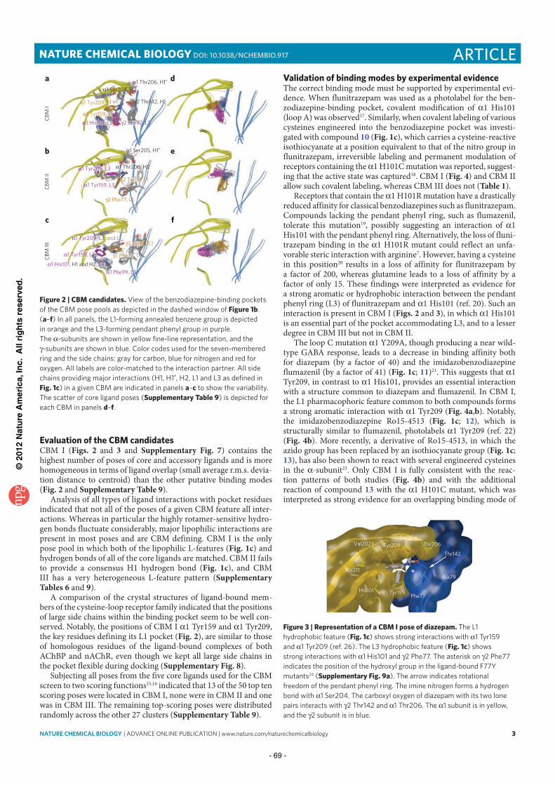
©20
12 N
atu
re A
mer
ica,
Inc.
All
rig
hts
res
erve
d.
NATURE CHEMICAL BIOLOGY | ADVANCE ONLINE PUBLICATION | www.nature.com/naturechemicalbiology 3
ARTICLENATURE CHEMICAL BIOLOGY DOI: 10.1038/NCHEMBIO.917
Evaluation of the CBM candidatesCBM I (Figs. 2 and 3 and Supplementary Fig. 7) contains the highest number of poses of core and accessory ligands and is more homogeneous in terms of ligand overlap (small average r.m.s. devia-tion distance to centroid) than the other putative binding modes (Fig. 2 and Supplementary Table 9).
Analysis of all types of ligand interactions with pocket residues indicated that not all of the poses of a given CBM feature all inter-actions. Whereas in particular the highly rotamer-sensitive hydro-gen bonds fluctuate considerably, major lipophilic interactions are present in most poses and are CBM defining. CBM I is the only pose pool in which both of the lipophilic L-features (Fig. 1c) and hydrogen bonds of all of the core ligands are matched. CBM II fails to provide a consensus H1 hydrogen bond (Fig. 1c), and CBM III has a very heterogeneous L-feature pattern (Supplementary Tables 6 and 9).
A comparison of the crystal structures of ligand-bound mem-bers of the cysteine-loop receptor family indicated that the positions of large side chains within the binding pocket seem to be well con-served. Notably, the positions of CBM I 1 Tyr159 and 1 Tyr209, the key residues defining its L1 pocket (Fig. 2), are similar to those of homologous residues of the ligand-bound complexes of both AChBP and nAChR, even though we kept all large side chains in the pocket flexible during docking (Supplementary Fig. 8).
Subjecting all poses from the five core ligands used for the CBM screen to two scoring functions15,16 indicated that 13 of the 50 top ten scoring poses were located in CBM I, none were in CBM II and one was in CBM III. The remaining top-scoring poses were distributed randomly across the other 27 clusters (Supplementary Table 9).
Validation of binding modes by experimental evidenceThe correct binding mode must be supported by experimental evi-dence. When flunitrazepam was used as a photolabel for the ben-zodiazepine-binding pocket, covalent modification of 1 His101 (loop A) was observed17. Similarly, when covalent labeling of various cysteines engineered into the benzodiazepine pocket was investi-gated with compound 10 (Fig. 1c), which carries a cysteine-reactive isothio cyanate at a position equivalent to that of the nitro group in flunitrazepam, irreversible labeling and permanent modulation of receptors containing the 1 H101C mutation was reported, suggest-ing that the active state was captured18. CBM I (Fig. 4) and CBM II allow such covalent labeling, whereas CBM III does not (Table 1).
Receptors that contain the 1 H101R mutation have a drastically reduced affinity for classical benzodiazepines such as flunitrazepam. Compounds lacking the pendant phenyl ring, such as flumazenil, tolerate this mutation19, possibly suggesting an interaction of 1 His101 with the pendant phenyl ring. Alternatively, the loss of fluni-trazepam binding in the 1 H101R mutant could reflect an unfa-vorable steric interaction with arginine7. However, having a cysteine in this position20 results in a loss of affinity for flunitrazepam by a factor of 200, whereas glutamine leads to a loss of affinity by a factor of only 15. These findings were interpreted as evidence for a strong aromatic or hydrophobic interaction between the pendant phenyl ring (L3) of flunitrazepam and 1 His101 (ref. 20). Such an interaction is present in CBM I (Figs. 2 and 3), in which 1 His101 is an essential part of the pocket accommodating L3, and to a lesser degree in CBM III but not in CBM II.
The loop C mutation 1 Y209A, though producing a near wild-type GABA response, leads to a decrease in binding affinity both for diazepam (by a factor of 40) and the imidazobenzodiazepine flumazenil (by a factor of 41) (Fig. 1c; 11)21. This suggests that 1 Tyr209, in contrast to 1 His101, provides an essential interaction with a structure common to diazepam and flumazenil. In CBM I, the L1 pharmacophoric feature common to both compounds forms a strong aromatic interaction with 1 Tyr209 (Fig. 4a,b). Notably, the imidazobenzodiazepine Ro15-4513 (Fig. 1c; 12), which is structurally similar to flumazenil, photolabels 1 Tyr209 (ref. 22) (Fig. 4b). More recently, a derivative of Ro15-4513, in which the azido group has been replaced by an isothiocyanate group (Fig. 1c; 13), has also been shown to react with several engineered cysteines in the -subunit23. Only CBM I is fully consistent with the reac-tion patterns of both studies (Fig. 4b) and with the additional reaction of compound 13 with the 1 H101C mutant, which was interpreted as strong evidence for an overlapping binding mode of
Ser204
Thr206Val202
Val211
Tyr209
His101 Tyr159Phe77
Ala79
Thr142
Figure 3 | Representation of a CBM I pose of diazepam. The L1
hydrophobic feature (Fig. 1c) shows strong interactions with 1 Tyr159
and 1 Tyr209 (ref. 26). The L3 hydrophobic feature (Fig. 1c) shows
strong interactions with 1 His101 and 2 Phe77. The asterisk on 2 Phe77
indicates the position of the hydroxyl group in the ligand-bound F77Y
mutants24 (Supplementary Fig. 9a). The arrow indicates rotational
freedom of the pendant phenyl ring. The imine nitrogen forms a hydrogen
bond with 1 Ser204. The carboxyl oxygen of diazepam with its two lone
pairs interacts with 2 Thr142 and 1 Thr206. The 1 subunit is in yellow,
and the 2 subunit is in blue.
1 Ser204, H2
2 Thr142, H1
1 Ser205, H1
1 Tyr159, L3
2 Phe77, L1
1 Thr206, H2
1 Tyr159, L3
1 Phe99, L3
1 His101, L3
2 Tyr58, L1
2 Tyr58, L1
2 Phe77, L1
1 His101, H1 and H2, L3
a
b
c
d
e
f
CB
M I
CB
M I
IC
BM
III
1 Tyr209, L3
1 Thr206, H1
1 Tyr209, L1
1 Tyr159, L1
2 Phe77, L3
1 Tyr209, L3 and L1
Figure 2 | CBM candidates. View of the benzodiazepine-binding pockets
of the CBM pose pools as depicted in the dashed window of Figure 1b.
(a–f) In all panels, the L1-forming annealed benzene group is depicted
in orange and the L3-forming pendant phenyl group in purple.
The -subunits are shown in yellow fine-line representation, and the
-subunits are shown in blue. Color codes used for the seven-membered
ring and the side chains: gray for carbon, blue for nitrogen and red for
oxygen. All labels are color-matched to the interaction partner. All side
chains providing major interactions (H1, H1 , H2, L1 and L3 as defined in
Fig. 1c) in a given CBM are indicated in panels a–c to show the variability.
The scatter of core ligand poses (Supplementary Table 9) is depicted for
each CBM in panels d–f.
- 69 -

©20
12 N
atu
re A
mer
ica,
Inc.
All
rig
hts
res
erve
d.
4 NATURE CHEMICAL BIOLOGY | ADVANCE ONLINE PUBLICATION | www.nature.com/naturechemicalbiology
ARTICLE NATURE CHEMICAL BIOLOGY DOI: 10.1038/NCHEMBIO.917
diazepam and flumazenil23. Together, these results suggest that only CBM I can provide a single structural hypothesis for all of these covalently incorporated derivatives of diazepam, flunitrazepam and flumazenil.
Compounds with a pendant phenyl ring suffer an affinity loss in the loop D 2 F77Y mutant, which is relatively less pronounced (by a factor of 250) for diazepam but is much stronger in diaze-pam analogs with chlorine substituents (monochlorodiazepam, by a factor of 530; dichlorodiazepam (Fig. 1c), by a factor of 700 (ref. 24)) that decrease the flexibility of the pendant phenyl ring. Thus, 2 Phe77 has been proposed to interact with the pendant phenyl ring of diazepam and its analogs24. CBM I features precisely this interaction and has a sufficiently narrow L3 pocket, defined mostly by 2 Phe77 and 1 His101, to possibly cause steric pro-blems when phenylalanine is replaced by tyrosine. Diazepam might be able to accommodate to the loss of space by its pendant phenyl ring adapting another rotational state (Fig. 3) that is probably not possible for its more rigid analogs. Neither CBM II nor CBM III offer interactions consistent with these findings.
Other mutations in 2 Phe77 have subtle effects on the affinities of diazepam and flumazenil. The point mutation 2 F77L is toler-ated well by both ligands, whereas 2 F77I leads to a loss of affin-ity for flumazenil by a factor of 2,000 but to a loss of affinity for diazepam25 or Ro15-8670 by a factor of only 4–5 (Supplementary Fig. 9a,b). Docking of flumazenil into the final models indicated that flumazenil, much like Ro15-8670, can be accommodated in all three CBM geometries, thus supporting the postulated CBM between diazepam and flumazenil23 by direct docking. The CBM I pose (Fig. 4b) features a strong hydrophobic interaction between 2 Phe77 and flumazenil. After replacement of phenylalanine with
leucine in our flumazenil-bound wild-type CBM I structures, both the hydrophobic interaction and local ligand burial of flumazenil are reduced, although the pocket structure tolerates the point
mutation without detectable unfavorable interactions. When isoleu-cine replaces phenylalanine, the loss of hydrophobic interaction and ligand burial are much more pronounced. In addition, steric clashes are observed and thus suggest that this mutation would induce local rearrangements in the pocket involving both subunits. CBM I structures are thus fully compatible with experimental data on the F77X mutants.
Several other experimental findings, such as the importance of 1 Tyr159 (loop B) for the action of diazepam, flunitrazepam and
flumazenil23,26 or of subtle effects of the 1 T206V substitution in loop C for the potency of diazepam and Ro15-8670 (ref. 24), together can only be explained by CBM I and suggest a close approximation between the ester group of Ro15-8670 (8) and 1 Thr206 (Table 1 and Supplementary Fig. 9a,c). This conclusion is supported by the effects of 1 S205N substitutions on compounds with or without this ester group27 as well as by covalent labeling of 1 S205C and 1 T206C mutants by a nitrazepam derivative carrying a reactive iso-thiocyanate in the seven-membered ring’s 3C position28 (Fig. 1c; 14; also Fig. 4b). Finally, other experiments indicated that the ester group of 3 -ester–substituted imidazobenzodiazepines must be located close to 2 Ala79 (ref. 29) (Fig. 4b and Supplementary Fig. 9c). All of these results are consistent with CBM I poses of these ligands (Table 1) and thus again support an overlapping orientation of benzodiazepines and imidazobenzodiazepines within the benzo-diazepine binding pocket.
Alternative workflow leads to the same binding modeTo avoid the assumption of a common binding mode, we also sub-jected the initial pool of 30 pose clusters to an alternative workflow (Supplementary Scheme 3) and evaluated all poses by geometric restraints (distance between a pocket residue and a ligand atom) derived from experiments 2, 3, 7, 8, 10 and 11 cited in Table 1. Only two clusters met all criteria at different cutoff levels for the
* 1 Val202
* 1 Val211
* 1 Gly157
* 1 His101 1 Tyr159
1 Ser204
1 Thr206
1 Ser205
2 Thr142
2 Phe77
2 Ala79
X
§1 Tyr209
1 Val211
$1 His101
1 Tyr159
1 Val202 1 Tyr209
1 Ser204
#1 Thr206
#1 Ser205
2 Thr142
2 Phe77
2 Ala79
R1
R3
R2
1 Ser204
1 Tyr209
1 His101
1 Tyr1592 Phe77
2 Thr142N
S
N
O
20
a1 Ser204
1 Tyr209
1 His1011 Tyr159
2 Phe77
2 Thr142
b
c
d
N
NO
R1
R2
R3
2 Thr142# 1 Thr206
# 1 Ser205
1 Ser204$ 1 His101
N
N
O
X
N O
O
2 Thr1421 Thr206 2 Ala79
1 Ser205
1 Ser204
§ 1 Tyr209
* 1 Val202
* 1 Val211
* 1 Gly157
* 1 His101
N N
NH
191: R1
= Cl; R2 = H; R3
= H3: R1
= NO2; R2 = H; R3
= F10: R1
= N=C=S; R2 = H; R3
= H14: R1
= NO2; R2 = N=C=S; R3
= H
11: X = F; 12: X = N3; 13: X = N=C=S
Figure 4 | CBM I reference binding modes of diazepam and flumazenil compared to docking poses of virtual screening hits 19 and 20, selected by IFP scoring. Representation shows the -subunit (yellow) and -subunit (blue). Ligand and key residues are rendered in stick
representation, and hydrogen-bond interactions are indicated by cyan dotted lines. (a) Two-dimensional scheme shows diazepam 1 and
analogs 3, 10 and 14 in CBM I. Position of covalent incorporation of 7-NCS18 is indicated by ($), and the sites of 3-NCS28 reactivity are indicated
by (#). (b) Two-dimensional scheme shows flumazenil (11) and analogs 12 and 13 in CBM I. Residues whose respective cysteine mutants react
with the imid 13 (ref. 23) are marked (*), and the 1 Tyr209 whose bovine homolog is photolabeled by 12 (ref. 22) is marked (§). The ester
group is localized between loops C and D. (c,d) 19 (c) and 20 (d) place their phenyl and thiazol groups in the same tight aromatic binding pocket
between 1 His101, 1 Tyr159 and 1 Tyr209 as that occupied by the L1 ring of 1 in a and 11 in b, and they stack with the same 2 Phe77 as the
reference ligands.
- 70 -

©20
12 N
atu
re A
mer
ica,
Inc.
All
rig
hts
res
erve
d.
NATURE CHEMICAL BIOLOGY | ADVANCE ONLINE PUBLICATION | www.nature.com/naturechemicalbiology 5
ARTICLENATURE CHEMICAL BIOLOGY DOI: 10.1038/NCHEMBIO.917
selected distances. These two clusters are the parent clusters of CBM I and CBM II. When all poses were scored by two scoring functions as above, the group of poses containing CBM I once again emerged as a strong candidate.
Neither of the two approaches led to a single bound-state model. The bound-state hypotheses derived in this work represent groups of poses with some uncertainty associated with atomic positions and protein-ligand interactions. The pose groups derived from the CBM analysis are smaller and more homogeneous, as we used their interaction pattern in defining the binding mode (Supplementary Tables 6–8).
Retrospective validation of CBM I by virtual screeningIf CBM I structures are indeed binding competent, they should be suitable for enriching other benzodiazepine-site ligands from different structural classes out of a library of decoy molecules. To investigate this possibility, we generated a ‘validation’ library in analogy to the directory of useful decoys (DUD) database30 (Supplementary Scheme 4) by extracting 41 benzodiazepine- binding-site ligands from different structural classes from the lit-erature (Supplementary Table 10). We property-matched decoys on the basis of the five terms used to construct the DUD database, plus net formal charge31. Thus, for each of the 41 ligands, we gener-ated all protonation states near pH 7.0, resulting in 48 unique ligand protomers. For each of them, we created 50 decoy molecules that are physically similar but topologically distinct and thus should not bind the benzodiazepine site, resulting in a library of 2,400 decoys. After seeding the 48 unique ligand protomers into the decoy library, the resulting validation library was used for virtual screening using structure-based pharmacophore models. These we generated using the software program LigandScout32 using bound-state complexes from all three CBM pools as our basis (Supplementary Fig. 10). We sorted the compounds in the validation library for those that best matched these pharmacophore models . In the sorted libraries, we computed33 the enrichment of known binders in the top 0.5%, 1%, 2%, 5% and 10% for each CBM. CBM I performed much better than CBM II and CBM III (Supplementary Table 9), resulting in an
enrichment over random selection of 24.8 when the top 0.5% was analyzed. With the exception of purely two-dimensional similarity searching, this screen outperforms those conducted with a ligand-based pharmacophore model and shape-based similarity search (Supplementary Table 11). Such enrichment factors are comparable to the average enrichment factors obtained for the 40 crystal struc-tures of the original DUD database30, clearly indicating that CBM I complexes are sufficiently binding competent to identify known benzodiazepine-site ligands from different structural classes.
CBM I binding hypotheses predict new ligandsWe then investigated whether CBM I–derived pharmacophore models (Supplementary Fig. 10) can be used for the discovery of new ligands (Supplementary Scheme 5). As a screening library we used the large DUD databank30, which contains 95,357 compounds and covers a large chemical space. After the screening run, we grouped the top 0.5% of compounds in the DUD screen accord-ing to their chemical scaffolds. Some of the compound classes in the top 0.5% were found to have anticonvulsive activity in animal models but were never investigated for a possible interaction with GABAARs. Representatives from four of these compound classes were commercially available and were tested for displacement of [3H]flunitrazepam binding from mouse cerebellum membrane preparations (Table 2).
In contrast to compounds 16–18, the 3-hydroxyoxindole 15 at a 10- M concentration was able to inhibit [3H]flunitrazepam binding to 18 7% of the control values, indicating that it interacts with the benzodiazepine-binding site of GABAARs. This compound at 1- M and 10- M concentrations also significantly (P < 0.001) stimulated GABA-induced currents in recombinant 1 3 2 receptors expressed in Xenopus laevis oocytes (Table 2). Notably, the ligand-based screening approaches would have missed this new hit compound (Supplementary Table 12).
A subsequent similarity search revealed that other 3-hydroxy-oxindoles were also present in the top 0.5%, four of which (15a–15d) were commercially available. To further increase the number of com-pounds for the follow-up study, we bought five additional analogs
Table 1 | CBM candidates in light of experimental dataLigandsa CBM I CBM II CBM III
1 His101
�(1) Flunitrazepam photolabeling17 3 All ++ All ++ None −
�(2) 7-NCS covalent labeling18 10 All ++ All ++ None −
�(3) 1 His101 interacts with L3 (ref. 20)b 3 13/16c ++ None − 3/6c +
1 Tyr209
�(4) 1 Tyr209 interacts with L1 (ref. 21) 1 All ++ None − All ++
�(5) Ro15-4513 photolabeling22 12 All ++ None − All ++
1 His101, 1 Gly157, 1 Val202 and 1 Val211
�(6) ‘Imid-NCS’ compound covalent labeling23 13 All ++ None − None −
2 Phe77
�(7) 2 Phe77 interacts with L3 (ref. 24)b 1–3 All ++ 3/8c + 7/12c +
1 Thr206
�(8) 1 Thr206 hydrogen bonding24 1,3 34/37c ++ 3/7c + None −
�(9) 1 Thr206 contacts ester24 8 All ++ 1/3c + None −
1 Ser205 and 1 Thr206
�(10) 3-NCS covalent labeling28 14 All ++ All ++ None −
2 Ala79
�(11) 2 Ala79 near ester29 8 All ++ All ++ None −
aOnly poses of the defined ligands (Fig. 1c) were used for evaluation. bInteractions were calculated using SCORING.SVL. cNumber of poses fulfilling the respective criterion in relation to number of total
poses in a CBM (Supplementary Table 7 lists the total number of poses per ligand within a CBM).
- 71 -

©20
12 N
atu
re A
mer
ica,
Inc.
All
rig
hts
res
erve
d.
6 NATURE CHEMICAL BIOLOGY | ADVANCE ONLINE PUBLICATION | www.nature.com/naturechemicalbiology
ARTICLE NATURE CHEMICAL BIOLOGY DOI: 10.1038/NCHEMBIO.917
Table 2 | Effects of top 0.5% compounds on [3H]flunitrazepam binding to cerebellar membranes and on GABA-stimulated currents in recombinant a1b3g2 receptors
Cmpd. 15, 15a–15i
N
R3
O
O
R2OH
R1
Percentage of flunitrazepam
binding left at 10 mM
Percentage of GABA EC3 stimulation at a1b3g2
receptors
Cmpd. ZINC code R1 R2 R3 1 mM 10 mM
15 ZINC00657218a,b H Ethyl p-Bromophenyl 18 7 130 4 300 28
15a ZINC00106837b H Methyl
S
69 7 124 9 140 5
15b ZINC01320283b H Methyl
S
34 5 118 3 168 9
15c ZINC02854078b H Bromo
O
77 6 134 9 165 16
15d ZINC01108532b 2-Ethinyl Bromo 4-Pyridyl Inactive n.d. n.d.
15e ZINC17744128c H H Methyl Inactive n.d. n.d.
15f ZINC00037760c Methyl H Phenyl Inactive n.d. n.d.
15g ZINC00299974c Isopropyl H Phenyl Inactive n.d. n.d.
15h ZINC00089290c O
NH2
H Phenyl Inactive n.d. n.d.
15i ZINC00299984c O
O
H Phenyl Inactive n.d. n.d.
16 ZINC00421595a
NN
NH
S
N
NH
Inactive n.d. n.d.
17 ZINC00627050a
N
N
S HN
OO
F
Inactive n.d. n.d.
18 ZINC00099714a
F NH
O
N N
N
O
H2N Inactive n.d. n.d.
19 IOTA0004d
N
N
NH
25.6 9.6 Me
20 IOTA0431d
N
S
N
O
34.7 2.6 Me
aCompounds ranked within the top 0.5% in score-sorted DUD decoys database that belong to a compound class in which anticonvulsive activity was shown in literature. bCompounds ranked within top
0.5% in score-sorted DUD decoys database that belong to the 3-hydroxyoxindole class. c3-hydroxyoxindoles not contained in DUD decoys database. dCompounds ranked within the top 30 of the IFP
score–sorted fragment library capable of displacing [3H]flunitrazepam in radioligand assays. eIC50 values were measured by the competition for [3H]flunitrazepam binding to mouse brain membranes.
Data are means s.d. from three independent [3H]flunitrazepam binding assays performed in triplicate. n.d., not determined.
- 72 -
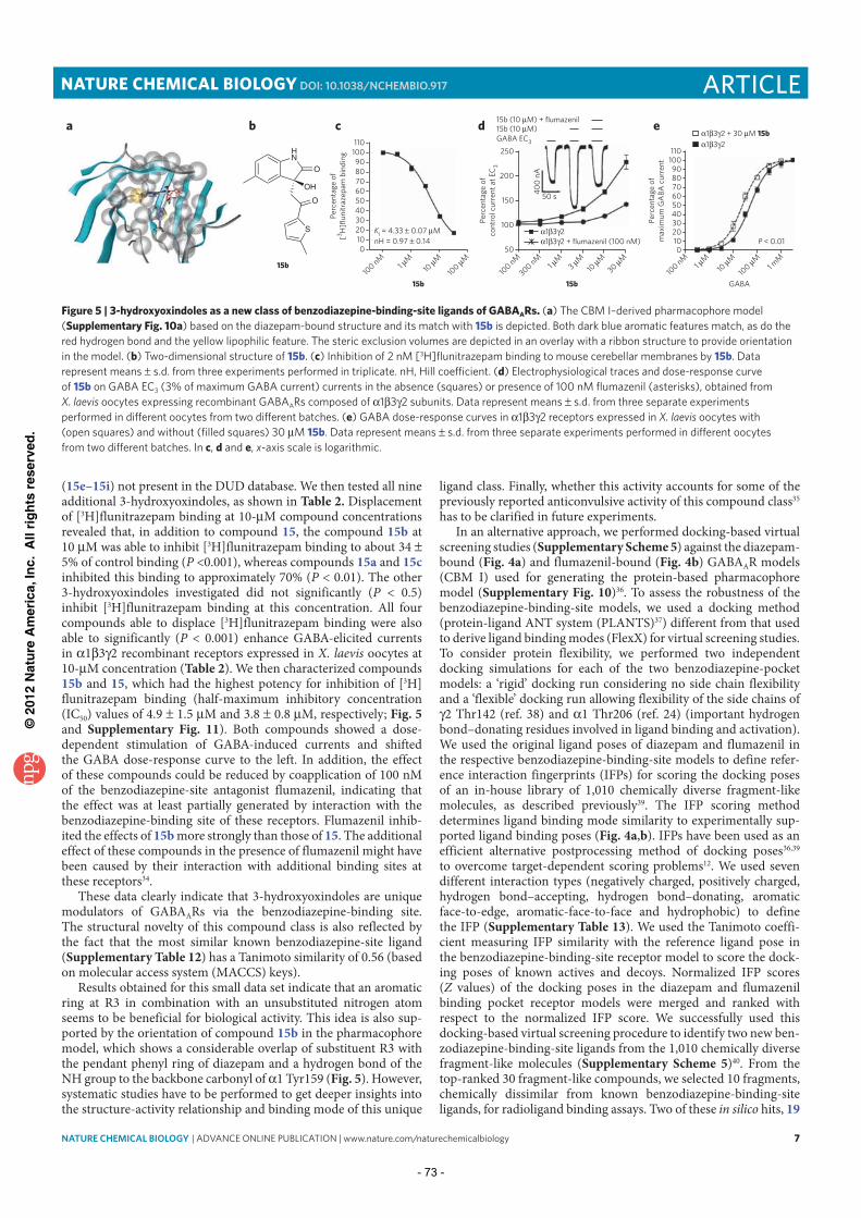
©20
12 N
atu
re A
mer
ica,
Inc.
All
rig
hts
res
erve
d.
NATURE CHEMICAL BIOLOGY | ADVANCE ONLINE PUBLICATION | www.nature.com/naturechemicalbiology 7
ARTICLENATURE CHEMICAL BIOLOGY DOI: 10.1038/NCHEMBIO.917
(15e–15i) not present in the DUD database. We then tested all nine additional 3-hydroxyoxindoles, as shown in Table 2. Displacement of [3H]flunitrazepam binding at 10- M compound concentrations revealed that, in addition to compound 15, the compound 15b at 10 M was able to inhibit [3H]flunitrazepam binding to about 34 5% of control binding (P <0.001), whereas compounds 15a and 15c inhibited this binding to approximately 70% (P < 0.01). The other 3-hydroxyoxindoles investigated did not significantly (P < 0.5) inhibit [3H]flunitrazepam binding at this concentration. All four compounds able to displace [3H]flunitrazepam binding were also able to significantly (P < 0.001) enhance GABA-elicited currents in 1 3 2 recombinant receptors expressed in X. laevis oocytes at 10- M concentration (Table 2). We then characterized compounds 15b and 15, which had the highest potency for inhibition of [3H]flunitrazepam binding (half-maximum inhibitory concentration (IC50) values of 4.9 1.5 M and 3.8 0.8 M, respectively; Fig. 5 and Supplementary Fig. 11). Both compounds showed a dose-dependent stimulation of GABA-induced currents and shifted the GABA dose-response curve to the left. In addition, the effect of these compounds could be reduced by coapplication of 100 nM of the benzodiazepine-site antagonist flumazenil, indicating that the effect was at least partially generated by interaction with the benzodiazepine-binding site of these receptors. Flumazenil inhib-ited the effects of 15b more strongly than those of 15. The additional effect of these compounds in the presence of flumazenil might have been caused by their interaction with additional binding sites at these receptors34.
These data clearly indicate that 3-hydroxyoxindoles are unique modulators of GABAARs via the benzodiazepine-binding site. The structural novelty of this compound class is also reflected by the fact that the most similar known benzodiazepine-site ligand (Supplementary Table 12) has a Tanimoto similarity of 0.56 (based on molecular access system (MACCS) keys).
Results obtained for this small data set indicate that an aromatic ring at R3 in combination with an unsubstituted nitrogen atom seems to be beneficial for biological activity. This idea is also sup-ported by the orientation of compound 15b in the pharmacophore model, which shows a considerable overlap of substituent R3 with the pendant phenyl ring of diazepam and a hydrogen bond of the NH group to the backbone carbonyl of 1 Tyr159 (Fig. 5). However, systematic studies have to be performed to get deeper insights into the structure-activity relationship and binding mode of this unique
ligand class. Finally, whether this activity accounts for some of the previously reported anticonvulsive activity of this compound class35 has to be clarified in future experiments.
In an alternative approach, we performed docking-based virtual screening studies (Supplementary Scheme 5) against the diazepam-bound (Fig. 4a) and flumazenil-bound (Fig. 4b) GABAAR models (CBM I) used for generating the protein-based pharmacophore model (Supplementary Fig. 10)36. To assess the robustness of the benzodiazepine-binding-site models, we used a docking method (protein-ligand ANT system (PLANTS)37) different from that used to derive ligand binding modes (FlexX) for virtual screening studies. To consider protein flexibility, we performed two independent docking simulations for each of the two benzodiazepine-pocket models: a ‘rigid’ docking run considering no side chain flexibility and a ‘flexible’ docking run allowing flexibility of the side chains of 2 Thr142 (ref. 38) and 1 Thr206 (ref. 24) (important hydrogen
bond–donating residues involved in ligand binding and activation). We used the original ligand poses of diazepam and flumazenil in the respective benzodiazepine-binding-site models to define refer-ence interaction fingerprints (IFPs) for scoring the docking poses of an in-house library of 1,010 chemically diverse fragment-like molecules, as described previously39. The IFP scoring method determines ligand binding mode similarity to experimentally sup-ported ligand binding poses (Fig. 4a,b). IFPs have been used as an efficient alternative postprocessing method of docking poses36,39 to overcome target-dependent scoring problems12. We used seven different interaction types (negatively charged, positively charged, hydrogen bond–accepting, hydrogen bond–donating, aromatic face-to-edge, aromatic-face-to-face and hydrophobic) to define the IFP (Supplementary Table 13). We used the Tanimoto coeffi-cient measuring IFP similarity with the reference ligand pose in the benzodiazepine-binding-site receptor model to score the dock-ing poses of known actives and decoys. Normalized IFP scores (Z values) of the docking poses in the diazepam and flumazenil binding pocket receptor models were merged and ranked with respect to the normalized IFP score. We successfully used this docking-based virtual screening procedure to identify two new ben-zodiazepine-binding-site ligands from the 1,010 chemically diverse fragment-like molecules (Supplementary Scheme 5)40. From the top-ranked 30 fragment-like compounds, we selected 10 fragments, chemically dissimilar from known benzodiazepine-binding-site ligands, for radioligand binding assays. Two of these in silico hits, 19
a110
c d e
100
90
80
70
60
50
40
30
20 Ki = 4.33 0.07 M
Pe
rce
nta
ge
of
co
ntr
ol
cu
rre
nt
at
EC
3
Pe
rce
nta
ge
of
[3H
]flu
nit
raze
pa
m b
ind
ing
nH = 0.97 0.1410
0
110
100
90
80
70
60
50
40
Pe
rce
nta
ge
of
ma
xim
um
GA
BA
cu
rre
nt
30
20
10
0
100 n
M1
M
10
M
15b10
0 M
50
100 n
M
300 nM
15b
1 M
3 M
10
M
30 M
100 n
M
GABA
1 M
1 mM
10
M
100
M
P < 0.01
100
150
200
250
15b (10 M) + flumazenil
15b (10 M)
GABA EC3
40
0 n
A
50 s
1 3 2
1 3 2
1 3 2 + 30 M 15b
1 3 2 + flumazenil (100 nM)
b
HN
O
O
OH
S
15b
Figure 5 | 3-hydroxyoxindoles as a new class of benzodiazepine-binding-site ligands of GABAARs. (a) The CBM I–derived pharmacophore model
(Supplementary Fig. 10a) based on the diazepam-bound structure and its match with 15b is depicted. Both dark blue aromatic features match, as do the
red hydrogen bond and the yellow lipophilic feature. The steric exclusion volumes are depicted in an overlay with a ribbon structure to provide orientation
in the model. (b) Two-dimensional structure of 15b. (c) Inhibition of 2 nM [3H]flunitrazepam binding to mouse cerebellar membranes by 15b. Data
represent means s.d. from three experiments performed in triplicate. nH, Hill coefficient. (d) Electrophysiological traces and dose-response curve
of 15b on GABA EC3 (3% of maximum GABA current) currents in the absence (squares) or presence of 100 nM flumazenil (asterisks), obtained from
X. laevis oocytes expressing recombinant GABAARs composed of 1 3 2 subunits. Data represent means s.d. from three separate experiments
performed in different oocytes from two different batches. (e) GABA dose-response curves in 1 3 2 receptors expressed in X. laevis oocytes with
(open squares) and without (filled squares) 30 M 15b. Data represent means s.d. from three separate experiments performed in different oocytes
from two different batches. In c, d and e, x-axis scale is logarithmic.
- 73 -

©20
12 N
atu
re A
mer
ica,
Inc.
All
rig
hts
res
erve
d.
8 NATURE CHEMICAL BIOLOGY | ADVANCE ONLINE PUBLICATION | www.nature.com/naturechemicalbiology
ARTICLE NATURE CHEMICAL BIOLOGY DOI: 10.1038/NCHEMBIO.917
and 20, inhibited [3H]flunitrazepam binding (IC50 values of 25.6 9.6 M and 34.7 2.6 M, respectively; Table 2). Remarkably, the new ligands are proposed to make the same interactions with the benzodiazepine-binding-site as diazepam and flumazenil 11, but most of the functional groups (hydrogen bond acceptors and aro-matic ring systems) mediating these conserved interactions are located at positions different than those of the corresponding func-tional groups in the reference ligands (Fig. 4). The newly identified chemical scaffolds (the MACCS Tanimoto similarity of 0.58 was the closest to any of the 41 benzodiazepine-binding-site ligands; Supplementary Table 12) illustrate the suitability of this molecular IFP approach for scaffold-hopping purposes41.
CBM I models are similar to the GluCl structureAfter submission of this study, a nematode glutamate-gated chloride channel (GluCl) structure was reported (PDB code 3RIF)42. This cysteine-loop receptor features 28%, 35% and 29% sequence iden-tity in the extracellular domain with 1, 2 and 2 subunits, respec-tively, and thus it is clearly the template of choice for future studies. Careful analysis of this structure and homology models derived from it (Supplementary Table 4) indicated that our CBM I models indeed are more similar to the structure with PDB code 3RIF than the AChBP structures used in modeling and, more importantly, much more so than the models that we rejected through our work-flow. This again highlights the validity of our approach.
DISCUSSIONIn the absence of a crystal structure of the ligand-bound benzo-diazepine-binding-site of GABAARs, protein homology modeling and computational ligand dockings are the only bases on which structure-based hypotheses for the interaction of benzodiazepines with their binding site can be formed. However, accurate predic-tion of membrane-protein structures and ligand interactions remains a challenge43 and requires accurate modeling of structurally divergent regions and extensive use of experimental evidence, as shown recently in a community-wide G protein–coupled receptor structure-prediction assessment. Here we compensated for the local uncertainty resulting from structurally divergent regions of related cysteine-loop receptor family members by constructing a variety of homology models from different templates. We then exploited the given structure of diazepam and its derivatives to select those models best accommodating these compounds. To do so, we developed a workflow that allowed handling of multiple models and several thousand diazepam docking poses. The only selection criteria used were a reasonable lipophilic interaction with the hydrophobic parts of the diazepam structure as well as simultaneous contacts of diazepam with the 1 and the 2 subunit of GABAARs. With the assumption that diazepam and its close structural analogs have a CBM within the benzodiazepine-binding pocket and by clustering of ligand poses according to a similar orientation of their common structure and according to similar interactions with pocket residues, we were finally led to three CBM geometries, CBMs I–III.
CBM I binding geometry and interactions are supported by a large variety of structural, computational and experimental evidence. Furthermore, CBM I can provide a single structural hypothesis for seemingly discrepant results obtained with various covalently incorporated compounds and supports the hypoth-esis that diazepam and the imidazobenzodiazepines Ro15-4513 or fluma zenil show a similar binding mode within the benzodiazepine pocket. Finally, CBM I can also explain subtle changes in affinity of various ligands for various amino acid substitutions. Taken together, this abundance of evidence indicates that CBM I is the correct binding mode of diazepam in the benzodiazepine-binding pocket.
Previous studies also featured docking poses of diazepam or its analogs. Overall, however, the conclusions remained contradictory. In one study5, a flunitrazepam orientation similar to the CBM III
poses was reported. Another study6 identified a diazepam pose vaguely similar to the CBM I pose, but apparently it had a very different interaction pattern owing to a different pocket topology. A pharmacophore-derived diazepam pose4 is in a position inter-mediate between that of CBM II and CBM I. Most recently, bind-ing modes vaguely resembling our CBM II were proposed based on modeling structures from a single template and covalently incorporated ligands7. In a study more concerned with zolpidem8, flunitrazepam poses featured the P conformation, which has been demonstrated to be inactive at the benzodiazepine-binding site (Supplementary Table 2).
A correct structure, however, should also be able to accommodate other ligands of this site from different structural classes. Thus, by using structure-based pharmacophore models derived from CBM I, we identified 5 out of 41 benzodiazepine-site ligands from different structural classes within the top 0.5% of hits of a benzodiazepine- focused set of decoy molecules (validation library), and we found five additional compounds within the top 2% and eight more in the top 10% of compounds (Supplementary Tables 9 and 10). Such enrichment factors, as well as the remaining false negatives (bind-ers that are not ranked in the top range) are comparable to those obtained from docking into ligand-bound crystal structures30.
Virtual screening with the DUD database30 identified not only additional known benzodiazepine-site ligands present in this database but also a variety of other anticonvulsive compounds that so far have not been associated with GABAARs. By investi-gating a commercially available subset for a possible interaction with the benzodiazepine-binding site of GABAARs, we identified several 3-hydroxyoxindole derivatives that were able to modu-late GABA-induced currents in recombinant receptors via the benzodiazepine-binding site. Furthermore, in a complementary approach, docking-based virtual screening studies using a mole-cular protein-ligand IFP scoring method identified two additional new chemical scaffolds, which were proven active in [3H]flunitraze-pam displacement assays. These data further support the validity of our diazepam-bound structure of the benzodiazepine-binding site and demonstrate its suitability for structure-based drug discovery.
Further exploration of our in silico screening results presum-ably will identify additional new ligand classes for the benzodiaz-epine-binding site. Information from docking studies with other benzodiazepine-site ligands as well as molecular dynamics studies will identify the flexible and rigid parts of the pocket and define their ligand-pocket interactions as well as the mechanism of allo-steric modulation of positive, negative and neutral interactors. The present structural models can also be used for modeling of the benzodiazepine-binding sites of other GABAAR subtypes, and dock-ing of unselective and subtype-selective ligands into their binding sites will ultimately lead to appropriate structural hypotheses that allow lead optimization and fragment-based drug design. Finally, our structures can be used for the modeling of similar extra cellular pockets of GABAARs34, again enabling lead optimization and drug discovery.
METHODSTemplate characterization and preparation. Six AChBP structures (PDB codes 1I9B, 1UW6, 2BYN, 2BYQ, 2BYR and 2BYS)3 and two nAChR structures (PDB codes 2BG9 and 2QC1)3 were used as GABAA modeling templates (Supplementary Table 1) and structurally aligned with ProFit (http://www.bioinf.org.uk/software/profit/). Only the extracellular domains of the structures with PDB codes 2BG9 and 2QC1 were considered, and both complete pentamers and individual subunits, reassembled into multitemplates, were used.
Alignment and model building. A multiple-sequence alignment with all templates’ extracellular domains and the GABAAR 1, 2 and 2 extracellular domains’ sequences was constructed with clustalX44 (Supplementary Scheme 1 and Supplementary Fig. 1). All templates were structurally aligned with secondary-structure matching10. Individual pairwise sequence-to-structure alignments between the GABAAR subunits and all template subunits were obtained from the
- 74 -

©20
12 N
atu
re A
mer
ica,
Inc.
All
rig
hts
res
erve
d.
NATURE CHEMICAL BIOLOGY | ADVANCE ONLINE PUBLICATION | www.nature.com/naturechemicalbiology 9
ARTICLENATURE CHEMICAL BIOLOGY DOI: 10.1038/NCHEMBIO.917
Fugue server45. From these data, alignment variants of variable segments were con-structed46, and homology models were built using Modeller. For the two subunits that contribute to the benzodiazepine-binding site, multiple template combinations were used to sample the structural variations in the templates.
Creation of input ligands. Seven 1,4-benzodiazepines and three 1,4-imidazo-benzodiazepines were built in Molecular Operating Environment (MOE) version 2007.09 (http://www.chemcomp.com), using the M conformation of the seven-membered ring that is supported by experimental studies (Supplementary Table 2 and Supplementary Fig. 2).
FlexX docking. Diazepam was docked into all models using FlexX v2.0.3 (ref. 11). RING_MODE was set to 0 (constraining the ring conformation), and all other parameters were left in their default values. The pose output limit was set to 100 for each run for extensive conformational sampling.
FlexE docking. FlexE13 allows protein flexibility through an ensemble of super-posed protein structures where similar parts of the structures are merged and dissimilar areas are treated as separate alternatives. To prepare such ensembles, we explored the rotameric states of 1 His101, 1 Tyr159, 1 Val202, 1 Ser204,
1 Ser205, 1 Thr206, 1 Tyr209, 2 Tyr58, 2 Phe77 and 2 Thr142 with the MOE tool Rotamer Explorer. In cases in which more than ten rotamers were obtained for a certain side chain, the ten most diverse rotamers were retained. Each benzodiazepine-binding-site ligand was docked into the ensemble structures of the corresponding homology models, generating 100 poses for each ligand. Finally, each ligand-receptor complex of the final pose pool was refined using the MOE tool LigX energy minimize. Lipophilic interactions of L1 and L3 both in docked and energy-minimized poses were calculated with the Scientific Vector Language (SVL)-exchange tool SCORING.SVL (http://svl.chemcomp.com).
CBM candidate selection. We defined the CBM of a molecular scaffold to require common orientation of the scaffold and common binding-site topology surrounding the common scaffold. Poses were superposed on the backbone atoms of the conserved protein segments using MOE to determine ligands’ common scaffold r.m.s. deviation. The SVL-exchange script MOL_RMSD.SVL (http://svl.chemcomp.com) was used for the r.m.s. deviation computation of the common ligand scaffolds. Then, the Microsoft Excel add-in XLSTAT (http://www.xlstat.com) was used for hierarchical clustering of the r.m.s. deviation dissimilarity matrix using the WARD method.
All protein heavy atoms within 4 Å of any heavy atom of the molecular scaffold in more than 90% of all poses of the cluster were considered part of the binding site of the common scaffold. The identified atoms and the common scaffold were used for the calculation of the bound-complex r.m.s. deviation, which was used to cluster the poses to candidate binding modes within their parent cluster.
Evaluating covalent incorporation and mutagenesis data. Poses were considered to fit experimental data if the smallest distance between the atoms in any of the following combinations was <6 Å: (i) any atom of the nitro group (flunitrazepam) and any atom of 1 His101 (ref. 17), (ii) the photoreactive nitrogen of the arylazido group (Ro15-4513) and 1 Tyr209 (ref. 22), (iii) any atom of the ester moiety (Ro15-8670) and side chain atoms of 1 Thr206 or the C of 2 Ala79 (ref. 29) or (iv) the carbon of isothiocyanate (NCS)-substituted ligands and the sulfur of the respective cysteine mutant.
Generation and validation of pharmacophore models. Two structure-based pharmacophore models were created for each CBM, one derived from the top-scoring diazepam pose (pharmacophore D) and the other from the flumazenil pose, which showed the lowest bound-complex r.m.s. deviation from the diazepam-selected pose (pharmacophore F). Pharmacophore models were created using LigandScout 3.0 (ref. 32) (Supplementary Methods).
Validation library. For each of the 41 known benzodiazepine-binding-site ligands (Supplementary Table 10), protonation states near pH 7.0 were generated, resulting in 48 unique ligand protomers. For each of them, we created 50 decoy molecules that are physically similar but topologically distinct and thus should not bind the benzodiazepine site, resulting in a library of 2,400 decoys (Supplementary Methods). A conformational database was created with OMEGA v2.3.3 (http://www.eyesopen.com/omega) and screened against pharmacophore D and pharmacophore F using LigandScout 3.0. Hits of each pharmacophore model were sorted according to scoring value, and enrichment factors33 were determined for the top 0.5%, 1%, 2%, 5% and 10% for each CBM.
Discovery library. To test the ability of the CBM I–derived pharmacophore model to identify new ligands, we generated three-dimensional conformations for the DUD30 databank (using OMEGA v2.4.1 (http://www.eyesopen.com/omega) and screened the resulting database of 93,597 compounds against pharmacophore D and pharmacophore F using LigandScout 3.0 (ref. 32). Compounds 15, 15a–15i and 18 were bought from Ambinter, and compounds 16 and 17 were bought from Asinex.
PLANTS docking and IFP scoring. PLANTS37 speedup settings were used twice to generate 15 poses for each compound in two independent docking runs: one run considering no side chain flexibility and another run allowing flexibility of the 2 Thr142 and 1 Thr206 side chains. The original ligand poses in the respective
GABAA models were used to define reference IFPs, as described previously36,39. The cavity used for the IFP analysis consisted of all of the residues within 5 Å of the reference ligand. Standard IFP scoring parameters36 and a Tanimoto coefficient measuring IFP similarity with the reference ligand pose were used to rank the docking poses.
Experimental section: Recombinant GABAARs were expressed in X. laevis oocytes, and HEK cells and compound modulatory effects were investigated using the two-electrode voltage-clamp technique at a GABA concentration eliciting 3% of the maximum current34. Binding of compounds was investigated by [3H]flunitrazepam displacement studies47 in mouse cerebellar membrane preparations.
Accession codes. PDB: the previously determined crystal structures for A. californica AChBP, L. stagnalis AChBP, mouse nAChR, a nematode glutamate-gated chloride channel, an nAChR structure and four AChBP structures are deposited under accession codes 2BYQ, 1UW6, 2QC1, 3RIF, 2BG9, 1I9B, 2BYN, 2BYR and 2BYS, respectively.
Received 24 March 2011; accepted 2 February 2012; published online 25 March 2012
References1. Sieghart, W. Structure and pharmacology of -aminobutyric acid A receptor
subtypes. Pharmacol. Rev. 47, 181–234 (1995).2. Sigel, E. Mapping of the benzodiazepine recognition site on GABAA receptors.
Curr. Top. Med. Chem. 2, 833–839 (2002).3. Miller, P.S. & Smart, T.G. Binding, activation and modulation of Cys-loop
receptors. Trends Pharmacol. Sci. 31, 161–174 (2010).4. Clayton, T. et al. An updated unified pharmacophore model of the
benzodiazepine binding site on -aminobutyric acidA receptors: correlation with comparative models. Curr. Med. Chem. 14, 2755–2775 (2007).
5. Mokrab, Y. et al. Exploring ligand recognition and ion flow in comparative models of the human GABA type A receptor. J. Mol. Graph. Model. 26, 760–774 (2007).
6. Ci, S., Ren, T. & Su, Z. Investigating the putative binding-mode of GABA and diazepam within GABAA receptor using molecular modeling. Protein J. 27, 71–78 (2008).
7. Berezhnoy, D., Gibbs, T.T. & Farb, D.H. Docking of 1,4-benzodiazepines in the 1/ 2 GABAA receptor modulator site. Mol. Pharmacol. 76, 440–450 (2009).
8. Sancar, F., Ericksen, S.S., Kucken, A.M., Teissére, J.A. & Czajkowski, C. Structural determinants for high-affinity zolpidem binding to GABAA receptors. Mol. Pharmacol. 71, 38–46 (2007).
9. Evers, A., Gohlke, H. & Klebe, G. Ligand-supported homology modelling of protein binding-sites using knowledge-based potentials. J. Mol. Biol. 334, 327–345 (2003).
10. Krissinel, E. & Henrick, K. Secondary-structure matching (SSM), a new tool for fast protein structure alignment in three dimensions. Acta Crystallogr. D Biol. Crystallogr. 60, 2256–2268 (2004).
11. Rarey, M., Kramer, B., Lengauer, T. & Klebe, G. A fast flexible docking method using an incremental construction algorithm. J. Mol. Biol. 261, 470–489 (1996).
12. Moitessier, N., Englebienne, P., Lee, D., Lawandi, J. & Corbeil, C.R. Towards the development of universal, fast and highly accurate docking/scoring methods: a long way to go. Br. J. Pharmacol. 153 (suppl 1): S7–S26 (2008).
13. Claussen, H., Buning, C., Rarey, M. & Lengauer, T. FlexE: efficient molecular docking considering protein structure variations. J. Mol. Biol. 308, 377–395 (2001).
14. Chema, D., Eren, D., Yayon, A., Goldblum, A. & Zaliani, A. Identifying the binding mode of a molecular scaffold. J. Comput. Aided Mol. Des. 18, 23–40 (2004).
15. Wang, R., Lai, L. & Wang, S. Further development and validation of empirical scoring functions for structure-based binding affinity prediction. J. Comput. Aided Mol. Des. 16, 11–26 (2002).
16. Labute, P. The generalized Born/volume integral implicit solvent model: estimation of the free energy of hydration using London dispersion instead of atomic surface area. J. Comput. Chem. 29, 1693–1698 (2008).
17. Duncalfe, L.L., Carpenter, M.R., Smillie, L.B., Martin, I.L. & Dunn, S.M. The major site of photoaffinity labeling of the -aminobutyric acid type A receptor by [3H]flunitrazepam is histidine 102 of the subunit. J. Biol. Chem. 271, 9209–9214 (1996).
18. Berezhnoy, D. et al. On the benzodiazepine binding pocket in GABAA receptors. J. Biol. Chem. 279, 3160–3168 (2004).
- 75 -

©20
12 N
atu
re A
mer
ica,
Inc.
All
rig
hts
res
erve
d.
10 NATURE CHEMICAL BIOLOGY | ADVANCE ONLINE PUBLICATION | www.nature.com/naturechemicalbiology
ARTICLE NATURE CHEMICAL BIOLOGY DOI: 10.1038/NCHEMBIO.917
19. Wieland, H.A., Lüddens, H. & Seeburg, P.H. A single histidine in GABAA receptors is essential for benzodiazepine agonist binding. J. Biol. Chem. 267, 1426–1429 (1992).
20. Davies, M., Bateson, A.N. & Dunn, S.M. Structural requirements for ligand interactions at the benzodiazepine recognition site of the GABAA receptor. J. Neurochem. 70, 2188–2194 (1998).
21. Buhr, A., Schaerer, M.T., Baur, R. & Sigel, E. Residues at positions 206 and 209 of the 1 subunit of -aminobutyric acidA receptors influence affinities for benzodiazepine binding site ligands. Mol. Pharmacol. 52, 676–682 (1997).
22. Sawyer, G.W., Chiara, D.C., Olsen, R.W. & Cohen, J.B. Identification of the bovine -aminobutyric acid type A receptor subunit residues photolabeled by the imidazobenzodiazepine [3H]Ro15–4513. J. Biol. Chem. 277, 50036–50045 (2002).
23. Tan, K.R. et al. Proximity-accelerated chemical coupling reaction in the benzodiazepine-binding site of -aminobutyric acid type A receptors: superposition of different allosteric modulators. J. Biol. Chem. 282, 26316–26325 (2007).
24. Sigel, E., Schaerer, M.T., Buhr, A. & Baur, R. The benzodiazepine binding pocket of recombinant 1 2 2 -aminobutyric acidA receptors: relative orientation of ligands and amino acid side chains. Mol. Pharmacol. 54, 1097–1105 (1998).
25. Buhr, A., Baur, R. & Sigel, E. Subtle changes in residue 77 of the subunit of 1 2 2 GABAA receptors drastically alter the affinity for ligands of the
benzodiazepine binding site. J. Biol. Chem. 272, 11799–11804 (1997).26. Amin, J., Brooks-Kayal, A. & Weiss, D.S. Two tyrosine residues on the
subunit are crucial for benzodiazepine binding and allosteric modulation of -aminobutyric acidA receptors. Mol. Pharmacol. 51, 833–841 (1997).
27. Derry, J.M.C., Dunn, S.M.J. & Davies, M. Identification of a residue in the -aminobutyric acid type A receptor subunit that differentially affects
diazepam-sensitive and -insensitive benzodiazepine site binding. J. Neurochem. 88, 1431–1438 (2004).
28. Tan, K.R., Baur, R., Charon, S., Goeldner, M. & Sigel, E. Relative positioning of diazepam in the benzodiazepine-binding-pocket of GABAA receptors. J. Neurochem. 111, 1264–1273 (2009).
29. Kucken, A.M., Teissére, J.A., Seffinga-Clark, J., Wagner, D.A. & Czajkowski, C. Structural requirements for imidazobenzodiazepine binding to GABAA receptors. Mol. Pharmacol. 63, 289–296 (2003).
30. Huang, N., Shoichet, B.K. & Irwin, J.J. Benchmarking sets for molecular docking. J. Med. Chem. 49, 6789–6801 (2006).
31. Mysinger, M.M. & Shoichet, B.K. Rapid context-dependent ligand desolvation in molecular docking. J. Chem. Inf. Model. 50, 1561–1573 (2010).
32. Wolber, G. & Langer, T. LigandScout: 3-D pharmacophores derived from protein-bound ligands and their use as virtual screening filters. J. Chem. Inf. Model. 45, 160–169 (2005).
33. Wei, B.Q., Baase, W.A., Weaver, L.H., Matthews, B.W. & Shoichet, B.K. A model binding site for testing scoring functions in molecular docking. J. Mol. Biol. 322, 339–355 (2002).
34. Ramerstorfer, J. et al. The GABAA receptor + - interface: a novel target for subtype selective drugs. J. Neurosci. 31, 870–877 (2011).
35. Popp, F.D., Parson, R. & Donigan, B.E. Synthesis of potential anticonvulsants: condensation of isatins with acetone and related ketones. J. Pharm. Sci. 69, 1235–1237 (1980).
36. Marcou, G. & Rognan, D. Optimizing fragment and scaffold docking by use of molecular interaction fingerprints. J. Chem. Inf. Model. 47, 195–207 (2007).
37. Korb, O., Stutzle, T. & Exner, T.E. Empirical scoring functions for advanced protein-ligand docking with PLANTS. J. Chem. Inf. Model. 49, 84–96 (2009).
38. Mihic, S.J., Whiting, P.J., Klein, R.L., Wafford, K.A. & Harris, R.A. A single amino acid of the human -aminobutyric acid type A receptor 2 subunit determines benzodiazepine efficacy. J. Biol. Chem. 269, 32768–32773 (1994).
39. de Graaf, C. & Rognan, D. Selective structure-based virtual screening for full and partial agonists of the 2 adrenergic receptor. J. Med. Chem. 51, 4978–4985 (2008).
40. Verheij, M.H. et al. Fragment library screening reveals remarkable similarities between the G protein-coupled receptor histamine H and the ion channel serotonin 5-HTA. Bioorg. Med. Chem. Lett. 21, 5460–5464 (2011).
41. Venhorst, J., Nunez, S., Terpstra, J.W. & Kruse, C.G. Assessment of scaffold hopping efficiency by use of molecular interaction fingerprints. J. Med. Chem. 51, 3222–3229 (2008).
42. Hibbs, R.E. & Gouaux, E. Principles of activation and permeation in an anion-selective Cys-loop receptor. Nature 474, 54–60 (2011).
43. Michino, M. et al. Community-wide assessment of GPCR structure modelling and ligand docking: GPCR Dock 2008. Nat. Rev. Drug Discov. 8, 455–463 (2009).
44. Larkin, M.A. et al. Clustal W and Clustal X version 2.0. Bioinformatics 23, 2947–2948 (2007).
45. Shi, J., Blundell, T.L. & Mizuguchi, K. FUGUE: sequence-structure homology recognition using environment-specific substitution tables and structure-dependent gap penalties. J. Mol. Biol. 310, 243–257 (2001).
46. Sali, A. & Blundell, T.L. Comparative protein modelling by satisfaction of spatial restraints. J. Mol. Biol. 234, 779–815 (1993).
47. Sieghart, W. & Schuster, A. Affinity of various ligands for benzodiazepine receptors in rat cerebellum and hippocampus. Biochem. Pharmacol. 33, 4033–4038 (1984).
48. Zhang, W., Koehler, K.F., Zhang, P. & Cook, J.M. Development of a comprehensive pharmacophore model for the benzodiazepine receptor. Drug Des. Discov. 12, 193–248 (1995).
AcknowledgmentsFinancial support from the Doctoral Fellowship Programme of the Austrian Academy of Science (L.R.), the Austrian Science Fund grant P19653 (M.E.) and the European Commission Seventh Framework Programme grant HEALTH-F4-2008-202088 (W.S. and I.J.P.d.E.) is gratefully acknowledged. We also thank Inte:Ligand for providing us a free license for LigandScout 3.0 and Open Eye for an Omega license. We thank M. Mysinger and J. Irwin from the Shoichet laboratory for generating the focused ‘benzodiazepine’ decoy set. In addition we thank E. Sigel for helpful suggestions and discussions during generation of this work; M. Stojanovic for technical assistance in the binding assays; A. Chalikiopoulos and M. Verheij for assistance in fragment docking studies; and E. Urban, S. Haselmaier, J. König, H. Custers and A. van de Stolpe for providing HRMS and 1H- and 13C-NMR spectral data.
Author contributionsL.R., W.S., G.F.E., I.J.P.d.E., C.d.G. and M.E. contributed to the design and evaluation of experiments. Docking and virtual screening experiments were performed by L.R. (supervised by G.F.E.) and C.d.G. GABAAR models were generated and evaluated by M.E. and M.M. Radioligand and electrophysiological experiments were performed by Z.V. The manuscript was written by W.S., L.R., G.F.E., I.J.P.d.E., C.d.G. and M.E., and all authors commented on and helped to revise the text.
Competing financial interestsThe authors declare no competing financial interests.
Additional informationSupplementary information and chemical compound information are available online at http://www.nature.com/naturechemicalbiology/. Reprints and permissions information is available online at http://www.nature.com/reprints/index.html. Correspondence and requests for materials should be addressed to M.E.
- 76 -

Supplementary Information
Diazepam-bound GABAA receptor models identify novel benzodiazepine site ligands
Lars Richter, Chris de Graaf, Werner Sieghart, Zdravko Varagic, Martina Mörzinger,
Iwan J. P. de Esch, Gerhard F. Ecker, Margot Ernst
Nature Chemical Biology: doi:10.1038/nchembio.917
- 77 -

Supplementary Methods………………………………………...4 Supplementary Schemes……………………………………………………………....…4 Supplementary Scheme 1…………………………………………………………….........4
Supplementary Scheme 2……………………………………………………………...…...5
Supplementary Scheme 3…………………………………………………………….........6
Supplementary Scheme 4…………………………………………………………..…....…7
Supplementary Scheme 5……………………………………………………………...…...8
Supplementary Tables …………………………………………………………………....9 Supplementary Table 1……………………………………………………........................9
Supplementary Table 2 …………………………………………………….....................10
Supplementary Figures…………………………………………………………………..12 Supplementary Fig. 1…………………………………………………………….….……..12
Supplementary Fig. 2…………………………………………………………….….……..15
Supplementary Text…………………………………………………………………....…16 Generation of validation library ………………………………………………………......16
Structure-based pharmacophore models..…………………………………….……..….16
Ligand-based pharmacophore models….……………………………………...………..17
Compound characterization data ...........................................................................18
Nature Chemical Biology: doi:10.1038/nchembio.917
- 78 -

Supplementary Results …………………..……………………34 Supplementary Tables …….…………………….……………………………………...34 Supplementary Table 3 …………………………………………………………………...34
Supplementary Table 4 …………………………………………………………………...36
Supplementary Table 5 …………………………………………………………………...37
Supplementary Table 6 …………………………………………………………………...38
Supplementary Table 7 …………………………………………………………………...39
Supplementary Table 8 …………………………………………………………………...39
Supplementary Table 9 …………………………………………………………………...40
Supplementary Table 10 ………………………………………………………………….41
Supplementary Table 11 ………………………………………………………………….46
Supplementary Table 12 ………………………………………………………………….47
Supplementary Table 13 ………………………………………………………………….48
Supplementary Figures …………………………………………………………………49 Supplementary Fig. 3 ……………………………………………………………………..49
Supplementary Fig. 4 ……………………………………………………………………..50
Supplementary Fig. 5……………………………………………………………………...51
Supplementary Fig. 6 ……………………………………………………………………..52
Supplementary Fig. 7 ……………………………………………………………………..53
Supplementary Fig. 8 ……………………………………………………………………..54
Supplementary Fig. 9 ……………………………………………………………………..55
Supplementary Fig. 10 …………………………………………………………………....57
Supplementary Fig. 11 …………………………………………………………………....58
Supplementary Reference ....………………………………....59
Nature Chemical Biology: doi:10.1038/nchembio.917
- 79 -

Supplementary Methods - Supplementary Schemes
* In an initial step, the GABAA receptor sequences are aligned against each template structure with a sequence-to-structure alignment algorithm such as TCoffee49 or FUGUE50 yielding seed alignments. ** In the seed alignments, gap positions in variable regions are varied according to the User’s Manual of Modeller51, thus generating a variety of input alignments. *** The standard homology modelling script supplied with the program is combined with one or more template structures and input alignments to generate the homology models.
Nature Chemical Biology: doi:10.1038/nchembio.917
- 80 -

34 initial (in) models
FlexX, docking of diazepam against 34 rigid models
good L1L3.a1 and g2
contact
3 final models (f)
6 models (f + fv)(M1-M6)
+3 final model variants (fv)
14 intermediate (im) models
good L1L3.a1 and g2
contact
-20 models
-11 models
FlexE, docking of 6 ligands against 14 flexible models
Flexibilize tight regions
+3
-20
FlexE, docking of 9 ligands against 6 flexible models.
Energy minimization and L1L3 calculation.
L1 and L3 abovemedian
4997 poses
-3534 poses
L1 & L3 pool(1463 poses)
1463 poses
-11no
yes
no yes
yes
no
Supplementary Scheme 2 – Model selection workflow
(see also Supplementary Table 3)
(see also Supplementary Table 3)
(see also Supplementary Table 3)
(see Supplementary Table 7)
Nature Chemical Biology: doi:10.1038/nchembio.917
- 81 -
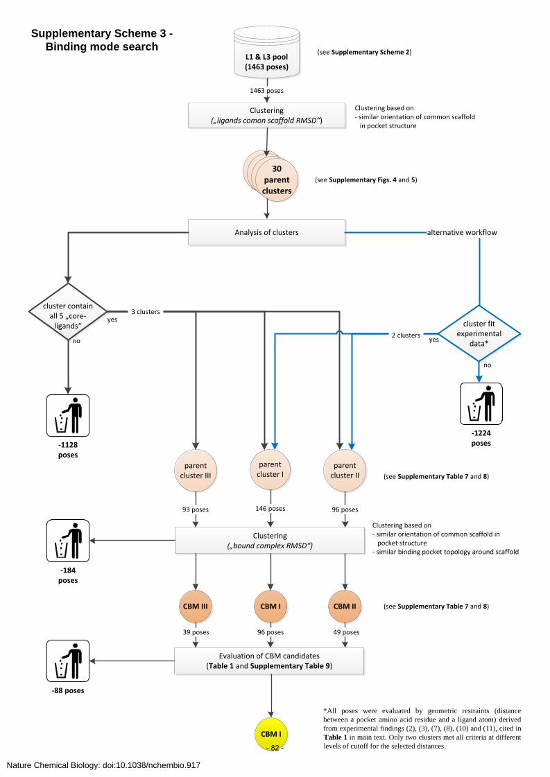
L1 & L3 pool(1463 poses)
Clustering(„ligands comon scaffold RMSD“)
30 parentclusters
Analysis of clusters alternative workflow
cluster containall 5 „core- ligands“ cluster fit
experimentaldata*
-1128 poses
CBM I
1463 poses
Clustering based on- similar orientation of common scaffold in pocket structure
yes
no yes
no
parentcluster III
parentcluster I
parent cluster II
Clustering („bound complex RMSD“)
-1224 poses
146 poses 96 poses
CBM III CBM II
Evaluation of CBM candidates (Table 1 and Supplementary Table 9)
CBM I
93 poses
96 poses39 poses 49 poses
Clustering based on- similar orientation of common scaffold in pocket structure- similar binding pocket topology around scaffold
*All poses were evaluated by geometric restraints (distance
between a pocket amino acid residue and a ligand atom) derived
from experimental findings (2), (3), (7), (8), (10) and (11), cited in
Table 1 in main text. Only two clusters met all criteria at different
levels of cutoff for the selected distances.
3 clusters
2 clusters
-184 poses
-88 poses
Supplementary Scheme 3 -
Binding mode search
(see Supplementary Figs. 4 and 5)
(see Supplementary Table 7 and 8)
(see Supplementary Table 7 and 8)
(see Supplementary Scheme 2)
Nature Chemical Biology: doi:10.1038/nchembio.917
- 82 -

CBM I, II, III
Pharmacophore D Pharmacophore F
Virtual screening of validation library against pharmacophore models and
subsequent ranking according to pharmacophore fit score.
Generation of structure based pharmacophore models based on:
Diazepam pose Flumazenil pose
Decoy set.2400 decoys
48 protomers of 41 actives
decoy setCBM I, II, III
Generation of a decoy set in analogy to DUD52. 41 benzodiazepine binding site ligands (48 unique ligand protomers*) were seeded in ZINC53 database. For each
ligand protomer, 50 physically similar but topologically distinct decoys were selected, resulting in 2400 (48x50) decoy molecules.
Validation library: The 41 benzodiazepine binding site ligands (48 unique ligand protomers*) were spiked into 2400 molecules comprising decoy set.
1. physically similar2. topologically distinct
Validation library: 48 protomers of 41 actives +
2400 decoys
ZINC53 database
Enrichment of actives in the top 0.5%, 1%, 2%, 5% and 10% was
computed for each CBM. (Supplementary Table 9)
CBM I CBM II CBM III
Top %
Score-sorted validation libraries
*As decoys were also property matched against net formal charge, for each of the 41 ligands all protonation states near pH 7.0 were generated, resulting in 48 unique ligand protomers. These 48 ligand protomers were seeded into the ZINC-database as well as into the validation library.
Supplementary Scheme 4 – Retrospective validation of CBMs by virtual screening
Nature Chemical Biology: doi:10.1038/nchembio.917
- 83 -

Pharmacophore D(Suppl. Fig. 10a)
Pharmacophore F(Suppl. Fig. 10b)
Virtual screening of DUD library (95 357 cpds.) against pharmacophore models and subsequent ranking according to
pharmacophore fit score.
Generation of structure based pharmacophore models based on:
Diazepam pose Flumazenil pose
Top 0.5% of score-sorted library
4 cpds. tested in assay
normalized IFP (diazepam) score
normalized IFP (flumazenil) score
Molecular docking based virtual screening of 1010 fragments and ranking
of library according to normalized IFP score.
Calculate Interaction Fingerprints (IFPs) for :
Diazepam pose Flumazenil pose
Top 30 hits of score-sorted library
10 cpds. tested in assay
DUD library (95 357 cpds.)
In-house db of 1010 fragment-like cpds.
1 novel benzodiazepine
binding site ligand identified
2 novel benzodiazepine
binding site ligands identified
Diazepam pose Flumazenil pose
CBM I
Flumazenil pose
Supplementary Scheme 5 – CBM I binding hypotheses predict novel ligands
Nature Chemical Biology: doi:10.1038/nchembio.917
- 84 -

Supplementary Methods - Supplementary Tables Supplementary Table 1– Templates used in model construction
PDB ID protein ligand
1I9B54 Lymnea AChBP HEPES bound
1UW655 Lymnea AChBP nicotine bound
2BG956 Torpedo nAChR apo/ undefined
2BYN57 Aplysia AChBP apo
2BYQ57 Aplysia AChBP epibatidine bound
2BYR57 Aplysia AChBP methyllycaconitine bound
2BYS57 Aplysia AChBP lobeline bound
2QC158 mouse nAChR alpha1
extracellular domain
alpha-bungarotoxin bound
3RIF59 Nematode glutamate gated
chloride channel
glutamate
Templates up to and including 2QC158 were considered in the original study. The
selection criteria were conformational diversity and resolution of the structures.
Bacterial homologs released after 2QC1 were examined as the study progressed,
and it was felt that models based on these would not alter the overall outcome. Upon
revision, 3RIF59and related structures were also integrated in retrospect and found to
be in good agreement with our model.
Nature Chemical Biology: doi:10.1038/nchembio.917
- 85 -

Supplementary Table 2 - Affinities of the two atropisomers/ enantiomers of 1,4-benzodiazepines at the BZ- binding site. reference: conformation M conformation P Salvadori et al. 199760
annotation in ref: cpd. 7a annotation in ref: cpd. 7b affinity: 93 nM affinity: 1750 nM
Gilman et al. 199061 annotation in ref: cpd. 11 annotation in ref: cpd. 12
affinity: 420 nM affinity: > 1000 nM
Gilman et al. 199362 annotation in ref: cpd. 31a annotaton in ref: cpd. 31b
affinity: 26.5 nM affinity: 540 nM
Gilman et al. 199362 annotation in ref: cpd. 25a annotation in ref: cpd. 25b
affinity: 7 nM affinity: 220 nM
Lee at al. 200863
annotation in ref: 1a-R annotation in ref: 1a-S affinity: 46 nM affinity: 2320 nM
Nature Chemical Biology: doi:10.1038/nchembio.917
- 86 -

reference: conformation M conformation P Blount et al. 198364 annotation in ref: 5 annotation in ref: 6
affinity: 6.4 nM affinity: 2320 nM
In principle two 1,4-diazepine ring conformations are energetically plausible for 1,4-
benzodiazepines (conformation M and conformation P, see also Supplementary Fig. 2) and are hence present in small molecule crystal structures (CSD reference
code: ANIXAX, FLDAZP).A literature search provides strong and coherent evidence
that conformation M of the 7-ring is present in the high affinity bound-state complex
of 1,4-benzodiazepines in the benzodiazepine binding site of GABAAreceptors.
Gilman et al.61,62 and Salvadori et al.60 applied a selective synthetic procedure that
leads to stable 1,4-benzodiazepines in either conformation M or conformation P.
The interconversion of the synthesized molecules into the other 7-ring conformation
was hindered by a bulky tert-butyl group either at N1 (1,4-benzodiazepines) or C1’
(imidazobenzodiazepines). Lee et al.63 used a different approach to isolate
atropisomers of 1,4-benzodiazepines. They first synthesized pyrimido[1,2-a][1,4]-
benzodiazepines that have a high interconversion energy. After synthesis, the
racemat of pyrimido[1,2-a][1,4]-benzodiazepines (containing both, conformation M
and conformation P) was then separated on a chiral column. Blount et al.64
synthesized pyrroloimidazobenzodiazepines that were constrained to conformation M (S enantiomer) or conformation P (R enantiomer) by ring annelation. The
isolated atropisomers/enantiomers deriving from either synthesis or chiral
separation, were then tested in radio-ligand binding assays. The experiments clearly
demonstrate that compounds in the conformation M exhibit a much higher affinity
for the GABAA receptor benzodiazepine site than compounds in conformation P.
Furthermore, studies based on 3-substituted 1,4-benzodiazepines came to the same
conclusion that conformation M is the 1,4-diazepine conformation 64-66 in the high
affinity bound-state complex.
Nature Chemical Biology: doi:10.1038/nchembio.917
- 87 -
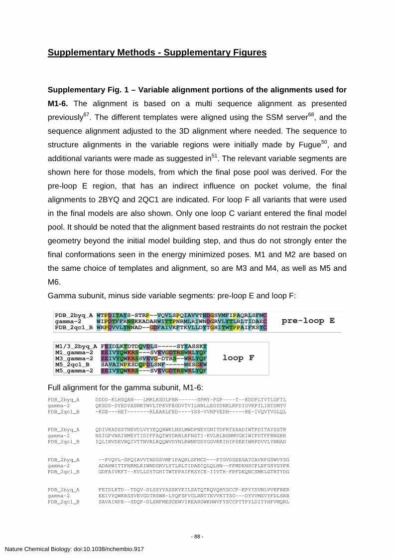
Supplementary Methods - Supplementary Figures
Supplementary Fig. 1 – Variable alignment portions of the alignments used for M1-6. The alignment is based on a multi sequence alignment as presented
previously67. The different templates were aligned using the SSM server68, and the
sequence alignment adjusted to the 3D alignment where needed. The sequence to
structure alignments in the variable regions were initially made by Fugue50, and
additional variants were made as suggested in51. The relevant variable segments are
shown here for those models, from which the final pose pool was derived. For the
pre-loop E region, that has an indirect influence on pocket volume, the final
alignments to 2BYQ and 2QC1 are indicated. For loop F all variants that were used
in the final models are also shown. Only one loop C variant entered the final model
pool. It should be noted that the alignment based restraints do not restrain the pocket
geometry beyond the initial model building step, and thus do not strongly enter the
final conformations seen in the energy minimized poses. M1 and M2 are based on
the same choice of templates and alignment, so are M3 and M4, as well as M5 and
M6.
Gamma subunit, minus side variable segments: pre-loop E and loop F:
Full alignment for the gamma subunit, M1-6: PDB_2byq_A DDDD-KLHSQAN---LMRLKSDLFNR------SPMY-PGP----T--KDDPLTVTLGFTL gamma-2 QKSDD-DYEDYASNKTWVLTPKVPEGDVTVILNNLLEGYDNKLRPDIGVKPTLIHTDMYV PDB_2qc1_B -KSE---HET-------RLEAKLFED----YSS-VVRPVEDH-----RE-IVQVTVGLQL PDB_2byq_A QDIVKADSSTNEVDLVYYEQQRWKLNSLMWDPNEYGNITDFRTSAADIWTPDITAYSSTR gamma-2 NSIGPVNAINMEYTIDIFFAQTWYDRRLKFNSTI-KVLRLNSNMVGKIWIPDTFFRNSKK PDB_2qc1_B IQLINVDEVNQIVTTNVRLKQQWVDYNLKWNPDDYGGVKKIHIPSEKIWRPDVVLYNNAD PDB_2byq_A --PVQVL-SPQIAVVTHDGSVMFIPAQRLSFMCD---PTGVDSEEGATCAVKFGSWVYSG gamma-2 ADAHWITTPNRMLRIWNDGRVLYTLRLTIDAECQLQLHN--FPMDEHSCPLEFSSYGYPR PDB_2qc1_B GDFAIVKFT--KVLLDYTGHITWTPPAIFKSYCE-IIVTH-FPFDEQNCSMKLGTRTYDG PDB_2byq_A FEIDLKTD--TDQV-DLSSYYASSKYEILSATQTRQVQHYSCCP-EPYIDVNLVVKFRER gamma-2 EEIVYQWKRSSVEVGDTRSWR-LYQFSFVGLRNTTEVVKTTSG---DYVVMSVYFDLSRR PDB_2qc1_B SAVAINPE--SDQP-DLSNFMESGEWVIKEARGWKHWVFYSCCPTTPYLDITYHFVMQRL
Nature Chemical Biology: doi:10.1038/nchembio.917
- 88 -

Alpha subunit, plus side variable segment: loop C:
Full alignment for the alpha subunit, M1-6: PDB_2byq_A ------DDDD--KLHSQANLMRLKSDLFNR-SPMY-PGPT--KDDPLTVTLGFTLQDIVK PDB_1uw6_A ----EFDRAD--ILYNIRQ--TSRPDVIP----------TQRDR-PVAVSVSLKFINILE alpha-1 QPSQ-DELKDNTTVFTR-----ILDRLLDGYDNRLR---PGLGERVTEVKTDIFVTSFGP PDB_2byq_A ADSSTNEVDLVYYEQQRWKLNSLMWDPNEYGNITDFRTSAADIWTPDITAYSSTR--PVQ PDB_1uw6_A VNEITNEVDVVFWQQTTWSDRTLAWNSS--HSPDQVSVPISSLWVPDLAAYNAIS--KPE alpha-1 VSDHDMEYTIDVFFRQSWKDERLKFKGPM-TVLRLNNLMASKIWTPDTFFHNGKKSVAHN PDB_2byq_A VL-SPQIAVVTHDGSVMFIPAQRLSFMCDPTG--VDSEEGATCAVKFGSWVYSGFEIDLK PDB_1uw6_A VL-TPQLARVVSDGEVLYMPSIRQRFSCDVSG--VDTESGATCRIKIGSWTHHSREISVD alpha-1 MTMPNKLLRITEDGTLLYTMRLTVRAECPMHLED-FPMDAHACPLKFGSYAYTRAEVVYE PDB_2byq_A TD---TDQV--DLSSYYASSKYEILSATQTRQVQHYSCCPEPYIDVNLVVKFRERR-- PDB_1uw6_A PT---T-ENSDDSEYFSQYSRFEILDVTQKKNSVTYSCCPEAYEDVEVSLNFRKKGRS alpha-1 WTREPARSVVVAE-DGSRLNQYDLLGQTVDSGIVQSS-TG-EYVVMTTHFHLKRKIG-
Below is a more up to date full alignment. The alignments used in this study can be
derived by simply repositioning the gaps in the variable segments as indicated in the
figure above. The full alignment is based on structural 3D alignment of the newest
crystal structure59 (PDB_3rif_A) with the templates used for this study, and the
GABAA receptor subunits then aligned against the structural alignment data. Future
modeling studies should consider the 3RIF and related structures when modeling
GABAA receptors.
CLUSTAL X (1.83) multiple sequence alignment PDB_2byq_A -DDDD-KLHSQAN-----LMR---------LKSDLFNRSPMY--PGPTK---D-D--PLT PDB_1uw6_A ----------EFD-----RAD---------ILYNIRQ--TSRPDVIPTQ---R-DR-PVA PDB_2qc1_B ---------KSEH-----ETR---------LEAKLFED-Y-SSVVRPVE---DHRE-IVQ PDB_3rif_A ------------S-----DSK---------ILAHLFTSGY-DFRVRPPTD--N-GG-PVV 1uw6_A ----------EFD-----RAD---------ILYNIRQ--TSRPDVIP----TQRDR-PVA 2byq_A -DDDD-KLHSQAN-----LMR---------LKSDLFNR-SPMY-PGPT------KDDPLT alpha-1 -QPSQDELKDNTT-----VFTR--------ILDRLLDG--YDNRLR-----PGLGERVTE minu2byq_A -DDDD-KLHSQAN-----LMR---------LKSDLFNR-SPMY-PGP----T--KDDPLT minu2qc1_B ---------KSE------HET--------RLEAKLFED-YSS-VVRPVEDH---RE-IVQ gamma-2 QKSDD-DYEDYASNKTWVLTPKVPEGDVTVILNNLLEG--YDNKLR-----PDIGVKPTL : : PDB_2byq_A VTLGFTLQDIVKADSSTNEVDLVYYEQQRWKLNSLMWD--PNEYGNITDFRTS---AADI PDB_1uw6_A VSVSLKFINILEVNEITNEVDVVFWQQTTWSDRTLAWN--SS--HSPDQVSVP---ISSL PDB_2qc1_B VTVGLQLIQLINVDEVNQIVTTNVRLKQQWVDYNLKWN--PDDYGGVKKIHIP---SEKI PDB_3rif_A VSVNMLLRTISKIDVVNMEYSAQLTLRESWIDKRLSYGVKGD--GQPDFVILTVGHQ--I 1uw6_A VSVSLKFINILEVNEITNEVDVVFWQQTTWSDRTLAWN--SS--HSPDQVSVP---ISSL 2byq_A VTLGFTLQDIVKADSSTNEVDLVYYEQQRWKLNSLMWD--PNEYGNITDFRTS---AADI alpha-1 VKTDIFVTSFGPVSDHDMEYTIDVFFRQSWKDERLKFKGPM-TVLRLNNLMAS-----KI minu2byq_A VTLGFTLQDIVKADSSTNEVDLVYYEQQRWKLNSLMWD--PNEYGNITDFRTS---AADI minu2qc1_B VTVGLQLIQLINVDEVNQIVTTNVRLKQQWVDYNLKWN--PDDYGGVKKIHIP---SEKI gamma-2 IHTDMYVNSIGPVNAINMEYTIDIFFAQTWYDRRLKFNSTI-KVLRLNSNMVG-----KI
Nature Chemical Biology: doi:10.1038/nchembio.917
- 89 -

: .: . : . * * : : PDB_2byq_A WTPDITAYSSTR--PVQVL-----SPQIAVVTHDGSVMFIPAQRLSFMCDPT------GV PDB_1uw6_A WVPDLAAYNAIS--KPEVL-----TPQLARVVSDGEVLYMPSIRQRFSCDVS------GV PDB_2qc1_B WRPDVVLYNNADGDFAIVK-----F-TKVLLDYTGHITWTPPAIFKSYCEIIVTHFPFD- PDB_3rif_A WMPDTFFPNEKQ--A-YKHTIDKPN-VLIRIHNDGTVLYSVRISLVLSCPMYLQYYPMD- 1uw6_A WVPDLAAYNAIS--KPEVL-----TPQLARVVSDGEVLYMPSIRQRFSCDVSG------V 2byq_A WTPDITAYSSTR--PVQVL-----SPQIAVVTHDGSVMFIPAQRLSFMCDPTG------V alpha-1 WTPDTFFHNGKKSVAHNMT----MPNKLLRITEDGTLLYTMRLTVRAECPMHLED----- minu2byq_A WTPDITAYSSTR--PVQVL-----SPQIAVVTHDGSVMFIPAQRLSFMCDPTG------V minu2qc1_B WRPDVVLYNNADGDFAIVKFT------KVLLDYTGHITWTPPAIFKSYCEIIVTH----- gamma-2 WIPDTFFRNSKKADAHWIT----TPNRMLRIWNDGRVLYTLRLTIDAECQLQLHN----- * ** . : * : : * PDB_2byq_A DSEEGATCAVKFGSWVYSGFEIDLKTD---TDQ--VDLSSYY-ASSK--YEILSATQTR- PDB_1uw6_A DTESGATCRIKIGSWTHHSREISVDPT---TENS-DDSEYFS-QYSR--FEILDVTQKK- PDB_2qc1_B ----EQNCSMKLGTRTYDGSAVAINPE---SDQ--PDLSNFM-ESGE--WVIKEARGWK- PDB_3rif_A ----VQQCSIDLASYAYTTKDIEYLWK---EHSP-LQL-KVGLSSSLPSFQLTNTSTTYC 1uw6_A DTESGATCRIKIGSWTHHSREISVDPT--T-ENS-DDSEYFS-QYSR--FEILDVTQKK- 2byq_A DSEEGATCAVKFGSWVYSGFEIDLKTD---TDQV--DLSSYY-ASSK--YEILSATQTR- alpha-1 FPMDAHACPLKFGSYAYTRAEVVYEWT---REPARSVVVAE-DGSRLNQYDLLGQTVDS- minu2byq_A DSEEGATCAVKFGSWVYSGFEIDLKTD---TDQV--DLSSYY-ASSK--YEILSATQTR- minu2qc1_B FPFDEQNCSMKLGTRTYDGSAVAINPE---SDQP--DLSNFM-ESGE--WVIKEARGWK- gamma-2 FPMDEHSCPLEFSSYGYPREEIVYQWKRS------SVEVG-DTRSWR-LYQFSFVGLRNT * :.:.: : : : : PDB_2byq_A QVQHYSCC-P-EPYIDVNLVVKFRER-R-------------------------------- PDB_1uw6_A NSVTYSCC-P-EAYEDVEVSLNFRKK-GRS------------------------------ PDB_2qc1_B HWVFYSCCPT-TPYLDITYHFVMQRL-P-------------------------------- PDB_3rif_A T-SVTNT----GIYSCLRTTIQLKREFSFYLLQLYIPSCMLVIVSWVSFWFDRTAIPARV 1uw6_A NSVTYSCC-P-EAYEDVEVSLNFRKK-GRS------------------------------ 2byq_A QVQHYSCC-P-EPYIDVNLVVKFRER-R-------------------------------- alpha-1 -GIVQSS-TG--EYVVMTTHFHLKRKIG-------------------------------- minu2byq_A QVQHYS-CCP-EPYIDVNLVVKFRER-R-------------------------------- minu2qc1_B HWVFYS-CCPTTPYLDITYHFVMQRLP--------------------------------- gamma-2 TEVVKTT-SG--DYVVMSVYFDLSRRMG-------------------------------- . * : . : .
Nature Chemical Biology: doi:10.1038/nchembio.917
- 90 -

Supplementary Fig. 2 - Depiction of the two possible 1,4-diazepine conformations in 5-aryl-1,4-benzodiazepines. The two conformations can be
distinguished by the dihedral angle of the atoms C2-C3-N4-C5. Conformation M
(Minus) has a negative dihedral angle while conformation P (Positive) has a positive
angle. A plethora of evidence summarized in Supplementary Table 2 showed that
conformation M of the 7-ring is present in the high affinity bound-state complex of
1,4-benzodiazepines in the benzodiazepine binding site of GABAA receptors.
*Natta projection was used for illustrative issues and does not indicate stereochemistry. Solid or dashed wedged bonds represent bonds pointing above-the-plane or below-the-plane of the fused benzene ring of 1,4-benzodiazepines, respectively
conformation P conformation M
conformation M conformationP
*
Nature Chemical Biology: doi:10.1038/nchembio.917
- 91 -

Supplementary Methods - Supplementary Text
Generation of the validation library (Bz-focused decoy set). The selection of
decoys was accomplished with an automated decoy generator (M.M.Mysinger and
John J. Irwin, unpublished results) in an analogous manner to the DUD52. First, 41
benzodiazepine binding site ligands (Supplementary Table 10) were seeded into
the ZINC database53. Then ZINC compounds with Tanimoto coefficient (Tc) less
than 0.5 (Daylight fingerprint) to any of the 41 ligands were selected, thus resulting in
a subset of ZINC compounds that are topologically dissimilar to the ligands.
Next, for each of the 41 seed compounds, all protonation states near pH 7.0 were
generated, resulting in 48 unique ligand protomers. For each of them, 50 decoy
molecules that property-match with the ligand protomer were selected from the
subset of the ZINC library. The following six physicochemical descriptors were used
for property-matching: molecular weight, number of hydrogen bond acceptors,
number of hydrogen bond donors, number of rotatable bonds, logP, and the net
formal charge69.
Thus for all 41 benzodiazepine binding site ligands, compromising 48 unique ligand
protomers, 2400 (48x50) decoys that are physically similar but topologically distinct
to the ligands, were generated. To generate finally the validation library, the 48
ligand protomers were added to the 2400 decoys (Supplementary Scheme 4).
Structure-based pharmacophore models. Pharmacophore models were created
using LigandScout 3.070. Feature specific feature ranges were 2.2 – 4.2 Angstrom for
H Bond Donor and H Bond Acceptor, respectively. Detected pharmacophoric
features which are also represented in Fig. 1c were set to obligatory features.
Exclusion volumes coats were added, using an Alpha C distance of 2-7 A and an
Alpha C exclusion tolerance of 1.75 A. In case a hydrophobic and an aromatic
feature were detected, only the more specific (aromatic) was kept. For the remaining
parameters default values were used.
Nature Chemical Biology: doi:10.1038/nchembio.917
- 92 -

Ligand-based pharmacophore models. The structure of diazepam was taken and
the following features were assigned using LigandScout 3.070: hydrophobic to the
fused benzene ring (L1), hydrophobic to the pendant phenyl ring (L3), H-bond
acceptor to the carbonyl oxygen atom (H1), and H-bond acceptor to the imino
nitrogen atom (H2) (see also Fig. 1c, main text and below). For flumazenil, the
pharmacophore feature assignment was as follows: hydrophobic to the fused
benzene ring (L1), H-bond acceptor to the imidazo-nitrogen (H1) and H-bond
acceptor to the carbonyl-oxygen (H2) (see also Fig. 1c, main text and below).
Depiction of assigned features (green = hydrophobic, red = H-bond acceptor) to the
structures of diazepam (left) and flumazenil (right), respectively.
Nature Chemical Biology: doi:10.1038/nchembio.917
- 93 -

Compound characterization data
Characterization data for [3H]-Flunitrazepam:
[3H]-Flunitrazepam was purchased from PerkinElmer with a purity of greater than 97% as described by the vendor.
Characterization data for cpd. 8 (Ro15-8670):
High field 1H NMR was performed for cpd. 8 (see p. 19), demonstrating high purity (>98%) and identity of cpd. 8. HRMS C20H16ClN3O2: [M+1]calc: 366.8189 [M+1]found: 366.8191.
Characterization data for cpds. 15, 15a-c (active compounds):
Compounds 15 and 15a-c were ordered from Ambinter. High field 1H NMR and 13C NMR were performed for compounds 15 and 15a-c (see p. 20-27), demonstrating high purity (>98%) and identity of the active compounds.
Characterization data for cpds. 15d-i, 16-18 (inactive compounds):
Compounds 15d-i and 18 were ordered from Ambinter, compounds 16 and 17 from Asinex. The inactive compounds 15d-i and 16-18 had a purity of greater than 90% as described by the vendors.
Characterization data for cpds. 19 and 20 (active compounds):
High field 1H NMR and 13C NMR was performed for cpd. 19 (p. 28 and 29). HRMS C14H14N3: [M+1]calc: 224.1187 [M+1]found: 224.1153. The purity of this compound was then verified in LCMS (see p. 30) analysis.
High field 1H NMR and 13C NMR was performed for cpd. 20 (p. 31 and 32). HRMS C14H15N2OS: [M+1]calc: 259.0905 [M+1]found: 259.0866. The purity of this compound was then verified in LCMS (see p. 33) analysis.
Nature Chemical Biology: doi:10.1038/nchembio.917
- 94 -

Nature
Chem
icalBiology:doi:10.1038/nchem
bio.917
- 95 -

Nature
Chem
icalBiology:doi:10.1038/nchem
bio.917
- 96 -

Nature
Chem
icalBiology:doi:10.1038/nchem
bio.917
- 97 -

Nature
Chem
icalBiology:doi:10.1038/nchem
bio.917
- 98 -

Nature
Chem
icalBiology:doi:10.1038/nchem
bio.917
- 99 -

Nature
Chem
icalBiology:doi:10.1038/nchem
bio.917
- 100 -

Nature
Chem
icalBiology:doi:10.1038/nchem
bio.917
- 101 -

Nature
Chem
icalBiology:doi:10.1038/nchem
bio.917
- 102 -

Nature
Chem
icalBiology:doi:10.1038/nchem
bio.917
- 103 -

Nature
Chem
icalBiology:doi:10.1038/nchem
bio.917
- 104 -

Nature
Chemememememememememememem
icicicicicicicicicicicicalBi
Bi
Bi
Bi
Bi
Bi
Bi
Bi
Bi
Bi
Bi
Biolololololololololololologoooooooooooy:dodododododododododododoi:i:i:i:i:i:i:i:i:i:i:10.1.10303030303030303030303038888/8888888nchehehehehehehehehehehehem
bmmmmmmmmmmm
io.9.9.9.9.9.9.9.9.9.9.9171717171717171717171717
- 105 -

Nature Chemical Biology: doi:10.1038/nchembio.917
- 106 -

Nature
Chem
icalBiology:doi:10.1038/nchem
bio.917
- 107 -

Nature
Ch
Ch
Ch
Ch
Ch
Ch
Ch
Ch
Ch
Chh
Chemememememememememememem
icalBi
Bi
Bi
Bi
Bi
Bi
Bi
Bi
Bi
Bi
Bi
Biolololololololololololology:dodododododododododododoi:10.1.1.1.1.1.1.1.1.1.1.1.10303030303030303030303038/ncncncncncncncncncncnchehehehehehehehehehehehem
bioioioioioioioioioio.9.9.9.9.9.9.9.9.9.9.9.9171717171717171717171717
- 108 -

Nature Chemical Biology: doi:10.1038/nchembio.917
- 109 -

Supplementary Results - Supplementary Tables
Supplementary Table 3 – modeling parameters
Main template(s)
Screening stagesa
Templates for the individual subunits and the interface geometryb Varied
parameters in im f fv alpha gamma interface
1I9B 4 3 1I9B 1I9B 1I9B Alignmentc
2BYN 2 1 2BYN 2BYN 2BYN Alignmentc
2BYS 3 2 2BYS 2BYS 2BYS Alignmentc
2BYQ 3 2 2d
2e 2BYQ
2BYQ/1UW6 2BYQ 2BYQ
2BYQ 2BYQ
Alignmentc
Loop Cf
2BG9 1 2BG9_C 2BG9_B 2BG9_CB
2BYQ/2BG9 5 4 2BYQ 2BYQ 2BG9_CB Alignmentc
2BYQ/2BG9 5 2BYQ 2BYQ 2BG9_BA Alignmentc
2BYQ/2QC1 1 1 1g
1h 2BYQ
2BYQ/1UW6 2QC1 2QC1
2BYQ 2BYQ
Loop Cf
2BYN/2BG9 1 2BYN 2BYN 2BG9_CB
2BYR/2BG9 5 1 2BYR 2BYR 2BG9_CB Alignmentc
2BYR/2BG9 4 2BYR 2BYR 2BG9_BA Alignmentc a From each set of main template(s), one or more model variants were derived by varying additional modeling parameters. Models used in the initial screen are denoted “in” (rigid docking of diazepam with FlexX71), “im” those that were retained for the intermediate screening (flexible docking of cpds. 1,3,4,5,8,9 with FlexE72, see text), “f” are the final models of the original set, and fv are the variants of the final models that were obtained by enhanced rotamer exploration and additional conformational sampling thus increasing the sampling density of the most promising pocket geometries (see also Supplementary Scheme 2). b Subunit templates were 3D-aligned with the indicated template for the interface geometry, in order to sample the relative orientation of the two subunits. This was necessary for including the monomeric structure 2QC1 as template, and for investigating the interface geometries seen in the 2BG9 heteropentameric structure. The chain IDs used for the interface are indicated as 2BG9_C, etc. c Alignment variants in the unconserved segments that contain “loops” C, E and F were constructed as recommended51 (see Supplementary Fig. 1) with standard procedures49,50,67,73 to generate a wide variety of initial geometries. d corresponds to models M1 and M3 in Supplementary Table 8. ecorresponds to models M2 and M4 in Supplementary Table 8. f In order to improve the sampling of the flexible C-loop tip, the alpha subunit was also modeled by local multi-template restraints based on two templates51. g corresponds to model M5 in Supplementary Table 8. h corresponds to model M6 in Supplementary Table 8.
Nature Chemical Biology: doi:10.1038/nchembio.917
- 110 -

Models were constructed from a broad variety of templates and parameters to
identify those best suited for the bound state complex of diazepam and analogues
(see also Supplementary Schemes 1 and 2). Some models were eliminated prior
to docking due to poor stereochemistry and are not included in the table. The full
breadth of structural variety in the pocket was then investigated with the initial model
pool, (in). Based on exploratory docking, models that were largely incapable of
providing promising poses of diazepam with sufficient hydrophobic interaction with
L1 and L3 and pocket forming residues were eliminated. To those that remained,
docking was extended to multiple ligands and parameters were added to consider
conformational and side chain flexibility (intermediate pool, im). Upon evaluation of
the intermediate pool using criteria as mentioned above for each of the investigated
ligands, three best performing final (f) models were selected. From those three
models, additional final variants (fv) were built to increase the sampling density of
the models of interest. This resulted in a total of six models providing the final pool of
docking poses, the three best performing of the intermediate pool, and one variant of
each of those. In the variants, a more extensive rotamer exploration at the subunit
interface (see Methods, main text) and a more detailed exploration of loop C
conformations were performed as indicated by RMSD analysis and visual inspection
of “empty” patches in the sampling of pocket geometries.
Nature Chemical Biology: doi:10.1038/nchembio.917
- 111 -

Supplementary Table 4 – Model variability in pocket forming segments total final compared to 3RIF-model 37 models 6 models 37 models 6 models
Segment ave.(max) ave. (max) ave.(max) ave. (max) A-Loop (α1F99 – α1N102) 1.9 (3.6) 0.3 (0.4) 1.5 (2.1) 1.3 (1.7) B-Loop (α1Y159 – α1Y161) 1.4 (2.5) 0.3 (0.5) 0.9 (3.0) 0.5 (0.8) C-Loop (α1V202 – α1V211) 3.1 (5.4) 0.5 (0.6) 2.6 (4.9) 1.1 (1.2) G-Loop (γ2D56 – γ2N60) 1.9 (3.8) 0.7 (1.0) 2.0 (3.5) 0.7 (0.7) D-Loop (γ2F77 – γ2A79) 1.6 (3.4) 0.6 (0.8) 1.5 (2.9) 0.8 (0.8) E-Loop (γ2M130 –γ2 I133 &γ2L140–γ2T142) 1.5 (2.5) 0.5 (0.7) 1.8 (2.3)
1.8 (1.8)
F-Loop (γ2185 - γ2194) 4.3 (11.9) 4.2 (6.1) 4.9 (8.8) 4.3 (6.1) Loops (A-E & G) 2.2 (3.3) 0.5 (0.8) 2.1 (3.3) 1.2 (1.3) Loops (A-F & G) 2.8 (6.0) 2.1 (3.0) Average and maximum backbone RMSD values of pocket forming segments. The
entire model pool has much higher variability than the final pool, which has
converged to optimally accommodate the ligands. Loop F is not engaged in any
obvious interactions with the ligands investigated, and thus remains variable. Since
loop F suffers from template uncertainty and undefined alignments, total pocket
RMSD is computed with and without loop F contribution. The value without loop F
contribution is a good measure for the variability obtained in the structurally
conserved pocket portions. The loop C value is a measure for the observed flexibility
(Supplementary Fig. 3).The total maximal RMSD for the final models is a measure
for combined overall protein variability that cannot be reduced with the workflow
used here. It contains uncertainty, possibly induced fit contributions and lack of
information to narrow down further. After submission of this work, a new structure
(PDB ID 3RIF) of a glutamate gated Cl- channel, which shares ~30% PID with
GABAA receptor subunits, was released. The “hindsight” validation against the
models based on the new 3RIF glutamate bound structure (last columns) shows that
our final models are more similar to this new structure than to some of the initial
models.
Thus, our workflow successfully identified models, that are “better” in the light of this
new X-ray structure over alternative models, that were equally valid in the light of the
older structural data.
Nature Chemical Biology: doi:10.1038/nchembio.917
- 112 -

Supplementary Table 5 – Compound information type cpd. Name affinity
(α1β2γ2) nM modulation
corea 1 Diazepam74 12 positive modulator core 2 Ro07-522074 2 (not known) core 3 Flunitrazepam74 3 positive modulator core 4 Flurazepam75 62 positive modulator core 5 Ro 7-1986/176 --b (not known) accessoryc 6 8,9-benzofused77 55 positive modulator accessory 7 7,8-benzofused77 120 (not known) accessory 8 Ro15-867074 34 positive modulator accessory 9 CNS705678 29 positive modulator not used for dockingd
10 7-NCS79 3170 positive modulator
docked only in f and fv modelse
11 Flumazenil74 0.61 null modulator
not used for dockingd
12 Ro15-451380 3.9 negative modulator
not used for dockingd
13 „Imid compound“81 10 null modulator
not used for dockingd
14 3-NCS82 340 --f
a core ligands were those for which a more strict definition of common binding mode was used: At the level of clustering by ligand RMSD, all core ligands were mandatory for a cluster of poses to be considered CBM-parent candidate. At the level of clustering with bound complex RMSD a tighter overlap (2 Å) was requested of those. b This compound is coupled to chromatography beads as affinity bait. c accessory ligands were those that were not considered mandatory in the first cluster analysis, and permitted larger bound complex RMSD in the second analysis (2.5 Å). If a CBM contained them, they were also used for CBM evaluation by experimental evidence, see Table 1 in the main text. d those ligands were aligned by the common atoms’ coordinates with docked ligands to evaluate the resulting position in the light of experimental evidence, see Table 1 in the main text. eFlumazenil was only docked into the final models (f) of the original set, and into the fv, the variants of the final models (see Supplementary Table 3) fdata for wildtype α1β2γ2 receptor not shown, positive allosteric modulator in α1S205Cβ2γ2 and α1T206Cβ2γ2.
Core ligands: In the workflow shown in Supplementary Scheme 3, only those
clusters were retained that contained all core ligands. Core ligands are assumed to
share common binding and interaction patterns due to similar structure and
biological profile Accessory ligands: were used additionally and less tight similarity was required in
the Supplementary Scheme 3 workflow to further restrict the possible binding
geometries by bulkier ligands.
Nature Chemical Biology: doi:10.1038/nchembio.917
- 113 -

Supplementary Table 6 – bound complex defining and major interaction forming residues alpha1 gamma2 Parent clusteraI
H101, Y159, S204, T206, Y209 Y58, N60, F77, T142
Parent cluster II
H101, S158, Y159, S204, S205, T206, Y209 Y58, N60, F77, T142
Parent cluster III
F99, H101, K155, G157, Y159, V202, Q203, S204, T206, Y209, V211
Y58, F77, T142
L1 L3 H1 H1’ H2 L1 L3 H1 CBM I Y159
100% Y209 100%
H101 66% γ2 T206 S204 α1 F77 100% Y58 19%
T142
CBM II γ2 Y159 100% Y209 100%
- S205 T206 Y58 100% F77 100%
α1 -
CBM III Y209 94%
F99 88% H101 48% Y159 74% Y209 100%
H101 - H101 Y58 59% F77 100%
α1 -
a Parent cluster was selected by ”ligands common scaffold RMSD” clustering (see Supplementary Scheme 3). For each putative common binding geometry (parent cluster), that was identified
based on the clustering by ligand overlap RMSD (“ligands common scaffold RMSD”),
the major bound complex forming atoms were determined. The upper three lines list
those amino acid residues that were selected, based on average distance to the
ligands’ common scaffold in the respective cluster, to compute the cluster’s individual
“bound complex RMSD” values. Many, but not all of those residues are found in the
final CBM poses as major interaction partners. The last three lines list the residues
that were subsequently found to provide major interactions in the final CBM poses,
for the lipophilic L interactions the percentage of core poses featuring the respective
interaction is also indicated. This shows the heterogeneity that is present in the
individual CBM pose pools. Please note: Our structural hypotheses do feature well defined geometries and interaction pattern, but also some residual scatter. Thus, no single pose can be considered “best” or “correct”, the final poses define a conformational space in which the “true structure” is assumed to be contained.
Nature Chemical Biology: doi:10.1038/nchembio.917
- 114 -

Supplementary Table 8 – distribution of poses in the final models Model identifiera model: total M1 M2 M3 M4 M5 M6 rawposepool 4997 886 827 843 790 856 795 L1 & L3pool 1463 114 239 237 389 236 248 Parent cluster I 146 4 23 5 48 12 54 Parent cluster II 96 8 4 8 24 48 4 Parent cluster III 93 20 14 13 3 12 31 CBM I 96 0b 23 0 28 0 45 CBM II 49 1 0 2 1 44 1 CBM III 39 4 7 2 0 4 22 aSeeSupplementary Table 3 and Supplementary Fig. 1 for the modeling parameters used in these six models. b It is worth noting that the final binding mode CBM I occurs only in the models M2, M4 and M6 which were obtained by extending the rotamer flexibility (see Methods), and were thus able to assume an energetically more favorable loop B conformation. This table thus illustrates that a binding mode can be missed by insufficient sampling of model flexibility. The Supplementary Tables 7 and 8 illustrate the effects of the different work steps
on the poses obtained with a given ligand (Supplementary Table 7) or a given
model (Supplementary Table 8). See also Supplementary Scheme 3.
Supplementary Table 7 – poses for each compound tracked through the workflow from pose pool to CBM Compound number total 1 2 3 4 5 6 7 8 9 rawposepoola 4997 600 444 600 537 588 595 510 538 585 L1 & L3poolb 1463 257 156 183 210 156 181 164 123 33 Parent cluster I 146 34 13 21 11 9 21 18 16 3 Parent cluster II 96 20 12 9 17 22 4 5 4 3 Parent cluster III 93 15 4 11 13 13 9 13 15 0 CBM I 96 21 5 16 11 9 14 6 12 2 CBM II 49 5 1 2 11 22 3 1 3 1 CBM III 39 4 2 6 6 13 5 1 2 0 a The raw pose pool refers to the number of poses obtained with FlexE flexible docking and retaining up to 100 poses. b L1& L3 pool denotes the pool of models that satisfies the lipophilic saturation requirements.
Nature Chemical Biology: doi:10.1038/nchembio.917
- 115 -

Supplementary Table 9 – Characterization and computational evaluation of CBM candidates ligandsa CBM I CBM II CBM III
Homogeneity of poses number of core poses 1-5 62 41 31 average distance to centroid Å
1-5 1.2
1.24
1.45
number of acc. ligands 6-9 all all 3/4 number of acc. poses 6-9 34 8 8 average distance to centroid Å 6-9 1.55 1.81 1.14
Major interactionsb (see Fig. 2) L1 – hydrophobic feature 1-5 α1Y159,α1Y209 γ2Y58,γ2F77 α1Y209,γ2Y58,
L3 – hydrophobic feature 1-5 α1H101,γ2F77, γ2Y58
α1Y159,α1Y209 α1F99,α1H101, α1Y159,α1Y209
H1 – H-bond donor feature 1-5 γ2T142 none α1H101 H1’ – H-bond donor feature 1-5 α1T206 α1S205 none H2 – H-bond donor feature 1-5 α1S204 α1T206 α1H101 Poses exhibiting side chain positions conserved in superfamily (see Supplementary Fig. 8) Conserved α1Y159 1-5 all ++ none - none - Conserved α1Y209 1-5 all ++ 28/41c + 28/31c ++ Scoring performance Top10 X-Score83 1-5 13 ++ none - 1 - Top10 London dG84 1-5 13 ++ none - 1 -
Enrichment factors85 in Retrospective virtual screening (Number of actives in database fraction) (see Supplementary Scheme 4)
EF 0.5% 24.8 (5) ++ 14.9 (3) + 9.9 (2) + EF 1.0% 14.9 (6) ++ 7.4 (3) + 5.0 (2) + EF 2.0% 12.2 (10) ++ 3.7 (3) - 2.5 (2) - EF 5.0% 6.9 (14) ++ 3.0 (6) - 2.0 (4) - EF 10.0% 4.5 (18) ++ 1.7 (7) - 1.7 (7) - a only poses of the defined ligand(s) (Fig. 1c) are used for evaluation. b interactions were calculated using SCORING.SVL (http://svl.chemcomp.com). c number of poses fulfilling the respective criterion in relation to number of total poses in a CBM (see Supplementary Table 7 for total number of poses per ligand within a CBM).
The slightly smaller average distance to centroid for CBM III results from the fact that this binding mode accommodates only three of the four accessory ligands.
Nature Chemical Biology: doi:10.1038/nchembio.917
- 116 -

Supplementary Table 10 – 41 known binders used in enrichment computation cpd. 2D structure annotation in
reference
ScreeningCBMIa
affinity (nM)
11
Flumazenil86 Top 0.5%
0.8
1
Diazepam86 Top 0.5% 14
21
U-9777587 Top 0.5% 1.2
22
Clobazam88 Top 0.5% 170
23
Triazolam86 Top 0.5% 0.8
24
2b89 Top 1% 0.55
25
MRK-01690 - 0.92
26
Panadiplon91 - 1.56
27
3892 Top 10% 2.0
28
Eszopiclone93 Top 5% 50.1
29
1094 Top 5% 0.1
Nature Chemical Biology: doi:10.1038/nchembio.917
- 117 -

cpd. 2D structure annotation in reference
ScreeningCBMIa
affinity (nM)
30
7195 Top 2% 0.14
31
1k96 Top 2% 13
32
RWJ-5120497 - 0.37
33
2b98 - 2.36
34
U-8557587 Top 2% 0.3
35
4aR99 Top 10% 7.5
36
1a100 Top 10% 21
37
23101 - 0.26
38
4k102 - 0.5
39
CL218,87286 - 57
Nature Chemical Biology: doi:10.1038/nchembio.917
- 118 -

cpd. 2D structure annotation in reference
ScreeningCBMIa
affinity (nM)
40
Pyridodiindole86 - 1.1
41
CGS-821686 Top 10% 0.05
42
2i103 Top 2% 3.9
43
45104 - 2.8
44
41105 - 0.9
45
1e106 - 5.4
46
DU43028107 - 0.07
47
Ocinaplon108 Top 5% 1200
48
RU32698109 - 114
Nature Chemical Biology: doi:10.1038/nchembio.917
- 119 -

cpd. 2D structure annotation in reference
ScreeningCBMIa
affinity (nM)
49
74110 - 5.5
50
21111 - 2.3
51
Zolpidem86 - 156
52
35112 Top 5% 0.51
53
14113 - 4.3
54
SL-651498114 - 6.8
55
2b115 - 0.2
56
GBLD-345116 - 0.9
Nature Chemical Biology: doi:10.1038/nchembio.917
- 120 -

cpd. 2D structure annotation in reference
ScreeningCBMIa
affinity (nM)
57
Ro19-8022117 - 0.38
58
TPA023118 - 0.92
59
Pazinaclone119 - 0.7
aRanking of benzodiazepine binding site ligands in the top 0.5%, 1%, 2%, 5% or 10% of the score-sorted validation library. Rankings derive from virtual screening of the validation library (see Supplementary Methods) against the CBM I derived pharmacophore models (Supplementary Fig. 10).
These 41 known benzodiazepine binding site ligands from different chemical classes
were extracted from the literature. These ligands together with property-matched
decoys (validation library, Supplementary Methods) were used in virtual screening
against structure-based pharmacophore models of CBM I-III (Supplementary Scheme 4). For CBM I, data indicate that flumazenil, diazepam and benzodiazepine
binding site ligands from 8 additional compound classes were ranked within the top
2% of the score-sorted validation library by the structure based pharmacophores.
The enrichment obtained (see also Supplementary Table 11) is comparable to the
average enrichment obtained by docking of the DUD database into the 40 crystal
structures52.
Nature Chemical Biology: doi:10.1038/nchembio.917
- 121 -

Supplementary Table 11 - Retrospective virtual screening results of the validation library (Bz-focused decoy set) using different methodologies
EFa (%)
pharmacophore models 2D fingerprint (FP) search shape-similarity searchf structure-
basedb (CBM I)
ligand-basedc
pharmacophore FP
(GpiDAPH3)d
substructural FP
(MACCS)e 0.5% 24.8 (24.4) 14.9 (14.6) 24.9 (29.3) 29.8 (29.3) 19.8 (19.5) 1% 14.9 (17.1) 7.5 (7.5) 17.4 (17.1) 14.9 (14.6) 12.4 (12.2) 2% 12.2 (12.2) 5.0 (5.0) 11.2 (12.2) 8.5 (8.5) 6.1 (6.1) 5% 6.9 (6.8) 2.0 (2.0) 6.5 (6.8) 4.0 (3.9) 2.5 (2.4)
10% 4.5 (4.4) 1.7 (1.7) 4.0 (3.9) 3.0 (2.9) 2.2 (2.2) aEnrichment factors were calculated as described in Wei et al.85. Values in brackets represent enrichment factors calculated according to guideline of Nicholls et al.120. bStructure-based pharmacophore models were created using LigandScout 3.070 (see Supplementary Methods) based on CBM I diazepam (pharmacophore D, see Supplementary Fig. 10a) and flumazenil (pharmacophore F, Supplementary Fig. 10b) poses. Conformational database was created using default values within OMEGA toolkit release 2.3.3 (http://www.openeye.com/omega). Virtual screening was performed within LigandScout 3.070. cLigand-based pharmacophore models were generated based on the structures of diazepam and flumazenil (Supplementary Methods) using LigandScout 3.070. Conformational database was created using default values within OMEGA toolkit release 2.3.3 (http://www.openeye.com/omega). Virtual screening was performed within LigandScout 3.070. d2D-similarity search was performed with MOE (http://www.chemcomp.com) against diazepam and flumazenil, respectively, using the pharmacophore graph triangle fingerprints GpiDAPH3 and Tanimoto similarity measure. e2D-similarity search was performed with MOE (http://www.chemcomp.com) against diazepam and flumazenil, respectively, using MACCS fingerprint keys and Tanimoto similarity measure. fShape-similarity search was performed with Phase (http://www.schrodinger.com) using diazepam and flumazenil as templates, respectively. Conformations were generated ‘on-the-fly’ within Phase.
Nature Chemical Biology: doi:10.1038/nchembio.917
- 122 -

Supplementary Table 12 – Prospective virtual screening ranks of novel and experimentally validated benzodiazepine site ligands and fragment hits cpd pharmacophore
models 2D fingerprint (FP) search shape-
similarity searche
closest 2D-similarity to
BDZ site ligandf
structure-based
(CBM I)a
ligand basedb
pharmacophore FP
(GpiDAPH3c)
substructural FP
(MACCS)d 15 245 5108 53333 48446 2866 0.50 (cpd. 37)
15b 313 3441 58873 61808 597 0.56 (cpd. 46) 19 - g - g -g - g - g 0.58 (cpd. 39) 20 - g - g -g - g - g 0.55 (cpd. 55)
aRank in protein-based pharmacophore screening with LigandScout 3.070 based on CMB I diazepam (pharmacophore D, Supplementary Fig. 10a) and flumazenil (pharmacophore F, Supplementary Fig. 10b) poses. bLigand-based pharmacophore models were generated based on the structures of diazepam and flumazenil (Supplementary Methods) using LigandScout 3.070. Conformational database was created using default values within OMEGA toolkit release 2.3.3 (http://www.openeye.com/omega). Virtual screening was performed within LigandScout 3.070. cRank in 2D-similarity search of the DUD library against the template structures diazepam and flumazenil, using the pharmacophore graph triangle fingerprints GpiDAPH3 (http://www.chemcomp.com). dRank in 2D-similarity search of the DUD library against the template structures diazepam and flumazenil, based on MACCS fingerprint keys. eRank in Phase(www.schrodinger.com) shape-based similarity search based on diazepam and flumazenil structures. fHighest Tanimoto similarity to any of the 41 benzodiazepine binding site ligands (Supplementary Table 10) based on MACCS fingerprint keys using Tanimoto similarity measure (ligand name with highest Tanimoto similarity score is give between brackets). gHits15 and 15b derive from screening of DUD database against protein-based pharmacophore models of CBM I, while hits 19 and 20 are identified by docking-based virtual screening of another library of 1010 fragment-like compounds using CBM I derived bound complexes.
Nature Chemical Biology: doi:10.1038/nchembio.917
- 123 -

The interaction finger print (IFP) bit string of the poses of 19 and 20 are compared to
the reference IFP of diazepam and flumazenil. For reasons of clarity, the bit strings
of only seven residues (out of 31) are shown as an example. Compounds 19 and 20
place their phenyl and thiazol groups in the same tight aromatic binding pocket
between α1H101, α1Y159, and α1Y209 as occupied by the L1 ring (Fig. 1c) of
diazepam 1 and flumazenil 11, and stack with the same γ2F77 residue as the
reference ligands as indicated by the IFP bit-string. Interestingly, the amide carbonyl
oxygen atom of cpd 20 does not align with the ether carbonyl oxygen atom of
flumazenil 11 but accepts an H-bond from the same α1S204 residue. The interaction
types are color coded: gray – hydrophobic, dark green – aromatic face to face, bright
green – aromatic edge to face, pink – H-bond (donor- acceptor)
Supplementary Table 13 - Interaction Finger Prints
α1H101 α1Y159 α1S204 α1T206 diazepam 1 1 0 1 0 0 0 0 1 1 1 0 0 0 0 1 0 0 1 0 0 0 1 0 0 1 0 0 0 cpd 19 1 0 1 0 0 0 0 1 1 1 0 0 0 0 1 0 0 0 0 0 0 1 0 0 0 0 0 0 flumazenil 11 1 0 1 0 0 0 0 1 1 1 0 0 0 0 1 0 0 1 0 0 0 1 0 0 0 0 0 0 cpd 20 1 0 1 0 0 0 0 1 1 1 0 0 0 0 1 0 0 1 0 0 0 1 0 0 0 0 0 0
α1Y209 γ2F77 γ2T142 diazepam 1 1 1 0 0 0 0 0 1 0 1 0 0 0 0 1 0 0 1 0 0 0 cpd 19 1 1 0 0 0 0 0 1 0 1 0 0 0 0 1 0 0 0 0 0 0 flumazenil 11 1 1 0 0 0 0 0 1 0 1 0 0 0 0 1 0 0 1 0 0 0 cpd 20 1 1 0 0 0 0 0 1 0 1 0 0 0 0 1 0 0 0 0 0 0
Nature Chemical Biology: doi:10.1038/nchembio.917
- 124 -

Supplementary Results - Supplementary Figures
Supplementary Fig. 3 – View of the binding site from slightly below loop C to
illustrate model variability. The left panel shows the entire model pool (each model in
line ribbon mode), the right panel shows the final six models used in this study. By
selecting models that provide docking poses with high lipophilic saturation regardless
of the bound state geometry, the conformations of the loops A, B and C are reduced
from a wide variety to a set of nearly identical models. Only one interface geometry
(see Supplementary Table 3) was found to consistently feature well saturated
lipophilic ligand interactions. In contrast, the minus side remains more uncertain:
Segments D, E, and G are within 1 (Ångstrom) RMSD in all final models. But the
region pre-E loop and the loop F region still vary significantly. This is consistent with
the observation that these segments are not directly involved in diazepam binding.
See Supplementary Table 4 for more detailed information on the variability in final
models.
Nature Chemical Biology: doi:10.1038/nchembio.917
- 125 -

Supplementary Fig. 4 – Dendrogram of the cluster analysis by “ligands common
scaffold RMSDs” (resulting in 30 parent clusters). Poses are grouped according to
similar orientation of the common scaffold in the pocket structure. In order to depict
the variety in ligand orientation, six dissimilar clusters were selected (a, b, c, d, e
and f) and their respective centroid pose is depicted in Supplementary Fig. 5. Only
three clusters, parent clusters I-III, fulfil the criteria for a common binding mode
containing all core ligands. Parent cluster II corresponds to a, parent cluster I to d
and parent cluster III to e in Supplementary Figs. 4 and 5.
Nature Chemical Biology: doi:10.1038/nchembio.917
- 126 -

Supplementary Fig. 5 – Sampling space of the initial pose pool represented by the
centroids of six dissimilar clusters (a-f) shown in Supplementary Figure 4. The
letter on the images corresponds with that of the respective cluster. The alpha
subunits are shown in yellow ribbon representation, the gamma subunits are shown
in blue. In all panels, the 5-aryl-1,4-benzodizepine scaffold of the centroid pose is
depicted. The L1 forming annealed benzene group is depicted in red and the L3
forming pendant phenyl group in green. Color codes used for the 7-ring: gray for C
and blue for N.
Nature Chemical Biology: doi:10.1038/nchembio.917
- 127 -
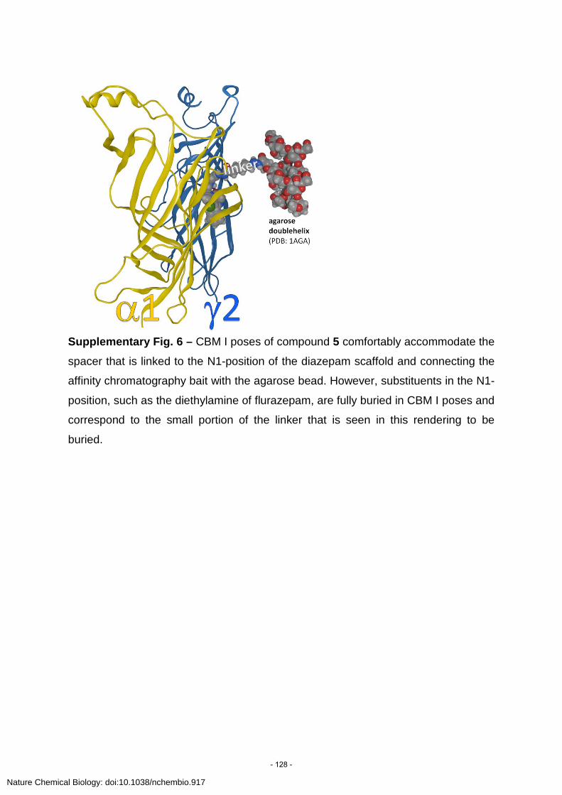
Supplementary Fig. 6 – CBM I poses of compound 5 comfortably accommodate the
spacer that is linked to the N1-position of the diazepam scaffold and connecting the
affinity chromatography bait with the agarose bead. However, substituents in the N1-
position, such as the diethylamine of flurazepam, are fully buried in CBM I poses and
correspond to the small portion of the linker that is seen in this rendering to be
buried.
Nature Chemical Biology: doi:10.1038/nchembio.917
- 128 -

Supplementary Fig. 7 – Representative CBM I poses of cpds. 1-9
Nature Chemical Biology: doi:10.1038/nchembio.917
- 129 -

Supplementary Fig. 8 – (a) Rotamer constellation of α1Y159/Y209 (stick
representation) as seen in CBM I poses,overlaid with all template structures used in
this study. The templates’ amino acid side chains that are homologous with
α1Y159/Y209 are shown in fine line. The Trp that is homologous with GABAAR
α1Y159 is seen to be in a highly conserved position. The Y on loop C appears to
feature a conserved rotamer throughout the superfamily, and probably serves as
transduction element for induced fit conformations in the flexible loop C tip. (b)
Rotamer constellation of α1Y159/Y209 (stick representation) as seen in CBM II
poses. The overlay shows that the pattern seen in other superfamily members is
lacking, this GABAA R’ model features a completely distinct topology. Quantitative
analysis of the entire pose pool that was used in this study revealed that the pattern
depicted in panel (a) is unique for CBM I poses, and not found in any other pose
group.
Nature Chemical Biology: doi:10.1038/nchembio.917
- 130 -

Supplementary Fig. 9 – (a) Representation of a CBM I Ro15-8670 pose.
Interestingly, in this pose, γ2F77 adapts a rotameric state that allows the
phenylalanine-ring to interact with the ester-moiety. Consequently, a γ2F77Y
mutation (hydroxyl-group position is indicated by the white star) would not clash the
pendant phenyl ring (compare with Fig. 3, main text). This observation is completely
consistent with the finding that in the γ2F77Y mutant only a 6x reduction was found
for Ro15-8670, while a 250x reduction in affinity was seen for diazepam74 which
lacks an ester-moiety. Thus, this structure offers a hypothesis for the observed
affinity changes in F77Y by an induced fit. In contrast to the γ2F77Y mutation, the
γ2F77I mutation should affect both ligands. Indeed this was shown by (b) inhibition of
[3H]flunitrazepam binding by Ro 15-8670 in wild-type α1β3γ2 (black curve) and point
mutated α1β3γ2F77I (red curve) receptors. Data represent means ± SD from an
experiment performed in triplicate. The affinity loss of Ro15-8670 by the mutation
γ2F77I is comparable to the one published for diazepam (see text), thus again
supporting similar interactions.
a
b
c
*
Nature Chemical Biology: doi:10.1038/nchembio.917
- 131 -

(c)The same CBM I Ro15-8670 from another perspective focusing on the position of
the ester moiety. This pose contains strong structural support for several findings. A
α1T206V substitution in loop C caused a 7-fold reduction in affinity of diazepam, but
a 16-fold increase in affinity of Ro15-867074. Assuming a common binding mode of
these compounds, in CBM I the imine nitrogen of the seven membered ring of
diazepam or Ro15-8670 forms a hydrogen bond with α1S204, and the imidazo-
group of Ro15-8670 interacts with γ2T142. The carbonyl oxygen of diazepam with its
two lone pairs interacts with α1T206, as previously proposed as well as with γ2T142
(see also main text Fig. 3). In receptors containing the α1T206V mutation, diazepam
is losing an H-bond partner and Ro15-8670 is gaining hydrophobic interactions from
the T206V mutant as the valine makes favourable contacts with the ester moiety.
This is visualized by an interaction potential map created with a MOE built-in
application which draws upon the work of GRID121,122. The map graphically illustrates
where a hydrophobic probe (dashed green) or an H-bond donating probe (dashed
magenta) has favourable interactions with the molecular surface, here depicted for
the ester-moiety of Ro15-8670. While the areas in plane of the ester-moiety favour
hydrophilic interactions, the areas above and below this moiety favour hydrophobic
interactions. Based on this map, the finding that the α1T206V mutation leads to a
16fold gain in affinity is fully consistent with the pose, as the –OH group of the wt
T206 “penetrates” the hydrophobic interaction surface, indicating an unfavourable
interaction potential.
Nature Chemical Biology: doi:10.1038/nchembio.917
- 132 -

Supplementary Fig. 10 – Depiction of structure-based pharmacophore models
based on CBM I diazepam (panel a, Pharmacophore D) and flumazenil (panel b,
Phamacophore F) bound structures. Yellow spheres represent hydrophobic features,
blue slices represent aromatic features and red arrows indicate h-bond acceptor
features. Grey spheres represent exclusion volumes and roughly resemble steric
binding site topology.
b
a
b
Nature Chemical Biology: doi:10.1038/nchembio.917
- 133 -

Supplementary Fig. 11 – Structure of the 3-hydroxy-oxindole 15, and its action on
GABAA receptors.(a) Chemical structure. (b) Inhibition of 2nM [3H]flunitrazepam
binding to mouse cerebellar membranes by 15. Data represent means±SD from
three experiment performed in triplicate (c) Dose response curve of 15 on GABA
EC3 currents in the absence (■), or presence of100nM flumazenil (●), obtained from
Xenopus laevis oocytes expressing recombinant GABAA receptors composed of
α1β3γ2 subunits. Data represent means±SD from three separate experiments
performed in different oocytes from 2 different batches. It can be seen that the
modulatory action of 15 is incompletely blocked by co-application of the
benzodiazepine null modulator flumazenil. (d) Effect of compound 15 at 1 µM and 10
µM concentration on α1β3 and α1β3γ2 (rat) receptors recombinantly expressed in
Xenopus laevis oocytes at GABA EC3 (n=4-6). The gamma-subunit independent
action seen in α1β3 receptors indicates a second site of interaction of this compound
with these receptors and is consistent with the incomplete flumazenil sensitivity of
modulation seen in (c).
Nature Chemical Biology: doi:10.1038/nchembio.917
- 134 -

Supplementary References 49. Notredame, C., Higgins, D.G. & Heringa, J. T-Coffee: A novel method for fast and
accurate multiple sequence alignment. J. Mol. Biol. 302, 205-217 (2000). 50. Shi, J., Blundell, T.L. & Mizuguchi, K. FUGUE: sequence-structure homology
recognition using environment-specific substitution tables and structure-dependent gap penalties. J. Mol. Biol. 310, 243-257 (2001).
51. Sali, A. & Blundell, T.L. Comparative protein modelling by satisfaction of spatial restraints. J. Mol. Biol. 234, 779-815 (1993).
52. Huang, N., Shoichet, B.K. & Irwin, J.J. Benchmarking sets for molecular docking. J. Med. Chem. 49, 6789-6801 (2006).
53. Irwin, J.J. & Shoichet, B.K. ZINC--a free database of commercially available compounds for virtual screening. J. Chem. Inf. Model. 45, 177-182 (2005).
54. Brejc, K. et al. Crystal structure of an ACh-binding protein reveals the ligand-binding domain of nicotinic receptors. Nature 411, 269-276 (2001).
55. Celie, P.H.N. et al. Nicotine and carbamylcholine binding to nicotinic acetylcholine receptors as studied in AChBP crystal structures. Neuron 41, 907-914 (2004).
56. Unwin, N. Refined structure of the nicotinic acetylcholine receptor at 4A resolution. J. Mol. Biol. 346, 967-989 (2005).
57. Hansen, S.B. et al. Structures of Aplysia AChBP complexes with nicotinic agonists and antagonists reveal distinctive binding interfaces and conformations. EMBO J. 24, 3635-3646 (2005).
58. Dellisanti, C.D., Yao, Y., Stroud, J.C., Wang, Z. & Chen, L. Crystal structure of the extracellular domain of nAChR alpha1 bound to alpha-bungarotoxin at 1.94 A resolution. Nat. Neurosci. 10, 953-962 (2007).
59. Hibbs, R.E. & Gouaux, E. Principles of activation and permeation in an anion-selective Cys-loop receptor. Nature 474, 54-60 (2011).
60. Salvadori, P., Bertucci, C., Ascoli, G., Uccello-Barretta, G. & Rossi, E. Direct resolution, characterization, and stereospecific binding properties of an atropisomeric 1,4-benzodiazepine. Chirality 9, 495-505 (1997).
61. Gilman, N.W., Rosen, P., Earley, J.V., Cook, C. & Todaro, L.J. Atropisomers of 1,4-benzodiazepines. Synthesis and resolution of a diazepam-related 1,4-benzodiazepine. J. Am. Chem. Soc. 112, 3969-3978 (1990).
62. Gilman, N.W. et al. Atropisomers of 1,4-benzodiazepines. 2. Synthesis and resolution of imidazo[1,5-a][1,4]benzodiazepines. J. Org. Chem. 58, 3285-3298 (1993).
63. Lee, S. et al. Axial chirality and affinity at the GABA(A) receptor of pyrimido[1,2-a][1,4]benzodiazepines and related compounds. Bioorg. Med. Chem. 16, 9519-9523 (2008).
64. Blount, J.F., Fryer, R.I., Gilman, N.W. & Todaro, L.J. Quinazolines and 1,4-benzodiazepines. 92. Conformational recognition of the receptor by 1,4-benzodiazepines. Mol. Pharmacol. 24, 425-428 (1983).
65. Simonyi, M.s. et al. Conformational recognition by central benzodiazepine receptors. Bioorg. Chem. 18, 1-12 (1990).
66. Maksay, G., Tegyey, Z. & Simonyi, M. Central benzodiazepine receptors: in vitro efficacies and potencies of 3-substituted 1,4-benzodiazepine stereoisomers. Mol. Pharmacol. 39, 725-732 (1991).
Nature Chemical Biology: doi:10.1038/nchembio.917
- 135 -

67. Ernst, M., Bruckner, S., Boresch, S. & Sieghart, W. Comparative models of GABAA receptor extracellular and transmembrane domains: important insights in pharmacology and function. Mol. Pharmacol. 68, 1291-1300 (2005).
68. Krissinel, E. & Henrick, K. Secondary-structure matching (SSM), a new tool for fast protein structure alignment in three dimensions. Acta Crystallogr. D Biol. Crystallogr. 60, 2256-2268 (2004).
69. Mysinger, M.M. & Shoichet, B.K. Rapid context-dependent ligand desolvation in molecular docking. J. Chem. Inf. Model. 50, 1561-73 (2010).
70. Wolber, G. & Langer, T. LigandScout: 3-D pharmacophores derived from protein-bound ligands and their use as virtual screening filters. J. Chem. Inf. Model. 45, 160-169 (2005).
71. Rarey, M., Kramer, B., Lengauer, T. & Klebe, G. A fast flexible docking method using an incremental construction algorithm. J. Mol. Biol. 261, 470-489 (1996).
72. Claussen, H., Buning, C., Rarey, M. & Lengauer, T. FlexE: efficient molecular docking considering protein structure variations. J. Mol. Biol. 308, 377-395 (2001).
73. Larkin, M.A. et al. Clustal W and Clustal X version 2.0. Bioinformatics 23, 2947-2948 (2007).
74. Sigel, E., Schaerer, M.T., Buhr, A. & Baur, R. The benzodiazepine binding pocket of recombinant alpha1beta2gamma2 gamma-aminobutyric acidA receptors: relative orientation of ligands and amino acid side chains. Mol. Pharmacol. 54, 1097-1105 (1998).
75. Sieghart, W. & Schuster, A. Affinity of various ligands for benzodiazepine receptors in rat cerebellum and hippocampus. Biochem. Pharmacol. 33, 4033-4038 (1984).
76. Sigel, E., Mamalaki, C. & Barnard, E.A. Isolation of a GABA receptor from bovine brain using a benzodiazepine affinity column. FEBS Lett. 147, 45-48 (1982).
77. Zhang, W., Koehler, K.F., Harris, B., Skolnick, P. & Cook, J.M. Synthesis of benzo-fused benzodiazepines employed as probes of the agonist pharmacophore of benzodiazepine receptors. J. Med. Chem. 37, 745-757 (1994).
78. Kilpatrick, G.J. et al. CNS 7056: a novel ultra-short-acting Benzodiazepine. Anesthesiology 107, 60-66 (2007).
79. Berezhnoy, D. et al. On the benzodiazepine binding pocket in GABAA receptors. J. Biol. Chem. 279, 3160-3168 (2004).
80. Kucken, A.M., Teissére, J.A., Seffinga-Clark, J., Wagner, D.A. & Czajkowski, C. Structural requirements for imidazobenzodiazepine binding to GABA(A) receptors. Mol. Pharmacol. 63, 289-296 (2003).
81. Tan, K.R. et al. Proximity-accelerated chemical coupling reaction in the benzodiazepine-binding site of gamma-aminobutyric acid type A receptors: superposition of different allosteric modulators. J. Biol. Chem. 282, 26316-26325 (2007).
82. Tan, K.R., Baur, R., Charon, S., Goeldner, M. & Sigel, E. Relative positioning of diazepam in the benzodiazepine-binding-pocket of GABA receptors. J. Neurochem. 111, 1264-1273 (2009).
83. Wang, R., Lai, L. & Wang, S. Further development and validation of empirical scoring functions for structure-based binding affinity prediction. J. Comput. Aided Mol. Des. 16, 11-26 (2002).
84. Labute, P. The generalized Born/volume integral implicit solvent model: Estimation of the free energy of hydration using London dispersion instead of atomic surface area. J. Comput. Chem. 29, 1693-1698 (2008).
Nature Chemical Biology: doi:10.1038/nchembio.917
- 136 -

85. Wei, B.Q., Baase, W.A., Weaver, L.H., Matthews, B.W. & Shoichet, B.K. A model binding site for testing scoring functions in molecular docking. J. Mol. Biol. 322, 339-355 (2002).
86. Huang, Q. et al. Pharmacophore/receptor models for GABA(A)/BzR subtypes (alpha1beta3gamma2, alpha5beta3gamma2, and alpha6beta3gamma2) via a comprehensive ligand-mapping approach. J. Med. Chem. 43, 71-95 (2000).
87. Im, W.B. et al. Differential affinity of dihydroimidazoquinoxalines and diimidazoquinazolines to the alpha 1 beta 2 gamma 2 and alpha 6 beta 2 gamma 2 subtypes of cloned GABAA receptors. Br. J. Pharmacol. 110, 677-680 (1993).
88. Ulrich Schacht, G.B. Effects of clobazam in benzodiazepine-receptor binding assays. Drug Dev. Res. S1, 83-93 (1982).
89. Jennings, A.S. et al. Imidazo[1,2-b][1,2,4]triazines as alpha2/alpha3 subtype selective GABA A agonists for the treatment of anxiety. Bioorg. Med. Chem. Lett. 16, 1477-1480 (2006).
90. Atack, J.R. et al. In vitro and in vivo properties of 3-tert-butyl-7-(5-methylisoxazol-3-yl)-2-(1-methyl-1H-1,2,4-triazol-5-ylme thoxy)-pyrazolo[1,5-d]-[1,2,4]triazine (MRK-016), a GABAA receptor alpha5 subtype-selective inverse agonist. J. Pharmacol. Exp. Ther. 331, 470-484 (2009).
91. Sethy, V.H. & Oien, T.T. Role of cGMP in the mechanism of anxiolytic activity of U-78875. Pharmacol. Biochem. Behav. 39, 379-382 (1991).
92. Mickelson, J.W. et al. High-affinity alpha-aminobutyric acid A/benzodiazepine ligands: synthesis and structure-activity relationship studies of a new series of tetracyclic imidazoquinoxalines. J. Med. Chem. 39, 4654-4666 (1996).
93. Hanson, S.M., Morlock, E.V., Satyshur, K.A. & Czajkowski, C. Structural requirements for eszopiclone and zolpidem binding to the gamma-aminobutyric acid type-A (GABAA) receptor are different. J. Med. Chem. 51, 7243-7252 (2008).
94. Lewis, R.T. et al. A pyridazine series of alpha2/alpha3 subtype selective GABA A agonists for the treatment of anxiety. J. Med. Chem. 49, 2600-2610 (2006).
95. Cox, E.D. et al. Synthesis and evaluation of analogues of the partial agonist 6-(propyloxy)-4-(methoxymethyl)-beta-carboline-3-carboxylic acid ethyl ester (6-PBC) and the full agonist 6-(benzyloxy)-4-(methoxymethyl)-beta-carboline-3-carboxylic acid ethyl ester (Zk 93423) at wild type and recombinant GABAA receptors. J. Med. Chem. 41, 2537-2552 (1998).
96. Primofiore, G. et al. 3-Aryl-[1,2,4]triazino[4,3-a]benzimidazol-4(10H)-ones: tricyclic heteroaromatic derivatives as a new class of benzodiazepine receptor ligands. J. Med. Chem. 43, 96-102 (2000).
97. Dubinsky, B. et al. 5-ethoxymethyl-7-fluoro-3-oxo-1,2,3,5-tetrahydrobenzo[4,5]imidazo[1,2a]pyr idine-4-N-(2-fluorophenyl)carboxamide (RWJ-51204), a new nonbenzodiazepine anxiolytic. J. Pharmacol. Exp. Ther. 303, 777-790 (2002).
98. Wegner, F. et al. GABAA receptor pharmacology of fluorinated derivatives of the novel sedative-hypnotic pyrazolopyrimidine indiplon. Eur. J. Pharmacol. 580, 1-11 (2008).
99. Guerrini, G. et al. Benzodiazepine receptor ligands. 8: synthesis and pharmacological evaluation of new pyrazolo[5,1-c] [1,2,4]benzotriazine 5-oxide 3- and 8-disubstituted: high affinity ligands endowed with inverse-agonist pharmacological efficacy. Bioorg. Med. Chem. 14, 758-775 (2006).
100. Primofiore, G. et al. Novel N-(arylalkyl)indol-3-ylglyoxylylamides targeted as ligands of the benzodiazepine receptor: synthesis, biological evaluation, and molecular
Nature Chemical Biology: doi:10.1038/nchembio.917
- 137 -

modeling analysis of the structure-activity relationships. J. Med. Chem. 44, 2286-2297 (2001).
101. Lager, E. et al. 4-quinolone derivatives: high-affinity ligands at the benzodiazepine site of brain GABA A receptors. synthesis, pharmacology, and pharmacophore modeling. J. Med. Chem. 49, 2526-2533 (2006).
102. Takada, S. et al. Synthesis and structure--activity relationships of fused imidazopyridines: a new series of benzodiazepine receptor ligands. J. Med. Chem. 39, 2844-2851 (1996).
103. Selleri, S. et al. Synthesis and benzodiazepine receptor affinity of pyrazolo[1,5-a]pyrimidine derivatives. 3. New 6-(3-thienyl) series as alpha 1 selective ligands. J. Med. Chem. 46, 310-313 (2003).
104. Primofiore, G. et al. High affinity central benzodiazepine receptor ligands: synthesis and biological evaluation of a series of phenyltriazolobenzotriazindione derivatives. J. Med. Chem. 48, 2936-2943 (2005).
105. Kahnberg, P. et al. Refinement and evaluation of a pharmacophore model for flavone derivatives binding to the benzodiazepine site of the GABA(A) receptor. J. Med. Chem. 45, 4188-4201 (2002).
106. De Sarro, G. et al. Benzodiazepine receptor affinities, behavioral, and anticonvulsant activity of 2-aryl-2,5-dihydropyridazino[4,3-b]indol- 3(3H)-ones in mice. Pharmacol. Biochem. Behav. 65, 475-487 (2000).
107. Marcel, D., Bardelay, C. & Hunt, P.F. Lesion of noradrenergic neurones with DSP4 does not modify benzodiazepine receptor binding in cortex and hippocampus of rat. Neuropharmacology 25, 283-286 (1986).
108. Lippa, A. et al. Selective anxiolysis produced by ocinaplon, a GABA(A) receptor modulator. Pro.c Natl. Acad. Sci. U S A 102, 7380-7385 (2005).
109. Ogris, W. et al. Affinity of various benzodiazepine site ligands in mice with a point mutation in the GABA(A) receptor gamma2 subunit. Biochem. Pharmacol. 68, 1621-1629 (2004).
110. Bare, T.M. et al. Synthesis and structure-activity relationships of a series of anxioselective pyrazolopyridine ester and amide anxiolytic agents. J. Med. Chem. 32, 2561-2573 (1989).
111. Szekeres, H.J. et al. 3,4-Dihydronaphthalen-1(2H)-ones: novel ligands for the benzodiazepine site of alpha5-containing GABAA receptors. Bioorg. Med. Chem. Lett. 14, 2871-2875 (2004).
112. Carling, R.W. et al. 3-phenyl-6-(2-pyridyl)methyloxy-1,2,4-triazolo[3,4-a]phthalazines and analogues: high-affinity gamma-aminobutyric acid-A benzodiazepine receptor ligands with alpha 2, alpha 3, and alpha 5-subtype binding selectivity over alpha 1. J. Med. Chem. 47, 1807-1822 (2004).
113. Chambers, M.S. et al. 6,7-Dihydro-2-benzothiophen-4(5H)-ones: a novel class of GABA-A alpha5 receptor inverse agonists. J. Med. Chem. 45, 1176-1179 (2002).
114. Griebel, G. et al. SL651498: an anxioselective compound with functional selectivity for alpha2- and alpha3-containing gamma-aminobutyric acid(A) (GABA(A)) receptors. J. Pharmacol. Exp. Ther. 298, 753-768 (2001).
115. Crawforth, J. et al. Tricyclic pyridones as functionally selective human GABAA alpha 2/3 receptor-ion channel ligands. Bioorg. Med. Chem. Lett. 14, 1679-1682 (2004).
116. Davies, L.P., Barlin, G.B., Ireland, S.J. & Ngu, M.M. Substituted imidazo[1,2-b]pyridazines. New compounds with activity at central and peripheral benzodiazepine receptors. Biochem. Pharmacol. 44, 1555-1561 (1992).
Nature Chemical Biology: doi:10.1038/nchembio.917
- 138 -

117. Mehta, A.K. & Shank, R.P. Interaction of abecarnil, bretazenil, and RO 19-8022 with diazepam-sensitive and -insensitive benzodiazepine sites in the rat cerebellum and cerebral cortex. Life Sci. 57, 2215-2222 (1995).
118. Atack, J.R. et al. TPA023 [7-(1,1-dimethylethyl)-6-(2-ethyl-2H-1,2,4-triazol-3-ylmethoxy)-3-(2-fluor ophenyl)-1,2,4-triazolo[4,3-b]pyridazine], an agonist selective for alpha2- and alpha3-containing GABAA receptors, is a nonsedating anxiolytic in rodents and primates. J. Pharmacol. Exp. Ther. 316, 410-422 (2006).
119. Wada, T. et al. Pharmacologic characterization of a novel non-benzodiazepine selective anxiolytic, DN-2327. Jpn. J. Pharmacol. 49, 337-349 (1989).
120. Jain, A.N. & Nicholls, A. Recommendations for evaluation of computational methods. J. Comput. Aided Mol. Des. 22, 133-139 (2008).
121. Boobbyer, D.N., Goodford, P.J., McWhinnie, P.M. & Wade, R.C. New hydrogen-bond potentials for use in determining energetically favorable binding sites on molecules of known structure. J.Med. Chem. 32, 1083-1094 (1989).
122. Goodford, P.J. A computational procedure for determining energetically favorable binding sites on biologically important macromolecules. J. Med. Chem. 28, 849-857 (1985).
Nature Chemical Biology: doi:10.1038/nchembio.917
- 139 -

II. A residue close to α1 loop F disrupts modulation of GABAA receptors by benzodiazepines while their
binding is maintained
Contribution of the thesis author to this article:
The author of the thesis performed a computational analysis on a homology model of the
α1β2γ2 GABAA receptor (homology models were generated by Margot Ernst and Werner
Sieghart). In more detail the intra molecular protein-protein H-bond networks in
proximity to the benzodiazepine binding site of the GABAA receptor model were
investigated. The results aided in the selection of α1Y168 as a candidate for receptor
mutation.
- 140 -

,1 ,1
*Institute of Biochemistry and Molecular Medicine, University of Bern, Bern, Switzerland
�Department of Medicinal Chemistry, University of Vienna, Vienna, Austria
In the family of ligand-activated ion channels that includesnicotinic acetylcholine receptors, GABA type A (GABAA)receptors, glycine receptors and serotonin 5-HT3 receptors,binding of the agonist is followed by a conformational changeleading to the opening of the inherent ion channel. Themechanism by which binding leads to channel opening ishighly controversial. One concept envisages a direct func-tional link between the binding site and the channel region bya random coil polypeptide stretch called loop F. The functionof loop F in linking binding and gating is disputed someobservations pointing in favor of the hypothesis (Lyford et al.2003; Newell and Czajkwski 2003; Hansen et al. 2005;Thompson et al. 2006; Williams et al. 2009) and othersagainst (Khatri et al. 2009; Pless and Lynch 2009). Thiscontroversy has recently been reviewed (Khatri and Weiss2010). Beside loop F, residues located at the outer membranesurface have been implicated in the binding/gating process(Sigel et al. 1999; Brejc et al. 2001; Kash et al. 2003;Lummis et al. 2005; Unwin 2005; Mercado and Czajkowski2006), but it is far from clear how the information of bindingsite occupancy is transferred to these residues.GABA type A receptors are pseudosymmetric proteins
consisting of five subunits, thus containing five F loops. The
pharmacological properties of a GABAA receptor depend onsubunit composition (Sieghart and Sperk 2002) and arrange-ment (Minier and Sigel 2004). In this study we concentratedon the major form of adult GABAA receptors a1b2c2(Macdonald and Olsen 1994; Rabow et al. 1995; Sieghart1995), whose subunits are arranged a1c2b2a1b2 counter-clockwise around the central ion channel (Baumann et al.2001, 2002; Baur et al. 2006). In the GABAA receptor, the FLoop of the a1 subunit as well as the F loop of the b2 subunithave been reported to play an important role in linkingbinding to gating of the GABAA receptor (Newell andCzajkwski 2003; Williams et al. 2009).
Many drugs act at GABAA receptors among thembenzodiazepines. Benzodiazepines are clinically important
Received August 31, 2010; revised manuscript received September 24,2010; accepted October 5, 2010.Address correspondence and reprint requests to Erwin Sigel, Institute
of Biochemistry and Molecular Medicine, University of Bern, Buhlst-rasse 28, CH-3012 Bern, Switzerland. E-mail: [email protected] authors contributed equally to this study.Abbreviations used: DMCM, methyl 6, 7-dimethoxy-4-ethly-beta-
carboline-2-carboxylate; GABAA receptor, GABA type A receptor;HEK, human embryonic kidney.
Abstract
Benzodiazepines act at the major isoforms of GABA type A
receptors where they potentiate the current evoked by the
agonist GABA. The underlying mechanism of this potentia-
tion is poorly understood, but hypothesized to be related to
the mechanism that links agonist binding to channel opening
in these ligand activated ion channels. The loop F of the a1and the b2 subunit have been implicated in channel gating,
and loop F of the c2 subunit in the modulation by ben-
zodiazepines. We have identified the conservative point
mutation Y168F located N-terminally of loop F in the a1subunit that fails to affect agonist properties. Interestingly, it
disrupts modulation by benzodiazepines, but leaves high
affinity binding to the benzodiazepine binding site intact.
Modulation by barbiturates and neurosteroids is also unaf-
fected. Residue a1Y168 is not located either near the binding
pockets for GABA, or for benzodiazepines, or close to the
loop F of the c2 subunit. Our results support the fact, that
broader regions of ligand gated receptors are conforma-
tionally affected by the binding of benzodiazepines. We infer
that also broader regions could contribute to signaling from
GABA agonist binding to channel opening.
Keywords: benzodiazepine binding, benzodiazepine modu-
lation, channel gating, F loop, GABA, GABAA receptor.
J. Neurochem. (2010) 115, 1478–1485.
JOURNAL OF NEUROCHEMISTRY | 2010 | 115 | 1478–1485 doi: 10.1111/j.1471-4159.2010.07052.x
1478 Journal of Neurochemistry � 2010 International Society for Neurochemistry, J. Neurochem. (2010) 115, 1478–1485� 2010 The Authors
- 141 -

drugs and are used mainly for their sedative/hypnotic andanxiolytic action. They bind to a high affinity binding site,located at the a/c subunit interface, that is homologous to thetwo GABA agonist sites, located at b/a subunit interfaces(for review see Sigel and Buhr 1997). Benzodiazepinespotentiate responses of the major GABAA receptor isoformsto the agonist GABA. In this case high affinity binding ofbenzodiazepines induces a structural change to the receptor,leading to potentiation. Again, the underlying structuralchange is poorly understood. In this case, loop F of the c2subunit has been suggested to be involved in this process(Hanson and Czajkowski 2008; Padgett and Lummis 2008).Residues located at the outer membrane surface may alsoplay a role (Boileau and Czajkowski 1999).We report here on a conservative point mutation located at
the N-terminal end of loop F in the a1 subunit of the GABAA
receptor, which does not affect the response to GABA andhigh affinity binding of benzodiazepines to their site, butstrongly decreases modulation by benzodiazepines. Thisresidue is distant from loop F of the c2 subunit, arguing forthe fact that receptor regions outside this loop F are essentialfor signaling from the benzodiazepine binding site to thechannel portion. We hypothesize that the same may be thecase for signaling from agonist binding to channel opening inthe ligand gated ion channel family.
Methods
Construction of the mutated receptor subunitThe mutant subunit a1Y168F was prepared using the QuikChangeTM
mutagenesis kit (Stratagene, Agilent Technologies, Basel, Switzer-
land). For cell transfection, the cDNAs were subcloned into the
polylinker of pBC/CMV (Bertocci et al. 1991). This expression
vector allows high-level expression of a foreign gene under control of
the cytomegalovirus promoter. The a subunit was cloned into the
EcoRI and the b and c subunits were subcloned into the SmaI site ofthe polylinker by standard techniques.
Expression in Xenopus oocytesCapped cRNAs were synthesized (Ambion, Applied Biosystems,
Rotkreuz, Switzerland) from the linearized vectors containing the
different subunits, respectively. A poly-A tail of about 400 residues
was added to each transcript using yeast poly-A polymerase (United
States Biologicals, Cleveland, OH, USA). The concentration of the
cRNAwas quantified on a formaldehyde gel using Radiant Red stain
(Bio-Rad Laboratories, Reinach, Switzerland) for visualization of
the RNA. Known concentrations of RNA ladder (Invitrogen, Basel,
Switzerland) were loaded as standard on the same gel. cRNAs were
precipitated in ethanol/isoamylalcohol 19 : 1, the dried pellet
dissolved in water and stored at )80�C. cRNA mixtures were
prepared from these stock solutions and stored at )80�C.Xenopus laevis oocytes were prepared, injected and defollicu-
lated as described previously (Sigel 1987; Sigel and Minier 2005).
They were injected with 50 nL of the cRNA solution containing
wild type or mutated a1 and wild type b2 and c2 subunits at a
concentration of 10 nM : 10 nM : 50 nM (Boileau et al. 2002), andthen incubated in modified Barth’s solution [10 mM HEPES, pH
7.5, 88 mM NaCl, 1 mM KCl, 2.4 mM NaHCO3, 0.82 mM
MgSO4, 0.34 mM Ca(NO3)2, 0.41 mM CaCl2, 100 units/mL
penicillin, 100 lg/mL streptomycin] at 18�C for at least 24 h before
the measurements.
Functional characterization in Xenopus oocytesCurrents were measured using a home-built two-electrode voltage
clamp amplifier in combination with a XY-recorder (90% response
time 0.1 s) or digitized at 100 Hz using a MacLab/200 (AD
Instruments, Spechbach, Germany). Tests with a model oocyte were
performed to ensure linearity in the larger current range. The
response was linear up to 20 lA. Electrophysiological experiments
were performed at a holding potential of )80 mV. The perfusion
medium contained 90 mM NaCl, 1 mM KCl, 1 mM MgCl2, 1 mM
CaCl2, and 5 mM Na-HEPES (pH 7.4). The perfusion solution
(6 mL/min) was applied through a glass capillary with an inner
diameter of 1.35 mm, the mouth of which was placed about 0.4 mm
from the surface of the oocyte (Baur and Sigel 2007). The
experiments were performed after the measured currents became
constant. Concentration response curves for GABA were fitted with
the equation I(c) = Imax/(1 + (EC50/c)n), where c is the concentration
of GABA, EC50 the concentration of GABA eliciting half maximal
current amplitude, Imax is the maximal current amplitude, I the
current amplitude and n the Hill coefficient. Allosteric modulation
via the benzodiazepine site were measured at a GABA concentration
eliciting 2–5% of the maximal GABA current amplitude. GABA
was applied for 20 s alone or in combination with allosteric
compound. The prototype positive allosteric modulator diazepam,
the prototype negative allosteric modulator methyl 6, 7-dimethoxy-
4-ethyl-beta-carboline-2-carboxylate (DMCM) and the prototype
benzodiazepine antagonist Ro15-1788 were used. Additionally, a
positive modulator with a different chemical structure from classic
benzodiazepines, the imidazobendiazepine zolpidem was tested.
Relative current potentiation by benzodiazepines (BZ) was deter-
mined as (I1 lM BZ + GABA/IGABA – 1) * 100%. The perfusion system
was cleaned between drug applications, by washing with dimeth-
ylsulfoxide to avoid contamination.
Transfection of recombinant GABAA receptor in cultured cellsThe cells were maintained in minimum essential medium
(Invitrogen, Basel, Switzerland) supplemented with 10% fetal calf
serum, 2 mM glutamine, 50 units/mL penicillin and 50 lg/mL
streptomycin by standard cell culture techniques. Equal amounts
(total of 20 lg DNA/90 mm dish) of plasmids coding for GABAA
receptor subunits were transfected into human embryonic kidney
(HEK) 293 cells (ATCC no. CRL 1573) by the calcium phosphate
precipitation method (Chen and Okayama 1987). After overnight
incubation, the cells were washed once with serum free medium and
reefed with medium.
Membrane preparationApproximately 60 h after transfection the cells were harvested by
washing with ice-cold phosphate-buffered saline, pH 7.0, and
centrifuged at 150 g. Cells were washed with buffer containing
10 mM K phosphate, 100 mM KCl, 0.1 mM K EDTA, pH 7.4.
Cells were homogenized by sonication in the presence of 10 lM
� 2010 The AuthorsJournal of Neurochemistry � 2010 International Society for Neurochemistry, J. Neurochem. (2010) 115, 1478–1485
a1Y168F disrupts benzodiazepine modulation of GABAA receptors | 1479
- 142 -
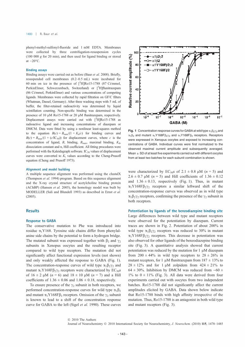
phenyl-methyl-sulfonyl-fluoride and 1 mM EDTA. Membranes
were collected by three centrifugation-resuspension cycles
(100 000 g for 20 min), and then used for ligand binding or stored
at )20�C.
Binding assaysBinding assays were carried out as before (Baur et al. 2008). Briefly,resuspended cell membranes (0.2–0.5 mL) were incubated for
60 min on ice in the presence of [3H]Ro15-1788 (87 Ci/mmol,
PerkinElmer, Schwerzenbach, Switzerland) or [3H]flunitrazepam
(86 Ci/mmol, PerkinElmer) and various concentrations of competing
ligands. Membranes were collected by rapid filtration on GF/C filters
(Whatman, Dassel, Germany). After three washing steps with 5 mL of
buffer, the filter-retained radioactivity was determined by liquid
scintillation counting. Non-specific binding was determined in the
presence of 10 lM Ro15-1788 or 20 lM flunitrazepam, respectively.
Displacement assays were carried out with [3H]Ro15-1788 as
radioactive ligand and increasing concentrations of diazepam or
DMCM. Data were fitted by using a nonlinear least-squares method
to the equation B(c) = Bmax/(1 + Kd/c) for binding curves and
B(c) = Bmax/(1 + (c/IC50)) for displacement curves, where c is the
concentration of ligand, B, binding, Bmax, maximal binding, Kd,
dissociation constant and n, Hill coefficient. All fitting procedures were
performed with the Kaleidagraph software. IC50 values of displacement
curves were converted to Ki values according to the Cheng-Prusoff
equation (Cheng and Prusoff 1973).
Alignment and model buildingA multiple sequence alignment was performed using the clustalX
(Thompson et al. 1994) program. Based on this sequence alignment
and the X-ray crystal structure of acetylcholine binding protein
(AChBP) (Hansen et al. 2005), the homology model was built by
MODELLER (Sali and Blundell 1993) as described in Ernst et al.(2005).
Results
Response to GABAThe conservative mutation to Phe was introduced intoresidue a1Y168. Tyrosine side chains differ from phenylal-anine side chains by the potential to form a hydrogen bridge.The mutated subunit was expressed together with b2 and c2subunits in Xenopus oocytes and the resulting receptorcompared to wild type receptors. The mutation did notsignificantly affect functional expression levels (not shown)and only weakly affected the response to GABA (Fig. 1).The concentration-response curves of wild type a1b2c2 andmutant a1Y168Fb2c2 receptors were characterized by EC50sof 16 ± 2 lM (n = 6) and 18 ± 10 lM (n = 7) and a Hillcoefficients of 1.36 ± 0.06 and 1.06 ± 0.18, respectively.To ensure presence of the c2 subunit in both receptors, we
performed concentration-response curves for wild type a1b2and mutant a1Y168Fb2 receptors. Omission of the c2 subunitis known to lead to a shift of the concentration responsecurve for GABA to the left (Sigel et al. 1990). These curves
were characterized by EC50s of 2.1 ± 0.8 lM (n = 5) and2.6 ± 0.7 lM (n = 5) and Hill coefficients of 1.36 ± 0.12and 1.36 ± 0.13, respectively (Fig. 1). Thus, in mutanta1Y168Fb2c2 receptors a similar leftward shift of theconcentration-response curves was observed as in wild typea1b2c2 receptors, confirming the presence of the c2 subunit inboth receptors.
Potentiation by ligands of the benzodiazepine binding siteLarge differences between wild type and mutant receptorswere observed for the potentiation by diazepam. Currenttraces are shown in Fig. 2. Potentiation of about 200% inwild type a1b2c2 receptors was reduced to 30% in mutanta1Y168Fb2c2 receptors. This decrease in potentiation wasalso observed for other ligands of the benzodiazepine bindingsite (Fig. 3). A quantitative analysis showed that currentpotentiation was reduced by the mutation for 1 lM diazepamfrom 200 ± 44% in wild type receptors to 28 ± 26% inmutant receptors, for 1 lM flunitrazepam from 187 ± 13% to28 ± 12% and for 1 lM zolpidem from 424 ± 21% to64 ± 30%. Inhibition by DMCM was reduced from )60 ±1% to 0 ± 11% (Fig. 3). All data were derived from fourexperiments carried out with oocytes from two independentbatches. Ro15-1788 did not significantly affect the currentamplitudes elicited by GABA. Data shown below indicatethat Ro15-1788 binds with high affinity irrespective of themutation. Thus, Ro15-1788 is an antagonist in both wild typeand mutant receptors (Fig. 3).
Fig. 1 Concentration response curves forGABAatwild type a1b2c2 and
a1b2 and mutant a1Y168Fb2c2 and a1Y168Fb2 receptors. Receptors
were expressed in Xenopus oocytes and exposed to increasing con-
centrations of GABA. Individual curves were first normalized to the
observed maximal current amplitude and subsequently averaged.
Mean ± SDof at least five experiments carried outwith different oocytes
from at least two batches for each subunit combination is shown.
Journal of Neurochemistry � 2010 International Society for Neurochemistry, J. Neurochem. (2010) 115, 1478–1485� 2010 The Authors
1480 | R. Baur et al.
- 143 -

The concentration dependence of the potentiation bydiazepam and zolpidem at wild type a1b2c2 and mutanta1Y168Fb2c2 receptors was also determined (Fig. 4). Poten-tiation by diazepam was fitted with an EC50 of 0.059 ±0.013 lM (n = 3) and a maximal potentiation of 285 ± 29%for wild type receptors, and 3.7 ± 2.4 lM (n = 4) and53 ± 9% for mutant receptors (Fig. 4a). Potentiation byzolpidem was fitted with an EC50 of 0.17 ± 0.01 lM (n = 3)and a maximal potentiation of 392 ± 29% for wild typereceptors, and 5.8 ± 2.6 lM (n = 3) and 64 ± 14% formutant receptors (Fig. 4b). Thus, for both ligands of the
benzodiazepine binding site, there was a decrease in apparentaffinity amounting to more than 30-fold, and a decrease inthe maximal potentiation amounting to more than 5-fold. Itwas interesting to see whether the apparent decrease inaffinity for function was reflected in a decrease in bindingaffinity.
Radioactive ligand binding to the benzodiazepine bindingsiteWild type a1b2c2 and mutant a1Y168Fb2c2 receptors wereexpressed in HEK293 cells and their benzodiazepine bindingsites probed with several ligands. Radioactive ligands weretested in binding assays and non-radioactive ligands indisplacement assays. Figure 5(a) shows as an example thebinding of [3H]Ro15-1788 to mutant receptors. The affinity
Fig. 3 Ligands of the benzodiazepine binding site potentiate
a1Y168Fb2c2 receptors less than wild type receptors. Similar experi-
ments as shown in Fig. 2 were carried out with 1 lM diazepam, 1 lM
flunitrazepam, 1 lM zolpidem, 1 lM DMCM or 1 lM Ro15-1788.
Means ± SD of experiments carried out with four oocytes from two
batches for each subunit combination are shown.
(a)
(b)
Fig. 4 Concentration-dependent potentiation of wild type a1b2c2 and
mutant a1Y168Fb2c2 receptors by diazepam and zolpidem. Receptors
were expressed in Xenopus oocytes and exposed to 0.5 lM or 1.0 lM
GABA in combination with increasing concentrations of diazepam (a)
or zolpidem (b), respectively. Individual curves were first normalized to
the observed maximal current amplitude and subsequently averaged.
Mean ± SD of experiments carried out with four oocytes from two
batches for each subunit combination are shown.
Fig. 2 Diazepam potentiates a1Y168Fb2c2 receptors less than wild
type receptors. Receptors were expressed in Xenopus oocytes and
exposed twice to 1 lM GABA alone followed by combined application
of 1 lM GABA and 1 lM diazepam.
� 2010 The AuthorsJournal of Neurochemistry � 2010 International Society for Neurochemistry, J. Neurochem. (2010) 115, 1478–1485
a1Y168F disrupts benzodiazepine modulation of GABAA receptors | 1481
- 144 -

of mutant receptors was with 0.8 nM in the range of that ofwild type receptors, indicating that the point mutation did notdisrupt the binding affinity. Displacement by diazepam of[3H]Ro15-1788 binding also remained unaffected by themutation (Fig. 5b). Table 1 summarizes these experiments.Wild type and mutant receptors were compared in theirrespective affinities for [3H]Ro15-1788, [3H]flunitrazepam,diazepam and DMCM. For none of the tested ligands morethan a 1.8-fold difference between wild type and mutantreceptors was observed.
Lack of an effect of mutation of the homologous residue inthe b2 subunitThe homologous residue to a1Y168 in the b2 subunit, F166was mutated to Y. a1b2F166Yc2 receptors were expressed inXenopus oocytes and functionally characterized. Neither theEC50 for GABA nor modulation by 1 lM diazepam wasaffected by the mutation (results not shown). Very similarresults were obtained with mutation of the neighboringresidues of b2F166, b2E165D and b2Y167F.
Lack of an effect of the mutation a1Y168Y on channelgating and modulation by pentobarbital, neurosteroids andinhibition by zinc ionsThe mutation a1Y168Ymutation did not affect channel gatingby GABA or binding of benzodiazepines, but disruptedmodulation of the channel by benzodiazepines. It wasinteresting to see if the action of other modulatory compoundswas affected by the mutation or whether the effect was specificfor modulation by benzodiazepines. Therefore, we investi-gated if channel modulation by pentobarbital, the neurosteroidTHDOC (3a,21-dihydroxy-5a-pregnan-20-one) or inhibitionby zinc ions was affected by the a1Y168Ymutation. As Fig. 6shows, neither concentration dependent gating (Fig. 6a) orpotentiation (Fig. 6b) by pentobarbital, nor concentrationdependent gating (Fig. 6c) or potentiation (Fig. 6d) of thechannel, nor its concentration dependent inhibition by zincions was affected (not shown). Inhibition by 1 lM and 1 mMZn2+ was 24 ± 11% (n = 3) and 86 ± 1% (n = 3) for wildtype receptors and 22 ± 10% (n = 3) and 82 ± 6% (n = 3) formutant receptors, respectively.
(a)
(b)
Fig. 5 Binding of [3H]Ro15-1788 to (a) and displacement of
[3H]Ro15-1788 by diazepam from (b) wild type a1b2c2 and mutant
a1Y168Fb2c2 receptors. Receptors were expressed in HEK293 cells,
membranes harvested and subjected to a radioactive ligand binding
assay. Individual experiments are shown. Fitted curves for wild type
and mutant receptor overlap. Mean ± SD from experiments carried
out in triplicate.
Table 1 Summary of the binding properties of the benzodiazepine binding site in wild type a1b2c2 and mutant a1Y168Fb2c2 receptors
Receptor [3H]Ro15-1788, Kd (nM) [3H]flunitrazepam, Kd (nM) Diazepam, Ki (nM) DMCM, Ki (nM)
a1b2c2 0.70 ± 0.11 2.0 ± 0.4 14.5 ± 3.7 1.3 ± 0.3
a1Y168Fb2c2 0.73 ± 0.05 1.6 ± 0.3 8.2 ± 2.0 2.1 ± 0.4
Binding of [3H]Ro15-1788 and of [3H]flunitrazepam to and displacement of [3H]Ro15-1788 by diazepam and DMCM was determined. Receptors
were expressed in HEK293 cells, membranes harvested and subjected to a radioactive ligand binding assays. Individual curves were first
normalized to the observed maximal binding level and subsequently averaged. Mean ± SD from two to four experiments each carried out in
triplicate.
Journal of Neurochemistry � 2010 International Society for Neurochemistry, J. Neurochem. (2010) 115, 1478–1485� 2010 The Authors
1482 | R. Baur et al.
- 145 -

Location of a1Y168 in the receptorFigure 7 is based on a homology model of part of theextracellular domain of the a1b2c2 GABAA receptor. The a1subunit is shown in yellow and the c2 subunit in bright blue.The amino acid residue a1Y168 (atoms as spheres) is neitherlocated in the binding site for GABA or that for benzodiaze-pines, nor is it located in the loop F of the c2 subunit (darkblue). It is located neighboring the N-terminal end of loop Fas defined in Hanson and Czajkowski (2008) in the a1subunit (orange).
Discussion
We made observations relevant for the question how thesignal induced by binding of benzodiazepines is propagatedto an altered channel function in GABAA receptors. As
amino acid residues located in the benzodiazepine bindingsite are homologous to amino acid residues located in theagonist site for GABA (reviewed in Sigel and Buhr 1997)our observations may be relevant for the question how thesignal induced by binding of GABA is propagated to the ionchannel. If this is the case, the experimental findings couldconcern the entire cys-loop ligand-activated receptor family.Introduction of the conservative point mutation a1Y168F
into a1b2c2 leaves channel gating by GABA and high affinitybinding of benzodiazepines unaltered while channel modu-lation by benzodiazepines is strongly reduced. Channelgating and modulation by pentobarbital and THDOC andcurrent inhibition by zinc ions are all unaffected by themutation, pointing to a specificity of the mutation forbenzodiazepine modulation. We observe a strong reductionof efficacy and potency of the current potentiation by
(a) (b)
(c) (d)
Fig. 6 Modulation of wild type a1b2c2 and mutant a1Y168Fb2c2receptors by pentobarbital and THDOC (3a,21-dihydroxy-5a-pregnan-
20-one) (a) Increasing concentrations of pentobarbital were applied to
wild type and mutant receptors in the absence GABA. (b) Increasing
concentrations of pentobarbital were applied to wild type and mutant
receptors in combination with 1.5 lM GABA. (c) Increasing concen-
trations of THDOC were applied to wild type and mutant receptors in
the absence GABA. (d) Increasing concentrations of THDOC were
applied to wild type and mutant receptors in combination with 1.5 lM
GABA. Mean ± SD of experiments carried out with three oocytes for
each subunit combination are shown.
� 2010 The AuthorsJournal of Neurochemistry � 2010 International Society for Neurochemistry, J. Neurochem. (2010) 115, 1478–1485
a1Y168F disrupts benzodiazepine modulation of GABAA receptors | 1483
- 146 -
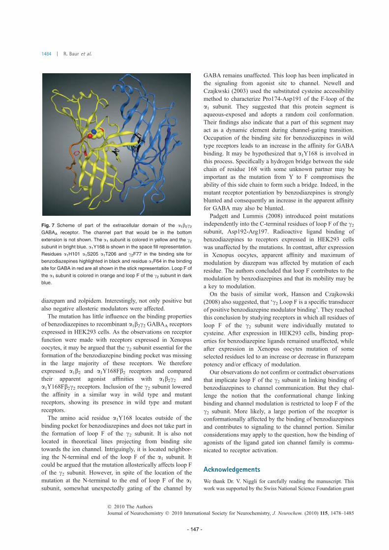
diazepam and zolpidem. Interestingly, not only positive butalso negative allosteric modulators were affected.The mutation has little influence on the binding properties
of benzodiazepines to recombinant a1b2c2 GABAA receptorsexpressed in HEK293 cells. As the observations on receptorfunction were made with receptors expressed in Xenopusoocytes, it may be argued that the c2 subunit essential for theformation of the benzodiazepine binding pocket was missingin the large majority of these receptors. We thereforeexpressed a1b2 and a1Y168Fb2 receptors and comparedtheir apparent agonist affinities with a1b2c2 anda1Y168Fb2c2 receptors. Inclusion of the c2 subunit loweredthe affinity in a similar way in wild type and mutantreceptors, showing its presence in wild type and mutantreceptors.The amino acid residue a1Y168 locates outside of the
binding pocket for benzodiazepines and does not take part inthe formation of loop F of the c2 subunit. It is also notlocated in theoretical lines projecting from binding sitetowards the ion channel. Intriguingly, it is located neighbor-ing the N-terminal end of the loop F of the a1 subunit. Itcould be argued that the mutation allosterically affects loop Fof the c2 subunit. However, in spite of the location of themutation at the N-terminal to the end of loop F of the a1subunit, somewhat unexpectedly gating of the channel by
GABA remains unaffected. This loop has been implicated inthe signaling from agonist site to channel. Newell andCzajkwski (2003) used the substituted cysteine accessibilitymethod to characterize Pro174-Asp191 of the F-loop of thea1 subunit. They suggested that this protein segment isaqueous-exposed and adopts a random coil conformation.Their findings also indicate that a part of this segment mayact as a dynamic element during channel-gating transition.Occupation of the binding site for benzodiazepines in wildtype receptors leads to an increase in the affinity for GABAbinding. It may be hypothesized that a1Y168 is involved inthis process. Specifically a hydrogen bridge between the sidechain of residue 168 with some unknown partner may beimportant as the mutation from Y to F compromises theability of this side chain to form such a bridge. Indeed, in themutant receptor potentiation by benzodiazepines is stronglyblunted and consequently an increase in the apparent affinityfor GABA may also be blunted.Padgett and Lummis (2008) introduced point mutations
independently into the C-terminal residues of loop F of the c2subunit, Asp192-Arg197. Radioactive ligand binding ofbenzodiazepines to receptors expressed in HEK293 cellswas unaffected by the mutations. In contrast, after expressionin Xenopus oocytes, apparent affinity and maximum ofmodulation by diazepam was affected by mutation of eachresidue. The authors concluded that loop F contributes to themodulation by benzodiazepines and that its mobility may bea key to modulation.On the basis of similar work, Hanson and Czajkowski
(2008) also suggested, that ‘c2 Loop F is a specific transducerof positive benzodiazepine modulator binding’. They reachedthis conclusion by studying receptors in which all residues ofloop F of the c2 subunit were individually mutated tocysteine. After expression in HEK293 cells, binding prop-erties for benzodiazepine ligands remained unaffected, whileafter expression in Xenopus oocytes mutation of someselected residues led to an increase or decrease in flurazepampotency and/or efficacy of modulation.Our observations do not confirm or contradict observations
that implicate loop F of the c2 subunit in linking binding ofbenzodiazepines to channel communication. But they chal-lenge the notion that the conformational change linkingbinding and channel modulation is restricted to loop F of thec2 subunit. More likely, a large portion of the receptor isconformationally affected by the binding of benzodiazepinesand contributes to signaling to the channel portion. Similarconsiderations may apply to the question, how the binding ofagonists of the ligand gated ion channel family is commu-nicated to receptor activation.
Acknowledgements
We thank Dr. V. Niggli for carefully reading the manuscript. This
work was supported by the Swiss National Science Foundation grant
Fig. 7 Scheme of part of the extracellular domain of the a1b2c2GABAA receptor. The channel part that would be in the bottom
extension is not shown. The a1 subunit is colored in yellow and the c2subunit in bright blue. a1Y168 is shown in the space fill representation.
Residues a1H101 a1S205 a1T206 and c2F77 in the binding site for
benzodiazepines highlighted in black and residue a1F64 in the binding
site for GABA in red are all shown in the stick representation. Loop F of
the a1 subunit is colored in orange and loop F of the c2 subunit in dark
blue.
Journal of Neurochemistry � 2010 International Society for Neurochemistry, J. Neurochem. (2010) 115, 1478–1485� 2010 The Authors
1484 | R. Baur et al.
- 147 -

3100A0-105272/2. L.R. is grateful to the Austrian Academy of
Sciences for financial support under the framework of the DOC
programme (grant 2021).
References
Baumann S. W., Baur R. and Sigel E. (2001) Subunit arrangement of c-aminobutyric acid type A receptors. J. Biol. Chem. 276, 36275–36280.
Baumann S. W., Baur R. and Sigel E. (2002) Forced subunit assembly ina1b2c2 GABAA receptors. J. Biol. Chem. 277, 46020–46025.
Baur R. and Sigel E. (2007) Replacement of histidine in position 105 inthe a5 subunit by cysteine stimulates zolpidem sensitivity ofa5b2c2 GABAA receptors. J. Neurochem. 103, 2556–2564.
Baur R., Minier F. and Sigel E. (2006) A GABAA receptor of definedsubunit composition and positioning, Concatenation of fivesubunits. FEBS Lett. 580, 1616–1620.
Baur R., Tan K. R., Luscher B. P., Gonthier A., Goeldner M. and Sigel E.(2008) Covalent modification of GABAA receptor isoforms by adiazepam analogue provides evidence for a novel benzodiazepinebinding site that prevents modulation by these drugs. J. Neuro-chem. 106, 2353–2356.
Bertocci B., Miggiano V., Da Prada M., Dembic Z., Lahm H.-W. andMalherbe P. (1991) Human catechol-O-methyltransferase: cloningand expression of the membrane-associated form. Proc. Natl Acad.Sci. USA 88, 1416–1420.
Boileau A. J. and Czajkowski C. (1999) Identification of transductionelements for benzodiazepine modulation of the GABA(A) receptor:three residues are required for allosteric coupling. J. Neurosci. 19,10213–10220.
Boileau A. J., Baur R., Sharkey L. M., Sigel E. and Czajkowski C.(2002) The relative amount of cRNA coding for c2 subunits affectsstimulation by benzodiazepines in GABAA receptors expressed inXenopus oocytes. Neuropharmacology 43, 695–700.
Brejc K., van Dijk W. J., Klaassen R. V., Schuurmans M., van Der OostJ., Smit A. B. and Sixma T. K. (2001) Crystal structure of an ACh-binding protein reveals the ligand-binding domain of nicotinicreceptors. Nature 411, 269–276.
Chen C. and Okayama H. (1987) High-efficiency transformation ofmammalian cells by plasmid DNA. Mol. Cell. Biol. 7, 2745–2752.
Cheng Y. C. and Prusoff W. H. (1973) Relationship between the inhi-bition constant (K1) and the concentration of inhibitor whichcauses 50 per cent inhibition (I50) of an enzymatic reaction.Biochem. Pharmacol. 22, 3099–3108.
Ernst M., Bruckner S., Boresch S. and Sieghart W. (2005) Comparativemodels of GABAA receptor extracellular and transmembrane do-mains: important insights in pharmacology and function. Mol.Pharmacol. 68, 1291–1300.
Hansen S. B., Sulzenbacher G., Huxford T., Marcot P., Taylor P. andBourne Y. (2005) Structures of Aplysia AChBP complexes withnicotinic agonists and antagonists reveal distinctive binding inter-faces and conformations. EMBO J. 24, 3635–3646.
Hanson S. M. and Czajkowski C. (2008) Structural mechanismsunderlying benzodiazepine modulation of the GABA(A) receptor.J. Neurosci. 28, 3490–3499.
Kash T. L., Jenkins A., Kelley J. C., Trudell J. R. and Harrison N. L.(2003) Coupling of agonist binding to channel gating in theGABAA receptor. Nature 421, 272–275.
Khatri A. and Weiss D. S. (2010) The role of Loop F in the activation ofthe GABA receptor. J. Physiol. 588, 59–66.
Khatri A., Sedelnikova A. and Weiss D. S. (2009) Structural re-arrangements in loop F of the GABA receptor signal ligandbinding, not channel activation. Biophys. J. 96, 45–55.
Lummis S. C., Beene D. L., Lee L. W., Lester H. A., Broadhurst R. W.and Dougherty D. A. (2005) Cis-trans isomerization at a prolineopens the pore of a neurotransmitter-gated ion channel. Nature438, 248–252.
Lyford L. K., Sproul A. D., Eddins D., McLaughlin J. T. and RosenbergR. L. (2003) Agonist-induced conformational changes in theextracellular domain of alpha 7 nicotinic acetylcholine receptors.Mol. Pharmacol. 64, 650–658.
Macdonald R. L. and Olsen R. W. (1994) GABAA receptor channels.Annu. Rev. Neurosci. 17, 569–602.
Mercado J. and Czajkowski C. (2006) Charged residues in the a1 and b2pre-M1 regions involved in GABAA receptor activation. J. Neu-rosci. 26, 2031–2040.
Minier F. and Sigel E. (2004) Positioning of the alpha-subunit isoformsconfers a functional signature to gamma-aminobutyric acid type Areceptors. Proc. Natl Acad. Sci. USA 101, 7769–7774.
Newell G. J. and Czajkwski C. (2003) The GABAA receptor a1 subunitPro174-Asp191 is involved in GABA Binding and channel gating.J. Biol. Chem. 278, 13166–13172.
Padgett C. L. and Lummis S. C. (2008) The F-loop of the GABA Areceptor gamma2 subunit contributes to benzodiazepine modula-tion. J. Biol. Chem. 283, 2702–2708.
Pless S. A. and Lynch J. W. (2009) Ligand-specific conformationalchanges in the alpha1 glycine receptor ligand-binding domain.J. Biol. Chem. 284, 15847–15856.
Rabow L. E., Russek S. J. and Farb D. H. (1995) From ion currents togenomic analysis, recent advances in GABAA receptor research.Synapse 21, 189–227.
Sali A. and Blundell T. L. (1993) Comparative protein modelling bysatisfaction of spatial restraints. J. Mol. Biol. 234, 779–815.
Sieghart W. (1995) Structure and pharmacology of gamma-aminobutyricacidA receptor subtypes. Pharmacol. Rev. 47, 181–233.
Sieghart W. and Sperk G. (2002) Subunit composition, distribution andfunction of GABAA receptor subtypes. Curr. Top. Med. Chem. 2,795–816.
Sigel E. (1987) Properties of single sodium channels translated byXenopus oocytes after injection with messenger ribonucleic acid.J. Physiol. 386, 73–90.
Sigel E. and Buhr A. (1997) The benzodiazepine binding site ofGABAA receptors. Trends Pharmacol. Sci. 18, 425–429.
Sigel E. and Minier F. (2005) Educational paper: the Xenopus oocyte:system for the study of functional expression and modulation ofproteins. Mol. Nutr. Food Res. 49, 228–234.
Sigel E., Baur R., Trube G., Mohler H. and Malherbe P. (1990) Theeffect of subunit combination of rat brain GABAA receptors onchannel function. Neuron 5, 703–711.
Sigel E., Buhr A. and Baur R. (1999) The conserved lysine residue in themiddle of the predicted extracellular loop between M2 and M3 inthe GABAA receptor is involved in channel gating. Neurochem 73,1758–1764.
Thompson J. D., Higgins D. G. and Gibson T. J. (1994) CLUSTALW: improving the sensitivity of progressive multiple sequencealignment through sequence weighting, position-specific gappenalties and weight matrix choice. Nucleic Acids Res. 22,4673–4680.
Thompson A. J., Padgett C. L. and Lummis S. C. (2006) Mutagenesisand molecular modeling reveal the importance of the 5-HT3receptor F-loop. J. Biol. Chem. 281, 16576–17582.
Unwin N. (2005) Refined structure of the nicotinic acetylcholinereceptor at 4A resolution. J. Mol. Biol. 346, 967–989.
Williams C. A., Bell S. V. and Jenkins A. (2009) A Residue of theb2-subunit stabilizes the closed state of the GABAA receptor.J. Biol. Chem. 285, 7281–7287.
� 2010 The AuthorsJournal of Neurochemistry � 2010 International Society for Neurochemistry, J. Neurochem. (2010) 115, 1478–1485
a1Y168F disrupts benzodiazepine modulation of GABAA receptors | 1485
- 148 -

C. Concluding Discussion The main part of this thesis (Richter et al., 2012, B.I) is concerned with the elucidation of the
binding mode of 1,4-benzodiazepines at the benzodiazepine binding site in α1β2γ2 GABAA
receptors. In the absence of a crystal structure of the ligand-bound benzodiazepine binding
site of GABAA receptors, protein homology modeling and computational ligand docking are
the only source of structure-based hypotheses for the interaction of benzodiazepines with their
binding site. However, accurate prediction of membrane protein structures and ligand
interactions remains a challenge1, and requires accurate modeling of structurally divergent
regions and extensive use of experimental evidence, as shown recently in a community-wide
GPCR structure prediction assessment. In the article (Richter et al., 2012, B.I) the workflow
for identifying the binding mode of 1,4-benzodiazepines can be divided into two steps in
order to overcome the obstacles of "homology model ambiguity" (exploration step) and
"docking scoring deficiences" (selection step).
In the first exploration step, the local uncertainty resulting from structurally divergent regions
of related cys-loop receptor family members was compensated by constructing a variety of
homology models from different templates, resulting in an array of homology models with
varying backbone conformations. Further variability was added by enabling flexible
sidechains during the docking procedure and retaining the 100 best ranked poses per ligand
per model. Inevitable this ends up with a multitude of possible binding modes, but with a
high likelihood of generating the correct ligand-receptor complex.
The great challenge of the subsequent step, the selection step, was the identification of this
correctly generated ligand-receptor complex out of an ocean of docking poses. The first
selection criterion was based on the common binding mode (CBM) hypothesis. The CBM
assumes that diazepam and its close structural analogues exhibit a common binding mode
within the benzodiazepine binding pocket. Indeed,clustering of ligand poses according to a
similar orientation of their common structure as well as according to similar interactions with
pocket amino acid residues finally led to three common binding mode geometries, CBM I –
III. The CBM hypothesis accomplished to integrate experimental knowledge (mutagenesis
data, labeling data, structure-activity relationships) deriving from different ligands and even
from an additional ligand class (flumazenil, belonging to the class of imidazobenzodiazepines
) for binding mode evaluation.
- 149 -

Here, CBM I is convincingly supported by a large variety of structural, computational
and experimental evidence and taken together, overwhelming evidence indicates that CBM
I is the correct binding mode of diazepam in the benzodiazepine binding pocket.
Previous studies also featured docking poses of diazepam or its analogues. Overall, however,
conclusions remained contradictory. Thus, in one study2 a flunitrazepam orientation similar
to CBM III poses was reported. Another study3 identified a diazepam pose vaguely similar to
CBM I, but apparently with a very different interaction pattern due to a different pocket
topology. A pharmacophore derived diazepam pose4 is in a position intermediate between
CBM II and I. Most recently, binding modes vaguely resembling our CBM II were proposed
based on modeling structures from a single template and covalently incorporated ligands.5 In
a study more concerned with zolpidem6, flunitrazepam poses featured the P-conformation,
which has been demonstrated to be inactive at the benzodiazepine binding site.
Ultimately, two virtual screening runs were applied based on the structures of CBM I ligand-
receptor complexes that finally lead to the identification of three new, experimentally
validated, benzodiazepine binding site ligand classes. The first screen was based on a
structure-based pharmacophore model derived from CBM I while the second was a docking-
based virtual screening run against CBM I structures. This was, for the very first time, the
application of structure-based methods in the successful identification of new benzodiazepine
binding site ligands. Further exploration of in silico screening results presumably will identify
additional novel ligand classes for the benzodiazepine binding site.
Next to the identification of the binding mode, the structural model of the benzodiazepine
binding site was also used for investigating the mechanism of the allosteric transduction from
modulator binding to altered channel gating. For this reason, a computational analysis of the
intra-molecular protein-protein interactions in the proximity to the benzodiazepine binding
site was undertaken. This procedure finally aided in the selection of α1Y68 as a candidate for
receptor mutation. Subsequent experimental studies then proved that α1Y168 plays an
important role in the allosteric coupling mechanism. The whole study was published in the
article of Baur et al.7(B.II), with the thesis author contributing as a co-author.
In order to gain more detailed insights into the allosteric coupling mechanism, molecular
dynamic studies will identify the flexible and rigid parts of the pocket and define their ligand-
- 150 -

pocket interactions as well as the mechanism of allosteric modulation of positive, negative
and neutral interactors.
In a broader view, the gained structural model of the benzodiazepine binding site (Richter et
al., 2012)8 can also be used for modelling of the benzodiazepine-binding sites of other
GABAA subtypes. Docking of unselective and subtype-selective ligands into their binding
sites will ultimately lead to an improvement of our understanding of the molecular
determinants for subtype selectivity of ligands on GABAA receptors. This knowledge will aid
the development of subtype selective benzodiazepine binding site ligands, avoiding
interactions with subtypes known to be responsible for benzodiazepine addiction9.
Finally, the underlying computational workflow can stimulate other groups, aiming to
integrate various information sources for binding mode evaluation of structurally unresolved
membrane proteins.
References:
1. Michino, M. et al. Community-wide assessment of GPCR structure modelling and ligand docking: GPCR Dock 2008. Nat. Rev. Drug Discov. 8, 455-463 (2009).
2. Mokrab, Y. et al. Exploring ligand recognition and ion flow in comparative models of the human GABA type A receptor. J. Mol. Graph. Model. 26, 760-774 (2007).
3. Ci, S., Ren, T. & Su, Z. Investigating the putative binding-mode of GABA and diazepam within GABA A receptor using molecular modeling. Protein J. 27, 71-78 (2008).
4. Clayton, T. et al. An updated unified pharmacophore model of the benzodiazepine binding site on gamma-aminobutyric acid(a) receptors: correlation with comparative models. Curr. Med. Chem. 14, 2755-2775 (2007).
5. Berezhnoy, D., Gibbs, T.T. & Farb, D.H. Docking of 1,4-benzodiazepines in the alpha1/gamma2 GABA(A) receptor modulator site. Mol. Pharmacol. 76, 440-450 (2009).
6. Sancar, F., Ericksen, S.S., Kucken, A.M., Teissére, J.A. & Czajkowski, C. Structural determinants for high-affinity zolpidem binding to GABA-A receptors. Mol. Pharmacol. 71, 38-46 (2007).
7. Baur, R., Luscher, B.P., Richter, L. & Sigel, E. A residue close to alpha1 loop F disrupts modulation of GABAA receptors by benzodiazepines while their binding is maintained. J Neurochem 115, 1478-85 (2010).
8. Richter, L. et al. Diazepam-bound GABA(A) receptor models identify new benzodiazepine binding-site ligands. Nat Chem Biol (2012).
9. Tan, K.R., Rudolph, U. & Luscher, C. Hooked on benzodiazepines: GABAA receptor subtypes and addiction. Trends Neurosci 34, 188-97 (2011).
- 151 -

- 152 -

C UR R I C U L UM V I T A E L A R S R I C H T E R
Postal address Sprögnitz 1 3910 Zwettl Telephone number 0664/ 183 72 60 E-Mail address [email protected] Date of birth 23.02.1982 Place of birth Hannover Citizenship Deutsch Marital status ledig E D U C A T I O N 10/2007 Beginning with "Doktoratsstudium Pharmazie" at the University of Vienna 05/2007 Completion of pharmacy study at the University of Vienna 05/2007 3.Diplomprüfung with excellence, grade: 1,0 03/2007 2.Diplomprüfung with excellence, grade: 1,25 01/2004 1.Diplomprüfung with excellence, grade: 1,5 10/2002 Beginning with pharmacy study at the University of Vienna 1993 – 2002 Bundesrealgymnasium Zwettl / lower austria 1989 – 1993 Volksschule Groß Globnitz / lower austria T E A C H I N G A C T I V I T I E S 10/2008 – 03/2012 Teaching activity at the Department of Pharmaceutical Chemistry 03/2007 – 09/2008 Tutor at the Department of Pharmaceutical Chemistry 08/2006 – 09/2008 Tutor at the Department of Pharmacology and Toxicology A W A R D S & P R I Z E S 01/2008 DOC-fellowship from the Austrian Academy of Sciences 12/2007 Würdigungspreis des Bundesministeriums für Wissenschaft und Forschung 11/2007 Leistungsstipendium der Pharmazeutischen Gehaltskasse S P E C I A L K N O W L E D G E A N D S K I L L S foreign language english very good knowledge foreign language portuguese good knowledge computer knowledges operating systems: Windows, Linux programs: MOE, FlexX, Schrödinger programming languages: Scientific Vector Language (SVL)
- 153 -

L E B E NSL A UF L A R S R I C H T E R
Adresse Sprögnitz 1 3910 Zwettl Telefon 0664/ 183 72 60 E-Mail [email protected] Geburtsdatum 23.02.1982 Geburtsort Hannover Staatsbürgerschaft Deutsch Familienstand ledig A U S B I L D U N G 10/2007 Beginn Doktoratsstudium Pharmazie 05/2007 Abschluss des Pharmaziestudiums an der Universität Wien 05/2007 3.Diplomprüfung mit Auszeichnung, Note: 1,0 03/2007 2.Diplomprüfung mit Auszeichnung, Note: 1,25 01/2004 1.Diplomprüfung mit Auszeichnung, Note: 1,5 10/2002 Beginn des Pharmaziestudiums an der Universität Wien 1993 – 2002 Bundesrealgymnasium Zwettl / NÖ 1989 – 1993 Volksschule Groß Globnitz / NÖ L E H R T Ä T I G K E I T E N 10/2008 – 03/2012 Lehrtätigkeit am Department für Medizinische Chemie 03/2007 – 09/2008 Tutor am Department für Medizinische Chemie 08/2006 – 09/2008 Tutor am Department für Pharmakologie und Toxikologie A U S Z E I C H N U N G E N & P R E I S E 01/2008 DOC-Stipendium der Österreichischen Akademie der Wissenschaften 12/2007 Würdigungspreis des Bundesministeriums für Wissenschaft und Forschung 11/2007 Leistungsstipendium der Pharmazeutischen Gehaltskasse B E S O N D E R E K E N N T N I S S E U N D F Ä H I G K E I T E N Fremdsprache Englisch sehr gute Kenntnisse Fremdsprache Portugiesisch gute Kenntnisse Computerkenntnisse Betriebssystem: Windows, Linux Programme: MOE, FlexX, Schrödinger Programmiersprachen: Scientific Vector Language (SVL)
- 154 -


![DISSERTATION - univie.ac.atothes.univie.ac.at/37768/1/2015-06-03_1048279.pdf · smooth time functions on Cauchy developments, see also [FS12]. One of the main ingredients for studying](https://img.dokumen.tips/doc/110x75/5f105b397e708231d448b4b8/dissertation-smooth-time-functions-on-cauchy-developments-see-also-fs12-one.jpg)