Embed Size (px)
Citation preview
7/30/2019 Disjuncao cirurgicamente assistida
http://slidepdf.com/reader/full/disjuncao-cirurgicamente-assistida 1/6
European Journal of Orthodontics 25 (2003) 353–358 2003 European Orthodontic Society
Introduction
Orthopaedic rapid palatal expansion (RPE) has been
used routinely in children to widen the maxilla andcorrect transverse discrepancies between the upper andlower arches. The rationale is that the orthopaedic forceexerted from the expander can open the mid-palatalsuture, which is usually patent in young children, andthus the maxilla is expanded (Haas, 1970). In adults orskeletally mature adolescents where the mid-palatalsuture has fused and there is additional resistance fromthe circummaxillary sutures, RPE is often achieved inaugmentation with a Le Fort I osteotomy with a mid-palatal cut (Kennedy et al., 1976; Bell, 1982; Betts et al .,1995). Nowadays, surgically assisted RPE (SARPE) hasbecome a popular treatment modality as the number of
adults seeking orthodontic treatment has increasedsignificantly.
After the maxilla is expanded by orthopaedic RPE orSARPE, would the abutment teeth change their inclin-ation (buccal or lingual tipping) or occlusal orientation(rotation)? There have been studies trying to answerthis question. Ladner and Muhl (1995) reported, in30 patients with an average age of 11.8 years, 4.6 ± 11.3degrees of lingual tipping and 16.5 ± 15.4 degreesof mesiobuccal rotation at the first molars utilizingthe orthopaedic RPE. However, in their study, thepost-expansion models used to measure the tipping
or rotation were taken after orthodontic treatment,not immediately following RPE. On the other hand,Ciambotti et al . (2001) found 6.08 ± 6.25 degrees of
buccal tipping and 1.58 ± 2.74 degrees of mesiobuccalrotation at the first molars using orthopaedic RPE in12 patients with an average age of 11.1 years.
In terms of dental tipping or rotational effects result-ing from SARPE, Kuo and Will (1990) evaluated thepostero-anterior (PA) cephalograms of 21 patients andfound that the mean maxillary skeletal expansion fromthe expansion procedure was 84 per cent of the meanmolar expansion, with a wide range. Thus, they suggestedthat there was some tipping effect on the molars withthis expansion. Northway and Meade (1997) evaluated thedental tipping effects resulting from SARPE and a fullcourse of edgewise orthodontic treatment. They reported
nearly 5 degrees of buccal tipping at the first premolarsand approximately 3 degrees of lingual tipping at thefirst molars in the buccal corticotomy with mid-palatalsplit group. However, they also stressed that much of the tipping from SARPE may be eliminated or reducedas a result of the following edgewise orthodontics. Simi-larly, Bays and Greco (1992) examined the dental tippingeffects from SARPE followed by orthodontic treatment.They were unable to reach any conclusion because of the significant variation between patients. No reportsregarding the effects of dental rotation from SARPEare available in the literature.
Dental tipping and rotation immediately after surgically assisted
rapid palatal expansion
Chun-Hsi Chung and Adena M. GoldmanDepartment of Orthodontics, School of Dental Medicine, University of Pennsylvania, Philadelphia, USA
SUMMARY The purpose of this investigation was to evaluate the effects of dental tipping and rotationimmediately after surgically assisted rapid palatal expansion (SARPE). Fourteen patients (10 females,four males; mean age 25.6 years) who required a SARPE procedure were available for this study. Apalatal expander appliance was cemented on four abutment teeth (first premolars and first molars) of each patient 1 week prior to surgery. Maxillary study models were taken before surgery and 2–3 weeksafter full expansion (7 mm). Each model was trimmed to have the base parallel to its occlusal plane.From an occlusal view, measurements were made to determine if the abutment teeth underwentrotation from SARPE. From a postero-anterior view, the abutment teeth were examined for any tippingeffect due to SARPE.
The results showed that from pre- to post-expansion, the two first premolars displayed 2.32 ± 8.29
degrees of mesiobuccal rotation (P
> 0.05) and the two first molars displayed 3.09 ± 5.89 degrees of mesiobuccal rotation (P > 0.05). Each first premolar showed 6.48 ± 2.29 degrees of buccal tipping(P < 0.05) and each first molar 7.04 ± 4.58 degrees of buccal tipping (P < 0.05). SARPE induced a slightmesiobuccal rotation (P > 0.05) and significant buccal tipping of the first premolars and the first molars(P < 0.05). Some overexpansion is suggested to counteract the relapse effect of buccal tipping of theposterior teeth that takes place during SARPE.
7/30/2019 Disjuncao cirurgicamente assistida
http://slidepdf.com/reader/full/disjuncao-cirurgicamente-assistida 2/6
The purpose of this investigation was to evaluate thedental tipping and rotation effects on the abutmentteeth of a palatal expander (first premolars and firstmolars) immediately after SARPE.
Subjects and methodsSubjects
Fourteen patients (10 females and four males) from twoprivate practices and the Orthodontic Clinic of theUniversity of Pennsylvania who required a SARPEprocedure were available for this study. The mean age of the patients was 25.6 years, ranging from 14 to 46 years.
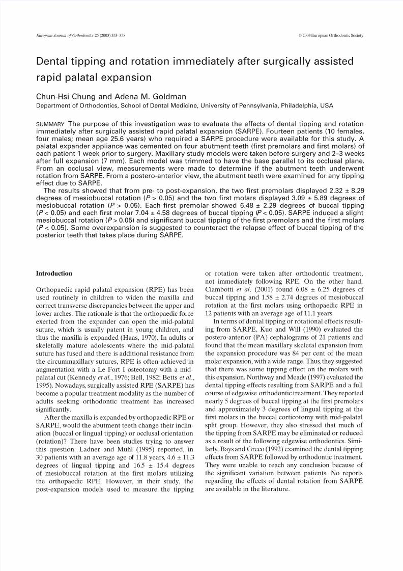
Pre-treatment (T1) study models were taken forall patients. One week before the scheduled SARPEprocedure, each patient had a palatal expansion appli-ance with a 7 mm jackscrew (Figure 1) cemented onfour abutment teeth (maxillary first premolars and first
molars). All palatal expanders were made at the sameorthodontic laboratory.
Three surgeons, using a similar technique, performedthe surgery on all the patients. Essentially, the techniqueconsisted of a subtotal Le Fort I osteotomy and a mid-palatal cut, as reported by Betts et al . (1995).
After surgery, the patients were instructed to activatethe palatal expander two turns (0.25 mm expansion eachturn) per day until the jackscrew was fully opened(7 mm). The expander was then inactivated by tying off the jackscrew with ligature wire or self-cure acrylic. Post-expansion (T2) maxillary study models were obtained for
all patients 2–3 weeks later. All the impressions weretaken with alginate and none of them showed anydistortion. From T1 to T2, no brackets or wires wereplaced in the maxillary dentition and none of the teethhad any occlusal adjustment.
Study models
For each T1 and T2 maxillary model, the base wastrimmed with a model trimmer (Great Lakes
Orthodontics, Tonawanda, New York, USA) to parallelits occlusal plane. The occlusal plane was registered atthe incisal edge of the right central incisor and thecentral occlusal fossa of the right and left first molars.
On each T1 model, a 0.020 inch stainless steel wire,approximately 2–3 cm in length, was fixed from the
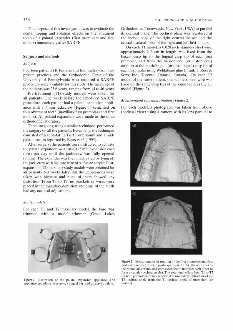
buccal cusp tip to the lingual cusp tip of each firstpremolar, and from the mesiobuccal (or distobuccal)cusp tip to the mesiolingual (or distolingual) cusp tip of each first molar using Weldabond glue (Frank T. Ross &Sons, Inc., Toronto, Ontario, Canada). On each T2model of the same patient, the stainless steel wire wasfixed on the same cusp tips of the same teeth as the T1model (Figure 2).
Measurement of dental rotation (Figure 2)
For each model, a photograph was taken from above(occlusal view) using a camera with its lens parallel to
354 C.-H. CHUNG AND A. M. GOLDMAN
Figure 1 Illustration of the palatal expansion appliance. Theappliance includes a jackscrew, a lingual bar, and an acrylic palate.
Figure 2 Measurements of rotation of the first premolars and firstmolars from pre- (T1, a) to post-expansion (T2, b). The wire lines onthe premolars (or molars) were extended to intersect each other toform an angle (occlusal angle). The rotational effect from T1 to T2for both premolars (or molars) was determined by subtraction of theT2 occlusal angle from the T1 occlusal angle of premolars (ormolars).
7/30/2019 Disjuncao cirurgicamente assistida
http://slidepdf.com/reader/full/disjuncao-cirurgicamente-assistida 3/6
the occlusal plane. On each picture, the wire lines on thefirst premolars were extended to intersect each other toform an angle (occlusal premolar angle). Similarly, anangle was formed by extending the wire lines on thefirst molars (occlusal molar angle). Each premolar ormolar occlusal angle was measured by two examiners
independently and an average was calculated. The rotationaleffect from T1 to T2 for the two first premolars(or molars)was determined by the subtraction of the T2 occlusalpremolar (or molar) angle from the T1 occlusal premolar(or molar) angle. An increase in the occlusal premolaror molar angle from T1 to T2 was defined as positive (+)and mesiobuccal rotation, whereas a decrease in the angleswas defined as negative (–) and mesiolingual rotation.
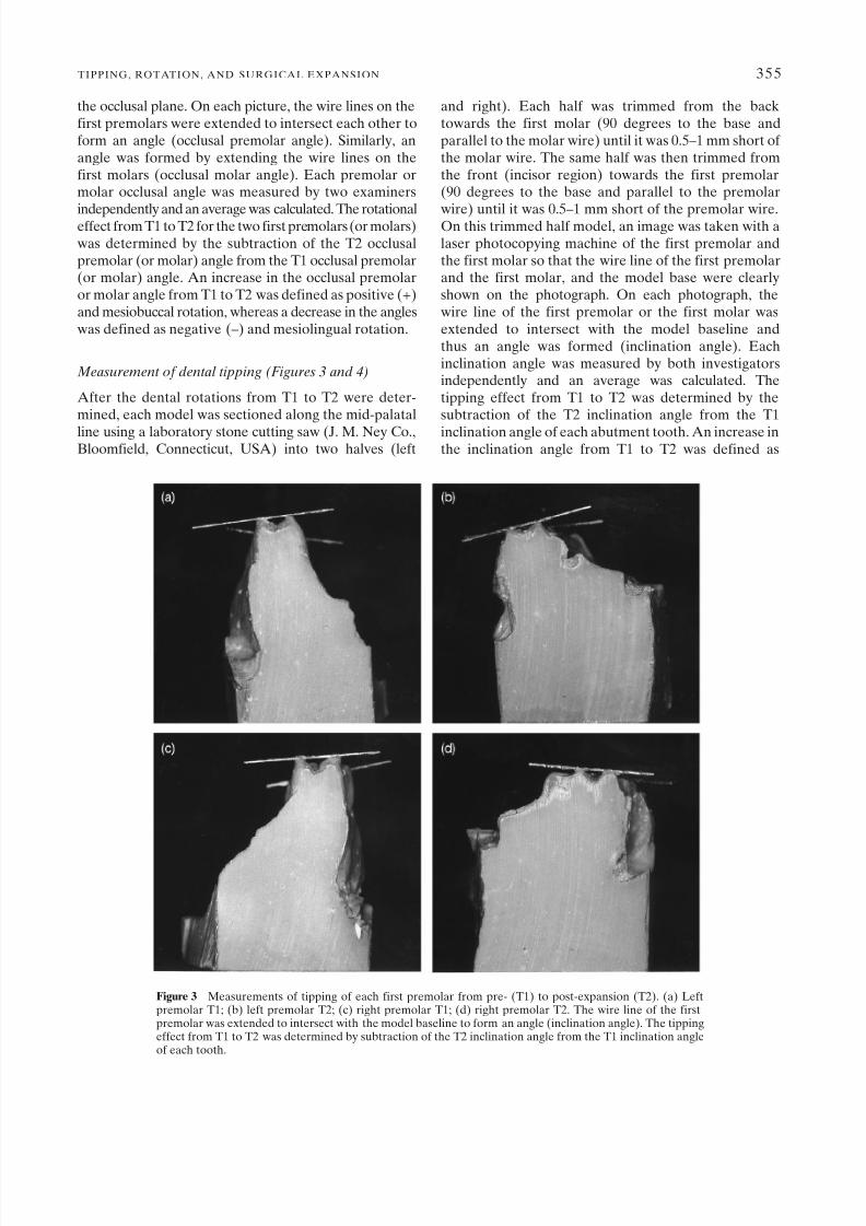
Measurement of dental tipping (Figures 3 and 4)
After the dental rotations from T1 to T2 were deter-mined, each model was sectioned along the mid-palatal
line using a laboratory stone cutting saw (J. M. Ney Co.,Bloomfield, Connecticut, USA) into two halves (left
and right). Each half was trimmed from the backtowards the first molar (90 degrees to the base andparallel to the molar wire) until it was 0.5–1 mm short of the molar wire. The same half was then trimmed fromthe front (incisor region) towards the first premolar(90 degrees to the base and parallel to the premolar
wire) until it was 0.5–1 mm short of the premolar wire.On this trimmed half model, an image was taken with alaser photocopying machine of the first premolar andthe first molar so that the wire line of the first premolarand the first molar, and the model base were clearlyshown on the photograph. On each photograph, thewire line of the first premolar or the first molar wasextended to intersect with the model baseline andthus an angle was formed (inclination angle). Eachinclination angle was measured by both investigatorsindependently and an average was calculated. Thetipping effect from T1 to T2 was determined by thesubtraction of the T2 inclination angle from the T1
inclination angle of each abutment tooth. An increase inthe inclination angle from T1 to T2 was defined as
TIPPING, ROTATION, AND SURGICAL EXPANSION 355
Figure 3 Measurements of tipping of each first premolar from pre- (T1) to post-expansion (T2). (a) Leftpremolar T1; (b) left premolar T2; (c) right premolar T1; (d) right premolar T2. The wire line of the firstpremolar was extended to intersect with the model baseline to form an angle (inclination angle). The tippingeffect from T1 to T2 was determined by subtraction of the T2 inclination angle from the T1 inclination angleof each tooth.
7/30/2019 Disjuncao cirurgicamente assistida
http://slidepdf.com/reader/full/disjuncao-cirurgicamente-assistida 4/6
positive (+) and lingual tipping, whereas a decrease inthe inclination angle was defined as negative (–) andbuccal tipping.
Statistical analysis
Descriptive statistical analyses, including the mean andstandard deviation, were calculated for all measurements.A Student’s paired t-test was used to determine if therewas any significant difference in the angular measure-
ments between the two examiners (inter-examinerreliability test), and if the tipping and rotation changesfrom pre- to post-expansion were statistically significant.Significance for all statistical tests was predetermined atP < 0.05.
Results
The inter-examiner reliability test showed no statisticallysignificant difference in the measurement of the degreeof tipping and rotation between the two examiners(P = 0.95).
Dental rotation measurements
Table 1 shows the rotation of the two first premolars andthe two first molars from T1 to T2. Compared with thepre-expansion models, the post-expansion premolarsdisplayed a mean of 2.32 ± 8.29 degrees of mesiobuccalrotation (P > 0.05). Similarly, the post-expansion twofirst molars displayed a mean of 3.09 ± 5.89 degrees of mesiobuccal rotation resulting from SARPE (P > 0.05).No statistically significant differences in rotation from
T1 to T2 were found between the first premolars andthe first molars.
Dental tipping measurements
Table 2 shows the changes in the inclination (tipping) of each first premolar and first molar and an average of both premolars and molars from T1 to T2. The leftfirst premolar displayed 6.31 ± 3.17 degrees of buccaltipping (P < 0.05) and the right first premolar displayed6.64 ± 4.64 degrees of buccal tipping (P < 0.05). Theaverage buccal tipping of the left and right first
356 C.-H. CHUNG AND A. M. GOLDMAN
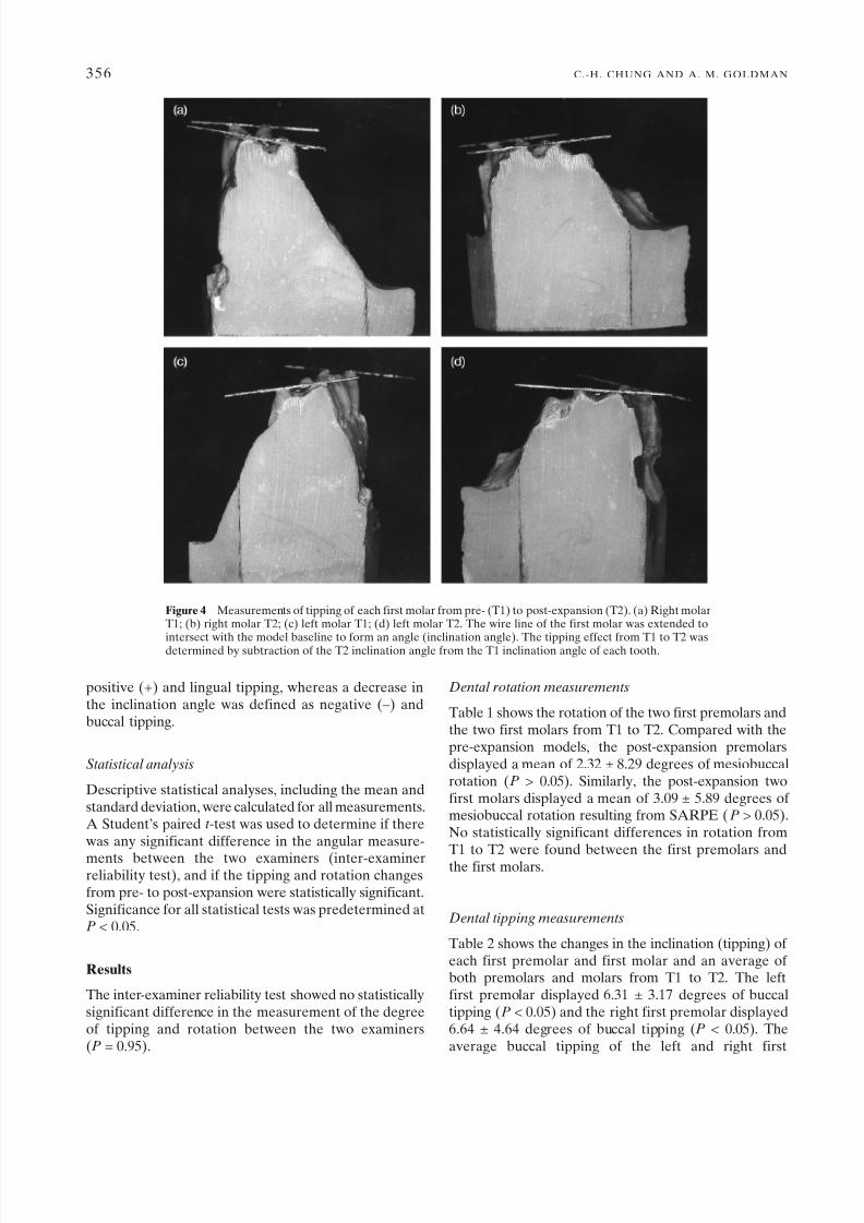
Figure 4 Measurements of tipping of each first molar from pre- (T1) to post-expansion (T2). (a) Right molarT1; (b) right molar T2; (c) left molar T1; (d) left molar T2. The wire line of the first molar was extended to
intersect with the model baseline to form an angle (inclination angle). The tipping effect from T1 to T2 wasdetermined by subtraction of the T2 inclination angle from the T1 inclination angle of each tooth.

7/30/2019 Disjuncao cirurgicamente assistida
http://slidepdf.com/reader/full/disjuncao-cirurgicamente-assistida 5/6
premolars from T1 to T2 was 6.48 ± 2.29 degrees(P < 0.05). The left first molar displayed 7.94 ± 3.88degrees of buccal tipping (P < 0.05) and the right first
molar 6.14 ± 6.15 degrees of buccal tipping (P < 0.05).The average buccal tipping of the left and right firstmolars from T1 to T2 was 7.04 ± 4.58 degrees (P < 0.05).When the left and the right sides and also the premolarsand the molars were compared, no significant differ-ences were found (P > 0.05).
Discussion
These results clearly show that there was significantbuccal tipping of the first premolars and the first molarsimmediately after SARPE (each first premolar tippedby 6.48 degrees, P < 0.05 and each first molar by 7.04
degrees, P < 0.05). The data are similar to a previousstudy by Kuo and Will (1990), who examined PAcephalograms of 21 patients and found more intermolarexpansion than maxillary skeletal width expansion result-ing from RPE in conjunction with a modified lateralmaxillary corticotomy. They suggested that the differencewas due to molar buccal tipping. The present resultscannot be compared with the findings of Northwayand Meade (1997), who examined the study models of 15 patients and found 5 degrees of buccal tipping at thefirst premolars and 3 degrees of lingual tipping at thefirst molars after SARPE and full edgewise orthodontic
treatment. The reason is that their post-expansionmodels were taken after orthodontic treatment and inthe present investigation they were taken 2–3 weeks
after SARPE.A slight mesiobuccal rotation on the abutment teethwas found after SARPE (2.32 degrees for the two firstpremolars and 3.09 degrees for the two first molars).Although the amount of rotation was not statisticallysignificant (P > 0.05), a large standard deviation (± 8.29degrees for the premolars and ± 5.89 degrees for themolars) and a wide range (–17.2 to +16.5 degrees for thepremolars and –15.8 to +6.5 degrees for the molars)was present, which might be due to the small samplesize (n = 14). Thus, it would be difficult to draw aconclusion on the effects of dental rotation. A largersample size would be needed to answer this question. To
our knowledge, this is the first report of dental rotationresulting from SARPE, although a study evaluating thedental rotational changes after orthopaedic RPE hasbeen reported (Ciambotti et al ., 2001).
The T1 and T2 models of each patient were trimmedwith their bases parallel to the occlusal plane, which wasregistered at the incisal edge of the central incisor andthe central occlusal fossa of the right and left firstmolars. It should be noted that there might have beensome minor changes in the occlusal plane resultingfrom SARPE. This limitation may have affected theinclination readings of individual right or left abutment
TIPPING, ROTATION, AND SURGICAL EXPANSION 357
Table 1 Effects of rotation on both maxillary first premolars and maxillary first molars from pre- (T1) to post-surgicallyassisted rapid palatal expansion (T2).
Abutment tooth n Changes in occlusal angle SD Range Significancefrom T1 to T2 (degrees) (degrees) (degrees)
Both first premolars 14 +2.32 8.29 –17.2 to +16.5 NS
Both first molars 14 +3.09 5.89 –15.8 to +6.5 NS
SD, standard deviation; NS, not statistically significant, P > 0.05.A positive change indicates mesiobuccal rotation and a negative change indicates mesiolingual rotation.
Table 2 Changes in inclination (tipping) of each abutment tooth from pre- (T1) to post-surgically assisted rapid palatalexpansion (T2).
Abutment tooth n Changes in inclination angle SD Range Significancefrom T1 to T2 (degrees) (degrees) (degrees)
Left first premolar 14 –6.31 3.17 –12.3 to –1.5 *Right first premolar 14 –6.64 4.64 –17.5 to –0.3 *
Averaged premolar 14 –6.48 2.29 –10.1 to –2.7 *Left first molar 14 –7.94 3.88 –14.0 to –2.6 *Right first molar 14 –6.14 6.15 –18.1 to +3.3 *
Averaged molar 14 –7.04 4.58 –16.1 to –1.6 *
SD, standard deviation.*Statistically significant, P < 0.05.A positive change indicates lingual tipping and a negative change indicates buccal tipping.There was no significant difference in tipping between right and left premolars, right and left molars, premolars and molars (P > 0.05).
7/30/2019 Disjuncao cirurgicamente assistida
http://slidepdf.com/reader/full/disjuncao-cirurgicamente-assistida 6/6
teeth. However, it should have minimal effects on theaverage of the inclination readings of the left and rightindividual abutment teeth. This is the reason why theaverage inclination measurements for both the right andleft first premolars and molars are provided.
PA cephalograms have been used by other investi-
gators to evaluate the dental tipping effects frommaxillary expansion devices (Kuo and Will, 1990;Asanza et al ., 1996). In the present study, models wereused because it is difficult to locate and trace the firstmolars and first premolars precisely with PA cephalo-grams, as there is much overlapping of anatomicalstructures.
The inclination angle of each first premolar and firstmolar was measured separately instead of the angle of intersection of the lines drawn through the mesiobuccaland mesiolingual cusp tips of both the first molars or thefirst premolars together, as suggested by McNamaraand Brudon (1993). The reason is that the cusp tips of
the two first premolars and the two first molars areoften not on the same plane due to rotation (this can bedemonstrated by the formation of the occlusal premolarangle or occlusal molar angle). Thus, from the PA view,the angle of intersection of cusp tips cannot representthe inclination of the two teeth. This is why each castwas sectioned into two halves, each half trimmed tothe abutment tooth and the inclination angle of eachabutment tooth measured.
Orthopaedic expansion has been suggested tooverexpand to the point where the lingual cusps of theupper posterior teeth are in contact with the buccal
cusps of the lower posterior teeth, due to the relapsepotential after appliance removal (Krebs, 1964; Haas,1973). However, for SARPE, Kraut (1984) suggestedthat only 1.0–1.5 mm of extra expansion be accomplished,as surgically assisted expansions are stable. The resultsof this investigation showed significant buccal tipping atthe first premolars and molars after SARPE, which maytend to relapse. Thus, some overexpansion may benecessary. Further studies are needed to determinethe amount of overexpansion that is required withSARPE.
Conclusions1. There was a slight mesiobuccal rotation of the
maxillary first premolars and first molars resultingfrom SARPE (P > 0.05).
2. SARPE induced significant buccal tipping of themaxillary first premolars and first molars (P < 0.05).
3. Some overexpansion is suggested to counteract therelapse effect of buccal tipping of the posterior teeththat takes place during SARPE.
Address for correspondence
Chun-Hsi ChungDepartment of OrthodonticsSchool of Dental MedicineUniversity of Pennsylvania4001 Spruce Street
Philadelphia, PA 19104–6003USA
Acknowledgements
We acknowledge the contributions of Drs SolomonKatz, Peter Greco, Robert Vanarsdall, Todd Welsh, andMs René Lukasiewicz.
References
Asanza S, Cisneros G J, Nieberg L G 1996 Comparison of Hyrax andbonded expansion appliances. Angle Orthodontist 67: 15–22
Bays R A, Greco J M 1992 Surgically assisted rapid palatalexpansion: an outpatient technique with long-term stability.Journal of Oral and Maxillofacial Surgery 50: 110–113
Bell R 1982 A review of maxillary expansion in relation to rate of expansion and patient’s age. American Journal of Orthodontics81: 32–37
Betts N J, Vanarsdall R L, Barber H D, Higgins-Barber K, FonsecaR J 1995 Diagnosis and treatment of transverse maxillarydeficiency. International Journal of Adult Orthodontics andOrthognathic Surgery 10: 75–96
Ciambotti C, Ngan P, Durkee M, Kohli K, Kim H 2001 Acomparison of dental and dentoalveolar changes between rapidpalatal expansion and nickel–titanium palatal expansionappliances. American Journal of Orthodontics and Dentofacial
Orthopedics 119: 11–20Haas A J 1970 Palatal expansion: just the beginning of dentofacial
orthopedics. American Journal of Orthodontics 57: 219–255
Haas A J 1973 JCO/Interviews. Journal of Clinical Orthodontics 7:227–245
Kennedy J W III, Bell W H, Kimbrough O L, James W B 1976Osteotomy as an adjunct to rapid maxillary expansion. AmericanJournal of Orthodontics 70: 123–137
Kraut R A 1984 Surgically assisted maxillary expansion by openingthe midpalatal suture. Journal of Oral and Maxillofacial Surgery42: 651–655
Krebs A 1964 Mid-palatal expansion studied by the implant methodover a seven-year period. Transactions of the EuropeanOrthodontic Society, pp. 131–142
Kuo P C, Will L A 1990 Surgical–orthodontic treatment of maxillaryconstriction. Oral and Maxillofacial Surgery Clinics of NorthAmerica 2: 751–759
Ladner P T, Muhl Z F 1995 Changes concurrent with orthodontictreatment when maxillary expansion is a primary goal. AmericanJournal of Orthodontics and Dentofacial Orthopedics 108: 184–193
McNamara J A, Brudon W L 1993 Orthodontic and orthopedictreatment in the mixed dentition. Needham Press, Ann Arbor
Northway W M, Meade J B Jr 1997 Surgically assisted rapidmaxillary expansion: a comparison of technique, response, andstability. Angle Orthodontist 67: 309–320
358 C.-H. CHUNG AND A. M. GOLDMAN