Embed Size (px)
Citation preview

Submitted 13 March 2018Accepted 16 May 2018Published 5 June 2018
Corresponding authorHoe-Han Goh, [email protected]
Academic editorVladimir Uversky
Additional Information andDeclarations can be found onpage 14
DOI 10.7717/peerj.4914
Copyright2018 Ravee et al.
Distributed underCreative Commons CC-BY 4.0
OPEN ACCESS
Discovery of digestive enzymes incarnivorous plants with focus onproteasesRishiesvari Ravee, Faris ‘Imadi Mohd Salleh and Hoe-Han GohInstitute of Systems Biology (INBIOSIS), Universiti Kebangsaan Malaysia, Bangi, Selangor, Malaysia
ABSTRACTBackground. Carnivorous plants have been fascinating researchers with their uniquecharacters and bioinspired applications. These include medicinal trait of some carniv-orous plants with potentials for pharmaceutical industry.Methods. This review will cover recent progress based on current studies on digestiveenzymes secreted by different genera of carnivorous plants:Drosera (sundews),Dionaea(Venus flytrap), Nepenthes (tropical pitcher plants), Sarracenia (North Americanpitcher plants), Cephalotus (Australian pitcher plants), Genlisea (corkscrew plants), andUtricularia (bladderworts).Results. Since the discovery of secreted protease nepenthesin in Nepenthes pitcher,digestive enzymes from carnivorous plants have been the focus of many studies. Recentgenomics approaches have accelerated digestive enzyme discovery. Furthermore,the advancement in recombinant technology and protein purification helped in theidentification and characterisation of enzymes in carnivorous plants.Discussion. These different aspects will be described and discussed in this review withfocus on the role of secreted plant proteases and their potential industrial applications.
Subjects Biochemistry, Biotechnology, Genomics, Plant ScienceKeywords Carnivorous plants, Enzyme discovery, Digestive enzyme, Secreted protease, Industrialapplications, Protein characterisation
INTRODUCTIONNitrogen is the most crucial mineral nutrient required by plants but its availability islargely limited in many terrestrial ecosystems (Behie & Bidochka, 2013). For adaptation tosuch unfavourable environment, carnivorous plants have developed the ability to attract,capture, and digest preys into simpler mineral compounds, which are then absorbed forplant growth and reproduction (Ellison, 2006). The first evidence on the ability of the plantto capture and digest insects was provided over 140 years ago (Darwin, 1875). Since then,more than 700 carnivorous species from 20 genera of 12 families (Givnish, 2015) have beenidentified with captivating morphological and physiological traits linked to carnivory (Królet al., 2011).
There are a few reviews on the evolution of carnivorous plants and their biotechnologicalapplications (Król et al., 2011; Miguel, Hehn & Bourgaud, 2018). However, a systematicreview with focus on digestive enzyme discovery and characterisation from all families ofcarnivorous plants is lacking. Furthermore, the pharmacological potentials of some of these
How to cite this article Ravee et al. (2018), Discovery of digestive enzymes in carnivorous plants with focus on proteases. PeerJ 6:e4914;DOI 10.7717/peerj.4914

carnivorous plants have also been largely overlooked. With the advent of omics technologywhich accelerated enzyme discovery in carnivorous plants for the past few years, there is apressing need for a timely review on current progress of studies in this field. This reviewwill be useful not only to researchers working on carnivorous plants, but also those withinterest in commercially useful enzymes and natural products.
SURVEY METHODOLOGYIn this review, we provide perspectives on the latest research of different carnivorous plants,namely Cephalotus, Drosera, Dionaea, Genlisea, Nepenthes, Sarracenia, and Utricularia, ontheir digestive enzyme discovery and characterisation. In earlier studies, research intereston carnivorous plants was centred on axenic culture, ultrastructure of specialised trappingorgans, foliar absorption of nutrients derived from preys, and the enzymatic studies ofprey digestion (Adamec, 1997; Gorb et al., 2004; Farnsworth & Ellison, 2008). Thus, thisreview summarises the previous findings with focus on digestive enzymes discovered incarnivorous plants, especially proteases and their industrial applications. The literaturesurvey was performed exhaustively online using Google search engine and SCOPUS. Thediscussion will be mainly based on recent studies.
Different families of carnivorous plantsThe emergence of carnivorous syndrome requires significant functional adaption in plantmorphology and physiology. Carnivory trait has evolved independently in different ordersof flowering plants, namely Caryophyllales, Ericales, Lamiales, Oxalidales, and Poales(Müller et al., 2004; Ellison & Gotelli, 2009; Król et al., 2011). This comprised of 12 differentfamilies of carnivorous plants with five distinct trapping mechanisms, including flypapertrap, snap trap, pitfall trap, suction trap, and eel trap (Table 1). The development of uniquetraps is one of the major indicators of carnivorous syndrome. These traps originate fromthe leaves specialised in trapping, digesting and absorbing nutrients from prey at the costof reduced photosynthesis (Ellison & Gotelli, 2009). The modified leaves of carnivorousplants often form either an active or passive trap (Bauer et al., 2015). An active trap involvesmovementmechanics to aid prey capture, whereas a passive trap relies on its morphologicalstructure to trap prey.
In Caryophylles, Droseraceae is one of the most species-rich families of carnivorousplants comprising over 160 species in Drosera genus of sundews with flypaper trap (Ellison& Gotelli, 2009). Earlier studies have reported the application of sundew plants as a remedyfor pulmonary illnesses and coughs (Didry et al., 1998), in the form of tincture (Caniato,Filippini & Cappelletti, 1989). Compounds of pharmaceutical interest in Drosera includeflavonoids, phenolic compounds, and anthocyanins. Drosera herbs have antispasmodic,diuretic, and expectorant properties (Banasiuk, Kawiak & Krölicka, 2012). Additionally, invitro culture extracts of Drosera were reported with antibacterial and anticancer properties(Banasiuk, Kawiak & Krölicka, 2012). Interestingly, a crystal-like pigment from D. peltatacan also be used as a dye in silk industry (Patel, 2014).
Venus flytrap (Dionaea muscipula) is another well-known member of Droseraceae dueto its unique snap-trapping mechanism to capture small preys, primarily insects or spiders.
Ravee et al. (2018), PeerJ, DOI 10.7717/peerj.4914 2/22

Table 1 Different carnivorous plant families and trapping mechanisms. Modified fromKról et al.(2011) andGivnish (2015).
Order Family Genus Trap
Dioncophyllaceae Triphyophyllum FlypaperDrosophyllaceae Drosophyllum Flypaper
Drosera FlypaperAldrovanda Snap
Droseraceae
Dionaea Snap
Caryophyllales
Nepenthaceae Nepenthes PitfallRoridulaceae Roridula Flypaper
Darlingtonia PitfallHeliamphora Pitfall
EricalesSarraceniaceae
Sarracenia PitfallPlantaginaceae Philcoxia FlypaperByblidaceae Byblis FlypaperLentibulariaceae Pinguicula Flypaper
Utricularia Suction
Lamiales
Genlisea EelOxalidales Cephalotaceae Cephalotus Pitfall
Bromeliaceae Brocchinia PitfallCatopsis Pitfall
Poales
Eriocaulaceae Paepalanthus Pitfall
Interestingly, the trapping signal ofDionaea is the fastest ever reported in the plant kingdomover 140 years ago (Darwin, 1875). The secretion of digestive fluid is highly induced bytouch stimulation of ‘trigger hairs’ on the trap sticky surface. Naphthoquinones werediscovered from in vitro culture extract of Venus flytrap which is a traditional medicine forcough (Banasiuk, Kawiak & Krölicka, 2012). Plumbagin is another promising antitumorcompound among the abundant beneficial secondary metabolites found in D. muscipula(Gaascht, Dicato & Diederich, 2013).
Cephalotaceae, Nepenthaceae, and Sarraceniaceae are three families of carnivorousplants which develop modified leaves shaped like a pitcher as a passive pitfall trap. Adigestive zone is located at the lowest inner wall of the pitcher with abundant digestiveglands responsible for the secretion of hydrolytic enzymes. In contrast, Bromeliaceae andEriocaulaceae of Poales forms tube-like pitfall trap from overlapping erect leaves insteadof a modified leaf organ. Most studies showed low production of enzymes in Brocchinia,Catopsis, and Paepalanthus in the absence of abundant specialised glands (Givnish et al.,1984; Adlassnig, Peroutka & Lendl, 2010). Some pitchers of Nepenthes and Sarracenia areso big that larger prey, such as frog and rodent are frequently found partially digestedinside the pitcher (Adlassnig, Peroutka & Lendl, 2010). This phenomenon shows that preysof carnivorous plants are not restricted to only insects.
For Sarracenia, its pitcher acts as rainwater storage and at the same time secreteshydrolytic enzymes and other proteins for prey digestion. The secretions formed at thehood of pitcher lure insect prey, which eventually fall and drown in the pitcher fluid
Ravee et al. (2018), PeerJ, DOI 10.7717/peerj.4914 3/22

(Ellison & Gotelli, 2001). The prey is digested by the digestive enzymes, such asphosphatases, proteases, and nucleases in the pitcher fluid (Chang & Gallie, 1997).Interestingly, Sarracenia has been used as a traditional remedy for childbirth and as adiuretic agent (Patel, 2014). Moreover, tea made from its dried foliage can be used to treatfever and cold; whereas its roots can be consumed as a remedy for lung, liver, and smallpoxdiseases (Patel, 2014).
Nepenthes is a genus of tropical pitcher plants from the species-rich Nepenthaceaefamily with fascinatingly diverse pitcher structures adapted to different ecological nichesand feeding habits. Despite the lack of a complete genome from this family, there arequite a few reports on transcriptome sequences. Recently, Zulkapli et al. (2017) reportedthe first single molecule real time sequencing of full-length transcriptome sequences forN. ampullaria, N. rafflesiana, and N. ×hookeriana. Metabolomics approach has also beentaken for the first time in these three species to profile compounds in pitcher tissue (Rosliet al., 2017). Ethnomedicinal properties of Nepenthes are well documented with boiledroots act as a remedy for stomach ache. The pitcher fluid can be consumed to cure urinarydiseases and used as eye drops to treat itchy eyes. Besides, the root and stem can serveas building materials for housing construction in place of rattan due to its elasticity andenduring property (Miguel, Hehn & Bourgaud, 2018). Besidews that, Nepenthes pitchershave a distinct use in traditional cooking of glutinous rice snacks, which is practised byBidayuh and Kadazan-Dusun people in Malaysia using N. ampullaria and N. mirabilis(Schwallier et al., 2015). Furthermore, Nepenthes also has a great potential as pest controlagent in agriculture due to their ability to capture and kill insects, such as flies, ants, bees, andbeetles; some even kill small animals like frog and rats (Miguel, Hehn & Bourgaud, 2018).
Genlisea and Utricularia are carnivorous plants under the family of Lentibulariaceae.These plants feed on microscopic preys and digest them in a closed trap under water.Utricularia spp. have reported usage for dressing wounds and as a remedy for urinaryinfections and cough (Patel, 2014). To date, Genlisea aurea (Leushkin et al., 2013) andUtricularia gibba (Lan et al., 2017) are among the four carnivorous plants with genomesequences publicly available, apart from Drosera capensis (Butts, Bierma & Martin, 2016)and Cephalotus follicularis (Fukushima et al., 2017). The availability of genome sequenceshas contributed greatly to enzyme discovery and improving our understanding of carnivorymechanisms and evolution in different carnivorous plant families.
DIGESTIVE ENZYME DISCOVERY, IDENTIFICATION ANDCHARACTERISATIONDigestion of prey in carnivorous plants relies on enzymes which could be associated withmorphologically diverse trapping mechanisms. There are a few studies which reported thatthe secretion of the digestive enzymes is strongly induced by prey capture. However, thereare also certain digestive enzymes which are readily secreted in the absence of prey. Thisindicates plant regulation of enzyme secretion because the production and secretion ofenzymes incur energetic costs.
To date, numerous studies had reported the discovery of distinct digestive enzymesin carnivorous plants (Table 2). Similar enzymes with various enzymatic properties were
Ravee et al. (2018), PeerJ, DOI 10.7717/peerj.4914 4/22
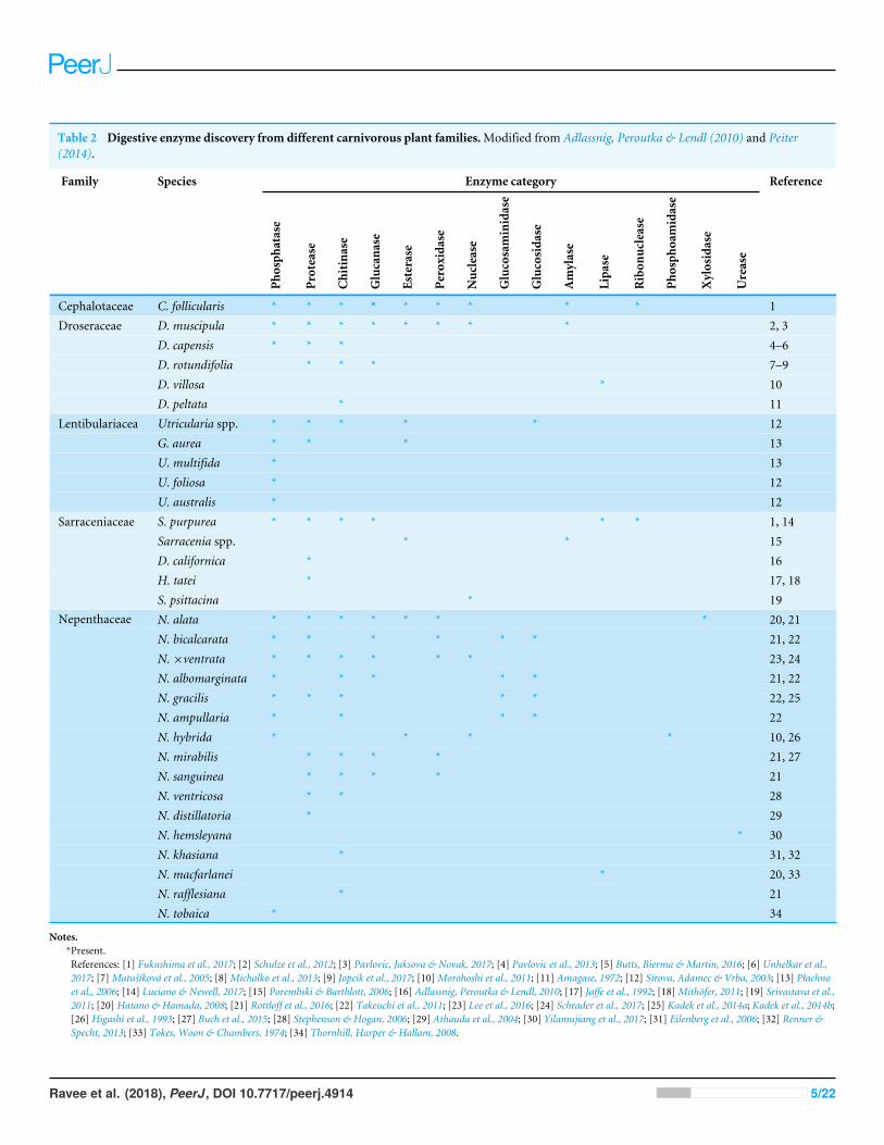
Table 2 Digestive enzyme discovery from different carnivorous plant families.Modified from Adlassnig, Peroutka & Lendl (2010) and Peiter(2014).
Family Species Enzyme category Reference
Phosph
atase
Protease
Chitina
se
Glucana
se
Esterase
Peroxida
se
Nuclease
Glucosaminidase
Glucosida
se
Amylase
Lipa
se
Ribon
uclease
Phosph
oamidase
Xylosidase
Urease
Cephalotaceae C. follicularis * * * * * * * * * 1D. muscipula * * * * * * * * 2, 3D. capensis * * * 4–6D. rotundifolia * * * 7–9D. villosa * 10
Droseraceae
D. peltata * 11Utricularia spp. * * * * * 12G. aurea * * * 13U. multifida * 13U. foliosa * 12
Lentibulariacea
U. australis * 12S. purpurea * * * * * * 1, 14Sarracenia spp. * * 15D. californica * 16H. tatei * 17, 18
Sarraceniaceae
S. psittacina * 19N. alata * * * * * * * 20, 21N. bicalcarata * * * * * * 21, 22N. ×ventrata * * * * * * 23, 24N. albomarginata * * * * * 21, 22N. gracilis * * * * * 22, 25N. ampullaria * * * * 22N. hybrida * * * * 10, 26N. mirabilis * * * * 21, 27N. sanguinea * * * * 21N. ventricosa * * 28N. distillatoria * 29N. hemsleyana * 30N. khasiana * 31, 32N. macfarlanei * 20, 33N. rafflesiana * 21
Nepenthaceae
N. tobaica * 34
Notes.*Present.References: [1] Fukushima et al., 2017; [2] Schulze et al., 2012; [3] Pavlovic, Jaksova & Novak, 2017; [4] Pavlovic et al., 2013; [5] Butts, Bierma & Martin, 2016; [6] Unhelkar et al.,2017; [7]Matušíková et al., 2005; [8]Michalko et al., 2013; [9] Jopcik et al., 2017; [10]Morohoshi et al., 2011; [11] Amagase, 1972; [12] Sirova, Adamec & Vrba, 2003; [13] Płachnoet al., 2006; [14] Luciano & Newell, 2017; [15] Porembski & Barthlott, 2006; [16] Adlassnig, Peroutka & Lendl, 2010; [17] Jaffe et al., 1992; [18]Mithöfer, 2011; [19] Srivastava et al.,2011; [20] Hatano & Hamada, 2008; [21] Rottloff et al., 2016; [22] Takeuchi et al., 2011; [23] Lee et al., 2016; [24] Schrader et al., 2017; [25] Kadek et al., 2014a; Kadek et al., 2014b;[26] Higashi et al., 1993; [27] Buch et al., 2015; [28] Stephenson & Hogan, 2006; [29] Athauda et al., 2004; [30] Yilamujiang et al., 2017; [31] Eilenberg et al., 2006; [32] Renner &Specht, 2013; [33] Tokes, Woon & Chambers, 1974; [34] Thornhill, Harper & Hallam, 2008.
Ravee et al. (2018), PeerJ, DOI 10.7717/peerj.4914 5/22

shared among different carnivorous families. With the genome sequencing of Cephalotusfollicularis, various digestive enzymes were discovered, namely esterases, proteases,nucleases, phosphatases, glucanases, and peroxidases (Takahashi et al., 2009; Fukushimaet al., 2017). Similar classes of enzymes were also detected in other carnivorous families,such as Droseraceae, Lentibulariacea, Sarraceniaceae, and Nepenthaceae. This suggestssignificant role of common hydrolytic enzymes, especially phosphatases, proteases, andchitinases, in prey digestion of various carnivorous plants regardless of different families ortrapping mechanisms. Recently, Yilamujiang et al. (2017) reported the presence of a noveldigestive enzyme urease in N. hemsleyana which has developed a symbiosis relationshipwith bat.
However, investigation related to the identification of proteins found in the pitcherfluid is highly challenged by unusual amino acid composition and limited carnivorousplant genome or protein sequence database (Lee et al., 2016). Early study byAmagase (1972)utilised zymography technique to determine the protease activity found in fluid ofNepenthesspp. and D. peltata. The fluids were purified and characterised for acid protease anddemonstrated similar protease activity from two distinct families. Later,Hatano & Hamada(2008) conducted proteomic analysis on the digestive fluid of N. alata in which secretedchitinase, glucanase, and xylosidase were identified through in-gel trypsin digestion, de novopeptide assembly, and homology search using public databases. Recently, a transcriptomicapproach was taken for N. ampullaria, N. rafflesiana, N. × hookeriana, and N. × ventrata(Wan Zakaria et al., 2016a;Wan Zakaria et al., 2016b; Zulkapli et al., 2017), which can serveas reference sequences for identifying more digestive enzymes through proteomics analysis(Wan Zakaria et al., 2018). A proteomics informed by transcriptomics approach was takenby Schulze et al. (2012) to determine the proteins highly expressed in the digestive fluidof Venus flytrap. They discovered a coordinated prey digestion mechanism facilitated byvarious enzymes, such as chitinases, lipases, phosphatases, peroxidases, glucanases, andpeptidases. Fluorescent resonance energy transfer (FRET) based technique can be utilisedas an efficient and rapid detection of proteolytic activities in the pitcher fluid of variousNepenthes species (Buch et al., 2015). Rey et al. (2016) applied a similar approach to assessproteolytic efficiency of the protein secreted in the pitcher fluid of Nepenthes species.
On the other hand, purification of digestive enzymes from carnivorous fluid isextremely challenging due to low amount of secreted fluid and enzyme. Furthermore,pitcher fluids are often diluted with rainwater and even contaminated by decomposingprey. Nevertheless, there are studies which manage to purify and characterise digestiveenzymes from carnivorous plants (Table 3). Based on the reported purification andcharacterisation studies, proteases are the most abundant enzymes characterised from thedigestive fluid of carnivorous plant. The very first purification of protease from pitcherfluid ofNepenthes species was performed by Steckelberg, Lüttge & Weigl (1967)using Ecteolacolumn chromatography and its optimum activity was detected at pH 2.2 with stability at50 ◦C. To date, the common purification strategies applied by various studies are columnchromatography, affinity chromatography, ultrafiltration, and dialysis. Although manydigestive enzymes have been identified from carnivorous plants, only few studies havepurified and characterised the enzymes. Therefore, further studies on the purification andcharacterisation of various digestive enzymes are needed.
Ravee et al. (2018), PeerJ, DOI 10.7717/peerj.4914 6/22

Table 3 Characterisation and purification of digestive enzymes from carnivorous plants.
Enzyme Species Protein purificationmethod
Substrate Condition Reference
pH T (◦C)
Proteinase N. mixta, N. dormanniana,N.neuvilleana
Ecteola cellulosecolumn chromatography
Casein 2.2 50 Steckelberg, Lüttge &Weigl (1967)
Nepenthesin Nepenthes sp. DEAE-Sephadex A-50 Casein 2.8 40 Amagase, Nakayama &Tsugita (1969)
Proteinase D. muscipula Sephadex G-150column
Congocoll 5.5 37 Scala et al. (1969)
N.maxima, N. rafflesiana,N. ampullaria
Nepenthesin
N. dyeriana, N. mixta, D.peltata
Sephadex G-75,Sephadex G-200
Casein 3.0 40 Amagase (1972)
Nepenthesin Nepenthes sp. Sephadex G-75 & G-50,DEAE-Sephadex A-50
Casein 2.9 40 Jentsch (1972)
Bovine fibrin NA 37Bovine serum albumin NA 37
Nepenthesin N. macfarlanei Sephadex G-75 gel filtration
Horse-heartcytochrome c
2.2 37
Tokes, Woon &Chambers (1974)
Aspartic protease N. alata Not purified Bovine serum albumin 3.0 37 An, Fukusaki &Kobayashi (2002)
DEAE cellulose column,Sephacryl S-200
Nepenthesin I & II N. distillatoria
Pepstatin–Sepharosecolumn, Mono Qcolumn
Acid-denaturedhaemoglobin
2.8 50 Athauda et al. (2004)
*Cysteine protease*Aspartic protease
N. ventricosa Not purified Gelatin 3.0 NA Stephenson & Hogan(2006)
*Cysteine protease D. muscipula Hi-Trap Column 7-amino-4-methylcoumarin
3.6 60 Risør et al. (2016)
N. alata, C. follicularis, Haemoglobin 2.5 47–57D. muscipula Haemoglobin 3.0 60
Nepenthesin I & II
D. capensis
Not purified
Oxidised insulin Bchain
3.5 47
Takahashi et al. (2009)
(continued on next page)
Ravee
etal.(2018),PeerJ,DOI10.7717/peerj.4914
7/22
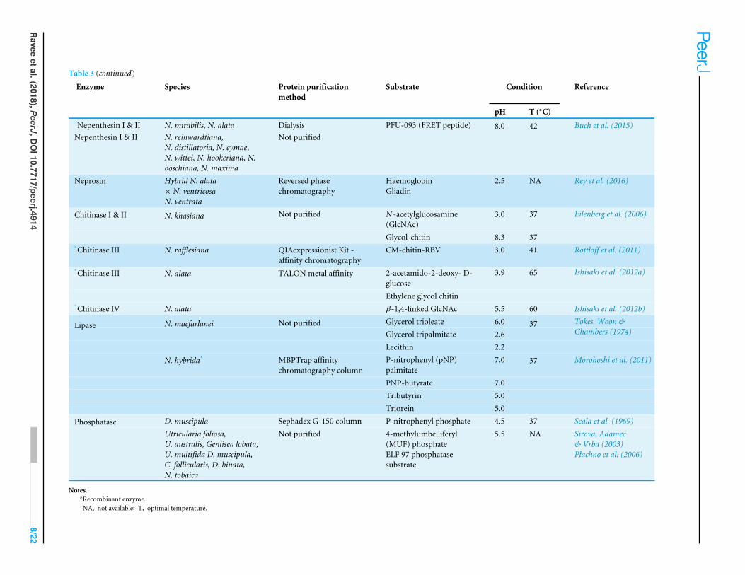
Table 3 (continued)
Enzyme Species Protein purificationmethod
Substrate Condition Reference
pH T (◦C)*Nepenthesin I & II N. mirabilis, N. alata DialysisNepenthesin I & II N. reinwardtiana,
N. distillatoria, N. eymae,N. wittei, N. hookeriana, N.boschiana, N. maxima
Not purifiedPFU-093 (FRET peptide) 8.0 42 Buch et al. (2015)
Neprosin Hybrid N. alata× N. ventricosaN. ventrata
Reversed phasechromatography
HaemoglobinGliadin
2.5 NA Rey et al. (2016)
N -acetylglucosamine(GlcNAc)
3.0 37Chitinase I & II N. khasiana Not purified
Glycol-chitin 8.3 37
Eilenberg et al. (2006)
*Chitinase III N. rafflesiana QIAexpressionist Kit -affinity chromatography
CM-chitin-RBV 3.0 41 Rottloff et al. (2011)
2-acetamido-2-deoxy- D-glucose
*Chitinase III N. alata
Ethylene glycol chitin
3.9 65 Ishisaki et al. (2012a)
*Chitinase IV N. alata
TALON metal affinity
β-1,4-linked GlcNAc 5.5 60 Ishisaki et al. (2012b)Glycerol trioleate 6.0Glycerol tripalmitate 2.6
N. macfarlanei Not purified
Lecithin 2.2
37 Tokes, Woon &Chambers (1974)
P-nitrophenyl (pNP)palmitate
7.0
PNP-butyrate 7.0Tributyrin 5.0
Lipase
N. hybrida* MBPTrap affinitychromatography column
Triorein 5.0
37 Morohoshi et al. (2011)
D. muscipula Sephadex G-150 column P-nitrophenyl phosphate 4.5 37 Scala et al. (1969)PhosphataseUtricularia foliosa,U. australis, Genlisea lobata,U. multifida D. muscipula,C. follicularis, D. binata,N. tobaica
Not purified 4-methylumbelliferyl(MUF) phosphateELF 97 phosphatasesubstrate
5.5 NA Sirova, Adamec& Vrba (2003)Płachno et al. (2006)
Notes.*Recombinant enzyme.NA, not available; T, optimal temperature.
Ravee
etal.(2018),PeerJ,DOI10.7717/peerj.4914
8/22
Most of the characterised enzymes can catalyse various substrates and activities ofthe same category of enzymes from different carnivorous plants are similar in terms ofoptimum pH, temperature, and substrate specificity (Table 3). For instance, most of thecharacterised proteases from different families function optimally at acidic condition.Interestingly, there are a few proteases reported to function optimally at high temperatureranging from 40−60 ◦C. Additionally, the secreted enzymes demonstrate higher stabilityagainst various chemicals and denaturing agents than similar enzymes from other sources.This is because prey digestion often occurs over long period under varied conditions,thus digestive enzymes are important to be active and stable (Butts, Bierma & Martin,2016). Subtle variations in enzymatic characteristics of digestive enzymes from differentcarnivorous plants remain to be explored. Furthermore, nomenclature of enzymes reportedfrom different carnivorous plants need to be standardised for comparative studies.
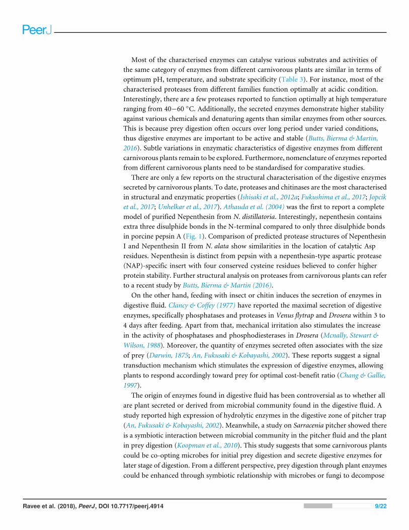
There are only a few reports on the structural characterisation of the digestive enzymessecreted by carnivorous plants. To date, proteases and chitinases are the most characterisedin structural and enzymatic properties (Ishisaki et al., 2012a; Fukushima et al., 2017; Jopciket al., 2017; Unhelkar et al., 2017). Athauda et al. (2004) was the first to report a completemodel of purified Nepenthesin from N. distillatoria. Interestingly, nepenthesin containsextra three disulphide bonds in the N-terminal compared to only three disulphide bondsin porcine pepsin A (Fig. 1). Comparison of predicted protease structures of NepenthesinI and Nepenthesin II from N. alata show similarities in the location of catalytic Aspresidues. Nepenthesin is distinct from pepsin with a nepenthesin-type aspartic protease(NAP)-specific insert with four conserved cysteine residues believed to confer higherprotein stability. Further structural analysis on proteases from carnivorous plants can referto a recent study by Butts, Bierma & Martin (2016).
On the other hand, feeding with insect or chitin induces the secretion of enzymes indigestive fluid. Clancy & Coffey (1977) have reported the maximal secretion of digestiveenzymes, specifically phosphatases and proteases in Venus flytrap and Drosera within 3 to4 days after feeding. Apart from that, mechanical irritation also stimulates the increasein the activity of phosphatases and phosphodiesterases in Drosera (Mcnally, Stewart &Wilson, 1988). Moreover, the quantity of enzymes secreted often associates with the sizeof prey (Darwin, 1875; An, Fukusaki & Kobayashi, 2002). These reports suggest a signaltransduction mechanism which stimulates the expression of digestive enzymes, allowingplants to respond accordingly toward prey for optimal cost-benefit ratio (Chang & Gallie,1997).
The origin of enzymes found in digestive fluid has been controversial as to whether allare plant secreted or derived from microbial community found in the digestive fluid. Astudy reported high expression of hydrolytic enzymes in the digestive zone of pitcher trap(An, Fukusaki & Kobayashi, 2002). Meanwhile, a study on Sarracenia pitcher showed thereis a symbiotic interaction between microbial community in the pitcher fluid and the plantin prey digestion (Koopman et al., 2010). This study suggests that some carnivorous plantscould be co-opting microbes for initial prey digestion and secrete digestive enzymes forlater stage of digestion. From a different perspective, prey digestion through plant enzymescould be enhanced through symbiotic relationship with microbes or fungi to decompose
Ravee et al. (2018), PeerJ, DOI 10.7717/peerj.4914 9/22

Figure 1 Comparison of the aspartic protease structures. (A) porcine pepsin (P00791), (B) NepenthesinI (Q766C3) and (C) Nepenthesin II (Q766C2) of Nepenthes gracilis. Active site (colour-shaded) is shownwith conserved catalytic Asp residues (arrowheads). Disulfide bonds are marked with asterisks. Box show-ing the conserved nepenthesin-type aspartic protein (NAP)-specific region with four conserved cysteineresidues. Models generated in SWISS-MODEL.
Full-size DOI: 10.7717/peerj.4914/fig-1
prey into simpler form of nutrients. Thismutualistic interaction withmicrobial communityin the digestive fluid will boost digestion and nutrient absorption. However, there mustbe a balancing point or even selection of microbial community (Takeuchi et al., 2015) toprevent competitive loss of nutrients as indicated by various defence-related proteins (Leeet al., 2016; Rottloff et al., 2016) and antimicrobial naphthoquinones (Buch et al., 2012)found in the pitcher fluid.
Ravee et al. (2018), PeerJ, DOI 10.7717/peerj.4914 10/22
SECRETED PROTEASES IN DIFFERENT FAMILIES OFCARNIVOROUS PLANTSCarnivorous plants attain substantial amount of nitrogen from prey through specialisedtrapping organs which accumulate acidic fluid containing protease. Early reports ofdigestive enzymes involved in carnivorous plants were initiated by Sir Joseph Hooker’sstudies of protease activity in the pitcher fluid ofNepenthes plants (Renner & Specht, 2013).Independent evolution of carnivorous plants might have resulted in convergent evolutionof diverse digestive enzymes serving similar functions (Fukushima et al., 2017).
Aspartic proteases (APs), such as nepenthesin, are one of the most abundant andwell characterised enzymes found in the digestive fluid (An, Fukusaki & Kobayashi,2002; Rottloff et al., 2016). AP have been purified and characterised from sterile pitcherfluid of several Nepenthes species (Jentsch, 1972; Tokes, Woon & Chambers, 1974). Ina study conducted by Nakayama & Amagase (1968), a protease from pooled pitcherfluids of N. mixta and N. maxima was only partially purified and characterised due toinsufficient amount.Amagase (1972) investigated aspartic proteases found inN. ampullaria,N. mixta, N. rafflesiana, N. maxima, andN. dyeriana compared to leaf extract fromDroserapeltata. Lately, acid protease from Nepenthes and Drosera genus are partially purifiedand characterised (Takahashi, Tanji & Shibata, 2007; Tokes, Woon & Chambers, 1974).Surprisingly, both the purified proteases from Nepenthes and Drosera share commoncharacteristics. An, Fukusaki & Kobayashi (2002) cloned homologous AP genes andexamined their expression in N. alata. The protease secreted in the pitcher fluid is pepsin-like and active at acidic condition (Rudenskaya et al., 1995). Although they have beencategorised as APs, none of the native enzymes was purified to homogeneity, mainly dueto difficulty in obtaining sufficient amount of pitcher fluid. Later, Athauda et al. (2004) forthe first time purified and characterised two APs, namely Nep1 and Nep2, from pitcherfluid N. distillatoria. They also characterised the amino acid sequences of the enzymesby cloning the cDNAs from pitcher tissue of N. gracilis. Recently, five nepenthesins werereported to be secreted in Nepenthes pitcher fluid (Lee et al., 2016). However, little isknown about why there are various AP genes expressed in Nepenthes pitcher fluid andtheir differential regulations if any. It is key to a better understanding of the regulation ofnitrogen-acquisition mechanism in Nepenthes plants.
Apart from aspartic proteases, there is also presence of cysteine proteases in carnivorousplants. Lately, it also has been found that cysteine protease is the primary proteasefound in digestive fluid of Dionaea (Venus flytrap). Prey proteins found in the digestivefluid of Dionaea are degraded by cysteine endopeptidases in association with serinecarboxypeptidases (Risør et al., 2016). This is highly distinct to the digestive fluids foundin Nepenthes and Drosera with aspartic proteases (Athauda et al., 2004). However, there isalso the presence of both aspartic and cysteine proteases in N. ventricosa as reported byStephenson & Hogan (2006). Takahashi, Tanji & Shibata (2007) conducted comparativeenzymatic characterisation of acid proteases from crude digestive fluid of variouscarnivorous plants namely Nepenthes, Chepalotus, Drosera, and Dionaea, with distincttrapping mechanisms. The study demonstrated significant variations between them, which
Ravee et al. (2018), PeerJ, DOI 10.7717/peerj.4914 11/22
might be due to the presence of different classes of proteases in different families. Thisreflects the phylogenetic diversity of these carnivorous plants.
There are attempts on the recombinant expression of the enzymes from carnivorousplants (Morohoshi et al., 2011; Ishisaki et al., 2012b; Kadek et al., 2014b). Kadek et al.(2014b) reported an efficient way to obtain high amount of Nepenthesin I (Nep1) fromN. gracilis through heterologous expression in Escherichia coli. The characteristics of therecombinant protein obtained are similar to the native enzyme isolated from the pitcherfluid. More recently, Nep1 from N. gracilis was successfully purified and crystallised(Fejfarová et al., 2016).
On the other hand, the evolution of different trapping mechanisms for carnivorousplants to survive in harsh environments with limited nutrients may result in enzymeswith novel properties. For instance, a novel class of prolyl endopeptidase called neprosin1 and neprosin 2 (Npr1 & Npr2) was recently discovered in Nepenthes species to bedistinct from commonly known proline-cleaving enzymes, which consists of two novelneprosin domains (Lee et al., 2016). Schrader et al. (2017) characterised neprosin to be aproline-cleaving enzyme through recombinant approach and demonstrated that it hasthe potential to be utilised for whole proteomic profiling and histone mapping. This isbecause neprosin is a low molecular weight prolyl endopeptidase and extremely activeat low concentration and pH. Combined actions of a neprosin and nepenthesin fromNepenthes pitcher fluid showed potential of effective gluten detoxification, which broadenthe prospects for enzyme supplementation approach to circumvent celiac disease (Reyet al., 2016).
Although the proteolytic activity in the digestive fluid is of great interest, low yields ofsecreted enzymes make it very challenging for native enzyme purification. Furthermore,prey digestion is likely to be concerted activities of various proteases and other enzymesin the digestive fluid, hence it is interesting to compare the enzyme assays between crudedigestive fluid extracts and individual purified proteases.
Applications of proteases from carnivorous plantsThe metabolic activity of most living organisms including plants, animals, fungi, bacteria,and viruses requires proteolytic enzymes. Proteases are one of the largest groups ofhydrolytic enzymes that cleave the peptide bonds in the polypeptide chains. The twomajor groups of proteases are endopeptidases that cleave non-terminal peptide bonds, andexopeptidases that can be classified to carboxypeptidases or aminopeptidases based on theirability to cleave the C or N terminal peptide bonds respectively. The four major classes ofproteases are aspartic proteases, serine proteases, cysteine proteases, and metalloproteases.
Proteases are the dominant class of industrial enzymes with diverse applications, suchas leather products, detergents, meat tenderisers, food products, as well as pharmaceuticaland waste processing industry (Rao et al., 1998; Lakshmi & Hemalatha, 2016). Almost60% of the total worldwide production of the enzymes are dominated by proteases(Usharani & Muthuraj, 2010). Microbes and animals are currently the major source ofproteases with only a few commercialised plant proteases. Interest has been growing inplant proteases, which have significant commercial values due to high stability in extreme
Ravee et al. (2018), PeerJ, DOI 10.7717/peerj.4914 12/22
Table 4 Applications of proteases from different plant sources.
Source Protease Application/functional properties Reference
Nepenthesin I & II Tool for digestion in H/D ExchangeMass Spectrometry
Kadek et al. (2014a), Kadek et al.(2014b) and Yang et al. (2015)
Proteomic analysis / Histone mapping Schrader et al. (2017)
NepenthesNeprosin
Gluten digestion Rey et al. (2016)Papaya Papain Meat tenderiser Amri & Mamboya (2012)
Denture cleaner Canay, Erguven & Yulug (1991)Detergent, healing burn wound,textiles, cosmestics industry
Choudhury et al. (2009)
Caricain Gluten-free food processing Buddrick, Cornell & Small (2015)Pineapple Bromelain Anti-inflammatory and anti-cancer
agentChanalia et al. (2011)
Fig (Ficus carica) Ficin Pharmaceutical industry Mazorra-Manzano, Ramírez-Suarez &Yada (in press)
Kiwifruit, Banana,Pineapple, Mango
Actinidin Dietary supplement Malone et al. (2005)
Zinger Zingipain Anti-proliferative agent Karnchanatat et al. (2011)Musk melon Cucumisin Hydrolysis of protein Feijoo-Siota & Villa (2011)Cardoon Cardosin A Milk clotting, manufacturing of
traditional cheeseFrazao et al. (1999)
Rice Oryzasin Milk clotting Simões & Faro (2004)Barley Phytepsin Milk clotting Runeberg-Roos & Saarma (1998)
conditions (Canay, Erguven & Yulug, 1991; Houde, Kademi & Leblanc, 2004; Karnchanatatet al., 2011). Examples of proteases from plant sources are listed in Table 4.
Broad substrate specificity, high activity in wide range of pH, temperature, and highstability in the presence of organic compounds are the major factors that attributedfor special attention towards proteolytic enzymes from plant sources. Furthermore,ethical/religious reasons and/or regulatory limitations, which restrict the applicationsof non-plant proteases (animal and recombinant sources) in certain countries pose aneed for new plant proteases. In plants, aspartic proteases are widely distributed in theseed, flower, leaf, as well as in the digestive fluid of carnivorous plants. Several plantaspartic proteases, such as oryzasin from rice and phytepsin from barley have been purifiedand well characterised. Proteases found in the digestive fluid of carnivorous plants arethe only extracellular proteinase of plant origin. Most plant proteases are known tobe intracellular vacuolar enzymes. Kadek et al. (2014a) and Yang et al. (2015) successfullyimmobilised nepenthesin-1 and nepenthesin-2 respectively as amolecular tool for digestionin hydrogen/deuterium exchange mass spectrometry (HXMS) to track exchange patternsin protein structure, especially useful for biopharmaceutical industry. Nep1 is shown toexhibit wide substrate cleavage specificity and high stability towards denaturing reagentscompared with pepsin for digesting protein into small peptides with overlapping fragmentsto provide necessary coverage of protein sequences.
Therefore, carnivorous plants signify a unique source of proteases for variousbiotechnological applications. The proteases discovered in the trap secretions could be
Ravee et al. (2018), PeerJ, DOI 10.7717/peerj.4914 13/22

distinct and provide wide range of functional temperature, stability and pH activityprofiles. Furthermore, differential substrate specificity among the proteases could providespecialised applications, such as that of demonstrated for a new mass spectrometrytechnique. The common plant proteases, such as bromelain and papain, denote only smallpopulation of plant proteases which are yet to be discovered. On the other hand, inhibitingprotease activity in digestive fluid will be critical when using carnivorous plants as hostsfor expressing functional plant-made proteins.
CONCLUSIONSThe search for new industrially viable plant enzymes is a continuous effort in whichcarnivorous plants serve as great resources for exploration. There are numerous studieson the properties of digestive fluid of carnivorous plants that contribute to a betterunderstanding of carnivory mechanism and evolution. Further extensive biochemicaland morphological studies on carnivorous plants will still be needed to help in furtherunderstanding the regulation of hydrolytic enzyme secretion. In addition, successfulpurification and characterisation of the secreted enzymes will encourage their exploitationfor industrial applications. Future research efforts in studying regulatory mechanisms ofdigestive enzymes ormetabolites responsible for attracting prey will not only be useful to fillin current gaps in knowledge, but also advancing novel utilisation of carnivorous plants forproducing plant-made proteins. Comparative genomics approach will help in elucidatingthe evolutionary history of these fascinating plants. With the advent of omics technologies,a holistic understanding on the molecular mechanisms of carnivory in various carnivorousplants will be achievable along with more exciting discoveries.
ADDITIONAL INFORMATION AND DECLARATIONS
FundingResearch was supported by Universiti KebangsaanMalaysia Research Grants DIP-2014-008andGUP-2017-057, and alsoMalaysiaMinistry ofHigher Education Fundamental ResearchGrant Scheme FRGS/2/2014/SG05/UKM/02/4. The funders had no role in study design,data collection and analysis, decision to publish, or preparation of the manuscript.
Grant DisclosuresThe following grant information was disclosed by the authors:Universiti Kebangsaan Malaysia Research Grants: DIP-2014-008, GUP-2017-057.Malaysia Ministry of Higher Education Fundamental Research Grant Scheme:FRGS/2/2014/SG05/UKM/02/4.
Competing InterestsThe authors declare there are no competing interests.
Ravee et al. (2018), PeerJ, DOI 10.7717/peerj.4914 14/22

Author Contributions• Rishiesvari Ravee conceived and designed the experiments, performed the experiments,analyzed the data, prepared figures and/or tables, authored or reviewed drafts of thepaper, approved the final draft.
• Faris ‘Imadi Mohd Salleh conceived and designed the experiments, performed theexperiments, analyzed the data, authored or reviewed drafts of the paper.
• Hoe-Han Goh conceived and designed the experiments, contributed reagents/material-s/analysis tools, prepared figures and/or tables, authored or reviewed drafts of the paper,approved the final draft.
Data AvailabilityThe following information was supplied regarding data availability:
The research in this article did not generate any data or code; this is a literature review.
REFERENCESAdamec L. 1997.Mineral nutrition of carnivorous plants: a review. The Botanical Review
63:273–299 DOI 10.1007/BF02857953.AdlassnigW, PeroutkaM, Lendl T. 2010. Traps of carnivorous pitcher plants as a
habitat: composition of the fluid, biodiversity and mutualistic activities. Annals ofBotany 107:181–194 DOI 10.1093/aob/mcq238.
Amagase S. 1972. Digestive enzymes in insectivorous plants: acid proteases inthe genus Nepenthes and Drosera peltata. Journal of Biochemistry 72:73–81DOI 10.1093/oxfordjournals.jbchem.a129899.
Amagase S, Nakayama S, Tsugita A. 1969. Acid protease in Nepenthes: study on thespecificity of Nepenthesin. Journal of Biochemistry 66:431–439.
Amri E, Mamboya F. 2012. Papain, a plant enzyme of biological importance: a review.American Journal of Biochemistry and Biotechnology 8:99–104DOI 10.3844/ajbbsp.2012.99.104.
An C-I, Fukusaki E, Kobayashi A. 2002. Aspartic proteinases are expressed inpitchers of the carnivorous plant Nepenthes alata Blanco. Planta 214:661–667DOI 10.1007/s004250100665.
Athauda SBP, Matsumoto K, Sanath R, Rajapakshe S, Kuribayashi M, KojimaM,Kubomura N, Inoue H, Shibata C, Takahashi K. 2004. Enzymic and structuralcharacterization of nepenthesin, a unique member of a novel subfamily of asparticproteinases. Journal of Biochemistry 381:295–306 DOI 10.1042/BJ20031575.
Banasiuk R, Kawiak A, Krölicka A. 2012. In vitro cultures of carnivorous plants fromthe Drosera and Dionaea genus for the production of biologically active secondarymetabolites. Journal of Biotechnology, Computational Biology and Bionanotechnology93:87–96 DOI 10.5114/bta.2012.46572.
Bauer U, Paulin M, Robert D, Sutton GP. 2015.Mechanism for rapid passive-dynamicprey capture in a pitcher plant. Proceedings of the National Academy of Sciences112:13384–13389 DOI 10.1073/pnas.1510060112.
Ravee et al. (2018), PeerJ, DOI 10.7717/peerj.4914 15/22

Behie SW, BidochkaMJ. 2013. Insects as a nitrogen source for plants. Insects 4:413–424DOI 10.3390/insects4030413.
Buch F, KamanWE, Bikker FJ, Yilamujiang A, Mithöfer A. 2015. Nepenthesin proteaseactivity indicates digestive fluid dynamics in carnivorous Nepenthes plants. PLOSONE 10:e0118853 DOI 10.1371/journal.pone.0118853.
Buch F, Rott M, Rottloff S, Paetz C, Hilke I, Raessler M, Mithöfer A. 2012. Secretedpitfall-trap fluid of carnivorous Nepenthes plants is unsuitable for microbial growth.Annals of Botany 111:375–383 DOI 10.1093/aob/mcs287.
Buddrick O, Cornell HJ, Small DM. 2015. Reduction of toxic gliadin contentof wholegrain bread by the enzyme caricain. Food Chemistry 170:343–347DOI 10.1016/j.foodchem.2014.08.030.
Butts CT, Bierma JC, Martin RW. 2016. Novel proteases from the genome of thecarnivorous plant Drosera capensis: structural prediction and comparative analysis.Proteins 84:1517–1533 DOI 10.1002/prot.25095.
Canay S, Erguven S, Yulug N. 1991. The function of enzyme in removing candidaaccumulated on denture plaque. Journal of Islamic Academy of Sciences 4:87–89.
Caniato R, Filippini R, Cappelletti EM. 1989. Naphthoquinone contents of culti-vated Drosera species. International Journal of Crude Drug Research 27:129–136DOI 10.3109/13880208909053952.
Chanalia P, Gandhi D, Jodha D, Singh J. 2011. Applications of microbial proteases inpharmaceutical industry: an overview. Reviews in Medical Microbiology 22:96–101DOI 10.1097/MRM.0b013e3283494749.
Chang SC, Gallie DR. 1997. RNase activity decreases following a heat shock in wheatleaves and correlates with its posttranslational modification. Plant Physiology113:1253–1263 DOI 10.1104/pp.113.4.1253.
Choudhury D, Roy S, Chakrabarti C, Biswas S, Dattagupta JK. 2009. Production andrecovery of recombinant propapain with high yield. Phytochemistry 70:465–472DOI 10.1016/j.phytochem.2009.02.001.
Clancy FG, Coffey MD. 1977. Acid phosphatase and protease release by the insec-tivorous plant Drosera rotundifolia. Canadian Journal of Botany 55:480–488DOI 10.1139/b77-058.
Darwin C. 1875. Insectivorous plants. New York: D. Appleton and Company.Didry N, Dubreuil L, Trotin F, Pinkas M. 1998. Antimicrobial activity of aerial parts
of Drosera peltata Smith on oral bacteria. Journal of Ethnopharmacology 60:91–96DOI 10.1016/S0378-8741(97)00129-3.
Eilenberg H, Pnini-cohen S, Schuster S, Movtchan A, Zilberstein A, Aviv R. 2006.Isolation and characterization of chitinase genes from pitchers of the carnivo-rous plant Nepenthes khasiana. Journal of Experimental Botany 57:2775–2784DOI 10.1093/jxb/erl048.
Ellison AM. 2006. Nutrient limitation and stoichiometry of carnivorous plants. PlantBiology 8:740–747 DOI 10.1055/s-2006-923956.
Ellison AM, Gotelli NJ. 2001. Evolutionary ecology of carnivorous plants. Trends inEcology and Evolution 16:623–629 DOI 10.1016/S0169-5347(01)02269-8.
Ravee et al. (2018), PeerJ, DOI 10.7717/peerj.4914 16/22

Ellison AM, Gotelli NJ. 2009. Energetics and the evolution of carnivorous plants—Darwin’s ‘‘most wonderful plants in the world’’. Journal of Experimental Botany60:19–42 DOI 10.1093/jxb/ern179.
Farnsworth EJ, Ellison AM. 2008. Prey availability directly affects physiology, growth,nutrient allocation and scaling relationships among leaf traits in 10 carnivorous plantspecies. Journal of Ecology 96:213–221 DOI 10.1111/j.1365-2745.2007.01313.x.
Feijoo-Siota L, Villa TG. 2011. Native and biotechnologically engineered plant pro-teases with industrial applications. Food and Bioprocess Technology 4:1066–1088DOI 10.1007/s11947-010-0431-4.
Fejfarová K, Kádek A, Mrázek H, Hausner J, Tretyachenko V, Koval’ T, Man P, HašekJ, Dohnálek J. 2016. Crystallization of nepenthesin I using a low-pH crystallizationscreen. Acta Crystallographica Section: F Structural Biology Communications 72:24–28DOI 10.1107/S2053230X15022323.
Frazao C, Bento I, Costa J, Soares CM, Veríssimo P, Faro C, Pires E, Cooper J, Car-rondoMA. 1999. Crystal structure of cardosin A, a glycosylated and Arg-Gly-Asp-containing aspartic proteinase from the flowers of Cynara cardunculus L. The Journalof Biological Chemistry 274:27694–27701 DOI 10.1074/jbc.274.39.27694.
Fukushima K, Fang X, Alvarez-Ponce D, Cai H, Carretero-Paulet L, Chen C, ChangTH, Farr KM, Fujita T, Hiwatashi Y, Hoshi Y, Imai T, Kasahara M, Librado P,Mao L, Mori H, Nishiyama T, NozawaM, Pálfalvi G, Pollard ST, Rozas J, Sánchez-Gracia A, Sankoff D, Shibata TF, Shigenobu S, Sumikawa N, Uzawa T, Xie M,Zheng C, Pollock DD, Albert VA, Li S, HasebeM. 2017. Genome of the pitcherplant Cephalotus reveals genetic changes associated with carnivory. Nature Ecologyand Evolution 1:Article 0059 DOI 10.1038/s41559-016-0059.
Gaascht F, DicatoM, DiederichM. 2013. Venus flytrap (Dionaea muscipula solanderex Ellis) contains powerful compounds that prevent and cure cancer. Frontiers inOncology 3:30–34 DOI 10.3389/fonc.2013.00202.
Givnish TJ. 2015. New evidence on the origin of carnivorous plants. Proceedings of theNational Academy of Sciences 112:10–11 DOI 10.1073/pnas.1422278112.
Givnish TJ, Burkhardt EL, Happel RE,Weintraub JD. 1984. Carnivory in the BromeliadBrocchinia reducta, with a cost/benefit model for the general restriction of car-nivorous plants to sunny, moist, nutrient-poor habitats. The American Naturalist124:479–497.
Gorb E, Kastner V, Peressadko A, Arzt E, Gaume L, Rowe N, Gorb S. 2004. Structureand properties of the glandular surface in the digestive zone of the pitcher in thecarnivorous plant Nepenthes ventrata and its role in insect trapping and retention.Journal of Experimental Biology 207:2947–2963 DOI 10.1242/jeb.01128.
Hatano N, Hamada T. 2008. Proteome analysis of pitcher fluid of the carnivorous plantNepenthes alata. Journal of Proteome Research 7:809–816 DOI 10.1021/pr700566d.
Higashi S, Nakashima A, Ozaki H, AbeM, Uchiumi T. 1993. Analysis of feedingmechanism in a pitcher of Nepenthes hybrida. Journal of Plant Research 106:47–54DOI 10.1007/BF02344372.
Ravee et al. (2018), PeerJ, DOI 10.7717/peerj.4914 17/22
Houde A, Kademi A, Leblanc D. 2004. Lipases and their industrial applications: anoverview. Applied Biochemistry and Biotechnology 118:155–170DOI 10.1385/ABAB:118:1-3:155.
Ishisaki K, Arai S, Hamada T, Honda Y. 2012a. Biochemical characterization of a recom-binant plant class III chitinase from the pitcher of the carnivorous plant Nepenthesalata. Carbohydrate Research 361:170–174 DOI 10.1016/j.carres.2012.09.001.
Ishisaki K, Honda Y, Taniguchi H, Hatano N, Hamada T. 2012b.Heterogonous expres-sion and characterization of a plant class IV chitinase from the pitcher of the carniv-orous plant Nepenthes alata. Glycobiology 22:345–351 DOI 10.1093/glycob/cwr142.
Jaffe K, Michelangeli F, Gonzalez JM, Miras B, Ruiz MC. 1992. Carnivory in pitcherplants of the genus Heliamphora (Sarraceniaceae). New Phytologist 122:733–744DOI 10.1111/j.1469-8137.1992.tb00102.x.
Jentsch J. 1972. Enzymes from carnivorous plants (nepenthes). Isolation of proteasenepenthacin. FEBS Letters 21:273–276 DOI 10.1016/0014-5793(72)80181-9.
Jopcik M, Moravcikova J, Matusikova I, Bauer M, Rajninec M, Libantova J.2017. Structural and functional characterisation of a class I endochitinaseof the carnivorous sundew (Drosera rotundifolia L.). Planta 245:313–327DOI 10.1007/s00425-016-2608-1.
Kadek A, Mrazek H, Halada P, ReyM, Schriemer DC, Man P. 2014a. Aspartic proteasenepenthesin-1 as a tool for digestion in hydrogen/deuterium exchange massspectrometry. Analytical Chemistry 86:4287–4294 DOI 10.1021/ac404076j.
Kadek A, Tretyachenko V, Mrazek H, Ivanova L, Halada P, ReyM, Schriemer DC, ManP. 2014b. Expression and characterization of plant aspartic protease nepenthesin-1 from Nepenthes gracilis. Protein Expression and Purification 95:121–128DOI 10.1016/j.pep.2013.12.005.
Karnchanatat A, TiengburanatamN, Boonmee A, Puthong S, Sangvanich P. 2011.Zingipain, a cysteine protease from Zingiber ottensii valeton rhizomes with an-tiproliferative activities against fungi and human malignant cell lines. PreparativeBiochemistry and Biotechnology 41:138–153 DOI 10.1080/10826068.2011.547347.
KoopmanMM, Fuselier DM, Hird S, Carstens BC. 2010. The carnivorous pale pitcherplant harbors diverse, distinct, and time-dependent bacterial communities. Appliedand Environmental Microbiology 76:1851–1860 DOI 10.1128/AEM.02440-09.
Król E, Płachno BJ, Adamec L, Stolarz M, Dziubińska H, Trebacz K. 2011. Quite a fewreasons for calling carnivores ‘‘the most wonderful plants in the world’’. Annals ofBotany 109:47–64 DOI 10.1093/aob/mcr249.
Lakshmi BKM, Hemalatha KPJ. 2016. Eco friendly recovery of silver from used X-rayfilms by alkaline protease of Bacillus Cereus strain S8. Frontiers in EnvironmentalMicrobiology 2:45–48 DOI 10.11648/j.fem.20160206.14.
Lan T, Renner T, Ibarra-Laclette E, Farr KM, Chang T-H, Cervantes-Pérez SA, ZhengC, Sankoff D, Tang H, Purbojati RW, Putra A, Drautz-Moses DI, SchusterSC, Herrera-Estrella L, Albert VA. 2017. Long-read sequencing uncovers theadaptive topography of a carnivorous plant genome. Nature 114:E5483–E5483DOI 10.1073/pnas.1709197114.
Ravee et al. (2018), PeerJ, DOI 10.7717/peerj.4914 18/22

Lee L, Zhang Y, Ozar B, Sensen CW, Schriemer DC. 2016. Carnivorous nutrition inpitcher plants (Nepenthes spp.) via an unusual complement of endogenous enzymes.Journal of Proteome Research 15:3108–3117 DOI 10.1021/acs.jproteome.6b00224.
Leushkin EV, Sutormin RA, Nabieva ER, Penin AA, Kondrashov AS, LogachevaMD. 2013. The miniature genome of a carnivorous plant Genlisea aurea containsa low number of genes and short non-coding sequences. BMC Genomics 14:476DOI 10.1186/1471-2164-14-476.
Luciano CS, Newell SJ. 2017. Effects of prey, pitcher age, and microbes on acid phos-phatase activity in fluid from pitchers of Sarracenia purpurea (Sarraceniaceae). PLOSONE 12:e0181252 DOI 10.1371/journal.pone.0181252.
Malone LA, Todd JH, Burgess EPJ, Philip BA, Christeller JT. 2005. Effects of kiwifruit(Actinidia deliciosa) cysteine protease on growth and survival of Spodoptera lituralarvae (Lepidoptera: Noctuidae) fed with control or transgenic avidin-expressingtobacco. New Zealand Journal of Crop and Horticultural Science 33:99–105DOI 10.1080/01140671.2005.9514337.
Matušíková I, Salaj J, Moravčíková J, Mlynárová L, Nap JP, Libantová J. 2005. Tentaclesof in vitro-grown round-leaf sundew (Drosera rotundifolia L.) show induction ofchitinase activity upon mimicking the presence of prey. Planta 222:1020–1027DOI 10.1007/s00425-005-0047-5.
Mazorra-ManzanoMA, Ramírez-Suarez JC, Yada RY. 2017. Plant proteases forbioactive peptides release: a review. Critical Reviews in Food Science and Nutrition1–17 In Press DOI 10.1080/10408398.2017.1308312.
Mcnally SF, Stewart A,Wilson UE. 1988. The stimulation of acid phosphatase ac-tivity in the stalked gland of Drosera-rotundifolia. Annals of Botany 61:289–292DOI 10.1093/oxfordjournals.aob.a087556.
Michalko J, Socha P, Mészáros P, Blehová A, Libantová J, Moravčíková J, Matušíková I.2013. Glucan-rich diet is digested and taken up by the carnivorous sundew (Droserarotundifolia L.): implication for a novel role of plant β-1,3-glucanases. Planta238:715–725 DOI 10.1007/s00425-013-1925-x.
Miguel S, Hehn A, Bourgaud F. 2018. Nepenthes: state of the art of an inspiring plant forbiotechnologists. Journal of Biotechnology 265:109–115DOI 10.1016/j.jbiotec.2017.11.014.
Mithöfer A. 2011. Carnivorous pitcher plants: insights in an old topic. Phytochemistry72:1678–1682 DOI 10.1016/j.phytochem.2010.11.024.
Morohoshi T, OikawaM, Sato S, Kikuchi N, Kato N, Ikeda T. 2011. Isolation andcharacterization of novel lipases from a metagenomic library of the microbialcommunity in the pitcher fluid of the carnivorous plant Nepenthes hybrida. Journalof Bioscience and Bioengineering 112:315–320 DOI 10.1016/j.jbiosc.2011.06.010.
Müller K, Borsch T, Legendre L, Porembski S, Theisen I, Barthlott W. 2004. Evolu-tion of carnivory in lentibulariaceae and the lamiales. Plant Biology 6:477–490DOI 10.1055/s-2004-817909.
Nakayama S, Amagase S. 1968. Acid Protease in Nepenthes. Proceedings of the JapanAcademy 44:358–362.
Ravee et al. (2018), PeerJ, DOI 10.7717/peerj.4914 19/22
Patel NR. 2014. Carnivory in pitcher plants: an enigmatic meat eating plant. Research &Review in BioScience 8:94–106.
Pavlovic A, Jaksova J, Novak O. 2017. Triggering a false alarm: wounding mimics preycapture in the carnivorous Venus flytrap (Dionaea muscipula). New Phytologist216:927–938 DOI 10.1111/nph.14747.
Pavlovic A, KrauskoM, LibiakováM, Adamec L. 2013. Feeding on prey increasesphotosynthetic efficiency in the carnivorous sundew Drosera capensis. Annals ofBotany 113:69–78 DOI 10.1093/aob/mct254.
Peiter E. 2014. Chapter 11: mineral deficiencies. In: Krauss G-J, Nies DH, eds. Ecologicalbiochemistry: environmental and interspecies interactions. Weinheim: Wiley-VCHVerlag GmbH & Co. KGaA, 208–235 DOI 10.1002/9783527686063.
Płachno BJ, Adamec L, Lichtscheidl IK, PeroutkaM, AdlassnigW, Vrba J. 2006.Fluorescence labelling of phosphatase activity in digestive glands of carnivorousplants. Plant Biology 8:813–820 DOI 10.1055/s-2006-924177.
Porembski S, Barthlott W. 2006. Advances in carnivorous plants research. Plant Biology8:737–739 DOI 10.1055/s-2006-924669.
RaoMB, Tanksale AM, Ghatge MS, Deshpande VV. 1998.Molecular and biotechno-logical aspects of microbial proteases.Microbiology and Molecular Biology Reviews62:597–635.
Renner T, Specht CD. 2013. Inside the trap: gland morphologies, digestive enzymes,and the evolution of plant carnivory in the Caryophyllales. Current Opinion in PlantBiology 16:436–442 DOI 10.1016/j.pbi.2013.06.009.
ReyM, YangM, Lee L, Zhang Y, Sheff JG, Sensen CW,Mrazek H, Halada P, Man P,Mccarville JL, Verdu EF, Schriemer DC. 2016. Addressing proteolytic efficiencyin enzymatic degradation therapy for celiac disease. Scientific Reports 6:30980DOI 10.1038/srep30980.
Risør MW, Thomsen LR, Sanggaard KW, Nielsen TA, Thøgersen IB, LukassenMV,Rossen L, Garcia-Ferrer I, Guevara T, Scavenius C, Meinjohanns E, Gomis-Rüth FX, Enghild JJ. 2016. Enzymatic and structural characterization of the majorendopeptidase in the Venus flytrap digestion fluid. Journal of Biological Chemistry291:2271–2287 DOI 10.1074/jbc.M115.672550.
Rosli MA, Azizan KA, Baharum SN, Goh H-H. 2017.Mass spectrometry dataof metabolomics analysis of Nepenthes pitchers. Data in Brief 14:295–297DOI 10.1016/j.dib.2017.07.068.
Rottloff S, Miguel S, Biteau F, Nisse E, Hammann P, Kuhn L, Chicher J, BazileV, Gaume L, Mignard B, Hehn A, Bourgaud F. 2016. Proteome analysis ofdigestive fluids in Nepenthes pitchers. Annals of Botany 117:479–495DOI 10.1093/aob/mcw001.
Rottloff S, Stieber R, Maischak H, Turini FG, Heubl G, Mithöfer A. 2011. Functionalcharacterization of a class III acid endochitinase from the traps of the carnivorouspitcher plant genus, Nepenthes. Journal of Experimental Botany 62:4639–4647DOI 10.1093/jxb/err173.
Ravee et al. (2018), PeerJ, DOI 10.7717/peerj.4914 20/22
Rudenskaya GN, Bogdanova EA, Revina LP, Golovkin BN, Stepanov VM. 1995.Macluralisin—a serine proteinase from fruits ofMaclura pomifera (Raf.) Schneid.Planta 196:174–179 DOI 10.1007/BF00193231.
Runeberg-Roos P, SaarmaM. 1998. Phytepsin, a barley vacuolar aspartic proteinase, ishighly expressed during autolysis of developing tracheary elements and sieve cells.The Plant Journal 15:139–145 DOI 10.1046/j.1365-313X.1998.00187.x.
Scala J, Iott K, Schwab DW, Semersky FE. 1969. Digestive secretion of Dionaea muscip-ula (Venus’s flytrap). Plant Physiology 44:367–371 DOI 10.1104/pp.44.3.367.
Schrader CU, Lee L, ReyM, Sarpe V, Man P, Sharma S, Zabrouskov V, Larsen B,Schriemer DC. 2017. Neprosin, a selective prolyl endoprotease for bottom-upproteomics and histone mapping.Molecular & Cellular Proteomics 16:1162–1171DOI 10.1074/mcp.M116.066803.
SchulzeWX, Sanggaard KW, Kreuzer I, Knudsen AD, Bemm F, Thøgersen IB,Bräutigam A, Thomsen LR, Schliesky S, Dyrlund TF, Escalante-Perez M, BeckerD, Schultz J, Karring H,Weber A, Højrup P, Hedrich R, Enghild JJ. 2012. Theprotein composition of the digestive fluid from the venus flytrap sheds lighton prey digestion mechanisms.Molecular & Cellular Proteomics 11:1306–1319DOI 10.1074/mcp.M112.021006.
Schwallier R, De Boer HJ, Visser N, Van Vugt RR, Gravendeel B. 2015. Traps as treats: atraditional sticky rice snack persisting in rapidly changing Asian kitchens. Journal ofEthnobiology and Ethnomedicine 11:Article 24 DOI 10.1186/s13002-015-0010-x.
Simões I, Faro C. 2004. Structure and function of plant aspartic proteinases. EuropeanJournal of Biochemistry 271:2067–2075 DOI 10.1111/j.1432-1033.2004.04136.x.
Sirova D, Adamec L, Vrba J. 2003. Enzymatic activities in traps of four aquaticspecies of the carnivorous genus Utricularia. New Phytologist 159:669–675DOI 10.1046/j.1469-8137.2003.00834.x.
Srivastava A, RogersWL, Breton CM, Cai L, Malmberg RL. 2011. Transcrip-tome analysis of Sarracenia, an insectivorous plant. DNA Research 18:253–261DOI 10.1093/dnares/dsr014.
Steckelberg R, Lüttge U,Weigl J. 1967. Nepenthes.Kannensaft. Planta 76:238–241DOI 10.1007/BF00409815.
Stephenson P, Hogan J. 2006. Cloning and characterization of a Ribonuclease, a cysteineproteinase, and an aspartic proteinase from pitchers of the carnivorous plantNepenthes ventricosa Blanco. International Journal of Plant Sciences 167:239–248DOI 10.1086/499284.
Takahashi K, Matsumoto K, Nishii W, MuramatsuM, Kubota K. 2009. Comparativestudies on the acid proteinase activities in the digestive fluids of Nepenthes, Cephalo-tus, Dionaea, and Drosera. Carnivorous Plant Newsletter 38:75–82.
Takahashi K, Tanji M, Shibata C. 2007. Variations in the content and isozymic composi-tion of Nepenthesin in the pitcher fluids among Nepenthes species. Carnivorous PlantNewsletter 36:73–76.
Takeuchi Y, Chaffron S, Salcher MM, Shimizu-inatsugi R, Kobayashi MJ, Diway B,VonMering C, Pernthaler J, Shimizu KK. 2015. Bacterial diversity and composition
Ravee et al. (2018), PeerJ, DOI 10.7717/peerj.4914 21/22

in the fluid of pitcher plants of the genus Nepenthes. Systematic and Applied Microbi-ology 38:330–339 DOI 10.1016/j.syapm.2015.05.006.
Takeuchi Y, Salcher MM, UshioM, Shimizu-Inatsugi R, Kobayashi MJ, Diway B, VonMering C, Pernthaler J, Shimizu KK. 2011. In situ enzyme activity in the dissolvedand particulate fraction of the fluid from four pitcher plant species of the genusNepenthes. PLOS ONE 6:e25144 DOI 10.1371/journal.pone.0025144.
Thornhill AH, Harper IS, HallamND. 2008. The development of the digestive glandsand enzymes in the pitchers of three nepenthes species: N. alata, N. tobaica, andN. ventricosa (Nepenthaceae). International Journal of Plant Science 169:615–624DOI 10.1086/533599.
Tokes ZA,WoonWC, Chambers SM. 1974. Digestive enzymes secreted by the carnivo-rous plant Nepenthes macferlanei L. Planta 119:39–46 DOI 10.1007/BF00390820.
Unhelkar MH, Duong VT, Enendu KN, Kelly JE, Tahir S, Butts CT, Martin RW. 2017.Structure prediction and network analysis of chitinases from the Cape sundew,Drosera capensis. Biochimica et Biophysica Acta—General Subjects 1861:636–643DOI 10.1016/j.bbagen.2016.12.007.
Usharani B, Muthuraj M. 2010. Production and characterization of protease enzymefrom Bacillus laterosporus. Journal of Microbiology Research 4:1057–1063.
Wan ZakariaWNA, AizatWM, Goh HH, Noor NM. 2018. Proteomic analy-sis of pitcher fluid from Nepenthes × ventrata. Data in Brief 17:517–519DOI 10.1016/j.dib.2018.01.037.
Wan ZakariaWNA, Loke KK, Goh HH,Mohd Noor N. 2016a. RNA-seq analysis forplant carnivory gene discovery in Nepenthes × ventrata. Genomics Data 7:18–19DOI 10.1016/j.gdata.2015.11.007.
Wan ZakariaW-N-A, Loke KK, Zulkapli MM,Mohd Salleh F-’I, Goh HH,Mohd NoorN. 2016b. RNA-seq analysis of Nepenthes ampullaria. Frontiers in Plant Science6:Article 1229 DOI 10.3389/fpls.2015.01229.
YangM, Hoeppner M, ReyM, Kadek A, Man P, Schriemer DC. 2015. Recombinantnepenthesin II for hydrogen/deuterium exchange mass spectrometry. AnalyticalChemistry 87:6681–6687 DOI 10.1021/acs.analchem.5b00831.
Yilamujiang A, Zhu A, Ligabue-Braun R, Bartram S,Witte CP, Hedrich R, HasabeM,Schöner CR, Schöner MG, Kerth G, Carlini CR, Mithöfer A. 2017. Coprophagousfeatures in carnivorous Nepenthes plants: a task for ureases. Scientific Reports7:11647 DOI 10.1038/s41598-017-11999-z.
Zulkapli MM, Rosli MAF, Salleh FIM, Mohd Noor N, AizatWM, Goh HH. 2017.Iso-Seq analysis of Nepenthes ampullaria, Nepenthes rafflesiana and Nepenthes ×
hookeriana for hybridisation study in pitcher plants. Genomics Data 12:130–131DOI 10.1016/j.gdata.2017.05.003.
Ravee et al. (2018), PeerJ, DOI 10.7717/peerj.4914 22/22