Embed Size (px)
Citation preview

Systematic Entomology (2009), 34, 113–136
Discheramocephalini, a new pantropical tribe offeatherwing beetles (Coleoptera: Ptiliidae): descriptionof new taxa and phylogenetic analysis
VA S I LY V . GREB ENN IKOV 1 , 2
1Entomology Research Laboratory, Ottawa Plant Laboratories, Canadian Food Inspection Agency, Ottawa, Ontario,Canada and 2Entomology Group, Institut fur Spezielle Zoologie und Evolutionbiologie, Friedrich-Schiller-Universitat,
Jena, Germany
Abstract. Two new genera and eight new species of featherwing beetles (Cole-optera: Ptiliidae) possessing a remarkable horizontal perforation of the mesoven-tral keel are described: Skidmorella vietnamensis sp.n. (Vietnam), S. memorabilissp.n. (Indonesia), S. serrata sp.n. (Vietnam), Fenestellidium capensis gen. et sp.n.(South Africa, type species), F. kakamegaensis sp.n. (Kenya), Cissidium okuensissp.n. (Cameroon), Dacrysoma usambarensis gen. et sp.n. (Tanzania, type species)and D. felis sp.n. (Madagascar). A phylogenetic analysis of 24 taxa and 37parsimony-informative characters supports the hypothesis of a single origin of themesoventral perforation, thus uniting Discheramocephalus, Skidmorella, Africop-tilium, Fenestellidium, Cissidium and Dacrysoma into a pantropically distributedclade, for which a new tribe Discheramocephalini (type genusDischeramocephalus)is proposed. Identification keys to Discheramocephalini genera and, in some cases,to species are provided. Each new species is illustrated with scanning electronmicroscopy images.
Introduction
The family Ptiliidae comprises beetles with the smallest bodylength and volume: the length of most species is between 0.6
and 1.2 mm, with a reported minimum of about 0.40 mm(Sorensson, 1997) or even 0.34 mm (Hall, 1999). Such extre-mely small organisms have suffered taxonomic neglect com-pared with other beetle families of similar diversity. This
family includes about 635 species in 89 genera: 45 genera wereestablished for a single species, and only 13 genera have 10 ormore species (Johnson, 2007a; Newton & Thayer, 2007a; this
paper). These numbers are very close to those for thehymenopteran family Trichogrammatidae, which also com-prises small and very small insects: 30 of 80 genera are
monotypic, and only 14 genera have 10 or more species(Pinto, 1997). Such a disproportionately high number ofmonotypic genera in families with smaller-than-average spe-cies is probably an artefact reflecting the taxonomic obscurity
of such groups. This implies that among small insects there
are many new taxa to be discovered and described.This assumption of taxonomic obscurity was correct for
the Ptiliidae I collected recently in South Africa, Kenya,
Tanzania and Cameroon. All specimens were united bypossessing a perforation of the mesoventral keel betweenand slightly anterior of the mesocoxae (Figs 2I; 3I; 4I; 5E;6E; 7G; 9H; 10F). Moreover, some could not be assigned to
any known genus. The character of the ventrally extendedmesoventral keel bearing a transparent opening in lateralview is a morphological attribute recorded just recently in
the genusDischeramocephalus Johnson, 2007 (Grebennikov,2008). A more extensive search has revealed that specieswith this character have been collected, but never described,
from many tropical and subtropical parts of the world, in-cluding Florida, Central and South America, Africa, Ma-dagascar, and the Asian-Australian tropics from Vietnam toQueensland in Australia. Furthermore, at least three pre-
viously known genera, namely Africoptilium Johnson, 1967,Cissidium Motschulsky, 1855 and Skidmorella Johnson,1971, possess this remarkable feature. It was plausible to
assume that such a novel and unique character was unlikelyto be acquired more than once within Ptiliidae, thus offeringa potentially valuable synapomorphy to define a previously
Correspondence: Vasily V. Grebennikov, Entomology Research
Laboratory, Ottawa Plant Laboratories, Canadian Food Inspection
Agency, K.W. Neatby Bldg., 960 Carling Avenue, Ottawa, Ontario
K1A 0C6, Canada. E-mail: [email protected]
# 2009 Her Majesty the Queen in Right of Canada (Canadian Food Inspection Agency)Journal compilation # 2009 The Royal Entomological Society 113
SystematicEntomology

undetected clade. Thus, following descriptive work, a phy-logenetic analysis was undertaken.
This paper presents: (i) descriptions and illustrations ofeight new species of Ptiliidae from Africa, Madagascar andSoutheast Asia possessing such perforated mesoventral keels;
(ii) assignation to three previously known and two newlyestablished genera; (iii) an identification key to genera, and, inmost cases, to species, of all taxa sharing this morphologicalpeculiarity; (iv) a cladistic analysis of Ptiliidae focused on
those taxa with a perforated mesoventral keel, testing mono-phyly; and (v) the formal introduction of a new tribecomprising species with a perforated mesoventral keel.
Material and methods
Museum abbreviations
All specimens, including types of the new species (unless
otherwise stated), are stored in the Canadian NationalCollection of Insects, Arachnids and Nematodes, Ottawa,Canada (CNC). The following abbreviations of entomolog-
ical collections in various museums are used throughout thetext (with the name/s of the contact person/s, when appli-cable, in parentheses):
FMNH – Field Museum of Natural History, Chicago,U.S.A. (Alfred Newton);
MMUE–ManchesterUniversityMuseum,Manchester,U.K.;MNHN – Museum National d’Histoire Naturelle, Paris,France (Louis Deharveng, Anne Bedos);
MVMA – Museum of Victoria, Melbourne, Australia;MRAC – Musee Royal de l’Afrique Centrale, Tervuren,Belgium (Marc De Meyer);
NHM –Natural HistoryMuseum, London, U.K. (MaxwellV. L. Barclay);NMW – Naturhistorisches Museum Wien, Wien, Austria(Harald Schillhammer, Heinrich Schonmann);
NZAC – New Zealand Arthropod Collection, Auckland,New Zealand;OPU – Entomological Laboratory, Osaka Prefecture
University, Osaka, Japan;TMSA – Transvaal Museum, Pretoria, South Africa;UQIC – University of Queensland Insect Collection,
Brisbane, Australia;ZMMU – Zoological Museum, Moscow University,Moscow, Russia;
ZMUC – Zoological Museum, University of Copenhagen,Copenhagen, Denmark.
Specimen collecting, handling and imaging
Most new species of Ptiliidae species described werecollected by sifting forest leaf litter. All material was storedoriginally in 70% ethanol and then examined under a dissect-
ing microscope to assign specimens to morpho-species. Afterpreliminary assessment, a few (two to six, depending on theavailability of material) specimens of each new species were
cleared with hot potassium hydroxide (KOH) to dissolve allnon-cuticular parts, treated with isopropanol overnight, and
then mounted in Euparal under a cover slip on microscopeslides. For scanning electron microscopy (SEM), beetle speci-mens were allowed to dry and then glued by their elytra to the
point of a fine entomological pin. This technique allowedrotation and tilting of the object in the SEM chamber, thuspermitting many different SEM views, which are impossibleto achieve with a regular table-mounting technique. Beetles
were not coated for SEM, and therefore remain available formicroscope slides or dry-mounts. KOH-treated whole-mounts in Euparal on microscope slides were studied with
a compound microscope with magnification up to 900�.Morphological line drawings were made with the aid ofa camera lucida attached to a compound microscope. Images
of free or whole-mounted beetles were captured by a digitalcamera and then assembled in Photoshop onto a single plate.Some images were captured at different focal depths to createa combined image with the maximum depth of focus using
COMBINEZ5 software (Hadley, 2007).
Rationale and implementation of phylogenetic analysis
The ingroup taxa include all new and previously knownspecies possessing a horizontally perforated mesoventral
keel (or a semi-perforated one, because it seems that insome species a thin membrane separates the bases of the twofossae in the vertical plane). This notable and previouslyundescribed feature initially was hypothesized to be a syna-
pomorphy of a monophyletic assemblage of ptiliid genera.The choice of the outgroup taxa was less straightforward,because the basal branching events within Ptiliidae have
never been reconstructed using phylogenetic analysis. Thisambiguity necessitated the inclusion of other Ptiliid taxa asmultiple outgroups to test the monophyly of the species with
perforated mesoventral keels. For example, some ptiliidswith a seemingly non-perforatedmesoventral keel (Cissidium)closely resemble others with a perforated keel (members of
the new genus Dacrysoma described below), suggestinginclusion of the former genus in the analysis. In addition,habitually dissimilar Millidium minutissimus (Ljungh, 1804)possesses differently shaped foveae in the mesoventrum,
necessitating the inclusion of this species. Members of thetwo recognized groups of genera, namely the tribe Ptiliini (¼Pterycini of Hall, 2003), the subfamily Acrotrichinae, and
some other distantly related taxa of Ptiliidae were includedto represent different branches of the family (see below forspecies names and label data). Members of two genera with
wide hindwing membranes, believed to be a sister-group (ortwo consequent sister-groups) to the rest of the family, wereused to root the tree:Motschulskium sinuatocolle Matthews,1872 and Nossidium pilosellum (Marsham, 1802).
Label data for outgroup taxa included in the analysis
Ptenidium gressneri Erichson, 1845: Sweden, Ostergotland,Nykvarnparken, 14.xi.2001 (Jansson).
114 V. V. Grebennikov
# 2009 Her Majesty the Queen in Right of Canada (Canadian Food Inspection Agency)Journal compilation # 2009 The Royal Entomological Society, Systematic Entomology, 34, 113–136

Ptiliola kunzei (Heer, 1841): Sweden, SodermanlandBylsjons norra inlopp, 10.ix.1997 (Viklund).
Ptilium modestum Wankowicz, 1869: Sweden, Skane,Hallands Vadero, 13.x.1984 (Sorensson).Ptiliolum schwarzi (Flach, 1887): Sweden, Skane,Maltesholm,
28.vii.1984 (Sorensson).Millidium minutissimus (Ljungh, 1804): U.S.A., Illinois, WillCo., 1mi E of Frankfort, 6.ix.1952 (Dybas). (FMNH). U.S.A.,Illinois, KenoshaCo., Kenosha, 16.vi.1966 (Suter). (FMNH).
Limulopteryx loebli Hall, 2003: data from Hall, 2003.Ptinella aptera (Guerin-Meneville, 1839): Germany, Groß-schonaus Umgebung, 27.vii.1985 (Sieberg).
Nephanes titan (Newman, 1834): Sweden, Hoor, 5.xi.2000(Ericson).Acrotrichis atomaria (DeGeer, 1774): Sweden, Maltesholm,
11.viii.1985 (Sorensson).MotschulskiumsinuatocolleMatthews,1872:U.S.A.,California,SanMateoCo.,PillarPoint,31.x.2005(Seago&Shepard).U.S.A.,California, Diego Co., San Diego, 30.v.1975 (Baranowski).
U.S.A., California, Del Norte Co., Del Norte Coast RedwoodsState Park, FalseKlamathCove, 17.vi.2000 (Newton&Thayer).Nossidium pilosellum (Marsham), 1802: Bulgaria, Albena,
24.viii.1986 (Leiler).Cissidium sp.: Kenya, Kakamega forest, 10–12.xi.2002(Grebennikov).
Phylogenetic analysis was performed using a matrix
(Table 1) comprising 27 terminal taxa and 37 parsimony-informative characters compiled inWINCLADA version 1.00.08(Nixon, 2002), and analysed using HENNIG86 (Farris, 1988).Two different analyses were implemented. For analysis one,
all characters were equally weighted and the most parsimo-nious trees were searched using the exhaustive search option(implicit enumeration, command ‘ie*’ in HENNIG86). For
analysis two, the successive approximation approach (Farris,1969) was implemented by the cyclical use of the commands‘mh*’, ‘bb*’ and ‘xs w’ until the tree statistics stabilized. For
both analyses, the multi-state characters were not ordered.Examination of the obtained trees, their consensus, andbootstrap analysis using 1000 replications were performed
with WINCLADA.
Terminology
Most of the terms used in this paper are those generallyadopted for Ptiliidae (see, for example, Sorensson, 1997). The
terms ‘mesoventrite’ and ‘metaventrite’, however, followLawrence (1999) for the misapplied terms ‘mesosternum’ and‘metasternum’; ‘mesosternal’ and ‘metasternal’ lines of Ptilii-
dae are therefore called ‘mesoventral’ and ‘metaventral’ lines.
Discheramocephalini trib.n.
Type genus. Discheramocephalus Johnson, 2007.
Diagnosis. Members of the tribe Discheramocephaliniare unique among Ptiliidae for their horizontally oriented
deep fossa on each side of the mesoventral keel openinglaterad, each fossa deeper than its external diameter, thus
seemingly perforating the mesoventral keel and making ittransparent in lateral view. Only in Cissidium is the layer ofthe exoskeleton separating the two fossae not transparent in
lateral view, and the fossae are not deeper than the diameterof their external openings. Most of the species’ body israther high or circular in cross-section, with height/widthratio 0.85–0.95. Many, but not all, Discheramocephalini
species have the posterior edge of the pronotum concave atmiddle, and an externally obliterate suture between themeso- and metaventrum.
Description. Body 0.4–1.1 mm in length; elongate, nearlycylindrical and not flattened dorso-ventrally; eyes present,
in some species large and with large protruding facets;antennae 11-segmented; head with or without two deepvertical fossae or transverse grooves behind eyes; pronotumwith or without longitudinal grooves, with or without
transversely oriented line of depressions along posterioredge, posterior margin in some species concave at middle;procoxae almost contiguous; proventrum without propleu-
ral suture; mesocoxae not contiguous; mesoventral keel withdeep horizontally oriented fossae opening laterally withtheir bottoms contiguous inside beetle, thus forming trans-
parent opening in lateral view; mesoventrum in some withdeep fossa in anterior lateral corners; suture separatingmeso- and metaventrum between mesocoxae and laterad
of them either visible externally or completely obliterated;meso- and metaventral lines absent or present; elytracovering entire abdomen, not truncate apically; scutellumwith or without longitudinal keel at middle, with or without
deep fossa on each side; alacrista without spur; hindwingspresent, abdominal glands absent; pygidial teeth present,variable in shape, or absent; parameres absent; spermatheca
and aedeagus of variable shape.
Monophyly and phylogenetic relationships. The tribe
Discheramocephalini was supported consistently as amonophyletic group (Fig. 14) in the phylogenetic analysis.No attempt was made to identify sister-group relation-
ships of Discheramocephalini within the apparently non-monophyletic subfamily Ptiliinae.
Composition and geographical distribution. The tribe
Discheramocephalini currently includes six genera and28 species (of these, two genera and eight species arenewly described here): Discheramocephalus Johnson, 2007
(D. semisulcatus Johnson, 2007 from Solomon Islands;D. brucei Grebennikov, 2008 from Cameroon; D. elisabe-thae Grebennikov, 2008 from Cameroon; D. mikaeli
Grebennikov, 2008 from Tanzania;D. stewartiGrebennikov,2008 from Bolivia; D. jarmilae Grebennikov, 2008 fromBolivia; D. minutissimus Grebennikov, 2008 from Indone-sia); Skidmorella Johnson, 1971 (S. magnifica Johnson, 1971
from Papua New Guinea, the Solomon Islands and Japan;S. amamiana Sawada & Hirowatari, 2003 from Japan; S.quadrisulucia Sawada & Hirowatari, 2003 from Japan;
Discheramocephalini, a new tribe of Ptiliidae 115
# 2009 Her Majesty the Queen in Right of Canada (Canadian Food Inspection Agency)Journal compilation # 2009 The Royal Entomological Society, Systematic Entomology, 34, 113–136

S. vietnamensis sp.n. from Vietnam; S. memorabilis sp.n.
from Indonesia; S. serrata sp.n. from Vietnam); Africoptilium
Johnson, 1967 (A. marginatum Johnson, 1967 from theDemocratic Republic of Congo; A. mimicum Johnson,1967 from Tanzania; A. concinnum Johnson, 1967 from
the Democratic Republic of Congo); Fenestellidium gen.n.
(F. capensis sp.n. from South Africa; F. kakamegaensissp.n. from Kenya); Cissidium Motschulsky, 1855 (C. basaleMotschulsky, 1855 from Panama; C. rufescens Motschulsky,
1855 from Panama; C. matthewsi Johnson, 2007 fromJapan; C. adustipenne Motschulsky, 1869 from India;C. scutellaris (Deane, 1931) from Australia; C. crowsoni
Johnson, 1982 from New Zealand; C. foveolatum Johnson,1982 from New Zealand; C. okuensis sp.n. from Cameroon);Dacrysoma gen.n. (D. usambarensis sp.n. from Tanzania;
D. felis sp.n. fromMadagascar). I have seen other undescribedspecies of the genera Discheramocephalus and Cissidium (fordetails, see below under the respective genera), and repre-sentatives of presumably undescribed genera belonging to
this tribe. The true diversity of Discheramocephalini taxaremains to be discovered.The tribe Discheramocephalini is pantropical in distribu-
tion, including at least two speciose genera, Discheramoce-phalus and Cissidium, that are recorded from all the maintropical regions of the world. Each of the remaining genera
seems to be confined to a single zoogeographical region,although this pattern is likely to change as more speciesbecome known. A few species of the genera Discheramoce-
phalus, Skidmorella and Cissidium were found just north ofthe Tropic of Cancer (U.S.A.: Florida; Japan: mainland andthe Okinawa Archipelago), and one species of the genusFenestellidium gen.n. was recorded just south of the Tropic
of Capricorn (southern part of South Africa).
Bionomics. All specimens of Discheramocephalini with
known habitat data were collected in flight intercept traps orby sifting leaf litter in primary, or mature secondary, wetand semi-wet forests. At least one specimen of Africoptilium
sp. had fungal spores in the gut, suggesting mycophagyamong Discheramocephalini, which is widespread in Ptilii-dae (Hammond & Lawrence, 1989). All known Discher-
amocephalini have large eyes, which implies that vision mayplay an important role in their behaviour. No data aboutDischeramocephalini suggest parthenogenesis, wing reduc-tion, wing polymorphism, or strong association with social
insects, fungal sporocarps, running water, endogean orsubcortical habits.
Identification key to the genera of the tribeDischeramocephalini
1. Pronotum on each side with one or two longitudinallyoriented grooves extending for at least half of pronotallength (Figs 1D; 2D) ................................................. 2
– Pronotum without longitudinally oriented grooves
(Fig. 5A), or at maximum, with round basal depressionsthat extend for less than half of pronotal length(Fig. 8F) .................................................................... 3
2. Hind coxae almost contiguous and separated by lessthan one-tenth of metaventral width (Fig. 1B); head
posterior of eyes with deep transverse groove extendingventrad on lateral surface of head (Fig. 1D, G, E);scutellum and dorsal surface of head between eyes
each without two deep fossae in a transverse row(Fig. 1D) .............. Discheramocephalus Johnson, 2007
– Hind coxae not contiguous, clearly separated by morethan one-quarter of mesoventral width (Fig. 3D); head
posterior of eyes with deep transverse groove not ex-tending ventrad on lateral surface of head (Fig. 3E);scutellum and dorsal surface of head between eyes
each with two deep fossae in a transverse row (Fig. 11A–C) ................................. Skidmorella Johnson, 1971
3. Pronotom with a transverse row of four to eight poorly
delimited depressions along posterior margin as inFig. 8F (not the much smaller, clearly delimited andround punctures each bearing a small seta as in Fig. 7Ffound in some Fenestellidium); external perimeter of
mesocoxal cavities with serrations extending halfwaylaterad along clearly delimited meso-metaventral suture(Fig. 8E, H) .................. Cissidium Motschulsky, 1855
– Pronotom without a transverse row of depressionsalong posterior margin (Fig. 9E); external perimeter ofmesocoxal cavities regularly shaped and without serra-
tion (Fig. 9H); meso- and metaventral junction lateradof mesocoxae obliterated externally and only visibleas a line of cuticular thickness in transparent light
(Fig. 9H) ................................................................... 44. Body behind pronotum about 1.8� as wide (or as high)
as pronotum (Fig. 9A); apical antennomere withmedian constriction separating basal and distal swollen
parts and thus resembling a dumbbell (Fig. 10B);the meso-metaventral suture between middle coxaeforming a clearly delimited border between two sclerites
(Fig. 10F) ........................................ Dacrysoma gen.n.
– Body behind pronotum at most about 1.5� as wide (oras high) as pronotum (Fig. 7A); apical antennomere
without median constriction (Fig. 7G); the meso-meta-ventral suture between middle coxae not or poorlyvisible externally, thus not forming a clearly delimited
border between two sclerites (Fig. 7I) ....................... 55. Pronotum without punctures in basal half, not
pubescent, with a few (8–12) long and symmetricallylocated setae on each side (Fig. 5G); perforation of
mesoventral keel circular in lateral view (Fig. 5F);meso- and metaventral longitudinal lines present(Fig. 5C); hind margin of metaventrum between
metacoxae without teeth (Fig. 5I) ...... AfricoptiliumJohnson, 1967
– Pronotum with at least some punctures in basal half,
pubescent, without long and symmetrically located setaeon each side (Fig. 7F); perforation of mesoventral keelellipsoid in lateral view, about 3� higher than wide(Fig. 7I); meso- and metaventral longitudinal lines
absent (Fig. 7C); hind margin of metaventrum betweenmetacoxae with two large teeth pointed posteriorly(Fig. 6D) ................................... Fenestellidium gen.n.
116 V. V. Grebennikov
# 2009 Her Majesty the Queen in Right of Canada (Canadian Food Inspection Agency)Journal compilation # 2009 The Royal Entomological Society, Systematic Entomology, 34, 113–136

Discheramocephalus Johnson, 2007 (Fig. 1)
Type species. Discheramocephalus semisulcatus Johnson,2007, original designation.
Diagnosis. Members of the genus Discheramocephalusare easily recognizable within all of Ptiliidae by havinga deep transverse groove on the head behind the eyes, which
crosses the dorsal surface of the head (Fig. 1D), extendingto the ventral surface (Fig. 1E). The possession of two ormore deep cavities on sternite VIII is also unique among
Ptiliidae (Fig. 1H, I). Within the tribe Discheramocephalini,
species of Discheramocephalus are unique in possessingalmost contiguous metacoxae (Fig. 1B, C, I).
Monophyly and phylogenetic relationships. The genusDischeramocephalus is a strongly supported monophyleticgroup with a bootstrap value above 95% and five synapo-
morphies, as indicated in Fig. 14. It is a member of theDischeramocephalus þ Skidmorella clade, which mayinclude Africoptilium (Fig. 14).
Remarks. This pantropical genus has seven recentlydescribed species; for generic re-description, keys to species,
bionomics and distribution, see Grebennikov (2008).
Fig. 1. Discheramocephalus brucei (A–H) and D. jarmilae (I) (Coleoptera: Ptiliidae) from Cameroon and Bolivia, respectively; paratypes,
scanning electron microscopy images (from Grebennikov, 2008). (A) habitus, dorsal view; (B) habitus, ventral view; (C) habitus, left ventro-
lateral view (left arrow indicates lack of visible meso-metaventral suture, right arrow indicates markedly transverse metacoxae); (D) anterior
half of body, dorsal view (arrow indicates transverse postocellar groove); (E) anterior half of body, ventral view; (F) abdomen, posterior view
(arrow indicates elytral setae arranged in longitudinal rows); (G) anterior half of body, left lateral view (upper arrow indicates vertical fossa in
anterior corner of mesoventrum, lower arrow indicates horizontal fossa in mesoventral keel); (H) abdomen, right latero-ventral view (arrows
indicate three of five cavities on abdominal sternite VIII); (I) posterior half of body, ventral view (left arrow indicates serration on pygidium,
right arrow indicates one of two large cavities on abdominal sternite VIII).
Discheramocephalini, a new tribe of Ptiliidae 117
# 2009 Her Majesty the Queen in Right of Canada (Canadian Food Inspection Agency)Journal compilation # 2009 The Royal Entomological Society, Systematic Entomology, 34, 113–136
Skidmorella Johnson, 1971
Type species. Skidmorella magnifica Johnson, 1971, by
original designation.
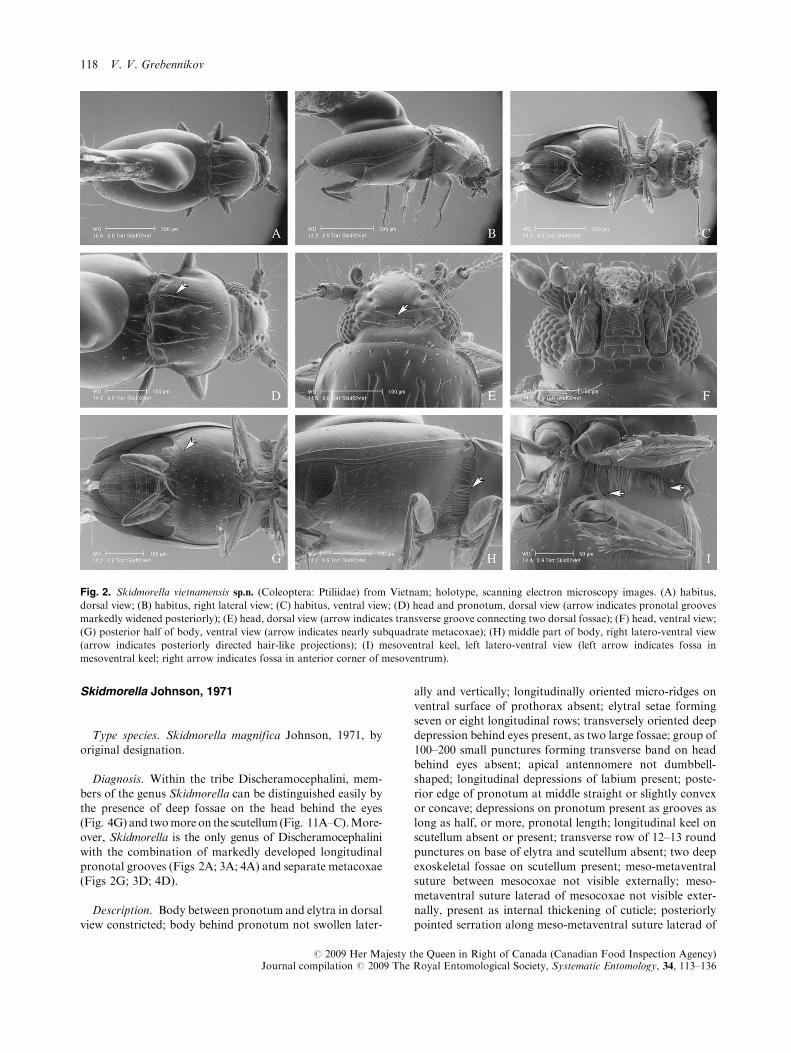
Diagnosis. Within the tribe Discheramocephalini, mem-
bers of the genus Skidmorella can be distinguished easily bythe presence of deep fossae on the head behind the eyes(Fig. 4G) and twomoreon the scutellum (Fig. 11A–C).More-over, Skidmorella is the only genus of Discheramocephalini
with the combination of markedly developed longitudinalpronotal grooves (Figs 2A; 3A; 4A) and separate metacoxae(Figs 2G; 3D; 4D).
Description. Body between pronotum and elytra in dorsalview constricted; body behind pronotum not swollen later-
ally and vertically; longitudinally oriented micro-ridges on
ventral surface of prothorax absent; elytral setae formingseven or eight longitudinal rows; transversely oriented deepdepression behind eyes present, as two large fossae; group of
100–200 small punctures forming transverse band on headbehind eyes absent; apical antennomere not dumbbell-shaped; longitudinal depressions of labium present; poste-
rior edge of pronotum at middle straight or slightly convexor concave; depressions on pronotum present as grooves aslong as half, or more, pronotal length; longitudinal keel onscutellum absent or present; transverse row of 12–13 round
punctures on base of elytra and scutellum absent; two deepexoskeletal fossae on scutellum present; meso-metaventralsuture between mesocoxae not visible externally; meso-
metaventral suture laterad of mesocoxae not visible exter-nally, present as internal thickening of cuticle; posteriorlypointed serration along meso-metaventral suture laterad of
Fig. 2. Skidmorella vietnamensis sp.n. (Coleoptera: Ptiliidae) from Vietnam; holotype, scanning electron microscopy images. (A) habitus,
dorsal view; (B) habitus, right lateral view; (C) habitus, ventral view; (D) head and pronotum, dorsal view (arrow indicates pronotal grooves
markedly widened posteriorly); (E) head, dorsal view (arrow indicates transverse groove connecting two dorsal fossae); (F) head, ventral view;
(G) posterior half of body, ventral view (arrow indicates nearly subquadrate metacoxae); (H) middle part of body, right latero-ventral view
(arrow indicates posteriorly directed hair-like projections); (I) mesoventral keel, left latero-ventral view (left arrow indicates fossa in
mesoventral keel; right arrow indicates fossa in anterior corner of mesoventrum).
118 V. V. Grebennikov
# 2009 Her Majesty the Queen in Right of Canada (Canadian Food Inspection Agency)Journal compilation # 2009 The Royal Entomological Society, Systematic Entomology, 34, 113–136

mesocoxae absent; metaventral longitudinal lateral linesabsent; horizontal perforation of mesoventral keel as visiblein lateral view round, transparent in lateral view; mesoven-
trum without transverse grooves; alacrista of metathorax atmiddle without short setae along margins; metacoxae nottransverse, separated by one-quarter of metaventral width;posteriorly oriented projection of metaventral plate between
metacoxae without two large lateral teeth or with two re-latively large and sharply pointed lateral teeth; transverselyoriented group of about 50–70 closely adjacent round
micropores along posterior edge of tergite VIII absent;cavities on abdominal sternum VIII absent; single elongateinternal sclerite alongside aedeagus absent; spermatheca
mainly globular (as in Fig. 13J), rarely tube-shaped (Fig. 13K).
Composition and geographical distribution. The genus
Skidmorella includes, alongside three new species fromVietnam and Indonesia described herein, three other valid
species: S. magnifica Johnson, 1971: 44 (from BismarckArchipelago of Papua New Guinea, the Solomon Islands,and Shikoku, Japan; holotype in ZMUC; examined);
Skidmorella amamiana Sawada & Hirowatari, 2003: 312(from the Okinawa Archipelago, Japan; holotype in OPU;not examined); and Skidmorella quadrisulucia Sawada &Hirowatari, 2003: 313 (from the Okinawa Archipelago,
Japan; holotype in OPU; not examined).
Monophyly and phylogenetic relationships. The genus Skid-
morella, as presently defined, is certainly a non-monophyleticassemblage incorporating all Discheramocephalini specieswith longitudinal furrows on the pronotum that do not belong
to Discheramocephalus. Two Skidmorella species, namely S.vietnamensis sp.n. and S. serrata sp.n., differ from the rest ofthe genus in having differently shaped pronotal furrows,
serrate pronotal lateral margins (S. serrata), non-globularspermatheca (S. serrata; state of this character is unknown in
Fig. 3. Skidmorella memorabilis sp.n. (Coleoptera: Ptiliidae) from Indonesia; paratype, scanning electron microscopy images. (A) habitus,
dorsal view; (B) habitus, right lateral view; (C) habitus, right latero-ventral view; (D) habitus, ventral view; (E) head and pronotum, dorsal view;
(F) head, dorsal view (arrow indicates left dorsal fossa); (G) head, ventral view (arrow indicates left longitudinal groove on labium); (H)
posterior half of body, ventral view; (I) meso- and metaventrites, right lateral view (arrow indicates fossa in mesoventral keel).
Discheramocephalini, a new tribe of Ptiliidae 119
# 2009 Her Majesty the Queen in Right of Canada (Canadian Food Inspection Agency)Journal compilation # 2009 The Royal Entomological Society, Systematic Entomology, 34, 113–136

S. vietnamensis), a transverse groove connecting dorsal fossae
on the head (S. vietnamensis) and an extra pair of deep fossaein the anterior lateral corners of the mesoventrum (S. vietna-mensis).Most probably, the genus Skidmorella is paraphyleticwith respect to Discheramocephalus. Exclusion of the two
above-named species would probably leave the rest of Skid-morella monophyletic; such action is, however, postponeduntil more Discheramocephalini species are discovered.
Bionomics. All known specimens of this genus were
collected from forest leaf litter.
Identification key to Skidmorella species(modified from Sawada & Hirowatari, 2003)
1. Disc of pronotum with two longitudinal furrows(Fig. 2A) ................................................................. 2
– Disc of pronotum with four or six longitudinal furrows
(Fig. 4A) ................................................................... 52. Head behind eyes with two deep fossae connected by
transverse groove (Fig. 2E); pronotal furrows at baseabout 4� wider than at apex (Fig. 2A); mesoventrum
on each side with deep fossa in anterior lateral corners(Fig. 2I) ...................... S. vietnamensis sp.n. (Vietnam)
– Head behind eyes with two deep fossae not connected
by transverse groove; pronotal furrows at base notwider than at apex; mesoventrum on each side withoutdeep fossa in anterior lateral corners ........................ 3
3. Base of pronotal furrows nearly contiguous to basaledge on pronotum; pronotal furrows sinuate, long,extending for some 80% of pronotal length ............. 4
– Base of pronotal furrows clearly separated from basaledge of pronotum by distance subequal to eye diameter(Fig. 3E); pronotal furrows nearly straight, slightly
Fig. 4. Skidmorella serrata sp.n. (Coleoptera: Ptiliidae) from Vietnam; paratype, scanning electron microscopy images. (A) habitus, dorsal
view; (B) habitus, left lateral view; (C) habitus, left latero-ventral view; (D) habitus, ventral view; (E) head and pronotum, dorsal view; (F)
middle part of body, left lateral view (arrow indicates fossa in mesoventral keel); (G) head, dorsal view (arrow indicates dorsal fossa); (H) head,
ventral view (arrow indicates left longitudinal groove on labium); (I) mesoventral keel, right ventro-lateral view (arrow indicates fossa in
mesoventral keel).
120 V. V. Grebennikov
# 2009 Her Majesty the Queen in Right of Canada (Canadian Food Inspection Agency)Journal compilation # 2009 The Royal Entomological Society, Systematic Entomology, 34, 113–136

convergent anteriorly, short, of about 50% of pronotallength (Fig. 3E) ......... S. memorabilis sp.n. (Indonesia)
4. Pronotal furrows sinuate and distance between themsmallest at their middle; aedeagus without sclerite inter-nally .... S. magnifica Johnson, 1971 (Bismarck Archi-
pelago of Papua New Guinea, the Solomon Islands,Shikoku, Japan)
– Pronotal furrows sinuate and distance between themsmallest distally; aedeagus with sinuate sclerite in-
ternally ...... S. amamiana Sawada et Hirowatari, 2003(Okinawa Archipelago, Japan)
5. Disc of pronotum with four longitudinal furrows, the
medial being half the length of lateral ones; posteriorhalf of pronotal margins not serrate; spermatheca glo-bular ...... S. quadrisulucia Sawada et Hirowatari, 2003
(Okinawa Archipelago, Japan)– Disc of pronotum with six longitudinal furrows sub-equal in length (Fig. 4E); posterior half of pronotalmargins finely serrate (Fig. 4E); spermatheca a long and
coiled tube (Fig. 13K) ......... S. serrata sp.n. (Vietnam)
Skidmorella vietnamensis sp.n.
(Figs 2; 11A; 13A)
Diagnostic description. Body length 0.80 mm (n ¼ 1);two transverse fossae on head dorsally behind eyes con-
nected by groove; disc of pronotum on each side with onelongitudinal furrow not shorter than half of pronotal length;pronotal margins in posterior half not serrate; setae in
anterior part of pronotum not longer than those inposterior part; mesoventrum with deep fossa on each sideat anterior corners; posteriorly oriented projection of
metaventral plate between metacoxae without two teethadjacent to, and mediad of, coxae; aedeagus Fig. 13E;spermatheca unknown.
Etymology. The specific epithet vietnamensis derives from
Vietnam, the type locality of this species.
Type material. Holotype (male; deposited in MNHN)
mounted on Euparal microscope slide and previouslyused for SEM (antennomeres 3–11 were lost after SEM):VIETNAM, Lam Dong Dalat, Bi Doup, Nui Gia Rich,
1440 m, 18.xii.1998 (L. Deharveng & A. Bedos).
Skidmorella memorabilis sp.n.(Fig. 3; 11B; 13C, J)
Diagnostic description. Body length 0.69–0.70 mm (n ¼2); two transverse fossae on head dorsally behind eyes notconnected by groove; disc of pronotum on each side withone longitudinal furrow not shorter than half of pronotal
length; pronotal margins in posterior half not serrate; setaein anterior part of pronotum not longer than those inposterior part; mesoventrum without deep fossa on each
side at anterior corners; posteriorly oriented projection ofmetaventral plate between metacoxae with two teeth adja-
cent to, and mediad of, coxae; aedeagus Fig. 13C; sperma-theca Fig. 13J.
Type material. Holotype (female; deposited in MNHN)mounted on Euparal microscope slide: INDONESIA,Sulawesi Selatan, Latimojong, 1980 m, 28.vii.1990 (L.Deharveng & A. Bedos). Paratypes: two, with the same
data as holotype; one unsexed specimen in 70% ethanoland another male mounted on Euparal microscope slide(CNC).
Etymology. The specific epithet is the Latin adjectivememorabilis, -e (remarkable, worthy to be remembered)
and refers to the history of the species discovery.Reading about Skidmorella in the advanced stages ofpreparation of this paper I suspected that it might bea member of the clade here called the tribe Discheramo-
cephalini. Because a perforated mesoventral keel was notmentioned in the description of this genus, I needed torequest the types to check for this character. Almost
simultaneously I remembered the existence of a largesample of unsorted Ptiliidae from Southern Asia receivedfrom Louis Deharveng, MNHN. Some minutes later I
was going through the samples and much to my joy onevial indeed contained specimens of the genus Skidmor-ella. Moreover, a clear perforation of the mesoventral
keel was observed immediately.
Skidmorella serrata sp.n.
(Figs 4; 11C; 13B, K)
Diagnostic description. Body length 0.78–0.80 mm (n ¼2); two transverse fossae on head dorsally behind eyes notconnected by groove; disc of pronotum on each side with
three longitudinal furrows not shorter than half of pronotallength; pronotal margins in posterior half serrate; setae inanterior part of pronotum 2–3� longer than those inposterior part; mesoventrum without deep fossa on each
side at anterior corners; posteriorly oriented projection ofmetaventral plate between metacoxae with two teeth adja-cent to, and mediad of, coxae; aedeagus Fig. 13B; sperma-
theca Fig. 13K.
Etymology. The specific name is the Latin adjective
serratus, -a, -um (saw-shaped, serrated), referring to theserrated margins of the pronotum in this species.
Type material. Holotype (male; deposited in MNHN)mounted on Euparal microscope slide: VIETNAM, LamDongDalat, Lang Bian, 2130 m, 21.xii.1998 (L. Deharveng &A. Bedos). Paratypes: 21, with the same data as holotype;
five are mounted on four Euparal microscope slides (twoslides in MNHN, two in CNC) and 16 in 70% ethanol (eightin MNHN and eight in CNC).
Discheramocephalini, a new tribe of Ptiliidae 121
# 2009 Her Majesty the Queen in Right of Canada (Canadian Food Inspection Agency)Journal compilation # 2009 The Royal Entomological Society, Systematic Entomology, 34, 113–136

Africoptilium Johnson, 1967(Figs 5; 11D–F; 12A–C)
Type species. Africoptilium marginatum Johnson, 1967,by original designation.
Diagnosis. Species of Africoptilium have a constrictionbetween the pronotum and elytra (Fig. 12A–C) resemblingthat in Discheramocephalus specimens; those of Africo-ptilium differ from the latter by their clearly separated
metacoxae and by lacking pronotal grooves. Among Dis-cheramocephalini genera without pronotal grooves (Fenes-tellidium, Cissidium, Dacrysoma), Africoptilium species are
recognizable by their elongate and constricted body alongwith sparsely located and short dorsal pubescence, whichgives Africoptilium specimens a ‘non-hairy’ appearance.
Other unique features of Africoptilium within the tribe are
the pygidium with two apices (Fig. 5I) and (among thegenera with non-contiguous metacoxae) the lack of a poste-riorly oriented sharp projection of the metaventrum adja-
cent to the mesal side of each metacoxal plate.
Description. Body between pronotum and elytra in dorsal
view constricted; body behind pronotum not swollen later-ally and vertically; longitudinally oriented micro-ridges onventral surface of prothorax absent; elytral setae formingseven or eight longitudinal rows; transversely oriented deep
depression behind eyes absent; group of 100–200 smallpunctures forming transverse band on head behind eyesabsent; apical antennomere not dumbbell-shaped; longitu-
dinal depressions of labium absent; posterior edge ofpronotum at middle concave; depressions on pronotumabsent; longitudinal keel on scutellum present, short, about
Fig. 5. Africoptilium sp. (Coleoptera: Ptiliidae) from DRC, scanning electron microscopy images. (A) habitus, dorsal view; (B) habitus, right
lateral view; (C) habitus, ventral view (arrow indicates longitudinal metaventral line); (D) habitus, right ventro-lateral view; (E) middle part of
body, right lateral view (upper arrow indicates fossa in anterior corners of mesoventrum; lower arrow indicates fossa of mesoventral keel); (F)
anterior part of body, left lateral view; (G) pronotum, dorsal view; (H) head, ventro-frontal view; (I) hind legs and abdomen, ventral view
(arrow indicates one of two pygidial teeth).
122 V. V. Grebennikov
# 2009 Her Majesty the Queen in Right of Canada (Canadian Food Inspection Agency)Journal compilation # 2009 The Royal Entomological Society, Systematic Entomology, 34, 113–136

one-third of its length; transverse row of 12–13 roundpunctures on base of elytra and scutellum absent; two deep
exoskeletal fossae on scutellum absent; meso-metaventralsuture between mesocoxae not visible externally; meso-metaventral suture laterad of mesocoxae not visible exter-
nally, present as internal thickening of cuticle; posteriorlypointed serration along meso-metaventral suture laterad ofmesocoxae absent; metaventral longitudinal lateral linespresent; horizontal perforation of mesoventral keel circular
and transparent in lateral view; mesoventrum withouttransverse grooves; alacrista of metathorax at middle with-out short setae along margins; metacoxae not transverse,
separated by one-eighth of metaventral width; posteriorlyoriented projection of metaventral plate between metacoxaewithout two large lateral teeth; transversely oriented group
of about 50–70 closely adjacent round micropores alongposterior edge of tergite VIII absent; cavities on abdominalsternum VIII absent; single elongate internal sclerite along-side aedeagus absent; spermatheca as a coiled tube.
Composition and geographical distribution. The genusAfricoptilium is known only from Eastern Africa (Tanzania
and eastern part of the Democratic Republic of Congo).
Monophyly and phylogenetic relationships. Monophyly
of Africoptilium was not tested in the present analysis. Theremarkable similarity among Africoptilium species, how-ever, indirectly suggests that this genus might indeed be
monophyletic (or, alternatively, a highly conservativeparaphyletic grade). A position of Africoptilium as a mem-ber of the Skidmorella þ Discheramocephalus clade(Fig. 14) could be an artefact resulting from presumably
convergent evolution of similarly located structures such asmetaventral longitudinal lateral lines and exoskeletal fos-sae in anterior lateral corners of mesoventrum. These
character states were scored as homologous for Africopti-lium and some members of the Skidmorella þ Dischera-mocephalus clade, even though some minor consistent
differences were observed. Acceptance of the evolutionaryscenario (Fig. 14) implies that the deep longitudinalfurrows on the pronotal disc synapomorphic for the
Skidmorella þ Discheramocephalus clade could becomesecondarily obsolete in Africoptilium, which appearsunlikely.
Bionomics. All specimens of Africoptilium with knownbiological information were collected by sifting leaf litter intropical forests. One specimen from the West Usambara
Mountains in Tanzania mounted on a microscope slidecontained fungal spores in its gut (Fig. 11E).
Remarks. Johnson (1967) described the genus Africopti-lium and all of its three species, A. marginatum, A. mimicumand A. concinnum, based on 13, 3 and 1 specimen, respec-tively, and provided an identification key. No new findings
of Africoptilium have been reported. I studied the short typeseries (including holotypes) of A. marginatum (Fig. 12A)and A. mimicum (Fig. 12B) stored in MRAC and NHM,
respectively (the allotype of A. marginatum is mounted ona Euparal microscope slide). The holotype of A. concinnum
was reportedly stored in NMW (Johnson, 1967); however,curators’ attempts to locate it were unsuccessful. I have seenat least five moreAfricoptilium specimens in the collection of
MRAC, all collected during the colonial period in theeastern part of the former Belgian Congo. One of thesespecimens, labelled ‘Congo Belge: P.N.A., 31–iii–1–iv–1955,P.Vanschuytbroeck 12.733–42, Secteur Nord, riv. May ya
Moto, 1.040 m, ex P.N.A.’, was used for SEM (Fig. 5) andthen mounted on a Euparal microscope slide (MRAC). Ialso collected two unidentified specimens of Africoptilium
while sifting leaf litter in partly human-disturbed forest inthe Lushoto district of the West Usambara Mountains,Tanzania, in October 2002 at an altitude of 1660 m (Figs 11
E; 12C, D). Of these two specimens, the female is mountedin a microscope slide in Euparal and stored in CNC, and themale is stored in the collection of M. Sorensson, Lund,Sweden. I did not make an effort to delimit species
boundaries of Africoptilium owing to the inadequate num-ber of available specimens. Specimens of this genus havenever been collected en masse.
Fenestellidium gen.n.
Type species. Fenestellidium capensis sp.n., here desig-
nated.
Diagnosis. This genus is unique among Ptiliidae byhaving the largest known internal size of the horizontally
oriented perforation of the mesoventral keel. This perfora-tion is oblong in vertical dimension (Figs 6E; 7G) and, inlateral view, its internal size is subequal to the cross-section
of the mesofemur in its thickest part. Among Discheramo-cephalini without pronotal longitudinal grooves, membersof Fenestellidium can be distinguished from those of Afri-
coptilium by not having meso- and metaventral longitudinallines; and from those of Cissidium and Dacrysoma by theshape of the body, which is much less swollen in both
horizontal and vertical dimensions. Moreover, males of theknown Fenestellidium species have groups of setae onabdominal sternite VIII (Fig. 7C, H), which is apparentlya case of sexual dimorphism.
Description. Body constricted between pronotum andelytra in dorsal view; body behind pronotum not swollen
laterally and vertically; longitudinally oriented micro-ridgeson ventral surface of prothorax absent; elytral setae notforming clearly demarked longitudinal rows; transversely
oriented deep depression behind eyes absent; group of 100–200 small punctures forming transverse band on headbehind eyes absent; apical antennomere not dumbbell-shaped; longitudinal depressions of labium absent; posterior
edge of pronotum at middle straight or slightly convex;depressions on pronotum absent; longitudinal keel onscutellum absent; transverse row of 12–13 round punctures
Discheramocephalini, a new tribe of Ptiliidae 123
# 2009 Her Majesty the Queen in Right of Canada (Canadian Food Inspection Agency)Journal compilation # 2009 The Royal Entomological Society, Systematic Entomology, 34, 113–136

on base of elytra and scutellum present; two deep exo-
skeletal fossae on scutellum absent; meso-metaventralsuture between mesocoxae not visible externally; meso-metaventral suture laterad of mesocoxae clearly visibleexternally as projecting keel; posteriorly pointed serration
along meso-metaventral suture laterad of mesocoxaeabsent; metaventral longitudinal lateral lines absent; hori-zontal perforation of mesoventral keel as visible in lateral
view vertically elongate, almost parallel-sided, about 2�longer than wide; mesoventrum with transverse groovesoriginating from fossae of mesoventral keel and extending
laterad absent; alacrista of metathorax at middle with shortsetae along margins; metacoxae not transverse, separated byone-sixth of metaventral width; posteriorly oriented pro-
jection of metaventral plate between metacoxae with tworelatively large and sharply pointed lateral teeth; trans-versely oriented group of about 50–70 closely adjacentround micropores along posterior edge of tergite VIII
absent; cavities on abdominal sternum VIII absent; singleelongate internal sclerite alongside aedeagus absent; sper-matheca either globular (Fig. 13I) or as a coiled tube
(Fig. 13H).
Etymology. The generic epithet Fenestellidium is a combi-
nation of the Latin noun fenestella, -ae, f., (a small openingin the wall, a little window) and the suffix –idium. The namerefers to the remarkably large horizontal perforation of the
mesoventral keel, which somewhat resembles a window.Gender neuter.
Composition and geographical distribution. The genus
Fenestellidium is established for F. kakamegaensis sp.n. andF. capensis sp.n. from Kenya and South Africa, respectively.
Monophyly and phylogenetic relationships. The genus
Fenestellidium is a well-supported clade with a bootstrapvalue above 95% supported by four synapomorphies asindicated in Fig. 14. Its sister-group relationships, however,
have not been clarified.
Bionomics. All known specimens of Fenestellidium were
sifted from the forest leaf litter.
Fenestellidium capensis sp.n.
(Figs 6; 11D; 12E, F; 13E, G, I )
Diagnostic description. Body length 0.78–0.86 mm (n¼ 3).Body densely pubescent dorsally and ventrally; individual
hairs 1.2–1.7� longer than the distance between them;pronotum more transverse; disc of pronotum not punctate;mesoventrite on each side with oblique groove originating
from perforation of mesoventral keel and extending postero-laterad; males without deep depression along mid-line ofapical abdominal sternites; aedeagus Fig. 13E; spermatheca
Fig. 13I.
Etymology. The specific epithet capensis derives from the
name of the Cape Region in South Africa, the type localityof this species.
Fig. 6. Fenestellidium capensis sp.n. (Coleoptera: Ptiliidae) from South Africa; paratype, scanning electron microscopy images. (A) habitus,
dorsal view; (B) habitus, right lateral view; (C) anterior half of body, dorsal view; (D) abdomen, left ventro-lateral view (arrow indicates
posterior tooth of metaventrum medially of metacoxa); (E) thorax, left ventro-lateral view (arrow indicates fossa in mesoventral keel); (F)
thorax, left ventro-lateral view (arrow indicates oblique groove on mesoventrum).
124 V. V. Grebennikov
# 2009 Her Majesty the Queen in Right of Canada (Canadian Food Inspection Agency)Journal compilation # 2009 The Royal Entomological Society, Systematic Entomology, 34, 113–136

Type material. Holotype (female) mounted on a Euparalmicroscope slide: SOUTH AFRICA, Western Cape, Cape
Town, Table Mt., 10–11.viii.2002 (V. Grebennikov). Paratypes:12; seven specimens are mounted on four Euparal slides.(Three females are mounted on three slides, and the remaining
three males and one female are mounted all together on thesame slide; this slide has an additional non-congeneric specimenof Ptiliidae mounted among four paratypes of Fenestellidium
capensis and bears an additional statement on the label:‘Single teneral male/mounted does not belong to the genusFenestellidium’.) Five specimens are stored in 70% ethanol.
Remarks. This species was collected on the eastern slopeof Table Mountain, Cape Town, South Africa, 200–300 mhigher than the campus of the University of Cape Town.
Two males and one female of a species closely resemblingF. capensis were collected in Grahamstown in the EasternCape, South Africa are mounted on Euparal (Fig. 11H).
Their only difference from the Cape Town population ofF. capensis is found in the shape of the spermatheca. At this
point it is impossible to assess whether these three specimensfrom Grahamstown represent a conspecific population ofF. capensis or a congeneric species.
Fenestellidium kakamegaensis sp.n.(Figs 7; 11I; 12G–H; 13D, H)
Diagnostic description. Body length 0.73–0.76 mm (n ¼2). Body sparsely pubescent with length of individual hairs0.5–0.3� distance between them; pronotum less transverse;
disc of pronotum punctate with each puncture bearinga short hair; mesoventrite on each side without obliquegroove originating from perforation of mesoventral keel;
males with deep depression along mid-line of apical abdom-inal sternites flanked by dense groups of setae; aedeagusFig. 13D, spermatheca Fig. 13H.
Fig. 7. Fenestellidium kakamegaensis sp.n. (Coleoptera: Ptiliidae) from Kenya; paratype, scanning electron microscopy images. (A) habitus,
dorsal view; (B), habitus, right lateral view; (C) habitus, ventral view (arrow indicates male secondary sexual features on abdomen); (D) habitus,
right ventro-lateral view; (E) anterior part of body, frontal view; (F) pronotum, dorsal view (arrow indicates transverse row of round punctures
on base of elytra and scutellum); (G) thorax, right lateral view (arrow indicates fossa in mesoventral keel); (H) abdomen, left ventro-lateral
view; (I) thorax, left ventro-lateral view.
Discheramocephalini, a new tribe of Ptiliidae 125
# 2009 Her Majesty the Queen in Right of Canada (Canadian Food Inspection Agency)Journal compilation # 2009 The Royal Entomological Society, Systematic Entomology, 34, 113–136

Etymology. The specific epithet kakamegaensis derives
from the name of the Kakamega forest in western Kenya,the type locality of this species.
Type material. Holotype (male) mounted on Euparal
microscope slide: KENYA, Kakamega forest, Udo’s ban-das, 10–12.xi.2001 (V. Grebennikov). Paratypes: six, with thesame data as holotype; three in 70% ethanol and three more
mounted on three Euparal microscope slides.
Cissidium Motschulsky, 1855
Type species. Cissidium basale Motschulsky, 1855, desig-
nated by Lucas, 1920: 187 (not seen; cited after Newton &Thayer, 2007b).
¼ CamptodiumMotschulsky, 1869: 189; type species Camp-todium adustipenneMotschulsky, 1869. Synonymy estab-
lished by Johnson (1982: 358).
¼ Dimorphella A. Matthews, 1889: 190; type species Di-
morphella reitteri A. Matthews, 1889. Synonymy estab-lished by Johnson (1982: 358).
¼ Epibaptus Deane, 1931: 228; type species Epibaptusscutellaris Deane, 1931. Synonymy established by
Johnson (1982: 358).
Diagnosis. Members of the genusCissidium have a prono-tum with a transverse row of four to eight poorly delimited
depressions along posterior margin (Fig. 8F). Anotherunique feature is their posteriorly pointed serration alongthe meso-metaventral suture laterad of the mesocoxae
(Fig. 8E). Within Discheramocephalini, species of Cissidiumare unique by having poorly developed horizontal perfora-tion of the mesoventral keel, which is not transparent in
lateral view. Most of the Cissidium and all the Dacrysomaspecies have dumbbell-shaped antennomere 11. Unlike Da-crysoma, Cissidium species have the meso-metaventral suture
clearly delimited and not obliterated externally (Fig. 8H).
Fig. 8. Cissidium okuensis sp.n. (Coleoptera: Ptiliidae) from Cameroon; scanning electron microscopy images. (A) habitus, dorsal view; (B)
habitus, right dorso-lateral view; (C) habitus, right lateral view; (D) habitus, right ventro-lateral view; (E) habitus, ventral view (arrow indicates
serration); (F) pronotum and head, dorsal view; (G) head, ventral view; (H) thorax, left lateral view; (I) meso- (to the left) and metacoxae (to the
right), left ventro-lateral view (arrow indicates clearly visible meso-metaventral suture between mesocoxae).
126 V. V. Grebennikov
# 2009 Her Majesty the Queen in Right of Canada (Canadian Food Inspection Agency)Journal compilation # 2009 The Royal Entomological Society, Systematic Entomology, 34, 113–136

Description. Body between pronotum and elytra in dorsal
view not or slightly constricted; body behind pronotumswollen laterally and vertically; longitudinally orientedmicro-ridges on ventral surface of prothorax absent; elytral
setae not forming clearly demarked longitudinal rows;transversely oriented deep depression behind eyes absent;group of 100–200 small punctures forming transverse bandon head behind eyes absent; apical antennomere dumbbell-
shaped; longitudinal depressions of labium absent; posterioredge of pronotum at middle concave; depressions onpronotum present as rounded basal depression; longitudinal
keel on scutellum absent; transverse row of 12–13 roundpunctures on base of elytra and scutellum absent; two deepexoskeletal fossae on scutellum absent; meso-metaventral
suture between mesocoxae clearly visible forming delimitedborder between plates; meso-metaventral suture laterad ofmesocoxae clearly visible externally as projecting keel;
posteriorly pointed serration along meso-metaventral suture
laterad of mesocoxae present; metaventral longitudinal
lateral lines absent; horizontal perforation of mesoventralcircular in lateral view, not transparent; mesoventrumwithout transverse grooves; alacrista of metathorax at mid-
dle without short setae along margins; metacoxae nottransverse, separated by one-quarter of metaventral width;posteriorly oriented projection of metaventral plate betweenmetacoxae with two relatively large and sharply pointed
lateral teeth; transversely oriented group of about 50–70closely adjacent round micropores along posterior edge oftergite VIII present; cavities on abdominal sternum VIII
absent; single elongate internal sclerite alongside aedeagusabsent; spermatheca small and nearly spherical in shape;aedeagus as in Johnson (1982: fig. 57).
Composition and geographical distribution. The genusCissidium includes eight valid species, including the new
species described below: C. basale Motschulsky, 1855
Fig. 9. Dacrysoma usambarensis sp.n. (Coleoptera: Ptiliidae) from Tanzania; holotype, scanning electron microscopy images. (A) habitus,
dorsal view; (B) habitus, right lateral view; (C) habitus, right latero-dorsal view; (D) habitus, ventral view; (E) pronotum and head, dorsal view;
(F) head, ventral view; (G) posterior part of body, ventral view; (H) thorax, right ventro-lateral view (left arrow indicates lack of visible meso-
metaventral suture; right arrow indicates fossa in mesoventral keel); (I) pro- (to the right) and mesocoxae (to the left), left ventral view (arrows
indicate lateral openings of fossae in mesoventral keel).
Discheramocephalini, a new tribe of Ptiliidae 127
# 2009 Her Majesty the Queen in Right of Canada (Canadian Food Inspection Agency)Journal compilation # 2009 The Royal Entomological Society, Systematic Entomology, 34, 113–136

(from Panama; holotype in ZMMU; not examined); C.rufescens Motschulsky, 1855 (from Panama; holotypepresumably in ZMMU; not examined); C. matthewsi
Johnson, 2007b: 22 (¼ C. matthewsi Johnson, 2004:25,nomen nudum) (from Japan; holotype in MMUE; notexamined); C. adustipenne Motschulsky, 1869 (nec adus-
tipenne A. Matthews, 1884) (from eastern India; holotypepresumably in ZMMU; not examined); C. scutellarus(Deane, 1931: 228) (from Australia: Victoria; holotype
either in MVMA or UQIC as cited by Johnson, 1982; notexamined); C. crowsoni Johnson, 1982:359 (from NewZealand; holotype in NZAC; not examined); and C.
foveolatum Johnson, 1982:359 (from New Zealand; holo-type in NZAC; not examined). Johnson (1982: 359) notedthat there were about 70 additional species from tropicaland subtropical areas of the world. I have seen at least one
undescribed species of this genus from Kenya, one fromCameroon, and another from Bolivia.
Monophyly and phylogenetic relationships. The genusCissidiumwas consistently supported as monophyletic basedon four synapomorphies with bootstrap values above 95%
(Fig. 14). Only two of its species were included in theanalysis, which might be seen as an inadequate samplingfor arguing its monophyly. The genus Cissidium is hypoth-
esized to be a sister-group of Dacrysoma.
Cissidium okuensis sp.n.
(Figs 8; 11J; 13N)
Diagnostic description. Body length 1.03–1.09 mm (n ¼3). Pronotum marginate, sides subparallel in posterior half.
With external morphological features as depicted in Figs 8;11J; aedeagus unknown; spermatheca Fig. 13N.
Etymology. The specific epithet okuensis derives from thename of Oku Mountain in Cameroon, the type locality ofthis species.
Type material. Holotype (female) mounted on Euparalmicroscope slide: CAMEROON, North-West province, Mt.
and lake Oku, N06812.2189, E010827.5969, 6.v.2006, 2200 m(V. Grebennikov). Paratypes: four, with the same data asholotype; two in 70% ethanol and two mounted on two
Euparal microscope slides. The specimen used for SEM wassubsequently lost.
Dacrysoma gen.n.
Type species. Dacrysoma usumbarensis sp.n., here desig-nated.
Diagnosis. Members of Dacrysoma share with at leastsome species of Cissidium a unique feature within Ptiliidae,namely the dumbbell-shaped antennomere 11 (Fig. 10B).
Dacrysoma species differ from those ofCissidium by a clearlyvisible horizontal perforation of the mesoventral keel(Figs 9H; 10F) and by the lack of serration of the meso-
metaventral suture laterad of mesocoxae (Figs 9G; 10F).
Description. Body between pronotum and elytra in
dorsal view constricted; body behind pronotum swollen
Fig. 10. Dacrysoma felis sp.n. (Coleoptera: Ptiliidae) from Madagascar; holotype, scanning electron microscopy images. (A) habitus, dorsal
view; (B) habitus, right lateral view (arrow indicates dumbbell-shaped apical antennomere); (C) habitus, right latero-ventral view; (D) habitus,
ventral view; (E) pronotum and head, dorsal view; (F) thorax, right ventro-lateral view (arrow indicates fossa in mesoventral keel).
128 V. V. Grebennikov
# 2009 Her Majesty the Queen in Right of Canada (Canadian Food Inspection Agency)Journal compilation # 2009 The Royal Entomological Society, Systematic Entomology, 34, 113–136

laterally and vertically; longitudinally oriented micro-ridges on ventral surface of prothorax absent; elytral setaenot forming clearly demarked longitudinal rows; trans-
versely oriented deep depression behind eyes absent; group
of 100–200 microscopic punctures forming transverse bandon head behind eyes present; apical antennomere dumb-bell-shaped; longitudinal depressions of labium absent;
posterior edge of pronotum at middle concave; depressions
Fig. 11. Discheramocephalini spp. (Coleoptera: Ptiliidae); light compound microscopy images, dorsal views. (A) Skidmorella vietnamensis
sp.n., holotype; (B) Skidmorella memorabilis sp.n., holotype; (C) Skidmorella serrata sp.n., holotype; (D) Africoptilium marginatum, paratype;
(E) Africoptilium sp. (specimen used for SEM in Fig. 5); (F) Africoptilium sp. (Tanzania: W. Usambara Mts); (G) Fenestellidium capensis sp.n.,
holotype; (H) Fenestellidium pr. capensis (South Africa: Grahamstown); (I) Fenestellidium kakamegaensis sp.n., holotype; (J)Cissidium okuensis
sp.n., holotype; (K) Dacrysoma usambarensis sp.n., paratype; (L) Dacrysoma felis sp.n., paratype.
Discheramocephalini, a new tribe of Ptiliidae 129
# 2009 Her Majesty the Queen in Right of Canada (Canadian Food Inspection Agency)Journal compilation # 2009 The Royal Entomological Society, Systematic Entomology, 34, 113–136

on pronotum absent; longitudinal keel on scutellumabsent; transverse row of 12–13 round punctures on baseof elytra and scutellum absent; two deep exoskeletal fossae
on scutellum absent; meso-metaventral suture betweenmesocoxae clearly visible externally forming delimitedborder between plates; meso-metaventral suture lateradof mesocoxae not visible externally, present as internal
thickening of cuticle; posteriorly pointed serration alongmeso-metaventral suture laterad of mesocoxae absent;metaventral longitudinal lateral lines absent; horizontal
perforation of mesoventral keel as visible in lateral viewround, transparent in lateral view; mesoventrum withtransverse grooves originating from fossae of mesoventral
keel and extending laterad; alacrista of metathorax at
middle without short setae along margins; metacoxae nottransverse, separated by one-quarter of metaventral width;posteriorly oriented projection of metaventral plate
between metacoxae with two relatively large and sharplypointed lateral teeth; transversely oriented group of about50–70 closely adjacent round micropores along posterioredge of tergite VIII absent; cavities on abdominal sternum
VIII absent; single elongate internal sclerite alongsideaedeagus absent; spermatheca globular.
Etymology. The generic epithet Dacrysoma is a combina-tion of two Greek words dakryon (a tear) and svma
(body). The name refers to the somewhat tear-shaped body
of these beetles. Gender neuter.
Fig. 12. Africoptilium spp. and Fenestellidium spp. (Coleoptera: Ptiliidae: Discheramocephalini); light dissecting microscopy images. (A)
Africoptilium marginatum, holotype, dorsal view; (B) Africoptilium mimicum, holotype, dorsal view; (C–D) Africoptilium sp. (Tanzania: W.
Usambara Mts.), dorsal (C) and ventral (D) view; (E–F) Fenestellidium capensis sp.n., dorsal (E) and ventral (F) view; (G–H) Fenestellidium
kakamegaensis sp.n., dorsal (G) and left lateral (H) view. Scale bars: 0.1 mm.
130 V. V. Grebennikov
# 2009 Her Majesty the Queen in Right of Canada (Canadian Food Inspection Agency)Journal compilation # 2009 The Royal Entomological Society, Systematic Entomology, 34, 113–136

Composition and geographical distribution. The genus
Dacrysoma is established for one African (Tanzania) andone Madagascar species.
Monophyly and phylogenetic relationships. The genusDacrysoma was supported consistently as a monophyleticwith three synapomorphies and bootstrap values above
90%, as indicated in Fig. 14. The genus Dacrysoma is hypo-thesized to be a sister-group of Cissidium.
Dacrysoma usambarensis sp.n.(Figs 9; 11K; 13M)
Diagnostic description. Body length 0.97–1.03 mm (n ¼2); pronotal disk and elytra sparsely pubescent (Fig. 9E);
aedeagus unknown; spermatheca Fig. 13M.
Etymology. The specific epithet usambarensis derives
from the name of the Usambara Mountains in Tanzania,the type locality of this species.
Type material. Holotype (sex unknown) dry-mounted onthe point of an entomological pin and used for SEM:TANZA-NIA, E. UsambaraMts., 12–16.x.2002, 950 m, Amani Nature
Reserve, headquarters (V. Grebennikov).Paratypes: three, withthe same data as holotype; two specimens are mounted on twoEuparal microscope slides and one in 70% ethanol.
Dacrysoma felis sp.n.(Figs 10; 11L; 13F, L)
Diagnostic description. Body length 0.88–0.89 mm (n ¼3); pronotal disk and elytra densely pubescent (Fig. 10E);
aedeagus Fig. 13F; spermatheca Fig. 13L.
Fig. 13. Discheramocephalini spp. (Coleoptera: Ptiliidae); ventral (A–F, H–N) and dorsal (G) views; male genitalia and abdominal sternites
VIII and IX (A–F), spermatheca (H–N), left half of alacrista (G). (A) Skidmorella vietnamensis; (B, K) Skidmorella serrata; (C, J) Skidmorella
memorabilis; (D, H) Fenestellidium kakamegaensis; (E, G, I) Fenestellidium capensis; (F, L) Dacrysoma felis; (M) Dacrysoma usambarensis; (N)
Cissidium okuensis.
Discheramocephalini, a new tribe of Ptiliidae 131
# 2009 Her Majesty the Queen in Right of Canada (Canadian Food Inspection Agency)Journal compilation # 2009 The Royal Entomological Society, Systematic Entomology, 34, 113–136

Etymology. The specific epithet is the Latin noun felis, -is,f (cat), in apposition.
Type material. Holotype (sex unknown; deposited inFMNH) dry-mounted on the point of an entomological pinand used for SEM: MADAGASCAR, province Antsira-
nana, reserve Manongarivo, 20.4 km SW2198 Antanambao,1860 m, 1482.73S, 48824.069E, 3.x.1999, FMNH#98–375
(B.L. Fischer), BLF#1990. Paratypes: 22, with the same dataas holotype; 16 of them in 70% ethanol (six in CNC and ten inFMNH) and the other six mounted on six Euparal micro-scope slides (three in CNC and the other three in FMNH).
Fig. 14. Phylogeny of Ptiliidae: Discheramocephalini inferred from exhaustive parsimony analysis of 37 morphological characters scored for
27 terminal taxa. Basal nodes of the tree (except for inside the Discheramocephalini clade) represent strict consensus topology found in both
Analysis 1 and Analysis 2. Note that the tribe Ptiliini is markedly non-monophyletic. Internal topology of the Discheramocephalini clade is
represented by one of 3387 shortest trees with tree length 66; consistency index 66, retention index 87. Discheramocephalini taxa are in bold;
generalized habitus of a Discheramocephalus species illustrates the type genus of the tribe. Only unambiguously optimized evolutionary events
are mapped on the internodes; closed black squares represent unique evolutionary events; white open squares represent convergences or
subsequent reversals; character numbers are given above; the direction of character state changes is indicated by two subsequent numbers
below. Bootstrap values for Discheramocephalini clades above the 50% threshold are indicated as two values separated by a slash for
unweighted and successively weighted analysis, respectively; bootstrap values below 50% are indicated by a dash ‘-’.
132 V. V. Grebennikov
# 2009 Her Majesty the Queen in Right of Canada (Canadian Food Inspection Agency)Journal compilation # 2009 The Royal Entomological Society, Systematic Entomology, 34, 113–136
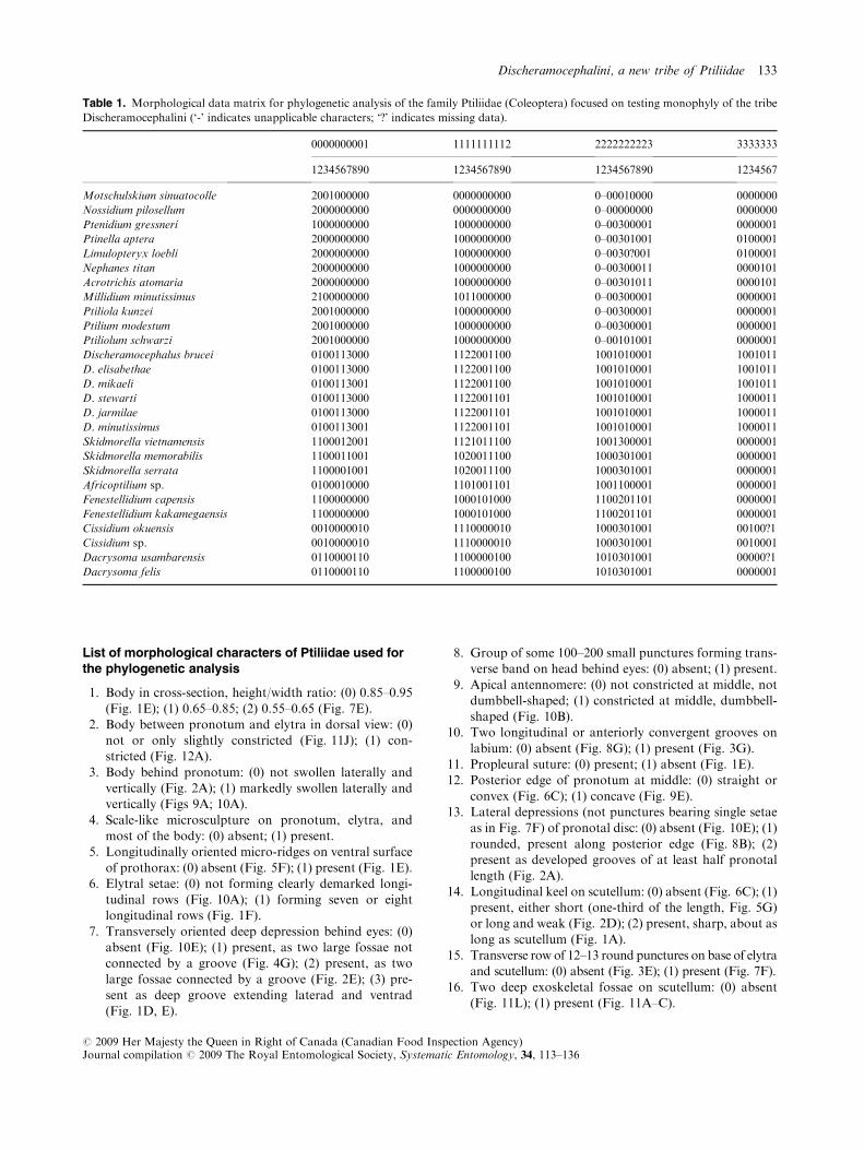
List of morphological characters of Ptiliidae used forthe phylogenetic analysis
1. Body in cross-section, height/width ratio: (0) 0.85–0.95
(Fig. 1E); (1) 0.65–0.85; (2) 0.55–0.65 (Fig. 7E).2. Body between pronotum and elytra in dorsal view: (0)
not or only slightly constricted (Fig. 11J); (1) con-stricted (Fig. 12A).
3. Body behind pronotum: (0) not swollen laterally andvertically (Fig. 2A); (1) markedly swollen laterally andvertically (Figs 9A; 10A).
4. Scale-like microsculpture on pronotum, elytra, andmost of the body: (0) absent; (1) present.
5. Longitudinally oriented micro-ridges on ventral surface
of prothorax: (0) absent (Fig. 5F); (1) present (Fig. 1E).6. Elytral setae: (0) not forming clearly demarked longi-
tudinal rows (Fig. 10A); (1) forming seven or eightlongitudinal rows (Fig. 1F).
7. Transversely oriented deep depression behind eyes: (0)absent (Fig. 10E); (1) present, as two large fossae notconnected by a groove (Fig. 4G); (2) present, as two
large fossae connected by a groove (Fig. 2E); (3) pre-sent as deep groove extending laterad and ventrad(Fig. 1D, E).
8. Group of some 100–200 small punctures forming trans-verse band on head behind eyes: (0) absent; (1) present.
9. Apical antennomere: (0) not constricted at middle, notdumbbell-shaped; (1) constricted at middle, dumbbell-shaped (Fig. 10B).
10. Two longitudinal or anteriorly convergent grooves on
labium: (0) absent (Fig. 8G); (1) present (Fig. 3G).11. Propleural suture: (0) present; (1) absent (Fig. 1E).12. Posterior edge of pronotum at middle: (0) straight or
convex (Fig. 6C); (1) concave (Fig. 9E).13. Lateral depressions (not punctures bearing single setae
as in Fig. 7F) of pronotal disc: (0) absent (Fig. 10E); (1)
rounded, present along posterior edge (Fig. 8B); (2)present as developed grooves of at least half pronotallength (Fig. 2A).
14. Longitudinal keel on scutellum: (0) absent (Fig. 6C); (1)present, either short (one-third of the length, Fig. 5G)or long and weak (Fig. 2D); (2) present, sharp, about aslong as scutellum (Fig. 1A).
15. Transverse row of 12–13 round punctures on base of elytraand scutellum: (0) absent (Fig. 3E); (1) present (Fig. 7F).
16. Two deep exoskeletal fossae on scutellum: (0) absent
(Fig. 11L); (1) present (Fig. 11A–C).
Table 1. Morphological data matrix for phylogenetic analysis of the family Ptiliidae (Coleoptera) focused on testing monophyly of the tribe
Discheramocephalini (‘-’ indicates unapplicable characters; ‘?’ indicates missing data).
0000000001 1111111112 2222222223 3333333
1234567890 1234567890 1234567890 1234567
Motschulskium sinuatocolle 2001000000 0000000000 0–00010000 0000000
Nossidium pilosellum 2000000000 0000000000 0–00000000 0000000
Ptenidium gressneri 1000000000 1000000000 0–00300001 0000001
Ptinella aptera 2000000000 1000000000 0–00301001 0100001
Limulopteryx loebli 2000000000 1000000000 0–0030?001 0100001
Nephanes titan 2000000000 1000000000 0–00300011 0000101
Acrotrichis atomaria 2000000000 1000000000 0–00301011 0000101
Millidium minutissimus 2100000000 1011000000 0–00300001 0000001
Ptiliola kunzei 2001000000 1000000000 0–00300001 0000001
Ptilium modestum 2001000000 1000000000 0–00300001 0000001
Ptiliolum schwarzi 2001000000 1000000000 0–00101001 0000001
Discheramocephalus brucei 0100113000 1122001100 1001010001 1001011
D. elisabethae 0100113000 1122001100 1001010001 1001011
D. mikaeli 0100113001 1122001100 1001010001 1001011
D. stewarti 0100113000 1122001101 1001010001 1000011
D. jarmilae 0100113000 1122001101 1001010001 1000011
D. minutissimus 0100113001 1122001101 1001010001 1000011
Skidmorella vietnamensis 1100012001 1121011100 1001300001 0000001
Skidmorella memorabilis 1100011001 1020011100 1000301001 0000001
Skidmorella serrata 1100001001 1020011100 1000301001 0000001
Africoptilium sp. 0100010000 1101001101 1001100001 0000001
Fenestellidium capensis 1100000000 1000101000 1100201101 0000001
Fenestellidium kakamegaensis 1100000000 1000101000 1100201101 0000001
Cissidium okuensis 0010000010 1110000010 1000301001 00100?1
Cissidium sp. 0010000010 1110000010 1000301001 0010001
Dacrysoma usambarensis 0110000110 1100000100 1010301001 00000?1
Dacrysoma felis 0110000110 1100000100 1010301001 0000001
Discheramocephalini, a new tribe of Ptiliidae 133
# 2009 Her Majesty the Queen in Right of Canada (Canadian Food Inspection Agency)Journal compilation # 2009 The Royal Entomological Society, Systematic Entomology, 34, 113–136

17. Meso-metaventral suture between mesocoxae: (0)clearly visible and forming a delimited border between
sclerites (Fig. 10F); (1) obliterated, both sclerites fullyamalgamated (Fig. 1E).
18. Meso-metaventral suture laterad of mesocoxae: (0)
present and clearly visible externally as an externallyprojecting keel (Fig. 8H); (1) present only as internalthickening of cuticle, not visible externally (Fig. 9H).
19. Serration along meso-metaventral suture laterad of
mesocoxae: (0) absent (Fig. 9D); (1) present (Fig. 8E).20. Metaventral longitudinal lateral lines: (0) absent (Fig. 1
E); (1) present (Fig. 5C).
21. Horizontally oriented perforation ofmesoventral keel: (0)absent; (1) present (Figs 2I; 3I; 4I; 5E; 6E; 7G; 9H; 10F).
22. Shape of the horizontal perforation of mesoventral keel
as visible in lateral view: (0) round (Fig. 5E); (1)vertically elongate, almost parallel-sided, about 2�longer than wide (Figs 6E; 7G).
23. Grooves on mesoventrum originating from fossae of
mesoventral keel and extending laterad: (0) absent(Fig. 5E); (1) present (Fig. 10F).
24. Fossae in anterior lateral corners of mesoventrum: (0)
absent (Fig. 6F); (1) present (Figs 1G; 2I; 5E).25. Metacoxae: (0) separated by one-fifteenth of metaven-
tral width (Fig. 1B); (1) separated by one-eighth of
metaventral width (Fig. 5C); (2) separated by one-sixthof metaventral width (Fig. 7C); (3) separated by one-qurater of metaventral width (Fig. 8E).
26. Metacoxae: (0) not transverse, less that 2� as wide aslong (Fig. 2G); (1) transverse, more than 2� as wide aslong (Fig. 1C).
27. Posteriorly oriented projection of metaventral plate
between metacoxae: (0) without two lateral teeth (Fig. 5I); (1) with two lateral teeth (Fig. 6D).
28. Alacrista of metathorax at middle: (0) without setae
along margins; (1) with short setae along margins(Fig. 13G).
29. Metascutellar spur on alacrista (Hall, 2003: 97): (0)
absent (Fig. 13G); (1) present.30. Hindwing membrane: (0) not or partly narrowed,
individual trichia of the fringe not longer than wing
width; (1) narrowed, individual trichia of the fringelonger than wing width.
31. Cavities on abdominal sternum VIII: (0) absent (Fig. 6D); (1) present (Fig. 1H, I).
32. Abdominal glands (Hall, 2003: 95): (0) absent; (1) present.33. Group of about 50–70 closely adjacent round micro-
pores transversely oriented along posterior edge of
tergite VIII: (0) absent; (1) present.34. Transverse rows of teeth-like serration of abdominal
sternites: (0) absent (Fig. 3H); (1) present (Grebenni-
kov, 2008: fig. 14).35. Tergites XI and X: (0) free, separated from each other;
(1) merged together into a single plate.36. Single elongate internal sclerite parallel to aedeagus: (0)
absent (Fig. 13A–F); (1) present (Grebennikov, 2008:fig. 75).
37. Parameres: (0) present; (1) absent.
Results
The present phylogenetic analysis with equally weightedunordered characters aimed to test the monophyly ofDischeramocephalini resulted in the overflow of 3387þmost-parsimonious trees of 66 steps (consistency index 66,retention index 87). A second analysis with successivecharacter weighting resulted in 305 most-parsimonious treesof 402 steps long (consistency index 82, retention index 94).
The strict consensus tree of both analyses retained Discher-amocephalini as a monophyletic group. Bootstrap values forthis clade were 91 and 68 in analysis 1 and 2, respectively.
The grouping of taxa inside Discheramocephalini wasconsistent and mainly resolved by both analyses. The generaCissidium and Dacrysoma form a clade supported by three
synapomorphies and a bootstrap value varying between 79and 94% (Fig. 14). Monophyly of Skidmorella and Discher-amocephalus is moderately well supported by foursynapomorphies (Fig. 14); however, the inclusion of Afri-
coptilium in this clade is apparently an artefact, as discussedabove under Africoptilium. Therefore, the most conservativecurrent evaluation of the internal relationships of the
Discheramocephalini taxa is that the tribe has the basalpolytomy of four clades: (i)Africoptilium, (ii) Fenestellidium,(iii) Cissidium þ Dacrysoma and (iv) Skidmorella þDischeramocephalus.
Discussion of phylogenetic analysis and currentclassification of Ptiliidae
Projecting these results to the family Ptiliidae, the internal
relationships and classification remain disordered. Thisproblem is mainly because no comprehensive attempt wasmade to make a phylogenetic analysis of the whole family or
to propose a cladogram-based classification. The classifica-tion adopted in this work derives from that used by Newton& Thayer (2007a) and reflects a conservative approach
aimed at minimizing the number of non-monophyletic taxa.This classification recognizes three subfamilies: Ptiliiinae,Acrotrichinae and Cephaloplectinae. Subfamily Acrotrichi-
nae includes the 12 genera listed by Hall (2005) arrangedinto two tribes, namely Acrotrichini and Nephanini; none ofthese three taxa has been demonstrated to be monophyletic.Subfamily Cephaloplectinae includes five genera (Hall,
2005); monophyly of this subfamily has also never beendemonstrated. Subfamily Ptiliinae contains the rest of thefamily and is a non-monophyletic assemblage of four tribes:
Nanosellini, Ptinellini, Discheramocephalini trib.n. andPtiliini. Among them, only Nanosellini and Discheramoce-phalini have been hypothesized to be monophyletic as
a result of phylogenetic analyses (Hall, 1999 and the presentpaper, respectively), whereas Ptinellini (under the namePterycini, nomen nudum) has been extensively discussed(Hall, 2003) and also hypothesized to represent a clade. The
tribe Ptiliini remains, however, a taxonomic ‘dumpingground’ for members of the family that share no apomor-phies of the remaining three tribes and cannot be assigned to
134 V. V. Grebennikov
# 2009 Her Majesty the Queen in Right of Canada (Canadian Food Inspection Agency)Journal compilation # 2009 The Royal Entomological Society, Systematic Entomology, 34, 113–136

Acrotrichinae or Cephalopectinae. Johnson (2004) and Hall(2005) resurrected from synonymy the tribe Ptenidiini with-
out providing cladistic justification; this taxonomic action isnot followed here and all its genera listed by these twoauthors are considered to belong to the non-monophyletic
Ptiliini.The bootstrap support statistics for monophyly of the
tribe Discheramocephalini might appear unconvincingfor validating the existence of a new tribe. Monophyly of
Discheramocephalini is, however, the only consistentlysupported hypothesis within the framework of the presentanalysis. With the present level of knowledge it is hardly
possible to rely exclusively on high and convincing values ofthe formalized cladistic approach when inferring the inter-nal branching pattern of Ptiliidae. The family remains
remarkably neglected taxonomically, with the majority ofnon-monotypic genera neither having been revised norhaving had their monophyly tested phylogenetically. Onlyone attempt to combine a taxonomic review of a subgroup
of Ptiliidae with phylogenetic analysis (tribe Nanosellini(Hall, 1999)) resulted in the tribe Nanosellini hypothesizedto be monophyletic, including 41 species in 26 genera, 21 of
the latter being monotypic. Such a high number of mono-typic genera suggests that research into the taxonomy ofPtiliidae still requires description and documentation of the
diversity of these beetles. It also means that any phylogeny-based internal groupings of Ptiliidae at the present stage willbe hampered by the lack of a pre-existing phylogenetic
hypothesis to root the trees and by the absence of a frame-work of well-understood morphological characters surveyedthroughout a wide spectrum of taxa. In other words, theattempt of phylogenetic reconstruction implemented here is
meant to serve mainly as a preliminary basis for futurestudies, rather than to provide definitive answers, in partic-ular regarding the monophyly of the new tribe.
Acknowledgements
Mikael Sorensson (Lund, Sweden) introduced me to thePtiliidae. He was the first to recognize the new peculiar
African genus named here as Fenestellidium, coining thisgeneric epithet. He also identified and supplied all species ofall non-Discheramocephalini ptiliids used in the presentanalysis, hosted my two visits to Lund, and commented on
two early drafts of this manuscript. Louis Deharveng andAnne Bedos (Paris, France) collected and made availablePtiliidae from Berlese samples taken in Vietnam and In-
donesia, which contained new species of Skidmorella. BruceD. Gill (Ottawa, Canada) accompanied me during thefieldwork in Cameroon, which resulted in the discovery of
a new Cissidium species. Elisabeth Hintelmann (Munich,Germany) funded my fieldwork in Cameroon througha scientific foundation which she established at the Zoo-logische Staatssammlung Munchen in memory of her late
husband, Robert J. H. Hintelmann (http://www.zsm.mwn.de/events/wiss_preise.htm). My 2001–2002 fieldwork inSouth Africa, Kenya and Tanzania was performed during
my appointment as a postdoctoral fellow at the Universityof Pretoria (South Africa) and was partly supported by the
following grants: Visiting Fellowship from the CanadianMuseumofNature (administrated throughRobertAnderson,Ottawa, Canada), Visiting Fellowship from the Field
Museum of Natural History (Chicago, IL), and Ernst MayrVisiting Fellowship from the Museum of ComparativeZoology (Cambridge, MA). This study was initiated duringmy tenure as an Alexander von Humboldt Fellow at the
Friedrich-Schiller-Universitat Jena hosted by Rolf G. Beutel(Jena,Germany).MaxwellV.L.Barclay (TheNaturalHistoryMuseum, London, U.K.) and Marc De Meyer (Royal
Museum for Central Africa, Tervuren, Belgium) made Afri-coptilium holotypes available for re-examination. HaraldSchillhammer and Heinrich Schonmann (Naturhistorisches
Museum, Wien, Austria) searched (unsuccessfully) throughthe Franz material in their museum for the Africoptiliumconcinnum holotype. Ainsley Seago (Berkeley, CA) suppliedspecimens ofMotschulskium sinuatocolle used for the analysis.
Hume Douglas (Ottawa, Canada) commented on an earlydraft of this manuscript. All the above people are sincerelyacknowledged for their contributions to the completion of this
project.
References
Deane, C. (1931) Trichopterygidae of Australia and adjacent lands.
Proceedings of the Linnean Society of New South Wales, 56,
227–242.
Farris, J.S. (1969) A successive approximations approach to
character weighting. Systematic Zoology, 18, 374–385.
Farris, J.S. (1988) Hennig86. Documentation for Version 1.5.
Published by the author, Port Jefferson Station, New York.
Grebennikov, V.V. (2008) How small you can go: factors limiting
body miniaturization in winged insects with a review of the
pantropical genus Discheramocephalus and description of six new
species of the smallest beetles (Pterygota: Coleoptera: Ptiliidae).
European Journal of Entomology, 105, 313–328.
Hadley, A. (2007) CombineZ, Version 5. Published by the author.
URL http://www.hadleyweb.pwp.blueyonder.co.uk/CZ5/combi-
nez5.htm [accessed/downloaded on 21 November 2006].
Hall, E.W. (1999)Generic revision of the tribeNanosellini (Coleoptera:
Ptiliidae: Ptiliinae). Transactions of the American Entomological
Society, 125, 39–126.
Hall, E.W. (2003) Limulopteryx, a new genus of Neotropical
featherwing beetles (Coleoptera: Staphylinoidea: Ptiliidae) and
comments on pterycine ptiliids. Systematics of Coleoptera: Papers
Celebrating the Retirement of Ivan Lobl (ed. by G. Cuccodoro and
R. A. B. Leschen), pp. 85–102. Memoirs on Entomology Interna-
tional, 17. Associated Publishers, Gainesville, FL.
Hall, E.W. (2005) 11.2. Ptiliidae Erichson, 1845. Coleoptera, Vol. 1:
MorphologyandSystematics (Archostemata,Adephaga,Myxophaga,
Polyphaga partim). Handbook of Zoology, Vol. IV, Arthropoda, Part
II, Insecta (ed. by R. G. Beutel and R. A. B. Leschen), pp. 251–261.
Walter De Gruyter, Berlin.
Hammond, P.M. & Lawrence, J.F. (1989) Appendix: mycophagy in
insects: a summary. Insect-Fungus Interactions. 14th Symposium
of the Royal Entomological Society of London in Collaboration
With the British Mycological Society (ed. by N. Wilding, N. M.
Collins, P. M. Hammond and J. F. Webber), pp. 275–319.
Academic Press, London.
Discheramocephalini, a new tribe of Ptiliidae 135
# 2009 Her Majesty the Queen in Right of Canada (Canadian Food Inspection Agency)Journal compilation # 2009 The Royal Entomological Society, Systematic Entomology, 34, 113–136

Johnson, C. (1967) Studies on Ethiopian Ptiliidae. 1, Africoptilium
gen. n. from Central Africa. Entomologist, 100, 288–292.
Johnson, C. (1971) Some Ptiliidae from the Philippine, Bismarck
and Solomon Islands (Insecta, Coleoptera). Steenstrupia, 2,
39–47.
Johnson, C. (1982) An introduction to the Ptiliidae (Coleoptera) of
New Zealand. New Zealand Journal of Zoology, 9, 333–376.
Johnson, C. (2004) Ptiliidae. Catalogue of Palaearctic Coleoptera.
Volume 2. Hydrophiloidea – Histeroidea – Staphylinoidea (ed. by I.
Lobl and A. Smetana), pp. 122–131. Apollo Books, Stenstrup.
Johnson, C. (2007a) Studies on Ptiliidae (Col.) from the Solomon
Islands, 1. The Entomologist’s Monthly Magazine, 143, 213–226.
Johnson, C. (2007b) Errata for Volume 2. Catalogue of Palaearctic
Coleoptera. Volume 4. Elateroidea – Derodontoidea – Bostrichoidea
– Lymexyloidea – Cleroidea – Cucujoidea (ed. by I. Lobl and A.
Smetana), pp. 22–24. Apollo Books, Stenstrup.
Lawrence, J.F. (1999) The Australian Ommatidae (Coleoptera:
Archostemata): new species, larva and discussion of relation-
ships. Invertebrate Taxonomy, 13, 369–390.
Lucas, R. (1920) Catalogus alphabeticus generum et subgenerum
Coleopterorum orbis terrarum totius (famil., trib., subtr., sec.
incl.). Pars I. Archiv fur Naturgeschichte, 84(A), xxxiþ696pp. (not
seen; cited after Newton & Thayer, 2007b).
Matthews, A. (1884) Trichopterygidae found in Japan by Mr G.
Lewis. Cistula Entomologica, 3, 77–84 (not seen; cited after
Newton & Thayer, 2007b).
Matthews, A. (1889) New genera and species of Trichopterygidae.
The Annals and Magazine of Natural History, 3, 188–195 (not
seen; cited after Newton & Thayer, 2007b).
Motschulsky, V. (1855) Voyages. Lettre de M. deMotschulsky a M.
Menetries. No 2. A bord du bateau a vapeur United-States, 20
Mars 1854. Etudes Entomologiques, 4, 8–25 (not seen; cited after
Newton & Thayer, 2007b).
Motschulsky, V. (1869) Enumeration des nouvelles especes de
coleopteres rapportes des ses voyages. 6-ieme article. Bulletin de
la Societe Imperiale des Naturalistes de Moscou, 41, 163–200 (not
seen; cited after Newton & Thayer, 2007b).
Newton, A.F. & Thayer, M.K. (2007a) Catalog of higher taxa,
genera and subgenera of Staphyliniformia [online]. Chicago:
Field Museum of Natural History [updated November 03,
2005]. URL http://www.fieldmuseum.org/peet_staph/
db_1a.html [accessed/downloaded on 20 July 2007].
Newton, A.F. & Thayer, M.K. (2007b) Bibliography supporting
catalogs of Staphyliniformia family-, genus- and species-group
names [online]. Chicago: Field Museum of Natural History
[posted date of last update]. URL http://www.fieldmuseum.org/
peet_staph/db_1d.html [accessed/downloaded on 20 July 2007].
Nixon, K.C. (2002) Winclada, Version 1.00.08. Published by the
author, Ithaca, New York.
Pinto, J.D. (1997) Chapter 22. Trichogrammatidae. Annotated Key
to Genera of Nearctic Chalcidoidea (Hymenoptera) (ed. by G. A. P.
Gibson, J. T. Huber and J. B. Wooley), pp. 726–752. NRC
Research Press, Ottawa.
Sawada, Y. & Hirowatari, T. (2003) Discovery of the genus
Skidmorella Johnson (Coleoptera: Ptiliidae) in Japan, with descrip-
tion of two new species. Entomological Science, 6, 309–314.
Sorensson, M. (1997) Morphological and taxonomical novelties in
the world’s smallest beetles, and the first Old World record of
Nanosellini (Coleoptera: Ptiliidae). Systematic Entomology, 22,
257–283.
Accepted 15 May 2008
136 V. V. Grebennikov
# 2009 Her Majesty the Queen in Right of Canada (Canadian Food Inspection Agency)Journal compilation # 2009 The Royal Entomological Society, Systematic Entomology, 34, 113–136