Embed Size (px)
Citation preview

J. Exp. Biol. (1970), 52, 139-165 139With 19 text-figures
Printed in Great Britain
DISCHARGE PATTERNS OFCOXAL LEVATOR AND DEPRESSOR MOTONEURONES OF
THE COCKROACH, PERIPLANETA AMERICANA
BY K. G. PEARSON* AND J. F. ILES.t
University Laboratory of Physiology, Oxford
(Received 9 July 1969)
INTRODUCTION
The control of rhythmic behaviour has been intensively studied in a number ofarthropod systems, namely insect flight (Wilson, 1961, 1968; Wyman, 1965, 1966),insect ventilation (Miller, 1965, 1966; Mill & Hughes, 1966) and crayfish and lobsterswimmeret (Hughes & Wiersma, i960; Davis, 1968, 1969). The patterning of moto-neuronal activity in these systems is to a large extent independent of sensory feed back,depending primarily upon the properties and connectivity of cells within the centralnervous system. In contrast to these systems the control of rhythmic leg movementsduring insect walking is poorly understood and there is currently no information as tothe extent of central patterning of activity in leg motoneurones.
Recent studies on the locust have shown that sensory input from the femoralchordotonal organ and tarsal hairs is important in co-ordinating leg movements andcontrolling excitatory and inhibitory motoneuronal activity when the animal is makingpostural adjustments and walking (Usherwood, Runion & Campbell, 1968; Runion&Usherwood, 1968). These studies have therefore suggested, for the locust at least, thatsensory input from leg receptors is very important for controlling motoneuronalactivity during walking. Currently there is no direct evidence to indicate that there isany significant central patterning of motoneuronal activity in locust walking. In-vestigations on the cockroach, Periplaneta americana, have also shown that sensoryinput could be important in patterning activity in motoneurones of a single leg, andfor co-ordinating the activity in motoneurones in different legs (Pringle, 1940, 1961;Wilson, 1965). However, in this animal there is indirect evidence suggesting that duringwalking motoneuronal activity may be patterned independent of sensory input(Milburn, 1963; Wilson, 1966a).
The aim of the present investigation has been to determine the extent of centralpatterning of activity in motoneurones innervating some of the metathoracic coxallevator and depressor muscles by recording the activity of identifiable motoneuronesin preparations in which all sensory input from leg receptors has been eliminated.
• Present address: Dept. of Physiology, University of Alberta, Edmonton, Canada,t M.R.C. Scholar.

140 K. G. PEARSON AND J. F. ILES
PREPARATIONS AND METHODS1. Anatomy
The positions of the nerves studied in this investigation are shown in Fig. 1. Themetathoracic posterior coxal levator muscle (number 182, notation of Carbonell, 1947)is innervated by axons contained in nerve 6Br4 (notation of Pipa & Cook, 1959).There are twelve motor axons in nerve 6Br4. These axons have been numbered from1 to 12 with increasing size (Pearson, Stein & Malhotra, 1969). Axons 3, 4, 5 and 6innervate the posterior coxal levator muscles 182C and 182D. The innervation patternsof these muscles by axons 3-6 have been described by Pearson & Bergman (1969).Axon 3 is one branch of a common inhibitory neurone having a powerful inhibitoryeffect on the slow contractions produced by axon 4. Activity in axons 5 and 6 pro-duces strong tonic contractions of the posterior coxal levator muscles (i82C,D)(Fig. 7), which are much less affected by the common inhibitory neurone.
The coxal depressor muscles (177D, E, 178 and 179) are innervated by motoraxons contained in nerve $TI. Nerve 5ri arises soon after nerve 5 enters the coxa andruns only a short distance before branching, branch 5ri a running ventrally and 5nbrunning dorsally.
2. Preparations
All experiments were carried out on the right side of the metathoracic segment ofadult male cockroaches, Periplaneta americana. Animals were lightly anaesthetizedwith carbon dioxide and pinned ventral side up to a cork board. One pin was placed
176
Fig. 1. Diagram showing the position of the nerves and muscles relevant to the investigationon the discharge patterns of the coxal levator and depressor motoneurones. Both metathoraciclegs have been rotated to expose their dorsal coxal surfaces.

Activity in cockroach leg motoneurones 141
•>ff centre near the last abdominal segment and another through the dorsal cuticlejust anterior to the prothoracic segment. The right metathoracic coxa was rotated toexpose its dorsal surface and fixed with a fine pin mid-way along the medial cuticle.Care was taken to ensure that the femur, tibia and tarsus of this leg were completelyfree and able to move to their full extent. The prothoracic and mesothoracic legswere therefore pulled forward and fixed so as not to interfere with any part of therotated metathoracic leg. The metathoracic leg not being studied was pinned ventralside up, the pin passing through the lateral edge of the coxa. Again care was taken toensure that the femur, tibia and tarsus of this leg were completely free.
The dissection to expose nerve 6Br4 has been described elsewhere (Pearson &Bergman, 1969). To expose nerve 5ri the coxa was first rotated to reveal its dorsalsurface. The dorsal coxal rim was cut both sides of the attachment of muscle 178 andthe dorsal coxal cuticle broken in an arc between these two cuts. This arc extendedabout two-thirds the length of the coxa. Muscle 178 was detached from this smallpiece of cuticle. By moving the detached muscle 178 laterally, and carefully removingthe underlying tracheal system, nerve 5ri b was exposed. A fine branch of this nerveis sent to muscle 178. The approximate position of nerve 5nb is shown in Fig. 1(muscle 178 is not shown connected to the nerve in this diagram). By moving nerve 5laterally nerve 5ri a is exposed. This dissection allowed recordings to be made fromnerve 5ri, 50 a or srib.
(a) Headless preparation. The head was removed before pinning the animal to thecork board and nerves 6Br4 and 5ri were exposed as described above. No peripheralnerves were cut in this preparation.
(b) De-qfferentated preparation. This preparation was designed to eliminate allsensory input to the central nervous system from the legs. After head removal all thelateral nerve trunks of the prothoracic and mesothoracic ganglia were cut. Thisremoved all sensory input from the legs of these segments. The cuticle above themetathoracic ganglion was then cut and lifted to expose the nerve trunks leaving theganglion. The trunks, 2, 3 and 4 were cut close to the ganglion on both sides and trunks5 and 6 on the side not being studied (left) were also cut. The cuticle flap was replacedover the ganglion and a thin layer of petroleum jelly was applied to the edges to preventdrying. The right leg was rotated to expose its dorsal coxal surface and fixed with afine pin through the medial coxal wall. Nerves 6B and 5r 1 were exposed. Nerve branches5ria, 5r2, 6A, 6Br2 and 6Br3 were cut. Electrodes were then placed under nerves6Br4 and 5rib and both these nerves were cut distal to the recording electrode.Finally, cutting nerve 5 distal to the branch 50 gave a preparation in which allsensory input from the legs was eliminated, and one in which recordings could bemade simultaneously from the coxal levator and depressor motoneurones. Strictlythis preparation was not de-afferentated because sensory information was probablycoming in the abdominal connectives. However, none of this input could be signallingany tactile or proprioceptive information from the legs.
(c) Cut thoracic connectives (c.T.c.) preparations. The connectives between themesothoracic and metathoracic ganglia were cut, doing minimal damage to the trachealsystem. In de-afferentated c.T.c. preparations the meso-metathoracic connectives werecut after de-afferentating the metathoracic ganglion as described in (b) above.

142 K. G. PEARSON AND J. F. ILES
3. Recording and analysing equipment
All recordings from the nerves were made with single 75 /i silver wire electrodes.The nerve was lifted clear of the haemolymph and coated with petroleum jelly toprevent drying. The two pre-amplifiers used to record simultaneously from nerve 6Br4and nerve 50 (or 5ri a or 5ri b) were a Tektronix type 122 and an Isleworth type A101.These amplifiers were connected to a Tektronix 502A oscilloscope and the outputfrom the cathode followers of this oscilloscope was fed to a Thermionic ProductsT3000 four-channel tape recorder. All experiments were recorded on magnetic tape.This allowed a more detailed analysis of the discharge patterns at a later time. Italso allowed the activity to be slowed down so that pen recordings and punched papertape records could be made of rapid events.
Reset
oCRO p.h.a.
d.a.c. 1Pen
recorder
HISTARi t...'!. I
Fig. 2. Equipment layout for analysing activity recorded from nerve 6Br4 and 5ri. p.h.a., pulseheight analyser; d.a.c, digital to analog converter; HISTAR, high speed timer and recorder.
The equipment layout for analysing the records made from nerves 6Br4 and 5riis shown in Fig. 2. Pulse-height analysing equipment was used to separate the spikesrecorded from the different motor axons in nerve 6Br4. Pulse-height analysing wasnot necessary for separating the spikes from motor axons in nerve 5ri, for generallythe largest spike corresponded to activity in the motor axon most intensively studied.Digital-to-analog converters were used to count the number of impulses in a givenaxon for a unit of time (from o-i to 10 sec). The output voltage from these deviceswas proportional to the number of events occurring in that unit of time (Stein, 1968)and was displayed on a Devices four-channel pen recorder. The result was a plot ofaverage frequency over the unit time interval against time. An on-line pen record ofthe activity in the different motoneurones was also made during the experiments usingthis equipment allowing a more rapid selection of interesting activity when analysing.
The activity of up to four motoneurones could be recorded on punched paper tapefor subsequent computer analysis, using the device labelled 'HISTAR' in Fig. 2(Stein, 1965).
Activity in cockroach leg motoneurones
4. Computing procedure
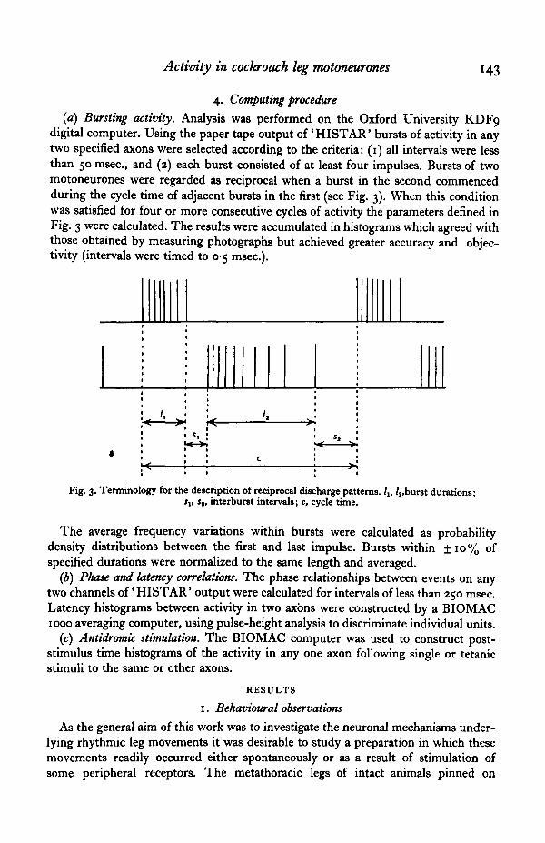
(a) Bursting activity. Analysis was performed on the Oxford University KDF9digital computer. Using the paper tape output of 'HISTAR' bursts of activity in anytwo specified axons were selected according to the criteria: (1) all intervals were lessthan 50 msec, and (2) each burst consisted of at least four impulses. Bursts of twomotoneurones were regarded as reciprocal when a burst in the second commencedduring the cycle time of adjacent bursts in the first (see Fig. 3). When this conditionwas satisfied for four or more consecutive cycles of activity the parameters defined inFig. 3 were calculated. The results were accumulated in histograms which agreed withthose obtained by measuring photographs but achieved greater accuracy and objec-tivity (intervals were timed to 0-5 msec).
••
1
'1
1
si i< >•
Fig. 3. Terminology for the description of reciprocal discharge patterns. /,, /,,burst durations;sv st, interburst intervals; c, cycle time.
The average frequency variations within bursts were calculated as probabilitydensity distributions between the first and last impulse. Bursts within ±10% ofspecified durations were normalized to the same length and averaged.
(b) Phase and latency correlations. The phase relationships between events on anytwo channels of' HISTAR' output were calculated for intervals of less than 250 msec.Latency histograms between activity in two axbns were constructed by a BIOMAC1000 averaging computer, using pulse-height analysis to discriminate individual units.
(c) Antidromic stimulation. The BIOMAC computer was used to construct post-stimulus time histograms of the activity in any one axon following single or tetanicstimuli to the same or other axons.
RESULTS
1. Behavioural observations
As the general aim of this work was to investigate the neuronal mechanisms under-lying rhythmic leg movements it was desirable to study a preparation in which thesemovements readily occurred either spontaneously or as a result of stimulation ofsome peripheral receptors. The metathoracic legs of intact animals pinned on
144 K. G. PEARSON AND J. F. ILES
their backs periodically showed spontaneous rhythmic movements but these usuall^did not last more than a few seconds. In headless animals, however, thesespontaneous movements occurred more often and sometimes lasted longer than10 sec. Stimulation of a cereal hair by a single mechanical shock in quiescentheadless preparations usually elicited strong rhythmic movements of the ipsilateralmetathoracic leg. Sometimes this stimulus also elicited movements of the contra-lateral leg, but this usually was neither as prolonged nor as rapid as the ipsilateralresponse. Cereal stimulation in intact preparations also produced rhythmic legmovements but, as with the spontaneous movements, these generally did not lastfor more than a few seconds.
Of particular interest in this investigation were the alternate flexion and extensionmovements at the coxa-trochanter-femur joint. For normal walking Hughes (1952)has shown that there is extension at this joint during leg retraction (depression) andflexion during leg protraction (Ievation). The muscles that can produce flexion at thisjoint are the anterior coxal levator (180) innervated by axons in nerve 3b, and themain and posterior coxal levators (181, 182) innervated by axons in nerve 6Br4. Themuscles that can produce extension at the trochanter joint are the main depressormuscles (177 A, B, C) innervated by axons in nerve 4, and the coxal depressor muscles(177 D, E, 178, 179) innervated by axons in nerve 5ri. To determine which of thesesets of muscles were mainly responsible for producing the alternate flexion and exten-sion movements of the metathoracic femur in an intact animal pinned on its back,either nerve 6Br4 or nerve 4 to one leg was cut and the movements of that leg werecompared with the movements of the other metathoracic leg. (Cutting either nerve6Br4 or nerve 4 does not remove any important sensory input to the central nervoussystem as Pipa & Cook (1959) have shown that both these nerves are alitiost entirelymotor; sensory fibres from the legs are contained in nerves 3b and 5.) Cutting nerve 4did not abolish, or have any obvious effect upon, the extension movements at thetrochanter joint. Thus the branches A, B and C of the main depressor muscle 177 donot appear to be involved in producing the femoral extension movements. Cuttingnerve 6Br4 considerably reduced flexion movements of the femur, and in all prepara-tions the strong flexion characteristic of walking did not occur. Thus the anterior coxallevator muscle, 180, does not produce the strong femur flexion seen during rhythmicleg movements.
The conclusions from these lesion experiments were that the rhythmic movementsof the femur relative to the coxa are produced by activity in the motor axons containedin nerve 6Br4 to the main and posterior coxal levator muscles (181, 182) giving flexionmovements, and nerve 5ri to the coxal depressor muscles (177 D, E, 178, 179) givingextension movements. From these observations it was therefore decided to examinethe discharge patterns of the motor axons contained in nerves 6Br4 and 5ri to deter-mine which of these axons were responsible for producing the alternate flexion andextension movements at the trochanter joint. Initially these discharge patterns werestudied in headless preparations as the rhythmic leg movements occurred more oftenand could easily be elicited by cereal stimulation.

Activity in cockroach leg motoneurones 145
2. Identification of motor axons
(a) Levator motor axons. Of the twelve motor axons contained in nerve 6Br4 thesix smallest (axons 1-6) could be reliably identified by the amplitude of the extra-cellularly recorded spikes and their discharge patterns. In intact preparations theaxons larger than axon 6 fired in bursts and interaction of the spikes from individualaxons made it impossible to identify confidently any of these larger axons. Whenrecordings were made using a single electrode on a section of uncut nerve liftedinto petroleum jelly, the other electrode being in the haemolymph close to therestricted length of nerve, the mean peak-to-peak amplitudes of the triphasic spikesrecorded from axons 1 to 6 were 0-073, 0-I3> I-2» I'3> 4'1 a nd 97 m V respectively.The variation in these amplitudes between preparations was very small, the coefficient
^ 0
"mJin
10 sec.
Fig. 4. Effects of femur flexion on the spontaneous discharge rate of the second largest axonin nerve 5ri. The onset of flexion is indicated by the upward going arrows and release fromflexion by the downward going arrows.
of variation being about o-i. The spike amplitudes from axons 3 and 4 were verysimilar and these axons could not always be identified by spike amplitude alone.However, they could be distinguished when recordings were also made from nerve4r2a or 5 r 1, for axon 3 is one branch of a common inhibitory neurone other branchesof which are contained in nerves 4«a and 51:1. Further criteria for identification ofaxons 5 and 6 were that both axons fired in bursts usually lasting less than 0-5 sec,and that axon 6 was never active without axon 5 being active. Usually axon 6 becameactive when the discharge frequency of axon 5 exceeded about 80 impulses/sec.
(b) Depressor motor axons. Recordings from the cut end of nerve 5n showed that upto four different motor axons could be spontaneously active at different times. In anintact preparation the spontaneous discharge rate of the largest of these four axons(peak monophasic amplitude of about 6 mV) was usually between 5 and 30 impulses/sec. This resting discharge rate corresponds to that observed by Pringle (1940) forthe slow depressor motor axon to the coxal depressor muscles. Further evidence thatthis motoneurone was in fact the slow depressor comes from the observation thatflexion of the coxa-trochanter-femur joint caused an increase in firing rate of thisneurone (Fig. 4), and bursts of activity in this neurone corresponded to extensionmovements of the femur. Also in the resting undissected animal, when this neuronewas presumably active, there were no visible signs of twitch contraction in any of thecoxal depressor muscles. This slow depressor motor axon will be referred to as axon
D..The amplitudes of the monophasic potentials recorded from the three small axons
IO EXB52

146 K. G. PEARSON AND J. F. ILES
were all about equal (approximately o-8 mV). When simultaneous recordings werdtaken from nerves 5ri and 6Br4 it was found that one of these three small axons wasa branch of the common inhibitory neurone as shown in Fig. 5. The records shown inthis figure were taken during a small burst of activity in axon 5 and it is seen that allthree small axons became more active during this burst of activity. Generally it wasobserved that the discharge rate of these axons only increased when the excitatoryaxons 5 and 6 to the levator muscles became active. Thus the discharge patterns ofthese neurones suggested they could be inhibitory in function. Intracellular recordingsfrom single fibres of muscle 177E have confirmed that all three small axons are inhibi-tory (Iles & Pearson, 1969).
• 5r1
• 6Br4
T T T T T0-2 sec.
Fig. 5. Discharge pattern of the three smaller axons in nerve 5ri during a small burst of activityin motoneurone 5. Top trace, extracellular record from nerve sr i ; bottom trace, extracellularrecord from nerve 6Br4. In the record from nerve 6 Br4 the large spike is from axon 5 and the twosmaller spikes from axons 3 and 4 (the spikes from axon 3 are marked with arrows). Note that oneof the smaller axons in nerve sri is a branch of the common inhibitory neurone, signified by the1:1 correspondence of its discharge with that of axon 3. Note also that all three small axonsin nerve sri increased in activity during the burst in axon 5. The large spike in the top recordis from the slow depressor axon D,.
When the ipsilateral cercus was lightly touched in the intact animal, the responserecorded from nerve 5n was often similar to that shown in Fig. 6. Apart from axonDs being activated, a single large axon (monophasic amplitude of about 25 mV) couldalso become active. This large axon, when active, gave twitch contractions and cor-responds to the fast axon described by Pringle (1939). The large axon will be referredto as axon Df. The response in axon Df rapidly declined on repeated stimulation of thecercus, whereas the response in axon Ds, although decreasing, never habituated com-pletely. In some preparations axon Df could not be activated.
One consistent finding was that on cereal stimulation axon Da was always activatedbefore axon Df, and the interval between the first impulse in axon Ds and the firstimpulse in axon Df varied from 10 to 150 msec. If axon Df is responsible for initiatingthe escape response then this finding may be of relevance in explaining the behaviouralobservation that the interval between cereal stimulation and the initiation of theescape response is quite variable, ranging from 28 to 90 msec. (Roeder, 1959).Another interesting observation was that a single stimulus to the cercus oftengave rise to repeated bursts of activity in axons Da and Df, as shown in Fig. 6.Removal of the head, or cutting the thoracic connectives, reduced the response inaxon Df to cereal stimulation. Thus there appears to be some sort of descending

Activity in cockroach leg motoneurones 147
excitatory pathway to control transmission from interneurones in the abdominal cordto axon D/. Roeder (1948) also reported that transmission through the metathoracicganglion is reduced by cutting any of the anterior connectives, and behaviourally it isobserved that the animal's escape response is reduced (Hughes, 1965 a).
Nerve 5n is short and branches soon after it leaves the main trunk of nerve 5.Recordings from either nerve 5ri a or 5n b showed potentials corresponding to all thefive axons described above. Thus all five axons branch at the first branch point ofnerve 5ri. Estimates of axon diameter from monophasic amplitude measurementsfrom nerve 5rib (method described by Pearson, Stein & Malhotra, 1969) gave the
0 2 sec.
Fig. 6. Response of the slow and fast excitatory axons D. and Dt in nerve fri to cereal stimula-tion. The ipsilateral cercus was touched just before the burst of activity on the left. The small•pike is from motor axon D, and the larger from Dr.
diameters of the five axons as 28 fi for axon Dt, 13 fi for axon D,, and approximately5 ft, for the three smaller axons. In the mesothoracic segment Dresden & Nijenhuis(1958) reported that nerve 5ri contains five axons larger than 5 /f all of which branchedand ran in nerve 5ri a and 5ri b. The diameters of these five axons measured by theseworkers were one larger than 2O/t, one in the 10 to 20 fi range, and three in the5-10 (i range. The good correlations of the number, diameter and initial branchingof the motor axons contained in nerve 5n of the mesothoracic and metathoracicsegments suggests that the functional properties of these motor axons may be thesame in both segments. Dresden & Nijenhuis (1958) reported that the largest axon(corresponding to axon Df) innervated only the posterior and anterior coxal depressormuscles (136 and 137) and that the four smaller axons innervated only the two coxalbranches of the main depressor muscle (135D and 135E). Usherwood (1962) con-firmed that the largest axon innervated only muscles 136 and 137, and showed thatthis axon gave twitch contractions. He also showed that the other four axons innervatedonly muscles 135D and 135E and could be classified as two fast and two slow axons.The largest of these four axons (corresponding to axon D,) was a slow axon. Thusaxons Dt and Df have functional equivalents in the mesothoracic segment. On theother hand the functional properties of the three smaller axons do not appear to bethe same since all three of these axons in the metathoracic segment are inhibitory(lies & Pearson, 1969). However, the discharge characteristics of the three smalleraxons in the two segments are identical and, furthermore, at least one of these axonsin the mesothoracic segment is a branch of a common inhibitory neurone, this neuronehaving similar distribution to that described by Pearson & Bergman (1969) for the

148 K. G. PEARSON AND J. F. ILES
metathoracic segment. These observations therefore do not support Usherwood'sclassification of the three small axons as one slow and two fast.
3. Motoneuronal activity
(a) Reciprocal patterning. The activity of the motor axons contained in nerve 6Br4could be monitored during rhythmic movements of the femur without damage to anyof the coxal muscles. This was achieved by recording from the proximal part of nerve6B which was exposed by removing the soft cuticle between the dorsal coxal rim andthe abdomen. During rhythmic leg movements high-intensity bursts of activity inmotor axons 5 and 6 corresponded to the flexion movements about the trochanterjoint. Motor axons larger than 6 became active during the more vigorous flexionmovements and at least four of these larger axons have been confidently identified ina single experiment. (Interaction of spikes during high-intensity bursts, and thesimilarity of spike amplitudes from the large axons, meant that it could not be deter-mined whether all six larger axons ever became active during vigorous leg-flexion
1 sec.
Fig. 7. Strong graded contractions produced in the posterior coxal levator muscles 182C and182D by bursts of activity in motoneurones 5 and 6. Top trace, tension record; bottom trace,extracellular record from nerve 6B. The smaller spike is from motor axon 5 and the larger fromaxon 6.
movements.) Since strong flexion movements of the femur occurred when only motoraxons 5 and 6 became active, and since cutting nerve 6Br4 abolished these movements,it was concluded that activity in motoneurones 5 and 6 is largely responsible forproducing the femoral flexion movements. Bursts of activity in these two moto-neurones produced strong graded contractions in branches C and D of the posteriorcoxal levator muscle, 182, as shown in Fig. 7. At least three of the six axons largerthan axon 6 produce twitch contractions in the main coxal levator muscle (181), andthe posterior coxal levator muscle is also innervated by one fast axon (Pearson &Bergman, 1969). Twitch responses seen in the tension records from the coxal levatormuscles indicated that some of these large fast axons were recruited to produce thevery rapid and strong femoral flexion movements.
The activity in the slow depressor motoneurone Ds was strongly reciprocal with thebursts of activity in the levator motoneurones, as shown in Fig. 8. The reciprocalrelationship was strongest between motoneurone Ds and the levator motoneurone 5.Only rarely were both simultaneously active and then only when each was dischargingat low frequencies (a few impulses per second). To record from nerve 5rib meantdamaging at least muscle 178 and usually muscle 177D. Even so the rhythmic move-ments of the femur about the trochanter still occurred but, because of the damage tothe extensor muscles, the extension movements were not as strong. The activity of

Activity in cockroach leg motoneurones 149
inotoneurone Da could easily be monitored without damage to any of these musclesby recording extracellularly from the coxal depressor muscles. Recordings were madesimultaneously from nerve 6B and extracellularly from the coxal depressor musclesduring normal rhythmic movements of the femur. No consistent differences havebeen found in the patterns of activity in the levator motoneurone Da using theserecording conditions compared to those seen when recordings were made from nerves6Br4 and 5ri b. Therefore, although some of the coxal depressor muscles were damagedin exposing nerve 5rib, leading to an alteration in the movements about the tro-chanter, this did not significantly alter the discharge patterns in motoneurone Da and
, | |6Br4
5r1b
6Br4
5r1b
1 sec.
Fig. 8. Reciprocal patterns of activity recorded from nerves 6Br4 and 5ri b in a headlesspreparation. The bottom pair of records is continuous with the top. The single spike recordedfrom nerve 5ri b is from motor axon D,. Motor axons 5 and 6 discharged throughout thelevator bursts (the spike from motor axon 5 being the smaller in the record from nerve 6Br4).At least one levator motor axon larger than axon 6 discharged one or two times during thelevator bursts.
the levator motoneurones. This observation suggested that sensory input from re-ceptors at the trochanter was not of importance in the generation of the reciprocalpatterns of activity. A further indication that sensory input was unimportant was thatthe activity did not depend on whether the leg was completely free to move, or fixedin one position. This was not conclusive, however, for with the leg fixed in one positionthe possibility that the isometric muscular contractions gave phasic excitation ofvarious groups of leg receptors (e.g. campaniform sensilla), and that this sensoryinput was important in the generation of the reciprocal patterns of activity, could notbe excluded. For this reason recordings were made in the de-afferentated preparation.
The patterns of activity in these de-afferentated preparations were similar in allrespects to those seen in the headless animal. Figure 9 shows spontaneous bursts ofreciprocal activity seen in one of these de-afferentated preparations. The spontaneousperiods of reciprocal activity occurred less often in these preparations but, as withthe headless animals, could be readily elicited by stimulation of the cerci.
(b) Characteristics of the reciprocal activity. As mentioned above, the patterns ofactivity in the headless and de-afferentated headless preparations were very similarand, apart from the decrease in spontaneous reciprocal activity in de-afferentated

K. G. PEARSON AND J. F. ILES
6Br4
5r1b
6Br4
5r1b
0 5 sec.
Fig. 9. Reciprocal patterns-of activity recorded from nerves 6Br4 and 5ri b in a de-afferentatedheadless preparation. The bottom pair of records are continuous with the top. Motor axons 5and 6 discharged throughout the levator bursts while at least one larger axon became activeduring some of these bursts (record from nerve 6Br4). Interaction of the spikes from thesethree axons account for the variability of spike amplitudes in some of the levator bursts. Thesingle spike recorded from nerve 5ri b is from motor axon D,.
501-
75
50 r
O L 1 1
!5 0|
25msec.
(b)
|50
I75
Fig. 10. Histograms of interburst intervals. Top, interval between the last impulse of theburst in motoneurone 5 and first impulse in the burst in motoneurone D,, and vice versa(bottom), for reciprocal patterns of activity in (a) three de-afferentated and (6) five headlesspreparations. Negative values of these intervals when the bursts overlap.

Activity in cockroach leg motoneurones 151
preparations, no consistent differences between the reciprocal activity has so far beenfound. Therefore the characteristics of this activity discussed in this section apply toboth preparations.
Non-overlap. One obvious characteristic was that the bursts in the levator moto-neurones were generally non-overlapping with activity in motoneurone Ds. Thehistograms in Fig. 10 show the interval between the last impulse in motoneurone 5and the first impulse in motoneurone Ds (top), and vice versa (bottom), observed inthree de-afferentated (Fig. 10a) and five headless preparations (Fig. 10b). Thesehistograms show two commonly observed features: first, there was less overlap between
100 100
1000)
. 0E3
100
400msec.
800
100
0
1001-
0 -
Cycle time
400 800msec.
Fig. 11. Histograms of the burst durations in motoneurone 5 (top) and motoneurone D,(middle), and cycle times (bottom) in (a) three de-afferentated headless, and (b) five headlesspreparations. All durations greater than 600 msec, have been placed in the column to the rightin each histogram.
the end of activity in motoneurone De and the beginning of the burst in motoneurone 5(see Figs. 8 and 9); and, secondly, the peak of both histograms was always in therange of 10-30 msec, the mean interval between the end of the levator burst and thebeginning of the depressor burst being usually smaller than the reverse.
Non-overlapping of reciprocal activity has also been found in insect ventilatingsystems (Mill & Hughes, 1966; Miller, personal communication) and the locust flightsystem (Wilson, 1961). The finding that reciprocal patterns of activity are non-overlapping excludes the possibility that activity in inhibitory collateral feedbackpathways between the two sets of motoneurones is responsible for terminating thebursts, for considerable overlap in activity would be expected if this were the case.Non-overlapping patterns of reciprocal activity do not indicate, however, that mutualinhibitory feedback pathways between the two sets of motoneurones do not exist;Wilson (19666) has shown that reciprocal non-overlapping activity may be generated
152 K. G. PEARSON AND J. F. ILES
by two groups of negatively coupled motoneurones if positive coupling exists betweensynergistic motoneurones such that the bursts are initiated by activation of the positivefeedback pathways and terminated by fatigue in these pathways. In the cockroach legsystem, however, no coupling between synergistic motoneurones has yet been foundand often reciprocal patterns could be generated with only motoneurones 5 and Ds
becoming active. Therefore, although it cannot be definitely excluded, Wilson's modeldoes not seem to be appropriate for this system.
50
T ^ ^ * l V*
50
r™^—T—r"
50 50
J50 r
01-
50r-
o1-
Cycle time
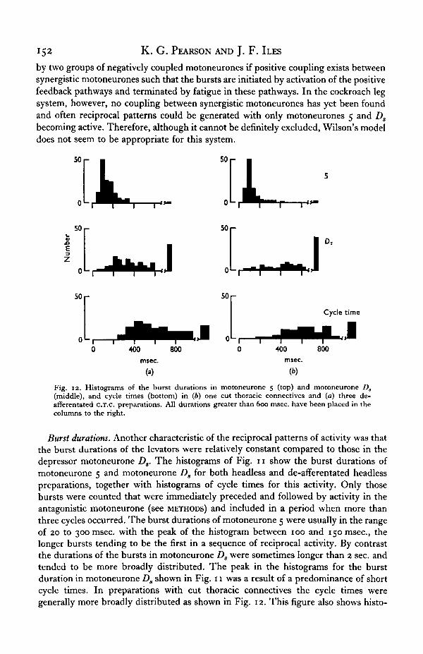
Fig. 12. Histograms of the burst durations in motoneurone 5 (top) and motoneurone D,(middle), and cycle times (bottom) in (6) one cut thoracic connectives and (a) three de-afferentated c.T.c. preparations. All durations greater than 600 msec, have been placed in thecolumns to the right.
Burst durations. Another characteristic of the reciprocal patterns of activity was thatthe burst durations of the levators were relatively constant compared to those in thedepressor motoneurone Ds. The histograms of Fig. 11 show the burst durations ofmotoneurone 5 and motoneurone Ds for both headless and de-afferentated headlesspreparations, together with histograms of cycle times for this activity. Only thosebursts were counted that were immediately preceded and followed by activity in theantagonistic motoneurone (see METHODS) and included in a period when more thanthree cycles occurred. The burst durations of motoneurone 5 were usually in the rangeof 20 to 300 msec, with the peak of the histogram between 100 and 150 msec, thelonger bursts tending to be the first in a sequence of reciprocal activity. By contrastthe durations of the bursts in motoneurone Ds were sometimes longer than 2 sec. andtended to be more broadly distributed. The peak in the histograms for the burstduration in motoneurone Ds shown in Fig. 11 was a result of a predominance of shortcycle times. In preparations with cut thoracic connectives the cycle times weregenerally more broadly distributed as shown in Fig. 12. This figure also shows histo-

Activity in cockroach leg motoneurones 153
grams of burst duration in motoneurones 5 and Da observed in one C.T.c. and threede-afferentated c.T.c. preparations. These histograms clearly show the relativeconstancy of the burst durations in motoneurone 5 compared to those in moto-neurone Ds. The plots in Fig. 13 show the relationship between the burst durations
500
Cycle time (msec.)
1000
Fig. 13. Scatter diagrams of the burst durations in motoneurone 5 (top) and motoneurone D,(bottom) against cycle time for three de-afferentated cut thoracic connectives preparations.

154 K. G. PEARSON AND J. F. ILES
in motoneurones 5 and Ds and cycle time for the three de-afferentated c.T.c. prepara-tions. These diagrams show more distinctly that longer cycle times are stronglycorrelated with long bursts in motoneurone Ds whereas, although there is a slighttendency for the levator burst lengths to increase with cycle time, these remain fairlyconstant.
In all preparations the burst durations in the levator motoneurone 6 were alwaysshorter than those in motoneurone 5 while the larger levator motoneurones, if activeduring the levator bursts, discharged only a few times.
Since activity in motoneurones 5 and 6 gives rise to tonic contractions it would beexpected that the duration of the mechanical effect produced by the levator burstswould be shorter than the duration of the activity in motoneurone 5. That this is socan be seen from Fig. 7. The finding that the levator burst durations were relativelyconstant compared with depressor burst durations corresponds to the behaviouralobservations of relatively constant leg protraction time during normal walking.Recently Delcomyn (personal communication) has shown that during walking theleg protraction time in Periplaneta varies from 20 msec, when running to 160 msec,when slowly walking. Therefore the observed durations of the levator bursts cor-respond reasonably well with these behavioural observations and suggest that the legprotraction phase of walking is predominantly under central control.
Burst shapes. During reciprocal activity the average frequency during a burst ofmotoneurone 5 varied from 30 to 150 impulses/sec. The discharge frequency ofmotoneurone 5 remained fairly constant throughout the burst, but showed a slightacceleration and deceleration at the beginning and end of the burst respectively. Thepattern of activity in motoneurone 6 was very similar to that of motoneurone 5.Motoneurone 6 only became active when the discharge frequency of motoneurone 5increased above about 80 impulses/sec. One interesting feature of the levator bursts,was that they did not appear to be dependent upon the activity in motoneurone Ds.Figure 15 shows two very similar levator bursts in one preparation with the activityin motoneurone Ds being very weak before and after one of the bursts and very strongbefore and after the other. This figure also shows that when the intensity of the burstswas such that motoneurone 6 only fired once, the frequency of discharge in moto-neurone 5 was about 80 impulses/sec.
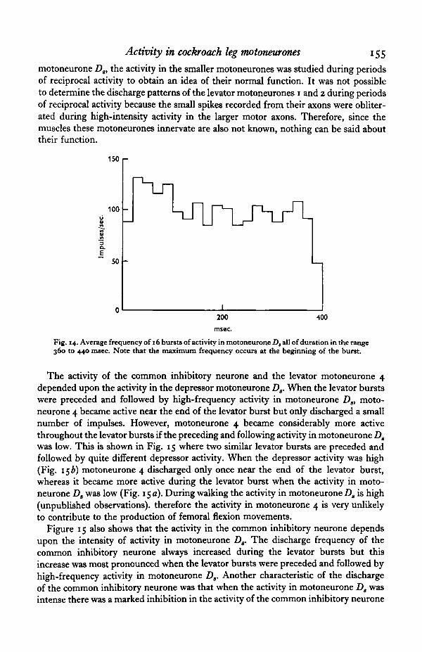
The frequency distribution throughout the bursts of activity in the depressormotoneurone Ds was quite distinctive and differed considerably from that seen in thelevator motoneurones 5 and 6. Figure 14 shows the average shape of 16 bursts ofactivity in motoneurone D3 all of which had durations within the range 360 to440 msec. The obvious feature of this activity was that the maximum frequencyoccurred at the beginning of the burst. This decreased to a plateau for long burstsfollowed by a deceleration at the end of the burst. The peak frequency varied from100 to 180 impulses/sec, while the plateau was in the range of 60 to 140 impulses/sec.The duration of the initial peak was about 150 msec, and independent of burst length.For shorter bursts the plateau did not occur. One possible explanation for the initialhigh-frequency activity in motoneurone Ds is that it is produced by post-inhibitoryrebound, similar to the rebound effect described by Chalazonitis & Arvanitaki (1961)in motoneurones of the abdominal ganglion of Aplysia.
Smaller axons. Apart from the levator motoneurones 5 and 6 and the depressor
Activity in cockroach leg motoneurones 155
motoneurone Ds, the activity in the smaller motoneurones was studied during periodsof reciprocal activity to obtain an idea of their normal function. It was not possibleto determine the discharge patterns of the levator motoneurones 1 and 2 during periodsof reciprocal activity because the small spikes recorded from their axons were obliter-ated during high-intensity activity in the larger motor axons. Therefore, since themuscles these motoneurones innervate are also not known, nothing can be said abouttheir function.
150 1-
100
3
a.E 50
200 400
Fig. 14. Average frequency of 16 bursts of activity in motoneurone D, all of duration in the range360 to 440 msec. Note that the maximum frequency occurs at the beginning of the burst.
The activity of the common inhibitory neurone and the levator motoneurone 4depended upon the activity in the depressor motoneurone Ds. When the levator burstswere preceded and followed by high-frequency activity in motoneurone Ds, moto-neurone 4 became active near the end of the levator burst but only discharged a smallnumber of impulses. However, motoneurone 4 became considerably more activethroughout the levator bursts if the preceding and following activity in motoneurone Ds
was low. This is shown in Fig. 15 where two similar levator bursts are preceded andfollowed by quite different depressor activity. When the depressor activity was high(Fig. 156) motoneurone 4 discharged only once near the end of the levator burst,whereas it became more active during the levator burst when the activity in moto-neurone D8 was low (Fig. 15 a). During walking the activity in motoneurone Dg is high(unpublished observations), therefore the activity in motoneurone 4 is very unlikelyto contribute to the production of femoral flexion movements.
Figure 15 also shows that the activity in the common inhibitory neurone dependsupon the intensity of activity in motoneurone Ds. The discharge frequency of thecommon inhibitory neurone always increased during the levator bursts but thisincrease was most pronounced when the levator bursts were preceded and followed byhigh-frequency activity in motoneurone Ds. Another characteristic of the dischargeof the common inhibitory neurone was that when the activity in motoneurone Da wasintense there was a marked inhibition in the activity of the common inhibitory neurone

156 K. G. PEARSON AND J. F. ILES
after the levator bursts; this is shown in Fig. 156. For weaker activity in the moto-neurone Ds this inhibition was not as marked (Fig. 15 a).
The maximal activity in the other two smaller depressor motoneurones (the thirdsmall depressor motor axon is a branch of the common inhibitory neurone) occurredduring the levator bursts as shown in Figs. 4 and 15. The intensity of activity in thesetwo motoneurones increased with increasing intensity of the levator bursts.
44LJ 1 I J . I
{*)
UJl^
(b)0 25 sec.
Fig. 15. Activity in levator motoneurone 4 and the common inhibitory neurone during twosimilar bursts of activity in the levator motoneurones 5 and 6 but different levels of activityin the depressor motoneurone D,. Top traces, records from nerve 6Br4; bottom traces, recordfrom nerve sri b. The two small spikes in the records from nerve 6Br4 are from axons 3 and 4.These could be distinguished because the firing of axon 3 showed a 1:1 correspondence to thefiring of another axon in nerve s n (axon 3 is one branch of a common inhibitory neurone anotherbranch of which is contained in nerve 5ri b). The spikes from axon 4 have been marked withdots. The two larger spikes recorded from nerve 6Br4 are from axons 5 and 6, axon 6 dischargingonly once during each burst. The large spike in the record from nerve sri b is from axon Ds.The three small axons in nerve 5ri b discharge maximally during the levator bursts. Note alsothat when the activity in motoneurone D, is high, (b), the activity in motoneurone 4 is low,while the common inhibitory neurone discharges at a higher rate throughout the levator burstsand is inhibited at the end of this burst.
(c) Non-reciprocal activity. One type of non-reciprocal activity was that the levatormotoneurones 5 and 6 would discharge in bursts without motoneurone Ds becomingactive, as shown in Fig. 16. The reverse situation, i.e. motoneurone Ds firing in normalbursts without motoneurones 5 and 6 becoming active, has never been seen. Thegeneration of levator bursts without activity in motoneurone Ds is similar to thefinding in the locust flight system that depressor bursts may be generated withoutlevator activity (Waldron, 1967). Also in the insect ventilation system expiratory burstscan occur without any activity in the inspiratory motoneurones (Miller, personalcommunication). Often levator bursts were immediately followed by a small numberof impulses in motoneurone Z)s, but the depressor activity was not maintained overthe entire interval between the levator bursts (Fig. 17). These patterns of activity

Activity in cockroach leg motoneurones 157
support the proposal of a rebound effect on motoneurone Ds giving rise to the increasein excitation after a levator burst (cf. Fig. 8 in Chalazonitis & Arvanitaki, 1961).Again the reverse pattern has never been seen. Thus three patterns of burst activityhave been observed: bursts in only the levator motoneurones, bursts in the levatormotoneurones followed by a short burst of activity in motoneurone Ds, and fullyreciprocal activity.
•6Br4
ftHMWM 5r1b
>6Br4
'5r1b
1sec.
Fig. 16. Bursts of activity in levator motoneurones 5 and 6 without activity in motoneurone D,.The bottom pair of records is continuous with the top. The two largest spikes in the recordfrom nerve 6Br4 are from axons 5 and 6, and the small spike from axon 3. The reciprocalactivity shown in Fig. 9 was from the same preparation.
05 sec.
Fig. 17. Bursts of activity in levator motoneurone 5 (large spike in top record) followedimmediately by short bursts of activity in motoneurone D8 (lower record).
Another type of non-reciprocal activity was that motoneurones 5 and Ds would besimultaneously active for long periods. This occurred very rarely and then only wheneach motoneurone was discharging at low frequencies (a few impulses/sec). Slowfluctuations in the average frequency of the two motoneurones were negatively cor-related ; an increase in the frequency of motoneurone 5 corresponded with a decreasein frequency of motoneurone D8, and vice versa. During these periods no latency orphase correlations have been found between the two trains of impulses as might havebeen expected if there was any direct coupling between the motoneurones.

158 K. G. PEARSON AND J. F. ILES
(d) Antidromic stimulation. Another test for coupling at the motoneuronal level wasto stimulate antidromically either the levator motor axons or the depressor motoraxons and observe the effect of this stimulation on the activity in the other.
Nerve 6Br4 was sufficiently long to allow both stimulating and recording electrodesto be placed on the nerve (the stimulating electrode being the distal pair). When thestimulus was adjusted so that all axons larger than axon 2 were excited, a singlestimulus re-set the activity in all the larger motoneurones. This re-setting effect onthe activity of motor axons 3, 4 and 5 is shown in Fig. 18. The interval immediatelyafter the stimulus was significantly longer than the mean interval before the stimulus,which was probably due to an accumulation of refractoriness (Wilson, 1964). Miller(1967) has also found the same effect on antidromically stimulating the motor axonsinnervating locust spiracle muscles.
1 sec.
Fig. 18. Re-setting effect of single antidromic stimuli on the activity in the motor axons 3, 4and 5 of nerve 6Br4. Stimuli were delivered at 1 /sec. (dots) distal to the recording electrode.The two small spikes are from axons 3 and 4 (the spike from axon 3 being slightly larger) whilethe large spike is from axon 5.
High-frequency (> 50/sec.) antidromic stimulation of nerve 6Br4 led to a de-pression of all spontaneous activity in motor axons contained in that nerve whichrecovered with a time constant of about 0-5 sec. Kennedy, Evoy & Fields (1966) reporta similar finding for the motor axons innervating the flexor muscles of the crayfishabdomen. These depression effects only occurred when the axon under study wasstimulated, thus, if the inhibition was mediated via collateral feedback pathways(similar to Renshaw inhibition in the mammalian spinal cord), then these collateralsonly produce inhibition in the motoneurones from which they originate. Rather thancollateral feedback pathways mediating the depression effects a simpler possibility isthat the high-frequency antidromic stimulation leads to some form of inactivation ofthe spike-generating region which slowly recovers at the end of stimulation.
High-frequency antidromic stimulation of the motor axons larger than axon 2 innerve 6Br4 was without effect on the activity of motoneurone Ds. (Another control forstimulus strength was that the branch of the common inhibitory neurone in nerve 5ri bwas activated by each stimulus.) Similarly antidromic stimulation of the axons innerve 50 b was without effect on the activity in the levator motoneurones (it wasassumed that if the branch of the common inhibitory neurone in nerve 6Br4 wasactivated then motor axons Ds and D,, which are larger than the branch of the commoninhibitory neurone in nerve sri b, were antidromically activated).

Activity in cockroach leg motoneurones 159
These negative results exclude the existence of collateral pathways that can beactivated antidromically. The re-setting effect did show that the spike-initiating zonewas invaded. Therefore, if collaterals do exist, they must have a separate spike-initiating zone.
DISCUSSION
1. Central patterning of motoneuronal activity
The patterns of activity seen in the de-afferentated headless preparations were inall respects similar to those recorded in the headless preparations. The reciprocalpatterning of motoneuronal activity is therefore largely independent of sensory feed-back from leg receptors. Wilson (1966, 1967) has postulated the existence of centralneuronal oscillators in each half-ganglion responsible for patterning activity inantagonistic motoneurones. The findings presented in this paper provide strongevidence for the existence of such oscillators in the metathoracic ganglion. Similaroscillators also exist in the mesothoracic ganglion, and the ipsilateral oscillators in themesothoracic and metathoracic ganglia are centrally coupled in such a way that thereis a negative correlation between activity in the two sets of levator motoneurones(lies & Pearson, in preparation).
Central patterning of motoneuronal activity has been demonstrated in many otherarthropod systems where rhythmic movements occur, e.g. flight in locust and fly(Wilson, 1961, 1968; Wyman, 1965, 1966), ventilation in locust dragonfly and cock-roach (Miller, 1965; Mill & Hughes, 1966; Farley, Case & Roeder, 1967), and move-ments of swimmerets in crayfish and lobster (Hughes & Wiersma, i960; Davis, 1968,1969). One feature of the insect ventilation and crustacean swimmeret systems is thatthe duration of movement in one direction is relatively constant for varying cycletimes (the inspiratory phase of insect ventilation and the power stroke of swimmeretmovements). This is also a feature of insect walking where the leg-protraction timeis relatively constant compared to leg-retraction time. (Hughes, 19656, has reportedthat for the cockroach, Periplaneta americana, the ratio of leg-protraction time toretraction time varies from 0-07 for very slow walking to 1 for rapid running.) Thefinding that the durations of centrally generated levator bursts are relatively constantcompared to the durations of depressor bursts parallels this behavioural observation(during leg-protraction there is flexion of the femur as a result of contractions in thecoxal levator muscles) and is similar to the findings that the centrally generated burstsof activity in locust inspiratory motoneurones and the lobster swimmeret moto-neurones remain fairly constant for varying cycle times (Miller, 1965; Davis, 1969).
There are two aspects of the centrally generated motoneuronal activity that requireexplanations in terms of the connectivity and properties of cells within the centralnervous system; the first is the generation of the levator burst, and the second is thestrong reciprocal relationship between activity in the levator motoneurones, particularlymotoneurone 5, and activity in motoneurone Ds. There are a number of different butnot mutually exclusive methods by which levator bursts could be generated. Thesimplest is that burst activity is an intrinsic property of the motoneurones themselves.If this is the mechanism for levator-burst formation then inhibitory collateral feed-back pathways must exist from motoneurone 5 to motoneurone Ds in order to accountfor the strong reciprocal relationship between the bursts of activity in these neurones.

160 K. G. PEARSON AND J. F . ILES
Since no evidence for the existence of such pathways has been suggested by antidromic stimulation experiments, by the non-overlapping of reciprocal activity, or bythe absence of phase and latency correlation between the spike trains from moto-neurones 5 and Ds when simultaneously active, the possibility that levator bursts aredue to the intrinsic properties of the motoneurones seems unlikely. The same diffi-culty arises with the proposal that the levator bursts are generated as a result ofpositive coupling between the synergistic motoneurones, analogous to that proposedby Wilson (1968) for the locust flight system. Another point against this secondpossibility for burst generation is that levator bursts could often be generated withonly motoneurone 5 becoming active. Moreover, when the larger levator motoneuroneswere also active there were no signs of coupling of activity in different motor axonssimilar to that for coupling of flight motoneurones in the locust where strong latencycorrelations have been found between activity in synergistic motoneurones (Wilson,1968).
Rather than levator bursts being generated at the motoneuronal level a more likelypossibility is that their generation results from phasic driving of the levator moto-neurones by a single (or set of) bursting interneurone(s). Davis & Murphy (1969) havemade a somewhat similar suggestion for the lobster swimmeret system by postulatingthat motoneurones are driven by a sinusoidal input, this input arising from activityin driver interneurones. This mechanism is known to be largely responsible for theburst generation in motoneurones of the lobster cardiac ganglion (Hagiwara &Bullock, 1957) but in this system there is also coupling between motoneurones (Watan-abe, 1958) and positive feedback from motoneurones to the pacemaker interneurones(Watanabe & Bullock, i960). One characteristic of the levator bursts to be explainedis their relative constancy in duration. Little can be said on this problem at themoment as no direct evidence from intracellular records is available and nothing canbe inferred from the discharge patterns. One possibility is that bursting driver inter-neurones have properties similar to those described by Chalazonitis (1963) for thebursting interneurone in the abdominal ganglion of Aplysia where the interburstinterval in this neurone could be decreased or increased by passing depolarizing orhyperpolarizing current respectively but the burst durations remained fairly constant.
Often levator bursts were generated without motoneurone Ds becoming active(Fig. 16) or with motoneurone Ds discharging a small burst of impulses at the end ofthe levator burst (Fig. 17). The reverse patterns have never been seen which suggeststhat the bursts of activity in motoneurone Ds are not produced by independentlybursting driver interneurones. The maximum rate of firing in motoneurone Ds
occurred at the beginning of its bursts (Fig. 14) and as mentioned earlier, this couldhave been due to a post-inhibitory rebound effect. If inhibitory collateral feedbackpathways between the antagonistic sets of motoneurones do not exist then thesimplest model for explaining the bursts of activity in motoneurone Ds is that thebursting driver interneurones to the levator motoneurones also inhibit the ongoingactivity in motoneurone D8, as shown schematically in Fig. 19. In this model it isproposed that a post-inhibitory rebound in the activity of motoneurone Ds follows itsrelease from inhibition at the end of the burst in the levator driver interneurones.By comparison with some other invertebrate systems this proposal seems reasonablefor post-inhibitory rebound excitation has been clearly demonstrated by Chalazonitis

Activity in cockroach leg motoneurones 161
& Arvanitaki (1961) in motoneurones of Aplysia, while the termination of hyper-polarizing currents leads to rebound excitation of the giant cells in the leech abdominalganglion (Eckert, 1963) and in some of the follower cells in the lobster cardiac ganglion(Hagiwara & Bullock, 1957). The independent bias on motoneurone Ds shown inFig. 19 accounts for the observation that for very similar levator bursts the depressormotoneurone Ds can be almost completely inactive or firing reciprocally at very highfrequencies (Fig. 15). For a certain range of low levels of bias on motoneurone Ds thisneurone will not fire continuously between levator bursts but will be excited rebound-wise on release from inhibition after a burst of activity in the levator driver inter-neurone. Therefore this model also accounts for the type of activity where a burst inmotoneurone 5 is immediately followed by a small burst of activity in motoneurone Ds.
BiasCommand
1 Excitatory
O Inhibitory
Fig. 19. Hypothetical model for the generation of reciprocal patterns of activity. Levatormotoneurones 5 and 6 are phasically driven by a bursting interneurone, b.i., which also inhibitsactivity in depressor motoneurone D,. The bias on motoneurone D, accounts for its varyinglevels of activity for similar levator bursts. See text for further details.
The intensity of the burst in motoneurone D8 generally increased with decreasingcycle time (unpublished observation), and strong reciprocal patterns of activity areusually initiated without any of the motoneurones 5, 6 or Ds previously being active.To account for both these findings it is proposed that activity in command inter-neurones (shown in Fig. 19 as the common input to the bursting interneurone, b.i.,and motoneurone Ds) leads to burst generation in the initially quiescent interneuronesdriving the levator motoneurones, and that an increase in the level of the commandinput decreases the interburst intervals and increases the intensity of activity inmotoneurone Ds. One unpublished observation of relevance to this proposal is thatregular stimulation of filaments of the ipsilateral meso-metathoracic connective hassometimes led to generation of reciprocal patterns of activity which only persisted solong as the filament was being stimulated, and the cycle time decreased with increasingstimulus frequencies.
X I EXB52
162 K. G. PEARSON AND J. F . ILES
The above model is the simplest to account for our experimental results to date.With the lack of other evidence it is pointless to speculate about other more complexmodels. Only by directly recording from cells within the ganglion, together withhistological studies, will the cellular events and cell connectivity leading to thepatterning of motoneuronal activity in this and other arthropod systems be determined(Bentley, 1969 a, b). The results presented in this paper show that the centrallygenerated patterns of activity in the coxal levator and depressor motoneurones havemany similarities with those seen in other systems, which raises the general question asto what extent do common principles of neuronal organization underlie the patterningof motoneuronal activity.
2. Functional significance of the motoneuronal activity
En passant recordings from nerve 6B during rhythmic leg movements have shownthat the activity of motoneurones contained in this nerve are very similar to thoseobserved in the de-afFerentated headless preparation (en passant recording from nerve6B allowed the recording of motoneuronal activity without damage to any coxalmuscles). Motor axons 5 and 6 were always active throughout flexion movements of thefemur while some of the larger axons were often recruited during very rapid move-ments. Since femur flexion movements were produced when only axons 5 and 6became active, and since cutting nerve 6Br4 abolished these movements, then thebursts of activity in motoneurones 5 and 6 contribute significantly to the flexionmovements of the femur by producing rapid graded contractions in the posteriorcoxal levator muscle (Fig. 7). The durations of the levator bursts occurring in the de-afferentated headless preparation during periods of reciprocal activity were relativelyconstant compared to the burst durations in motoneurone Ds. Furthermore, theselevator-burst durations correspond to the behaviourally observed leg-protractiontimes. These findings suggest that the motoneuronal activity producing the leg-protraction phase of walking is almost entirely centrally generated, i.e. generatedlargely independent of sensory feedback from leg receptors. Although direct recordingshave not been made of activity in levator motoneurones during walking behaviour,it is likely that the activity in these cells is similar to that occurring during the rhythmicleg movements of restrained inverted animals, and therefore similar to the centrallygenerated levator bursts shown in Figs. 8 and 9. This is a reasonable possibilitybecause the sensory input from leg receptors during the protraction phase of walkingwill be similar to that during flexion movements of the femur and tibia in the invertedanimal. Sensory input from the other legs which would not occur in restrained animalscould affect levator motoneuronal activity during leg protraction. However, theseeffects will be small (Pringle, 1940; Wilson, 1965) and unlikely to alter significantlythe patterning of the levator motoneuronal activity.
Extracellular recordings from the coxal depressor muscles have shown that duringextension movements of the femur in both restrained inverted preparations and inunrestrained freely moving animals, the slow depressor motoneurone Ds dischargesat rates of up to 200 impulses/sec (unpublished observations). These bursts were verysimilar to those seen in the de-afferentated headless preparations. As bursts of activityin motoneurone D8 give rise to strong rapid graded contractions of the coxal depressormuscles, a significant part of the femur extension movements during walking is

Activity in cockroach leg motoneurones 163
probably produced by activity in this motoneurone, and the generation of this activitymay, to a large extent, be independent of sensory feedback from leg receptors.
Apart from activity in motoneurones 5, 6 and Ds and the other larger axons, thefunctional significance of the patterns of activity in the smaller levator and depressormotoneurones must be considered. The muscles innervated by levator motoneurones 1and 2 have not been determined, nor has it been possible to investigate the dischargepatterns in these cells as the small size of their spikes prevented study of their patternsof activity when the larger motor axons were active. Thus nothing can be said abouttheir function. The common inhibitory neurone discharged maximally during thelevator bursts and was inhibited at the beginning of the depressor bursts (Fig. 156).Stimulation of the common inhibitory neurone produces inhibition of contractionsproduced by motoneurone D8 in the coxal depressor muscles (unpublished observa-tions) and may therefore function to produce a more rapid relaxation of the depressormuscles after a burst of activity in motoneurone Dg as suggested by Pearson & Bergman(1969). The relaxation time-constant in the coxal depressor muscles 135D and 135Eof the mesothoracic segment after high-frequency stimulation of the axon equivalentto motor axon Ds is about 300 msec. (Usherwood, 1962). Since the leg protraction timeis less than 160 msec, some form of active relaxation of the depressor muscles wouldtherefore seem desirable. The other two small depressor axons are also inhibitory(lies & Pearson, 1969) and discharge maximally throughout the levator bursts. Theincreased activity in these two neurones during levator bursts would assist the commoninhibitory neurone in producing a faster relaxation of the coxal depressor muscles.Levator motoneurone 4 produces very slow graded contractions in the posterior coxallevator muscles (Pearson & Bergman, 1969) but is not strongly activated during periodsof reciprocal activity (Fig. 15 b). Thus this motoneurone most probably functions toproduce slow flexion movements of the femur when the animal is making posturaladjustments.
We wish to thank Dr D. C. S. White and Dr P. L. Miller of the University Depart-ment of Zoology for their helpful suggestions and criticisms of this paper.
SUMMARY
1. Observation of movements of the metathoracic legs of the cockroach before andafter section of peripheral nerves allowed identification of muscles involved in flexionand extension of the femur.
2. Extracellular recordings from the nerves to these coxal muscles show that duringrhythmic leg movements bursts of activity in a number of levator motor axons werestrongly reciprocal and generally non-overlapping with those of a slow depressormotor axon.
3. These reciprocal patterns persisted after removal of all sensory input from thelegs.
4. The durations of levator bursts were relatively constant compared to those ofthe depressor, corresponding to the behavioural observations on leg protraction time.The pattern was asymmetric: levator bursts could be generated without depressoractivity, but never the reverse.
164 K. G. PEARSON AND J. F. ILES
5. No evidence was found for inhibitory collateral pathways between antagonist!motoneurones.
6. It is proposed that levator motoneurones are driven by a group of burstinginterneurones which simultaneously inhibit the ongoing depressor activity.
REFERENCES
BENTLEY, D. R. (1969a). Intracellular activity in cricket neurons during the generation of behaviourpatterns. J. Insect Physiol. 15, 677-700.
BENTLEY, D. R. (19696). Intracellular activity in cricket neurons during generation of song patterns.Z. vergl. Physiol. 6a, 267-83.
CARBONELL, C. S. (1947). The thoracic muscles of the cockroach, Periplaneta americana. SmithsonianInst. Misc. Coll. 107 (2), 1-23.
CHALAZONITIS.N. (1963). Effects of changes inPco2 and P o , in rhythmic potentials from giant neurons.Ann. N.Y. Acad. Sci. 109, 451-479.
CHALAZONITIS, N. & ARVANITAKI, A. (1961). Slow changes during and following repetitive synapticactivation in ganglion nerve cells. Bull. Inst. Oceanogr. 58, No. 1225.
DAVIS, W. J. (1968). The neuromuscular basis of lobster swimmeret beating. J. exp. Zool. 168, 363-78.DAVIS, W. J. (1969). The neural control of swimmeret beating in the lobster. J. exp. Biol. 50, 99-118.DAVIS, W. J. & MURPHEY, R. K. (1969). Discharge patterns of swimmeret motoneurones in the lobster,
simulated with a digital computer. J. exp. Biol. 50, 119-28.DRESDEN, D. & NIJENHUIS, E. D. (1958). Fibre analysis of the nerves of the second thoracic leg in
Periplaneta americana. Proc. K. Ned. Akad. Wet. C61, 213-33.ECKERT, R. (1963). Electrical interaction of paired ganglion cells in the leech. J. gen. Physiol. 46, 573-88.FARLEY, R. D., CASE, J. F. & ROEDER, K. D. (1967). Pacemaker of tracheal ventilation in the cockroach,
Periplaneta americana. J. Insect Physiol. 13, 1713-1728.HAGIWARA, S. & BULLOCK, R. H. (1957). Intracellular potentials in pacemaker and integrative neurons
of the lobster cardiac ganglion. J. cell. comp. Physiol. 50, 25-47.HUGHES, G. M. (1952). The co-ordination of insect movements. 1. The walking movements of insects.
J. exp. Biol. 29, 267-84.HUGHES, G. M. (1965 a). Neuronal pathways in the insect central nervous system. In The Physiology
of the Insect Central Nervous System, ed. J. E. Treherne & J. W. L. Beament.HUGHES, G. M. (19656). Locomotion: terrestrial. In The Physiology of Insects, 11,227-254, ed. M. Rock-
stein.HUGHES, G. M. & WIERSMA, C. A. G. (i960). The co-ordination of swimmeret movements in the cray-
fish, Procambarus clarkii. J. exp. Biol. 37, 657-70.ILES, J. F. & PEARSON, K. 6.(1969). Triple inhibitory innervation of insect muscle. J. Physiol. 204, 125-6 P.KENNEDY, D., EVOY, W. H. & FIELDS, H. L. (1966). The unit basis of some crustacean reflexes. Symp.
Soc. exp. Biol. 20, 75-110.MILL, P. J. & HUGHES, G. M. (1966). The nervous control of ventilation in dragonfly larvae, jf. exp.
Biol. 44, 297-316.MlLBURN, N. S. (1963). Sensitivity of cockroach campaniform sensilla to adrenergic drugs. Am. Zool.
3. 513-MILLER, P. L. (1965). The central nervous control of respiratory movements. In The Physiology of the
Insect Central Nervous System, ed. J. E. Treherne & J. W. L. Beament.MILLER, P. L. (1966). The regulation of breathing in insects. Adv. Insect Physiol. 3, 279-344.MILLER, P. L. (1967). The derivation of motor command to the spiracles of the locust. Jf. exp. Biol. 46,
349-71.PEARSON, K. G. & BERGMAN, S. J. (1969). Common inhibitory motoneurones in insects.^, exp. Biol. 50,
445-72.PEARSON, K. G., STEIN, R. B. & MALHOTRA, S. K. (1969). Properties of action potentials from insect
motor nerve fibres (in preparation).PIPA, R. L. & COOK, E. F. (1959). Studies on the hexapod nervous system. I. The peripheral distribu-
tion of the thoracic nerves of the adult cockroach, Periplaneta americana. Ann. Entomol. Soc. Am.52(6), 695-710.
PRINGLE, J. W. S. (1939). The motor mechanism of the insect leg. J. exp. Biol. 16, 220-231.PRINGLE, J. W. S. (1940). The reflex mechanism of the insect leg. J. exp. Biol. 17, 8-17.PRINGLE, J. W. S. (1961). Proprioception in arthropods. In The Cell and the Organism, ed. J. A. Ramsay
& V. B. Wigglesworth.ROEDER, K. D. (1948). Organization of the ascending giant fibre system in the cockroach, Periplaneta
americana,. J. exp. Zool. 108, 243-61.

Activity in cockroach leg motoneurones 165AOEDER, K. (1959). A physiological approach to the relation between prey and predator. Smithsonian
Inst. Misc. Coll. 137, 287-306.RUNION, H. I. & USHERWOOD, P. N. R. (1968). Tarsal receptors and leg reflexes in the locust and grass-
hopper. J. exp. Biol. 49, 421-36.STEIN, R. B. (1965). A high speed timer and recorder for computer analysis of physiological data. J.
Physiol. 181, s-6 P.STEIN, R. B. (1968). Modules for neurophysiology using integrated circuits. ,7. Physiol. 197, 1-2 P.USHERWOOD, P. N. R. (1962). The nature of 'fast' and 'slow' contractions in the coxal muscles of the
cockroach. J. Insect Physiol. 8, 31-52.USHERWOOD, P. N. R., RUNION, H. I. & CAMPBELL, J. I. (1968). Structure and physiology of a chordo-
tonal organ in the locust leg. J. exp. Biol. 48, 305-324.WALDRON, I. (1967). Mechanisms for the production of the motor output pattern in flying locusts.
J. exp. Biol. 47, 201-12.WATANABE, A. (1958). The interaction of electrical activity among neurons of lobster cardiac ganglion.
Jap. J. Physiol. 8, 305-18.WATANABE, A. & BULLOCK, T. H. (i960). Modulation of activity of one neuron by subthreshold slow
potentials in another in lobster cardiac ganglion. J. gen. Physiol. 43, 1031-45.WILSON, D. M. (1961). The central nervous control of flight in a locust. J. exp. Biol. 38, 471-90.WILSON, D. M. (1964). Relative refractoriness and patterned discharge of locust flight motoneurones.
J. exp. Biol. 41, 191-205.WILSON, D. M. (1965). Proprioceptive leg reflexes in cockroaches. J. exp. Biol. 43, 397-409.WILSON, D. M. (1966a). Insect walking. Ann. Rev. Entomol. u , 103-22.WILSON, D. M. (19666). Central nervous mechanisms for the generation of rhythmic behaviour in
arthropods. Symp. Soc. exp. Biol. zo, 199-228.WILSON, D. M. (1967). An approach to the problem of control of rhythmic behaviour. In Invertebrate
Nervous Systems, ed. C. A. G. Wiersma.WILSON, D. M. (1968). The nervous control of insect flight and related behaviour. Adv. Insect Physiol.
5, 289-338.WYMAN, R. J. (1965). Probabilistic characterization of simultaneous nerve impulse sequences controlling
dipteran flight. Biophys. J. 5, 447-71.WYMAN, R. J. (1966). Multistable firing patterns among several neurons. J. Neurophysiol. 29, 807-33.