Embed Size (px)
Citation preview

27Disasters and Public Health: Planning and ResponseCopyright © 2009 by Academic Press. Inc. All rights of reproduction in any form reserved.
Bioterrorism
Objectives of This ChapterDifferentiate • bioterrorism from emerging infectious disease outbreaks.Discuss some of the controversies surrounding bioterrorism • preparedness.List the highest priority biological threat agents.•Identify the epidemiological clues indicating a possible bioterrorism attack.•Describe the common characteristics of Category A biological agents.•Explain the criteria used to classify bioterrorism agent threats.•Identify diagnosis and treatment recommendations for critical agents.•Describe various types of bioterrorism surveillance approaches.•Explain how environmental surveillance is used to identify the presence of •suspicious organisms.Recognize the infection control precautions needed for responding to bioterrorism.•
They were two men going about their daily lives. Both had reputations as pillars of the community in the Washington, DC, area. Joseph Curseen, Jr. (Figure 2–1b), was 47 and had followed in his father’s footsteps. He enjoyed his job with the postal service and had not used a day of sick leave in 15 years (Becker and Toner, 2001). His coworker and friend, Thomas Morris, Jr. (Figure 2–1a), was 55. Mr. Morris was the first to experience mysteri-ous symptoms after suspicious letters with anthrax threats were processed at the Brentwood postal facility where both men worked. After several days of a “flu-like” illness and a trip to his doctor’s office, Mr. Morris had been told by his physician that his illness was likely due to a virus. On October 21, 2001, he placed a 911 phone call when his health took another turn for the worst. He had begun vomiting while at work and called for help. He told the 911 operator that he thought he may have been exposed to anthrax. Unfortunately, it was too late to intervene (CNN.com, 2001; Morris, 2001). Mr. Morris died that day and Mr. Curseen’s death followed the next day. These were two of the five people who died following exposure to anthrax-laced letters sent to U.S. Senators and media outlets in the weeks following the terrorist attacks of September 11, 2001 (Figure 2–2). Another 17 people were sickened by associated anthrax exposures. The Brentwood postal facility was decontaminated and reopened in 2003 under the name: The Joseph Curseen, Jr., and Thomas Morris, Jr., Processing and Distribution Center. A dedication plaque on the building says: “We are poorer for their loss but richer for having been touched by these dedicated, hard-working heroes. We will never forget.”
The “anthrax letters” of 2001 set public health preparedness initiatives in motion that continue today. Billions of dollars have been spent to develop new drugs and vac-cines, place biothreat agent detection monitors in major cities and at important events,
2
28 DISASTERS AND PUBLIC HEALTH: PLANNINg AND RESPONSE
enhance laboratory capabilities, expand epidemiological monitoring, increase hospital decontamination and surge capacity, and accomplish planning, training, and exercises across a wide range of related professions. Details have recently emerged suggesting that Dr. Bruce Ivins, an employee of the U.S. Army Medical Research Institute of Infectious Diseases (USAMRIID) at Fort Detrick, Maryland, was responsible for the letters. Although there is strong circumstantial and forensic evidence pointing to Dr. Ivins, his recent suicide will keep the details cloaked in mystery (Lichtblau and Wade, 2008).
Figure 2–1 Thomas Morris, Jr. (left) and Joseph Curseen, Jr. (right).
Source: U.S. Postal Service. Used with permission.
Bioterrorism DefinitionsBacteremia: The presence of bacteria in the blood. Fever, chills, tachycardia, and tachypnea
are common manifestations of bacteremia.Bubonic: Relating to an inflamed, enlarged lymph gland.Encephalitis: Inflammation of the brain due to infection, toxins, and other conditions.Fever: An abnormal elevation of body temperature (>100 °F), usually as a result of an infection.Hemorrhagic: Related to bleeding or hemorrhage.Hypotension: Abnormally low blood pressure; seen in shock.Maculopapular: A rash of the skin consisting of both spots (macules) and elevations of the
skin (papules).Papular: Characterized by the presence of small, circumscribed, solid elevations of the skin.Petechiae: Purplish or brownish red discoloration, visible through the epidermis, caused by
bleeding into the tissues.Pneumonic: Related to an inflammation of the lungs.Prodrome: An early symptom of a disease.

Chapter 2•Bioterrorism 29
Figure 2–2 One of the anthrax-tainted letters that passed through the Brentwood postal facility.
Source: Federal Bureau of Investigation. http://www.fbi.gov/page2/august08/anthrax_gallery1.html.
Basic Facts about Bioterrorism ThreatsBioterrorism is the use or threatened use of biological agents as weapons of terror. Current U.S. laws make the threat alone, even in the absence of dangerous biological material possession or use, a severe crime. The biological material used in an act of bio-terrorism may be lethal or nonlethal, a common bacteria or virus, the toxic by-product of a pathogen, a rare organism, or even a specially engineered organism, never before diagnosed or treated. Although most public concerns focus on the threat to humans, bioterrorists may also attack crops or livestock. Even a small attack on these resources may result in serious economic consequences for any nation targeted. Regardless of the focus of an attack or the agent used, the ultimate goal of bioterrorism is the same as con-ventional terrorism. These acts are intended to instill fear in the targeted population in support of terrorist goals. It continues to be one of our most complex preparedness chal-lenges. It is distinct from the other disasters described in this book. Most disasters have a focal point. Resources are clearly needed in a relatively well-defined area following an earthquake, hurricane, or other natural disaster. The hazardous chemical exposures of greatest concern are typically acute and those affected are quickly seen among those exposed. Naturally occurring diseases often have a relatively slow moving timeframe where an epidemic unfolds over weeks or months. Conversely, bioterrorism can cause a large, delayed surge of severely ill people spread out across multiple communities or even multiple continents. Organisms or other biological materials can be released in the air,
Sepsis: The presence of microorganisms or their toxins in tissues or in the blood. Systemic disease caused by the spread of the microorganisms or their by-products via the circulat-ing blood is commonly called septicemia.
Septicemic: Related to a systemic disease associated with the presence of their by-products in the blood.
Septic shock: Shock due to circulatory inadequacy caused by gram-negative bacteria (bacter-emia). It is less often the result of the presence of other microorganisms (fungus or virus) in the blood (fungemia; viremia).
Shock: An imbalanced condition of the hemodynamic equilibrium, usually manifested by failure to oxygenate vital organs.

30 DISASTERS AND PUBLIC HEALTH: PLANNINg AND RESPONSE
or placed in food or water sources. With incubation periods of days or weeks for many agents, it is difficult to find all who may have been exposed to an intentional release and successfully treat them to avert full blown disease. As a result, it is likely that the response will include an enormous number of potentially exposed individuals in multiple regions. Unlike many other disasters, a window of opportunity exists for a bioterrorism response. Most conditions can be successfully averted if they are caught early and pro-phylactically treated. As treatment is delayed, morbidity and mortality increase daily as the end of the incubation period is reached. The challenge of responding increases with some high-threat organisms that are spread from person-to-person. These materials can also be engineered to optimize release effectiveness or even make them impossible to treat with standard therapies. A unique aspect of the public health and healthcare response to these scenarios is that their organizations are placed in the vanguard of a response. In most other disaster scenarios, the public health and healthcare roles are in support of emergency management and first responders. In the case of bioterrorism, the majority of response will be identifying and treating those at risk.
There is a long history associated with biological weapons. (See Table 2–1) Thousands of years ago, before any understanding of pathogenic organisms, there was an initial observation that various biological materials could cause illness. A variety of plants were known to be poisonous; but in addition there was a growing understanding that feces, blood, tissue from dead bodies, or entire dead bodies of sick humans or animals, were able to cause illness and could be used as weapons. This crude use of filth and car-casses was the beginning of biological warfare and bioterrorism. The twentieth century saw a rapid acceleration in life sciences but behind the scenes, known to relatively few, was a growing group of scientific professionals, including microbiologists, physicians, and many other disciplines working covertly on the development of biological weapons. Although most nations acknowledge that these weapons should be banned and signed the Biological Weapons Convention in 1972 stating they would no longer pursue them for offensive purposes, the life sciences advances and technology continue to trickle down. As these amazing advances move us forward with new pharmaceuticals and powerful tools to manipulate microorganisms, it also places a growing body of potentially dangerous information within reach of those who may intend to use it for harm. A new arms race is on. Rather than competing super powers, it is criminals, terrorists, and rogue nations against the world. Rather than detecting an incoming threat on radar screens, as it was during the Cold War, this time it may be detected through the lens of a microscope.
U.S. Bioterrorism Preparedness Controversies
Bioterrorism became a well recognized part of American vernacular after five anthrax-tainted letters circulated on the East Coast of the United States in September and October of 2001. The letters were responsible for 22 cases of anthrax disease and five deaths (Mina et al., 2002). These events led to serious consideration of other possible biological threats and led to strong debates on issues, such as who should receive smallpox vaccinations and how available should antibiotics be to the public for preparedness purposes. Federal, state, and local public health preparedness efforts accelerated to unprecedented levels in the years immediately following 2001. The growing apprehension of Americans over these emerging threats resulted in bioterrorism preparedness becoming a moral imperative. In recent years, indifference has crept in and bioterrorism preparedness has become a much lower priority.

Chapter 2•Bioterrorism 31
Table 2–1 Select Historical Events Involving Biological Weapons and Bioterrorism
<1000 c. BC Scythian archers tipped arrows with blood, manure, and tissue from dead bodies.
5th c. BC Assyrians poisoned enemy wells with rye ergot (Claviceps purpurea), a fungus containing
mycotoxins.
590 BC Athenians poisoned enemy water supplies with hellebore, an herb purgative, during the
Siege of Krissa.
3rd c. BC Persian, Greek, and Roman literature describes the use of dead animals being used to
contaminate enemy water supplies.
184 BC Carthaginian General Hannibal ordered his sailors to hurl clay pots filled with poisonous
snakes onto the decks of enemy ships during a naval battle. Hannibal won the battle.
1155 Emperor Barbarossa poisons wells with decomposing human bodies.
1346 Tartur army catapulted deceased bodies of plague victims over city walls during the siege of
Caffa.
1495 Spanish sell wine mixed with the blood of lepers to their enemies.
1763 British distribute variola virus contaminated blankets to Native Americans resulting in a
smallpox outbreak.
1797 Napoleon floods fields around Mantua to promote malaria.
1915–18 Germans attempt to infect Allied horses with anthrax and glanders.
1932–45 Japanese operate Unit 731 in Manchuria conducting experiments that included infecting
prisoners with a variety of lethal pathogens.
1942 British test anthrax bombs on Gruinard Island off the coast of Scotland.
1950–69 US and USSR grow offensive biological weapons programs.
1969 US President Nixon ends the US offensive biological weapons program.
1972 US and USSR sign the Biological Weapons Convention agreeing to an end to offensive
programs.
1978 Assassination of Bulgarian exile Georgi Markov in London with an injected ricin pellet.
1979 Accidental anthrax release from a secret Soviet facility in Sverdlovsk kills 66.
1984 In the Dalles, Oregon, the Rajneeshee cult contaminated local salad bars with salmonella
sickening more than 750 people.
1990 Japanese Aum Shinrikyo cult unsuccessfully attempts botulinum toxin releases in Tokyo.
1991 US troops receive anthrax vaccinations.
1991 After the first Gulf War, UN inspectors begin inspections of biological weapons capability
in Iraq. Iraqi government officials confirm they had researched the use of anthrax and
botulism.
1993 Aum Shinrikyo cult unsuccessfully attempts a second botulinum toxin attack on the
wedding of the Crown Prince. Later the same month they unsuccessfully attempted to
release anthrax from a Tokyo high rise.
(Continued)

32 DISASTERS AND PUBLIC HEALTH: PLANNINg AND RESPONSE
This is most apparent in the declining public health preparedness budgets that provide the needed infrastructure to quickly identify a bioterrorism attack and respond effectively to it.
The past several years of U.S. bioterrorism preparedness initiatives have suffered increasing scrutiny as well. The growing bio-preparedness research infrastructure is con-sidered by some to be unnecessary and possibly even increase the risks of future incidents. This is difficult to dispute in light of the discovery that the source of the 2001 anthrax let-ters was a worker at the USAMRIID research facility (Figure 2–3) (Regaldo et al., 2002). Other safety and security problems have surfaced at the U.S. Army Fort Detrick labs, as well as other research labs across the country (U.S. general Accounting Office, 2008; Weiss and Snyder, 2002; Williamson, 2003). This raises questions concerning the poten-tial for increasing bioterrorism and biocrime risks from new facilities being established to research and develop measures to defend against these threats. However, there is adequate precedent for safe handling and management of extremely hazardous materials. In a wide range of academic and industrial endeavors, safe-handling of dangerous products occurs daily with few problems. Should an accident or security breach occur, it is generally felt
Figure 2–3 Researcher at the U.S. Army Medical Research Institute of Infectious Diseases (USAMRIID) at Fort Detrick, Maryland.
Source: Department of Defense, USAMRIID. http://www3.niaid.nih.gov/NR/rdonlyres/E68D5347-2172-4983-81AC-CD5739E8B8B0/0/BD_BSL_1.JPG.
Table 2–1 (Continued)
2001 Anthrax contaminated letters mailed to US Senate offices and media outlets sickening 22
and killing five.
2004 Ricin sent to US Senate Majority Leader Bill Frist’s office.
Adapted from Frischknecht F. (2008). The History of Biological Warfare. In Richardt A, Blum M (eds.) Decontamination of Warfare Agents: Enzymatic Methods for Removal of B/C Weapons, Wiley-VCH and Eitzen, E. and Takafuji, E. (1997). Historical Overview of Biological Warfare. In Office of the Surgeon General, Department of the Army (ed.) Textbook of Military Medicine: Medical Aspects of Chemical and Biological Warfare.
Chapter 2•Bioterrorism 33
that the benefits outweigh the risks. As with other risky activities, research of dangerous pathogens is essential and has important benefits that must be weighed against the risks. Sufficient measures must be in place and enforced to ensure adequate screening and secu-rity in the life sciences industry, as there are in other industries or initiatives. We cannot make preparedness decisions based upon suspicion of ourselves and cease important work that could eventually diminish the impact of even the worst case bioterrorism scenarios.
There is growing criticism of the U.S. investment in public health preparedness. The large infusion of bioterrorism preparedness funding had mixed results in the years immediately following the 2001 terrorist attacks. Many public health programs are historically underfunded. Although many of these public health programs such as public health laboratories, communicable disease, and environmental health benefited from preparedness resources, accusations of supplanting and misappropriation of resources persist. Supplanting is the use of federal preparedness funding to replace state or local resources. The problem is if the preparedness dollars are focused appropriately to culti-vate the public health infrastructure that identifies and responds to bioterrorism, it turns out to be the same infrastructure that detects and responds to usual outbreaks such as food borne and seasonal illnesses. Although some supplanting violations may be obvious, the distinction can be tricky. When preparedness spending is done wisely, there is typi-cally a dual use of resources. This is a hallmark of an effective preparedness program. If the preparedness function of public health is stove piped and not integrated into daily public health activities, it will not be effective during times of crisis.
Bioterrorism preparedness activities include the development and practice of a mass emergency distribution of pharmaceuticals, risk communication training, and Incident Command System training for public health and healthcare workers. All these prepared-ness activities came into play during Rhode Island’s December 2006 response to a deadly outbreak of Mycoplasma pneumoniae. This infection causes a serious neurological illness. A cluster of five children at the same school were identified with the infection and one died. Although this was not the result of an act of bioterrorism, the local and state public health authorities used the same process developed for mass dispensing of antibiotics for an anthrax or plague response to identify other children at risk, communicate the risk to the public, and dispense prophylactic medications to over 1000 people over a holiday weekend. They reached 100% of those at risk (Association of State and Territorial Health Officials, 2008).
Overall, the effort was a great example of an effective partnership and col-laboration between the state, CDC, EMA, Department of Education, and the towns. If it weren’t for the emergency preparedness funds that CDC has provided us and training all our staff have received in ICS (which we utilized extensively during this episode), I don’t believe we could have accomplished everything we did over a holiday weekend . . . .
David gifford, MD, MPH, Director, Rhode Island Department of Health
Chain of Infection
The human routes of entry for bioterrorism agents depend upon the characteristics of the agent and the method of release. Some agents can only cause infection when entering
34 DISASTERS AND PUBLIC HEALTH: PLANNINg AND RESPONSE
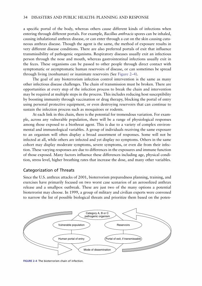
a specific portal of the body, whereas others cause different kinds of infections when entering through different portals. For example, Bacillus anthracis spores can be inhaled, causing inhalational anthrax disease, or can enter through a cut on the skin causing cuta-neous anthrax disease. Though the agent is the same, the method of exposure results in very different disease conditions. There are also preferred portals of exit that influence transmissibility of pathogenic organisms. Respiratory diseases usually exit an infectious person through the nose and mouth, whereas gastrointestinal infections usually exit in the feces. These organisms can be passed to other people through direct contact with symptomatic or asymptomatic human reservoirs of disease, or can sometimes be spread through living (nonhuman) or inanimate reservoirs (See Figure 2–4).
The goal of any bioterrorism infection control intervention is the same as many other infectious disease challenges. The chain of transmission must be broken. There are opportunities at every step of the infection process to break the chain and intervention may be required at multiple steps in the process. This includes reducing host susceptibility by boosting immunity through vaccination or drug therapy, blocking the portal of entry using personal protective equipment, or even destroying reservoirs that can continue to sustain the infection process such as mosquitoes or rodents.
At each link in this chain, there is the potential for tremendous variation. For exam-ple, across any vulnerable population, there will be a range of physiological responses among those exposed to a biothreat agent. This is due to a variety of complex environ-mental and immunological variables. A group of individuals receiving the same exposure to an organism will often display a broad assortment of responses. Some will not be infected at all, while others are infected and yet display no symptoms. Others in the same cohort may display moderate symptoms, severe symptoms, or even die from their infec-tion. These varying responses are due to differences in the exposures and immune function of those exposed. Many factors influence these differences including age, physical condi-tion, stress level, higher breathing rates that increase the dose, and many other variables.
Categorization of Threats
Since the U.S. anthrax attacks of 2001, bioterrorism preparedness planning, training, and exercises have primarily focused on two worst case scenarios of an aerosolized anthrax release and a smallpox outbreak. These are just two of the many options a potential bioterrorist may choose. In 1999, a group of military and civilian experts were convened to narrow the list of possible biological threats and prioritize them based on the poten-
Category A, B or Cpathogenic organism
Vulnerable population
Human portal of entry
Mode of dissemination
Portal of exit, if transmissable
Reservoirs
Figure 2–4 The bioterrorism chain of infection.

Chapter 2•Bioterrorism 35
tial impact they could have when used as a weapon. All available open source literature as well as classified information was reviewed. The list that emerged from their efforts included three categories of pathogens that were designated as Category A, B, and C (Rotz et al., 2002) (Table 2–2). Category A pathogens are high priority organisms and toxins posing the greatest threat to public health. This category includes agents that cause the highest morbidity and mortality with a likelihood of subsequent public panic. They are capable of being spread over a large area and are sometimes also transmitted eas-ily from person-to-person. The scenarios associated with Category A agents necessitate extraordinary actions on the part of public health and healthcare organizations making them the focus of many public health preparedness activities. Throughout the early cold-war era of the 1950s and 60s, most of these organisms were successfully weaponized in the offensive biological weapons programs of the United States and the former Soviet Union. Category B agents are fairly easy to disperse but have lower morbidity and mor-tality than the Category A agents and can be successfully addressed through enhancing laboratory capabilities and epidemiological monitoring. Category C agents are emerging
Table 2–2 Critical Biological Agents for Public Health Preparedness
Disease Biological Agent(s)
Category A
Anthrax Bacillus anthracis
Botulism Clostridium botulinum (botulinum toxins)
Plague Yersinia pestis
Smallpox Variola major
Tularemia Francisella tularensis
Viral hemorrhagic fevers Filoviruses and Arenaviruses (e.g. Ebola, Lassa Fever)
Category B
Brucellosis Brucella spp.
Food safety threats Salmonella spp., Escherichia coli O157:H7
Glanders Burkholderia mallei
Melioidosis Burkholderia pseudomallei
Psittacosis Chlamydia psittaci
Q fever Coxiella burnetii
Ricin toxin Ricinus communis (Castor Beans)
Staphylococcal enterotoxin B Exotoxin from Staphylococcus aureus bacterium
Typhus fever Rickettsia prowazekii
Viral encephalitis Alphaviruses (*VEE, EEE, WEE)
Water safety threats Vibrio cholerae, Cryptosporidium parvum
Category C
Nipah virus encephalitis Paramyxoviridae family
Hendra virus encephalitis Paramyxoviridae family
Hantavirus pulmonary syndrome Hantavirus
*Venezuelan equine (VEE), eastern equine (EEE), and western equine encephalomyelitis (WEE) viruses.Adapted from Centers for Disease Control and Prevention, Bioterrorism Agents/Diseases, By Category. http://www.bt.cdc.gov/agent/agentlist-category.asp.

36 DISASTERS AND PUBLIC HEALTH: PLANNINg AND RESPONSE
infectious organisms that could become easily available at some point in the future and used as a weapon. These agents may be used as weapons and could have a major public health impact (Centers for Disease Control and Prevention, Bioterrorism agents/diseases by category, http://emergency.cdc.gov/agent/agentlistcategory.asp).
Health Threats: Category A OrganismsAnthrax (Bacillus anthracis)
Anthrax disease is caused by the gram-positive, spore-forming bacterium Bacillus anthracis (See Figure 2–7). The disease occurs naturally among mammals such as cattle, sheep, goats, and camels. However, it can also occur in humans when they are exposed to infected ani-mals or to anthrax spores used as a weapon. B. anthracis is considered, by many biologi-cal warfare experts, to be the perfect biological weapon because of its ability to change into a spore when it is subjected to adverse environmental conditions that destroy other pathogenic organisms. Once in the spore form, it can survive for decades under extreme hot or cold conditions. Historically, most cases of anthrax result from occupational expo-sures among leather and wool industry workers. The organism and disease get their name from the greek word “anthrakitis,” which is a kind of coal. The name originated from the appearance of cutaneous anthrax that causes a black lesion on the skin.
There are three forms of anthrax disease. These distinct disease presentations depend upon the route of exposure. Cutaneous anthrax is the most common form of anthrax disease and is fatal in about 5–20% of those who are not treated. It occurs following the exposure of compromised skin to anthrax spores. Inhalational anthrax disease is the most lethal form of disease with more than 80% mortality for those who are not treated. It occurs following the inhalation of anthrax spores and is the most likely form of disease to be seen following an act of bioterrorism. gastrointestinal anthrax disease is highly lethal but very rare. It usually occurs following the ingestion of live (nonspore) B. anthracis in
Figure 2–5 Cutaneous anthrax lesion on the neck. Photo Courtesy of CDC/Public Health Image Library PHIL ID#1934.

Chapter 2•Bioterrorism 37
contaminated, undercooked meat. Ingestion of the spore form of B. anthracis is unlikely to result in a fatal disease because of the transport time in the gastrointestinal system. The spores are likely to pass through the gI system before they have the opportunity to germinate. However, vegetative, nonspore B. anthracis organisms can quickly begin to release toxins damaging the gI tract and can quickly cause illness when ingested.
Cutaneous anthrax is the most common form of anthrax disease. It is a localized skin infection that results from compromised skin exposure to anthrax spores. It is typi-cally seen on commonly exposed skin such as the head, neck, hands, and arms. Areas of compromised skin, such as cuts, abrasions, or chronic dermatological conditions, are more prone to infection than the intact skin. Once the spores become embedded in the skin, they germinate and begin to release a toxin. The toxin causes an edema. The edema progresses into an ulcer over the course of several days and the ulcer develops into a depressed, pain-less lesion called an eschar (See Figure 2–5). Though it is sometimes mistaken for a spider bite, the anthrax eschar remains painless whereas a spider bite causes severe pain. The eschar develops a black leathery appearance for a week or two and then loosens and falls off. Cutaneous anthrax is the most common form of occupational anthrax disease and may also result from an intentional release of spores. Cutaneous anthrax comprised 11 of the 22 cases of disease resulting from the 2001 anthrax letters and none were fatal (Inglesby et al., 2002). If it is untreated, it may lead to a systemic disease with up to 20% mortality. If treated, this form of anthrax disease has very low (<1%) mortality (Lew, 1995).
Inhalational anthrax has the highest mortality of any form of anthrax disease. It results from inhaling a sufficient number of anthrax spores to initiate the disease process. This form of anthrax disease is very rare and even a single case requires notification of both public health and law enforcement authorities. In the 20 years preceding the 2001 anthrax attacks, there had not been a single case of inhalation anthrax in the United States (Inglesby et al., 2002). When anthrax spores are inhaled, they are transported by pulmonary macrophages, part of the lungs’ defense system, to the mediastinal lymph nodes in the center of the chest. Over a period of several days, the spores germinate into vegetative bacilli and begin to release a toxin. This produces a two-stage illness that includes prodromal and fulminant phases. The prodromal phase lasts from a few hours to several days. It includes nonspecific, flu-like symptoms including fever, malaise, shortness of breath, nonproductive cough, and nausea. This stage of the disease is easily confused with influenza or other common infections. However, inhalational anthrax does not normally include runny nose or sore throat. Many flu-like illnesses have those symp-toms (Centers for Disease Control and Prevention, 2001). In addition, influenza does not normally include shortness of breath, vomiting, and mediastinal pain. These are common symptoms of inhalational anthrax. This prodromal period is sometimes followed by a brief improvement before progressing to the next phase of illness. The second phase is a high-grade bacteremia called the fulminant stage. It includes symptoms such as fever, respiratory distress, profuse sweating, cyanosis, and shock. Patients displaying these symptoms usually progress to death within days. If an outbreak is known or suspected, initial diagnosis of inhalational anthrax is made using the described signs and symptoms. A widened mediastinum on a chest X-ray, as displayed in Figure 2–6, is considered highly suspect for inhalational anthrax. However, this clue may or may not be present at the initial patient evaluation or can result from other respiratory conditions. Confirmation testing may be accomplished using blood cultures and polymerase chain reaction (PCR) of blood or pleural fluid.

38 DISASTERS AND PUBLIC HEALTH: PLANNINg AND RESPONSE
The gastrointestinal form of the anthrax disease is caused by the ingestion of vegeta-tive B. anthracis. This may occur from consumption of undercooked contaminated meat from an infected animal and can be very difficult to diagnose. Initial presentation may include nausea and vomiting quickly progressing to bloody diarrhea and sepsis. It can lead to the same sort of fatal sepsis as untreated cutaneous and inhalational anthrax. Although gastrointestinal anthrax may result from an intentional release of anthrax, the spore form of anthrax is far less likely to generate gastrointestinal cases (Inglesby et al., 2002).
Treatment recommendations for inhalational anthrax are based on animal testing and a very small number of human cases. Antibiotic therapy should be initiated as soon as possible when exposure is suspected. The 2001 anthrax cases included 11 inhalational anthrax cases. Six survived due to the rapid initiation of multiple antibiotic therapies and aggressive supportive care (Jernigan et al., 2001). If an epidemiological or criminal inves-tigation identifies populations at risk, postexposure prophylaxis should be provided to all potentially exposed persons. If the right antibiotics are administered quickly enough, espe-cially before symptoms begin, the disease can be prevented. This is the greatest challenge in managing a mass exposure to B. anthracis. getting the right antibiotics into the exposed population quickly enough to halt the progression from exposure to disease is very diffi-cult. Although patient contacts (e.g., family, friends, healthcare providers) and others that are not originally exposed to a B. anthracis release do not require prophylaxis, there are likely to be difficulties in concisely identifying all at risk, particularly if it is an aerosolized urban release scenario. Between the need to expand the potential exposure footprint to include everyone at risk and the demands of the “worried well” that are not necessarily at
Figure 2–6 Chest radiograph 22 hours prior to death showing widened mediastinum due to inhalation anthrax. Photo Courtesy of CDC/Public Health Image Library PHIL ID#1118.

Chapter 2•Bioterrorism 39
Figure 2–7 Under a very high magnification of 31,207X, this scanning electron micrograph (SEM) depicted spores from Bacillus anthracis Sterne strain bacteria. Photo Courtesy of CDC/Public Health Image Library PHIL ID#2266.
The greatest short-term challenge in managing a response to a large scale anthrax release is the identification and prophylaxis of the entire population at risk. Although this preparedness scenario has been exercised by most major U.S. cities, these exercises have shown that there is insufficient capacity to provide prophylaxis to the entire population, many known gaps, and many other things that are simply unknown. This kind of mass prophylaxis has never been done before on this scale. The long-term challenges may even prove to be greater. It will require the safe and complete remediation of a contaminated urban area. Although the inside remedia-tion of several anthrax contaminated buildings has been accomplished, a wide area urban restoration project has never been done before.
There is a licensed, cell-free anthrax vaccine called Biothrax (Anthrax Vaccine Adsorbed) manufactured by Emergent Biosolutions (formerly BioPort) in Lansing, Michigan, the only current U.S. manufacturer of anthrax vaccine. Use of this vaccine has been restricted to military personnel and to groups with occupational exposure risks, including lab person-nel required to handle suspicious powders for testing, and veterinarians or others exposed to potentially contaminated animals or animal products (Emergent Biosolutions, 2002). However, it is considered as an investigational new drug (IND) for mass prophylaxis during public health emergencies. The Advisory Committee on Immunization Practices (ACIP) recommends using anthrax vaccine in combination with a prophylactic antimicrobial regi-men to reduce the recommended time of antibiotic therapy from 60 days to 30 days. This
risk but insist upon prophylaxis anyway, the list of those receiving prophylactic antibiotics may easily be in the hundreds of thousands within a 2- to 3-day timeframe.

40 DISASTERS AND PUBLIC HEALTH: PLANNINg AND RESPONSE
includes a three-dose vaccine regimen (0, 2, and 4 weeks) in combination with 30 days of antimicrobial prophylaxis (Centers for Disease Control and Prevention, 2002). Following the mailed anthrax incidents of 2001, more than 10,000 people were placed on a 60-day regimen of antibiotic therapy. However, adherence to the full 60-day antibiotic regimen was low. Less than half (42%) of the individuals placed on this prophylactic therapy were able to com-plete it (Shepard et al., 2002). Combining anthrax vaccinations with antimicrobial prophy-laxis reduces the course of prophylactic therapy and may facilitate complete prophylaxis for those at risk by shortening the length of time antibiotics are taken to a tolerable timeframe.
Although there is a new anthrax vaccine in development, the existing vaccine formula-tion was developed in the 1960s and has not changed much since it was licensed by the FDA in 1970. One of the problems with the vaccine is the dosing schedule. The initial regimen includes six injections over the first 18 months with an annual booster thereafter. As a result, when large cohorts of people are vaccinated over an extended period of time, nearly any health issue arising in the group during that time may be erroneously associated with the vac-cine. It has been an ongoing issue with this vaccine. Many illnesses and side effects that are biologically implausible from any vaccine have been attributed to Biothrax (Anthrax Vaccine Adsorbed). These debates have been fueled by the added controversies of the vaccine pro-duction facility transition from being a state-owned operation to being privately held, poor facility inspection results, and questions about vaccine efficacy and safety. Recent efforts of the Department of Health and Human Services to speed the production of the next genera-tion anthrax vaccine led to the failed contract effort with a small California company called Vaxgen. According to a recent government Accounting Office report, the contract was premature and had impracticable expectations (U.S. general Accounting Office, 2007).
Those at greatest risk of infection are individuals exposed to the initial release of anthrax spores. Depending upon the weather and other environmental conditions, the spores will usually settle within several hours. They will then pose ongoing risks of secondary aerosolization from passing foot traffic, vehicles, and other disruptions. If a B. anthracis release occurs in an urban area, the environmental decontamination of sur-faces will be a complicated and extremely expensive process. The clean-up will last for years and require many millions of dollars in direct cost; this is in addition to the high costs of business disruption and the personal impact of possible loss of life and required aban-donment of property for years during clean-up. The social impact will be far reaching.
Exposed individual and patient decontamination for B. anthracis is only warranted in the immediate aftermath of a known release. Human decontamination simply consists of clothing removal and soap and water shower. Bleach and other harsh chemicals are not necessary. It is improbable that a release would be detected for at least 24 hours. During that time, those exposed will likely change clothes and shower. The chances of mass B. anthracis decontamination are remote. If a covert release is successfully accomplished, patients seen days or weeks later will also not pose a risk to providers and do not require decontamination. The one positive aspect of managing the aftermath of a B. anthracis attack is that the disease does not pose a risk of person-to-person transmission.
Botulism (Clostridium botulinum Toxin)
Botulism is caused by a group of neurotoxins produced by the anaerobic, spore forming, gram-positive bacillus Clostridium botulinum. This family of botulinum toxins cause paralytic disease and are regarded as the most potent poisons in the world (gill, 1982).
Chapter 2•Bioterrorism 41
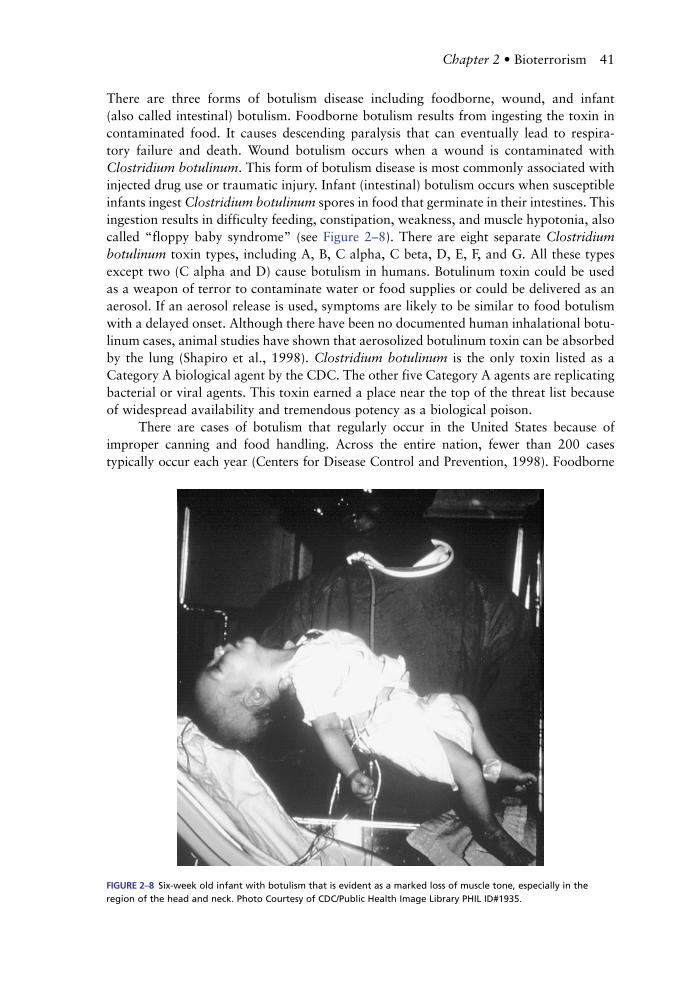
There are three forms of botulism disease including foodborne, wound, and infant (also called intestinal) botulism. Foodborne botulism results from ingesting the toxin in contaminated food. It causes descending paralysis that can eventually lead to respira-tory failure and death. Wound botulism occurs when a wound is contaminated with Clostridium botulinum. This form of botulism disease is most commonly associated with injected drug use or traumatic injury. Infant (intestinal) botulism occurs when susceptible infants ingest Clostridium botulinum spores in food that germinate in their intestines. This ingestion results in difficulty feeding, constipation, weakness, and muscle hypotonia, also called “floppy baby syndrome” (see Figure 2–8). There are eight separate Clostridium botulinum toxin types, including A, B, C alpha, C beta, D, E, F, and g. All these types except two (C alpha and D) cause botulism in humans. Botulinum toxin could be used as a weapon of terror to contaminate water or food supplies or could be delivered as an aerosol. If an aerosol release is used, symptoms are likely to be similar to food botulism with a delayed onset. Although there have been no documented human inhalational botu-linum cases, animal studies have shown that aerosolized botulinum toxin can be absorbed by the lung (Shapiro et al., 1998). Clostridium botulinum is the only toxin listed as a Category A biological agent by the CDC. The other five Category A agents are replicating bacterial or viral agents. This toxin earned a place near the top of the threat list because of widespread availability and tremendous potency as a biological poison.
There are cases of botulism that regularly occur in the United States because of improper canning and food handling. Across the entire nation, fewer than 200 cases typically occur each year (Centers for Disease Control and Prevention, 1998). Foodborne
Figure 2–8 Six-week old infant with botulism that is evident as a marked loss of muscle tone, especially in the region of the head and neck. Photo Courtesy of CDC/Public Health Image Library PHIL ID#1935.
42 DISASTERS AND PUBLIC HEALTH: PLANNINg AND RESPONSE
botulism is the most common form of botulism disease and is caused by eating foods that are contaminated with Clostridium botulinum that has released some of the toxin. If inten-tional food contamination becomes the delivery method selected by a terrorist or criminal, it would likely be limited to a small-scale attack. The toxin is easily destroyed with normal cooking and is not stable enough to survive for long-time periods in bulk food items. Regardless of the size or source of a botulism outbreak, any cases are considered to be a public health emergency that must be reported and investigated immediately. The source of the case or cases may still be accessible to the public and able to cause additional cases.
Intestinal botulism occurs when Clostridium botulinum spores are ingested resulting in intestinal colonization. The germinating spores release the illness-causing toxin. This form of the disease typically occurs in infants and is often attributed to feeding them honey. Although adults have immune systems that can tolerate and manage the minuscule num-ber of botulinum spores that often exist in honey, infants do not. However, in children or adults, when an infected wound is not apparent and a specific food source cannot be iden-tified, it is typically attributed to intestinal colonization. If an infected wound is observed, it may be wound botulism. This form of disease occurs when Clostridium botulinum infects a wound, multiplies, and releases the toxin causing systemic illness. Like foodborne, intestinal, and wound botulism occur naturally. However, the fourth type of disease, inha-lational botulism, does not occur naturally and is the most likely form expected to result from an aerosolized botulinum toxin release. It has never been seen in humans before, and there is limited animal data to provide clues on what to expect if it occurs.
The onset of botulism symptoms depends upon the exposure dose. Incubation for foodborne botulism ranges from several hours to just over a week (St. Louis et al., 1998). The lack of human data on inhalational botulism disease progression makes it impossible to predict exactly how these cases may progress. Initial botulism symptoms typically include blurred vision and dry mouth. For small exposures, these symptoms may be all that the symp-toms a patient experiences before recovery. Significant exposures may include slurred speech, difficulty swallowing, and descending peripheral muscle weakness. Severe disease involves the respiratory muscles, leading to respiratory failure and death. Presumptive diagnosis is based on the descending paralytic symptoms described. This diagnosis may be confirmed through testing available at reference labs but takes several days to complete. Botulism is sometimes mistaken for guillain-Barré syndrome, myasthenia gravis, or other diseases of the central nervous system (CNS) but differs from other CNS conditions in the descending symmetrical paralysis and absence of sensory nerve damage (Arnon et al., 2001).
The greatest challenge in managing a response to a large scale botulism outbreak will likely be a shortage of ventilators. As the descending paralysis moves down from the top of the head to the neck and chest of those exposed, it will eventually cause respiratory failure and require mechanical assistance to maintain respiration.
Botulism treatment requires supportive care with careful respiratory support and rapid administration of antitoxin. Fortunately, it is not transmitted from person to person, so patient care does not include a negative pressure patient room. In a mass exposure, these therapies pose tremendous challenges to an effective public health response. The need

Chapter 2•Bioterrorism 43
for ventilators may quickly exceed available capacity in an affected community. In addi-tion, the antitoxin efficacy is dependent upon how early it is started in the course of treat-ment (Tackett et al., 1984). This highlights the need for well-trained healthcare providers who maintain a high index of suspicion. It also shows how important it is to maintain a logistical system that can rapidly deliver antitoxin anywhere it is needed. The Centers for Disease Control and Prevention maintain the antitoxin supply and will release it for use through state health departments following a quick consultation. The botulism antitoxin cannot be used prophylactically for exposed individuals. It can only be used when initial symptoms appear. Although there is no botulism vaccine available, there are some promis-ing advances. A new Human Botulism Immune globulin Intravenous (BIg-IV) has shown great promise in reducing the length of treatment for infant botulism (Arnon et al., 2006). This could become the basis of new therapies for other forms of botulism disease as well.
Plague (Yersinia pestis)
Plague is a disease caused by Yersinia pestis, an anaerobic, gram-negative bacterium. The natural host for this organism is a rat and the disease is usually transmitted to humans through a flea bite from a flea that has fed on an infected rat and then on a human. There are about 10–15 human plague cases that naturally occur this way in the Western United States. Human cases also naturally occur in portions of South America, Africa, and Asia. Between 1000 and 3000 cases occur globally each year (Centers for Disease Control and Prevention (CDC), Division of Vector Borne Disease, Plague Home Page, www.cdc.gov/ncidod/dvbid/plague/index.htm). There are three types of plague disease including bubonic, pneumonic, and septicemic. The bubonic form of plague is the most likely form of the disease to be seen from naturally occurring infections. However, a terrorist attack could potentially involve the release of infected fleas resulting in bubonic cases. Pneumonic plague is more likely to be asso-ciated with an aerosolized release of Yersinia pestis during a bioterrorism attack. Sustained person-to-person transmission is possible through airborne droplets from infected individuals. This potential secondary transmission complicates the management of a large bioterrorism related outbreak and poses significant infection control challenges. Untreated pneumonic plague has a mortality rate of nearly 100%. It spreads through the lymph system and may involve multiple organ systems. All forms of plague can progress to the septicemic or pneu-monic forms of the disease, but the worst case bioterrorism scenario is an aerosolized release of Yersinia pestis that causes a large number of primary cases that continue to spread the infection to others before it is brought under control through public health interventions.
Yersinia pestis was isolated and characterized by Alexandre Yersin who traveled to Hong Kong in 1894 to study a plague outbreak that had taken tens of thousands of Chinese lives. The disease was eventually named after him. He seldom used his first name, referring to himself simply as Yersin and he refused to attend most medi-cal and scientific meetings of his day. He was an explorer and traveler trained in Europe who was fascinated by “Indo-China.” After discovering Yersinia Pestis, he went on to become the founder and director of the Medical School of Hanoi (Bibel and Chen, 1976).

44 DISASTERS AND PUBLIC HEALTH: PLANNINg AND RESPONSE
The word “plague” is often used to describe lethal outbreaks from a variety of infectious organisms. However, it is a specific disease that results from an infection by the gram-negative bacillus, Yersinia pestis. Plague has an infamous history killing millions of people in pandemic outbreaks occurring around the world across the centuries. Since plague still occurs naturally in many parts of the world, it is important to understand the baseline occurrence of naturally occurring plague so that you can recognize a suspicious case. If human cases are identified in a nonendemic area, in persons without risk factors, or in the absence of confirmed rodent cases, a terrorist release of Yersinia pestis should be considered as a possibility. There are three forms of plague disease: pneumonic, bubonic, and septicemic. Early symptoms for all forms of plague disease include classic flu-like symptoms such as fever, chills, myalgia, weakness, and headaches. Each form of plague poses different challenges and unique symptoms.
Pneumonic plague is the least common and most severe form of the plague dis-ease. This form of disease results from the inhalation of infectious particles either from an intentional aerosolized release or from a cough or sneeze of an infectious person. Although it sometimes occurs secondarily to bubonic plague, it will also cause primary pneumonic disease if it is released in an act of bioterrorism. This is the most likely form of plague disease resulting from aerosolized Yersinia pestis in an act of bioterrorism.
Yersinia pestis can only remain viable for about an hour as an aerosol and will quickly die when exposed to sunlight and heat outside of a living host. After exposure to aerosolized Yersinia pestis, individuals will display symptoms in 1–6 days (Inglesby et al., 2000). Based on limited epidemiological data, the mortality rate for pneu-monic plague in the United States is about 57% (Centers for Disease Control and Prevention, 1997). Patients will initially present with dizziness, chest discomfort, and a productive cough with blood-tinged sputum. By the second day, the symptoms may rapidly worsen and include chest pain, coughing, sputum increase and may include dyspnea, hemoptysis, cardiopulmonary complications, and circulatory collapse.
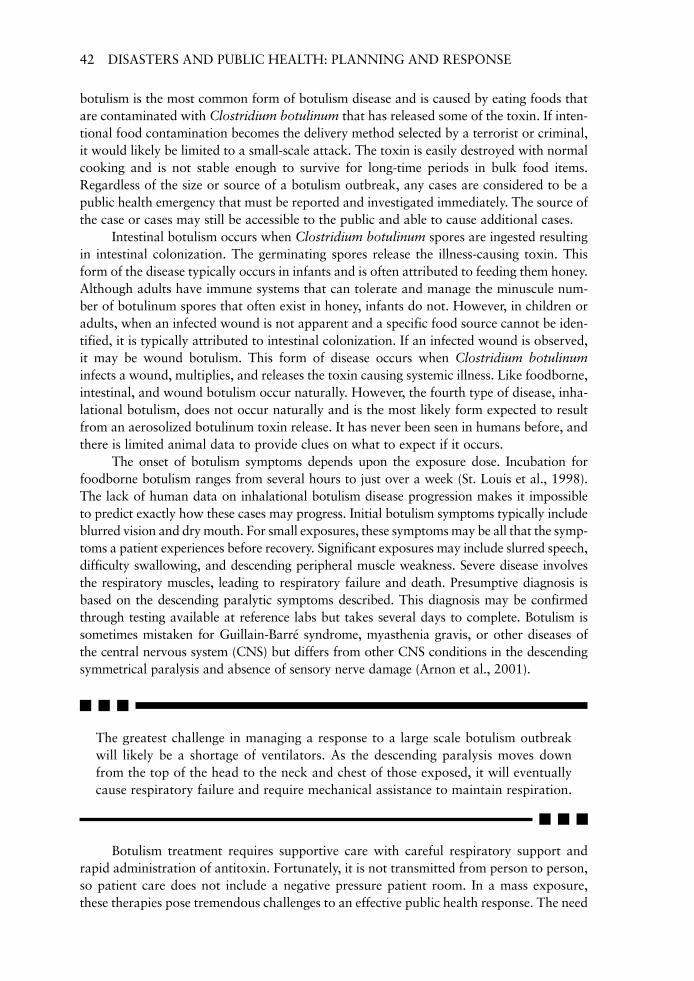
Bubonic plague is the most common form of plague disease. It is spread through the bite of an infected flea or by handling contaminated infected animals. Although this form of plague is less likely to be the result of bioterrorism, it is possible to intentionally spread bubonic plague through an intentional release of infected fleas. The mortality rate for this form of the disease is approximately 13% (CDC, 1997). Patients with bubonic plague will present with swollen, tender, and painful buboes (enlarged lymph nodes). These symptoms typically occur within 24 hours of the onset of early flu-like symptoms and are generally located near the area of inoculation. Most frequently, they occur in the inguinal or femoral lymph node region of the groin (See Figure 2–9). Buboes (swollen lymph glands) may erupt on their own or require incision and drainage.
Septicemic plague is the least common form of plague disease and it can result from either pneumonic or bubonic disease. It is a systemic infection that results from introduc-tion of Yersinia pestis into the bloodstream. The mortality rate for this form of the disease
Chapter 2•Bioterrorism 45
is about 22% (CDC, 1997). Patients with septicemic plague will initially present with nausea, vomiting, and diarrhea and may develop skin lesions, gangrene, and necrosis later in the infection process.
Rapid diagnosis and treatment are essential to reduce plague morbidity and mor-tality. Without quick antibiotic treatment, plague can cause death in several days. Initial diagnosis can be accomplished through microscopic examination of clinical samples of lymph node aspirates, sputum, or spinal fluid. Definitive diagnosis is accomplished using sputum or bubo cultures or IgM and Igg antibody testing available through reference labs. In addition, it is important to identify antibiotic resistance patterns to select the most effective treatment. Rapid diagnosis and antibiotic therapy are the keys to success-fully managing a plague outbreak. Once known or suspected cases of pneumonic plague occur, assume all patients presenting with flu-like symptoms are positive and treat them immediately. The rapid progression of the disease does not allow much time for definitive diagnosis during a possible mass exposure or outbreak. The antibiotic selection is based on the results of susceptibility testing and the age and condition of the patient. The aim of treatment includes both the treatment of active disease and the prophylactic treatment of suspected exposures. There was a killed whole-cell vaccine for use against bubonic plague but that vaccine was not very effective and has been discontinued. There are sev-eral research initiatives currently under way to develop a new plague vaccine.
Any identified cases of plague must be reported to the local public health agency. If an intentional release is suspected, local law enforcement and the Federal Bureau of Investigation (FBI) must also be notified. As the response is initiated, infection control for bubonic and septicemic forms of plague disease will consist of standard precautions. If pneumonic plague is identified, it requires patient isolation and droplet precautions until the patient has been on antibiotic therapy for at least 48 hours and shows signs of improvement.
Figure 2–9 This plague patient is displaying a swollen, ruptured inguinal lymph node, or bubo. Photo Courtesy of CDC/Public Health Image Library PHIL ID#2047.

46 DISASTERS AND PUBLIC HEALTH: PLANNINg AND RESPONSE
Smallpox (Variola Major, Variola Minor)
Smallpox disease is the most destructive infectious disease in human history. It has claimed more human lives than any other pathogen. It is caused by variola viruses, which are members of the orthopoxvirus family and it can only survive in humans. Smallpox was eradicated in the 1970s through a World Health Organization vaccination campaign. Since being declared eradicated over three decades ago, it now only exists in the govern-ment research labs of Moscow, Russia, and at the CDC in Atlanta, georgia. Unfortunately, there are suspicions that not all nations destroyed their lab supplies of the organisms. If a rogue nation still has some of the virus and supplies it to a terrorist organization, it could cause a devastating blow to international public health. For that reason, any single confirmed case today would be considered an international public health emergency.
There are two forms of this virus including variola major and variola minor. Variola major is the most likely form of the organism to be used as a weapon and is the causative agent for classic smallpox disease. It was the most common form of disease seen in historical epidemics and has a mortality of about 30%. Variola minor is a less common organism with a very low mortality rate of less than 1% (Fenner et al., 1988). The viruses are transmitted person to person through inhalation or direct contact with viral particles that are spread by infected individuals through their respiratory secretions or from pustules or scabs on their skin. Once an individual is exposed, an asymptomatic incubation period follows. It lasts for about 2 weeks. This incubation period is followed by a flu-like, prodromal illness for 2–5 days. Near the end of this prodromal period, the infected individual begins to develop pustules in the throat and mouth. They are then infectious, before any pustules appear on the skin. Once the rash begins on the skin, it has a characteristic, centrifugal pattern that appears mostly on the face, arms, and legs. It quickly becomes papular, pustular, and then scabs over. The scabs separate in 2–3 weeks. For those who do not survive, death often comes in the second week of this disease stage. If the patient has survived, the loss of the scabs is the end of their infectiousness. Survivors have a variety of complications, including severe scarring, encephalitis, secondary bacterial infections, conjunctivitis, and blindness.
During a confirmed smallpox outbreak, diagnosis should be made based on the clinical presentation of the patient alone. The greatest challenge will be the diagnosis of an index case. Most healthcare workers have never seen a smallpox case and many may miss the initial diagnosis. The most similar patient presentation is chickenpox. Smallpox lesions begin in the mouth and throat and then spread to the face and extremities. The lesions are concentrated centrifugally on the face, arms, and legs (See Figure 2–10). In addition, the lesions appear on the palms of the hands and the soles of the feet. Across the body, the lesions are uniform and in the same stage of development (vesicles, pustules, or scabs). In contrast, chickenpox con-centrates centrally on the trunk, not centrifugally. The chickenpox lesions are not seen on the palms of the hands or the soles of the feet and are not uniform in appearance. Chickenpox lesions are usually in various stages of development with pustules erupting next to scabs.
Smallpox is contagious person to person and usually spreads through respiratory droplets or direct contact with infected persons or contaminated materials. Historically, each primary case will infect three or four other people. The exception to this is if infected individuals have a severe cough, they may infect dozens of other susceptible contacts (Wehrle et al., 1970). Those at highest risk of secondary infection include house-hold contacts and healthcare providers. In a healthcare setting, airborne and contact precautions must be instituted. The patient should be in a negative pressure room and personnel and/or visitors need to wear proper respiratory protection, gown, and gloves.

Chapter 2•Bioterrorism 47
All suspected cases should be immediately isolated and observed. Confirmed cases should remain in hospital isolation until all the scabs separate. Home isolation may be the only option during a large outbreak. Over a century ago, there were separate hospitals desig-nated for smallpox patients to keep them out of the other facilities. That approach may need to be adopted again in the event of a new epidemic. Anyone identified as a contact of a smallpox case should be vaccinated immediately and then monitored twice daily for 17 days for a fever. If a fever greater than 38 °C (>101 °F) develops, it could be the pro-dromal stage of smallpox and the patient should be isolated (Fenner et al., 1988).
Smallpox vaccine does not contain Variola virus. It contains an attenuated-strain of vaccinia virus, a close cousin of Variola. It is genetically similar enough to smallpox that a small vaccinia infection on the skin resulting from the vaccination will provide protec-tion against smallpox as well. The original vaccine was made from calf lymph and is no longer produced. The decades old stockpile of this vaccine was being used by DOD and for homeland security initiatives for the past several years but in 2007, the Food and Drug Administration approved the next generation of smallpox vaccine (Food and Drug Administration, FDA approves new smallpox vaccine, www.fda.gov/consumer/updates/smallpox090407.html). It is very similar to the old vaccine in that it is still delivered with a
Figure 2–10 A young boy in Bangladesh with the classic maculopapular rash evident on his torso, arms, and face. 1974. Photo Courtesy of CDC/Public Health Image Library PHIL ID#10660.

48 DISASTERS AND PUBLIC HEALTH: PLANNINg AND RESPONSE
bifurcated needle to initiate a vaccinia skin infection (Figure 2–11). It is called ACAM2000 and the primary difference is in the production process. It is made using cell cultures. Although it will have fewer side effects than the original vaccine, there are serious risks of complications with the vaccine and it should not be offered to the general public unless there is an imminent threat of a smallpox attack or outbreak.
Vaccination may also be used prophylactically to reduce morbidity and mortality. It has been proven effective if it is provided within 4 days of exposure (Dixon, 1962). An exposure is defined as suspected inhalation of viral particles from an initial bioter-rorism release, face-to-face contact with a confirmed smallpox case, or direct contact with potentially contaminated materials or lab specimens. Other antiviral drugs, such as Cidofovir, may also improve patient outcomes if it is given within 1–2 days of an expo-sure (Huggins et al., 2001). It is impossible to say which prophylactic options may be most effective for humans in the absence of existing smallpox cases and exposures.
There are three smallpox disease variants. Each disease type is identified by the pre-sentation of the rash. Classical smallpox is the most common and historically accounts for >90% of cases (Henderson et al., 1999). The other types of smallpox disease are malig-nant and hemorrhagic. They appear much different than typical smallpox and are difficult to diagnose. Malignant smallpox cases have a velvety, non-pustule rash. It was associated with high mortality. Hemorrhagic smallpox looks like a diffuse erythematous rash leading to petechiae and hemorrhages. It was often misdiagnosed as meningococcemia and is also usually fatal. One of the planning challenges we face in preparing for the possibility of a smallpox attack is the question of how the initial patients may present symptoms. As our assumptions about variola major infections are based on the natural presentation of small-pox disease, it may be easy to misdiagnose individuals infected from an aerosolized inhala-tion exposure that results in a large number of individuals with atypical presentations.
Fortunately, the variola viruses are not stable or persistent in the environment. If they were released as an aerosol, they would naturally be inactivated within about 24 hours. Because the organism dies out rapidly in the external environment, the buildings and
Figure 2–11 CDC clinician demonstrates the use of a bifurcated needle during the 2002 Smallpox Vaccinator Workshop. Photo by James Gathany. Courtesy of CDC/Public Health Image Library PHIL ID#2815.

Chapter 2•Bioterrorism 49
areas in the path of an intentional release do not need decontamination and no long-term environmental recovery issues following a release (Henderson et al., 1999).
Tularemia (Francisella tularensis)
Tularemia is caused by Francisella tularensis, a very hardy and highly infectious aerobic organism. Exposure to as little as 10 organisms can result in the disease, also known as “rabbit fever” or “deer fly fever” (Saslaw et al., 1961). Naturally occurring disease is almost always rural and although it has occurred in every state in the U.S. except for Hawaii, it primarily occurs in the Midwest. It usually occurs as individual cases or as small, isolated outbreaks and has never been reported as a large epidemic. However, the infectiousness and availability of the organism makes it a potential weapon for terror-ists. Any cases in urban or nonendemic areas, or among populations without clear risk factors, should be investigated as a possible bioterrorism incident.
Humans become infected by Francisella tularensis in a several ways. As with other Category A organisms, the route of exposure plays an important role in determining the form of tularemia disease. It is a very complex disease that presents as pneumonic, typhoidal, ulceroglandular, glandular, oculoglandular, or oropharyngeal. Natural infec-tions usually occur from handling infected small mammals such as rabbits or rodents or through the bites of ticks, deerflies, or mosquitoes that are carrying the infection from feeding on infected animals. Most of these natural infections are ulceroglandular or glan-dular tularemia. If an infected animal is eaten without being thoroughly cooked, or if contaminated food or water is ingested, it may cause oropharyngeal tularemia. It is also possible to get the organism on the hands and inoculate the eye resulting in oculoglandu-lar tularemia. The types of tularemia disease likely to result from an intentional aerosol release of Francisella tularensis are pneumonic and typhoidal tularemia. Morbidity and mortality vary greatly depending upon the type of tularemia disease. The most common forms of the disease, such as ulceroglandular tularemia (50–85% of all cases), have a low mortality of less than 2% (See Figure 2–12) (Dennis, 1998). However, primary pneu-monic cases have a much higher mortality of 30–60% if untreated (Evans et al., 1985).
Francisella tularensis inhalation will usually cause flu-like symptoms after a 2–5 day incubation period. However, these symptoms can occur as soon as 1 day or as long as 14 days after exposure (Dennis et al., 2001). The flu-like clinical presentation may include the rapid onset of fever, chills, myalgia, weakness, and headaches. These symp-toms sometimes worsen to include a nonproductive cough, sore throat, and inflammation of the bronchi, lungs, and lymph nodes. Diagnosis is complicated by the fact that there are no rapid diagnostics available for tularemia. Although there are promising initiatives under way to develop this capability, the initial identification of an intentional tularemia outbreak will depend upon an astute physician using a presumptive diagnosis based on the clinical presentation. This is particularly difficult for clinicians that have never seen a case. If a large number of otherwise healthy individuals present with these symptoms, particularly in nonrural regions with low incidence of naturally occurring tularemia, the index of suspicion should be high that it could be tularemia from an intentional release. If inhalational tularemia is suspected, respiratory secretions and blood samples may be used for confirmatory testing. When a case of pneumonic tularemia is suspected or confirmed, providers should assume all patients presenting with flu-like symptoms are positive and treat appropriately with antibiotics. The antimicrobial selection is based on the results of

50 DISASTERS AND PUBLIC HEALTH: PLANNINg AND RESPONSE
susceptibility testing to be sure that there is no drug resistance, the age and condition of the patient, and the goal of the therapy. The goal may be treatment of suspected disease or prophylactic antimicrobial treatment for suspected exposures. A vaccine also exists for tularemia that uses an attenuated live vaccine strain (LVS) (Oyston and Quarry, 2005); however, it continues to be under review by the Food and Drug Administration. The strain used and the process of attenuating it have raised safety and efficacy questions that have slowed approval and it is not yet available to the general public.
Francisella tularensis can survive for long periods of time in a cool, moist environ-ment. However, it is unlikely that it could survive for a long-period of time in an outdoor environment following an aerosol release. It is also not likely to be reaerosolized. Therefore, environmental decontamination following aerosol release of Francisella tularensis is not necessary. Individuals suspected of being contaminated by an aerosol should launder cloth-ing and shower with soap and water. Even though Francisella tula rensis is highly infectious for individuals directly exposed to the organisms, it is not spread from person to person.
Viral Hemorrhagic Fevers (Filoviruses, Arenaviruses, Bunyaviruses, and Flaviviruses)
The term “viral hemorrhagic fevers” (VHFs) describes a severe multisystem syndrome caused by one of four different viral families (Arenaviruses, Filoviruses, Bunyaviruses, and Flaviviruses). These are all RNA viruses that naturally reside in animal hosts
Figure 2–12 Girl with ulcerating lymphadenitis colli due to tularemia, Kosovo, April 2000.
Source: Centers for Disease Control and Prevention, http://www.cdc.gov/ncidod/eid/vol8no1/01-0131G1.htm.

Chapter 2•Bioterrorism 51
and arthropod vectors. These hosts usually include rats and mice but for some VHF pathogens, such as Ebola and Marburg, it is still not certain where they naturally reside in nature. Transmission to humans results from infected hosts or vectors exposing humans through their body fluids and excretions. Some VHF viruses can spread from person to person through close contact with infected people or exposure to their body fluids. VHF illness begins as flu-like symptoms including fever, fatigue, dizziness, muscle aches, and weakness. The symptom become worse as vascular damage takes place. As the disease progresses, severe symptoms occur including hypotension, facial edema, pulmonary edema, and mucosal hemorrhages. Some patients will bleed from body orifices, internal organs, and even under the skin. Although the bleeding is sometimes severe, that is rarely the cause of death. VHF death is usually the result of multiorgan failure and cardiovascu-lar collapse. The case fatality rates for VHFs have a wide range that differs by organism and by outbreak. For example, it was only 0.5% for an Omsk hemorrhagic fever but as high as 90% for a Zaire Ebola outbreak (Cunha, 2000; Muyembe-Tamfum et al., 1999). None of these organisms are airborne transmitted in natural outbreaks, but the concern is that they may be delivered as an aerosol during a bioterrorism attack. If a particularly infectious and lethal VHF agent is used for an aerosol attack, it could be devastating.
The RNA viruses that cause VHFs are dependent upon animal and insect hosts for their survival, and the naturally occurring forms of these diseases are limited to the area immediately surrounding their primary hosts. Humans are not normally part of their lifecycle but are sometimes inadvertently included by encountering an animal reservoir or infectious vector. Outbreaks are erratic and unpredictable but usually isolated.
VHF pathogens were placed on the Category A list of threats because of their potential lethality and infectiousness at low doses when delivered as an aerosol. Although there has never been an aerosolized VHF attack, animal studies have demonstrated the severe risk of infection posed by aerosolization of these pathogens (Johnson et al., 1995; Stephenson et al., 1984). If this scenario occurs, there will be an incubation period rang-ing from a couple days to 3 weeks depending on the VHF pathogen used, the individual dose received, and other variables. A prodromal illness of flu-like symptoms occurs for about 1 week. Severe infections may also display bleeding from orifices, under the skin, and in internal organs, eventually leading to multiorgan failure. Initial diagnosis is based on the clinical presentation and requires a high clinical index of suspicion (Borio et al., 2002). Treatment is limited mostly to supportive care. There are no antivirals specifically approved for VHFs and it is not known if any will be effective.
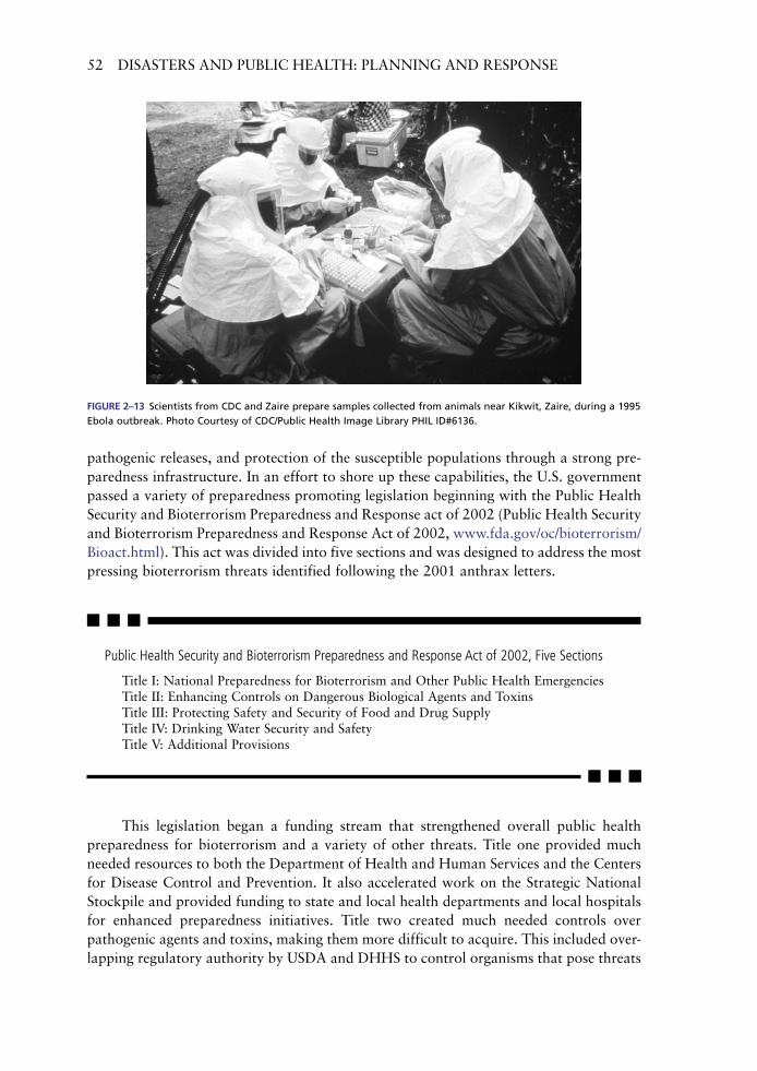
Those at greatest risk during historical outbreaks have been healthcare workers. To ensure their safety, infection control precautions must be strictly enforced with suspect VHF patients. This includes patient isolation in a negative pressure room, strict hand hygiene, and use of double gloves, impermeable gown, face shield, eye protection, and leg/shoe coverings (See Figure 2–13). In addition, HEPA respiratory protection should be worn by anyone enter-ing the patients room (Borio et al., 2002). The risk to prehospital contacts may be somewhat lower because the patients will likely be in the earlier stages of disease and less infectious.
Prevention and DetectionPrevention of bioterrorism threats begins with solid regulations to prevent acquisition of pathogens through strict control of potential biothreat agents, rapid detection of
52 DISASTERS AND PUBLIC HEALTH: PLANNINg AND RESPONSE
Public Health Security and Bioterrorism Preparedness and Response Act of 2002, Five Sections
Title I: National Preparedness for Bioterrorism and Other Public Health EmergenciesTitle II: Enhancing Controls on Dangerous Biological Agents and ToxinsTitle III: Protecting Safety and Security of Food and Drug SupplyTitle IV: Drinking Water Security and SafetyTitle V: Additional Provisions
pathogenic releases, and protection of the susceptible populations through a strong pre-paredness infrastructure. In an effort to shore up these capabilities, the U.S. government passed a variety of preparedness promoting legislation beginning with the Public Health Security and Bioterrorism Preparedness and Response act of 2002 (Public Health Security and Bioterrorism Preparedness and Response Act of 2002, www.fda.gov/oc/bioterrorism/ Bioact.html). This act was divided into five sections and was designed to address the most pressing bioterrorism threats identified following the 2001 anthrax letters.
Figure 2–13 Scientists from CDC and Zaire prepare samples collected from animals near Kikwit, Zaire, during a 1995 Ebola outbreak. Photo Courtesy of CDC/Public Health Image Library PHIL ID#6136.
This legislation began a funding stream that strengthened overall public health preparedness for bioterrorism and a variety of other threats. Title one provided much needed resources to both the Department of Health and Human Services and the Centers for Disease Control and Prevention. It also accelerated work on the Strategic National Stockpile and provided funding to state and local health departments and local hospitals for enhanced preparedness initiatives. Title two created much needed controls over pathogenic agents and toxins, making them more difficult to acquire. This included over-lapping regulatory authority by USDA and DHHS to control organisms that pose threats

Chapter 2•Bioterrorism 53
to plants, animals, and humans. This included a requirement for registration by anyone possessing hazardous biological material and it established strict controls for safety and security. Title three was established to protect food and drugs. It included enhanced inspection of imported foods, food safety and security, and upgrading of animal and plant health inspections. Title four increased security requirements for drinking water, and title five included several additional provisions primarily directed toward prescrip-tion drugs.
The laws instituted through the Public Health Security and Bioterrorism Preparedness and Response Act of 2002 to control pathogenic organisms were not only a result of the 2001 anthrax letters but in part a response to the exploits of microbiologist Larry Wayne Harris. He mail ordered and received pathogenic organisms. It was discovered later that he had ties to white supremacist groups and allegedly threat-ened to release anthrax in Las Vegas. Although the strain he purchased was a harm-less strain of Bacillus anthracis spores used in vaccine development, it highlighted a vulnerability that needed to be addressed.
In 2005, another major piece of public health legislation was passed. The Pandemic Preparedness and Response Act was a response to growing public concerns over pandemic influenza. Although the aims of the legislation were good, at least part of the implementation was not. Although requiring state and local public health orga-nizations to meet planning and preparedness goals for pandemics is an important step, and public health funding is always welcome, the pandemic dollars provided through this legislation came after a large cut to the basic public health preparedness budget previously established for bioterrorism. Even though the infrastructure required for pandemic response and bioterrorism are parallel, the cuts in basic preparedness funding meant losing trained staff who could meet the requirements of the pandemic prepared-ness funding. Unfortunately, the way the dollars were originally disbursed, some key staff needed to meet the pandemic preparedness objectives had already lost their jobs from the preparedness budget cuts. In other words, funding was given with one hand by federal health agencies to state and local public health agencies and taken with the other. To sustain adequate public health preparedness infrastructure, core funding needs to be sustained and not cut and then supplemented by additional funding for perceived emerging threats.
Cultivating the Perceptive Provider
Limiting the impact of a bioterrorism attack requires healthcare providers with sufficient training and support to remain diligent. Over an extended period of time, and in the absence of recent attacks, this diligence becomes more difficult to maintain. There are nonbioterrorism threats that are encountered regularly that draw the focus away from preparedness for unlikely events. Though this is understandable, it will not be excusable

54 DISASTERS AND PUBLIC HEALTH: PLANNINg AND RESPONSE
if a sentinel case is missed. The diversity of terrorist threats has resulted in substantial investments in public health and healthcare preparedness. The costs are well into the bil-lions and are likely to continue for many years. Much of this spending takes the form of high-tech solutions including new epidemiological surveillance software or new detection and monitoring devices, such as the BioWatch Program. Although this technology may compliment other preparedness initiatives, there is not a technological solution that can replace an astute, perceptive healthcare provider. Throughout history, astute physicians and other well-informed providers have been the sentinels of our nation’s public health. Typically, they identify emerging problems for public health, rather than public health identifying the problems in advance for providers (Thacker, 1994). This notion was con-firmed in 2001 when clinicians and laboratory professionals who had recently received preparedness training, diagnosed the index case of inhalational anthrax (Bush et al., 2001). The preparedness education and training of healthcare providers will continue to be one of the most important measures any nation can invest in to mitigate the impact of a bioterrorism attack through rapid detection. We must consistently challenge and sharpen the intuition and skill of healthcare providers so that they are able to recognize a bioterrorism attack early, and take the appropriate actions.
Disease Surveillance
Disease surveillance is an essential and familiar function of local and state public health agencies. Regular disease surveillance includes mandatory disease reporting by local healthcare providers, data entry and analysis by local or regional public health agen-cies, and additional analysis, reporting, and allocation of needed resources by state and federal public health agencies. However, the bioterrorism agents of concern have short incubation periods that require rapid identification to facilitate early treatment to reduce morbidity and mortality. Most local disease surveillance activities do not process and analyze information quickly enough to ensure a prompt identification of a suspicious trend. They often lack the sensitivity needed to detect small changes in population health, and are not specific enough to distinguish normal fluctuations in illness from unusual or suspicious cases. There is no single solution to this gap in surveillance capabilities. An ideal system will include a network of systems that is able to simultaneously analyze multiple variables, effectively compare changes and trends, and control confounders and background noise (Pavlin et al., 2002). The realization of a complete, integrated, effec-tive, uniform system throughout the United States or any other nation is many years away. In the United States, the largest system in development is called BioSense. It has a growing list of data sources including state and regional epidemiological surveillance systems, healthcare facilities, national labs, VA Hospitals, Department of Defense, and others. The plans are to eventually incorporate data from poison control center calls and-over-the counter drug sales (Centers for Disease Control and Prevention (CDC), BioSense program fact sheet, www.cdc.gov/biosense/files/fact_sheet.pdf). It is the hope of CDC officials overseeing the construction of this surveillance network that trends in a sufficient number of healthcare and public health data sets will be capable of detecting growing epidemics before current reporting systems are able to detect subtle clues. Until it is fully developed and implemented across the nation, we will continue to depend upon a patchwork of surveillance and detection systems.
Chapter 2•Bioterrorism 55
Active surveillance for special events is currently the most exhaustive and effective epidemiological surveillance available. This comprehensive, symptomatic surveillance is accomplished at the Olympic games, Superbowl, political conventions, and other high visibility events. This is a labor intensive and expensive endeavor requiring addi-tional staff placement in key points of healthcare delivery to gather information. The information collected focuses on respiratory, neurological, and dermatological clues that may quickly indicate an intentional biological release has occurred. Although this may appear to be an ideal method to gather accurate information in a timely manner, it is far too expensive to sustain on a daily basis. Other active surveillance approaches include the monitoring of available information in public health and healthcare databases. This data may include ambulance runs, chief complaints of 911 calls, hospital admissions, school or work absenteeism, or trends in pharmaceutical or laboratory demands. The challenge of surveillance is twofold. First, the correct baseline is often difficult to deter-mine. The success of surveillance depends upon the accurate determination of a trigger or threshold signaling an unusual event or trend. This is only successful if the numerous confounders in the data are controlled. For example, how can 911 calls or absenteeism trends distinguish between a suspicious event and the onset of “cold and flu season” or how can sales trends in certain pharmaceuticals distinguish between an increase in flu-like symptoms and price discounts involving those products? These are challenging problems but progress continues and important strides are being made to advance the surveillance process.
The trigger for activating a bioterrorism response is difficult to define. Bioterrorism encompasses such a wide array of pathogens and pathogenic by-products that it is impossible to define a “typical” bioterrorism event and establish appropri-ate triggers for response. In 1984, the Dalles, Oregon experienced a large outbreak of Salmonella gastroenteritis. More than 700 people became ill after eating at 10 local salad bars. The epidemiological investigation could not identify a single food as the source of the illnesses. Months later, the criminal investigation identified the origin of the outbreak as intentional contamination of area salad bars by members of a reli-gious cult (Torok et al., 1997). In this case, the agent used was not lethal and initially appeared as a large, but accidental foodborne outbreak. A bioterrorism scenario will unfold in a very different way if a lethal agent is used. If the agent is transmissible from person to person or if it is delivered by spraying an aerosol, the “footprint” or focused area of an outbreak could expand across large regions. Clues that should raise the suspicion of providers include anything that is out of the ordinary. Every com-munity has trends in seasonal illnesses with some variation from year to year. There may be obvious differences emerging, such as out of season “flu-like” outbreaks or more severe cases among groups that normally do not experience serious illness. An illness may be uncharacteristic for a particular region such as plague in New Jersey or the strain may be unusual or resistant to treatment. Clues that should raise suspicion may range from more subtle trends to the obvious clues that may be provided through claims made by aggressors or evidence identified by law enforcement authorities. Maintaining a high index of suspicion increases the likelihood of discovering an event sooner and the swift discovery of an attack translates into more lives saved. Examples of the epidemiological clues that may suggest an act of bioterrorism are listed in Table 2–3.

56 DISASTERS AND PUBLIC HEALTH: PLANNINg AND RESPONSE
Table 2–3 Bioterrorism Epidemiological Clues
1) A large epidemic, with greater case loads than expected, especially in a discrete population.
2) More severe disease than expected for a given pathogen, as well as unusual routes of exposure.
3) Unusual disease for a given geographical area, found outside the normal transmission season, or
impossible to transmit naturally in the absence of the normal vector for transmission.
4) Multiple, simultaneous epidemics of different diseases.
5) A zoonotic disease outbreak as well as a human outbreak, as many of the potential threat agents are
pathogenic to animals.
6) Unusual strains or variants, antimicrobial resistance patterns disparate from those circulating.
7) Higher attack rates in those exposed in certain areas, such as inside a building if the agent was released
indoors, or lower rates in those inside a sealed building if an aerosol was released outdoors.
8) Intelligence that an adversary has access to a particular agent or agents.
9) Claims by a terrorist of the release of a biological agent.
10) Direct evidence of the release of an agent, with findings of equipment, munitions, or tampering.
Source: Julie A. Pavlin, Walter Reed Army Institute of Research, Washington, D.C. (Pavlin, 1999).
Environmental Monitoring
Other forms of surveillance include environmental monitoring and standoff detection. Detector research and development continues to rapidly move forward. Biological moni-toring includes a range of technologies from basic bioaerosol collectors to advanced laser particle analyzers. There are a variety of point source detection technologies in development, including nucleic acid, antibody, whole cell, and mass spectrometry analysis technologies. These detectors may enhance preparedness for specific high-risk events or buildings but they will always have limitations. The greatest limitation in the foreseeable future is cost. This emerging technology is expensive and is not affordable for most state and local agencies. These detectors are not sensitive enough to detect the full range of biological threats at a low level or specific enough to distinguish between normal background biological material in the air and the pathogens of greatest concern. These devices also must be prepositioned in areas most likely to experience an attack. This requires comprehensive and accurate risk assessments of each community. Once a risk assessment is accomplished, it must be continually monitored and updated accord-ing to community activities, events, and changes. At their best, detectors can only cover a limited area. This includes portable detectors that may be used for special events and more stationary detectors such as the BioWatch system. BioWatch is a surveillance program in dozens of major cities across the United States, where discretely placed air samplers continually draw air through a filter that is collected daily and tested for DNA associated with biothreat agents. Although this is an important tool, it cannot be successful without and will never replace well-trained physicians or epidemiological surveillance systems.
Immediate ActionsOnce a biological threat has been detected, the biothreat agent and population at risk must be quickly identified. Because most of the severe illnesses resulting from an act of bioterrorism will have an incubation period, there is a window of opportunity for

Chapter 2•Bioterrorism 57
prophylactic treatment. This is true for even the most lethal agents. Decisions must be made rapidly and the response needs to begin immediately. Communication must be quickly established with the population at risk. If there are specific measures that may be taken to reduce their risk or the risk of others, it must be shared. For example, if pneumonic plague is the disease of concern, those at risk may be advised to isolate themselves from family members to avoid the risk of spreading the illness. At the same time, hospitals will be preparing for patient surges, whereas public health officials may be setting up Points of Dispensing or coordinating other strategies for mass prophy-laxis. However, each response will be unique according to the agent, method of release, population at risk, environmental conditions, etc. There are far too many variables to concisely summarize the immediate actions to take. However, the initial message will consist of the preparedness mantra promoted by the Red Cross and most public pre-paredness organizations. People should have a communication plan in case difficulties arise in the local communication infrastructure due to the crisis. They should maintain a home preparedness kit in case an elective or mandatory quarantine is put in place to limit the spread of disease. Most importantly, they should stay informed and follow the instructions provided.
Just as the public is being informed of the appropriate measures to take, those working in healthcare, public health, and the first response community will be provided with detailed instructions on how to respond. For example, infection control recommen-dations and requirements will be shared (See Table 2-4). If standard precautions are sug-gested, that will include isolating blood and all other body fluids, secretions, nonintact skin, and mucous membranes of those who may be infected. Use gloves and gowns to prevent exposure to blood and other potentially infectious fluids. Use mask and eye pro-tection or a face shield if there are activities that are likely to generate splashes or sprays of blood, body fluids, secretions, or excretions. Appropriate hand washing is always necessary. Other precautions may include:
Droplet Precautions: Provide private patient rooms or cohort patients with the same •infectious agent. Use a mask if within three feet of the patient.Contact Precautions: Provide private patient rooms or cohort patients with the •same infectious agent. Use gloves when entering the room and a gown if clothing is likely to have contact with an infectious patient, or contaminated surfaces or equipment.
• Airborne Precautions: Requires a negative pressure isolation room and appropriate respiratory protection such as the N95 respirator which must be fit-tested.
Recovery
Following any bioterrorism event, there are likely to be strong debates over the need for decontamination and clean-up. With the notable exception of anthrax, clean-up will not be difficult for most pathogenic organisms. To ensure that an effective clean-ing agent is used, the Environmental Protection Agency has established a listing of “antimicrobial products” under the Federal Insecticide, Fungicide, and Rodenticide Act (FIFRA). This is a much different antimicrobial than the antibiotic treatment of humans that often uses the same nomenclature. In the context of an environmental clean-up,

Tab
le 2
–4
Sam
ple
Pat
ien
t M
anag
emen
t G
uid
elin
es f
or
Bio
terr
ori
sm-R
elat
ed Il
lnes
ses
iMPO
rTA
NT
PHO
Ne
Nu
MB
erS:
infe
ctio
us
Dis
ease
s _
____
____
____
infe
ctio
n C
on
tro
l ___
____
____
___
er _
____
____
___
uSA
Mr
iiD 3
01-6
19-2
833,
CD
C e
mer
gen
cy r
esp
on
se O
ffic
e 77
0-48
8-71
00
BACTERIAL AGENTS
Anthrax
Brucellosis
Cholera
Glanders
Bubonic Plague
Pneumonic Plague
Tularemia
Q Fever
VIRUSES
Smallpox
Encephalitis
Viral Encephalitis
Viral Hemorrhagic Fever
BIOLOGICAL TOXINS
Botulism
Ricin
T-2 Mycotoxins
Staph. Enterotoxin B
iso
lati
on
Pre
cau
tio
ns
Stan
dard
Pre
caut
ions
for
all
aspe
cts
of p
atie
nt c
are
X
XX
XX
XX
X
XX
XX
X
XX
X
Con
tact
Pre
caut
ions
(go
wn
& g
love
s; w
ash
hand
s af
ter
each
pt
enco
unte
r)
X**
*X
*X
*
X
X
X*
Airb
orne
Pre
caut
ions
(ne
gativ
e pr
essu
re r
oom
& N
95 m
asks
for
all
indi
vidu
als
ente
ring
the
room
)
X
X**
Dro
plet
Pre
caut
ions
(su
rgic
al m
ask)
X
X
**
Pati
ent
Plac
emen
t
No
rest
rictio
ns
XX
XX
XX
XX
XX
XX
Coh
ort
‘like
’ pa
tient
s w
hen
priv
ate
room
una
vaila
ble
X
***
X*
XX
X
X
X*
Priv
ate
Room
X
***
X*
X*
X
X
X
X
*
Neg
ativ
e Pr
essu
re
X
X**
Doo
r cl
osed
at
all t
imes
X
X
**
Pati
ent
Tran
spo
rt
No
rest
rictio
ns
XX
XX
X
XX
XX
XX
XX
Lim
it m
ovem
ent
to e
ssen
tial m
edic
al p
urpo
ses
only
X
***
X*
X*
X
X
X
X
*
Plac
e m
ask
on p
atie
nt t
o m
inim
ize
disp
ersa
l of
drop
lets
X
X
X
**

Cle
anin
g a
nd
Dis
infe
ctio
n
Rout
ine
clea
ning
of
room
with
hos
pita
l app
rove
d di
sinf
ecta
nt
XX
XX
XX
XX
X
XX
XX
XX
Dis
infe
ct s
urfa
ces
with
10%
ble
ach
solu
tion
or p
heno
lic d
isin
fect
ant
X
Ded
icat
ed e
quip
men
t (d
isin
fect
prio
r to
leav
ing
room
)
X**
*X
*X
*
X
X
X*
Line
n m
anag
emen
t as
with
all
othe
r pa
tient
s
XX
XX
XX
XX
XX
X
XX
XX
Line
ns a
utoc
lave
d be
fore
laun
derin
g or
was
h in
hot
wat
er w
ith b
leac
h ad
ded
X
Post
mo
rtem
Car
e
Follo
w p
rinci
ples
of
Stan
dard
Pre
caut
ions
X
XX
XX
XX
X
XX
XX
X
XX
X
Dro
plet
Pre
caut
ions
(su
rgic
al m
ask)
X
Con
tact
Pre
caut
ions
(go
wn
& g
love
s)
X
*X
*
X
X
X*
Avo
id a
utop
sy o
r us
e A
irbor
ne P
reca
utio
ns &
HEP
A f
ilter
X
X
X
**
Rout
ine
term
inal
cle
anin
g of
roo
m w
ith h
ospi
tal a
ppro
ved
disi
nfec
tant
X
XX
XX
XX
X
XX
X
X
XX
X
Dis
infe
ct s
urfa
ces
with
10%
ble
ach
solu
tion
or p
heno
lic d
isin
fect
ant
X
Min
imal
han
dlin
g of
bod
y; s
eal b
ody
in le
af-p
roof
mat
eria
l
X
Cre
mat
e bo
dy w
hene
ver
poss
ible
X
Dis
con
tin
uat
ion
of
iso
lati
on
48 h
rs o
f ap
prop
riate
ant
ibio
tic a
nd c
linic
al im
prov
emen
t
X
Unt
il al
l sca
bs s
epar
ate
X
Unt
il sk
in d
econ
tam
inat
ion
com
plet
ed (
1 hr
con
tact
tim
e)
X
Dur
atio
n of
illn
ess
X
***
X*
X*
X
STA
ND
Ar
D P
reC
Au
TiO
NS—
Stan
dard
Pre
caut
ions
pre
vent
dire
ct c
onta
ct w
ith a
ll bo
dy f
luid
s (in
clud
ing
bloo
d),
secr
etio
ns,
excr
etio
ns,
non-
inta
ct s
kin
(incl
udin
g ra
shes
), an
d
muc
ous
mem
bran
es.
Sta
ndar
d Pr
ecau
tions
rou
tinel
y pr
actic
ed b
y he
alth
care
pro
vide
rs in
clud
e:
spla
sh/s
pray
and
gow
ns t
o pr
otec
t sk
in a
nd c
loth
ing
durin
g pr
oced
ures
.
*Con
tact
Pre
caut
ions
nee
ded
only
if t
he p
atie
nt h
as s
kin
invo
lvem
ent
(bub
onic
pla
gue:
dra
inin
g bu
bo)
or u
ntil
deco
ntam
inat
ion
of s
kin
is c
ompl
ete
(T
-2 M
ycot
oxin
s).
**A
sur
gica
l mas
k an
d ey
e pr
otec
tion
shou
ld b
e w
orn
if yo
u co
me
with
in 3
fee
t of
pt.
A
irbor
ne P
reca
utio
ns a
re n
eede
d if
patie
nt h
as c
ough
, vo
miti
ng,
diar
rhea
, or
hem
orrh
age.
**
* C
onta
ct P
reca
utio
ns n
eede
d on
ly if
the
pat
ient
is d
iape
red
or in
cont
inen
t.So
urce
: Sa
int
Loui
s U
nive
rsity
, C
ente
r fo
r th
e St
udy
of B
iote
rror
ism
and
Em
ergi
ng In
fect
ions
, O
ctob
er 2
001.
Ada
pted
with
per
mis
sion
fro
m a
n or
igin
al t
able
des
igne
d by
LTC
Suz
anne
E.
John
son,
RN
, M
SN,
CIC
, W
alte
r Re
ed A
rmy
Med
ical
Cen
ter.
60 DISASTERS AND PUBLIC HEALTH: PLANNINg AND RESPONSE
antimicrobials are substances with chemical or physical properties that destroy or repress the growth of pathogenic organisms. Thousands of products are registered with the U.S. Environmental Protection Agency (EPA) for various applications. Over half of the products registered are specifically for controlling harmful organisms in healthcare settings. These antimicrobial products fall into two broad categories including those that disinfect, sanitize, or limit the growth of organisms and those that protect various objects and surfaces from contamination. There is a regulatory distinction between products that are used directly on food, those that are used on or in a living human body, and those products that are used to control microbes in the environment. The Food and Drug Administration regulate those used directly on food or people. The EPA regulates the substances used for microbial control in the environment. They will have the final word on which antimicrobials will be used under different circumstances on different surfaces.
SummaryBioterrorism is a particularly challenging public health preparedness issue. It requires a robust public health infrastructure to detect and manage an incident, a healthcare system with astute providers and well managed surge capacity, and a variety of disciplines pre-pared to follow and enforce public health recommendations. Bioterrorism preparedness requires hard work and a solid infrastructure consisting of a system of systems. These preparedness systems will all have weaknesses and the only way to identify and solve those flaws is to conduct thorough training and conduct regular multiagency exercises of local plans. The list of potential pathogens that could be used by terrorists is long. Although there will be unique aspects to any scenario, the establishment of a solid infra-structure that is well practiced in some of the more challenging Category A scenarios will provide the foundation for what is needed.
WebsitesAmerican Society for Microbiology: www.asm.org/.
Association for Professionals in Infection Control and Epidemiology: www.apic.org/.
Association of Public Health Laboratories, Emerg-ency Preparedness and Response: www.aphl.org/aphlprograms/
ep/Pages/default.aspx.
Center for Biosecurity, University of Pittsburgh Medical Center: www.upmc-biosecurity.org/.
Center for Infectious Disease Research and Policy, University of Minnesota: www.cidrap.umn.edu/cidrap/.
Center for the Study of Bioterrorism, Saint Louis University: bioterrorism.slu.edu/.
Centers for Disease Control and Prevention, Emergency Preparedness and Response Program, Bioterrorism: www.
bt.cdc.gov/bioterrorism/.
The Food and Drug Administration — Drug Preparedness and Response to Bioterrorism: www.fda.gov/cder/
drugprepare/.
Infectious Diseases Society of America, Bioterrorism Resource Page: www.idsociety.org/BT/ToC.htm.
U.S. Army Medical Research Institute of Infectious Diseases: www.usamriid.army.mil/.

Chapter 2•Bioterrorism 61
ReferencesArnon, S. S., Schechter, R., Inglesby, T. V., et al. (2001). Botulinum toxin as a biological weapon: medical and
public health management. JAMA 285:1059–1070.
Arnon, S. S., Schechter, R., Maslanka, S. E., Jewell, N. P., & Hatheway, C. L. (2006). Human botulism immune
globulin for the treatment of infant botulism. N Engl J Med 354:462–471.
Association of State and Territorial Health Officials. (2008). States of preparedness: health agency progress.
2nd ed. Washington, DC: Association of State and Territorial Health Officials. www.astho.org/pubs/
StatesofPreparedness2008fin.pdf.
Becker, E., & Toner, R. (2001). A nation challenged: the victims; Postal workers’ illness set off no alarms. The New
York Times. October 21, 2001. http://query.nytimes.com/gst/fullpage.html?res=9C0DEED71431F937A1575
3C1A9679C8B63.
Bibel, D. J., & Chen, T. H. (1976). Diagnosis of plague: an analysis of the Yersin-Kitasato controversy. Bacteriol
Rev 40:633–651.
Borio, L., Inglesby, T., Peters, C. J., et al. (2002). Hemorrhagic fever viruses as biological weapons: Medical and
public health management. JAMA 287:2391–2405.
Bush, L. M., Abrams, B. H., Beall, A., et al. (2001). Index case of fatal inhalational anthrax due to bioterrorism in
the United States. N Engl J Med 345:1607–1610.
Centers for Disease Control and Prevention (CDC). (1997). Fatal human plague—Arizona and Colorado, 1996.
Morb Mortal Wkly Rep 46:617–620. www.cdc.gov/mmwr/preview/mmwrhtml/00048352.htm.
Centers for Disease Control and Prevention (CDC). (1998). Botulism in the United States 1899–1996: hand-
book for epidemiologists, clinicians, and laboratory workers. Atlanta, GA: CDC. www.cdc.gov/ncidod/dbmd/
diseaseinfo/botulism.pdf.
Centers for Disease Control and Prevention. (2001). Considerations for distinguishing influenza-like illness from
-inhalational anthrax. MMWR 50(44):985–987. www.cdc.gov/mmwR/PDF/wk/mm5044.pdf.
Centers for Disease Control and Prevention. (2002). Notice to readers: use of anthrax vaccine in response to
terrorism: supplemental recommendations of the Advisory Committee on Immunization Practices. MMWR
51(45):1024–1026. www.cdc.gov/mmwr/preview/mmwrhtml/mm5145a4.htm.
CNN.com. (2001). Postal worker suspected he had anthrax. November 8, 2001. http://archives.cnn.com/2001/
HEALTH/conditions/11/07/911.anthrax/index.html.
Cunha, B. A. (2000). Tickborne infectious diseases: diagnosis and management. New York, NY: Marcel Dekker.
Dennis, D. T. (1998). Tularemia. In: Wallace, R. B., ed. Maxcy-Rosenau last public health and preventive medicine.
14th ed. Stamford, Conn: Appleton & Lange; pp. 354–357.
Dennis, D. T., Inglesby, T. V., Henderson, D. A., et al. (2001). Tularemia as a biological weapon: medical and public
health management. JAMA 285:2763–2773.
Dixon, C. W. (1962). Smallpox. London: J & A Churchill Ltd; p. 1460.
Emergent Biosolutions. (2002). Anthrax Vaccine-Adsorbed (BIOTHRAX™). Manufacturer’s prescribing information.
January 31. www.bioport.com/pdf/emergent_biothrax_us.pdf.
Evans, M. E., Gregory, D. W., Schaffner, W., & McGee, Z. A. (1985). Tularemia: a 30-year experience with 88 cases.
Medicine (Baltimore) 64:251–269.
Fenner, F., Henderson, D. A., Arita, I., et al. (1988). Smallpox and its eradication. Geneva, Switzerland: World
Health Organization; pp. 1–276. www.who.int/emc/diseases/smallpox/Smallpoxeradication.html.
Gill, D. M. (1982). Bacterial toxins: a table of lethal amounts. Microbiol Rev 46:86–94.
Henderson, D. A., Inglesby, T. V., Bartlett, J. G., et al. (1999). Smallpox as a biological weapon: medical and public
health management. JAMA 281:1735–1745.
Huggins, J. W., Bray, M., Smee, D. F., et al. (2001). Potential antiviral therapeutics for smallpox, monkeypox,
and other orthopox virus infections. Presented at The WHO Advisory Committee on Variola Virus Research,
December 3–6, Geneva, Switzerland.

62 DISASTERS AND PUBLIC HEALTH: PLANNINg AND RESPONSE
Inglesby, T., Dennis, D., Henderson, D., et al. (2000). Plague as a biological weapon: medical and public health
management. JAMA 283:2281–2290.
Inglesby, T., O’Toole, T., Henderson, D., et al. (2002). Anthrax as a biological weapon, 2002: updated recommen-
dations for management. JAMA 287:2236–2252.
Jernigan, J. A., Stephens, D. S., Ashford, D. A., et al. (2001). Bioterrorism-related inhalational anthrax: the first
10 cases reported in the United States. Emerg Infect Dis 7:933–944. www.cdc.gov/ncidod/EID/vol7no6/pdf/
jernigan.pdf.
Johnson, E., Jaax, N., White, J., et al. (1995). Lethal experimental infections of rhesus monkeys by aerosolized
Ebola virus. Int J Exp Pathol 76(4):227–236.
Lew, D. (1995). Bacillus anthracis. In: Mandell, G. L., Bennett, J. E., & Dolin, R., eds. Principles and practice of
infectious disease. New York, NY: Churchill Livingstone Inc.; pp. 1885–1889.
Lichtblau, E., & Wade, N. (2008). F.B.I. details anthrax case, but doubts remain. The New York Times. August 18,
2008. www.nytimes.com/2008/08/19/us/19anthrax.html#.
Mina, B., Dym, J. P., Kuepper, F., et al. (2002). Fatal inhalational anthrax with unknown source of exposure in a
61-year-old woman in New York City. JAMA 287:858–862.
Morris, T. (2001). Tom Morris’ last call. TIME. November 19, 2001. www.time.com/time/magazine/
article/0,9171,1001251,00.html.
Muyembe-Tamfum, J. J., Kipasa, M., Kiyungu, C., & Colebunders, R. (1999). Ebola outbreak in Kikwit, Democratic
Republic of the Congo: discovery and control measures. J Infect Dis 179 (suppl. 1):S259–S262.
Oyston, P. C. F., & Quarry, J. E. (2005). Tularemia vaccine: past, present and future. Antonie van Leeuwenhoek
87:277–281.
Pavlin, J. A. (1999). Epidemiology of bioterrorism. Emerg Infec Dis 5:528–530.
Pavlin, J. A., Kelley, P., Mostashari, F., et al. (2002). Innovative surveillance methods for monitoring dangerous
pathogens. In: Knobler, S. L., Mahmoud, A. F., Pray, L. A., eds. Biological threats and terrorism: assessing the
science and response capabilities. Washington, DC: National Academy Press; pp. 185–191.
Regaldo, A., Fields, G., & Schoofs, M. (2002). FBI makes military labs key focus on anthrax. Wall Street Journal.
Retrieved Februrary 12, A4.
Rotz, L. D., Khan, A. S., & Lillibridge, S. R. (2002). Public health assessment of potential biological terrorism
agents. Emerg Infec Dis 8:225–230.
Saslaw, S., Eigelsbach, H. T., Prior, J. A., et al. (1961). Tularemia vaccine study. I. Intracutaneous challenge. Arch
Intern Med 107:121–133.
Shapiro, R. L., Hatheway, C., Swerdlow, D. L. (1998). Botulism in the United States: a clinical and epidemio-logic
review. Ann Intern Med 129(3):221–228.
Shepard, C. W., Soriano-Gabarro, M., Zell, E. R., et al. (2002). Antimicrobial postexposure prophylaxis for anthrax:
adverse events and adherence. Emerg Infect Dis 8:1124–1132. www.cdc.gov/ncidod/EID/vol8no10/02-0349.
htm.
St. Louis, M. E., Peck, S. H., Bowering, D., et al. (1988). Botulism from chopped garlic: delayed recognition of a
major outbreak. Ann Intern Med 108:363–368.
Stephenson, E. H., Larson, E. W., & Dominik, J. W. (1984). Effect of environmental factors on aerosol-induced
Lassa virus infection. J Med Virol 14:295–303.
Tackett, C. O., Shandera, W. X., Mann, J. M., et al. (1984). Equine antitoxin use and other factors that predict
outcome in type A foodborne botulism. Am J Med 76:794–798.
Thacker, S. B. (1994). Historical development. In: Teutsch, S. M., & Churchill, R. E., eds. Principles and practice of
public health surveillance. New York, NY: Oxford University Press; pp. 8–9.
Torok, T. J., Tauxe, R. V., Wise, R. P., et al. (1997). A large community outbreak of salmonellosis caused by inten-
tional contamination of restaurant salad bars. JAMA 278:389–395.
U.S. General Accounting Office. (2007). Actions needed to avoid repeating past problems with procuring new
anthrax vaccine and managing the stock-pile of licensed vaccine. www.gao.gov/new.items/d0888.pdf.

Chapter 2•Bioterrorism 63
U.S. General Accounting Office. (2008). Combating bioterrorism: actions needed to improve security at Plum
Island-Animal Disease Center. Retreived July 29. www.gao.gov/atext/d03847.txt.,.
Wehrle, P. F., Posch, J., Richter, K. H., et al. (1970). An airborne outbreak of smallpox in a German hospital and its
significance with respect to other recent outbreaks in Europe. Bull World Health Organ 43:669–679.
Weiss, R., & Snyder, D. (2002). 2nd leak of anthrax found at army lab. Washington Post. Retrieved April 24, B1.
Williamson, E. (2003). Ft. Detrick unearths hazardous surprises. Washington Post. Retrieved May 27, B1.