Embed Size (px)
Citation preview

Dnp
SPa
b
a
ARRAA
KSMBDHA
1
riptpkuptMste
k
h0
Sensors and Actuators B 202 (2014) 177–184
Contents lists available at ScienceDirect
Sensors and Actuators B: Chemical
journa l homepage: www.e lsev ier .com/ locate /snb
irect electrochemistry of myoglobin at silveranoparticles/myoglobin biocomposite: Application for hydrogeneroxide sensing
elvakumar Palanisamya, Chelladurai Karuppiaha, Shen-Ming Chena,∗, R. Emmanuelb,. Muthukrishnanb, P. Prakashb,∗∗
Department of Chemical Engineering and Biotechnology, National Taipei University of Technology, Taipei 106, Taiwan, ROCPost Graduate and Research Department of Chemistry, Thiagarajar College, Madurai 625009, Tamilnadu, India
r t i c l e i n f o
rticle history:eceived 21 March 2014eceived in revised form 13 May 2014ccepted 15 May 2014vailable online 23 May 2014
eywords:ilver nanoparticles
a b s t r a c t
Herein we report a novel hydrogen peroxide (H2O2) sensor based on direct electrochemistry of myoglobinat a silver nanoparticles/myoglobin (Ag-NPs/MB) biocomposite modified electrode. The green synthe-sized Ag-NPs and MB were used for the fabrication of Ag-NPs/MB biocomposite. The fabricated Ag-NPsand Ag-NPs/MB biocomposite were characterized by using TEM and SEM respectively. A good direct elec-tron transfer of MB is realized at Ag-NPs/MB biocomposite electrode rather than direct drop casting ofMB onto Ag-NPs modified electrode, which indicates that the native structure of MB is retained in thebiocomposite modified electrode. The apparent heterogenous electron transfer rate constant (ks) is calcu-
−1
yoglobiniocompositeirect electrochemistryydrogen peroxidemperometry
lated as 3.02 s , which confirms a fast direct electron transfer of MB at the biocomposite electrode. A fastamperometric response for H2O2 (<5 s) at Ag-NPs/MB biocomposite modified electrode indicates a goodcatalytic activity of the modified electrode. The H2O2 detection is found in the linear response range from1 �M to 3 mM with the detection limit of 0.088 �M. The sensitivity is calculated as 0.357 �A �M−1 cm−2.In addition, the fabricated biosensor shows a good consistency along with excellent precision.
© 2014 Elsevier B.V. All rights reserved.
. Introduction
The direct electrochemistry of redox active proteins haseceived extensive attention owing to its highly challenging task formmobilization at the electrode surface [1] and these proteins arelaying a key role in the biological process along with the applica-ion in biosensors and bioreactors [2]. Among various redox activeroteins, myoglobin (MB) is an ideal heme-protein because of itsnown structure, commercial availability and relatively low molec-lar weight, which plays an important function in the biologicalrocess [3]. However, the direct electron transfer of MB at conven-ional electrodes is quite difficult due to the redox active sites of
B (FeIII/FeII) which are deeply buried inside the protein molecular
tructure [4]. Hence, the modified electrodes have been employedo immobilize MB and attain a suitable microenvironment for directlectron transfer of MB. Hitherto, various materials have been used∗ Corresponding author. Tel.: +886 227017147; fax: +886 227025238.∗∗ Corresponding author. Tel.: +91 9842993931; fax: +91 4522312375.
E-mail addresses: [email protected] (S.-M. Chen),[email protected] (P. Prakash).
ttp://dx.doi.org/10.1016/j.snb.2014.05.069925-4005/© 2014 Elsevier B.V. All rights reserved.
for the immobilization of MB including carbon nanomaterials [5],metal oxides [6], metal nanoparticles [7], conducting polymer [8]and surfactants [9]. Metal nanoparticles are widely used in variousdisciplines because of their high reactivity, high surface volumeratio and tunable nature of their optical properties [10]. Manyphysical and chemical methods are adopted for the synthesis andstabilization of metal nanoparticles. But the drawbacks associatedwith these methods are: highly expensive, enormous consump-tion of energy, maintenance of high pressure and temperature, thelow yield of nanoparticles, contamination from precursor chemi-cals, use of toxic solvents and generation of hazardous by-products[11]. Therefore there is a vast need to develop a most promisingmethod for the synthesis of metal nanoparticles. Recently gold,silver, cadmium, silicon and titanium nanoparticles have been syn-thesized using a number of bio-organisms [12–16]. By using theirdiscrete physical, chemical and biological properties, the nanopar-ticles have been applied in various fields that include drug delivery[17], biosensors [18], bio imaging [19], anti-microbial activity [20],
food preservation [21], etc. Ag-NPs are special materials withgood biocompatibility, high surface volume ratio and fast elec-tron communication features [22]. In this study, Ag-NPs have beensynthesized using inexpensively and plentifully obtainable Acacia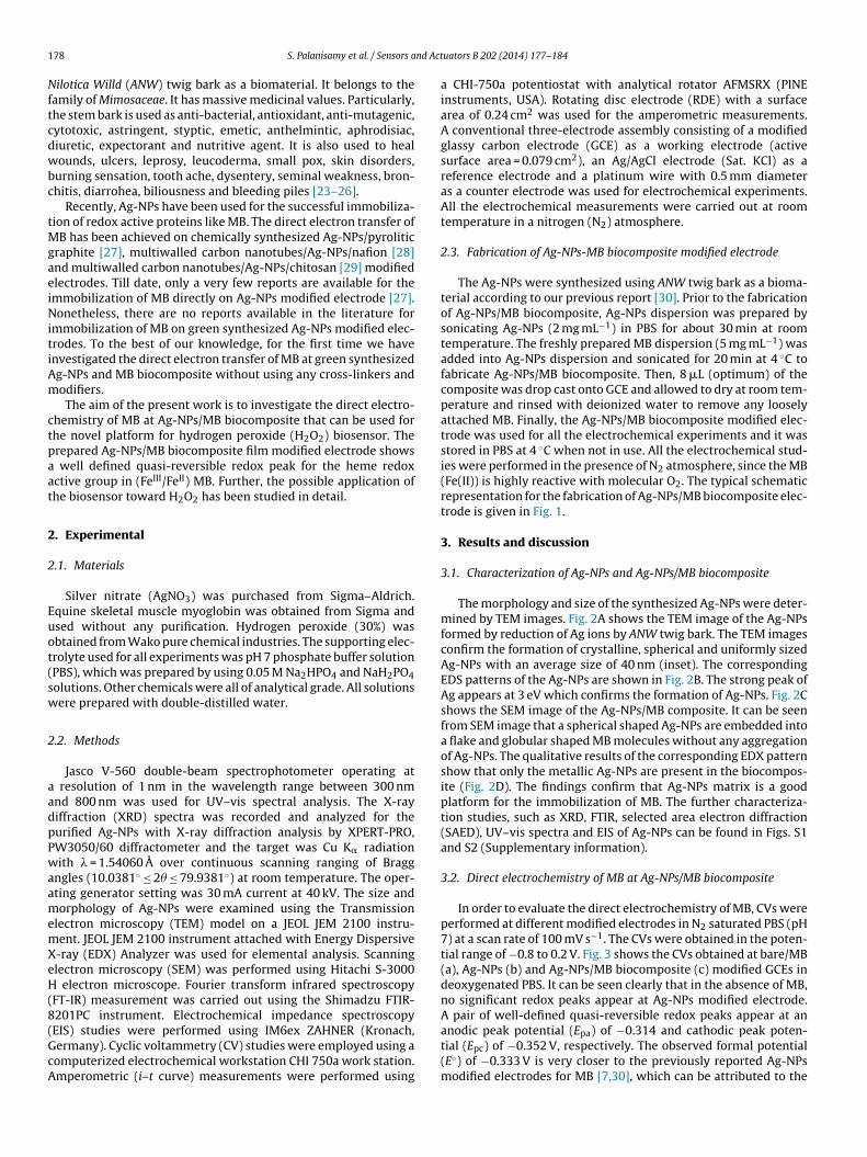
1 nd Act
Nftcdwbc
tMgaeiNitiAm
ctpaat
2
2
Euot(sw
2
aadpPwaamemXeH(8(GcA
78 S. Palanisamy et al. / Sensors a
ilotica Willd (ANW) twig bark as a biomaterial. It belongs to theamily of Mimosaceae. It has massive medicinal values. Particularly,he stem bark is used as anti-bacterial, antioxidant, anti-mutagenic,ytotoxic, astringent, styptic, emetic, anthelmintic, aphrodisiac,iuretic, expectorant and nutritive agent. It is also used to healounds, ulcers, leprosy, leucoderma, small pox, skin disorders,
urning sensation, tooth ache, dysentery, seminal weakness, bron-hitis, diarrohea, biliousness and bleeding piles [23–26].
Recently, Ag-NPs have been used for the successful immobiliza-ion of redox active proteins like MB. The direct electron transfer of
B has been achieved on chemically synthesized Ag-NPs/pyroliticraphite [27], multiwalled carbon nanotubes/Ag-NPs/nafion [28]nd multiwalled carbon nanotubes/Ag-NPs/chitosan [29] modifiedlectrodes. Till date, only a very few reports are available for themmobilization of MB directly on Ag-NPs modified electrode [27].onetheless, there are no reports available in the literature for
mmobilization of MB on green synthesized Ag-NPs modified elec-rodes. To the best of our knowledge, for the first time we havenvestigated the direct electron transfer of MB at green synthesizedg-NPs and MB biocomposite without using any cross-linkers andodifiers.The aim of the present work is to investigate the direct electro-
hemistry of MB at Ag-NPs/MB biocomposite that can be used forhe novel platform for hydrogen peroxide (H2O2) biosensor. Therepared Ag-NPs/MB biocomposite film modified electrode showswell defined quasi-reversible redox peak for the heme redox
ctive group in (FeIII/FeII) MB. Further, the possible application ofhe biosensor toward H2O2 has been studied in detail.
. Experimental
.1. Materials
Silver nitrate (AgNO3) was purchased from Sigma–Aldrich.quine skeletal muscle myoglobin was obtained from Sigma andsed without any purification. Hydrogen peroxide (30%) wasbtained from Wako pure chemical industries. The supporting elec-rolyte used for all experiments was pH 7 phosphate buffer solutionPBS), which was prepared by using 0.05 M Na2HPO4 and NaH2PO4olutions. Other chemicals were all of analytical grade. All solutionsere prepared with double-distilled water.
.2. Methods
Jasco V-560 double-beam spectrophotometer operating atresolution of 1 nm in the wavelength range between 300 nm
nd 800 nm was used for UV–vis spectral analysis. The X-rayiffraction (XRD) spectra was recorded and analyzed for theurified Ag-NPs with X-ray diffraction analysis by XPERT-PRO,W3050/60 diffractometer and the target was Cu K� radiationith � = 1.54060 A over continuous scanning ranging of Bragg
ngles (10.0381◦ ≤ 2� ≤ 79.9381◦) at room temperature. The oper-ting generator setting was 30 mA current at 40 kV. The size andorphology of Ag-NPs were examined using the Transmission
lectron microscopy (TEM) model on a JEOL JEM 2100 instru-ent. JEOL JEM 2100 instrument attached with Energy Dispersive-ray (EDX) Analyzer was used for elemental analysis. Scanninglectron microscopy (SEM) was performed using Hitachi S-3000
electron microscope. Fourier transform infrared spectroscopyFT-IR) measurement was carried out using the Shimadzu FTIR-201PC instrument. Electrochemical impedance spectroscopy
EIS) studies were performed using IM6ex ZAHNER (Kronach,ermany). Cyclic voltammetry (CV) studies were employed using aomputerized electrochemical workstation CHI 750a work station.mperometric (i–t curve) measurements were performed usinguators B 202 (2014) 177–184
a CHI-750a potentiostat with analytical rotator AFMSRX (PINEinstruments, USA). Rotating disc electrode (RDE) with a surfacearea of 0.24 cm2 was used for the amperometric measurements.A conventional three-electrode assembly consisting of a modifiedglassy carbon electrode (GCE) as a working electrode (activesurface area = 0.079 cm2), an Ag/AgCl electrode (Sat. KCl) as areference electrode and a platinum wire with 0.5 mm diameteras a counter electrode was used for electrochemical experiments.All the electrochemical measurements were carried out at roomtemperature in a nitrogen (N2) atmosphere.
2.3. Fabrication of Ag-NPs-MB biocomposite modified electrode
The Ag-NPs were synthesized using ANW twig bark as a bioma-terial according to our previous report [30]. Prior to the fabricationof Ag-NPs/MB biocomposite, Ag-NPs dispersion was prepared bysonicating Ag-NPs (2 mg mL−1) in PBS for about 30 min at roomtemperature. The freshly prepared MB dispersion (5 mg mL−1) wasadded into Ag-NPs dispersion and sonicated for 20 min at 4 ◦C tofabricate Ag-NPs/MB biocomposite. Then, 8 �L (optimum) of thecomposite was drop cast onto GCE and allowed to dry at room tem-perature and rinsed with deionized water to remove any looselyattached MB. Finally, the Ag-NPs/MB biocomposite modified elec-trode was used for all the electrochemical experiments and it wasstored in PBS at 4 ◦C when not in use. All the electrochemical stud-ies were performed in the presence of N2 atmosphere, since the MB(Fe(II)) is highly reactive with molecular O2. The typical schematicrepresentation for the fabrication of Ag-NPs/MB biocomposite elec-trode is given in Fig. 1.
3. Results and discussion
3.1. Characterization of Ag-NPs and Ag-NPs/MB biocomposite
The morphology and size of the synthesized Ag-NPs were deter-mined by TEM images. Fig. 2A shows the TEM image of the Ag-NPsformed by reduction of Ag ions by ANW twig bark. The TEM imagesconfirm the formation of crystalline, spherical and uniformly sizedAg-NPs with an average size of 40 nm (inset). The correspondingEDS patterns of the Ag-NPs are shown in Fig. 2B. The strong peak ofAg appears at 3 eV which confirms the formation of Ag-NPs. Fig. 2Cshows the SEM image of the Ag-NPs/MB composite. It can be seenfrom SEM image that a spherical shaped Ag-NPs are embedded intoa flake and globular shaped MB molecules without any aggregationof Ag-NPs. The qualitative results of the corresponding EDX patternshow that only the metallic Ag-NPs are present in the biocompos-ite (Fig. 2D). The findings confirm that Ag-NPs matrix is a goodplatform for the immobilization of MB. The further characteriza-tion studies, such as XRD, FTIR, selected area electron diffraction(SAED), UV–vis spectra and EIS of Ag-NPs can be found in Figs. S1and S2 (Supplementary information).
3.2. Direct electrochemistry of MB at Ag-NPs/MB biocomposite
In order to evaluate the direct electrochemistry of MB, CVs wereperformed at different modified electrodes in N2 saturated PBS (pH7) at a scan rate of 100 mV s−1. The CVs were obtained in the poten-tial range of −0.8 to 0.2 V. Fig. 3 shows the CVs obtained at bare/MB(a), Ag-NPs (b) and Ag-NPs/MB biocomposite (c) modified GCEs indeoxygenated PBS. It can be seen clearly that in the absence of MB,no significant redox peaks appear at Ag-NPs modified electrode.A pair of well-defined quasi-reversible redox peaks appear at an
anodic peak potential (Epa) of −0.314 and cathodic peak poten-tial (Epc) of −0.352 V, respectively. The observed formal potential(E◦) of −0.333 V is very closer to the previously reported Ag-NPsmodified electrodes for MB [7,30], which can be attributed to the
S. Palanisamy et al. / Sensors and Actuators B 202 (2014) 177–184 179
Fig. 1. A schematic representation of the typical procedure for fabrication of Ag-NPs/MB biocomposite.
Fig. 2. TEM images of green synthesized Ag-NPs (A), magnified view of Ag-NPs (inset A), corresponding EDX pattern of Ag-NPs (B), SEM image of Ag-NPs/MB biocomposite(C), inset shows the magnified view of C and consequent EDX profile of Ag-NPs/MB biocomposite (D).

180 S. Palanisamy et al. / Sensors and Act
Fig. 3. Cyclic voltammograms of bare/MB (a), Ag-NPs (b) and Ag-NPs/MB biocom-psc
pMipraiiHcpbwr
Faa(c
electrodes [28,33–37]. This result further indicates that Ag-NPsgreatly facilitate the direct electron transfer of MB toward theelectrode surface. The surface coverage concentration (� ) of MB
osite modified GCEs (c) in N2 saturated PBS at the scan rate of 100 mV s−1. Insethows the cyclic voltammetry response of Ag-NPs/MB modified GCE in the sameondition.
resence of redox active behavior of the heme couple (FeIII/FeII) inB. The peak-to-peak separation (�Ep) is found to be 38 mV, which
s smaller than that of previously reported MB immobilized com-osite modified electrodes [7,27–31]. Such a small value of �Ep
eveals that a fast and reversible electron transfer process occurst the composite modified electrode surface. This result furtherndicates that the good electron conductivity of Ag-NPs plays anmportant role for facilitating the direct electron transfer of MB.owever, bare/MB modified electrode does not show any electro-hemical signal for MB in the same potential window. We also
erformed direct drop casting of MB at Ag-NPs modified electrode,ut the appeared redox peak intensities are relatively very smallhen compared with Ag-NPs/MB biocomposite (inset). All theseesults clearly reveal that a good direct electron transfer of MB
ig. 4. Cyclic voltammogram of Ag-NPs/MB biocomposite modified GCE in thebsence (a) and presence of 25 �M (b) and 50 �M (c) of H2O2 in N2 saturated PBSt the scan rate of 100 mV s−1. Inset shows the cyclic voltammetry response of barea′), Ag-NPs (b′) and Ag-NPs/MB biocomposite (c′) modified GCEs in 100 �M of H2O2
ontaining N2 saturated PBS at the scan rate of 100 mV s−1.
uators B 202 (2014) 177–184
occurs only in Ag-NPs/MB biocomposite rather than dropping castdirectly on Ag-NPs modified electrode surface.
3.3. Effect of scan rate and pH
The effect of scan rate and pH at Ag-NPs/MB biocomposite modi-fied electrode was investigated using CV. Fig. S3A shows calibrationplot for the CV response of biocomposite modified GCE in PBS at dif-ferent scan rates (50–500 mV s−1). It can be seen that the redox peakcurrents (IPA and IPC) increase linearly with increasing scan rates.The obtained result suggests that MB immobilized Ag-NPs biocom-posite electrode undergoes a surface-controlled quasi-reversibleprocess [7,27]. When the peak to peak separation, �Ep > 200 mV,the average value of the heterogenous electron transfer rate con-stant (ks) at the Ag-NPs/MB biocomposite modified electrode iscalculated as 3.02 s−1 using Eq. (1) as stated by Laviron [32]:
Log ks = ˛ Log(1 − ˛) + (1 − ˛) − Log(
RT
nFv
)− ˛
(1 − ˛)nF�Ep
2.3RT(1)
where R = gas constant (8.314 J mol−1 K−1), T = the room tempera-ture (298.15 K), �Ep = peak-to-peak separation of the redox coupleand ˛ = 0.5. The number of electrons (n) transferred in the redoxreaction is 1. The calculated ks value is found to be higher thanthat of the previously reported MB immobilized other modified
Fig. 5. (A) Typical amperometric response of Ag-NPs/MB biocomposite modifiedRDE in N2 saturated PBS for different concentrations of H2O2 (1–3000 �M). Appliedpotential: −0.32 V. Inset shows the enlarged amperometric response of the biosen-sor in lower concentration additions. (B) Consequent calibration plot for reductionpeak current (Ipc) vs. [H2O2]. Error bar represents the relative standard deviation of3 replicate measurements.
nd Act
a1
�
wt((pua
A
tt(t5o0scnt
TC
S. Palanisamy et al. / Sensors a
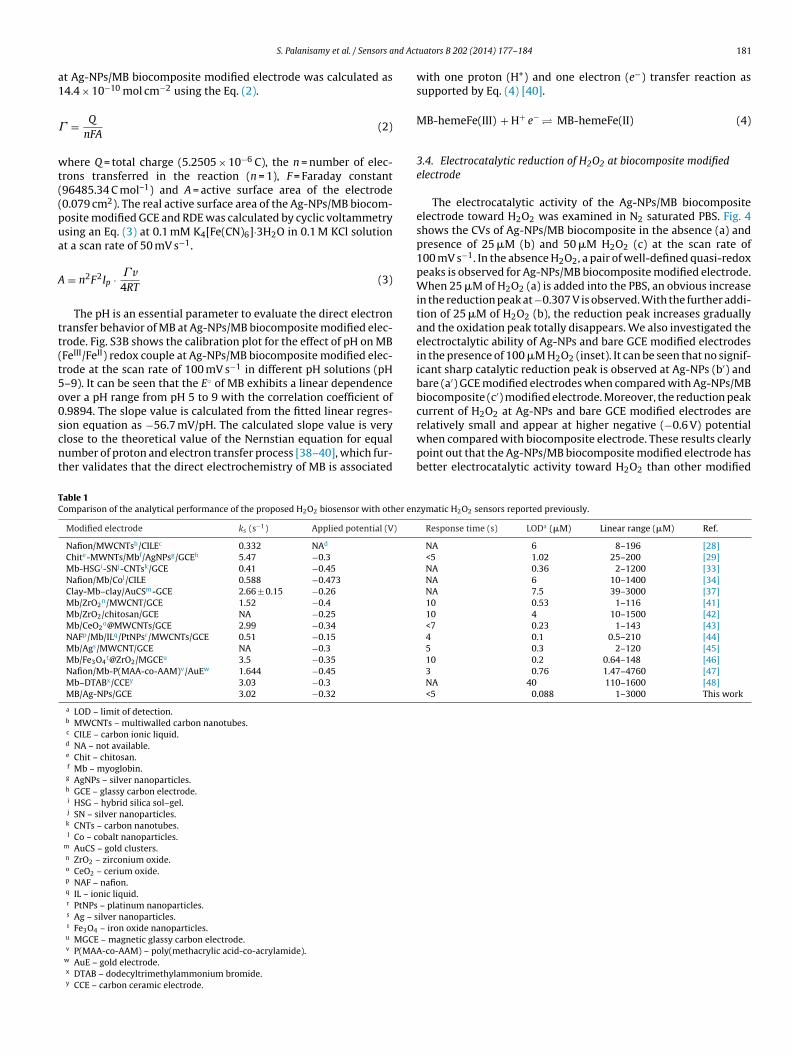
t Ag-NPs/MB biocomposite modified electrode was calculated as4.4 × 10−10 mol cm−2 using the Eq. (2).
= Q
nFA(2)
here Q = total charge (5.2505 × 10⁻⁶ C), the n = number of elec-rons transferred in the reaction (n = 1), F = Faraday constant96485.34 C mol–1) and A = active surface area of the electrode0.079 cm2). The real active surface area of the Ag-NPs/MB biocom-osite modified GCE and RDE was calculated by cyclic voltammetrysing an Eq. (3) at 0.1 mM K4[Fe(CN)6]·3H2O in 0.1 M KCl solutiont a scan rate of 50 mV s−1.
= n2F2Ip · � v4RT
(3)
The pH is an essential parameter to evaluate the direct electronransfer behavior of MB at Ag-NPs/MB biocomposite modified elec-rode. Fig. S3B shows the calibration plot for the effect of pH on MBFeIII/FeII) redox couple at Ag-NPs/MB biocomposite modified elec-rode at the scan rate of 100 mV s−1 in different pH solutions (pH–9). It can be seen that the E◦ of MB exhibits a linear dependencever a pH range from pH 5 to 9 with the correlation coefficient of.9894. The slope value is calculated from the fitted linear regres-
ion equation as −56.7 mV/pH. The calculated slope value is verylose to the theoretical value of the Nernstian equation for equalumber of proton and electron transfer process [38–40], which fur-her validates that the direct electrochemistry of MB is associatedable 1omparison of the analytical performance of the proposed H2O2 biosensor with other en
Modified electrode ks (s−1) Applied potential (V)
Nafion/MWCNTsb/CILEc 0.332 NAd
Chite-MWNTs/Mbf/AgNPsg/GCEh 5.47 −0.3Mb-HSGi-SNj-CNTsk/GCE 0.41 −0.45Nafion/Mb/Col/CILE 0.588 −0.473Clay-Mb–clay/AuCSm-GCE 2.66 ± 0.15 −0.26Mb/ZrO2
n/MWCNT/GCE 1.52 −0.4Mb/ZrO2/chitosan/GCE NA −0.25Mb/CeO2
o@MWCNTs/GCE 2.99 −0.34NAFp/Mb/ILq/PtNPsr/MWCNTs/GCE 0.51 −0.15Mb/Ags/MWCNT/GCE NA −0.3Mb/Fe3O4
t@ZrO2/MGCEu 3.5 −0.35Nafion/Mb-P(MAA-co-AAM)v/AuEw 1.644 −0.45Mb–DTABx/CCEy 3.03 −0.3MB/Ag-NPs/GCE 3.02 −0.32
a LOD – limit of detection.b MWCNTs – multiwalled carbon nanotubes.c CILE – carbon ionic liquid.d NA – not available.e Chit – chitosan.f Mb – myoglobin.g AgNPs – silver nanoparticles.h GCE – glassy carbon electrode.i HSG – hybrid silica sol–gel.j SN – silver nanoparticles.k CNTs – carbon nanotubes.l Co – cobalt nanoparticles.
m AuCS – gold clusters.n ZrO2 – zirconium oxide.o CeO2 – cerium oxide.p NAF – nafion.q IL – ionic liquid.r PtNPs – platinum nanoparticles.s Ag – silver nanoparticles.t Fe3O4 – iron oxide nanoparticles.u MGCE – magnetic glassy carbon electrode.v P(MAA-co-AAM) – poly(methacrylic acid-co-acrylamide).
w AuE – gold electrode.x DTAB – dodecyltrimethylammonium bromide.y CCE – carbon ceramic electrode.
uators B 202 (2014) 177–184 181
with one proton (H+) and one electron (e−) transfer reaction assupported by Eq. (4) [40].
MB-hemeFe(III) + H+ e− � MB-hemeFe(II) (4)
3.4. Electrocatalytic reduction of H2O2 at biocomposite modifiedelectrode
The electrocatalytic activity of the Ag-NPs/MB biocompositeelectrode toward H2O2 was examined in N2 saturated PBS. Fig. 4shows the CVs of Ag-NPs/MB biocomposite in the absence (a) andpresence of 25 �M (b) and 50 �M H2O2 (c) at the scan rate of100 mV s−1. In the absence H2O2, a pair of well-defined quasi-redoxpeaks is observed for Ag-NPs/MB biocomposite modified electrode.When 25 �M of H2O2 (a) is added into the PBS, an obvious increasein the reduction peak at−0.307 V is observed. With the further addi-tion of 25 �M of H2O2 (b), the reduction peak increases graduallyand the oxidation peak totally disappears. We also investigated theelectroctalytic ability of Ag-NPs and bare GCE modified electrodesin the presence of 100 �M H2O2 (inset). It can be seen that no signif-icant sharp catalytic reduction peak is observed at Ag-NPs (b′) andbare (a′) GCE modified electrodes when compared with Ag-NPs/MBbiocomposite (c′) modified electrode. Moreover, the reduction peakcurrent of H2O2 at Ag-NPs and bare GCE modified electrodes are
relatively small and appear at higher negative (−0.6 V) potentialwhen compared with biocomposite electrode. These results clearlypoint out that the Ag-NPs/MB biocomposite modified electrode hasbetter electrocatalytic activity toward H2O2 than other modifiedzymatic H2O2 sensors reported previously.
Response time (s) LODa (�M) Linear range (�M) Ref.
NA 6 8–196 [28]<5 1.02 25–200 [29]NA 0.36 2–1200 [33]NA 6 10–1400 [34]NA 7.5 39–3000 [37]10 0.53 1–116 [41]10 4 10–1500 [42]<7 0.23 1–143 [43]4 0.1 0.5–210 [44]5 0.3 2–120 [45]10 0.2 0.64–148 [46]3 0.76 1.47–4760 [47]NA 40 110–1600 [48]<5 0.088 1–3000 This work

1 nd Act
ec
M
2
3
AostaiepoTcoTrtcrc3rs0pmAeicstlwfs
3
ocaowSoaSo(tg
cr8id
[
[
[
[
82 S. Palanisamy et al. / Sensors a
lectrodes. The typical electrocatalytic reduction of H2O2 by MBan be expressed by following equations (Eqs. (5) and (6)).
B-Fe(III) + e → MB-HFe(II) (5)
MB-HFe(II) + H2O2 → 2MB-Fe(III) + 2H2O (6)
.5. Amperometric determination of H2O2
Fig. 5A shows the typical amperometric i–t curve obtained forg-NPs/MB biocomposite modified RDE upon successive additionf different concentrations of H2O2 in N2 saturated constantlytirred PBS. The electrode potential was held at −0.32 V. The reac-ion of common interference species like dopamine (DA), ascorbiccid (AA) and uric acid (UA) can be limited at this negative work-ng potential (−0.32 V). Moreover, the oxygen reduction in thelectrode surface can also be eliminated at such a low workingotential. Upon addition of H2O2, a good and well-defined amper-metric response is observed for the reduction of H2O2 by MB.he biocomposite modified electrode reaches its 95% steady stateurrent within 5 s, indicating the rapid electrocatalytic reductionf H2O2 by MB present in the biocomposite modified electrode.he Ag-NPs acts as a tunnel in the biocomposite between theedox active center (FeIII/FeII) of the MB and the electrode surfacehat leads to the sharp amperometric response and fast electro-atalytic reduction of H2O2 at the biocomposite electrode. Theeduction peak current increases linearly upon increasing H2O2oncentration and the linear response range is from 1 �M tomM (Fig. 5B). The sensitivity is calculated from the fitted linear
egression equation as 0.357 �A �M−1 cm−2 (slope value/effectiveurface area of RDE). A limit of detection (LOD) is found to be.088 �M [(3 × Sd of the blank response)/slope of the calibrationlot (Sd = 0.0025 �A and slope = 0.0856)]. The analytical perfor-ance (ks, response time, LOD and linear response range) of theg-NPs/MB biocomposite was compared with previously reportednzyme based H2O2 sensors and the comparison results are shownn Table 1. Table 1 is evident that the proposed H2O2 sensor is moreomparable with that of previously reported enzymatic H2O2 sen-ors [28,29,33,34,37,41–48]. The good electrocatalytic activity ofhe biosensor can be attributed to the excellent biocompatibility,arge active sites and good electron conducting ability of Ag-NPs
hich retain the native structure of the immobilized MB and greatlyacilitate the direct electron transfer of MB toward the electrodeurface.
.6. Selectivity of the biosensor
The selectivity of the electrode is more important for amper-metric measurements due to the interference of biologicaloexisting active species like ascorbic acid (AA), dopamine (DA)nd uric acid (UA). Therefore, we have investigated the selectivityf the proposed biosensor using the amperometic i–t method. Theorking conditions and parameters are similar to that described in
ection 3.5. The amperometric responses of 50 �M H2O2 (a), 1 mMf AA (b), 500 �M DA (c) and 1 mM UA (d) at different time intervalst Ag-NPs/MB biocomposite modified electrode are shown in Fig.4. It can be seen that a good reduction peak current is observedn addition of 10 �M H2O2 and the potentially interfering speciesAA, DA and UA) do not show any apparent current response afterheir injection into PBS. The obtained result clearly validates theood selectivity of fabricated biosensor.
The storage stability of the biosensor was investigated in PBSontaining 50 �M H2O2 at 4 ◦C and its reduction peak current
esponse was monitored periodically. The sensor retains about6.2% of its initial reduction peak current response after a week,ndicating the good storage stability of the sensor. The indepen-ently prepared 3 electrodes for the determination of 50 �M H2O2
[
[
uators B 202 (2014) 177–184
shows an acceptable reproducibility with the RSD of 3.2%. That therepeatability for 10 successive measurements with the RSD of 4.1%for the determination of 50 �M H2O2 indicates a good repeatabilityof the proposed biosensor.
4. Conclusions
In conclusion, a novel H2O2 biosensor using green synthesizedAg-NPs and MB as a model electrode has been demonstrated. Thefabricated Ag-NPs/MB biocomposite modified electrode shows agood direct electron transfer of MB and can be used for the construc-tion of H2O2 biosensor. A fast electron transfer of MB reveals thegood biocompatibility and tunable properties of green synthesizedAg-NPs. The fabricated biosensor exhibits a good linear response,quick response and better sensitivity for H2O2. Further, the supe-rior practicability of the biosensor can be used for the real timesensing of H2O2. In addition, the described procedure can be verymuch applied for immobilizing and investigating the other redoxactive enzymes in the near future.
Acknowledgments
This project was supported by the National Science Council andthe Ministry of Education of Taiwan (Republic of China). The author,PP, who is the second corresponding author of this article, thanksthe Management of Thiagarajar College, Madurai, Tamilnadu, Indiafor granting permission to have collaboration with National TaipeiUniversity of Technology, Taiwan.
Appendix A. Supplementary data
Supplementary material related to this article can be found, inthe online version, at http://dx.doi.org/10.1016/j.snb.2014.05.069.
References
[1] J.F. Rusling, Enzyme bioelectrochemistry in cast biomembrane-like films, Acc.Chem. Res. 31 (1998) 363–369.
[2] S.K. Arya, P.R. Solanki, M. Datta, B.D. Malhotra, Recent advances in self-assembled monolayers based biomolecular electronic devices, Biosens.Bioelectron. 24 (2009) 2810–2817.
[3] R. Mezzenga, J. Ruokolainen, Nanocomposites: nanoparticles in the right place,Nat. Mater. 8 (2009) 926–928.
[4] J.J. Feng, J.J. Xu, H.Y. Chen, Synergistic effect of zirconium phosphate and Aunanoparticles on direct electron transfer of hemoglobin on glassy carbon elec-trode, J. Electroanal. Chem. 585 (2005) 44–50.
[5] P.L. He, N.F. Hu, J.F. Rusling, Driving forces for layer-by-layer self-assembly offilms of SiO2 nanoparticles and heme proteins, Langmuir 20 (2004) 722–729.
[6] L. Zhang, D.B. Tia, J.J. Zhu, Direct electrochemistry and electrochemical catalysisof myoglobin–TiO2 coated multiwalled carbon nanotubes modified electrode,Bioelectrochemistry 74 (2008) 157–163.
[7] W. Suna, L. Li, B. Lei, T. Li, X. Ju, X. Wang, G. Li, Z. Sun, Fabrication ofgraphene–platinum nanocomposite for the direct electrochemistry and elec-trocatalysis of myoglobin, Mater. Sci. Eng. C 33 (2013) 1907–1913.
[8] Q. Lu, T. Zhou, S.S. Hu, Direct electrochemistry of hemoglobin in PHEA and itscatalysis to H2O2, Biosens. Bioelectron. 22 (2012) 899–904.
[9] X.Y. He, L. Zhu, Direct electrochemistry of hemoglobin in cetylpyridiniumbromide film: redox thermodynamics and electrocatalysis to nitric oxide, Elec-trochem. Commun. 8 (2006) 615–620.
10] C.S. Shan, H.F. Yang, D.X. Han, Q.X. Zhang, A. Ivaska, L. Niu,Graphene/AuNPs/chitosan nanocomposites film for glucose biosensing,Biosens. Bioelectron. 25 (2010) 1070–1074.
11] J.S. Kim, E. Kulk, K.N. Yu, J.H. Kim, S.J. Park, H.J. Lee, et al., Antimicrobial effectsof silver nanoparticles, Nanomedicine 3 (2007) 95–101.
12] A.M. Fayaz, M. Girilal, S.A. Mahdy, S.S. Somsundar, R. Venkatesan, P.T. Kalaichel-van, Vancomycin bound biogenic gold nanoparticles: a different perspective fordevelopment of anti VRSA agents, Process Biochem. 46 (2011) 636–641.
13] A.M. Fayaz, K. Balaji, P.T. Kalaichelvan, R. Venkatesan, Fungal based synthesis ofsilver nanoparticles – an effect of temperature on the size of particles, ColloidsSurf. B 74 (2009) 123–126.
14] A. Absar, M. Priyabrata, M. Deenadayal, S. Satyajyoti, M.I. Khan, K. Rajiv, et al.,Enzyme mediated extracellular synthesis of CdS nanoparticles by the fungus,Fusarium oxysporum, J. Am. Chem. Soc. 124 (2002) 12108–12109.
15] B. Vipul, R. Debabrata, A. Absar, S. Murali, Biosynthesis of zirconia nanoparticlesusing the fungus Fusarium oxysporum, J. Mater. Chem. 14 (2004) 3303–3305.
nd Act
[
[
[
[
[
[
[
[
[
[
[
[
[
[
[
[
[
[
[
[
[
[
[
[
[
[
[
[
[
[
[
[
[
thesis of nanoparticles for the application of antimicrobialactivity and bio-sensors. He is one of the active members
S. Palanisamy et al. / Sensors a
16] B. Vipul, R. Debabrata, B. Atul, A. Keda, S. Ambarish, A. Absar, et al., Fungus-mediated biosynthesis of silica and titania particles, J. Mater. Chem. 15 (2005)2583–2589.
17] S.O. Keun, S.K. Ree, L. Jinho, K. Dongmin, H.C. Sun, H.Y. Soon,Gold/chitosan/pluronic composite nanoparticles for drug delivery, J. Appl.Polym. Sci. 108 (2008) 3239–3244.
18] J.H. Amanda, C. Lei, L.K. William, P.V.D. Richard, Detection of a biomarker forAlzheimer’s disease from synthetic and clinical samples using a nanoscale opti-cal biosensor, J. Am. Chem. Soc. 127 (2005) 2264–2271.
19] A.M. Fayaz, C.S. Tiwary, P.T. Kalaichelvan, R. Venkatesan, Blue orange light emis-sion from biogenic synthesized silver nanoparticles using Trichoderma viride,Colloids Surf. B 75 (2009) 175–178.
20] A.M. Fayaz, K. Balaji, M. Girilal, R. Yadav, P.T. Kalaichelvan, R. Venkate-san, Biogenic synthesis of silver nanoparticles and their synergistic effectwith antibiotics: a study against Gram-positive and Gram-negative bacteria,Nanomedicine 6 (2010) 103–109.
21] A.M. Fayaz, K. Balaji, M. Girilal, P.T. Kalaichelvan, R. Venkatesan, Mycobasedsynthesis of silver nanoparticles and their incorporation into sodium algi-nate films for vegetable and fruit preservation, J. Agric. Food Chem. 57 (2009)6246–6252.
22] J. Zheng, G. Chumanov, T.M. Cotton, Photoinduced electron transfer at the sur-face of nanosized silver particles as monitored by EPR spectroscopy, Chem.Phys. Lett. 349 (2001) 367–370.
23] T. Kalaivani, L. Mathew, Free radical scavenging activity from leaves of Acacianilotica (L.) Wild. ex Delile, an Indian medicinal tree, Food Chem. Toxicol. 48(2010) 298–305.
24] K. Kaur, H. Michael, S. Arora, P. Harkonen, S. Kumar, In vitro bioactivity-guided fractionation and characterization of polyphenolic inhibitory fractionsfrom Acacia nilotica (L.) Willd. ex Del, J. Ethnopharmacol. 99 (2005)353–360.
25] B.N. Singh, B.R. Singh, R.L. Singh, D. Prakash, B.K. Sarma, H.B. Singh, Antioxidantand anti-quorum sensing activities of green pod of Acacia nilotica L., Food Chem.Toxicol. 47 (2009) 778–786.
26] S.K. Mitra, R. Sundaram, Antioxidant activity of ethyl acetate soluble fractionof Acacia arabica bark in rats, Indian J. Pharmacol. 39 (2007) 33–38.
27] X. Gan, T. Liu, J. Zhong, X. Liu, G. Li, Effect of silver nanoparticles on the elec-tron transfer reactivity and the catalytic activity of myoglobin, ChemBioChem5 (2004) 1686–1691.
28] W. Sun, X. Li, Y. Wang, X. Li, C. Zhao, K. Jiao, Electrochemistry of myoglobinin nafion and multi-walled carbon nanotubes modified carbon ionic liquidelectrode, Bioelectrochemistry 75 (2009) 170–175.
29] Y. Li, Y. Li, Y. Yang, Direct electrochemistry and electrocatalysis of myoglobin-based nanocomposite membrane electrode, Bioelectrochemistry 82 (2011)112–116.
30] C. Karuppiah, S. Palanisamy, S.M. Chen, R. Emmanuel, M.A. Ali, P. Muthukrish-nan, P. Prakash, F.M.A. Al-Hemaid, Green biosynthesis of silver nanoparticlesand nanomolar detection of p-nitrophenol, J. Solid State Electrochem. (2014),http://dx.doi.org/10.1007/s10008-014-2425-z.
31] L. Shen, N. Hu, Heme protein films with polyamidoamine dendrimer: directelectrochemistry and electrocatalysis, Biochim. Biophys. Acta 1608 (2004)23–33.
32] E. Laviron, Adsorption, autoinhibition and autocatalysis in polarography andin linear potential sweep voltammetry, J. Electroanal. Chem. Interfacial Elec-trochem. 52 (1979) 355–399.
33] C.Y. Liu, J.M. Hu, Hydrogen peroxide biosensor based on the direct electro-chemistry of myoglobin immobilized on silver nanoparticles doped carbonnanotubes film, Biosens. Bioelectron. 24 (2009) 2149–2154.
34] W. Sun, X.Q. Li, P. Qin, K. Jiao, Electrodeposition of Co nanoparticles on thecarbon ionic liquid electrode as a platform for myoglobin electrochemicalbiosensor, J. Phys. Chem. C 113 (2009) 11294–11300.
35] A.B. Moghaddam, M.R. Ganjali, R. Dinarvand, S. Ahadi, A.A. Saboury, Myoglobinimmobilization on electrodeposited nanometer-scale nickel oxide particlesand direct voltammetry, Biophys. Chem. 134 (2008) 25–33.
36] X.Q. Li, R.J. Zhao, Y. Wang, X.Y. Sun, W. Sun, C.Z. Zhao, K. Jiao, An electrochemicalbiosensor based on nafion-ionic liquid and a myoglobin-modified carbon pasteelectrode, Electrochim. Acta 55 (2010) 2173–2178.
37] X.J. Zhao, Z.B. Mai, X.H. Kang, Z. Dai, X.Y. Zou, Clay–chitosan–gold nanoparticlenanohybrid: preparation and application for assembly and direct electrochem-istry of myoglobin, Electrochim. Acta 53 (2008) 4732–4739.
38] E. Laviron, General expression of the linear potential sweep voltammogramin the case of diffusionless electrochemical systems, J. Electroanal. Chem. 101(1979) 19–28.
39] X.L. Chen, N.F. Hu, Y.H. Zeng, J.F. Rusling, J. Yang, Ordered electrochemicallyactive films of hemoglobin, didodecyldimethylammonium ions, and clay, Lang-muir 15 (1999) 7022–7030.
40] H. Sun, N.F. Hu, H.Y. Ma, Electrochemical and UV–vis spectroscopic measure-ments of nitric oxide in fibroblasts and astrocytes, Electroanalysis 12 (2000)1064–1070.
41] R. Liang, M. Deng, S. Cui, H. Chen, J. Qiu, Direct electrochemistry and electro-catalysis of myoglobin immobilized on zirconia/multi-walled carbon nanotubenanocomposite, Mater. Res. Bull. 45 (2010) 1855–1860.
42] G. Zhao, J.J. Feng, J.J. Xu, H.Y. Chen, Direct electrochemistry and electrocataly-sis of heme proteins immobilized on self-assembled ZrO2 film, Electrochem.Commun. 7 (2005) 724–729.
43] J.D. Qiu, S.G. Cui, R.P. Liang, Hydrogen peroxide biosensor based on the directelectrochemistry of myoglobin immobilized on ceria nanoparticles coated
uators B 202 (2014) 177–184 183
with multiwalled carbon nanotubes by a hydrothermal synthetic method,Microchim. Acta 171 (2010) 333–339.
44] A. Babaei, D.J. Garrett, A.J. Downard, Electrochemical investigations on athird generation biosensor for determination of hydrogen peroxide basedon immobilization of myoglobin on a novel platinum nanoparticle/carbonnanotube/ionic liquid/nafion composite, Int. J. Electrochem. Sci. 7 (2012)3141–3154.
45] B. Liu, M. Wang, Hydrogen peroxide biosensor based on myoglobin immo-bilized on silver nanoparticles decorated oxidized multi-walled carbonnanotubes, Int. J. Electrochem. Sci. 8 (2013) 9801–9810.
46] H.P. Peng, R.P. Liang, L. Zhang, J.D. Qiu, Sonochemical synthesis of magneticcore–shell Fe3O4@ZrO2 nanoparticles and their application to the highly effec-tive immobilization of myoglobin for direct electrochemistry, Electrochim. Acta56 (2011) 4231–4236.
47] K. Zhan, H. Liu, H. Zhang, Y. Chen, H. Ni, M. Wu, D. Sun, Y. Chen, Afacile method for the immobilization of myoglobin on multi-walled car-bon nanotubes: poly(methacrylic acid-co-acrylamide) nanocomposite and itsapplication for direct bio-detection of H2O2, J. Electroanal. Chem. (2014),http://dx.doi.org/10.1016/j.jelechem.2014.04.012.
48] Y.Z. Zhou, H. Wang, S.Y. Dong, A.X. Tian, Z.X. He, B. Chen, Direct electro-chemistry and electrocatalysis of myoglobin in dodecyltrimethylammoniumbromide film modified carbon ceramic electrode, Chin. Chem. Lett. 22 (2011)465–468.
Biographies
Selvakumar Palanisamy received his B.Sc. (2006), M.Sc.(2009) and M.Phil. (2010) degrees in Chemistry fromMadurai Kamaraj University, Tamilnadu, India. Now, heis a fourth year Ph.D. student in Chemical Engineer-ing and Biotechnology at National Taipei University ofTechnology. He is currently working on the synthesis ofnanomaterials and applications related to the electro-chemical sensors and biosensors.
Chelladurai Karuppiah received his B.Sc. Degree inChemistry from Bharathiar University, Tamilnadu, India,in 2007. He obtained his M.Sc. degree in 2009 and M.Phil.degree in 2011 from Madurai Kamaraj University. Now,he is a second year Ph.D. student in Chemical Engineer-ing and Biotechnology at National Taipei University ofTechnology. He specializes in nanomaterials synthesis forenzyme immobilization related to biosensor and biofuelcell applications.
Shen-Ming Chen received his B.S. degree in Chemistryin 1980 from National Kaohsiung Normal University,Taiwan. He received his M.S. degree (1983) and Ph.D.degree (1991) in Chemistry from National Taiwan Univer-sity, Taiwan. He is currently a professor at the Departmentof Chemical Engineering and Biotechnology, NationalTaipei University of Technology, Taiwan. His currentresearch interests include electroanalytical chemistry,bioelectrochemistry, fabrication of energy conservationand storage devices and nanomaterial synthesis for elec-trochemical applications. He has published more than 280research articles in SCI journals.
R. Emmanuel completed his M.Sc. Chemistry in Bharatid-hasan University, Tiruchirappalli, India. He is now workingas a full time research scholar in the Post Graduate andResearch Department of Chemistry, Thiagarajar College,Madurai, India. His research areas include the green syn-
of the Indian Nanobiologists Association.

1 nd Act
84 S. Palanisamy et al. / Sensors aP. Muthukrishnan is currently working as a full timeresearch scholar in the Post Graduate and Research
Department of Chemistry, Thiagarajar College, Madurai,India. He received his M.Phil. degree from Bharathid-hasan University, Tiruchirappalli, India. His research areasinclude corrosion mitigation and nanoparticles synthesisfor the application of biosensors.uators B 202 (2014) 177–184
P. Prakash completed his Ph.D. in Anna University,Chennai, India in the field of Biomimetics. He is work-ing as an Assistant Professor in the Post Graduate andResearch Department of Chemistry, Thiagarajar College,Madurai, India. His research areas include Biomimetics,Nanotechnology, Corrosion, Biosensors, Reaction Kineticsand Environmental Science. He is a visiting professor of
Dr. Shen-Ming Chen group in the Electroanalysis and Bio-chemistry Lab, Department of Chemical Engineering andBiotechnology at National Taipei University of Technol-ogy, Taipei, Taiwan. He is one of the Board of ExecutiveMembers of Indian Nanobiologists Association.