Embed Size (px)
Citation preview

1
BREAKTHROUGH REPORT 1 2
Direct and Indirect Visualization of Bacterial Effector Delivery into Diverse 3
Plant Cell Types During Infection 4
5
6 Elizabeth Henry1, Tania Y. Toruño1†, Alain Jauneau3†, Laurent Deslandes2, Gitta Coaker1* 7
8 1Department of Plant Pathology, University of California, Davis, California 95616, USA. 9 2LIPM, Université de Toulouse, INRA, CNRS, UPS, Castanet-Tolosan, France.10 3Institut Fédératif de Recherche 3450, Université de Toulouse, CNRS, UPS, Plateforme Imagerie 11
TRI-Genotoul, Castanet-Tolosan 31326, France 12
† These authors contributed equally to this work. 13
*Correspondence to: [email protected]
15 Short title: Visualizing effector delivery in plants 16
17 One-sentence summary: The GFP strand system enables spatial and temporal visualization of 18
bacterial effector delivery during infection. 19
20 The author responsible for distribution of materials integral to the findings presented in this 21
article in accordance with the policy described in the Instructions for Authors 22
(www.plantcell.org) is: Gitta Coaker ([email protected]). 23
24
ABSTRACT 25 26
To cause disease, diverse pathogens deliver effector proteins into host cells. Pathogen effectors 27
can inhibit defense responses, alter host physiology, and represent important cellular probes to 28
investigate plant biology. However, effector function and localization have primarily been 29
investigated after overexpression in planta. Visualizing effector delivery during infection is 30
challenging due to the plant cell wall, autofluorescence, and low effector abundance. Here, we 31
utilized a GFP strand system to directly visualize bacterial effectors delivered into plant cells 32
through the Type III secretion system. GFP is a beta barrel that can be divided into 11 strands. 33
We generated transgenic Arabidopsis thaliana plants expressing GFP1-10 (strands 1 to 10). 34
Multiple bacterial effectors tagged with the complementary strand 11 epitope retained their 35
biological function in Arabidopsis and tomato (Solanum lycopersicum). Infection of plants 36
expressing GFP1-10 with bacteria delivering GFP11-tagged effectors enabled direct effector 37
detection in planta. We investigated the temporal and spatial delivery of GFP11-tagged effectors 38
during infection with the foliar pathogen Pseudomonas syringae and the vascular pathogen 39
Ralstonia solanacearum. Thus, the GFP strand system can be broadly used to investigate effector 40
biology in planta. 41
42
43
44
INTRODUCTION 45
Plant Cell Advance Publication. Published on June 9, 2017, doi:10.1105/tpc.17.00027
©2017 American Society of Plant Biologists. All Rights Reserved

2
46
Plants can be infected by all classes of pathogens and rely on their innate immune system 47
to recognize and respond to invading organisms (Henry et al., 2013). An important aspect of 48
pathogenicity is the delivery of pathogen proteins, termed effectors, into host cells (Toruno et al., 49
2016). Effectors can modulate host metabolism, shut down host defense signaling, and suppress 50
cell death (Toruno et al., 2016). Gram-negative bacterial pathogens such as the foliar pathogen 51
Pseudomonas syringae and the vascular pathogen Ralstonia solanacearum use the Type III 52
secretion system (TTSS), a proteinaceous needle-like structure, to directly deliver effectors 53
inside host cells (Chang et al., 2014). Mutations in core components of the TTSS, such as the 54
homopolymeric ring hrcC, block the ability to cause disease (Deng et al., 1998; Vasse et al., 55
2000). Plants have evolved intracellular immune receptors with nucleotide binding leucine-rich 56
repeat (NLR) domain architecture that specifically recognize pathogen effectors leading to 57
effector-triggered immunity (ETI) (Chiang and Coaker, 2015). A hallmark of ETI is the 58
hypersensitive response (HR), a specialized form of programmed cell death. Much of our 59
understanding of the plant innate immune system has been gained through investigation of the 60
model plant Arabidopsis thaliana, which can be infected by pathogens with diverse tissue 61
preferences, including P. syringae and R. solanacearum. 62
Intracellular delivery of GFP-tagged effectors from the fungal pathogen, Magnaporthe 63
oryzae, has been successfully visualized inside plant cells (Khang et al., 2010). However, this 64
approach has not been successful for other fungal pathogens, possibly due to the GFP tag, which 65
can interfere with effector delivery or function, or due to low-level effector expression (Tanaka 66
et al., 2015). Despite the importance of bacterial effectors in the modulation of host-microbe 67
interactions, direct TTSS effector delivery has not been visualized in whole organisms. Recently, 68
delivery of the virulence protein VirE2 by Agrobacterium tumefaciens through the Type IV 69
secretion system was successfully visualized using the GFP strand system in yeast, Arabidopsis 70
and tobacco (Li et al., 2014; Li et al., 2017; Yang et al., 2017). The Type IV secretion system is 71
responsible for delivery and uptake of proteins and DNA, with a conduit diameter of about 18.5 72
nm (Wallden et al., 2010). Size constraints of the inner needle (2-3 nm conduit) of the TTSS 73
require proteins to be unfolded prior to secretion and fusion with a full-length fluorophore would 74
likely block delivery (Akeda and Galan, 2005; Chang et al., 2014). Bacterial effector delivery by 75
the TTSS has been studied indirectly in plant genotypes during ETI, via cell viability staining 76

3
during the HR (Torres et al., 2002; Greenberg and Yao, 2004). However, due to the spread of 77
defense signaling by apoplastic reactive oxygen species (ROS) and potentially other small 78
molecules that can move through plasmodesmata (including some pathogen effectors), it is 79
impossible to determine which cells are direct recipients of bacterial effectors, or which host 80
cells are capable of directly recognizing effectors (Allan and Fluhr, 1997; Torres et al., 2002; 81
Greenberg and Yao, 2004; Khang et al., 2010). Additionally, effector detection in the host has 82
relied primarily on overexpression in Nicotiana benthamiana or Arabidopsis, but these 83
approaches may not reflect their subcellular localization/targeting and accumulation under 84
natural infection. Thus, multiple questions remain regarding which cells are targeted for effector 85
delivery and where effectors localize within the host cell during infection. 86
Here, we investigated cell-specific immune responses and used the GFP strand system to 87
directly visualize the delivery of bacterial effectors in planta. This approach allowed direct 88
visualization of the P. syringae effectors AvrPto, AvrPtoB, and AvrB delivered into diverse leaf 89
cell types during natural infection and visualization of the R. solanacearum effector PopP2. The 90
GFP strand system enables the investigation of effector biology during natural infection in intact 91
plants and our findings provide insight to the overlap of cell-type-specific immune responses and 92
patterns of effector delivery. 93
94
RESULTS 95
Diverse cell types in Arabidopsis leaves recognize and respond to the bacterial effector 96
AvrB 97
To investigate the capacity of various leaf cell types to recognize and respond to 98
pathogenic bacteria, cell death was used as a proxy for effector recognition. The HR was 99
visualized using trypan blue, a vital stain that selectively accumulates in dead cells turning them 100
blue (van Wees, 2008). In Arabidopsis, the RPM1 NLR immune receptor recognizes 101
phosphorylation of the plant protein RIN4 induced in the presence of the AvrB and AvrRpm1 102
effectors (Chung et al., 2011; Liu et al., 2011). The RPS2 NLR immune receptor recognizes 103
cleavage and elimination of RIN4 by the AvrRpt2 effector protease (Axtell and Staskawicz, 104
2003; Mackey et al., 2003). Dip inoculation of Arabidopsis Col-0 with virulent P. syringae pv. 105
tomato (Pst) DC3000 carrying an empty vector (EV) did not result in trypan blue staining of leaf 106
cells 14 hours post-inoculation (hpi) (Figure 1A). In contrast, Col-0 infection with Pst carrying 107
4
the AvrB effector resulted in activation of RPM1-mediated resistance and accumulation of the 108
trypan blue stain in the epidermal pavement and guard cells as well as internal mesophyll cells 109
(Figure 1A). 110
To probe the ability of specific cell types to elicit ETI responses, we complemented the 111
Arabidopsis double mutant rps2-101C/rin4 (r2r4) with genomic T7-tagged RIN4 driven by 112
previously published cell-specific promoters (CSPs) for guard cells (pGC1), epidermal cells 113
(pCER6), and mesophyll cells (pCAB3) (Yang et al., 2008; Ranjan et al., 2011) (Figure 1B). 114
Expression of RIN4 in the CSP lines as compared to Col-0 was determined by anti-RIN4 115
immunoblotting (Supplemental Fig. 1). In order to enable detection of RIN4 in transgenic lines 116
using different CSPs, protein loading for the Col-0 control was diluted by half to avoid 117
overexposure. RIN4 expression in the cell-specific transgenic lines was still less than the diluted 118
Col-0 sample. Lines were dip inoculated with Pst DC3000 (avrB) and assessed for the ability to 119
elicit a microscopic HR by trypan blue staining (Figure 1B). In the r2r4 background, RPM1 is 120
not functional due to the absence of RIN4 (Mackey et al., 2002). As expected, the r2r4 line did 121
not elicit an HR and was not stained by trypan blue, whereas the rps2-101C single mutant (r2) 122
used as a positive control displayed an HR in all cell types (Figure 1C). When the rin4 mutation 123
is complemented in a cell-specific manner, single-cell HR is also detected in RIN4 expressing 124
cell types using trypan blue staining (Figure 1C). 125
In order to visualize macroscopic HR, we infiltrated the same Arabidopsis genotypes 126
with a high dose of Pst DC3000 (EV) or (avrB) (Figure 1D). After Pst DC3000 (avrB) 127
inoculation we could visualize macroscopic HR spread throughout the infiltrated area in all lines 128
containing RIN4 (Figure 1D). Infiltration with Pst DC3000 (EV) did not induce an HR in any 129
lines. To quantify macroscopic HR in the CSP lines, we measured ion leakage, a proxy for cell 130
death (Henry et al., 2015). Consistent with the limited expression of RIN4 in different cell types, 131
we found the ion leakage in the CSP lines was reduced with respect to the positive controls Col-0 132
and r2 after infiltration with Pst DC3000 (avrB) (Figure 1E). Together, these results indicate that 133
discrete cell types within the leaf tissue are capable of responding to recognized effectors and 134
may propagate cell death signals across tissues either through plasmodesmata linkages or by 135
apoplastic ROS signaling. Alternatively, there may be a small amount of RIN4 expressed in other 136
cells in CSP lines that is sufficient to induce cell death after high density inoculation. 137
138

5
The GFP strand system functions in Nicotiana benthamiana and effector function is 139
retained when fused with GFP strand 11 140
Although diverse cell types are able to recognize AvrB, this does not demonstrate direct 141
bacterial effector delivery into these cells (Figure 1). To visualize bacterial effector delivery in 142
planta we adopted the GFP strand system based on spontaneous assembly of two complementary 143
GFP fragments (Cabantous et al., 2005). GFP is a beta barrel protein and can be divided into 11 144
strands (Cabantous et al., 2005). The success of the GFP strand system for detection of type three 145
secreted proteins was previously demonstrated for the Salmonella effectors PpB2 and SteA into 146
HeLa cell culture, indicating that the strand 11 tag does not interfere with bacterial effector 147
delivery in Salmonella (Van Engelenburg and Palmer, 2010). We used a superfolder GFP 148
variant, which exhibits enhanced solubility, folding, and fluorescence (Cabantous et al., 2005; 149
Pedelacq et al., 2006). The small 16 amino acid eleventh strand (RDHMVLHEYVNAAGIT) 150
was fused via a flexible linker (GDGGSGGGS) on the C-terminus of the P. syringae effectors 151
AvrB, AvrPto, and AvrPtoB (Figure 2A). When the effector-GFP11 fusion is co-expressed with 152
GFP1-10, the two complementary GFP fragments should spontaneously associate/self-assemble to 153
form a functional GFP molecule (Figure 2A). First, we demonstrated proof-of-concept for the 154
GFP strand system using Agrobacterium-mediated transient plant expression system in N. 155
benthamiana. GFP1-10 and the individual effector-GFP11 constructs were transiently expressed in 156
N. benthamiana cells (Figure 2B). Expression of GFP1-10 alone did not result in fluorescence, 157
whereas co-expression with effector-GFP11 resulted in clear GFP fluorescence (Figure 2B). 158
Expression of full-length GFP1-11 also resulted in clear fluorescence. Plasmolysis revealed that 159
effector subcellular localization was consistent with previous reports: AvrB-GFP11 and AvrPto-160
GFP11 were detected at the plasma membrane with visible Hechtian strands after plasmolysis and 161
AvrPtoB-GFP11 exhibited cytosolic localization (Nimchuk et al., 2000; de Vries et al., 2006) 162
(Figure 2B). 163
Pst DC3000 infects both tomato and Arabidopsis, causing bacterial speck disease. In 164
resistant tomato genotypes such as the cultivar Rio Grande (RG) 76R, AvrPto and AvrPtoB both 165
interact with tomato protein kinase Pto, which is guarded by the tomato NLR Prf (Martin et al., 166
1993; Salmeron et al., 1996). In the susceptible tomato genotype RG prf3, AvrPto and AvrPtoB 167
promote bacterial virulence (Martin et al., 1993; Lin and Martin, 2005). In order to verify that 168
effector fusion to GFP strand 11 does not impact effector delivery and function, inoculations 169

6
were performed in tomato using Pst expressing AvrPto- GFP11 and AvrPtoB- GFP11. Effectors 170
were cloned into the pBBR1 plasmid under the control of their native promoters and expressed 171
with a C-terminal fusion to GFP strand 11 as described above (Kovach et al., 1995). The 172
resulting plasmids were transformed into Pst DC3000. Since AvrPto and AvrPtoB are 173
functionally redundant with respect to the host ETI response, they were singly complemented 174
into a Pst double deletion mutant for both effectors (DC3000 ΔavrPto/ΔavrPtoB) (Lin and 175
Martin, 2005). Pst DC3000 ΔavrPto/ΔavrPtoB caused disease on both RG and RG prf3 although 176
symptoms were attenuated compared to wild-type Pst DC3000 (Figure 3A, B). Complementation 177
of DC3000 ΔavrPto/ΔavrPtoB with either AvrPto-GFP11 or AvrPtoB-GFP11 resulted in recovery 178
of recognition and resistance in the RG background (Figure 3A, B). Bacterial titers were assessed 179
at four days post-dip inoculation. Complementation of Pst DC3000 ΔavrPto/ΔavrPtoB with 180
AvrPto-GFP11 leads to a complete recovery of virulence and bacterial titers in RG prf3 (Figure 181
3A-B). Complementation of Pst DC3000 ΔavrPto/ΔavrPtoB with AvrPtoB-GFP11 enhanced 182
disease symptoms, but did not result in a significant increase in bacterial virulence in RG prf3 183
(Figure 3A-B). Previously, AvrPtoB was demonstrated to enhance DC3000 disease symptoms 184
but not bacterial titers at four days post-inoculation (Lin and Martin, 2005). These data 185
demonstrate that both AvrPto and AvrPtoB effectors are able to be delivered into plant cells via 186
the TTSS and can be recognized by the plant innate immune system when fused to GFP strand 187
11. 188
In order to verify that fusion to GFP strand 11 does not impact AvrB delivery and 189
function, inoculations were performed in Arabidopsis using Pst DC3000 expressing AvrB-190
GFP11. AvrB is recognized by the RPM1 NLR in the Arabidopsis Col-0 ecotype (Boyes et al., 191
1998). The ability of AvrB-GFP11 to elicit RPM1-mediated responses was assessed using trypan 192
blue staining as well as bacterial growth assays. To assess macroscopic HR, leaves of Col-0 193
plants were syringe infiltrated with a bacterial suspension of either Pst DC3000 (EV) or Pst 194
DC3000 (avrB-GFP11). Infection with Pst DC3000 (avrB-GFP11) elicited a robust macroscopic 195
HR, visualized by trypan blue at 24hpi (Figure 3C). Bacterial growth assays demonstrate that 196
AvrB-GFP11 is delivered and recognized by RPM1, as the bacterial growth of Pst DC3000 197
(avrB-GFP11) was attenuated at 4 days post-inoculation compared to DC3000 EV on Col-0 but 198
not the rpm1-3 mutant line (Figure 3D-E). Taken together, these results indicate that effector-199

7
GFP11 fusions, when delivered by the TTSS of Pst DC3000, are recognized by host NLR 200
immune receptors and can promote bacterial virulence in susceptible genetic backgrounds. 201
202
Effectors are delivered into multiple cell types in Arabidopsis leaves 203
To visualize effector delivery during natural infection, we infiltrated Pst DC3000 strains 204
carrying the complementary GFP strand 11 tagged effectors into homozygous transgenic 205
Arabidopsis lines expressing 35S:GFP1-10 (Supplemental Figure 2). To avoid the cell death 206
elicited by RPM1-mediated recognition of AvrB in wild-type Col-0, rpm1-3/rps2-101C mutant 207
plants (r1r2) lacking both RPM1 and RPS2 NLR receptors expressing GFP1-10 were used to 208
analyze AvrB-GFP11 delivery. The following Pst genotypes were used to detect bacterial effector 209
delivery in the Col-0 background: DC3000 ΔavrPto/ΔavrPtoB + avrPto-GFP11, DC3000 210
ΔavrPto/ΔavrPtoB + avrPtoB-GFP11, DC3000 ΔavrPto/ΔavrPtoB (negative control), DC3000 211
ΔhrcC + avrPtoB-GFP11 (negative control), and DC3000 ΔhrcC + avrPto-GFP11 (negative 212
control). The following Pst genotypes were used to detect bacterial effector delivery in the r1r2 213
background: DC3000 + avrB-GFP11 and DC3000 EV (negative control). Confocal micrographs 214
were acquired with the Zeiss LSM710 confocal microscope. We analyzed four different 215
Arabidopsis plants for each bacterial strain. Two inoculation methods were assessed: syringe 216
(Figure 4) and surface inoculation (Figure 5). 217
Effector delivery was first assessed after syringe inoculation of four-week-old 218
Arabidopsis leaves with Pst DC3000 carrying each of the three GFP11 tagged effectors. We 219
examined and quantified confocal micrographs taken from four biological replicates to record the 220
temporal and spatial delivery of AvrB-GFP11, AvrPto-GFP11 and AvrPtoB-GFP11 at 24 and 221
48hpi. At 24hpi, effector delivery events were detected in mesophyll cells for AvrB-GFP11 and 222
AvrPto-GFP11 (Supplemental Figure 3, Figure 4D). Delivery events of AvrB-GFP11 and AvrPto-223
GFP11 effectors at this time point were visualized as small foci at the cell periphery in both 224
mesophyll and epidermal pavement cells (Supplemental Figure 3). At 24 hpi, AvrPtoB-GFP11 225
was not detected in mesophyll cells but a small amount of fluorescence was observed in the 226
epidermal pavement cells and the GFP signal had a more diffuse localization around the cell 227
periphery than the other two effectors (Supplemental Figure 3, Figure 4D). By 48hpi, we 228
observed robust delivery of all three effectors in both mesophyll and epidermal pavement cells 229
(Figure 4A, B). The number of cells positive for GFP fluorescence was greater for the effectors 230

8
AvrB-GFP11 and AvrPto-GFP11 than for AvrPtoB-GFP11, suggesting enhanced effector delivery 231
or stability (Figure 4D). At 48 hpi, fluorescent stretches of effectors were observed, indicating 232
effector accumulation over time (Figure 4A-B). Surprisingly, the highest frequency of delivery 233
events was not in the mesophyll, but the epidermal pavement cells (Figure 4D). GFP 234
fluorescence localized to the cell periphery was observed at both 24 and 48hpi (Supplemental 235
Figure 3 and Figure 4B). The AvrB-GFP11 signal was strongest of the three effectors 236
investigated, and was frequently detected at junctions between pavement cells. Delivery into 237
adjacent pavement cells could be detected as parallel stretches of GFP fluorescence (Figure 4B 238
inset). To clearly demonstrate separation of the GFP signal and chlorophyll autofluorescence, we 239
used the Zeiss ZEN lite software to create 3D projections of representative Z-stacks for each 240
effector (Figure 4C). These projections further establish a predominance of GFP signal in the 241
epidermal layer. This may reflect a preference for effector delivery at the leaf surface or it may 242
be a consequence of signal reduction when attempting to move the focal plane more deeply into 243
the leaf interior. Expression of GFP1-10 under the control of 35S promoter within individual cell 244
types may vary and contribute to an observational bias for effector delivery in cells with higher 245
expression of the transgene. We did not detect GFP fluorescence after inoculation with the 246
control Pst strains DC3000 EV, DC3000 ΔhrcC carrying effector-GFP11 constructs, or DC3000 247
ΔavrPto/ΔavrPtoB, indicating that the fluorescence signal was specifically detecting effector 248
delivery (Figure 4A, B). 249
Syringe inoculation delivers bacteria directly to the apoplast. Surface inoculation (dip or 250
spray inoculation) more closely mimics natural infection conditions, enables epiphytic growth on 251
the surface of leaves, and facilitates the detection of early invasion events into the leaf interior 252
(Katagiri et al., 2002). Due to the robust delivery of AvrB-GFP11 and AvrPto-GFP11 and their 253
plasma membrane localization (effectively concentrating the fluorescent signal), we chose to 254
focus on these two effectors for surface inoculation experiments. In surface-inoculated leaves, 255
effector delivery events were primarily detected in epidermal pavement and guard cells (Figure 256
5A-C). Guard cells can exhibit autofluorescence in their inner walls flanking the stomatal pore. 257
Therefore, guard cells only exhibiting fluorescence in their inner walls were not included in the 258
quantification of effector delivery. Effector delivery was detected when fluorescence occurred at 259
the guard cell outer edge, which is adjacent to the surrounding epidermal pavement cells. 260
Effector delivery into pavement cells gave characteristic stretches of GFP fluorescence at the cell 261

9
periphery, while delivery events into guard cells appeared as more discretely localized in puncta 262
(Figures 5B). It is possible that effector delivery is initially concentrated as foci at the membrane 263
near the tip of the TTSS needle before spreading along the plasma membrane, as previously 264
described in vitro (Jin and He, 2001). Although we were able to visualize AvrB-GFP11 delivered 265
into mesophyll cells at 24hpi, AvrPto-GFP11 could not be detected in mesophyll cells after 266
surface inoculation at 24 or 48hpi (Supplemental Figure 4, Figure 5A and C). Regardless of the 267
inoculation method, we observed that epidermal pavement cells exhibited the highest number of 268
cells positive for effector delivery (Figure 4C, 5C-D). These data indicate that pavement cells are 269
sites for effector delivery by Pseudomonas. These findings are supported by previously 270
published work, which demonstrated that transcription of avrPto in Pseudomonas was highest on 271
the leaf surface when bacteria were in contact with pavement cells (Lee et al., 2012). 272
Epidermal pavement cells in Arabidopsis display a wide size distribution with 273
dimensions ranging 10 µm to 200 µm, correlated with endopolyploidy of the cell (Melaragno et 274
al., 1993). In contrast, the average size of an epiphytically colonizing Pseudomonad is 1.2 µm 275
(Monier and Lindow, 2003a). The size disparity between bacterial and host cells at the leaf 276
surface could enable multiple bacterial cells to attach and deliver effectors into the same host cell 277
from discrete locations. In order to investigate the ability of Pst DC3000 to deliver effectors at 278
multiple sites within each cell, we quantified the number of distinct fluorescent foci within an 279
individual cell from confocal micrographs. The number of fluorescent foci per cell differed 280
depending on the effector and ranged from 1-25 (Supplemental Figure 5). Compared to other 281
effectors, AvrPto-GFP11 exhibited a significantly higher number fluorescent foci in pavement 282
cells at 48 hpi, but a significantly lower number of foci in mesophyll cells at 24 hpi 283
(Supplemental Figure 5). At 24 hpi, AvrB-GFP11 was not only delivered into more mesophyll 284
cells than AvrPto-GFP11, but the number of distinct foci in a single mesophyll cell was also 285
significantly higher (Supplemental Figure 5C-D). Taken together, these data demonstrate that the 286
GFP strand system allows analysis of native promoter driven effector delivery during natural 287
infection and the positive detection of cells targeted for effector delivery. 288
289
The GFP strand system allows visualization of effector delivery by the xylem colonizing 290
pathogen Ralstonia solanacearum 291

10
To demonstrate the utility of the GFP strand system across diverse bacterial pathogens, 292
we applied this technology to detect effector delivery from Ralstonia solanacearum. This soil-293
borne Gram-negative bacterial pathogen colonizes the xylem of infected plants, causing 294
devastating bacterial wilt disease in over 200 plant species (Schell, 2000). In contrast to P. 295
syringae, R. solanacerarum gains entry through the root apex or secondary root emergence sites. 296
After invading the root xylem vessels, R. solanacearum disseminates into the stem, where it 297
multiplies and induces wilting through excessive production of exopolysaccharides (Schell, 298
2000). Mutation of core R. solanacearum TTSS components renders the bacteria nonpathogenic 299
(Arlat et al., 1992). PopP2 is a well-characterized effector from the R. solanacearum strain 300
GMI1000. PopP2 contains a nuclear localization signal and is targeted to the nucleus where it 301
inactivates defensive plant transcription factors to dampen basal immunity (Deslandes et al., 302
2003). In the Arabidopsis Ws-0 ecotype, PopP2 is recognized by the NLR receptors RPS4 and 303
RRS1-R that cooperate molecularly to trigger resistance (Deslandes et al., 2003; Tasset et al., 304
2010; Le Roux et al., 2015; Sarris et al., 2015). 305
Using the GFP strand system, we investigated PopP2 delivery during natural infection 306
with R. solanacerarum GMI1000. PopP2-GFP11 was cloned into the integrative plasmid pRCT 307
under its native promoter and transformed into R. solanacearum GMI1000 ∆popP2 (Monteiro et 308
al., 2012). In order to determine if PopP2-GFP11 was functional when delivered from R. 309
solanacearum, susceptible Arabidopsis Col-0 and resistant Ws-0 plants were root-inoculated 310
(Figure 6A). Compared to the GMI1000 ∆popP2+popP2, ∆popP2 complemented with PopP2-311
GFP11 had similar levels of disease development in Col-0 and the Col-0 GFP1-10 transgenic lines 312
(Figure 6A). Similarly, both strains were recognized in the Ws-0 ecotype. Bacterial virulence of 313
PopP2-containing strains was recovered in the absence of NLR recognition in rrs1-1 and rps4-314
21/rrs1-1 mutant lines (Figure 6A). PopP2 is a member of the YopJ-like family of 315
acetyltransferases and the PopP2-C321A catalytic mutation abolishes effector recognition in Ws-316
0 (Tasset et al., 2010). The R. solanacearum ∆popP2+popP2-C321A catalytic mutant had a 317
slight reduction in disease severity in susceptible Col-0 and Col-0 35S:GFP1-10 lines compared to 318
PopP2 and PopP2-GFP11, using a previously described disease scale (Figure 6A) (Tasset et al., 319
2010). These results demonstrate that the PopP2-GFP11 is functionally similar to the wild-type 320
PopP2 in both susceptible and resistant Arabidopsis ecotypes. 321

11
To investigate delivery of PopP2-GFP11, four-week-old Col-0 GFP1-10 plants were root-322
inoculated with R. solanacearum GMI1000 ∆popP2+popP2-GFP11 as previously described 323
(Deslandes et al., 2003) and confocal micrographs were taken 7 days post-inoculation. R. 324
solanacearum PopP2-GFP11 was observed accumulating in nuclei of cells surrounding the sites 325
of lateral root emergence (Figure 6B-D). Roots inoculated with R. solanacerarum GMI1000 326
∆popP2+popP2 did not show fluorescence accumulation in the nuclei (Figure 6E-F). 327
Interestingly, PopP2-GFP11 nuclear accumulation was also observed in cells surrounding 328
vasculature in the shoot petiole (Figure 6G). These results demonstrate the functionality and 329
translatability of the GFP strand system for detecting effector delivery across pathogens 330
colonizing diverse host tissues. Effectors delivered to the cytoplasm, plasma membrane, and 331
nucleus can be visualized with the 35S:GFP1-10 system. However, effector visualization is 332
dependent on the subcellular distribution of GFP1-10 and it is likely that specific organelle-333
targeted variants of GFP1-10 will be necessary to visualize effector in other cellular organelles. 334
335
DISCUSSION 336
Over 30 effectors are delivered by P. syringae DC3000 and over 72 by R. solanacearum 337
GMI1000 (Alfano and Collmer, 2004; Coll and Valls, 2013). Pathogen effectors are virulence 338
factors that suppress diverse aspects of plant immunity and thus can be excellent cellular probes 339
for investigating plant innate immune responses (Toruno et al., 2016). Here, we have developed 340
the GFP strand system to facilitate investigation of effector delivery and function during natural 341
infection. We have demonstrated that four effector-GFP11 fusions are functional, delivered by the 342
TTSS, and can be visualized in planta using the GFP strand system. Thus, the GFP strand system 343
provides a powerful tool to examine effector biology, including temporal and spatial differences 344
in effector delivery. 345
P. syringae is a hemibiotrophic pathogen and it is currently unknown how the switch 346
from biotrophy to necrotrophy is regulated in planta (Xin and He, 2013). Filamentous pathogens 347
demonstrate spatial and temporal regulation of effector expression (Wang et al., 2011; Kleemann 348
et al., 2012). Effector transcription is temporally regulated in filamentous pathogens 349
corresponding to the switch between biotrophic and necrotrophic life stages. In filamentous 350
pathogens, cell death-suppressing effectors are expressed early and cell death-promoting 351
effectors are expressed later during infection (Wang et al., 2011; Kleemann et al., 2012; Jupe et 352
12
al., 2013). P. syringae effectors share a common hrp box in their promoter sequences which is 353
recognized by the alternative sigma factor HrpL (Xin and He, 2013). The presence of a 354
conserved hrp box indicates that the transcriptional regulation of individual bacterial effectors 355
may not differ. Using the GFP strand system, we detected differences in the quantity of 356
Pseudomonas effector delivery over time. For example, AvrB-GFP11 was most easily detected in 357
planta. Robust AvrB delivery is consistent with the robust activation of RPM1-mediated 358
resistance. RPM1 can trigger a macroscopic HR at 5-6 hpi, compared to other NLRs which 359
trigger an HR around 10-12 hpi (Boyes et al., 1998). The differences we detected in effector 360
abundance and number of delivery events per cell may reflect kinetics of effector maturation or 361
stability, or a difference in substrate preference of the TTSS for different effectors. Additionally, 362
variation in plasmid copy number or replication may have contributed to observed effector 363
delivery differences, although all three Pseudomonas effectors were expressed from the same 364
freely replicating plasmid, pBBR1, which should limit such variation. Future research will focus 365
on investigating effector delivery after integrating the GFP11 tag into endogenous sites at the C-366
termini of effectors in the genome. Our data indicate that variation in effector delivery may exist 367
for P. syringae and paves the way for future detailed investigations into the hierarchy of effector 368
delivery using the GFP strand system. 369
Effector proteins play an important role in epiphytic leaf colonization. Previous studies 370
using P. syringae pv. syringae B728a detected bacterial effector expression at 24 and 48hpi on 371
the leaf surface, and bacterial strains with mutations in the TTSS exhibited reduced epiphytic 372
growth (Lee et al., 2012). In a natural infection, P. syringae initially colonizes the leaf surface, 373
with aggregates forming at cell-cell junctions between pavement cells (Monier and Lindow, 374
2003b; Lee et al., 2012). We observed effector delivery at the pavement cell junctions (Figure 375
4B), supporting cell junctions as important environmental niches for P. syringae colonization 376
and initiation points for effector dissemination. Effector delivery into pavement cells occurred 377
after syringe or surface inoculation indicating P. syringae can directly deliver diverse effectors 378
into pavement cells on the cell surface or once inside the leaf from the apoplast. Pavement cells 379
have been shown to actively sense pathogen associated microbial patterns (PAMPs), triggering 380
immune responses such as actin filament rearrangements required for delivery of antimicrobial 381
compounds to infection sites, callose deposition or mobilization of immune response machinery 382
via the secretory pathway (Henty-Ridilla et al., 2013). Likewise, Pseudomonas effectors HopW1 383

13
and HopG1 have been shown to disrupt actin dynamics to suppress these host cell immune 384
responses (Kang et al., 2014; Shimono et al., 2016). Thus, targeting effector delivery into 385
pavement cells may be an attempt to inhibit or delay plant perception. 386
Stomatal pores on the leaf surface serve as ports of entry into the leaf interior for multiple 387
pathogens, including P. syringae (McLachlan et al., 2014). Several effectors (AvrB, HopX1, 388
HopZ1a, HopF2) have been shown to interfere with PAMP-induced stomatal closure and 389
promote pathogen entry to the leaf apoplast (Jiang et al., 2013; Gimenez-Ibanez et al., 2014; 390
Hurley et al., 2014; Zhou et al., 2015). Here, we demonstrated that AvrB and AvrPtoB can be 391
directly delivered into guard cells, which flank stomatal pores, upon surface inoculation using 392
the GFP strand system. Thus, guard cells can be directly targeted by pathogen effectors to 393
promote colonization. Consistent with this finding, AvrB has been demonstrated to promote 394
bacterial growth and stomatal opening upon surface inoculation (Zhou et al., 2015). Collectively, 395
these data indicate that effectors can be directly delivered into cells of the leaf surface in order to 396
enhance entry into the leaf interior and suppress defense responses. 397
Relatively little is known about the delivery of effectors by vascular pathogens such as R. 398
solanacearum. Xylem vessels are non-living water conduits within the plant, and are thus poor 399
reservoirs of nutrients to support a pathogenic microbial population (Yadeta and BP, 2013). 400
Effectors from xylem-limited pathogens are thought to be delivered into surrounding live tissues. 401
Multiple NLR immune receptors have been identified recognizing effectors from xylem-402
colonizing pathogens, including ZAR1 that recognizes AvrAC from Xanthomonas campestris, 403
and the RRS1-R/RPS4 pair recognizing PopP2 from R. solanacearum (Le Roux et al., 2015; 404
Sarris et al., 2015; Wang et al., 2015). Furthermore, the TTSS is absolutely required for ability of 405
R. solanacearum to cause bacterial wilt disease, highlighting the importance of effector delivery 406
(Schell, 2000). 407
We were able to visualize the R. solanacearum effector PopP2-GFP11 at lateral root sites, 408
suggesting effector delivery is important for early colonization. R. solanacearum invades plant 409
roots through wounds or cracks, primarily those caused by lateral root emergence (Vasse et al., 410
1995). Effector delivery at these sites of invasion could be important to suppress early defense 411
responses until R. solanacearum can gain entry into the xylem vessels. Once established in the 412
xylem vessels, the bacteria are able to enter the intercellular spaces of the parenchyma cells in 413
the cortex and pith in various areas of the plant (Deslandes et al., 1998). Consistent with this, we 414

14
were able to detect PopP2-GFP11 delivery and nuclear localization in cells surrounding 415
vasculature. Delivery of effectors from the xylem to surrounding live tissue cells may also 416
metabolically reprogram host cells to support pathogen proliferation. 417
The GFP strand system is versatile and we have demonstrated its capability to enable 418
visualization of effector delivery in a foliar, epiphytic bacterial pathogen in addition to a root-419
associated, xylem-colonizing vascular pathogen. This system could be used to investigate 420
effector delivery across kingdoms, and may work well for fungal, oomycete and viral protein 421
delivery. The short GFP strand 11 did not interfere with effector function, as opposed to a full-422
length fluorophore which would not be delivered by the TTSS and can also interfere with 423
effector function in planta (Akeda and Galan, 2005; Van Engelenburg and Palmer, 2010; Radics 424
et al., 2014). Natural infection allows visualization of effector localization within host cells. For 425
example, we observed discrete fluorescent puncta or short fluorescent stretches at the cell 426
periphery for AvrB-GFP11 and AvrPto-GFP11 in inoculated leaves, indicating effectors are 427
delivered in non-homogenous microenvironments within a cell (Figures 4, 5, Supplemental 428
Figures 3, 4). This is in contrast to robust fluorescence observed around the entire cell periphery 429
in our transient overexpression validation assays for the GFP strand system in N. benthamiana 430
(Figure 2). Creation of microenvironments within a host cell by delivering effectors into discrete 431
subcellular locations may play a key role in effector function during natural infection. Taken 432
together, the GFP strand system provides effector biologists in with a valuable tool to advance 433
the understanding plant-pathogen interactions. 434
435
METHODS 436
Plant Materials and Growth Conditions 437
Arabidopsis thaliana plants were grown in a controlled environment chamber at 23˚C, 75% 438
relative humidity, and a 10h/14h light/dark photoperiod with light intensity of 100 µE.m-2.s-1 439
using T12 high output bulbs. The rps2-101c/rpm1-3 and rps2-101c/rpm1-3/rin4 genotypes were 440
previously described (Mindrinos et al., 1994; Boyes et al., 1998; Mackey et al., 2002). Line r2 441
refers to rps2-101c, r1 refers to rpm1-3, and r4 refers to rin4 (Mindrinos et al., 1994; Boyes et 442
al., 1998). Seedlings used in detecting GFP1-10 expression in roots vs. shoots were grown on MS 443
plates for two weeks before harvesting. Nicotiana benthamiana plants were grown in a 444
controlled environmental chamber at 25°C, 85% relative humidity,16/8-hr light/dark photoperiod 445

15
with light intensity of 180µE.m-2. S-1. Solanum lycopersicum cv. Rio-Grande (RG) 76R lines 446
[RG (Pto/Pto, Prf/Prf), RG prf3 (Pto/Pto, prf3/prf3)] were grown as previously described 447
(Thapa et al., 2015). 448
449
Molecular Cloning 450
All primers used for cloning are described in Supplemental Table 1. The cell-specific promoters 451
pGC1 ((Yang et al., 2008), 1730bp), pCER6 ((Ranjan et al., 2011), 1230bp) and pCAB3 452
((Ranjan et al., 2011), 1550bp) were PCR amplified and cloned into pENTR (Invitrogen). RIN4’s 453
genomic DNA with an N-terminal fusion to the T7 epitope was cloned into pENTR as previously 454
described (Lee et al., 2015). Cell-specific promoter T7-gRIN4 constructs were moved into the 455
binary vector pGWB1 using gateway technology (Invitrogen, (Nakagawa et al., 2007)). The 456
Arabidopsis rps2-101c/rin4 mutant was used as the background genotype for all cell-specific 457
promoter transformations. All transgenic plants were generated using the floral-dip method and 458
homozygous T3 lines were used for all assays (Clough and Bent, 1998). For effector delivery 459
assays, Arabidopsis Col-0 and rpm1-3/rps2-101C (r1r2) backgrounds were transformed with 460
pZP222 carrying 35S:GFP1-10 by floral-dip method (Clough and Bent, 1998) to generate 461
transgenic plants with constitutive expression of GFP strands 1-10. 462
Bacterial effectors with C-terminal fusions to GFP strand 11 (GFP11) were cloned into 463
binary vectors for expression in N. benthamiana as well as broad host range vectors for 464
expression in Pseudomonas syringae pv. tomato (Pst) strain DC3000. In-fusion cloning 465
(Clontech) was used to seamlessly clone effector-GFP11 constructs into pGWB514 (Nakagawa et 466
al., 2007), followed by electroporation into Agrobacterium tumefaciens strain GV3101 for 467
transient co-expression with 35S:GFP1-10. The broad host range vector pBBR1 MCS5 (Kovach 468
et al., 1995) was linearized with XhoI and the gateway cassette B with C-terminal GFP11 was 469
ligated back in, creating a new vector named pBBR1GW-GFP11. The avrPtoB promoter and 470
coding sequence was PCR amplified from Pst, cloned into pENTR, and moved into pBBR1GW-471
GFP11. In-fusion cloning (Clontech) was used to seamlessly clone npro:avrB-GFP11 and 472
npro:avrPto-GFP11 into pBBR1-MCS5 without the gateway footprint (npro:avrB in pBBR1GW-473
GFP11 was non-functional in planta). The pBBR1 avrPto-GFP11 plasmid and pBBR1GW 474
avrPtoB-GFP11 plasmid were conjugated into Pto DC3000 ΔavrPto/ΔavrPtoB using tri-parental 475
mating (Lin and Martin, 2005). pBBR1GW avrPtoB-GFP11 was electroporated into Pst DC3000 476

16
ΔhrcC as a negative control for type three secretion of GFP11 tagged effector. The avrB promoter 477
and coding sequence was PCR amplified from a previously described pENTR plasmid (Lee et 478
al., 2015). The pBBR1 avrB-GFP11 plasmid was electroporated into Pst DC3000. Native 479
promoters for each effector were defined as follows: 85bp upstream ATG for AvrPtoB, 256bp 480
upstream ATG for AvrB and 116bp upstream of ATG for AvrPto. Promoters were designed to 481
include Hrp box and intergenic regions. 482
The PopP2-GFP11 sequence was generated by two-step PCR using PrimeStar HS DNA 483
polymerase from Takara Bio Inc. (Otsu, Japan). Primers used are listed in Supplementary Table 484
1: fragment 1 was generated using primers PopP2-Fw and PopP2-GFP11-rev, fragment 2 used 485
primers PopP2-GFP11-Fw and GFP11-Rev. PCR fragment 3, generated by mixing fragments 1 486
and 2 in presence of primers AttB1-PopP2 and AttB2-GFP11 was recombined into pDONR207 487
(Invitrogen) to generate the pENTR-PopP2-GFP11 entry clone. The PopP2-GFP11 insert was 488
recombined into the pRCT-GWY destination vector allowing the expression of PopP2 under the 489
control of its native promoter (383 pb upstream ATG) . Integrative pRCT-PopP2-GFP11 plasmid 490
allowing expression of the PopP2-GFP11 coding sequence under the control of native popP2 491
promoter was introduced in Ralstonia solanacearum GMI1000 ∆popP2 strain by natural 492
transformation. 493
494
Immunoblotting 495
SDS-PAGE and subsequent immunoblotting were performed according to standard 496
procedures (Harlow and Lane, 1988). RIN4 immunoblots were performed with Anti-RIN4 rabbit 497
polyclonal antibody at a concentration of 1:3,000 (Lee et al., 2015). GFP immunoblots were 498
performed with Anti-GFP (ab290, Abcam) rabbit polyclonal antibody at a concentration of 499
1:8,000. Secondary goat anti-rabbit IgG-HRP conjugate (Biorad) was used at a concentration of 500
1:3,000 for detection via enhanced chemiluminescence (Pierce). 501
502
Pathogen Assays 503
Microscopic cell death assays were performed 12 hpi with Pst DC3000 (avrB). Four-504
week-old Col-0, pGC1 and pCER6 promoter lines were dip inoculated with 1×10⁹ CFU/mL, 505
whereas pCAB3 lines were syringe inoculated with 5×10⁵ CFU/ml Pst DC3000 (avrB). Dead 506
cells were visualized using trypan blue staining (9.3mL Phenol (liquid), 10mL Lactic Acid, 507

17
10mL Glycerol, 10mL water, 10mg trypan blue). Leaves were covered with trypan blue stain 508
(Alfa Aesar) and incubated in a boiling water bath for 5-10min. Samples were allowed to cool at 509
room temp for 45-60 min before removing the trypan blue stain and washing three times with 510
water to remove excess stain. Tissue was cleared in chloral hydrate overnight (2.5g chloral 511
hydrate/ mL of water), then transferred to 60% glycerol for storage and microscopy. 512
For macroscopic cell death assays, Pst DC3000 carrying empty pBBR1 and Pst DC3000 513
(avrB-GFP11 in pBBR1) were syringe infiltrated into four-week-old Arabidopsis Col-0 leaves at 514
a concentration of 4×10⁷ CFU/ mL. After infiltration, plants were placed under a light bank and 515
macroscopic cell death (HR) was observed using trypan blue staining at 12hpi as described 516
above. For the ion leakage assay, leaf disks of infiltrated leaves were harvested with a cork borer 517
#4 and incubated in water for 30 min. Water was replaced and conductivity was measured 5 h 518
later using an Orion 3 Star conductivity meter (Thermo Electron Corporation, Beverly, MA). 519
Bacterial growth assays in Arabidopsis were performed using syringe inoculation, 520
whereby four-week-old Col-0 plants were syringe infiltrated with 2×10⁵ CFU/ml bacteria in 10 521
mM MgCl2 as described by Kim and colleagues (Kim et al., 2005). Experiments were repeated at 522
least twice, with a minimum of six biological replicates (six individual plants) per time point. 523
Bacterial growth assays were performed on three-week-old tomato plants. The S. lycopersicum 524
genotypes Rio Grande 76R (Pto/Pto, Prf/Prf), RG prf3 (Pto/Pto, prf3/prf3) were dip inoculated 525
with 5×10⁷ CFU/ml bacteria in 10mM MgCl2 with 0.01% Silwet L-77 as previously described 526
(Thapa et al., 2015). For inoculations with R. solanacerarum GMI1000 ∆popP2 strains, four 527
week-old plants were root inoculated as described before and disease symptoms were scored 7-8 528
dpi (Deslandes et al., 1998). 529
530
Microscopy 531
All microscopy for single cell HR trypan blue staining was performed using a Leica DM 532
5000B epifluorescent microscope under brightfield conditions. All confocal microscopy with P. 533
syringae infections were performed using a Zeiss LSM710 confocal microscope equipped with a 534
LDC-apochromat 40×/1.1W Korr M27 water-immersion objective (NA 1.1). GFP was excited at 535
488nm, emission gathered at 500-550nm. Chloroplast autofluorescence emission was gathered at 536
650-750nm. Imaging of transient co-expression of effector-GFP11 and GFP1-10 in Nicotiana 537
benthamiana was performed at 36 hpi. Agrobacterium tumefaciens strains (described above) 538

18
carrying 35S:GFP1-10 or avrPto-GFP11, avrPtoB-GFP11 and avrB-GFP11 were induced with 100 539
µM acetosyringone and co-infiltrated at an OD600 = 0.6 into N. benthamiana. Plasmolysis was 540
performed using 1M NaCl. 541
For effector delivery experiments where surface inoculation was used, two-week-old 542
seedlings of Arabidopsis Col-0, Col-0 GFP1-10 or r1r2 GFP1-10 were painted with 1×10⁹ 543
CFU/mL bacteria re-suspended in 10 mM MgCl2. Effector delivery assays using syringe 544
inoculation were performed on four-week-old plants using bacterial suspensions of 2×10⁶ 545
CFU/mL. The following bacterial strains were used: Pst DC3000, Pst DC3000 546
(ΔavrPto/ΔavrPtoB), Pst DC3000 (ΔavrPto/ΔavrPtoB +avrPtoB-GFP11), Pst DC3000 547
(ΔavrPto/ΔavrPtoB +avrPto-GFP11), Pst DC3000 (+avrB-GFP11), Pst DC3000 ∆hrcC 548
(+avrPtoB-GFP11), and Pst DC3000 ∆hrcC (+avrPto-GFP11). Plant leaves were scanned for the 549
presence of GFP fluorescence to detect effector delivery. Confocal micrographs were collected 550
24hpi and 48hpi. Micrographs taken from four independent plants per treatment were analyzed 551
for delivery into specific cells and discrete delivery events ranging from puncta to larger plasma 552
membrane or cytosolic sheets. A total of 107 micrographs were analyzed, with 63 from the three 553
combined effector treatments. Of the 63 micrographs, 26 were of Pst DC3000 (+avrB-GFP11), 554
27 of Pst DC3000 (ΔavrPto/ΔavrPtoB +avrPto-GFP11) and 10 were from Pst DC3000 555
(ΔavrPto/ΔavrPtoB +avrPtoB-GFP11) inoculated leaves. Pst DC3000 (ΔavrPto/ΔavrPtoB 556
+avrPtoB-GFP11) had fewer total micrographs because it was only tested using syringe 557
inoculation. 558
Detection of PopP2-GFP11 in root or petiole cells was performed seven days after 559
inoculation of four-week-old Arabidopsis 35S:GFP1-10 plants with R. solanacearum GMI1000 560
∆popP2 expressing PopP2-GFP11 or PopP2 (negative control). Entire roots were washed with 561
distillated water, mounted on a glass slide and covered with a coverslip. Plant petioles and roots 562
were scanned for the presence of GFP fluorescence to detect effector delivery. Images were 563
acquired with a confocal microscope (Leica SP2 AOBS, Mannheim) using a 40x water 564
immersion lens (N.A. 0.8). For excitation, a 405 nm ray line of a diode laser and the 488 nm ray 565
line of an argon laser were used and the emitted fluorescence collected in the blue range between 566
410 nm and 470 nm and in the green range between 500 nm and 530 nm. Maximal projections of 567
20-25 confocal planes were acquired in z-dimension. From the z-stack of confocal images, the 568
maximal projections of the two color channels were then computed and overlaid. 569

19
570
Accession Numbers 571
Sequence data from this article can be found in the Arabidopsis Genome Initiative or 572
GenBank/EMBL databases under the following accession numbers: GC1 (At1g22690), CER6 573
(At1g68530), CAB3 (At1g29910), RIN4 (At3g25070), RPM1 (At3g07040), RPS2 (At4g26090), 574
PTO (101268866), PRF (101263413), AvrPto (1185679), AvrPtoB (1184744), AvrB (3366713), 575
and PopP2 (16105295). 576
577
Supplemental Data 578
Supplemental Figure 1. RIN4 expression in Arabidopsis tissue-specific lines (Supports 579
Figure 1). 580
Supplemental Figure 2. GFP1-10 is detected by immunoblot using anti-GFP after expression 581
in Arabidopsis and Nicotiana benthamiana (Supports Figure 2). 582
Supplemental Figure 3. Visualization of Pseudomonas syringae DC3000 effector delivery in 583
Arabidopsis 24 hours post-syringe inoculation (Supports Figure 4). 584
Supplemental Figure 4. Visualization of Pseudomonas syringae DC3000 effector delivery in 585
Arabidopsis 24 hours post-surface inoculation (Supports Figure 5). 586
Supplemental Figure 5. Bacterial effectors are delivered at multiple foci per cell (Supports 587
Figures 4 and 5). 588
Supplemental Table 1. Primers used in experiments listed 5'-3'. 589
Supplemental Table 2. Analysis of variance of conductivity in Arabidopsis cell-specific 590
promoter lines (Supports Figure 1E). 591
Supplemental Table 3. Analysis of variance of bacterial growth in tomato (Supports Figure 592
3B). 593
Supplemental Table 4. Analysis of variance of bacterial growth in Arabidopsis (Supports 594
Figure 3E). 595
596
AUTHOR CONTRIBUTIONS 597
EH, TYT, LD, and GC designed experiments. EH, TYT, AJ performed experiments. GC and LD 598
supervised the study. EH, TYT, and GC wrote the manuscript with input from AJ and LD. 599
600
ACKNOWLEDGEMENTS 601

20
EH was supported by a USDA predoctoral fellowship (2014-67011-21563). TYT and GC were 602
supported by a NIH grant (RO1GM092772) awarded to GC. LD was supported by an ANR grant 603
(RADAR, ANR-15-CE20-0016-01). AJ and the TRI-Genotoul platform were supported by the 604
région Occitanie/Pyrénées-Méditerranée (PRISM-Project). 605
606
607
21
REFERENCES 608
609
Akeda, Y., and Galan, J.E. (2005). Chaperone release and unfolding of substrates in type III 610
secretion. Nature 437, 911-915. 611
Alfano, J.R., and Collmer, A. (2004). Type III secretion system effector proteins: double agents 612
in bacterial disease and plant defense. Annual Review of Phytopathology 42, 385-414. 613
Allan, A.C., and Fluhr, R. (1997). Two Distinct Sources of Elicited Reactive Oxygen Species 614
in Tobacco Epidermal Cells. The Plant Cell 9, 1559-1572. 615
Arlat, M., Gough, C.L., Zischek, C., Barberis, P.A., Trigalet, A., and Boucher, C.A. (1992). 616
Transcriptional organization and expression of the large hrp gene cluster of Pseudomonas 617
solanacearum. Molecular Plant-Microbe Interactions 5, 187-193. 618
Axtell, M.J., and Staskawicz, B.J. (2003). Initiation of RPS2-specified disease resistance in 619
Arabidopsis is coupled to the AvrRpt2-directed elimination of RIN4. Cell 112, 369-377. 620
Boyes, D.C., Nam, J., and Dangl, J.L. (1998). The Arabidopsis thaliana RPM1 disease 621
resistance gene product is a peripheral plasma membrane protein that is degraded 622
coincident with the hypersensitive response. Proc Natl Acad Sci U S A 95, 15849-15854. 623
Cabantous, S., Terwilliger, T.C., and Waldo, G.S. (2005). Protein tagging and detection with 624
engineered self-assembling fragments of green fluorescent protein. Nat Biotechnol 23, 625
102-107. 626
Chang, J.H., Desveaux, D., and Creason, A.L. (2014). The ABCs and 123s of bacterial 627
secretion systems in plant pathogenesis. Annual Review of Phytopathology 52, 317-345. 628
Chiang, Y.-H., and Coaker, G. (2015). Effector Triggered Immunity: NLR Immune Perception 629
and Downstream Defense Responses. The Arabidopsis Book, e0183. 630
Chung, E.H., da Cunha, L., Wu, A.J., Gao, Z., Cherkis, K., Afzal, A.J., Mackey, D., and 631
Dangl, J.L. (2011). Specific threonine phosphorylation of a host target by two unrelated 632
type III effectors activates a host innate immune receptor in plants. Cell Host & Microbe 633
9, 125-136. 634
Clough, S.J., and Bent, A.F. (1998). Floral dip: a simplified method for Agrobacterium-635
mediated transformation of Arabidopsis thaliana. Plant J 16. 636
Coll, N.S., and Valls, M. (2013). Current knowledge on the Ralstonia solanacearum type III 637
secretion system. Microb Biotechnol 6, 614-620. 638

22
de Vries, J.S., Andriotis, V.M., Wu, A.J., and Rathjen, J.P. (2006). Tomato Pto encodes a 639
functional N-myristoylation motif that is required for signal transduction in Nicotiana 640
benthamiana. Plant J 45, 31-45. 641
Deng, W.L., Preston, G., Collmer, A., Chang, C.J., and Huang, H.C. (1998). Characterization 642
of the hrpC and hrpRS operons of Pseudomonas syringae pathovars syringae, tomato, and 643
glycinea and analysis of the ability of hrpF, hrpG, hrcC, hrpT, and hrpV mutants to elicit 644
the hypersensitive response and disease in plants. Journal of Bacteriology 180, 4523-645
4531. 646
Deslandes, L., Olivier, J., Peeters, N., Feng, D.X., Khounlotham, M., Boucher, C., Somssich, 647
I., Genin, S., and Marco, Y. (2003). Physical interaction between RRS1-R, a protein 648
conferring resistance to bacterial wilt, and PopP2, a type III effector targeted to the plant 649
nucleus. Proc Natl Acad Sci U S A 100, 8024-8029. 650
Deslandes, L., Pileur, F., Liaubet, L., Camut, S., Can, C., Williams, K., Holub, E., Beynon, 651
J., Arlat, M., and Marco, Y. (1998). Genetic characterization of RRS1, a recessive locus 652
in Arabidopsis thaliana that confers resistance to the bacterial soilborne pathogen 653
Ralstonia solanacearum. Molecular Plant-Microbe Interactions 11, 659-667. 654
Gimenez-Ibanez, S., Boter, M., Fernandez-Barbero, G., Chini, A., Rathjen, J.P., and 655
Solano, R. (2014). The bacterial effector HopX1 targets JAZ transcriptional repressors to 656
activate jasmonate signaling and promote infection in Arabidopsis. PLoS Biol 12, 657
e1001792. 658
Greenberg, J.T., and Yao, N. (2004). The role and regulation of programmed cell death in 659
plant-pathogen interactions. Cell Microbiol 6, 201-211. 660
Harlow, E., and Lane, D.P. (1988). Antibodies: A Laboratory Manual. (Cold Spring Harbor 661
(New York): Cold Spring Harbor Press). 662
Henry, E., Yadeta, K.A., and Coaker, G. (2013). Recognition of bacterial plant pathogens: 663
local, systemic and transgenerational immunity. New Phytologist 199, 908-915. 664
Henry, E., Fung, N., Liu, J., Drakakaki, G., and Coaker, G. (2015). Beyond glycolysis: 665
GAPDHs are multi-functional enzymes involved in regulation of ROS, autophagy, and 666
plant immune responses. PLoS Genet 11, e1005199. 667

23
Henty-Ridilla, J.L., Shimono, M., Li, J., Chang, J.H., Day, B., and Staiger, C.J. (2013). The 668
plant actin cytoskeleton responds to signals from microbe-associated molecular patterns. 669
PLoS Pathogens 9, e1003290. 670
Hurley, B., Lee, D., Mott, A., Wilton, M., Liu, J., Liu, Y.C., Angers, S., Coaker, G., 671
Guttman, D.S., and Desveaux, D. (2014). The Pseudomonas syringae type III effector 672
HopF2 suppresses Arabidopsis stomatal immunity. PLoS One 9, e114921. 673
Jiang, S., Yao, J., Ma, K.W., Zhou, H., Song, J., He, S.Y., and Ma, W. (2013). Bacterial 674
effector activates jasmonate signaling by directly targeting JAZ transcriptional repressors. 675
PLoS Pathogens 9, e1003715. 676
Jin, Q., and He, S.Y. (2001). Role of the Hrp pilus in type III protein secretion in Pseudomonas 677
syringae. Science 294, 2556-2558. 678
Jupe, J., Stam, R., Howden, A.J., Morris, J.A., Zhang, R., Hedley, P.E., and Huitema, E. 679
(2013). Phytophthora capsici-tomato interaction features dramatic shifts in gene 680
expression associated with a hemi-biotrophic lifestyle. Genome Biol 14, R63. 681
Kang, Y., Jelenska, J., Cecchini, N.M., Li, Y., Lee, M.W., Kovar, D.R., and Greenberg, J.T. 682
(2014). HopW1 from Pseudomonas syringae disrupts the actin cytoskeleton to promote 683
virulence in Arabidopsis. PLoS Pathogens 10, e1004232. 684
Katagiri, F., Thilmony, R., and He, S.Y. (2002). The Arabidopsis Thaliana-Pseudomonas 685
Syringae Interaction. The Arabidopsis Book, e0039. 686
Khang, C.H., Berruyer, R., Giraldo, M.C., Kankanala, P., Park, S.Y., Czymmek, K., Kang, 687
S., and Valent, B. (2010). Translocation of Magnaporthe oryzae effectors into rice cells 688
and their subsequent cell-to-cell movement. The Plant Cell 22, 1388-1403. 689
Kim, M.G., da Cunha, L., McFall, A.J., Belkhadir, Y., DebRoy, S., Dangl, J.L., and 690
Mackey, D. (2005). Two Pseudomonas syringae type III effectors inhibit RIN4-regulated 691
basal defense in Arabidopsis. Cell 121, 749-759. 692
Kleemann, J., Rincon-Rivera, L.J., Takahara, H., Neumann, U., Ver Loren van Themaat, 693
E., van der Does, H.C., Hacquard, S., Stuber, K., Will, I., Schmalenbach, W., 694
Schmelzer, E., and O'Connell, R.J. (2012). Sequential delivery of host-induced 695
virulence effectors by appressoria and intracellular hyphae of the phytopathogen 696
Colletotrichum higginsianum. PLoS Pathogens 8, e1002643. 697

24
Kovach, M.E., Elzer, P.H., Steven Hill, D., Robertson, G.T., Farris, M.A., Roop Ii, R.M., 698
and Peterson, K.M. (1995). Four new derivatives of the broad-host-range cloning vector 699
pBBR1MCS, carrying different antibiotic-resistance cassettes. Gene 166, 175-176. 700
Le Roux, C., Huet, G., Jauneau, A., Camborde, L., Tremousaygue, D., Kraut, A., Zhou, B., 701
Levaillant, M., Adachi, H., Yoshioka, H., Raffaele, S., Berthome, R., Coute, Y., 702
Parker, J.E., and Deslandes, L. (2015). A receptor pair with an integrated decoy 703
converts pathogen disabling of transcription factors to immunity. Cell 161, 1074-1088. 704
Lee, D., Bourdais, G., Yu, G., Robatzek, S., and Coaker, G. (2015). Phosphorylation of the 705
Plant Immune Regulator RPM1-INTERACTING PROTEIN4 Enhances Plant Plasma 706
Membrane H(+)-ATPase Activity and Inhibits Flagellin-Triggered Immune Responses in 707
Arabidopsis. The Plant Cell 27, 2042-2056. 708
Lee, J., Teitzel, G.M., Munkvold, K., del Pozo, O., Martin, G.B., Michelmore, R.W., and 709
Greenberg, J.T. (2012). Type III secretion and effectors shape the survival and growth 710
pattern of Pseudomonas syringae on leaf surfaces. Plant Physiol. 158, 1803-1818. 711
Li, X., Yang, Q., Tu, H., Lim, Z., and Pan, S.Q. (2014). Direct visualization of 712
Agrobacterium-delivered VirE2 in recipient cells. Plant J 77, 487-495. 713
Li, X., and Pan, S.Q. (2017). Agrobacterium delivers VirE2 protein into host cells via clathrin-714
mediated endocytosis. Science Advances 3, e1601528. 715
Lin, N.C., and Martin, G.B. (2005). An avrPto/avrPtoB mutant of Pseudomonas syringae pv. 716
tomato DC3000 does not elicit Pto-mediated resistance and is less virulent on tomato. 717
Molecular Plant-Microbe Interactions 18, 43-51. 718
Liu, J., Elmore, J.M., Lin, Z.J., and Coaker, G. (2011). A receptor-like cytoplasmic kinase 719
phosphorylates the host target RIN4, leading to the activation of a plant innate immune 720
receptor. Cell Host & Microbe 9, 137-146. 721
Mackey, D., Holt, B.F., 3rd, Wiig, A., and Dangl, J.L. (2002). RIN4 interacts with 722
Pseudomonas syringae type III effector molecules and is required for RPM1-mediated 723
resistance in Arabidopsis. Cell 108, 743-754. 724
Mackey, D., Belkhadir, Y., Alonso, J.M., Ecker, J.R., and Dangl, J.L. (2003). Arabidopsis 725
RIN4 is a target of the type III virulence effector AvrRpt2 and modulates RPS2-mediated 726
resistance. Cell 112, 379-389. 727

25
Martin, G.B., Brommonschenkel, S.H., Chunwongse, J., Frary, A., Ganal, M.W., Spivey, 728
R., Wu, T., Earle, E.D., and Tanksley, S.D. (1993). Map-based cloning of a protein 729
kinase gene conferring disease resistance in tomato. Science 262, 1432-1436. 730
McLachlan, D.H., Kopischke, M., and Robatzek, S. (2014). Gate control: guard cell regulation 731
by microbial stress. New Phytologist 203, 1049-1063. 732
Melaragno, J.E., Mehrotra, B., and Coleman, A.W. (1993). Relationship between 733
Endopolyploidy and Cell Size in Epidermal Tissue of Arabidopsis. Plant Cell 5, 1661-734
1668. 735
Mindrinos, M., Katagiri, F., Yu, G.L., and Ausubel, F.M. (1994). The A. thaliana disease 736
resistance gene RPS2 encodes a protein containing a nucleotide-binding site and leucine-737
rich repeats. Cell 78, 1089-1099. 738
Monier, J.M., and Lindow, S.E. (2003a). Pseudomonas syringae Responds to the Environment 739
on Leaves by Cell Size Reduction. Phytopathology 93, 1209-1216. 740
Monier, J.M., and Lindow, S.E. (2003b). Differential survival of solitary and aggregated 741
bacterial cells promotes aggregate formation on leaf surfaces. Proc Natl Acad Sci U S A 742
100, 15977-15982. 743
Monteiro, F., Sole, M., van Dijk, I., and Valls, M. (2012). A chromosomal insertion toolbox 744
for promoter probing, mutant complementation, and pathogenicity studies in Ralstonia 745
solanacearum. Molecular Plant-Microbe Interactions 25, 557-568. 746
Nakagawa, T., Kurose, T., Hino, T., Tanaka, K., Kawamukai, M., Niwa, Y., Toyooka, K., 747
Matsuoka, K., Jinbo, T., and Kimura, T. (2007). Development of series of gateway 748
binary vectors, pGWBs, for realizing efficient construction of fusion genes for plant 749
transformation. Journal of Bioscience and Bioengineering 104, 34-41. 750
Nimchuk, Z., Marois, E., Kjemtrup, S., Leister, R.T., Katagiri, F., and Dangl, J.L. (2000). 751
Eukaryotic fatty acylation drives plasma membrane targeting and enhances function of 752
several type III effector proteins from Pseudomonas syringae. Cell 101, 353-363. 753
Pedelacq, J.D., Cabantous, S., Tran, T., Terwilliger, T.C., and Waldo, G.S. (2006). 754
Engineering and characterization of a superfolder green fluorescent protein. Nat 755
Biotechnol 24, 79-88. 756
Radics, J., Konigsmaier, L., and Marlovits, T.C. (2014). Structure of a pathogenic type 3 757
secretion system in action. Nat Struct Mol Biol 21, 82-87. 758

26
Ranjan, A., Fiene, G., Fackendahl, P., and Hoecker, U. (2011). The Arabidopsis repressor of 759
light signaling SPA1 acts in the phloem to regulate seedling de-etiolation, leaf expansion 760
and flowering time. Development 138, 1851-1862. 761
Salmeron, J.M., Oldroyd, G.E., Rommens, C.M., Scofield, S.R., Kim, H.S., Lavelle, D.T., 762
Dahlbeck, D., and Staskawicz, B.J. (1996). Tomato Prf is a member of the leucine-rich 763
repeat class of plant disease resistance genes and lies embedded within the Pto kinase 764
gene cluster. Cell 86, 123-133. 765
Sarris, P.F., Duxbury, Z., Huh, S.U., Ma, Y., Segonzac, C., Sklenar, J., Derbyshire, P., 766
Cevik, V., Rallapalli, G., Saucet, S.B., Wirthmueller, L., Menke, F.L., Sohn, K.H., 767
and Jones, J.D. (2015). A Plant Immune Receptor Detects Pathogen Effectors that 768
Target WRKY Transcription Factors. Cell 161, 1089-1100. 769
Schell, M.A. (2000). Control of virulence and pathogenicity genes of Ralstonia solanacearum by 770
an elaborate sensory network. Annual Review of Phytopathology 38, 263-292. 771
Shimono, M., Lu, Y.J., Porter, K., Kvitko, B.H., Henty-Ridilla, J., Creason, A., He, S.Y., 772
Chang, J.H., Staiger, C.J., and Day, B. (2016). The Pseudomonas syringae Type III 773
Effector HopG1 Induces Actin Remodeling to Promote Symptom Development and 774
Susceptibility during Infection. Plant Physiol 171, 2239-2255. 775
Tanaka, S., Djamei, A., Presti, L.L., Schipper, K., Winterberg, S., Amati, S., Becker, D., 776
Buchner, H., Kumlehn, J., Reissmann, S., and Kahmann, R. (2015). Experimental 777
approaches to investigate effector translocation into host cells in the Ustilago 778
maydis/maize pathosystem. European Journal of Cell Biology 94, 349-358. 779
Tasset, C., Bernoux, M., Jauneau, A., Pouzet, C., Briere, C., Kieffer-Jacquinod, S., Rivas, 780
S., Marco, Y., and Deslandes, L. (2010). Autoacetylation of the Ralstonia solanacearum 781
effector PopP2 targets a lysine residue essential for RRS1-R-mediated immunity in 782
Arabidopsis. PLoS Pathogens 6, e1001202. 783
Thapa, S.P., Miyao, E.M., Michael Davis, R., and Coaker, G. (2015). Identification of QTLs 784
controlling resistance to Pseudomonas syringae pv. tomato race 1 strains from the wild 785
tomato, Solanum habrochaites LA1777. Theor Appl Genet 128, 681-692. 786
Torres, M.A., Dangl, J.L., and Jones, J.D. (2002). Arabidopsis gp91phox homologues 787
AtrbohD and AtrbohF are required for accumulation of reactive oxygen intermediates in 788
the plant defense response. Proc Natl Acad Sci U S A 99, 517-522. 789
27
Toruno, T.Y., Stergiopoulos, I., and Coaker, G. (2016). Plant-Pathogen Effectors: Cellular 790
Probes Interfering with Plant Defenses in Spatial and Temporal Manners. Annual 791
Review of Phytopathology 54, 419-441. 792
Van Engelenburg, S.B., and Palmer, A.E. (2010). Imaging type-III secretion reveals dynamics 793
and spatial segregation of Salmonella effectors. Nat Methods 7, 325-330. 794
van Wees, S. (2008). Phenotypic analysis of Arabidopsis mutants: trypan blue stain for fungi, 795
oomycetes, and dead plant cells. CSH protocols 2008, pdb prot4982. 796
Vasse, J., Frey, P., and Trigalet, A. (1995). Microscopic studies of intercellular infection and 797
protoxylem invasion of tomato roots by Pseudomonas solanacearum. Molecular Plant-798
Microbe Interactions 8, 241-251. 799
Vasse, J., Genin, S., Frey, P., Boucher, C., and Brito, B. (2000). The hrpB and hrpG 800
regulatory genes of Ralstonia solanacearum are required for different stages of the tomato 801
root infection process. Molecular Plant-Microbe Interactions 13, 259-267. 802
Wallden, K., Rivera-Calzada, A., and Waksman, G. (2010). Type IV secretion systems: 803
versatility and diversity in function. Cell Microbiol 12, 1203-1212. 804
Wang, G., Roux, B., Feng, F., Guy, E., Li, L., Li, N., Zhang, X., Lautier, M., Jardinaud, 805
M.F., Chabannes, M., Arlat, M., Chen, S., He, C., Noel, L.D., and Zhou, J.M. (2015). 806
The Decoy Substrate of a Pathogen Effector and a Pseudokinase Specify Pathogen-807
Induced Modified-Self Recognition and Immunity in Plants. Cell Host & Microbe 18, 808
285-295. 809
Wang, Q., Han, C., Ferreira, A.O., Yu, X., Ye, W., Tripathy, S., Kale, S.D., Gu, B., Sheng, 810
Y., Sui, Y., Wang, X., Zhang, Z., Cheng, B., Dong, S., Shan, W., Zheng, X., Dou, D., 811
Tyler, B.M., and Wang, Y. (2011). Transcriptional programming and functional 812
interactions within the Phytophthora sojae RXLR effector repertoire. The Plant Cell 23, 813
2064-2086. 814
Xin, X.F., and He, S.Y. (2013). Pseudomonas syringae pv. tomato DC3000: a model pathogen 815
for probing disease susceptibility and hormone signaling in plants. Annual 816
Review ofP 51, 473-498. 817
Yadeta, K.A., and BP, J.T. (2013). The xylem as battleground for plant hosts and vascular wilt 818
pathogens. Frontiers in Plant Science 4, 97. 819

28
Yang, Q., Li, X., Tu, H., and Pan, S.Q. (2017). Agrobacterium-delivered virulence protein 820
VirE2 is trafficked inside host cells via a myosin XI-K–powered ER/actin network. Proc 821
Natl Acad Sci U S A 114, 2982-2987. 822
Yang, Y., Costa, A., Leonhardt, N., Siegel, R.S., and Schroeder, J.I. (2008). Isolation of a 823
strong Arabidopsis guard cell promoter and its potential as a research tool. Plant Methods 824
4, 1-15. 825
Zhou, Z., Wu, Y., Yang, Y., Du, M., Zhang, X., Guo, Y., Li, C., and Zhou, J.M. (2015). An 826
Arabidopsis Plasma Membrane Proton ATPase Modulates JA Signaling and Is Exploited 827
by the Pseudomonas syringae Effector Protein AvrB for Stomatal Invasion. The Plant 828
Cell 27, 2032-2041. 829
830
831
832

29
FIGURE LEGENDS 833
834
Figure 1. Cell-specific immune responses in plant leaves. A) All leaf cell types are able to 835
elicit cell death upon effector recognition. Four-week-old Arabidopsis Col-0 plants were dip 836
inoculated with Pseudomonas syringae pv. tomato DC3000 expressing empty vector (EV) or the 837
bacterial effector avrB. Twelve hours post inoculation (hpi), microscopic cell death was 838
visualized using trypan blue staining. Circles indicate representative trypan blue staining in leaf 839
mesophyll, epidermal, and guard cells elicited by recognition of P. syringae avrB but not by 840
inoculation with P. syringae EV. B) Complementation of specific leaf cell types with the 841
immune regulator RIN4 to assess cell-specific immune responses. Transgenic lines were 842
generated expressing RIN4 under cell specific promoters in the rps2-101c/rin4 knockout genetic 843
background. r2 = rps2-101c, r4 = rin4. C) Four-week-old Arabidopsis plants expressing RIN4 in 844
a cell specific manner were dip inoculated with P. syringae (EV) and (avrB). Twelve hpi, trypan 845
blue staining was used to visualize cell death in r2r4, r2, and cell specific promoter lines. All cell 846
types were capable of eliciting cell death after inoculation with P. syringae (avrB). Bar = 100µm. 847
D) Four-week-old Arabidopsis plants of the indicated genotypes were subjected to half-leaf 848
syringe infiltration with P. syringae DC3000 (avrB) or (EV). Eight plants per genotype were 849
infiltrated with each bacterial strain and macroscopic cell death was recorded 16hpi. E) Four-850
week-old Arabidopsis plants of the indicated genotypes were syringe infiltrated as described in 851
(D) and ion leakage (µS/cm) was measured with a conductivity meter 5 hpi. Bars represent 852
means, n = 3 individual plants, error bars represent standard deviation. Statistical differences 853
were conducted by ANOVA followed by LSD mean separation, alpha = 0.05. Experiments were 854
repeated three times with similar results. 855
856
Figure 2. The GFP strand system is able to detect effector localization in Nicotiana 857
benthamiana. A) Overview of the GFP strand system for effector detection in planta. Superfold 858
GFP is a beta barrel protein consisting of eleven strands. GFP can be split into strands 1-10, 859
which are transformed into plant cells and constitutively expressed. GFP strand 11 is fused to the 860
C-terminus of a bacterial effector via a flexible linker. Here, the AvrB effector is C-terminally 861
tagged with GFP11. When AvrB-GFP11 is expressed in planta, GFP1-10 and AvrB-GFP11 862
spontaneously recombine to give a functional fluorescent GFP molecule. A protein model of 863

30
superfolder GFP and AvrB was generated from existing crystal structures (PDB: 2B3P and 864
1NH1, respectively). B) Validation of the GFP strand system in N. benthamiana using 865
Agrobacterium-mediated transient expression of GFP1-10 and the P. syringae effectors AvrB-866
GFP11, AvrPto-GFP11 and AvrPtoB-GFP11. GFP fluorescence was visualized by confocal 867
microscopy 48 h post-infiltration. Right panel: plant leaves were subjected to plasmolysis with 1 868
M NaCl for 5 minutes. Plasmolysis demonstrates that the subcellular localization of AvrB and 869
AvrPto effectors are at the plasma membrane and the AvrPtoB effector is cytosolic. GFP1-10 870
alone was used as a negative control and full-length GFP was used as a positive control for 871
plasmolysis. Bar = 20 µm. 872
873
Figure 3. Effectors fused to GFP11 retain their biological activity. Functional validation of 874
strand 11 tagged effectors delivered by Pseudomonas syringae pv. tomato DC3000 in tomato and 875
Arabidopsis. A) Disease symptoms on indicated tomato genotypes four days post-inoculation. 876
Four-week-old tomato plants were dip inoculated with the indicated strains. Effectors were 877
expressed from their native promoters in the pBBR1 broad host range vector. Tomato Rio 878
Grande (RG) 76R recognizes the AvrPto and AvrPtoB effectors and is resistant to Pst DC3000, 879
but not DC3000 ΔavrPtoΔavrPtoB (ΔΔ). The tomato line RG 76R prf3 is unable to recognize 880
the AvrPto and AvrPtoB effectors, and is susceptible to Pst DC3000. RG 76R prf3 exhibits 881
characteristic necrotic lesions when inoculated with DC3000 (+avrPto/+avrPtoB), DC3000 ΔΔ 882
(+avrPto-GFP11) and DC3000 ΔΔ (+avrPto-GFP11). B) Quantification of bacterial growth in 883
tomato. Bacterial inoculations were conducted as described in (A) and bacterial titers were 884
determined four days post-inoculation. RG 76R is able to recognize DC3000 ΔavrPtoΔavrPtoB 885
is complemented with either AvrPto-GFP11 or AvrPtoB-GFP11. The susceptible cultivar 76R prf3 886
cannot mount a response to AvrPto-GFP11 or AvrPtoB-GFP11. Bacterial titers are represented as 887
Log colony forming units per cm2 (Log CFU/cm2) of leaf tissue. Bars represent means, n =6 888
individual plants, error bars indicate standard deviation. Statistical differences were conducted 889
with by ANOVA followed by LSD mean separation, alpha = 0.05. The experiment was repeated 890
3 times, with similar results. C) The AvrB effector is recognized by the RPM1 immune receptor 891
in Arabidopsis Col-0. DC3000 carrying avrB-GFP11 or avrB-3xFLAG is recognized in 892
Arabidopsis Col-0 and elicits cell death, as visualized by trypan blue staining. The DC3000 893
empty vector (EV) control does not elicit cell death. Four-week-old plants were syringe 894

31
infiltrated in one leaf half with the indicated bacterial genotypes and leaves harvested for trypan 895
blue staining 12 h post-inoculation. D) Disease symptoms of Arabidopsis Col-0 four days post-896
syringe infiltration with DC3000 (EV), (avrB-GFP11), or (avrB-3XFLAG). E) Quantification of 897
bacterial growth from leaves infiltrated as described in (D). Bacterial titers demonstrate 898
significantly reduced growth of DC3000 (avrB-GFP11) compared to DC3000 (EV) on Col-0 but 899
not rpm1-3. Bars represent means, n = 4 individual plants at day 0 and n = 8 individual plants at 900
day 4. Error bars indicate standard deviation from the mean. Statistical differences were 901
conducted with by ANOVA followed by LSD mean separation, alpha = 0.05. The experiment 902
was repeated twice with similar results. 903
904
Figure 4. The GFP strand system enables visualization of type III delivered effectors into 905
diverse cell types. Four-week-old Arabidopsis plants expressing 35S:GFP1-10 were syringe 906
infiltrated with Pseudomonas syringae pv. tomato DC3000 expressing GFP11 tagged effectors 907
and effector delivery was visualized by confocal microscopy at 24h and 48h post-inoculation. A) 908
Effector delivery into mesophyll cells 48 hours post-syringe inoculation. AvrB-GFP11 was 909
visualized after inoculation onto rpm1-3/rps2-101c (r1r2) plants expressing GFP1-10. AvrPto-910
GFP11 and AvrPtoB-GFP11 were visualized after inoculation onto Col-0 expressing GFP1-10. 911
Effectors were expressed from their native promoters in the broad host range vector pBBR1. EV 912
= pBBR1 empty vector, ∆∆= DC3000 ∆avrPto/∆avrPtoB. Bar = 10 µm. B) Effector delivery 913
into the epidermal pavement cells of Col-0 and r1r2 expressing GFP1-10 48h post-syringe 914
inoculation. Inset and circle highlight plasma membrane GFP localization in two adjacent 915
epidermal cells for the AvrB-GFP11 effector. Bar = 20 µm. C) Cross sectional views of syringe 916
inoculated leaves indicate effector delivery predominately localizes to the epidermal cell layers. 917
Each effector cross section represents the time of maximal delivery for that effector, AvrB-GFP11 918
cross section is from 24 hpi and AvrPto-GFP11 and AvrPtoB-GFP11 cross sections are from 48 919
hpi. Cross sections were generated from 3D projections of Z-stacks ranging from 15-27µm in 920
thickness. Magenta indicates chlorophyll autofluorescence. Bar = 20 µm. D) Graph indicates the 921
overall temporal differences in delivery of three effectors across cell types in both surface and 922
syringe inoculations, n = 4 individual plants per treatment. Numbers reflect the sum of all 923
positive cells over four individual plant replicates at 24 and 48h post-inoculation after both 924
syringe and surface inoculation. 925

32
926
Figure 5. Visualization of DC3000 type III delivered effectors in Arabidopsis after surface 927
inoculation. Two-week-old Arabidopsis plants expressing 35S:GFP1-10 were painted with 928
Pseudomonas syringae pv. tomato expressing empty vector (EV), AvrPto-GFP11 and AvrB-929
GFP11. Effector delivery was visualized by confocal microscopy 24h and 48h post-inoculation. 930
AvrB-GFP11 was visualized after inoculation onto rpm1-3/rps2-101c (r1r2) plants expressing 931
GFP1-10. AvrPto-GFP11 was visualized after inoculation onto Col-0 expressing GFP1-10. Effectors 932
were expressed from their native promoters in the broad host range vector pBBR1. A) Effector 933
delivery into epidermal pavement cells 48h post-surface inoculation. EV = pBBR1 empty vector, 934
∆∆= DC3000 ∆avrPto/∆avrPtoB. Bar = 20 µm. B) Effector delivery into guard cells 48 h post-935
surface inoculation. Both AvrB-GFP11 and AvrPto-GFP11 were delivered into epidermal guard 936
cells. Bar = 5 µm. C) Temporal distribution of effector delivery into mesophyll, epidermal 937
pavement and guard cells differs for each effector. Graph indicates the total number of cells 938
where effector delivery was detected after surface inoculation, n = 4 individual plants per 939
treatment and time point. Numbers reflect the sum of all positive cells at 24 or 48 h post-940
inoculation. D) Graph indicates cell-type specific distribution of effector delivery, contrasting 941
syringe and surface inoculations n = 4 individual plants per treatment and time point. Numbers 942
reflect the sum of all positive cells at both 24 and 48 h post-inoculation. 943
944
945
Figure 6. The GFP strand system enables visualization of the Ralstonia solanacearum 946
PopP2 effector in the nuclei of root and petiole cells. A) Roots of four-week-old plants were 947
inoculated with the indicated R. solanacearum genotypes and disease symptoms were scored 948
eight days post-inoculation (dpi). The inoculated plant genotypes included: Col-0 (does not 949
recognize PopP2), transgenic Col-0 expressing GFP1-10, Ws-0 (recognizes PopP2), the rrs1-1 950
mutant, and the rps4-21/rrs1-1 double mutant. Plants were inoculated with R. solanacearum 951
GMI1000 ∆popP2 expressing wild-type PopP2, PopP2-C321A or PopP2-GFP11. The disease 952
index was scored based on the following scale: 0 = no wilting, 1 = 25%, 2 = 50%, 3 = 75%, and 953
4 = 100% of wilted leaves. Mean and SD values were calculated from scores of >7 plants. B-D) 954
Confocal micrographs showing the presence of PopP2-GFP11 effector in nuclei of root cells, 955
seven dpi of four-week-old transgenic Col-0 35S:GFP1-10 plants with R. solanacearum GMI1000 956
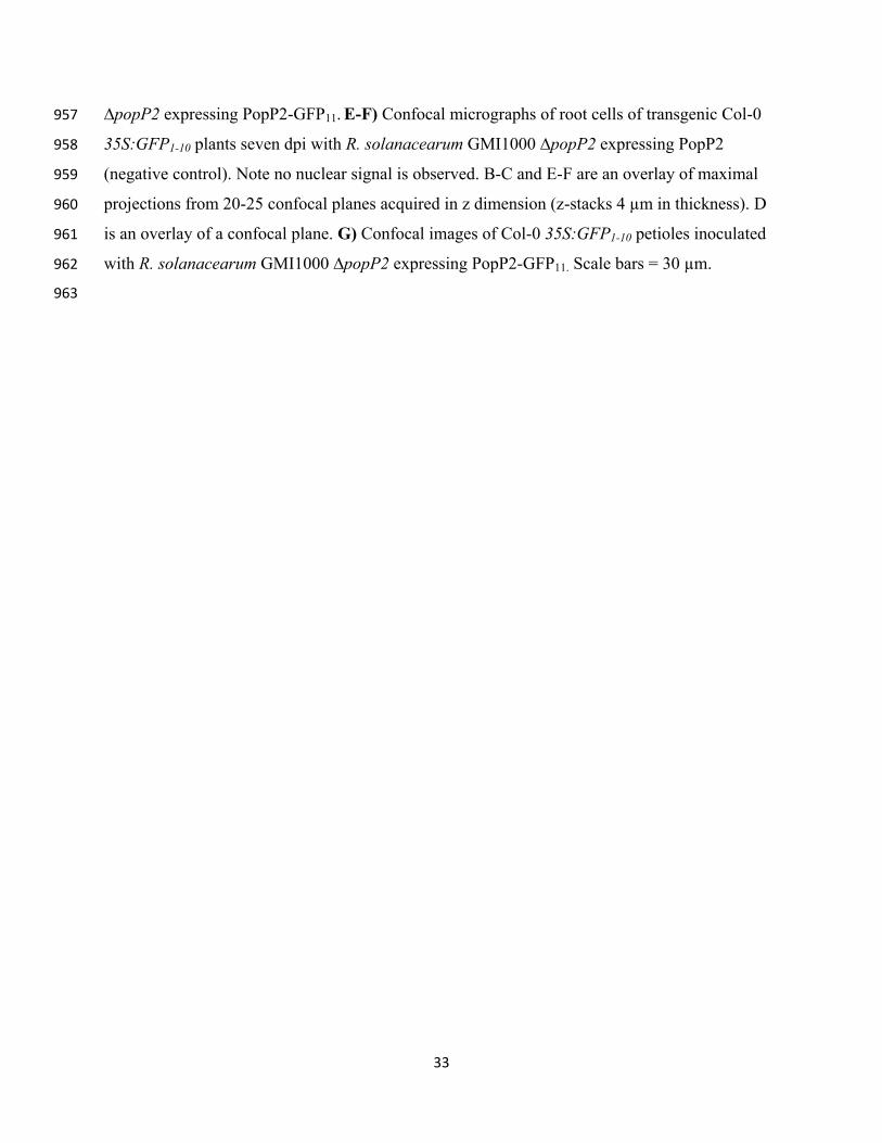
33
∆popP2 expressing PopP2-GFP11. E-F) Confocal micrographs of root cells of transgenic Col-0 957
35S:GFP1-10 plants seven dpi with R. solanacearum GMI1000 ∆popP2 expressing PopP2 958
(negative control). Note no nuclear signal is observed. B-C and E-F are an overlay of maximal 959
projections from 20-25 confocal planes acquired in z dimension (z-stacks 4 µm in thickness). D 960
is an overlay of a confocal plane. G) Confocal images of Col-0 35S:GFP1-10 petioles inoculated 961
with R. solanacearum GMI1000 ∆popP2 expressing PopP2-GFP11. Scale bars = 30 µm. 962
963

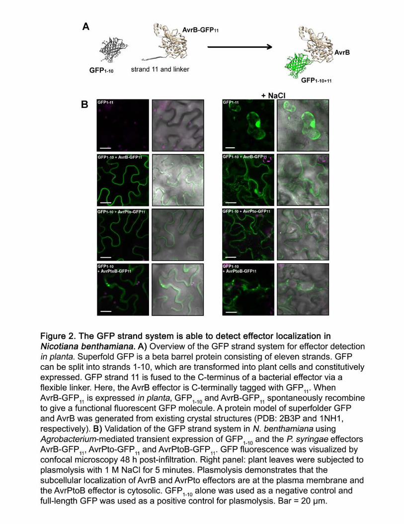


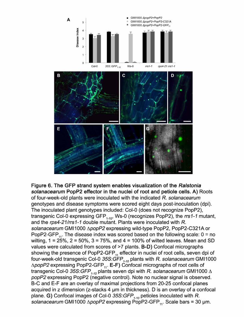

Parsed CitationsAkeda, Y., and Galan, J.E. (2005). Chaperone release and unfolding of substrates in type III secretion. Nature 437, 911-915.
Pubmed: Author and TitleCrossRef: Author and TitleGoogle Scholar: Author Only Title Only Author and Title
Alfano, J.R., and Collmer, A. (2004). Type III secretion system effector proteins: double agents in bacterial disease and plantdefense. Annual Review of Phytopathology 42, 385-414.
Pubmed: Author and TitleCrossRef: Author and TitleGoogle Scholar: Author Only Title Only Author and Title
Allan, A.C., and Fluhr, R. (1997). Two Distinct Sources of Elicited Reactive Oxygen Species in Tobacco Epidermal Cells. The PlantCell 9, 1559-1572.
Pubmed: Author and TitleCrossRef: Author and TitleGoogle Scholar: Author Only Title Only Author and Title
Arlat, M., Gough, C.L., Zischek, C., Barberis, P.A., Trigalet, A., and Boucher, C.A. (1992). Transcriptional organization andexpression of the large hrp gene cluster of Pseudomonas solanacearum. Molecular Plant-Microbe Interactions 5, 187-193.
Pubmed: Author and TitleCrossRef: Author and TitleGoogle Scholar: Author Only Title Only Author and Title
Axtell, M.J., and Staskawicz, B.J. (2003). Initiation of RPS2-specified disease resistance in Arabidopsis is coupled to the AvrRpt2-directed elimination of RIN4. Cell 112, 369-377.
Pubmed: Author and TitleCrossRef: Author and TitleGoogle Scholar: Author Only Title Only Author and Title
Boyes, D.C., Nam, J., and Dangl, J.L. (1998). The Arabidopsis thaliana RPM1 disease resistance gene product is a peripheralplasma membrane protein that is degraded coincident with the hypersensitive response. Proc Natl Acad Sci U S A 95, 15849-15854.
Pubmed: Author and TitleCrossRef: Author and TitleGoogle Scholar: Author Only Title Only Author and Title
Cabantous, S., Terwilliger, T.C., and Waldo, G.S. (2005). Protein tagging and detection with engineered self-assembling fragmentsof green fluorescent protein. Nat Biotechnol 23, 102-107.
Pubmed: Author and TitleCrossRef: Author and TitleGoogle Scholar: Author Only Title Only Author and Title
Chang, J.H., Desveaux, D., and Creason, A.L. (2014). The ABCs and 123s of bacterial secretion systems in plant pathogenesis.Annual Review of Phytopathology 52, 317-345.
Pubmed: Author and TitleCrossRef: Author and TitleGoogle Scholar: Author Only Title Only Author and Title
Chiang, Y.-H., and Coaker, G. (2015). Effector Triggered Immunity: NLR Immune Perception and Downstream Defense Responses.The Arabidopsis Book, e0183.
Pubmed: Author and TitleCrossRef: Author and TitleGoogle Scholar: Author Only Title Only Author and Title
Chung, E.H., da Cunha, L., Wu, A.J., Gao, Z., Cherkis, K., Afzal, A.J., Mackey, D., and Dangl, J.L. (2011). Specific threoninephosphorylation of a host target by two unrelated type III effectors activates a host innate immune receptor in plants. Cell Host &Microbe 9, 125-136.
Pubmed: Author and TitleCrossRef: Author and TitleGoogle Scholar: Author Only Title Only Author and Title
Clough, S.J., and Bent, A.F. (1998). Floral dip: a simplified method for Agrobacterium-mediated transformation of Arabidopsisthaliana. Plant J 16.
Pubmed: Author and TitleCrossRef: Author and TitleGoogle Scholar: Author Only Title Only Author and Title
Coll, N.S., and Valls, M. (2013). Current knowledge on the Ralstonia solanacearum type III secretion system. Microb Biotechnol 6,614-620.
Pubmed: Author and TitleCrossRef: Author and TitleGoogle Scholar: Author Only Title Only Author and Title
de Vries, J.S., Andriotis, V.M., Wu, A.J., and Rathjen, J.P. (2006). Tomato Pto encodes a functional N-myristoylation motif that isrequired for signal transduction in Nicotiana benthamiana. Plant J 45, 31-45.
Pubmed: Author and TitleCrossRef: Author and TitleGoogle Scholar: Author Only Title Only Author and Title

Deng, W.L., Preston, G., Collmer, A., Chang, C.J., and Huang, H.C. (1998). Characterization of the hrpC and hrpRS operons ofPseudomonas syringae pathovars syringae, tomato, and glycinea and analysis of the ability of hrpF, hrpG, hrcC, hrpT, and hrpVmutants to elicit the hypersensitive response and disease in plants. Journal of Bacteriology 180, 4523-4531.
Pubmed: Author and TitleCrossRef: Author and TitleGoogle Scholar: Author Only Title Only Author and Title
Deslandes, L., Olivier, J., Peeters, N., Feng, D.X., Khounlotham, M., Boucher, C., Somssich, I., Genin, S., and Marco, Y. (2003).Physical interaction between RRS1-R, a protein conferring resistance to bacterial wilt, and PopP2, a type III effector targeted tothe plant nucleus. Proc Natl Acad Sci U S A 100, 8024-8029.
Pubmed: Author and TitleCrossRef: Author and TitleGoogle Scholar: Author Only Title Only Author and Title
Deslandes, L., Pileur, F., Liaubet, L., Camut, S., Can, C., Williams, K., Holub, E., Beynon, J., Arlat, M., and Marco, Y. (1998). Geneticcharacterization of RRS1, a recessive locus in Arabidopsis thaliana that confers resistance to the bacterial soilborne pathogenRalstonia solanacearum. Molecular Plant-Microbe Interactions 11, 659-667.
Pubmed: Author and TitleCrossRef: Author and TitleGoogle Scholar: Author Only Title Only Author and Title
Gimenez-Ibanez, S., Boter, M., Fernandez-Barbero, G., Chini, A., Rathjen, J.P., and Solano, R. (2014). The bacterial effector HopX1targets JAZ transcriptional repressors to activate jasmonate signaling and promote infection in Arabidopsis. PLoS Biol 12,e1001792.
Pubmed: Author and TitleCrossRef: Author and TitleGoogle Scholar: Author Only Title Only Author and Title
Greenberg, J.T., and Yao, N. (2004). The role and regulation of programmed cell death in plant-pathogen interactions. CellMicrobiol 6, 201-211.
Pubmed: Author and TitleCrossRef: Author and TitleGoogle Scholar: Author Only Title Only Author and Title
Harlow, E., and Lane, D.P. (1988). Antibodies: A Laboratory Manual. (Cold Spring Harbor (New York): Cold Spring Harbor Press).Pubmed: Author and TitleCrossRef: Author and TitleGoogle Scholar: Author Only Title Only Author and Title
Henry, E., Yadeta, K.A., and Coaker, G. (2013). Recognition of bacterial plant pathogens: local, systemic and transgenerationalimmunity. New Phytologist 199, 908-915.
Pubmed: Author and TitleCrossRef: Author and TitleGoogle Scholar: Author Only Title Only Author and Title
Henry, E., Fung, N., Liu, J., Drakakaki, G., and Coaker, G. (2015). Beyond glycolysis: GAPDHs are multi-functional enzymes involvedin regulation of ROS, autophagy, and plant immune responses. PLoS Genet 11, e1005199.
Pubmed: Author and TitleCrossRef: Author and TitleGoogle Scholar: Author Only Title Only Author and Title
Henty-Ridilla, J.L., Shimono, M., Li, J., Chang, J.H., Day, B., and Staiger, C.J. (2013). The plant actin cytoskeleton responds tosignals from microbe-associated molecular patterns. PLoS Pathogens 9, e1003290.
Pubmed: Author and TitleCrossRef: Author and TitleGoogle Scholar: Author Only Title Only Author and Title
Hurley, B., Lee, D., Mott, A., Wilton, M., Liu, J., Liu, Y.C., Angers, S., Coaker, G., Guttman, D.S., and Desveaux, D. (2014). ThePseudomonas syringae type III effector HopF2 suppresses Arabidopsis stomatal immunity. PLoS One 9, e114921.
Pubmed: Author and TitleCrossRef: Author and TitleGoogle Scholar: Author Only Title Only Author and Title
Jiang, S., Yao, J., Ma, K.W., Zhou, H., Song, J., He, S.Y., and Ma, W. (2013). Bacterial effector activates jasmonate signaling bydirectly targeting JAZ transcriptional repressors. PLoS Pathogens 9, e1003715.
Pubmed: Author and TitleCrossRef: Author and TitleGoogle Scholar: Author Only Title Only Author and Title
Jin, Q., and He, S.Y. (2001). Role of the Hrp pilus in type III protein secretion in Pseudomonas syringae. Science 294, 2556-2558.Pubmed: Author and TitleCrossRef: Author and TitleGoogle Scholar: Author Only Title Only Author and Title
Jupe, J., Stam, R., Howden, A.J., Morris, J.A., Zhang, R., Hedley, P.E., and Huitema, E. (2013). Phytophthora capsici-tomatointeraction features dramatic shifts in gene expression associated with a hemi-biotrophic lifestyle. Genome Biol 14, R63.
Pubmed: Author and TitleCrossRef: Author and TitleGoogle Scholar: Author Only Title Only Author and Title

Kang, Y., Jelenska, J., Cecchini, N.M., Li, Y., Lee, M.W., Kovar, D.R., and Greenberg, J.T. (2014). HopW1 from Pseudomonassyringae disrupts the actin cytoskeleton to promote virulence in Arabidopsis. PLoS Pathogens 10, e1004232.
Pubmed: Author and TitleCrossRef: Author and TitleGoogle Scholar: Author Only Title Only Author and Title
Katagiri, F., Thilmony, R., and He, S.Y. (2002). The Arabidopsis Thaliana-Pseudomonas Syringae Interaction. The Arabidopsis Book,e0039.
Pubmed: Author and TitleCrossRef: Author and TitleGoogle Scholar: Author Only Title Only Author and Title
Khang, C.H., Berruyer, R., Giraldo, M.C., Kankanala, P., Park, S.Y., Czymmek, K., Kang, S., and Valent, B. (2010). Translocation ofMagnaporthe oryzae effectors into rice cells and their subsequent cell-to-cell movement. The Plant Cell 22, 1388-1403.
Pubmed: Author and TitleCrossRef: Author and TitleGoogle Scholar: Author Only Title Only Author and Title
Kim, M.G., da Cunha, L., McFall, A.J., Belkhadir, Y., DebRoy, S., Dangl, J.L., and Mackey, D. (2005). Two Pseudomonas syringae typeIII effectors inhibit RIN4-regulated basal defense in Arabidopsis. Cell 121, 749-759.
Pubmed: Author and TitleCrossRef: Author and TitleGoogle Scholar: Author Only Title Only Author and Title
Kleemann, J., Rincon-Rivera, L.J., Takahara, H., Neumann, U., Ver Loren van Themaat, E., van der Does, H.C., Hacquard, S.,Stuber, K., Will, I., Schmalenbach, W., Schmelzer, E., and O'Connell, R.J. (2012). Sequential delivery of host-induced virulenceeffectors by appressoria and intracellular hyphae of the phytopathogen Colletotrichum higginsianum. PLoS Pathogens 8,e1002643.
Pubmed: Author and TitleCrossRef: Author and TitleGoogle Scholar: Author Only Title Only Author and Title
Kovach, M.E., Elzer, P.H., Steven Hill, D., Robertson, G.T., Farris, M.A., Roop Ii, R.M., and Peterson, K.M. (1995). Four newderivatives of the broad-host-range cloning vector pBBR1MCS, carrying different antibiotic-resistance cassettes. Gene 166, 175-176.
Pubmed: Author and TitleCrossRef: Author and TitleGoogle Scholar: Author Only Title Only Author and Title
Le Roux, C., Huet, G., Jauneau, A., Camborde, L., Tremousaygue, D., Kraut, A., Zhou, B., Levaillant, M., Adachi, H., Yoshioka, H.,Raffaele, S., Berthome, R., Coute, Y., Parker, J.E., and Deslandes, L. (2015). A receptor pair with an integrated decoy convertspathogen disabling of transcription factors to immunity. Cell 161, 1074-1088.
Pubmed: Author and TitleCrossRef: Author and TitleGoogle Scholar: Author Only Title Only Author and Title
Lee, D., Bourdais, G., Yu, G., Robatzek, S., and Coaker, G. (2015). Phosphorylation of the Plant Immune Regulator RPM1-INTERACTING PROTEIN4 Enhances Plant Plasma Membrane H(+)-ATPase Activity and Inhibits Flagellin-Triggered ImmuneResponses in Arabidopsis. The Plant Cell 27, 2042-2056.
Pubmed: Author and TitleCrossRef: Author and TitleGoogle Scholar: Author Only Title Only Author and Title
Lee, J., Teitzel, G.M., Munkvold, K., del Pozo, O., Martin, G.B., Michelmore, R.W., and Greenberg, J.T. (2012). Type III secretionand effectors shape the survival and growth pattern of Pseudomonas syringae on leaf surfaces. Plant Physiol. 158, 1803-1818.
Pubmed: Author and TitleCrossRef: Author and TitleGoogle Scholar: Author Only Title Only Author and Title
Li, X., Yang, Q., Tu, H., Lim, Z., and Pan, S.Q. (2014). Direct visualization of Agrobacterium-delivered VirE2 in recipient cells. Plant J77, 487-495.
Pubmed: Author and TitleCrossRef: Author and TitleGoogle Scholar: Author Only Title Only Author and Title
Li, X., and Pan, S.Q. (2017). Agrobacterium delivers VirE2 protein into host cells via clathrin-mediated endocytosis. ScienceAdvances 3, e1601528.
Pubmed: Author and TitleCrossRef: Author and TitleGoogle Scholar: Author Only Title Only Author and Title
Lin, N.C., and Martin, G.B. (2005). An avrPto/avrPtoB mutant of Pseudomonas syringae pv. tomato DC3000 does not elicit Pto-mediated resistance and is less virulent on tomato. Molecular Plant-Microbe Interactions 18, 43-51.
Pubmed: Author and TitleCrossRef: Author and TitleGoogle Scholar: Author Only Title Only Author and Title
Liu, J., Elmore, J.M., Lin, Z.J., and Coaker, G. (2011). A receptor-like cytoplasmic kinase phosphorylates the host target RIN4,leading to the activation of a plant innate immune receptor. Cell Host & Microbe 9, 137-146.
Pubmed: Author and Title

CrossRef: Author and TitleGoogle Scholar: Author Only Title Only Author and Title
Mackey, D., Holt, B.F., 3rd, Wiig, A., and Dangl, J.L. (2002). RIN4 interacts with Pseudomonas syringae type III effector moleculesand is required for RPM1-mediated resistance in Arabidopsis. Cell 108, 743-754.
Pubmed: Author and TitleCrossRef: Author and TitleGoogle Scholar: Author Only Title Only Author and Title
Mackey, D., Belkhadir, Y., Alonso, J.M., Ecker, J.R., and Dangl, J.L. (2003). Arabidopsis RIN4 is a target of the type III virulenceeffector AvrRpt2 and modulates RPS2-mediated resistance. Cell 112, 379-389.
Pubmed: Author and TitleCrossRef: Author and TitleGoogle Scholar: Author Only Title Only Author and Title
Martin, G.B., Brommonschenkel, S.H., Chunwongse, J., Frary, A., Ganal, M.W., Spivey, R., Wu, T., Earle, E.D., and Tanksley, S.D.(1993). Map-based cloning of a protein kinase gene conferring disease resistance in tomato. Science 262, 1432-1436.
Pubmed: Author and TitleCrossRef: Author and TitleGoogle Scholar: Author Only Title Only Author and Title
McLachlan, D.H., Kopischke, M., and Robatzek, S. (2014). Gate control: guard cell regulation by microbial stress. New Phytologist203, 1049-1063.
Pubmed: Author and TitleCrossRef: Author and TitleGoogle Scholar: Author Only Title Only Author and Title
Melaragno, J.E., Mehrotra, B., and Coleman, A.W. (1993). Relationship between Endopolyploidy and Cell Size in Epidermal Tissueof Arabidopsis. Plant Cell 5, 1661-1668.
Pubmed: Author and TitleCrossRef: Author and TitleGoogle Scholar: Author Only Title Only Author and Title
Mindrinos, M., Katagiri, F., Yu, G.L., and Ausubel, F.M. (1994). The A. thaliana disease resistance gene RPS2 encodes a proteincontaining a nucleotide-binding site and leucine-rich repeats. Cell 78, 1089-1099.
Pubmed: Author and TitleCrossRef: Author and TitleGoogle Scholar: Author Only Title Only Author and Title
Monier, J.M., and Lindow, S.E. (2003a). Pseudomonas syringae Responds to the Environment on Leaves by Cell Size Reduction.Phytopathology 93, 1209-1216.
Pubmed: Author and TitleCrossRef: Author and TitleGoogle Scholar: Author Only Title Only Author and Title
Monier, J.M., and Lindow, S.E. (2003b). Differential survival of solitary and aggregated bacterial cells promotes aggregateformation on leaf surfaces. Proc Natl Acad Sci U S A 100, 15977-15982.
Pubmed: Author and TitleCrossRef: Author and TitleGoogle Scholar: Author Only Title Only Author and Title
Monteiro, F., Sole, M., van Dijk, I., and Valls, M. (2012). A chromosomal insertion toolbox for promoter probing, mutantcomplementation, and pathogenicity studies in Ralstonia solanacearum. Molecular Plant-Microbe Interactions 25, 557-568.
Pubmed: Author and TitleCrossRef: Author and TitleGoogle Scholar: Author Only Title Only Author and Title
Nakagawa, T., Kurose, T., Hino, T., Tanaka, K., Kawamukai, M., Niwa, Y., Toyooka, K., Matsuoka, K., Jinbo, T., and Kimura, T. (2007).Development of series of gateway binary vectors, pGWBs, for realizing efficient construction of fusion genes for planttransformation. Journal of Bioscience and Bioengineering 104, 34-41.
Pubmed: Author and TitleCrossRef: Author and TitleGoogle Scholar: Author Only Title Only Author and Title
Nimchuk, Z., Marois, E., Kjemtrup, S., Leister, R.T., Katagiri, F., and Dangl, J.L. (2000). Eukaryotic fatty acylation drives plasmamembrane targeting and enhances function of several type III effector proteins from Pseudomonas syringae. Cell 101, 353-363.
Pubmed: Author and TitleCrossRef: Author and TitleGoogle Scholar: Author Only Title Only Author and Title
Pedelacq, J.D., Cabantous, S., Tran, T., Terwilliger, T.C., and Waldo, G.S. (2006). Engineering and characterization of a superfoldergreen fluorescent protein. Nat Biotechnol 24, 79-88.
Pubmed: Author and TitleCrossRef: Author and TitleGoogle Scholar: Author Only Title Only Author and Title
Radics, J., Konigsmaier, L., and Marlovits, T.C. (2014). Structure of a pathogenic type 3 secretion system in action. Nat Struct MolBiol 21, 82-87.
Pubmed: Author and TitleCrossRef: Author and TitleGoogle Scholar: Author Only Title Only Author and Title

Ranjan, A., Fiene, G., Fackendahl, P., and Hoecker, U. (2011). The Arabidopsis repressor of light signaling SPA1 acts in the phloemto regulate seedling de-etiolation, leaf expansion and flowering time. Development 138, 1851-1862.
Pubmed: Author and TitleCrossRef: Author and TitleGoogle Scholar: Author Only Title Only Author and Title
Salmeron, J.M., Oldroyd, G.E., Rommens, C.M., Scofield, S.R., Kim, H.S., Lavelle, D.T., Dahlbeck, D., and Staskawicz, B.J. (1996).Tomato Prf is a member of the leucine-rich repeat class of plant disease resistance genes and lies embedded within the Ptokinase gene cluster. Cell 86, 123-133.
Pubmed: Author and TitleCrossRef: Author and TitleGoogle Scholar: Author Only Title Only Author and Title
Sarris, P.F., Duxbury, Z., Huh, S.U., Ma, Y., Segonzac, C., Sklenar, J., Derbyshire, P., Cevik, V., Rallapalli, G., Saucet, S.B.,Wirthmueller, L., Menke, F.L., Sohn, K.H., and Jones, J.D. (2015). A Plant Immune Receptor Detects Pathogen Effectors that TargetWRKY Transcription Factors. Cell 161, 1089-1100.
Pubmed: Author and TitleCrossRef: Author and TitleGoogle Scholar: Author Only Title Only Author and Title
Schell, M.A. (2000). Control of virulence and pathogenicity genes of Ralstonia solanacearum by an elaborate sensory network.Annual Review of Phytopathology 38, 263-292.
Pubmed: Author and TitleCrossRef: Author and TitleGoogle Scholar: Author Only Title Only Author and Title
Shimono, M., Lu, Y.J., Porter, K., Kvitko, B.H., Henty-Ridilla, J., Creason, A., He, S.Y., Chang, J.H., Staiger, C.J., and Day, B. (2016).The Pseudomonas syringae Type III Effector HopG1 Induces Actin Remodeling to Promote Symptom Development andSusceptibility during Infection. Plant Physiol 171, 2239-2255.
Pubmed: Author and TitleCrossRef: Author and TitleGoogle Scholar: Author Only Title Only Author and Title
Tanaka, S., Djamei, A., Presti, L.L., Schipper, K., Winterberg, S., Amati, S., Becker, D., Buchner, H., Kumlehn, J., Reissmann, S., andKahmann, R. (2015). Experimental approaches to investigate effector translocation into host cells in the Ustilago maydis/maizepathosystem. European Journal of Cell Biology 94, 349-358.
Pubmed: Author and TitleCrossRef: Author and TitleGoogle Scholar: Author Only Title Only Author and Title
Tasset, C., Bernoux, M., Jauneau, A., Pouzet, C., Briere, C., Kieffer-Jacquinod, S., Rivas, S., Marco, Y., and Deslandes, L. (2010).Autoacetylation of the Ralstonia solanacearum effector PopP2 targets a lysine residue essential for RRS1-R-mediated immunity inArabidopsis. PLoS Pathogens 6, e1001202.
Pubmed: Author and TitleCrossRef: Author and TitleGoogle Scholar: Author Only Title Only Author and Title
Thapa, S.P., Miyao, E.M., Michael Davis, R., and Coaker, G. (2015). Identification of QTLs controlling resistance to Pseudomonassyringae pv. tomato race 1 strains from the wild tomato, Solanum habrochaites LA1777. Theor Appl Genet 128, 681-692.
Pubmed: Author and TitleCrossRef: Author and TitleGoogle Scholar: Author Only Title Only Author and Title
Torres, M.A., Dangl, J.L., and Jones, J.D. (2002). Arabidopsis gp91phox homologues AtrbohD and AtrbohF are required foraccumulation of reactive oxygen intermediates in the plant defense response. Proc Natl Acad Sci U S A 99, 517-522.
Pubmed: Author and TitleCrossRef: Author and TitleGoogle Scholar: Author Only Title Only Author and Title
Toruno, T.Y., Stergiopoulos, I., and Coaker, G. (2016). Plant-Pathogen Effectors: Cellular Probes Interfering with Plant Defenses inSpatial and Temporal Manners. Annual
Pubmed: Author and TitleCrossRef: Author and TitleGoogle Scholar: Author Only Title Only Author and Title
Review of Phytopathology 54, 419-441.
Van Engelenburg, S.B., and Palmer, A.E. (2010). Imaging type-III secretion reveals dynamics and spatial segregation of Salmonellaeffectors. Nat Methods 7, 325-330.
Pubmed: Author and TitleCrossRef: Author and TitleGoogle Scholar: Author Only Title Only Author and Title
van Wees, S. (2008). Phenotypic analysis of Arabidopsis mutants: trypan blue stain for fungi, oomycetes, and dead plant cells. CSHprotocols 2008, pdb prot4982.
Pubmed: Author and TitleCrossRef: Author and TitleGoogle Scholar: Author Only Title Only Author and Title

Vasse, J., Frey, P., and Trigalet, A. (1995). Microscopic studies of intercellular infection and protoxylem invasion of tomato roots byPseudomonas solanacearum. Molecular Plant-Microbe Interactions 8, 241-251.
Pubmed: Author and TitleCrossRef: Author and TitleGoogle Scholar: Author Only Title Only Author and Title
Vasse, J., Genin, S., Frey, P., Boucher, C., and Brito, B. (2000). The hrpB and hrpG regulatory genes of Ralstonia solanacearum arerequired for different stages of the tomato root infection process. Molecular Plant-Microbe Interactions 13, 259-267.
Pubmed: Author and TitleCrossRef: Author and TitleGoogle Scholar: Author Only Title Only Author and Title
Wallden, K., Rivera-Calzada, A., and Waksman, G. (2010). Type IV secretion systems: versatility and diversity in function. CellMicrobiol 12, 1203-1212.
Pubmed: Author and TitleCrossRef: Author and TitleGoogle Scholar: Author Only Title Only Author and Title
Wang, G., Roux, B., Feng, F., Guy, E., Li, L., Li, N., Zhang, X., Lautier, M., Jardinaud, M.F., Chabannes, M., Arlat, M., Chen, S., He, C.,Noel, L.D., and Zhou, J.M. (2015). The Decoy Substrate of a Pathogen Effector and a Pseudokinase Specify Pathogen-InducedModified-Self Recognition and Immunity in Plants. Cell Host & Microbe 18, 285-295.
Pubmed: Author and TitleCrossRef: Author and TitleGoogle Scholar: Author Only Title Only Author and Title
Wang, Q., Han, C., Ferreira, A.O., Yu, X., Ye, W., Tripathy, S., Kale, S.D., Gu, B., Sheng, Y., Sui, Y., Wang, X., Zhang, Z., Cheng, B.,Dong, S., Shan, W., Zheng, X., Dou, D., Tyler, B.M., and Wang, Y. (2011). Transcriptional programming and functional interactionswithin the Phytophthora sojae RXLR effector repertoire. The Plant Cell 23, 2064-2086.
Pubmed: Author and TitleCrossRef: Author and TitleGoogle Scholar: Author Only Title Only Author and Title
Xin, X.F., and He, S.Y. (2013). Pseudomonas syringae pv. tomato DC3000: a model pathogen for probing disease susceptibility andhormone signaling in plants. Annual
Pubmed: Author and TitleCrossRef: Author and TitleGoogle Scholar: Author Only Title Only Author and Title
Review ofP 51, 473-498.
Yadeta, K.A., and BP, J.T. (2013). The xylem as battleground for plant hosts and vascular wilt pathogens. Frontiers in Plant Science4, 97.
Pubmed: Author and TitleCrossRef: Author and TitleGoogle Scholar: Author Only Title Only Author and Title
Yang, Q., Li, X., Tu, H., and Pan, S.Q. (2017). Agrobacterium-delivered virulence protein VirE2 is trafficked inside host cells via amyosin XI-K-powered ER/actin network. Proc Natl Acad Sci U S A 114, 2982-2987.
Pubmed: Author and TitleCrossRef: Author and TitleGoogle Scholar: Author Only Title Only Author and Title
Yang, Y., Costa, A., Leonhardt, N., Siegel, R.S., and Schroeder, J.I. (2008). Isolation of a strong Arabidopsis guard cell promoter andits potential as a research tool. Plant Methods 4, 1-15.
Pubmed: Author and TitleCrossRef: Author and TitleGoogle Scholar: Author Only Title Only Author and Title
Zhou, Z., Wu, Y., Yang, Y., Du, M., Zhang, X., Guo, Y., Li, C., and Zhou, J.M. (2015). An Arabidopsis Plasma Membrane Proton ATPaseModulates JA Signaling and Is Exploited by the Pseudomonas syringae Effector Protein AvrB for Stomatal Invasion. The Plant Cell27, 2032-2041.
Pubmed: Author and TitleCrossRef: Author and TitleGoogle Scholar: Author Only Title Only Author and Title

DOI 10.1105/tpc.17.00027; originally published online June 9, 2017;Plant Cell
Elizabeth Henry, Tania Y Toruño, Alain Jauneau, Laurent Deslandes and Gitta Laurel CoakerInfection
Direct and Indirect Visualization of Bacterial Effector Delivery into Diverse Plant Cell Types During
This information is current as of May 11, 2018
Supplemental Data
/content/suppl/2017/08/15/tpc.17.00027.DC3.html /content/suppl/2017/07/19/tpc.17.00027.DC2.html /content/suppl/2017/06/09/tpc.17.00027.DC1.html
Permissions https://www.copyright.com/ccc/openurl.do?sid=pd_hw1532298X&issn=1532298X&WT.mc_id=pd_hw1532298X
eTOCs http://www.plantcell.org/cgi/alerts/ctmain
Sign up for eTOCs at:
CiteTrack Alerts http://www.plantcell.org/cgi/alerts/ctmain
Sign up for CiteTrack Alerts at:
Subscription Information http://www.aspb.org/publications/subscriptions.cfm
is available at:Plant Physiology and The Plant CellSubscription Information for
ADVANCING THE SCIENCE OF PLANT BIOLOGY © American Society of Plant Biologists


![Proteasome Activity Imaging and Profiling Characterizes · PDF fileProteasome Activity Imaging and Profiling Characterizes Bacterial Effector Syringolin A1[W] Izabella Kolodziejek2,](https://img.dokumen.tips/doc/110x75/5a79e7cc7f8b9a5c3a8de66d/proteasome-activity-imaging-and-proling-characterizes-activity-imaging-and-proling.jpg)