Embed Size (px)
Citation preview

Dinesh Talwar, MD Center for Neurosciences

Idiopathic – 60%
• No clear etiology
• Presumed genetic
• Normal or near normal neurologically
• Idiopathic Generalized Epilepsy
• Childhood Absence Epilepsy
• Juvenile Absence Epilepsy
• Juvenile Myoclonic Epilepsy
• Idiopathic Partial Epilepsy
• Childhood Epilepsy with centro-temporal spikes
• Occipital Epilepsy of Childhood
Cryptogenic – 10%
• No clear etiology
• Abnormal neurologically
• Probable genetic etiology
• Severe Epilepsies of Childhood – the epileptic encephalopathies
• Infantile spasms
• Lennox Gastaut Syndrome
Symptomatic – 30%
• Identifiable etiology
• Variety of causes
• Structural brain abnormalities
• Developmental
• Genetic
• Acquired
• Complex epilepsy syndromes
• Genetic disorders affecting brain development and associated neurologic and systemic manifestations

• No clear etiology
• Presumed genetic
• Normal or near normal neurologically
• Idiopathic Generalized Epilepsy
• Childhood Absence Epilepsy
• Juvenile Absence Epilepsy
• Juvenile Myoclonic Epilepsy
• Idiopathic Partial Epilepsy
• Childhood Epilepsy with centro-temporal spikes
• Occipital Epilepsy of Childhood

Idiopathic Generalized/Partial Epilepsies Genetically complex
▪ Polygenic, inheritance of susceptibility genes (vast majority) ▪ Rapidly diminishing risks beyond first degree relatives ▪ High concordance between monozygotic twins ▪ Likely modify ion channels
Monogenic with variability in phenotypic presentation (less frequent, inherited or de novo) GEFS + (SCN1A, SCN1B, GABRG2, GABRD) GRIN 2A (Glutamate receptor, ionotropic, NMDA) Autosomal Dominant Nocturnal Frontal Lobe Epilepsy (CHRNA4, CHRNB2,
CHRNA2)

Symptomatic – 30%
• Identifiable etiology
• Variety of causes
• Structural brain abnormalities
• Developmental
• Genetic
• Acquired
• Complex epilepsy syndromes
• Genetic disorders affecting brain development and associated neurologic and systemic manifestations
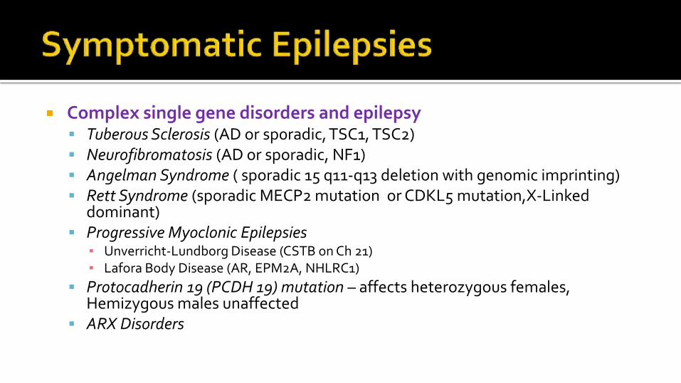
Complex single gene disorders and epilepsy Tuberous Sclerosis (AD or sp0radic, TSC1, TSC2) Neurofibromatosis (AD or sporadic, NF1) Angelman Syndrome ( sporadic 15 q11-q13 deletion with genomic imprinting) Rett Syndrome (sporadic MECP2 mutation or CDKL5 mutation,X-Linked
dominant) Progressive Myoclonic Epilepsies
▪ Unverricht-Lundborg Disease (CSTB on Ch 21) ▪ Lafora Body Disease (AR, EPM2A, NHLRC1)
Protocadherin 19 (PCDH 19) mutation – affects heterozygous females, Hemizygous males unaffected
ARX Disorders

Cryptogenic – 10%
• No clear etiology
• Abnormal neurologically
• Probable genetic etiology
• Severe Epilepsies of Childhood – the epileptic encephalopathies
• Infantile spasms
• Lennox Gastaut Syndrome

In which the underlying epilepsy and abnormal electrical discharges
contribute, at least in part to the abnormal neurologic dysfunction –
cognitive, motor dysfunction
Most epilepsies in children do not contribute to or cause neurologic dysfunction

Onset in early childhood
Severe Epilepsy Devastating Seizures are
difficult to control Relatively severe
EEG abnormalities
Brain dysfunction Developmental delay, arrest of
development and often regression Cognitive Dysfunction, Intellectual
disability, Autism Motor Dysfunction – delayed milestones,
coordination and balance difficulties, abnormal movements, features of cerebral palsy.
Behavioral difficulties

Epileptic
Encephalopathy
Epileptic events and
electrical abnormaliti
es as seen on the EEG
Electrical abnormalities
Critical Period of
Brain
Development
Age

Genetic causes Monogenic – de novo mutations
▪ Channelopathies ▪ Pathologic affectation of membrane bound proteins
Voltage gated
Ligand gated
▪ Gene mutations causing alterations of critical intracellular proteins
Acquired causes Autoimmune epilepsies
▪ Anti-NMDA receptor encephalitis
▪ Rasmussen’s encephalitis

Polygenic inheritance Idiopathic generalized epilepsies, Idiopathic focal epilepsies Relatively benign disorders
Monogenic inheritance Epileptic Encephalopathies, severe epilepsies of childhood Complex Epilepsy Syndromes, may also cause epileptic
encephalopathies, may have variability of presentation Epilepsy disorders of variable presentation (vary from
relatively benign to severe disorders)

Autosomal Dominant Autosomal Recessive X-Linked Mitochondrial Disorders De novo mutations
Variable presentation - GEFS+ - Autosomal
dominant nocturnal frontal lobe epilepsy
- Benign Neonatal convulsions
- Complex disorders such as Tuberous Sclerosis
Usually have significant neurologic impairment - GLUT1 deficiency - Various metabolic
and CNS degenerative disorders
Moderate to severe neurologic impairment often with severe epilepsy - Fragile X - Rett Syndrome - CDKL5 encephalo-
pathy - ARX syndromes
Variety of disorders with significant variability in presentation. - MERRF - MELAS - POLG related
syndromes
Some variability in epilepsy presentation but usually severe epilepsies - Epileptic encephalo-
pathies - Early onset severe
epilepsy with significant neurologic impairment

Comparative genetic architectures of susceptibility alleles in complex epilepsy, genes of large effect in monogenic epilepsy and epilepsy arising as a secondary feature of other Mendelian syndromes.
Mulley J C et al. Hum. Mol. Genet. 2005;14:R243-R249
© The Author 2005. Published by Oxford University Press. All rights reserved. For Permissions, please email: [email protected]

Hildebrand MS, et al. J Med Genet 2013
Same model applicable to intellectual disability and autism

GEFS+ spectrum arising from mutations in SCN1A with quantitative representation of associated epilepsy subsyndromes.
Mulley J C et al. Hum. Mol. Genet. 2005;14:R243-R249
© The Author 2005. Published by Oxford University Press. All rights reserved. For Permissions, please email: [email protected]

Prolonged (45 mins) GTC seizure within 24 hours of 4-month immunization.
Prolonged (45 mins) GTC seizure within 24 hours of 6-month immunization – positctal left hemiparesis.
Over next few months developed recurrent, prolonged focal (hemiclonic) or generalized onset tonic-clonic seizures requiring hospitalization multiple times. Often intubated.
Seizures were both febrile and non-febrile, although often precipitated by an illness.
Seizures often unresponsive to rectal diazepam.

Pregnancy and birth history unremarkable Early development till 6 months of age was normal.
Subsequently - developmental delay, slowing of developmental progress, regression with seizure episodes.
Family history + for infantile spasms in first cousin MRIs normal EEG – initial EEGs were normal. EEGs abnormal from 11
months of age – multifocal, predominantly frontal, spikes and diffuse slowing
Lab tests – normal metabolic and routine genetic testing Epileptic encephalopathy

Seizures refractory to medication therapy – Phenobarbital, valproate, zonisamide, levetiracetam,
Seizure types – hemiclonic (left or right), frontal lobe seizures characterized by hypermotor activity, GTC, frequent episodes of status epilepticus
Walked at 23 months – ataxic gait, rare words At 2 ½ years age – status epilepticus for 4 ½ hours, with
evidence for continuing electrical seizures subsequently on EEG. Went into ARDS and subsequently died.

Neonatal Period
• Early Myoclonic Encephalopathy
• Ohtahara Dyndrome
Infancy
• Epilepsy of Infancy with Migrating Focal Seizures
• West Syndrome
• Dravet Syndrome
• Myoclonic Encephalopathy in non-progressive disorders
Childhood
• Epileptic Encephalopathy with Continuous Spike-And-Wave during Sleep (CSWS) (including Landau Kleffner Syndrome)
• Lennox- Gastaut Syndrome
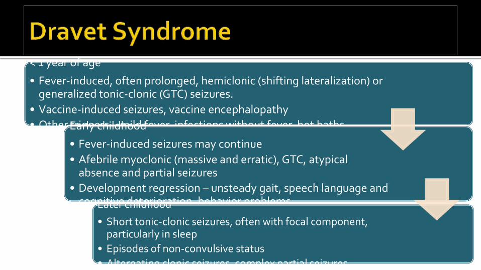
< 1 year of age
• Fever-induced, often prolonged, hemiclonic (shifting lateralization) or generalized tonic-clonic (GTC) seizures.
• Vaccine-induced seizures, vaccine encephalopathy
• Other triggers – mild fever, infections without fever, hot baths Early childhood
• Fever-induced seizures may continue
• Afebrile myoclonic (massive and erratic), GTC, atypical absence and partial seizures
• Development regression – unsteady gait, speech language and cognitive deterioration, behavior problems. Later childhood
• Short tonic-clonic seizures, often with focal component, particularly in sleep
• Episodes of non-convulsive status
• Alternating clonic seizures, complex partial seizures

EEG
Often normal in the first months/year of life
Second year of life – diffuse slowing, generalized fast spike-wave and polyspike-wave discharges, multifocal spikes
Later life – variable EEG changes including multifocal spikes, generalized spike and polyspike-wave discharges, diffuse frontally predominant slowing
Treatment
Seizures often intractable, difficult to treat
Avoid medications that may worsen seizures – carbamazepine, lamotrigine, vigabatrin, phenytoin
Beneficial meds – valproate, levetiracetam, zonisamide, clobazam, topiramate, stiripentol
Alternative treatments – ACTH, prednisone, IV IgG, ketogenic diet, VNS
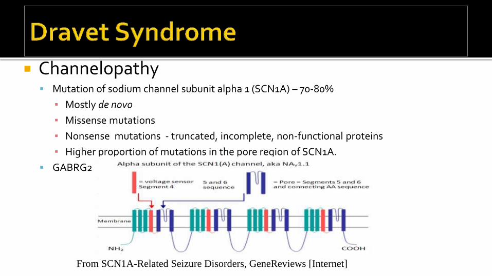
Channelopathy Mutation of sodium channel subunit alpha 1 (SCN1A) – 70-80%
▪ Mostly de novo
▪ Missense mutations
▪ Nonsense mutations - truncated, incomplete, non-functional proteins
▪ Higher proportion of mutations in the pore region of SCN1A.
GABRG2, PCDH19 (X-linked, females) mutations in some cases
From SCN1A-Related Seizure Disorders, GeneReviews [Internet]

Severe Myoclonic Epilepsy of Infancy (SMEI, Dravet syndrome)
Genetic testing
Direct sequencing of the SCN1A gene –
▪ novel missense mutation found not previously reported.
▪ Mutation resides in the region of the gene where other pathogenic mutations have been detected.
▪ Parents have not been tested
De novo mutations

15-years old, female Seizure onset – 6-7 months of age
Brief generalized seizures
Staring, repetitive eye blinking
Synchronous clonic jerks of arms/hands
2-10 seconds
40-50 per day
EEG: Brief bursts of GSW, bifrontal spikes
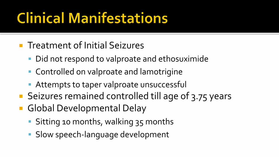
Treatment of Initial Seizures
Did not respond to valproate and ethosuximide
Controlled on valproate and lamotrigine
Attempts to taper valproate unsuccessful
Seizures remained controlled till age of 3.75 years Global Developmental Delay
Sitting 10 months, walking 35 months
Slow speech-language development

Recurrence of seizures at 45 months
Coincided with attempt to taper meds
Loss of muscle tone, unresponsiveness, inability to sit up, staring, eye blinking
Few seconds
Only partially controlled with meds
EEG at 46 months – Left centrotemporal spikes

Late-onset Epileptic spasms 4 ½ years Loss of tone, balance, up-rolling of eyes, tonic
stiffening of arms for 1-2 seconds recurring in a cluster 5-60 mins Persisted for next 11 years Continued with complex partial seizures, sometimes
preceding epileptic spasms Occasional tonic seizures Best response to Clobazam added to VPA + LTG

Decreased verbal abilities Diminished language comprehension Markedly diminished cognitive abilities Repetitive behaviors Slow slurred speech Hypotonia Scoliosis Coordination and balance difficulties, ataxia Jerky choreoathetotic movements

MRIs – Normal or nonspecific, non-diagnostic findings
Metabolic workup – lactic acid, ammonia, amino acids, organic acids, acylcarnitine profile, plasma guandinoacetate, CSF neurotransmitters
Genetic workup – High resolution chromosomes, Subteleomeric deletions, ARX mutation analysis, Rett syndrome sequencing
PET scan - Normal
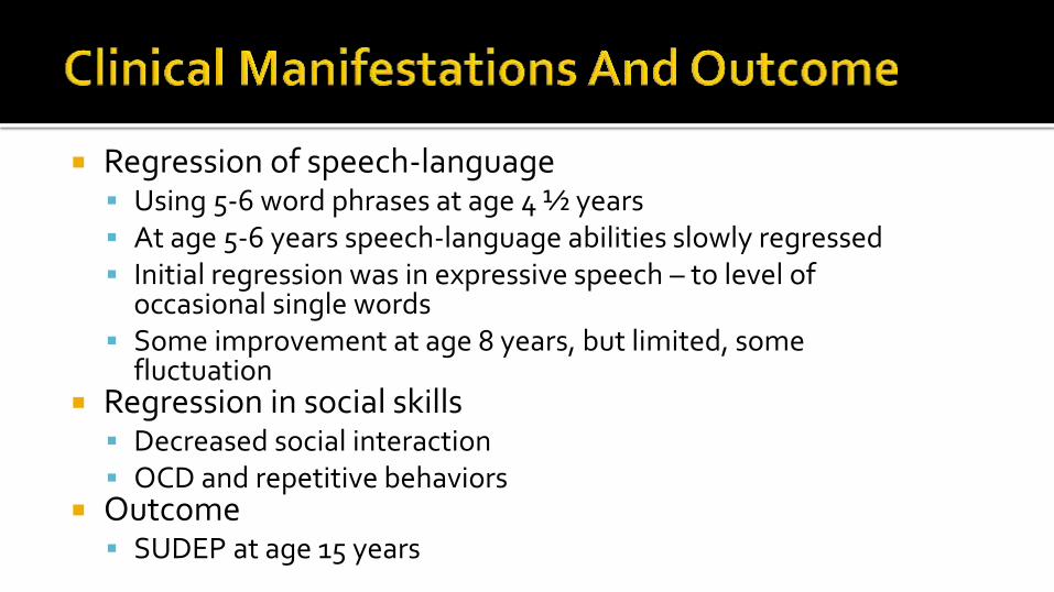
Regression of speech-language Using 5-6 word phrases at age 4 ½ years At age 5-6 years speech-language abilities slowly regressed Initial regression was in expressive speech – to level of
occasional single words Some improvement at age 8 years, but limited, some
fluctuation Regression in social skills Decreased social interaction OCD and repetitive behaviors
Outcome SUDEP at age 15 years

Vissers et al 2010 (Nat Gen) demonstrated the power of using trios (proband and parents) and Next Generation Sequencing (NGS)
Identified 10 trios with unexplained MR Sequenced exomes of all individuals Assumed dominant de novo model

Other exome screens in patients and their immediate families with neurological disorders have since demonstrated high rates of success
However, exome screening still has a few disadvantages
These can be improved by (more expensive) Whole Genome Sequencing (WGS)

WGS
Pathogenic dominant de novo variant
Differences from the human reference genome
Found in exome
Potential functional effect
Appear de novo
Validated
5,378,745
31,931
13,395
34
1
? ?

NGS still has a high error rate
~1 in every 100,000 nucleotides
Therefore most of our 34 are probably
sequencing errors
We removed 10 candidate variants present in
public databases (“normal” variation)
Performed Sanger sequencing on remaining 24
As expected, only 1 mutation was
successfully validated

The validated de novo variant was in the SCN8A gene c.5302A>G
Causes a non-synonymous change in Nav1.6 p.Asn1768Asp
SCN8A not previously associated with any human epilepsy disorders
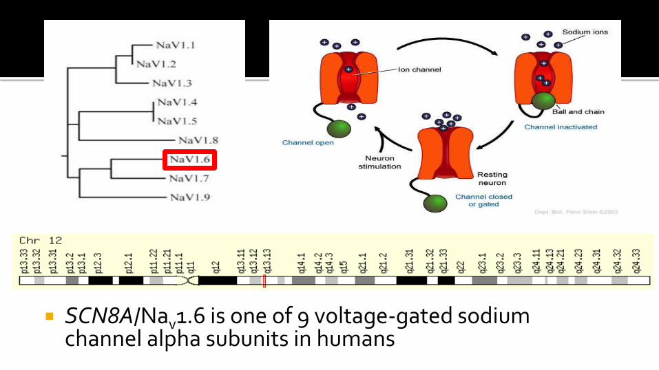
SCN8A/Nav1.6 is one of 9 voltage-gated sodium channel alpha subunits in humans
De Novo Pathogenic SCN8A Mutation Identified by Whole-Genome Sequencing of a Family Quartet Affected by Infantile Epileptic Encephalopathy and SUDEP Krishna R. Veeramah, Janelle E. O’Brien, Miriam H. Meisler, Xiaoyang Cheng Sulayman D. Dib-Hajj, Stephen G. Waxman, Dinesh Talwar, Santhosh Girirajan, Evan E. Eichler, Linda L. Restifo, Robert P. Erickson, and Michael F. Hammer.
The American Journal of Human Genetics 90, 1–9, March 9, 2012
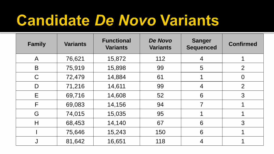
Family Variants Functional
Variants
De Novo
Variants
Sanger
Sequenced Confirmed
A 76,621 15,872 112 4 1
B 75,919 15,898 99 5 2
C 72,479 14,884 61 1 0
D 71,216 14,611 99 4 2
E 69,716 14,608 52 6 3
F 69,083 14,156 94 7 1
G 74,015 15,035 95 1 1
H 68,453 14,140 67 6 3
I 75,646 15,243 150 6 1
J 81,642 16,651 118 4 1

7 good candidate pathogenic de novo variants
based on gene function
2 probands have de novo variants possibly
related to phenotype
1 proband did not possess any de novo
mutations in exomes
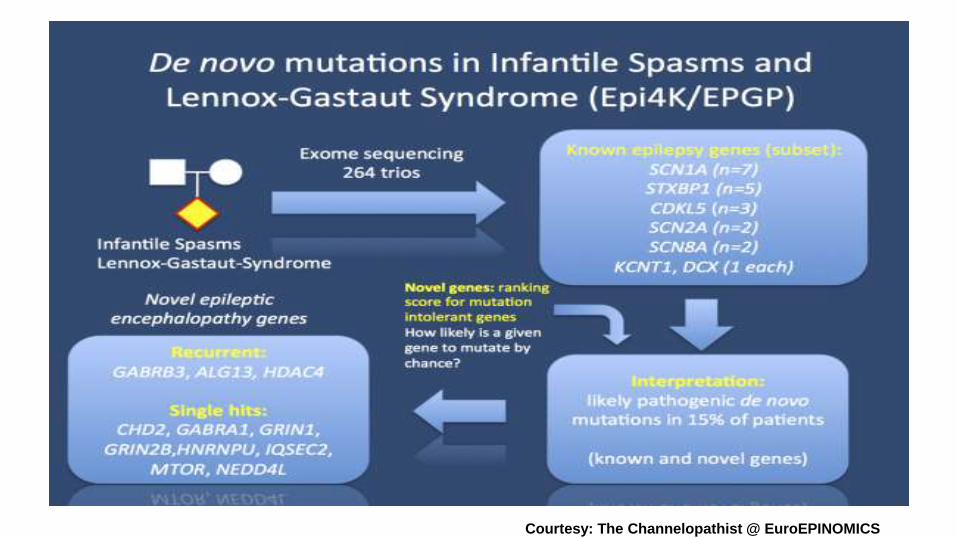
Courtesy: The Channelopathist @ EuroEPINOMICS
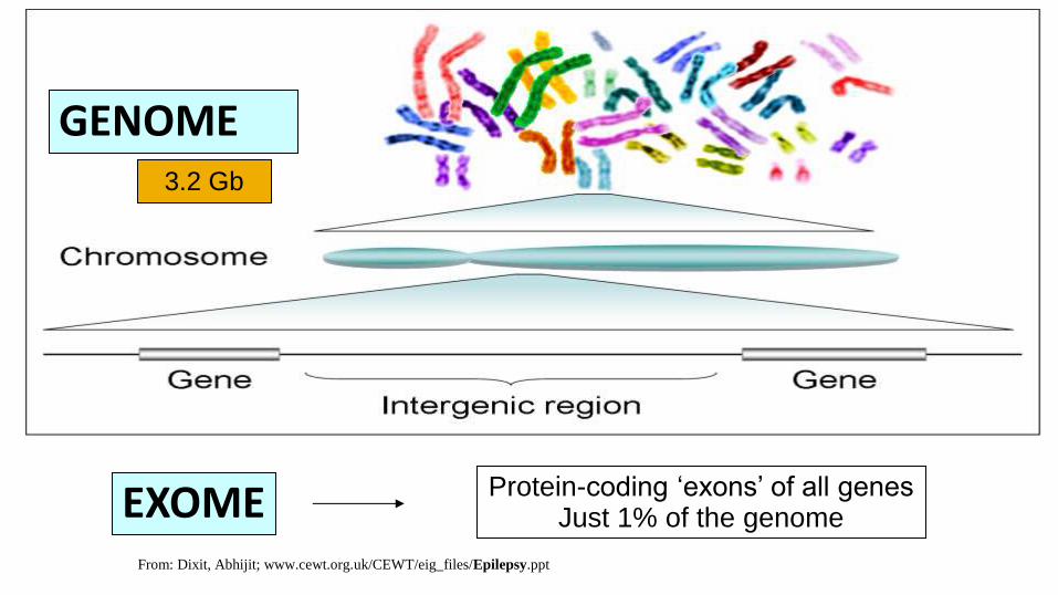
GENOME
EXOME Protein-coding ‘exons’ of all genes Just 1% of the genome
3.2 Gb
From: Dixit, Abhijit; www.cewt.org.uk/CEWT/eig_files/Epilepsy.ppt

ArrayCGH 1000X resolution
Karyotype
Sanger sequencing Next-gen sequencing
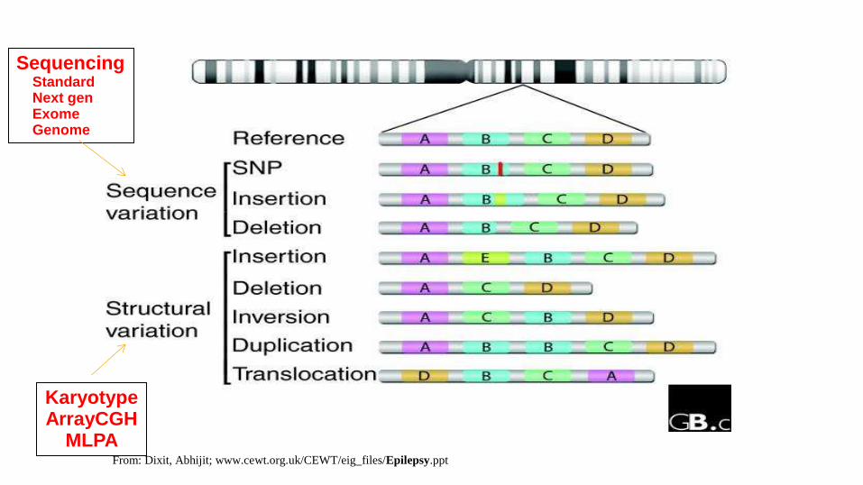
Karyotype ArrayCGH
MLPA
Sequencing Standard Next gen Exome Genome
From: Dixit, Abhijit; www.cewt.org.uk/CEWT/eig_files/Epilepsy.ppt

From: Dixit, Abhijit; www.cewt.org.uk/CEWT/eig_files/Epilepsy.ppt
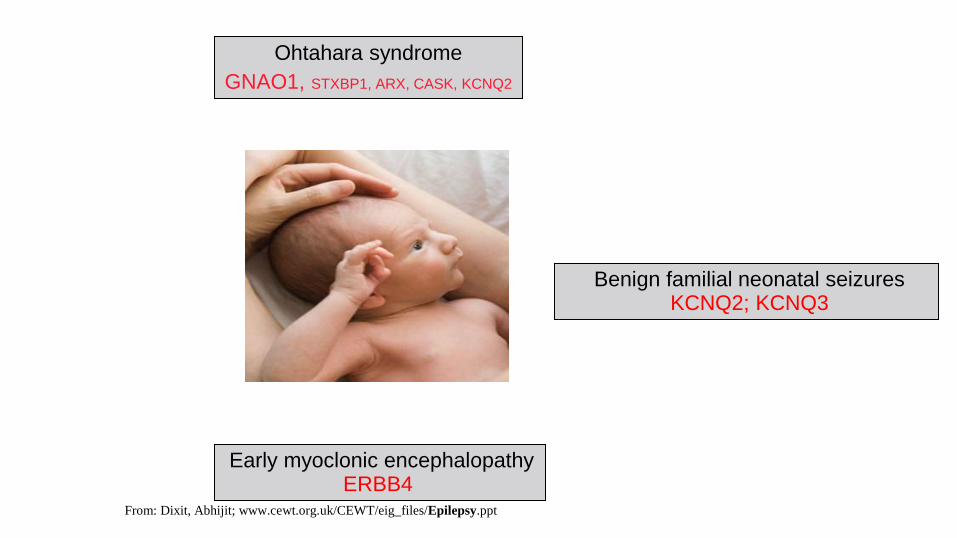
Benign familial neonatal seizures KCNQ2; KCNQ3
Early myoclonic encephalopathy ERBB4
Ohtahara syndrome
GNAO1, STXBP1, ARX, CASK, KCNQ2
From: Dixit, Abhijit; www.cewt.org.uk/CEWT/eig_files/Epilepsy.ppt

West syndrome multiple
Migrating partial seizures of infancy KCNT1
Benign familial infantile seizures PRRT2 Dravet syndrome
SCN1A
From: Dixit, Abhijit; www.cewt.org.uk/CEWT/eig_files/Epilepsy.ppt

Early onset benign childhood occipital epilepsy (Panayiotopoulos type)
Complex
Febrile seizures plus SCN1A
EE with continuous spike-and-wave during sleep (CSWS) Landau-Kleffner syndrome (LKS) GRIN2A
Lennox- Gastaut syndrome Multiple
Autosomal dominant nocturnal frontal lobe epilepsy
CHRNA4; CHRNB2; CHRNA2
Benign epilepsy with centro-temporal spikes GRIN2A
Childhood absence epilepsy Complex
From: Dixit, Abhijit; www.cewt.org.uk/CEWT/eig_files/Epilepsy.ppt
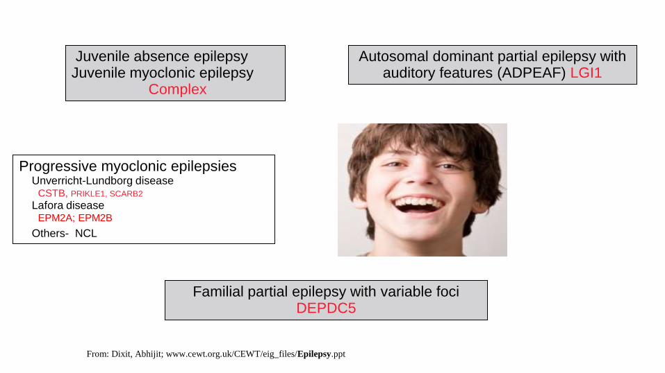
Progressive myoclonic epilepsies Unverricht-Lundborg disease CSTB, PRIKLE1, SCARB2
Lafora disease EPM2A; EPM2B
Others- NCL
Familial partial epilepsy with variable foci DEPDC5
Autosomal dominant partial epilepsy with auditory features (ADPEAF) LGI1
Juvenile absence epilepsy Juvenile myoclonic epilepsy
Complex
From: Dixit, Abhijit; www.cewt.org.uk/CEWT/eig_files/Epilepsy.ppt
Epilepsy Panels
Panels directed at detecting gene mutations known to be associated with epilepsy
Commercially available
▪ GeneDx
▪ Courtagen
▪ Transgenomic

Chromosomal Microarray (ArrayCGH) All patients with epilepsy plus intellectual disability and learning
difficulties Epilepsy Gene Panel Difficult to control seizures, intractable epilepsy often with other
associated neurologic impairment Epileptic encephalopathy
Whole exome sequencing Unexplained epileptic encephalopathy Intractable epilepsy with associated neurologic impairment of
unclear etiology

Many of the idiopathic childhood epilepsies have polygenic inheritance, with a few identified syndromes with monogenic inheritance
Monogenic inherited epilepsies Autosomal dominant conditions
Autosomal recessive conditions
X-linked disorders
De novo mutations causing epileptic encephalopathies

Epileptic encephalopathies are disorders in which intractable seizures and EEG abnormalities contribute to developmental and cognitive difficulties. Look for slowing, arrest or regression in development.
Heterogeneous etiologies New genetic tests (Exome sequencing) will help to
identify disorders not previously recognized Early recognition and treatment is important and helpful
in some of the disorders.

In epileptic encephalopathies seizures are often difficult to treat, may require treatment other than anti-epileptic medications
Appropriate diagnosis of channelopathies, genetic mutations with alteration of protein function, and acquired disorders (e.g. autoimmune disorders) will help guide future treatment directed specifically for the disorder.

1. The most common method of genetic inheritance in Idiopathic generalized epilepsies is
a. Polygenic inheritance
b. Monogenic inheritance
c. De-novo mutations
d. X-Linked inheritance
2. Autosomal dominant inheritance is seen in:
a. Generalized seizures with febrile seizures PLUS (GEFS+)
b. Tuberous Sclerosis
c. Autosomal dominant nocturnal frontal lobe epilepsy
d. All of the above
3. An epileptic encephalopathy is:
a. A benign epilepsy of childhood
b. A condition that usually starts in teenage years
c. A condition in which the abnormal EEG and ongoing seizures contribute to or cause neurologic deterioration
d. Is easy to treat