Embed Size (px)
Citation preview

Enjoy the digital experience
Digital microscopEs
�
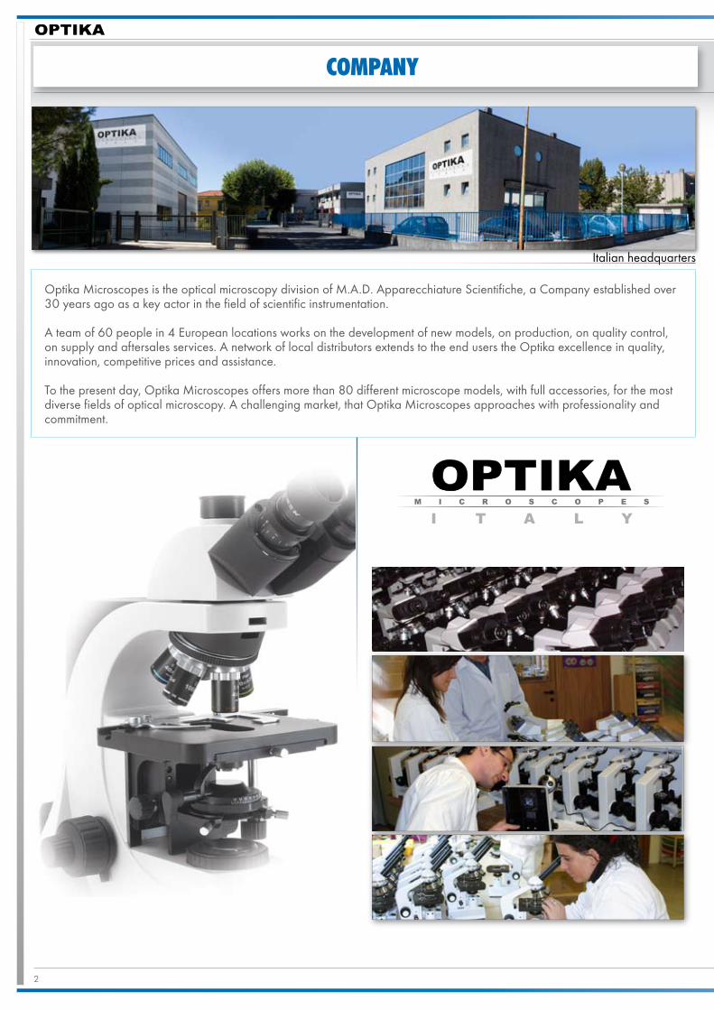
COMPANY
Optika Microscopes is the optical microscopy division of M.A.D. Apparecchiature Scientifiche, a Company established over 30 years ago as a key actor in the field of scientific instrumentation.
A team of 60 people in 4 European locations works on the development of new models, on production, on quality control, on supply and aftersales services. A network of local distributors extends to the end users the Optika excellence in quality, innovation, competitive prices and assistance.
To the present day, Optika Microscopes offers more than 80 different microscope models, with full accessories, for the most diverse fields of optical microscopy. A challenging market, that Optika Microscopes approaches with professionality and commitment.
Italian headquarters

3
Index
dm-5 pag.4
dm-10 pag.6
dm-15 pag.8
dm-20 pag.10
OPTIKAVISION®SOFTWARE pag.12

4 dIGITALmICROSCOPES
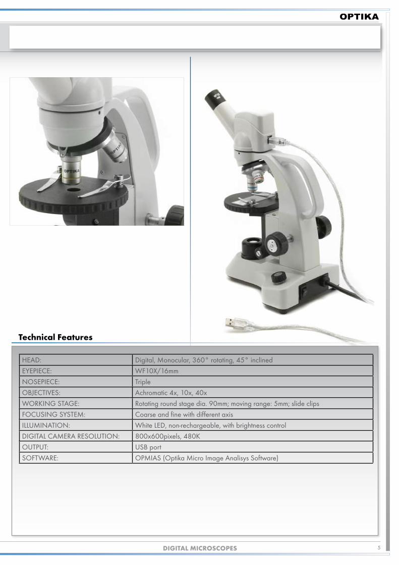
BIOLOGICAL DIGITAL MICROSCOPEDM-5
Themodel
Belonging to the “entry-level” models of the series of OPTIKA digital biological microscopes, the DM-5 is ideal for primary schools.
Easy to use, its sturdy structure and first-class optical system make the study of biology using the microscope a really plea-sant activity.
The microscope comes complete with all necessary equipment, thus allowing its immediate use with no need for additional accessories.
DM
-5
5dIGITALmICROSCOPES
TechnicalFeatures
HEAD: Digital, Monocular, 360° rotating, 45° inclined
EYEPIECE: WF10X/16mm
NOSEPIECE: Triple
OBJECTIVES: Achromatic 4x, 10x, 40x
WORKING STAGE: Rotating round stage dia. 90mm; moving range: 5mm; slide clips
FOCUSING SYSTEM: Coarse and fine with different axis
ILLUMINATION: White LED, non-rechargeable, with brightness control
DIGITAL CAMERA RESOLUTION: 800x600pixels, 480K
OUTPUT: USB port
SOFTWARE: OPMIAS (Optika Micro Image Analisys Software)

6
BIOLOGICAL DIGITAL MICROSCOPEDM-10
dIGITALmICROSCOPES
An intermediate model of the series of OPTIKA digital biological microscopes, the DM-10 is the best solution for secondary schools.
Its strengths are its optical and mechanical qualities, and its particularly rich outfit, as it consists, among other, of a mechani-cal stage with coaxial knobs and a set of 3 achromatic objectives ensuring magnification up to 400x.
DM
-10
Themodel

�
HEAD: Digital, Monocular, 360° rotating, 45° inclined
EYEPIECE: WF10X/18mm
NOSEPIECE: Quadruple, reversed
OBJECTIVES: Achromatic 4x, 10x, 40x.
WORKING STAGE: Double layer with mechanical sliding stage, 1�5x115mm, mov. range 50x30mm
CONDENSER: 1.�5 N.A. Abbe type
FOCUSING SYSTEM: Coaxial coarse and fine, with focusing stop mechanism
ILLUMINATION: White LED, non-rechargeable, with brightness control
DIGITAL CAMERA RESOLUTION: 800x600pixels, 480K
OUTPUT: USB port
SOFTWARE: OPMIAS (Optika Micro Image Analisys Software)
PACKING: Carton box with inner foam
dIGITALmICROSCOPES
DM-10
TechnicalFeatures
8
BIOLOGICAL DIGITAL MICROSCOPEDM-15
dIGITALmICROSCOPES
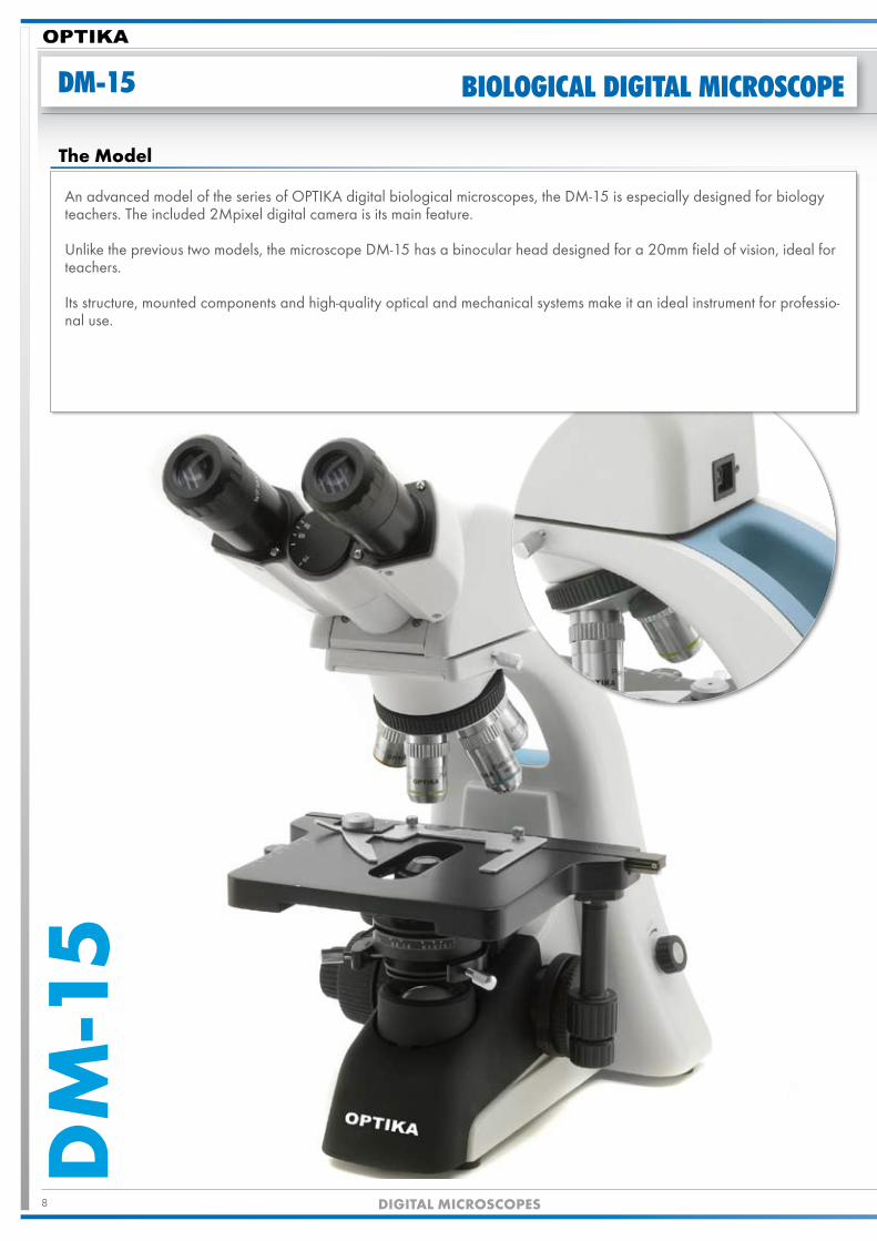
An advanced model of the series of OPTIKA digital biological microscopes, the DM-15 is especially designed for biology teachers. The included �Mpixel digital camera is its main feature.
Unlike the previous two models, the microscope DM-15 has a binocular head designed for a �0mm field of vision, ideal for teachers.
Its structure, mounted components and high-quality optical and mechanical systems make it an ideal instrument for professio-nal use.
DM
-15
Themodel

9dIGITALmICROSCOPES
DM-15
HEAD: Digital, Binocular, 360° rotating, 30° inclined
EYEPIECES: WF10X/�0mm
NOSEPIECE: Quadruple, reversed
OBJECTIVES: Achromatic 4x, 10x, 40x, 100x (oil immersion)
WORKING STAGE: Double layer with mechanical sliding stage, 160x14�mm, mov. range �6x5�mm
CONDENSER: 1.�5 N.A. Abbe type, with centring system
FOCUSING SYSTEM: Coarse and fine with different axis, with focusing stop mechanism
ILLUMINATION: White LED, non-rechargeable, with brightness control
DIGITAL CAMERA RESOLUTION: 1600x1�00pixels, �.0M
OUTPUT: USB �.0 port
SOFTWARE: OPMIAS (Optika Micro Image Analisys Software)
PACKING: Carton box with inner foam
TechnicalFeatures
10 dIGITALmICROSCOPES
DM-20 BIOLOGICAL DIGITAL MICROSCOPED
M-2
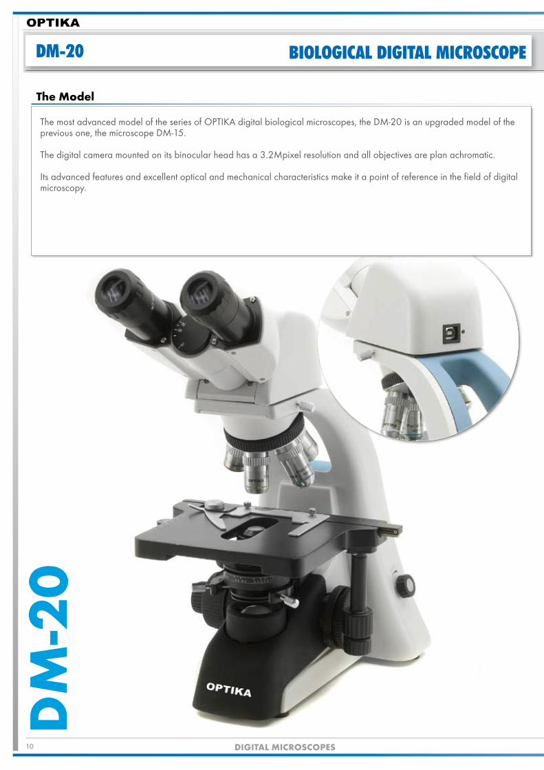
0The most advanced model of the series of OPTIKA digital biological microscopes, the DM-�0 is an upgraded model of the previous one, the microscope DM-15.
The digital camera mounted on its binocular head has a 3.�Mpixel resolution and all objectives are plan achromatic.
Its advanced features and excellent optical and mechanical characteristics make it a point of reference in the field of digital microscopy.
Themodel

11dIGITALmICROSCOPES
DM-20
HEAD: Digital, Binocular, 360° rotating, 30° inclined
EYEPIECES: WF10X/�0mm
NOSEPIECE: Quadruple, reversed
OBJECTIVES: PLAN Achromatic 4x, 10x, 40x, 100x (oil immersion)
WORKING STAGE: Double layer with mechanical sliding stage, 160x14�mm, mov. range �6x5�mm
CONDENSER: 1.�5 N.A. Abbe type, with centring system
FOCUSING SYSTEM: Coarse and fine with different axis, with focusing stop mechanism
ILLUMINATION: White LED, non-rechargeable, with brightness control
DIGITAL CAMERA RESOLUTION: �048x1536pixels, 3.�M
OUTPUT: USB �.0 port
SOFTWARE: OPMIAS (Optika Micro Image Analisys Software)
PACKING: Carton box with inner foam
Technical Features

1� dIGITALmICROSCOPES
CAMERA SPECIFICATIONS
dm-5/dm-10 dm-15 dm-20
Resolution 800 × 600 pixels (480 kilo pixels) 1600 × 1�00 pixels (�.0 mega pixels) �048 × 1536 pixels (3.1 mega pixels)
Sensor 1/4”CMOS 1/�”CMOS 1/�”CMOS
SensorImagingArea 4.1� mm × 3.0� mm 6.�� mm × 5.04 mm 6.59 mm × 4.90 mm
dataOutput(UncompressedVideo) 8 bit 8,1� bit 8,1� bit
PixelSize �.9 μm × �.9 μm 4.� μm × 4.� μm 3.� μm × 3.� μm
Resolution&FrameRate
“800 × 600 �5 frames/second”
“1600 × 1�00 10 frames/second”
“�048 × 1536 6 frames/second”
“800 × 600 �5 frames/second”
“1�80 × 10�4 15 frames/second”
“10�4 × �68 �0 frames/second”
Sensitivity�.3 V/Lux-sec 1.� V/Lux-sec 1.0 V/Lux-sec
(550 nm) (550 nm) (550 nm)
ElectronicShutter 1/16-1/1600 1/16-1/1600 1/16-1/1600
WhiteBalance Auto / Manual Auto / Manual Auto / Manual
S/NRatio -52 dB -52 dB -52 dB
dynamicRange -60 dB -60 dB -60 dB
digitalPort USB1.1 or USB�.0 USB�.0 USB�.0
ImagingSoftwareOPMIAS (OPTIKA Micro Image Analysis Software)
available in English and Italian
SystemRequirements
RAM: �56 MB (51� MB recommended)
Video card: Separate card (64 MB or higher)
Operating system: Windows �000 (SP4) or Windows XP (SP�)

13dIGITALmICROSCOPES
OPMIAS
OPmIAS (OPTIKA Micro Image Analysis Software) is especially developed for Optika digital microsco-pes series, DM. It contains powerful tools for image capturing, adjusting, operating and measuring. OPMIAS
can be used in many different fields, such as biology, medicine, education, mechanics, electronics and chemi-stry.
ImageAcquisition
OPMIAS lets you acquire high quality focussed video and still images from your digital microscope. You can grab still or multi real time images in BMP or JPG file format and add voice to the images or to different parts of the images in MIG and SFC for-mat. You can also record your own video AVI video format. There are many possibilities to control the image output accor-ding to your needs. There are functions such as white balance, automatic exposure, hue, saturation and intensity controls, to mention a few.
measurements,CountingandTagging
Using OPMIAS you can make various types of calibrated mea-surements such as distances between two points, angles and areas in images. Tools for tagging and counting objects in the images are also available to the user.
ImageElaboration
OPMIAS provides easy ways for opening, adjusting, rotating, and enlarging images. You can add texts and draw graphics in the acquired image in order to illuminate certain aspects. OP-MIAS can perform powerful digital image filtering to facilitate image analysis.You also have the possibility to compare and display your ima-ges in various views and merge them into one.

14 dIGITALmICROSCOPES
M.A.D. IberIca aparatos cIentIfIcos
C/ La Lluna, 11 - 08001- Barcelona - ESPAÑA
Tel.: +34 93 3�4868� Fax: +34 93 3�48683
Optika MikrOscOp ve LabOratuvar cihazLari ticaret Ltd. Şti.
Yürekli Adam Sokak N0:20 Kat:6 Daire:14 Kavac-k BEYKOZ/-STANBUL
Tel: 090 �16 413 4100 Fax: 090 �16 413 161�
alpha optIka MIcroscopes hungary �030 ÉRD, Kaktusz u. ��.- HUNGARY
Tel.: (�3) 5�0-0�� Fax: (�3) 3�4-965
M.A.D. APPARECCHIATURE SCIENTIFICHE SRLVia Rigla, 3� - �4010 Ponteranica (BG) - ITALIA
Tel.: +39 035 5�139� (6 linee r.a.) Fax: +39 035 5�1435 - [email protected]
OUR GROUP

15dIGITALmICROSCOPES
EXIBITIONS AND TRADE SHOWS
ACHEMA, GERMANYMEDICA, GERMANYANALYTICA, GERMANYWORLDDIDAC, SWITZERLANDRICH-MACH, ITALYRESTAURO, ITALYTED, ITALYARABLAB, UAEEXPOQUIMIA, SPAININTERDIDAC, SPAINLABTECH, TURKEY

WWW.OPTIKAMICROSCOPES.COM
M.A.D. IberIca aparatos cIentIfIcos
C/ La Lluna, 11 - 08001- Barcelona - ESPAÑATel.: +34 93 3248682 Fax: +34 93 3248683
Optika MikrOscOp ve LabOratuvar cihazLari ticaret Ltd. Şti.Yürekli Adam Sokak N0:20 Kat:6 Daire:14 Kavacık BEYKOZ/İSTANBUL
Tel: 090 216 413 4100 Fax: 090 216 413 1617
alpha optIka MIcroscopes hungary 2030 ÉRD, Kaktusz u. 22.- HUNGARYTel.: (23) 520-077 Fax: (23) 374-965
M.A.D. AppArecchiAture Scientifiche SrlVia Rigla, 32 - 24010 Ponteranica (BG) - ITALIA
Tel.: +39 035 571392 (6 linee r.a.) Fax: +39 035 571435 - [email protected]