Embed Size (px)
Citation preview
Digital Image Manipulation I
By Professor Stelmark
Image Histogram
Digital radiographic image histograms are very important for digital image production. However, they can be the source of bothersome digital radiographic image artifacts if they are not properly understood and manipulated.
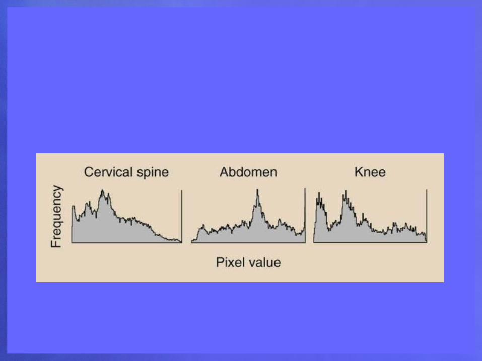
All digital radiographic imaging systems have the ability to evaluate the original image data through histogram analysis. A histogram is a plot of the frequency of appearance of a given object characteristic.
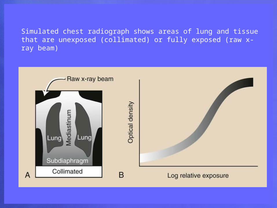
Simulated chest radiograph shows areas of lung and tissue that are unexposed (collimated) or fully exposed (raw x-ray beam)
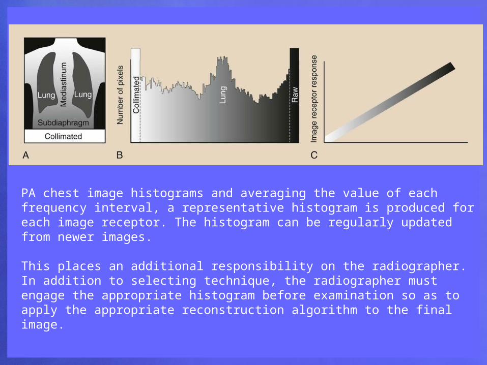
PA chest image histograms and averaging the value of each frequency interval, a representative histogram is produced for each image receptor. The histogram can be regularly updated from newer images.
This places an additional responsibility on the radiographer. In addition to selecting technique, the radiographer must engage the appropriate histogram before examination so as to apply the appropriate reconstruction algorithm to the final image.
Collimation and Partition
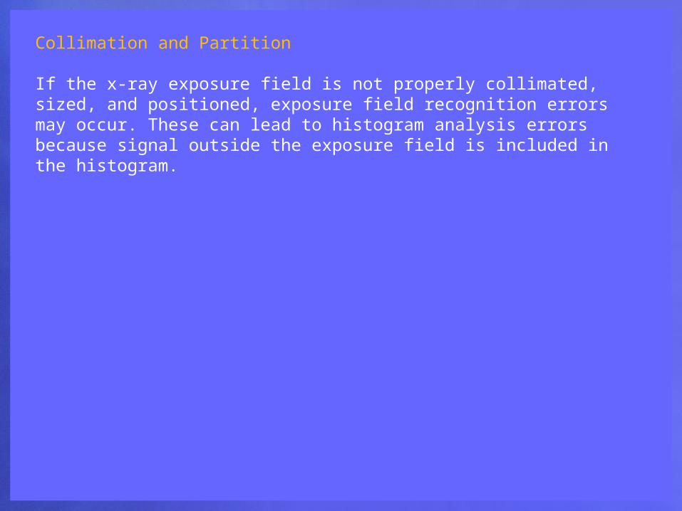
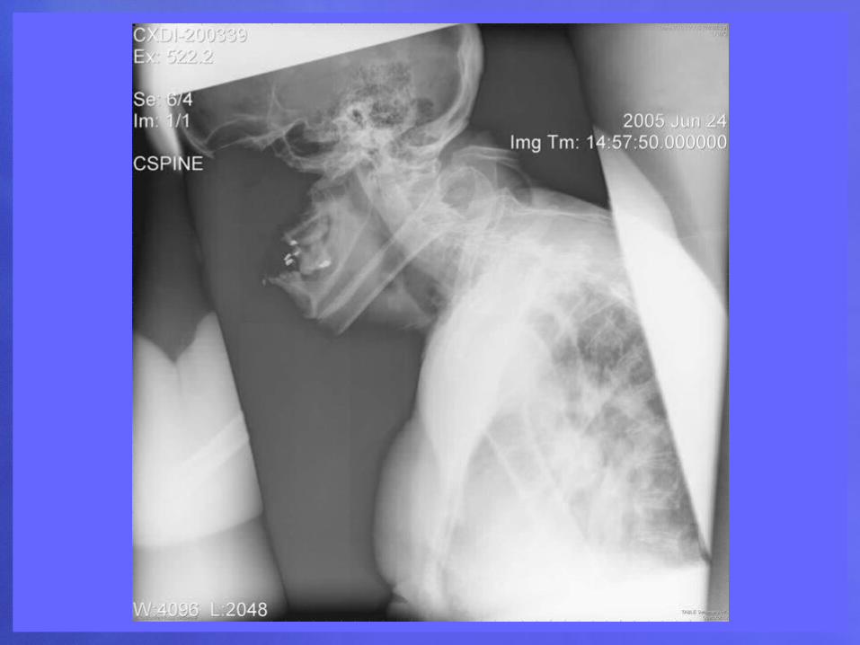
If the x-ray exposure field is not properly collimated, sized, and positioned, exposure field recognition errors may occur. These can lead to histogram analysis errors because signal outside the exposure field is included in the histogram.
Collimation of the projected area x-ray beam is important for patient radiation dose reduction and for improved image contrast in screen-film radiography. In DR, proper collimation has the added value of defining the image histogram. If improperly collimated, the histogram can be improperly analyzed.
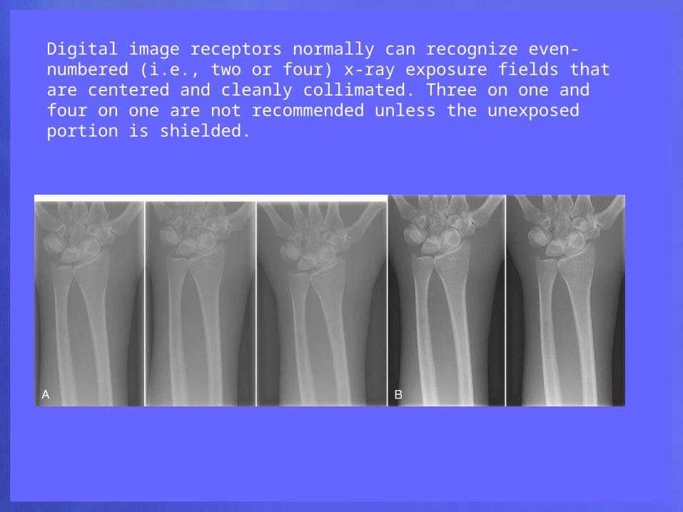
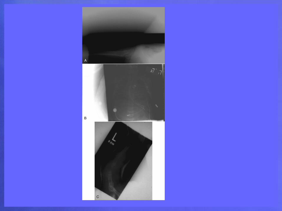
Digital image receptors normally can recognize even-numbered (i.e., two or four) x-ray exposure fields that are centered and cleanly collimated. Three on one and four on one are not recommended unless the unexposed portion is shielded.
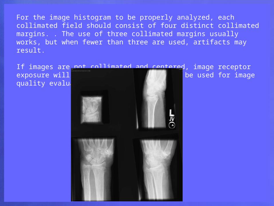
For the image histogram to be properly analyzed, each collimated field should consist of four distinct collimated margins. . The use of three collimated margins usually works, but when fewer than three are used, artifacts may result.
If images are not collimated and centered, image receptor exposure will not be accurate and cannot be used for image quality evaluation.
If multiple fields are projected onto a single IP, each must have clear, collimated edges and margins between each field. This process, called partitioning,
Alignment
Alignment of the exposure field on the IP is important in the same way and for the same reason as collimation.
Preprocessing
Before an image is prepared “for processing,” several manipulations of the output of an image receptor may be necessary to correct for potential artifacts. Such artifacts can occur because of dead pixels or dead rows or columns of pixels
Preprocessing of digital images is largely automatic.
Preprocessing is designed to produce artifact-free digital images. In this regard, preprocessing provides electronic calibration to reduce pixel-to-pixel, row-to-row, and column-to-column response differences. The processes of pixel interpolation and noise correction are automatically applied with most systems.
A single pixel or a single row or column normally will not interfere with diagnosis. However, many of these defects must be corrected. Correction algorithms specific to each type of digital image receptor use interpolation techniques to assign digital values to each dead pixel, row, or column. Interpolation is the mathematical process of assigning a value to a dead pixel based on the recorded values of adjacent pixels.
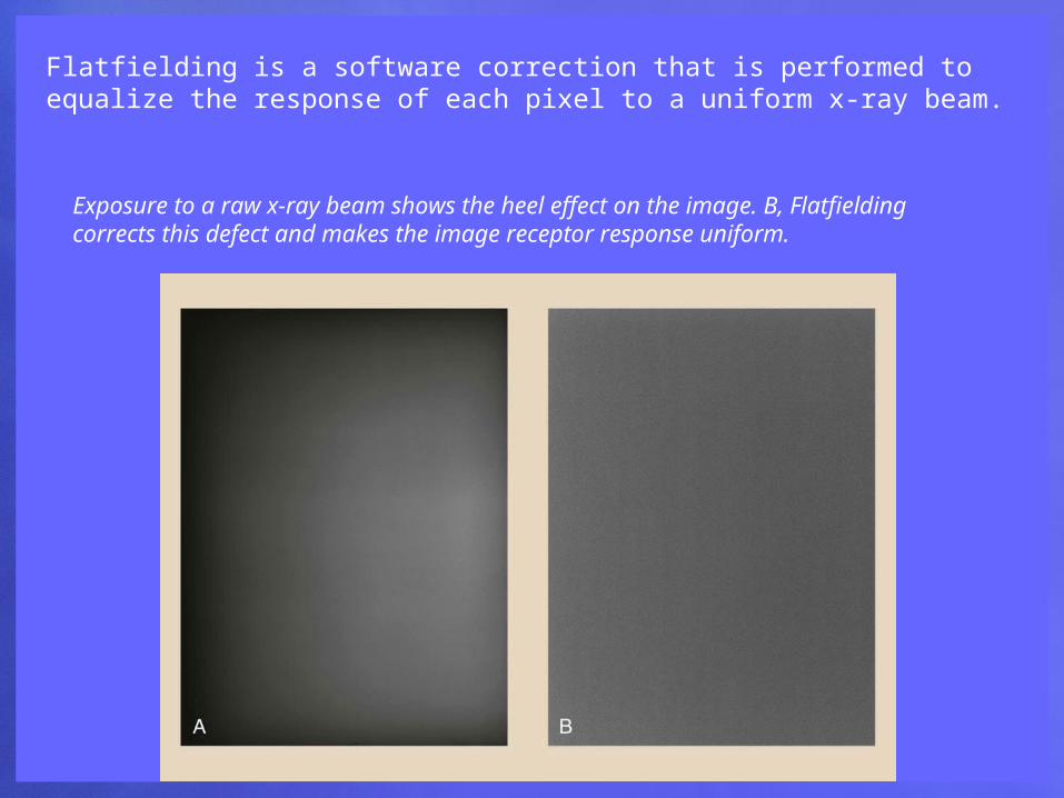
Flatfielding is a software correction that is performed to equalize the response of each pixel to a uniform x-ray beam.
Exposure to a raw x-ray beam shows the heel effect on the image. B, Flatfielding corrects this defect and makes the image receptor response uniform.
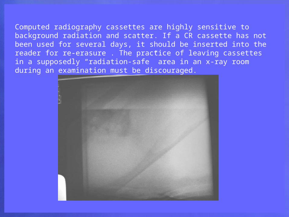
Computed radiography cassettes are highly sensitive to background radiation and scatter. If a CR cassette has not been used for several days, it should be inserted into the reader for re-erasure . The practice of leaving cassettes in a supposedly “radiation-safe” area in an x-ray room during an examination must be discouraged.