Embed Size (px)
Citation preview

Digestive System

Overview
● Basic digestive and functional processes
● The digestive organs● Chemical digestion● Absorption● Elimination

Basic digestive and functional processes
● Ingestion – food intake● Propulsion – moves food along tract● Digestion – break down of food● Absorption – into blood or lymph● Defecation – eliminate residue

Propulsion
●Swallowing●voluntary
●Peristalsis●involuntary

Digestion
● Mechanical – increase surface area of food particles● Teeth and tongue● Stomach churning● segmentation
● Chemical – breakdown of large molecules into smaller ones

Macromolecules and what they break down into
● Polysaccharides● Monosaccharides
● Lipids● Glycerol● Fatty acids
● Proteins● Amino acids
● Nucleic acids● Nucleotides

Gastrointestinaltract
The Digestive Tract Organs

Teeth, tongue,Salivary glands
pancreas
Gall bladderLiver
The Digestive Tract Accessory Organs

Peritoneal relationships
Peritoneum – slippery membranes that line the body cavity●Visceral●Parietal ●Mesentery – double layer of peritoneum
●Path for blood vessels and nerves
●Fat stored here
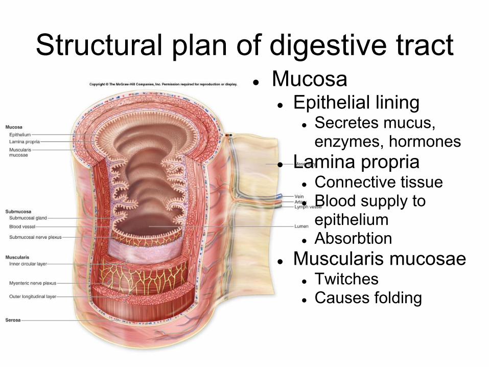
Structural plan of digestive tract● Mucosa
● Epithelial lining● Secretes mucus,
enzymes, hormones● Lamina propria
● Connective tissue● Blood supply to
epithelium● Absorbtion
● Muscularis mucosae● Twitches● Causes folding
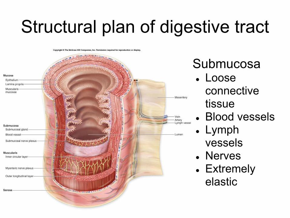
Structural plan of digestive tract
● Submucosa ● Loose
connective tissue
● Blood vessels● Lymph
vessels● Nerves● Extremely
elastic

Structural plan of digestive tract
● Muscularis● Functions
● Peristalis● Segmentation
● Inner layer● Circular ● Forms sphincters
● Prevents backflow● Controls passage
● Outer layer ● Longitudinal

Structural plan of digestive tract
● Serosa ● Visceral
peritoneum● Loose
connective tissue
● Blood vessels ● Produce
serous fluid

Enteric nervous system●Submucosal nerve plexus
●Glands and muscle in mucosa
●Myenteric nerve plexus
●Muscularis●Secretion of accessory organs
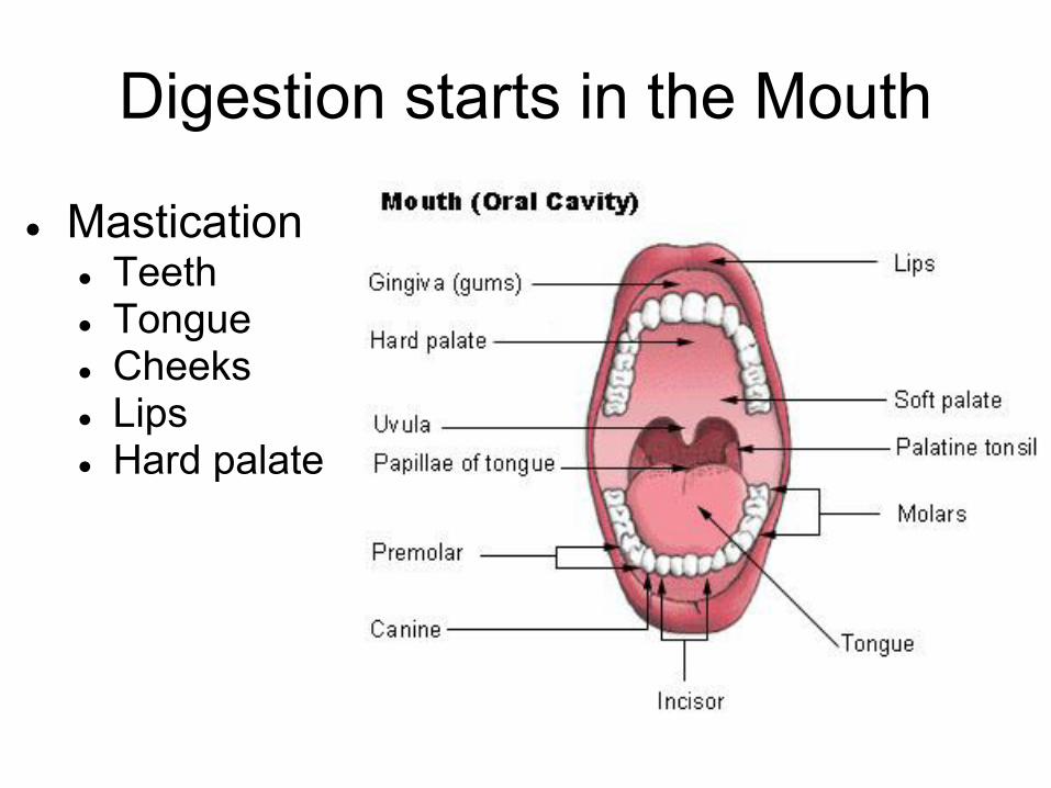
Digestion starts in the Mouth
● Mastication● Teeth● Tongue● Cheeks● Lips● Hard palate

Salivary Glands
● Intrinsic salivary glands● Dispersed within oral cavity
● Tongue● Lips ● Cheeks
● Keeps mouth moist● Extrinsic salivary glands
● Large, discrete organs● Parotid● Submandibular● sublingual
● secrete upon salivation
Secrete 1-1.5L of saliva each day!

Saliva● Water (98%)● Electrolytes
● Sodium, potassium, chloride, phosphate, bicarbonate
● pH – 6.8-7.0● Salivary amylase
● Begins starch digestion● most active in lower pH
● Lingual lipase● Activated by stomach acid● Digests fat
● Mucus● Lubricates food● Binds food into a bolus

Saliva continued
● Lysozyme● Kills bacteria
● Immunoglobulin A (IgA)● Inhibits bacterial growth
● a cyanide compound● inhibit bacteria
● defensins● antimicrobial proteins● cytokine - draws white blood cells into the
mouth
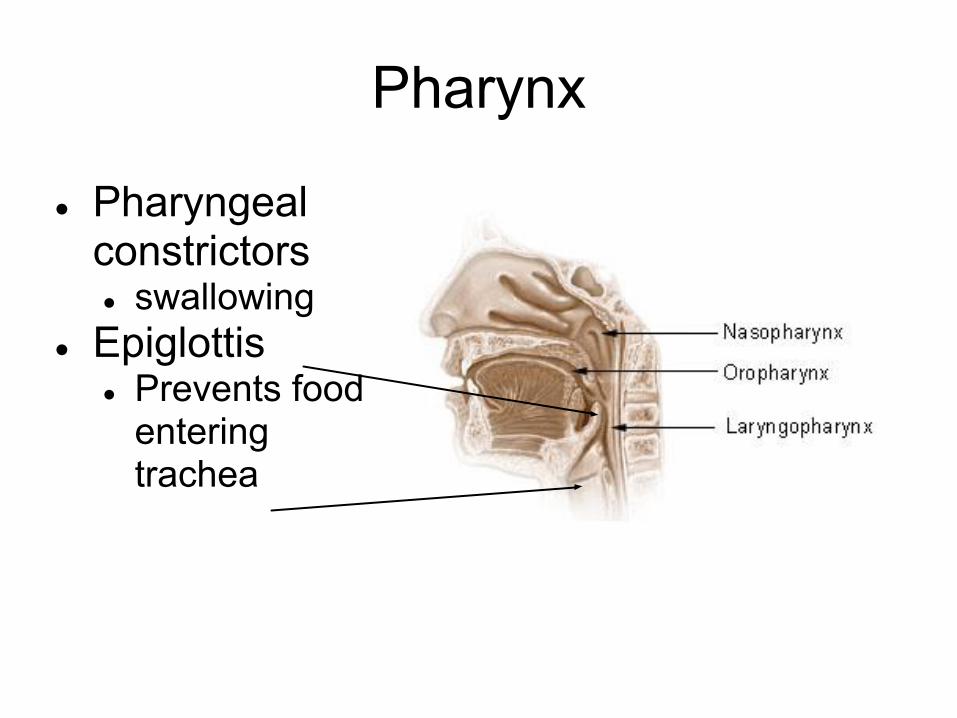
Pharynx
● Pharyngeal constrictors● swallowing
● Epiglottis ● Prevents food
entering trachea

● Requires 22 muscles!
● Tongue blocks oral cavity
● Soft palate blocks nasopharynx
● Epiglottis blocks trachea
Swallowing (or Deglutition)

● Tongue blocks oral cavity● Soft palate blocks
nasopharynx● Epiglottis blocks trachea
Swallowing (or Deglutition)

Esophagus● Straight,
muscular tube● about 10"● Peristalsis
● collapsed when empty
● Esophageal glands● Secrete mucus

Esophagus● Esophageal hiatus
● Penetration of diaphragm● Helps restrict backflow
● Lower esophageal sphincter● Not a separate muscle● Constriction of the esophagus
● Cardiac orifice● Entry into stomach● surrounded by a thickening
of the muscles - cardiac sphincter

Stomach● Volume
● 50ml empty● 1-1.5L full● Can extend to 4L
● Mechanical digestion● Due to churning action
● Chemical digestion● Fats● proteins
● Chyme ● Pasty soup of
semidigested food

Stomach regions
esophagus Cardiac orifice
Cardiac region Fundic region●Superior tocardiac orifice
Body – largest region
Pyloric region●Antrum – funnel like area●Canal – narrower area
Pylorus- Narrow passage

Stomach musculature
● Oblique ● Circular● Longitudinal
● Gastric rugae● Not muscles!● allow
stomach to expand

Stomach lining
● entirely mucus cells● 2 layers of
mucus● both alkaline● top is viscous,
insoluble● Gastric pits
● Open into 2-3 glands

Gastric glands
● Mucous cells● thin acidic mucus
● Regenerative (stem) cells● Lining is replaced every 3-
6 days● Parietal cells
● Secrete HCl and intrinsic factor
● Chief cells● Secrete pepsinogen
● Enteroendocrine cells● Secrete hormones● (G cell here) Produce 2-3L of gastric
juice each day!

Gastric secretions● HCl
● Activates lingual lipase and pepsin● Softens connective tissues and plant cell walls● denatures proteins● Destroys ingested pathogens● Converts ferric (Fe3+) ions to ferrous (Fe2+) ions
● More absorbable● Pepsinogen
● Converted to pepsin● Digests proteins
● Intrinsic factor● Binds with B12 to make it absorbable● Only indespensable function of the stomach!

Digestion and absorption in the stomach
● SMALL amounts of digestion● Starch● Fats● Proteins
● SMALL amounts of absorption● Aspirin● Some lipid-soluble drugs● alcohol

Liver● Body’s largest
gland (3 lbs)● Many functions -
only one important for digestion● Secretes bile

Liver● composed of
lobules● sesame sized● hexagonal
structures

Liver microanatomy-Hepatic lobules
Branch of hepatic portal vein
Bile ductule-receives bile from bile caniculi –narrow channels between hepatocytes
Sinusoids- Blood filled
Central vein
Liver cells (hepatocytes)Branch ofhepatic artery
Hepatic triad-artery-vein-bile duct

Liver microanatomy- Hepatic lobules
● filtration unit● blood from
arteriole and venule move through sinusoids
● blood moves toward central vein

Liver microanatomy- Hepatic lobules
● Kupffer cells○ macrophages
that remove bacteria, old blood cells and other debris

Liver microanatomy- Hepatic lobules● hepatocytes
○ store glucose as glycogen
○ make plasma proteins
○ store fat soluble vitamins
○ convert ammonia to urea
○ detoxifies blood
○ regenerate if injured
○ create bile

Liver microanatomy- Hepatic lobules● bile moves into
bile canaliculi to bile duct
● note blood and bile move countercurrent

Bile ducts● Bile flows from
bile ductules● to right and left
hepatic ducts● To common
hepatic duct● To common bile
duct● to
hepatopancreatic duct
● To gallbladder
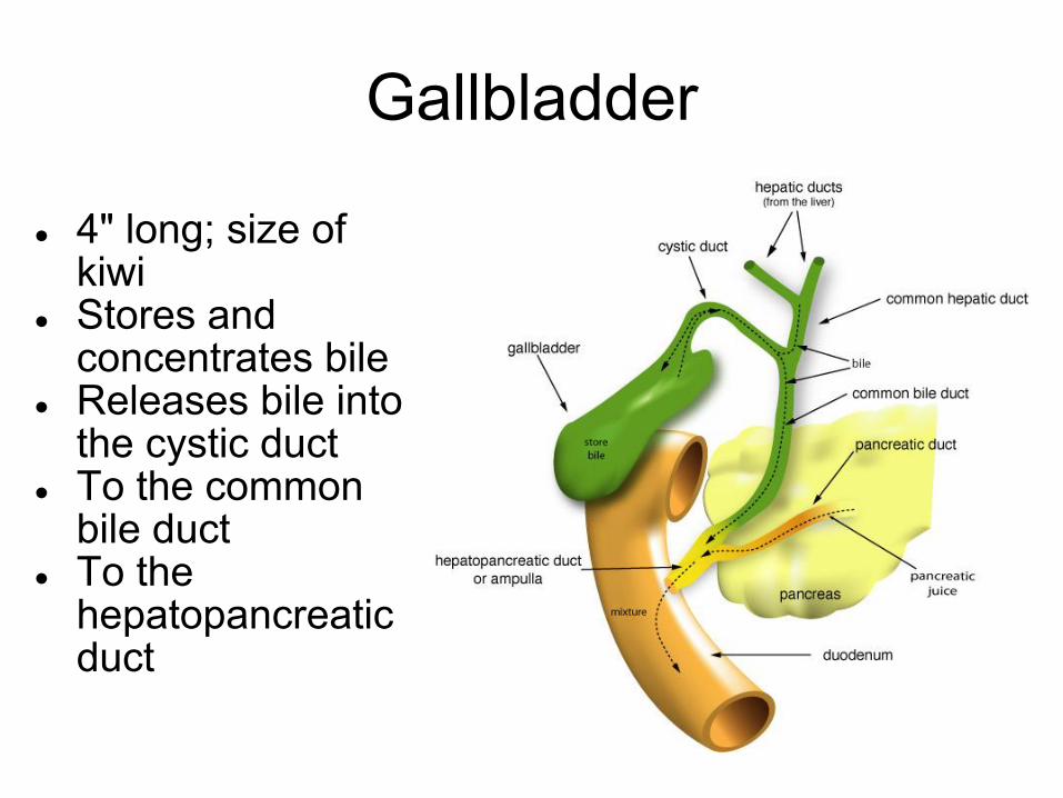
Gallbladder
● 4" long; size of kiwi
● Stores and concentrates bile
● Releases bile into the cystic duct
● To the common bile duct
● To the hepatopancreatic duct

Bile● Yellowish green fluid● Contains
● bile salts● cholesterol derivatives● emulsify fats● help keep cholesterol in solution● facilitate fat and cholesterol absorption● not excreted - are reabsorbed in the ileum
● Cholesterol● triglycerides ● Phospholipids● Bile pigments
● Bilirubin – decomposed hemoglobin● processed by bacteria to give feces their color

Pancreas
● Both an endocrine and exocrine gland
● Exocrine function relates to digestion

Pancreas
● Secretes 1.2-1.5L of pancreatic juice each day● Thru pancreatic
duct● To both the
hepatopancreatic duct and the accessory pancreatic duct
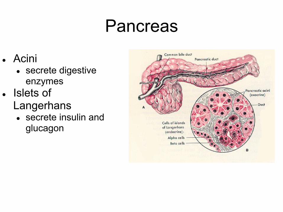
Pancreas
● Acini● secrete digestive
enzymes● Islets of
Langerhans● secrete insulin and
glucagon

Pancreatic juice
● Sodium bicarbonate● Buffers the HCl (exactly!)
● Enzymes precursors● Trypsinogen ● Chymotrypsinogen● procarboxylase
● Enzymes● Pancreatic amylase● Pancreatic lipase● Ribonuclease● Deoxyribonuclease
● Electrolytes (ions)
1.Trypsinogen is converted to trypsin by enteropeptidase (secreted by small intestine)
2.Trypsin converts chymotrypsinogen into chymotrypsin
3.Trypsin converts procarboxykpeptidase into carboxypeptidase

Small Intestine - regions● Duodenum – 10
inches● From pyloric valve to
duodenojejunal flexure
● Bile and pancreatic juice enter here
● glands produce alkaline mucus
● counteract HCl
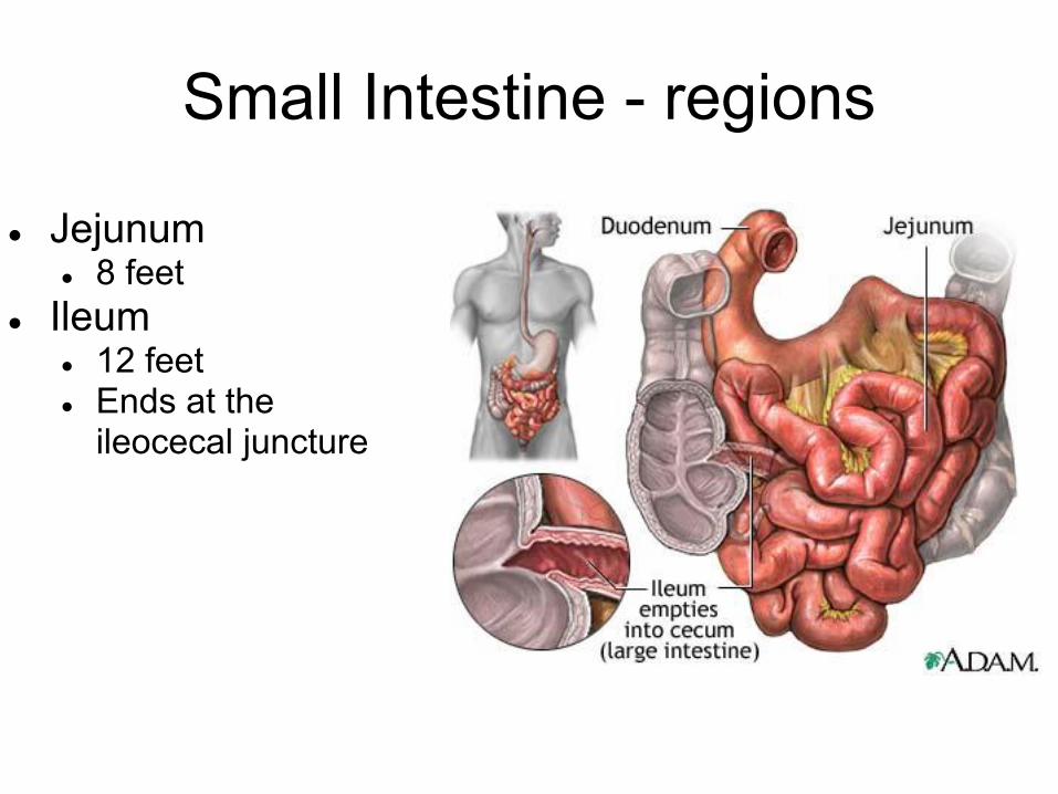
Small Intestine - regions
● Jejunum● 8 feet
● Ileum ● 12 feet● Ends at the
ileocecal juncture

● Circular folds● 1 cm tall● Duodenum through
middle of ileum● Force chyme in a
spiral path● slows movement of
chyme● increases contact of
chyme with intestine

● Villi● about 1mm tall● Largest in
duodenum, smallest in ileum
● 2 cell types● Absorptive cells● Goblet cells –
secrete mucus

● Villi● Arteriole,
capillary bed, venule
● Absorb most nutrients
● Lacteal● Lymphatic
capillary● Absorbs fat
● smooth muscle● shortens and
lengthens the villus
● moves lymph● increases
contact with chyme
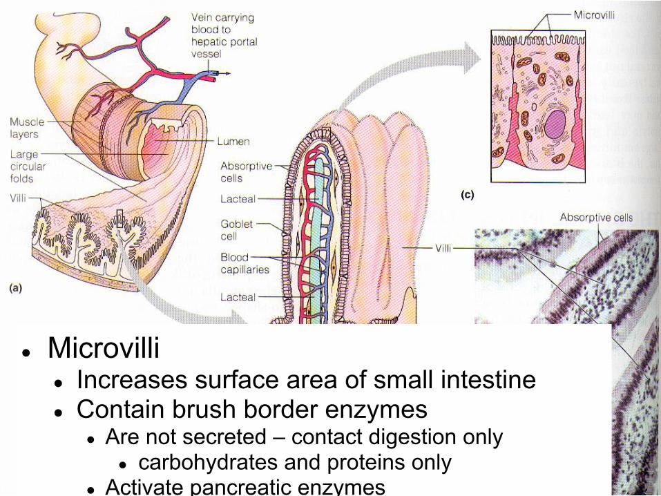
● Microvilli● Increases surface area of small intestine● Contain brush border enzymes
● Are not secreted – contact digestion only● carbohydrates and proteins only
● Activate pancreatic enzymes

Intestinal gland (or crypt)● Similar to gastric
gland● secrete intestinal
juice● Paneth cells
○ determine flora○ Secrete lysozyme,
phospholipase, defensins■ All antibacterial
●1-2 L of intestinal juice is secreted each day●pH of 7.4-7.8●Mostly water and mucus●no enzymes!

Intestinal gland (or crypt)● enteroendocrine
cells● secretin and
cholecystokinin● T cells
● bind with antigens and kill target cells
● Stem cells that resurface the villi● 2-4 days
●

Function of the small intestine
● Segmentation● occurs when "loaded"● massages the chyme back and forth● chyme moves very slowly along
● Peristalsis● occurs after most nutrients have been absorbed● first wave starts near duodenum● subsequent waves begin further along● all material (food, bacteria, debris) is moved out -
critical to prevent bacterial overgrowth● takes about 2 hours

Function of the small intestine
● Carbohydrate digestion overview● In mouth, salivary amylase
● Begins breakdown of starch● Continues in stomach until acid level reduces to pH 4.5● 50% of starch may be broken down
● In duodenum, pancreatic amylase● Finishes starch digestion (mostly to maltose) within 10
minutes● Brush border, maltase, sucrase, lactase
● dextrinase and glucoamylase - digest oligosaccharides● maltase, sucrase, lactose ● Converts remaining carbs to glucose

Function of the small intestine
● Carbohydrate absorption● glucose and
galactose: active transport (co-transport with Na+)
● fructose: facilitated diffusion
Function of small intestine
● Protein digestion● 3 sources of proteins
● Dietary● Digestive enzymes● Sloughed epithelial cells
● In stomach, pepsin● Digests 10-15% into short polypeptides (some amino acids)● Inactivated in duodenum due to increase in pH
● In duodenum, trypsin and chymotrypsin● Create short peptides
● Brush border, carboxypeptidase, aminopeptidase, dipeptidase
● Create amino acids

Function of small intestine
● Amino acids are absorbed ● active transport (co-transport with Na+)
● dipeptides and tripeptides● active transport (H+ dependent)● these are digested to amino acids within the epithelial
cells

Function of small intestine
● Lipid digestion● In mouth, lingual lipase
● Activated by acid in stomach● Digests 10% of ingested fat
● In duodenum, bile salts● Emulsifies fat to increase surface area
● In duodenum, pancreatic lipase● Digests fat in 1-2 minutes● Result of digestion is 2 fatty acids and a monoglyceride

Function of small intestine● fatty acids and monoglycerides
associate with bile salts and lecithin to create micelles
● At epithelium, fatty acids and monoglycerides are absorbed via simple diffusion
● inside epithelium, triglycerides reform● chylomicrons form: triglycerides +
phospholipids, lecithin, cholesterol● exocytosis of chylomicrons to lacteals● once in blood stream: triglycerides are
digested to fatty acids and glycerol● remaining substances are
processed by the liver

Function of small intestine
● Nucleic acid digestion● In duodenum, pancreatic nucleases
● Create nucleotides● Brush border, nucleosidases and
phosphatases● Create phosphate, ribose or deoxyribose,
nitrogenous bases● Phosphate, sugar and bases are absorbed
● all active transport

Function of small intestine● Vitamin absorption
● fat soluble (A, D, E, and K)● incorporate into micelles
● water soluble (B and C)● diffusion or active transport● B12 - very large
● intrinsic factor binds to it
● complex binds to mucosal receptors (in terminal ileum)
● uptake is via endocytosis

Function of small intestine● Electrolyte absorption
● most ions actively absorbed● Na+ - coupled to glucose and amino acid
absorption● K+ - facilitated diffusion● Iron
● active transport into mucosa where it binds to ferritin: this is a local iron storage
● women have 4x as much as men● If iron is needed, iron is transferred to transferrin - a
plasma protein● Calcium
● regulated by vitamin D dependent calcium binding protein
Function of small intestine
● Water is absorbed● Intestine receives 9L of water a day!
● 0.7L in food● 1.6L in drink● 6.7L in gastrointestinal secretions
● 9L is absorbed● 95% is absorbed via osmosis in small
intestine (remainder (all but 0.1L) is absorbed in large intestine

● 1.5m long● Functions
● absorption● formation of
feces● Haustra
● puckers due to muscle tone
Large Intestine

● Cecum● Blind pouch● Appendix
● Significant source of immune cells (lymphocytes)
Large Intestine

● Colon● Ascending
colon● Transverse
colon● Descending
colon● Sigmoid colon
Large Intestine

● Rectum● 3 Rectal valves
● Transverse folds
● Retain feces● Pass gas
Large Intestine
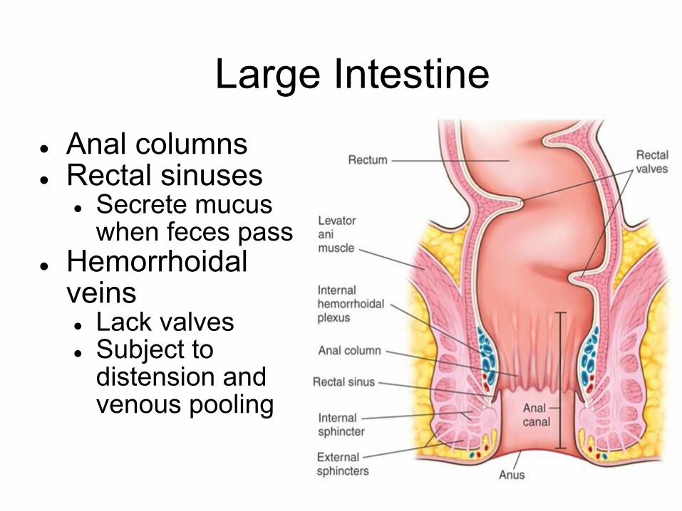
● Anal columns● Rectal sinuses
● Secrete mucus when feces pass
● Hemorrhoidal veins● Lack valves● Subject to
distension and venous pooling
Large Intestine

● Internal sphincter● involuntary
● External sphincter● voluntary
Large Intestine

Functions of the colon
● Passage takes 12-24 hours● haustral contractions
● segmental movement that last 1 min. and occur every 30 min.
● Mass movements● peristalsis over large areas that occur 3-4x daily
● Diverticula: breaks in the mucosa● if diet lacks bulk, colon narrows and these form
(usually in sigmoid colon)● diverticulitis = inflamed diverticula

Functions of the colon
● Bacterial flora● Ferment cellulose, xylan and other carbs (from food)● metabolize mucin, heparin, hyaluronic acid (produced
by our body)● Synthesize B vitamins● Synthesize vitamin K (necessary for blood clotting
proteins)● Absorbs vitamins● Reabsorbs water and electrolytes (NaCl)
● 0.8L water is absorbed
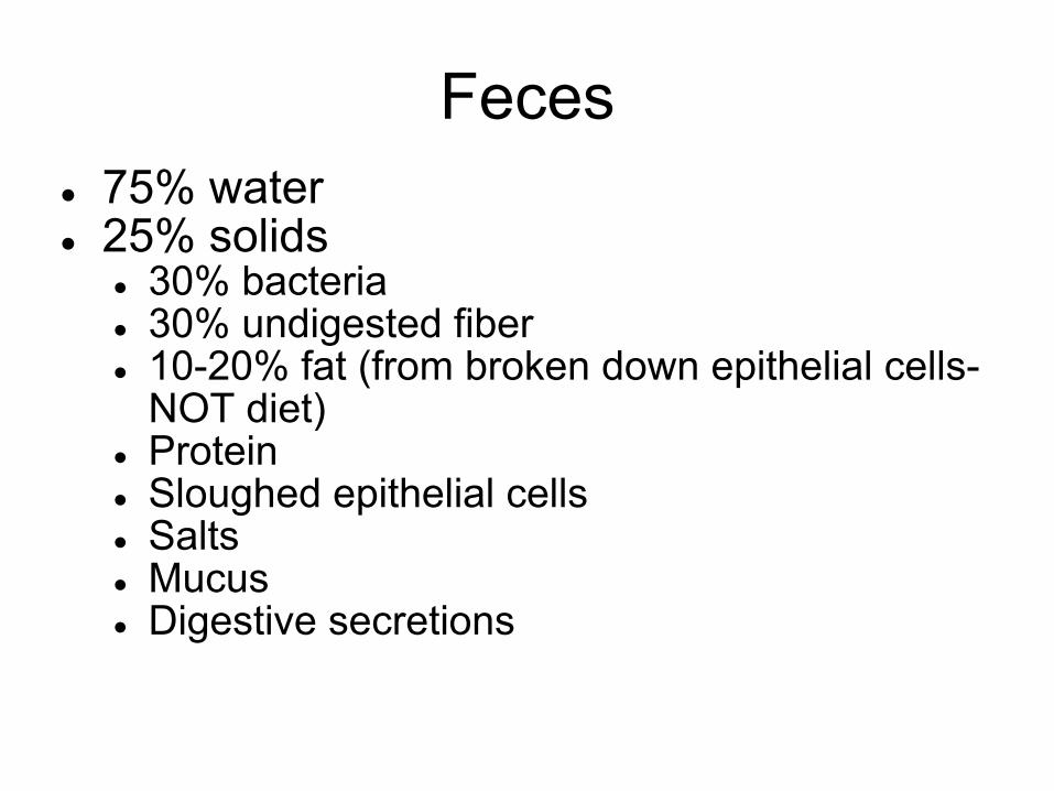
Feces● 75% water● 25% solids
● 30% bacteria● 30% undigested fiber● 10-20% fat (from broken down epithelial cells-
NOT diet)● Protein● Sloughed epithelial cells● Salts● Mucus● Digestive secretions