Embed Size (px)
Citation preview

Bull. Natl. Mus. Nat. Sci., Ser. A, 42(1), pp. 1–22, February 22, 2016
Digeneans Parasitic in Freshwater Fishes (Osteichthyes) of Japan VI. Lissorchiidae
Takeshi Shimazu
10486–2 Hotaka-Ariake, Azumino, Nagano 399–8301, Japan E-mail: [email protected]
(Received 30 November 2015; accepted 22 December 2015)
Abstract Digeneans (Trematoda, Monorchioidea, Lissorchiidae) parasitic in freshwater fishes of Japan are reviewed: Asymphylodora innominata (Faust, 1924), Asymphylodora japonica Yama-guti, 1938, Asymphylodora sp. of Shimazu, Urabe, and Grygier, 2011, Asymphylotrema monosty-loides (Ito, 1960) comb. nov., and Palaeorchis diplorchis (Yamaguti, 1936). Each species is described and figured with a summarized life cycle where known. Partial sequences (653 bp) of the cytochrome c oxidase I gene of the mitochondrial DNA (COI mtDNA) were identical between adults of Asymphylotrema hamajimai (Fujino and Kifune, 1991) (syn. Anapalaeorchis hamajimai Fujino and Kifune, 1991) and rediae of Cercaria monostyloides Ito, 1960. This result lends further support to the previous hypothesis that C. monostyloides is the cercaria of As. hamajimai. Since these two species are synonymous, the new combination As. monostyloides is made for As. hama-jimai. A key to the three genera and five species of the family Lissorchiidae in Japan is presented.Key words : Digeneans, Lissorchiidae, Asymphylodora, Asymphylotrema, Palaeorchis, Asymphy-lotrema monostyloides (Ito, 1960) comb. nov., partial COI sequences, freshwater fishes, Japan, review.
Introduction
This is the sixth paper of a series that reviews adult digeneans (Trematoda) parasitic in fresh-water fishes (Osteichthyes) of Japan (Shimazu, 2013). This contribution deals with the family Lissorchiidae Magath, 1917 sensu Bray (2008b) in the superfamily Monorchioidea Odhner, 1911 sensu Bray (2008a). Shimazu (2013) gave the Introduction and Materials and Methods for the review.
Abbreviations used in the figures. bp, birth pore; c, cercaria; cp, cirrus pouch; csd, common sperm duct; cvd, common vitelline duct; e, esophagus; ed, ejaculatory duct; egg, egg in uterus and metraterm; ep, excretory pore; ev, excretory vesicle; ga, genital atrium; gp, genital pore; i, intestine; Lc, Laurer’s canal; m, metra-term; md, male duct; me, metacercaria; Mg, Mehlis’ gland; o, ovary; od, oviduct; os, oral
sucker; ot, ootype; p, pharynx; pc, prostatic cells; pp, pars prostatica; pr; prepharynx; s, stylet; sd, sperm duct; sr, seminal receptacle; sv, seminal vesicle; t, testis; tnc, transverse nerve commis-sure; u, uterus; usr, uterine seminal receptacle; v, vitellarium; vd, vitelline duct; vs, ventral sucker.
Superfamily Monorchioidea Odhner, 1911 Family Lissorchiidae Magath, 1917 Genus Asymphylodora Looss, 1899
Asymphylodora innominata (Faust, 1924)
(Figs. 1–9)
Cercaria H: Kobayashi, 1918: 70–73, 1 pl., fig. 16.Cercaria VIII, or [Shin]: Ando, 1918: 616, 1 pl., fig. 8b.Cercariaeum A: Kobayashi, 1922: 266–267.Cercariaeum innominatum Faust, 1924: 295, table 1.Asymphylodora macrostoma Ozaki, 1925: 104–106, fig. 4;
Yamaguti, 1934: 393; Tang, 1962: 162–163, fig. 2; Shimazu, 1992: 8–10, figs. 6–11; Shimazu and Urabe, 2005: 11–12, figs. 18–20; Shimazu, 2008: 56–57, fig. 12.

2 Takeshi Shimazu
Cercaria T: Ishii in Ueno, Ishii, and Abe, 1930: 974, fig. 27.Parasymphylodora macrostoma: Szidat, 1943: 44–45,
table 1, fig. 12.Cercaria innominatum [sic, should be innominata]: Ito,
Mochizuki, and Noguchi, 1959: 918; Ito, 1960: 59–72, fig. 13; Ito, 1964: 501, fig. 140.
Asymphylodora (Asymphylodora) macrostoma: Yamaguti, 1971: 97.
Cercaria innominata: Shimazu, 2007: 18.Asymphylodora innominata: Shimazu, Urabe, and Grygier,
2011: 74, figs. 100–103.
Hosts in Japan. Odontobutis obscura (Tem-minck and Schlegel, 1845) (Odontobutidae) (type host) including “[Gori]” (Ozaki, 1925; Yamaguti, 1934; Shimazu, 1992, 1995, 2015; this paper), Opsariichthys uncirostris uncirostris (Temminck and Schlegel, 1846) (Cyprinidae) (Yamaguti, 1934; Shimazu, 1992; Shimazu et al., 2011), Tribolodon hakonensis (Günther, 1877) (Cyprinidae) (Yamaguti, 1934; Shimazu, 1992, 2007, 2008; Nakamura et al., 2000; Shimazu and Urabe, 2005; Shimazu et al., 2011; this paper), Hemibarbus barbus (Temminck and Schlegel, 1846) (Cyprinidae) (Yamaguti, 1934; Shimazu, 1992; Shimazu et al., 2011), Phoxinus steindach-neri Sauvage, 1883 (Cyprinidae) (Yamaguti, 1934; Shimazu, 1992), “[Ukikamatsuka?]” (Hemibarbus longirostris (Regan, 1908) [?]) (Shimazu 1992; Shimazu et al., 2011), “[Bote]” (an acheilognathine) (Cyprinidae) (Shimazu, 1992), and Gymnogobius isaza (Tanaka, 1916) (Gobiidae) (Shimazu, 1992; Shimazu et al., 2011).
Sites of infection. Intestine, rectum, and gut.Geographical distribution. (1) Ibaraki Pre-
fecture: Lake Kasumigaura at Tsuchiura City and Tsuchiura City (Yamaguti, 1934; Shimazu, 1992). (2) Saitama Prefecture: Oppe River at Ogose Town (Shimazu, 1992). (3) Toyama Pre-fecture: Namerikawa City (Yamaguti, 1934; Shimazu, 1992). (4) Nagano Prefecture: Hiroi River at Kotobuki, Iiyama City; Nogu River in Oomachi City; Torii River at Mure, Iizuna Town; and Lake Suwa at Suwa City (Shimazu, 1992, 2007; this paper). (5) Fukui Prefecture: Obama City (Shimazu, 1992). (6) Shiga Prefecture: Lake Biwa basin (Lake Biwa, Momose, Moriyama,
Omastu, Onoe, and Tenjin River) (Yamaguti, 1934; Shimazu, 1992; Shimazu et al., 2011; this paper). (7) Kyoto Prefecture: Yura River at Ayabe City and Lake Ogura (Yamaguti, 1934; Shimazu, 1992). (8) Nara Prefecture: Takami River at Kotsukawa, Higashiyoshino Village (Nakamura et al., 2000; Shimazu and Urabe, 2005). (9) Wakayama Prefecture: Tonda River at Kurisugawa, Nakahechi-cho, Tanabe City (Shimazu, 2008). (10) Osaka Prefecture (?): Yodo River (Yamaguti, 1934; Shimazu, 1992). (11) Hiroshima Prefecture: a brook (type local-ity) in the vicinity of Saijo-cho, Matsuita River at Umaki, Saijo-cho, and Irasuke River at Kane-sawa, Kurose-cho, Higashihiroshima City; Eno River at Yoshida, Yoshida-cho, Akitakata City; and Saijo River at Ooya, Saijo-cho, Shobara City (Ozaki, 1925; Shimazu, 1992; this paper). (12) Kochi Prefecture: Matsuda River at Idei, Hashikami-cho, Sukumo City (Shimazu, 2008). (13) Fukuoka Prefecture: Futatsu River at Taka-hatake, Mitsuhashi-machi, Yanagawa City (this paper). (14) Oita Prefecture: Chikugo River at Kobuchi Bridge, Miyoshikobuchi-machi, Hita City (this paper).
In Korea (cercaria) (Kobayashi, 1918) and China (e.g., Tang, 1962).
Material examined. (1) 3 specimens (Ozaki’s Collection, MPM Coll. No. 30028, species name not given, syntypes of Asymphylodora macro-stoma) of A. macrostoma, adult, whole-mounted, ex “[Gori]” (Odontobutis obscura [syn. Mogurnda obscura]) (other data not given) (Shimazu, 1992, 1995, 2015; this paper). (2) 1 (Yamaguti’s Collection, MPM Coll. No. 22280) of A. macrostoma, adult, whole-mounted, ex rec-tum of O. obscura, Lake Ogura, 4 May 1932 (Yamaguti, 1934; Shimazu, 1992). (3) 2 (NSMT-Pl 3683) of A. macrostoma, adult, whole-mounted, ex rectum of O. obscura (formalin-pre-served), Matsuita River, 16 April 1991 (Shimazu, 1992). (4) 11 (NSMT-Pl 5785, MPM Coll. No. 21161), adult, whole-mounted, ex intestine of O. obscura, Matsuita River, 18 June 2009, 9 June 2011. (5) 2 (NSMT-Pl 5786), immature, adult, whole-mounted, ex intestine of O. obscura,

Digeneans Parasitic in Freshwater Fishes of Japan 3
Irasuke River, 18 June 2009. (6) 8 (Urabe’s unpublished specimens) of A. mocrostoma, adult, whole-mounted, ex intestine of O. obscura, Futatsu River, 22–23 May 2003. (7) Specimens of A. innominata, whole-mounted, ex intestine of Opsariichthys uncirostris uncirostris (syn. Opsariichthys uncirostris), Lake Biwa basin (Yamaguti, 1934; Shimazu, 1992; Shimazu et al., 2011): 3 (Yamaguti’s Collection, MPM Coll. No. 22746), adult, Omatsu, 17 July 1927; 7 (NSMT-Pl 3700), adult, Omatsu, 30 April 1992; 119
(NSMT-Pl 3684–3686), immature, adult, Onoe, 5 May 1979, 3 June 1980, 11 November 1980; 9 (NSMT-Pl 3973), adult, Moriyama, 2 May 1992; and 58 (LBM 1-17 to -20), adult, Tenjin River, 21 May 1998; and 6 (LBM 5-8 to -10, hot forma-lin-fixed), adult, Momose, 1 May 2001. (8) 17 (Yamaguti’s Collection, MPM Coll. Nos. 22284, published; 22745, unpublished) of A. macro-stoma, adult, whole-mounted, ex intestine of Op. uncirostris uncirostris, Yodo River (locality not specified), 16 October 1929, 27 September 1928
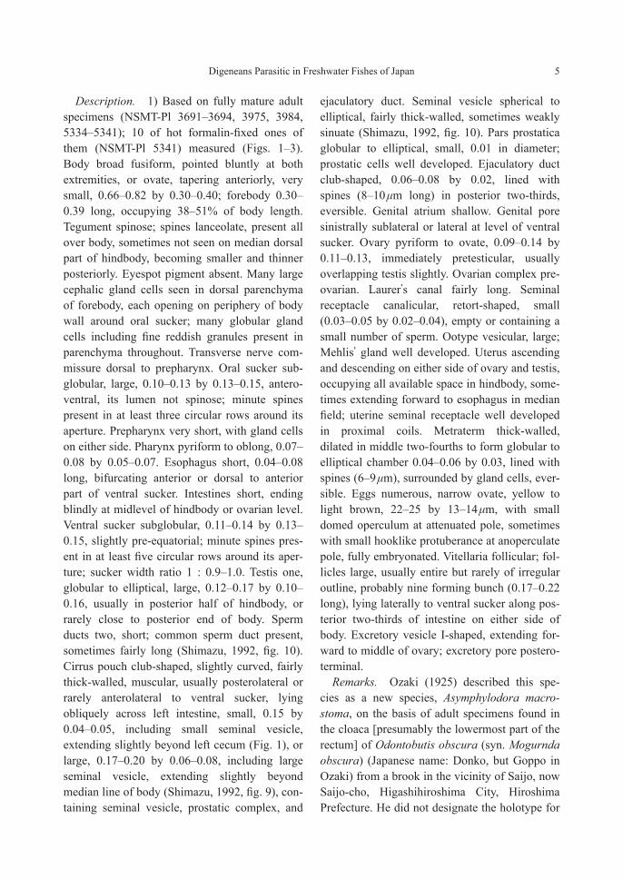
Figs. 1–4. Asymphylodora innominata, adult specimens. — 1, specimen (NSMT-Pl 3692) found in intestine of Tribolodon hakonensis, entire body, ventral view; 2, specimen (NSMT-Pl 5334) found in intestine of T. hako-nensis, terminal genitalia, ventral view; 3, specimen (NSMT-Pl 5334), ovarian complex, dorsal view; 4, speci-mens (NSMT-Pl 3685) found in intestine of Opsariichthys uncirostris uncirostris, vitelline follicles in young adult specimen (A) and old adult specimen (B), ventral view. Fig. 4 redrawn from Shimazu et al. (2011). Scale bars: 0.3 mm in Figs. 1 and 4; 0.1 mm in Figs. 2–3.

4 Takeshi Shimazu
(Yamaguti, 1934; Shimazu, 1992). (9) 2 (Yama-guti’s Collection, MPM Coll. No. 22278) of A. macrostoma, adult, whole-mounted, ex intestine of Tribolodon hakonensis (syn. Leuciscus hako-nensis), Namerikawa City, 28 October 1929 (Yamaguti, 1934; Shimazu, 1992). (10) 37 (NSMT-Pl 3691) of A. macrostoma, adult, whole-mounted, ex intestine of T. hakonensis, Nogu River, 1 July 1987 (Shimazu, 1992). (11) 247 (NSMT-Pl 3692, 5334, 5335) of A. macro-stoma, immature, adult, whole-mounted, ex intes-tine of T. hakonensis, Torii River at Mure Vil-lage, now Mure, Iizuna Town, 16 August 1987, 7 May 1994, 11 November 1995 (Shimazu, 1992, 2007). (12) 146 (NSMT-Pl 3693, 3694, 3975, 3984, 5336) of A. macrostoma, immature, adult, whole-mounted, ex intestine of T. hakonensis, Lake Suwa, 5 October 1991, 14 November 1991, 19 May 1992, 2 June 1992, 14 September 1992 (Shimazu, 1992, 2007). (13) 50 (NSMT-Pl 5337–5341, slightly flattened and hot formalin-fixed) of A. macrostoma, immature, adult, whole-mounted, ex intestine of T. hakonensis, Hiroi River, 16 October 1999, 13 November 1999, 8 October 2004, 4–5 November 2004, 1 and 3 December 2004 (Shimazu, 2007). (14) 6 (NSMT-Pl 3689) of A. macrostoma, adult, whole-mounted, ex intestine of T. hakonensis, Oppe River, 13 October 1976 (Shimazu, 1992). (15) 2 (Yamaguti’s Collection, MPM Coll. No. 22274, unpublished) of A. macrostoma, immature, adult, whole-mounted, ex intestine of T. hakonensis, Obama City, 27 March 1935 (Shimazu, 1992). (16) Specimens of A. innominata, adult, whole-mounted, ex intestine of T. hakonensis, Lake Biwa basin (Shimazu, 1992; Shimazu et al., 2011): 8 (NSMT-Pl 3632, 3690), Onoe, 3 June 1980, 4 February 1980; 8 (NSMT-Pl 3974), Omatsu, 30 April 1992. (17) 46 (NSMT-Pl 5274, 5275) of A. macrostoma, immature, adult, whole-mounted, ex intestine of T. hakonensis, Takami River, 26 and 30 July 1999, 16 August 2000 (Shimazu and Urabe, 2005; see also Shimazu, 2013 for measurements). (18) 6 (NSMT-Pl 5556) of A. macrostoma, adult, whole-mounted, ex intestine of T. hakonensis, Tonda River, 4 August
1999 (Shimazu, 2008). (19) 8 (NSMT-Pl 3687) of A. macrostoma, adult, whole-mounted, ex intestine of T. hakonensis, Eno River, 30 October 1976 (Shimazu, 1992). (20) 19 (NSMT-Pl 3688) of A. macrostoma, adult, whole-mounted, ex intestine of T. hakonensis, Saijo River, 31 Octo-ber 1976 (Shimazu, 1992). (21) 14 (NSMT-Pl 5557) of A. macrostoma, adult, whole-mounted, ex intestine of T. hakonensis, Matsuda River, 5 August 2000 (Shimazu, 2008). (22) 4 (Urabe’s unpublished specimens) of A. macrostoma, adult, whole-mounted, ex intestine of T. hakonensis, Chikugo River, 25 August 2003. (23) 4 (Yamagu-ti’s Collection, MPM Coll. Nos. 22277, 22743) of A. macrostoma, adult, whole-mounted, ex intestine of Hemibarbus barbus, Lake Kasumi-gaura at Tsuchiura, 16 April 1929 (Yamaguti, 1934; Shimazu, 1992). (24) 1 (NSMT-Pl 3976) of A. macrostoma, immature, whole-mounted, ex intestine of H. barbus, Onoe, 4 May 1992 (Shimazu, 1992; Shimazu et al., 2011). (25) 1 (Yamaguti’s Collection, MPM Coll. No. 22747, labeled “Asymphylodora,” unpublished) of A. innominata, immature, whole-mounted, ex intes-tine of “[Ukikamatsuka?]” (Hemibarbus longi-rostris [?]), Lake Biwa, 4 December 1938 (Shimazu, 1992; Shimazu et al., 2011). (26) 7 (NSMT-Pl 3977) of A. innominata, adult, whole-mounted, ex intestine of Gymnogobius isaza (syn. Chaenogobius isaza), Omatsu, 30 April 1992 (Shimazu, 1992; Shimazu et al., 2011). (27) 1 (Yamaguti’s Collection, MPM Coll. No. 22748, labeled “Asymphylodora,” unpublished) of A. macrostoma, immature, whole-mounted, ex “[Bote]” (an acheilognathine), immature, whole-mounted, Tsuchiura, 4 April 1940 (Shimazu, 1992). (28) 3 (Yamaguti’s Collection, MPM Coll. No. 22279) of A. macrostoma, adult, whole-mounted, ex intestine of Phoxinus steindachneri (syn. Moroco steindachneri), Yura River, 20 November 1929 (Yamaguti, 1934; Shimazu, 1992). (29) 10 (Yamaguti’s Collection, MPM Coll. No. 22774, unpublished) of A. macrostoma, adult, whole-mounted, ex intestine of Ph. stein-dachneri, Obama City (date not given) (Shimazu, 1992).
Digeneans Parasitic in Freshwater Fishes of Japan 5
Description. 1) Based on fully mature adult specimens (NSMT-Pl 3691–3694, 3975, 3984, 5334–5341); 10 of hot formalin-fixed ones of them (NSMT-Pl 5341) measured (Figs. 1–3). Body broad fusiform, pointed bluntly at both extremities, or ovate, tapering anteriorly, very small, 0.66–0.82 by 0.30–0.40; forebody 0.30–0.39 long, occupying 38–51% of body length. Tegument spinose; spines lanceolate, present all over body, sometimes not seen on median dorsal part of hindbody, becoming smaller and thinner posteriorly. Eyespot pigment absent. Many large cephalic gland cells seen in dorsal parenchyma of forebody, each opening on periphery of body wall around oral sucker; many globular gland cells including fine reddish granules present in parenchyma throughout. Transverse nerve com-missure dorsal to prepharynx. Oral sucker sub-globular, large, 0.10–0.13 by 0.13–0.15, antero-ventral, its lumen not spinose; minute spines present in at least three circular rows around its aperture. Prepharynx very short, with gland cells on either side. Pharynx pyriform to oblong, 0.07–0.08 by 0.05–0.07. Esophagus short, 0.04–0.08 long, bifurcating anterior or dorsal to anterior part of ventral sucker. Intestines short, ending blindly at midlevel of hindbody or ovarian level. Ventral sucker subglobular, 0.11–0.14 by 0.13–0.15, slightly pre-equatorial; minute spines pres-ent in at least five circular rows around its aper-ture; sucker width ratio 1 : 0.9–1.0. Testis one, globular to elliptical, large, 0.12–0.17 by 0.10–0.16, usually in posterior half of hindbody, or rarely close to posterior end of body. Sperm ducts two, short; common sperm duct present, sometimes fairly long (Shimazu, 1992, fig. 10). Cirrus pouch club-shaped, slightly curved, fairly thick-walled, muscular, usually posterolateral or rarely anterolateral to ventral sucker, lying obliquely across left intestine, small, 0.15 by 0.04–0.05, including small seminal vesicle, extending slightly beyond left cecum (Fig. 1), or large, 0.17–0.20 by 0.06–0.08, including large seminal vesicle, extending slightly beyond median line of body (Shimazu, 1992, fig. 9), con-taining seminal vesicle, prostatic complex, and
ejaculatory duct. Seminal vesicle spherical to elliptical, fairly thick-walled, sometimes weakly sinuate (Shimazu, 1992, fig. 10). Pars prostatica globular to elliptical, small, 0.01 in diameter; prostatic cells well developed. Ejaculatory duct club-shaped, 0.06–0.08 by 0.02, lined with spines (8–10 μm long) in posterior two-thirds, eversible. Genital atrium shallow. Genital pore sinistrally sublateral or lateral at level of ventral sucker. Ovary pyriform to ovate, 0.09–0.14 by 0.11–0.13, immediately pretesticular, usually overlapping testis slightly. Ovarian complex pre-ovarian. Laurer’s canal fairly long. Seminal receptacle canalicular, retort-shaped, small (0.03–0.05 by 0.02–0.04), empty or containing a small number of sperm. Ootype vesicular, large; Mehlis’ gland well developed. Uterus ascending and descending on either side of ovary and testis, occupying all available space in hindbody, some-times extending forward to esophagus in median field; uterine seminal receptacle well developed in proximal coils. Metraterm thick-walled, dilated in middle two-fourths to form globular to elliptical chamber 0.04–0.06 by 0.03, lined with spines (6–9 μm), surrounded by gland cells, ever-sible. Eggs numerous, narrow ovate, yellow to light brown, 22–25 by 13–14 μm, with small domed operculum at attenuated pole, sometimes with small hooklike protuberance at anoperculate pole, fully embryonated. Vitellaria follicular; fol-licles large, usually entire but rarely of irregular outline, probably nine forming bunch (0.17–0.22 long), lying laterally to ventral sucker along pos-terior two-thirds of intestine on either side of body. Excretory vesicle I-shaped, extending for-ward to middle of ovary; excretory pore postero-terminal.
Remarks. Ozaki (1925) described this spe-cies as a new species, Asymphylodora macro-stoma, on the basis of adult specimens found in the cloaca [presumably the lowermost part of the rectum] of Odontobutis obscura (syn. Mogurnda obscura) (Japanese name: Donko, but Goppo in Ozaki) from a brook in the vicinity of Saijo, now Saijo-cho, Higashihiroshima City, Hiroshima Prefecture. He did not designate the holotype for
6 Takeshi Shimazu
this species. Shimazu (2015) regarded the host fish “[Gori]” as O. obscura. I consider that the present three adult specimens of Ozaki (MPM Col. No. 30028) as syntypes of A. macrostoma. Shimazu (1992, fig. 7) briefly described and fig-ured them. The name of the brook has not yet been determined.
Tang (1962) erected a new genus, Oriento-trema, for Asymphylodora japonica Yamaguti, 1938 (type species) and A. macrostoma; but he did not make a new combination for A. macro-stoma. Shimazu (1992) and Bray (2008b) did not accept this new genus.
Kobayashi (1918) described a tailless cercaria (cercariaeum), Cercaria H, found in the gonads and around their ducts of Melania spp. [now Semisulcospira spp.] (Pleuroceridae) from Korea and added that this cercaria also occurred in Japan. Ando (1918) described Cercaria VIII, or [Shin], found in Semisulcospira libertina (Gould, 1859) (syn. Melania libertina) from Hiramaki Village, now in Kani City, and Kaminogo Vil-lage, now in Mitake Town, Gifu Prefecture, Japan. Cercaria H and Cercaria VIII are the same species (Ito, 1960, 1964). Faust (1924) named Cercaria H Cercariaeum innominatum. Since Shimazu (2007) had experimentally demon-strated that Cercariaeum innominatum or Cer-caria innominata was the larva of Asymphylo-dora macrostoma, Shimazu et al. (2011) made a new combination, A. innominata (Faust, 1924), for A. macrostoma. Ito (1964) also assigned the cercaria of Okumura (1919) (locality not given) and Cercaria T of Ishii in Ueno et al. (1930) (locality not given) to Cercaria innominata.
Kobayashi (1922) grouped Cercaria H and Cercaria VIII into Cercariaeum A and stated that Cercariaeum A was parasitic in Semisulcospira spp. and the bithyniid snail Parafossarulus man-chouricus japonicus (Pilsbry, 1901) (syn. Buli-mus striatulus japonicus) in Okayama, Shiga, and Gifu Prefectures and Korea without citing any references concerning the cercaria in Pa. manchouricus japonicus. Shimazu et al. (2011) questioned whether Cercariaeum innominatum and Cercariaeum A were the same species. It
seems evident that Kobayashi (1922) erroneously identified the cercaria of Asymphylodora japon-ica Yamaguti, 1938 in Pa. manchouricus japoni-cus with Cercariaeum A owing to a morphologi-cal similarity between these two cercariae (see the next species A. japonica).
Ozaki (1925) described that the oral sucker was larger than the ventral sucker, with a sucker width ratio of 1 : 0.8 (my calculation). The oral sucker was larger in cercariae (Fig. 6), metacer-cariae (Fig. 8) and barely matured adults, but sometimes slightly smaller in fully matured adults (Fig. 1) (Shimazu, 1992, 2008; Shimazu and Urabe, 2005; Shimazu et al., 2011; this paper). Ozaki (1925) described that the cirrus pouch was slender and slightly larger than an elongated spined vagina, or the spined chamber of the metraterm (this paper). However, the cir-rus pouch was about twice as long as the spined chamber (Shimazu, 1992, 2008; Shimazu and Urabe, 2005; Shimazu et al., 2011; this paper) and in Ozaki’s specimens (MPM Coll. No. 30028) (Shimazu, 1992, fig. 7) and the new spec-imens (NSMT-Pl 5785–5786) from Higashihiro-shima City. Ozaki (1925, fig. 4) described that the vitellaria consist of a number of follicles. Probably, nine follicles form a bunch on either side of the body. They are large and entire in young adult specimens, but lobed (of irregular outline) in old adult specimens (Shimazu, 1992; Shimazu et al., 2011; this paper, Fig. 4). The presence of minute spines around the apertures of both suckers has previously been overlooked in adult specimens. Ito (1960, 1964) described five and seven circular rows of pointed spines on the oral and ventral suckers, respectively, in the cercaria (Cercaria innominata). I counted at least three and five circular rows, respectively.
Yamaguti (1934) claimed to find A. macro-stoma in Pseudorasbora parva (Temminck and Schlegel, 1846) (Cyprinidae) from Lake Kobata [Kohata Pond or Kowata Pond] in Uji City, Kyoto Prefecture; but Yamaguti’s Collection con-tained no specimen from this fish species (Shimazu, 1992). Kuyama (1938) found adults, which were similar to a European species,

Digeneans Parasitic in Freshwater Fishes of Japan 7
Asymphylodora tincae (Modeer, 1790), in Ps. parva from an irrigation canal in Okita Village, now in Okayama City, Okayama Prefecture. It is possible that they may have belonged to A. macrostoma, though they have not been described. Tang (1962) found adults of A. macrostoma in a small cyprinid, Punctius sp., from Fujian Province, China, where Melania peregrinorum Heude was infected with cercariae.
Asymphylodora innominata and A. japonica (see below) have a single testis but two sperm ducts. Bray (2008b) regarded this as a reduction of two testes into one. However, it appears to me
that this is an undivision of a single testicular pri-mordium that fails to divide into two.
Life cycle. The first intermediate hosts in Japan are pleurocerid snails, Semisulcospira libertina (Japanese name: Kawanina), Semi-sulcospira dolorosa (Gould, 1859) (Japanese name: Kitano-kawanina), Semisulcospira (Biwa-melania) habei Davis, 1969 (Japanese name: Habe-kawanina), Semisulcospira (Biwamelania) niponica (Smith, 1876) (Japanese name: Yamato-kawanina), and Semisulcospira (Biwamelania) nakasekoae Kuroda, 1929 (Japanese name Nakaseko-kawanina) (e.g., Ando, 1918; Ito,
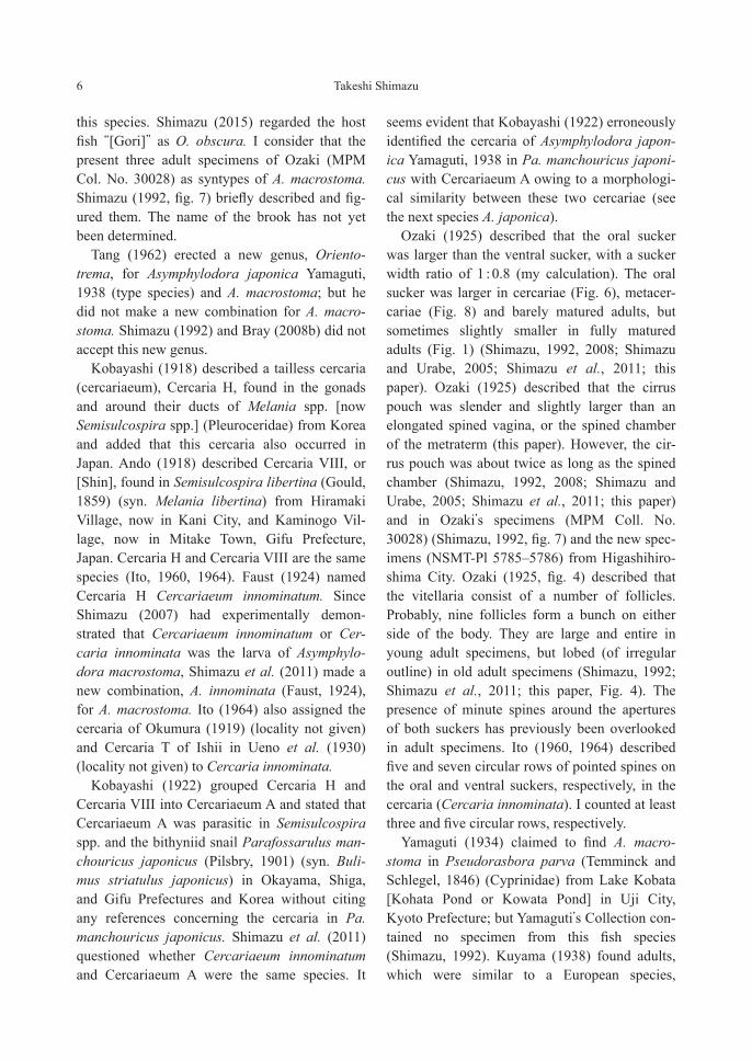
Figs. 5–8. Asymphylodora innominata, life cycle. — 5, daughter (?) redia (NSMT-Pl 5347) found in Semisulco-spira libertina; 6, cercaria (NSMT-Pl 5345), ventral view; 7, encysted metacercaria (NSMT-Pl 5352) found in connective tissue of mucous membrane of gill raker of Pseudorasbora parva, 16 days after experimental infection, surrounded by tissue of host origin (*); 8, excysted metacercaria (NSMT-Pl 5349), 14 days after experimental infection. Scale bars: 0.5 mm in Fig. 5; 0.2 mm in Figs. 6–8.

8 Takeshi Shimazu
1960, 1964; Urabe, 2003; Shimazu, 2007). Tail-less cercariae (Cercaria innominata) are pro-duced in [daughter (?)] rediae in the gonads and around their duct (e.g., Kobayashi, 1918; Ito, 1960; Shimazu, 2007, NSMT-Pl 5345–5347; this paper, Figs. 5–6). The testis, ovary, and terminal genitalia are already differentiated fairly well (Fig. 6). The flame-cell formula is 2[(3+3+3+3+3)+(3+3+3+3+3)]=60 (Ito, 1960). When leaving snail hosts, many cercariae make aggregations of various sizes (Fig. 9) in the man-tle cavity of the snails (Okumura, 1919; Shimazu, 2007).
The second intermediate hosts are small fish, such as Tribolodon hakonensis, Pseudorasbora parva, etc. (e.g., Yamaguti, 1934, 1938; Komiya, 1965; Shimazu, 2007). Metacercariae are found encysted, surrounded by a tissue of host origin, in the connective tissue of the mucous membrane mainly of the gill arches and gill rakers, rarely of the buccal cavity, and more rarely of the esopha-gus [not pharynx] and intestine of the hosts (e.g., Yamaguti, 1934, 1938; Shimazu, 2007, NSMT-Pl 5342–5344, 5348–5349; this paper, Figs. 7–8).
The final hosts are listed above. Adults live in the intestine of them. Tang (1962) studied the life cycle in China.
The aggregations of cercariae are flesh-colored and move slowly on the bottom of the water. Small fish (T. hakonensis and Ps. parva) ingested them quickly (Shimazu, 2007). Since cercariae
are bottom-dwellers and incapable of invading second intermediate host fish cutaneously, they must be ingested by the fish. Obviously, inges-tion of cercarial aggregations will be much more efficient than ingestion of individual cercariae for infection of cercariae to fish (Shimazu, 2007).
Adults have been reported from T. hakonensis (as small as 35 mm in standard body length), Gymnogobius isaza, “[Gote]” and Ps. parva (Shimazu, 1992; this paper). It is interesting how these fishes acquire infection with A. innominata, because they are unlikely to eat fish infected with metacercariae. Metacercariae are also found in the wall of the digestive tract (buccal cavity, esophagus, and intestine) as mentioned above. Possibly, metacercarial cysts may liberate from the wall of the digestive tract into the lumen by unknown mechanism after unknown days after encystment and are swallowed down into the intestinal lumen, excystment takes place there, and then now juveniles can attain sexual maturity there (see also Shimazu, 2007).
Asymphylodora japonica Yamaguti, 1938
(Figs. 10–14)
Metacercaria IV: Kurokawa, 1935: 1797–1798, fig. 4.Cercaria A: Kuyama, 1938: 351, figs. 36–37.Cercaria B: Abe in Ueno, Ishii, and Abe, 1930: 967, pl. 1,
fig. 3.Asymphylodora japonica Yamaguti, 1938: 87–88, fig. 47;
Yamaguti, 1942: 371; Shimazu, 1992: 3, 5, figs. 1–3; Shimazu et al., 2011: 75–77, figs. 105–107.
Orientotrema japonica [sic, should be japonicum]: Tang, 1962: 169, 183.
Parasymphilodora japonica [sic, misspelling of Parasym-phylodora]: Besprozvannykh, 2005: 138, 141, fig. 2A.
Host in Japan. Cyprinus carpio Linnaeus, 1758 (Cyprinidae) (type host) (Yamaguti, 1938, 1942; Shimazu, 1992; Shimazu et al., 2011).
Site of infection. Intestine.Geographical distribution. (1) Ibaraki Pre-
fecture: Lake Kasumigaura at Tsuchiura City (Yamaguti, 1942; Shimazu, 1992). (2) Shiga Pre-fecture: Lake Biwa (Yamaguti, 1938; Shimazu, 1992; Shimazu et al., 2011). (3) Okayama Pre-
Fig. 9. Asymphylodora innominata, life cycle, photograph of an aggregation of cercariae, scale unknown. Reproduced from Shimazu (2007).

Digeneans Parasitic in Freshwater Fishes of Japan 9
fecture (type locality): Minami-ku, Okayama City (Yamaguti, 1938; Shimazu, 1992; this paper).
In Primorsky Region, Russia (Besprozvan-nykh, 2005); and China (?) (e.g., Tang, 1962; Wang and Pan, 1984; Wang, 1991).
Material examined. (1) Many specimens (Yamaguti’s Collection, MPM Coll. No. 22271, type series, holotype and paratypes) of Asymphy-lodora japonica, immature, adult, whole-mounted, ex intestine of Cyprinus carpio,
Fukuda-son (Fukuda Village), now in Minami-ku, Okayama City, Okayama Prefecture, 26 November 1937 (Yamaguti, 1938; Shimazu, 1992). (2) Some 50 (Yamaguti’s Collection, MPM Coll. No. 22272, paratypes) of A. japon-ica, adult, whole-mounted, ex intestine of Cy. carpio, Lake Biwa (locality not specified), 1 June 1936 (Yamaguti, 1938; Shimazu, 1992; Shimazu et al., 2011). (3) 3 (Yamaguti’s Collection, MPM Coll. No. 22270-a, number changed) of A. japon-ica, adult, whole-mounted, ex small intestine
Figs. 10–12. Asymphydora japonica, adult, holotype (MPM Coll. No. 22271) found in intestine of Cyprinus car-pio — 10, entire body, possibly a protozoan mass (*), ventral view; 11, terminal genitalia, ventral view; 12, ovarian complex, dorsal view. Scale bars: 0.3 mm in Fig. 10; 0.1 mm in Figs. 11–12.

10 Takeshi Shimazu
[sic] of Cy. carpio, Lake Kasumigaura, 4 April 1940 (Yamaguti, 1942; Shimazu, 1992).
Description. Based on adult ones of type specimens (MPM Coll. No. 22271–22272), after Shimazu (1992) and Shimazu et al. (2011), slightly modified from the present study (Figs. 10–12). Similar to the foregoing Asymphylodora innominata in morphology, except for spinose lumen of oral sucker and bipartite seminal vesi-cle. Body fusiform, small, 0.75–1.31 by 0.26–0.48 (holotype 1.16 by 0.41); forebody 0.30–0.48, occupying 35–42% of body length. Tegumental spines triangular, seen all over body but on median dorsal part of hindbody, becoming smaller and thinner posteriorly and larger and thicker again at posterior extremity of body. (A possibly protozoan mass seen on right side of pharynx in parenchyma of holotype, Fig. 10, *). Oral sucker 0.12–0.18 by 0.13–0.19; minute spines present on internal wall of lumen; small spines present in at least seven semicircular rows
around anterior half of its aperture. Pharynx 0.06–0.09 by 0.05–0.07. Esophagus bifurcating usually anterodorsal but sometimes anterior to ventral sucker. Intestines extending usually to level of posterior border of testis or rarely to midlevel of post-testicular region. Ventral sucker 0.16–0.22 by 0.17–0.25; small spines present in at least five circular rows around its aperture; sucker width ratio 1 : 1.2–1.6. Testis 0.11–0.22 by 0.09–0.16, in middle third of hindbody. Sperm ducts two, short; common sperm duct absent. Cirrus pouch 0.19–0.27 by 0.07–0.09, extending a little farther than median line of body, usually reaching to ovary. Seminal vesicle bipartite, thin-walled, 0.09–0.12 by 0.06–0.10. Pars prostatica 0.02 in diameter. Ejaculatory duct 0.09–0.14 by 0.02–0.03, lined heavily with spines (14–24 μm long) in posterior four-fifths, eversible. Genital atrium fairly deep. Genital pore sinistrally lateral, at level slightly anterior to posterior border of ventral sucker. Ovary subglobular to pyriform,
Figs. 13–14. Asymphylodora japonica, life cycle. — 13, [daughter (?)] redia (MPM Coll. No. 22744) found in Parafossarulus manchouricus japonicus; 14, excysted metacercaria (MPM Coll. No. 22281) found in Pa. manchouricus japonicus, ventral view. Scale bars: 0.3 mm in Figs. 13–14.
Fig. 15. Asymphylodora sp., immature specimen (NSMT-Pl 3978) found in intestine of Tridentiger brevispinis, entire body, ventral view. Scale bar: 0.3 mm.

Digeneans Parasitic in Freshwater Fishes of Japan 11
sometimes of irregular outline, 0.09–0.14 by 0.06–0.16, dextrally submedian, immediately anterolateral to testis. Laurer’s canal short. Semi-nal receptacle 0.02–0.03 by 0.01–0.02, empty. Uterus occupying all available space in hind-body, sometimes extending to bifurcal level; metraterm short, dilated in middle two-fourths to form globular to elliptical chamber (0.04–0.06 by 0.03–0.05) lined heavily with spines (17–29 μm long). Eggs narrow ovate, symmetrical, yellow, 32–40 by 16–22 μm, fully embryonated. Vitelline follicles about seven on either side of body, lat-eral to intestines in gonadal zone of hindbody. Excretory vesicle reaching to anterior border of testis or slightly farther than it; excretory pore posteroterminal.
Remarks. Yamaguti (1938) described this species as a new species, Asymphylodora japon-ica, based on the type series found in the intes-tine of Cyprinus carpio from Fukuda-son, Okayama Prefecture, and Lake Biwa, Shiga Pre-fecture (see also Shimazu, 1992). Later, Yama-guti (1942) found the species in Cy. carpio from Lake Kasumigaura, Ibaraki Prefecture. Kobayashi (1918) briefly mentioned that numer-ous specimens of a digenean were found in the intestine of Cy. carpio from Lake Biwa in November, 1911. He said that they resembled Asymphylodora perlata (Nordmann, 1832), or now A. tincae; but he must have encountered A. japonica then (Shimazu et al., 2011). Shimazu (1992) redescribed the species on the basis of the holotype and paratypes and three of the seven specimens of Yamaguti (1942). Shimazu et al. (2011) redescribed the paratypes from Lake Biwa. Small spines around the apertures of both suckers and minute spines on the internal wall of the oral sucker have previously been overlooked.
Tang (1962) erected a new genus, Oriento-trema, for A. japonica (type species) and A. macrostoma. Shimazu (1992) and Bray (2008b) did not accept this new genus.
Yamaguti (1938) merely stated that A. japon-ica very closely resembled Asymphylodora tin-cae but differed from it markedly in egg size. Shimazu (1992) distinguished A. japonica from
A. tincae as described by Looss (1894) by not only the egg size (37–40 by 16–22 μm instead of 23–27 by 12–14 μm) but also the shape and size of the excretory vesicle (I-shaped and reaching to the ovary instead of sacciform and located poste-rior to the testis). Asymphylodora japonica mor-phologically differs from the foregoing A. macrostoma in that minute spines are present on the internal wall of the oral sucker; the intestines are longer, ending at the level of the posterior border of the testis instead of the level of the ovary; the sucker width ratio is higher, 1 : 1.2–1.6 instead of 1 : 0.9–1.3; the seminal vesicle is bipartite instead of unipartite; the vitelline folli-cles are posterior to the ventral sucker instead of lateral; and eggs are larger, 32–40 by 16–22 μm instead of 22–25 by 11–14 μm (see also Shimazu, 1992; Shimazu et al., 2011).
Yamaguti (1938, plate fig. 7) found cercariae and metacercarial cysts of A. japonica in the snail Parafossarulus manchouricus japonicus (syn. Bulimus striatulus japonicus) (Bithyniidae) (Japanese name: Mametanishi) (their site of infection not given) from Okayama. Shimazu (1992, figs. 4–5) reexamined Yamaguti’s speci-mens: [daughter (?)] rediae (MPM Coll. No. 22744, labeled “Asymphylodora”) and excysted metacercariae (MPM Coll. No. 22281, labeled “Asymphylodora”) from Kojo-son (Kojo Village), now in Minami-ku, Okayama City, Okayama Prefecture, on 15 September 1935 (see Life cycle below). The rediae each contained several devel-oping cercariae, one of which usually was fully formed, and one encysted metacercaria. The encysted metacercariae were too deeply stained to discern internal organs.
Nagano (1930) found cercariae and encysted metacercariae in Pa. manchouricus japonicus (locality not given). Further, he recovered adults from the intestine of Cy. carpio and crucian carp [Carassius sp.], to which he had fed the metacer-cariae. Similar rediae and metacercariae were reported from Pa. manchouricus japonicus: Metacercaria IV from Okayama Prefecture (Kurokawa, 1935) and Cercaria A (with the excretory vesicle reaching to the ventral sucker)

12 Takeshi Shimazu
from an irrigation canal in Okita Village, now in Naka-ku, Okayama City (Kuyama, 1938). Abe in Ueno et al. (1930) described Cercaria B found in “[Mame-mamedanishi],” possibly Gabbia kiusiu-ensis (Hirase, 1927) (Bithyniidae) (Japanese name: Hime-mametanishi), not Stenothyra japonica Kuroda as mentioned by Ito (1964), in Kumamoto Prefecture. Metacercariae were found encysted in some tissues and even in rediae (Nagano, 1930; Kurokawa, 1935; Kuyama, 1938) of both snails infected with the rediae and those free from them (Nagano, 1930). Nagano (1930), Kurokawa (1935), and Kuyama (1938) considered their parasites to be A. tincae. Although their rediae, cercariae, metacercariae, and adults lack detailed morphological descrip-tions, they are most likely referred to not A. tin-cae but A. japonica (Yamaguti, 1938; Shimazu, 1992, 1999, 2003; Shimazu et al., 2011). Fur-thermore, A. tincae in the Czech Republic uses pulmonate snails as intermediate hosts and devel-ops into adults, unusually without the metacer-carial stage, in Tinca tinca (Linnaeus, 1758) (Cyprinidae) when this final host ingests snails harboring rediae with mature cercariae (Našincová and Scholz, 1994).
Tang (1962), Wang and Pan (1984), and Besprozvannykh (2005) studied the life cycle of the digeneans under the species name of A. japonica in China and Primorsky Region, Russia, respectively. Wang and Pan (1984) found rediae, cercariae, and metacercariae in snails of three species including Parafossarulus sp. The digene-ans from China do resemble A. japonica from Japan in general morphology but differ from it in that the seminal vesicle in the adult is semicircu-lar (Tang, 1962) or oblong (Wang and Pan, 1984; Wang, 1991); adults were found in the intestine of Pseudorasbora parva (Tang, 1962); and adults, which were smaller than cercariae, were found in the intestine of fry of Ctenopharyngo-don idellus (Cyprinidae), etc., as small as 0.7–0.8 cm long (Wang and Pan, 1984). Since the above fishes are unlikely to eat snails harboring metacercariae (see Life cycle below), it is doubt-ful that their digeneans from China are assigned
to A. japonica (see also Shimazu, 1992; Shimazu et al., 2011).
Life cycle. The first and second intermediate hosts are Parafossarulus manchouricus japoni-cus and possibly Gabbia kiusiuensis (Nagano, 1930; Abe in Ueno et al., 1930; Kurokawa, 1935; Kuyama, 1938; Yamaguti, 1938; this paper). Tailless cercariae (cercariaea) are produced in [daughter (?)] rediae (Fig. 13) in the midgut gland of snails. Metacercariae encyst in some organs (tissues) of snails and even in rediae (Figs. 13–14) in them. The final hosts are Cypri-nus carpio (natural and experimental) and cru-cian carp (Carassius sp.) (experimental), in the intestine of which adults live (Kobayashi, 1918; Nagano, 1930; Yamaguti, 1938, 1942). Probably, Cy. carpio attains infection with A. japonica by eating snails harboring metacercariae (Shimazu, 1999, 2003).
Shimazu (1992) stated that cercariae encyst into metacercariae while still in rediae. However, it may be that, after emerging from snails infected with rediae, cercariae invade the same or other snails to become encysted metacercariae in some tissues of them and even in the rediae in them, because Nagano (1930) and Tang (1962) found metacercariae in not only snails infected with rediae but also those free from them.
Asymphylodora sp. of Shimazu, Urabe, and Grygier, 2011
(Fig. 15)
Asymphylodora macrostoma (not of Ozaki, 1925): Shimazu, 1992: 8.
Asymphyrodora sp.: Shimazu, Urabe, and Grygier, 2011: 78, fig. 104.
Host in Japan. Tridentiger brevispinis Katsu-yama, Arai, and Nakamura, 1972 (Gobiidae) (Shimazu, 1992; Shimazu et al., 2011).
Site of infection. Intestine.Geographical distribution. Shiga Prefecture:
Lake Biwa basin (Omatsuzaki Point, Mina-mikomatsu, Otsu City) (Shimazu, 1992; Shimazu et al., 2011).
Digeneans Parasitic in Freshwater Fishes of Japan 13
Material examined. 1 specimen (NSMT-Pl 3978) of Asymphylodora sp., immature, whole-mounted, ex intestine of Tridentiger brevispinis, Omatsuzaki Point, 5 May 1992 (Shimazu, 1992; Shimazu et al., 2011).
Description. Body broad elliptical, very small, 0.76 by 0.42; forebody 0.31 long, occupy-ing 41% of body length. Oral sucker 0.14 by 0.16, with many minute spines on anterior half of its aperture. Pharynx 0.07 by 0.09. Esophagus bifurcating anterodorsally to ventral sucker. Intestines ending at junction between middle and posterior thirds of hindbody. Ventral sucker 0.14 by 0.16, with many minute spines around its aperture; sucker width ratio 1 : 1.0. Testis oblong, 0.26 by 0.19, located some distance from poste-rior extremity of body. Cirrus pouch 0.13 by 0.06, lateral to ventral sucker, lying across left cecum, not reaching median line of body. Semi-nal vesicle spherical, 0.06 by 0.05. Pars prostat-ica 0.02 in diameter; prostatic cells well devel-oped. Ejaculatory duct 0.05 by 0.01, lined with spines (about 8 μm long). Genital atrium large. Genital pore sinistrally submedian at level of ventral sucker. Ovary oval, 0.14 by 0.11, dex-trally submedian, anteroventral to testis. Ovarian complex not clearly observed. Metraterm (or spined chamber) elongate, large, 0.16 by 0.06, lined heavily with small thornlike spines (about 6 μm long), slightly everted into genital atrium. Vitelline follicles in irregular and complicated shape between lateral margins of body and ven-tral sucker, slightly overlapping ventral sucker. Excretory vesicle extending forward to anterior border of testis; excretory pore posteroterminal.
Remarks. This immature specimen of Asym-phylodora morphologically differs from A. macrostoma and A. japonica in that the cirrus pouch is smaller than the metraterm; the metra-term is lined with much more numerous, much more smaller spines; and the vitelline follicles are in an irregular, much more complicated shape instead of globular. Furthermore, it is different from A. japonica in that the seminal vesicle is unipartite instead of bipartite and the vitelline follicles are lateral to the ventral sucker instead
of posterior. It may represent an undescribed spe-cies of Asymphylodora (see also Shimazu et al., 2011).
Life cycle. Not known.
Genus Asymphylotrema Dvoryadkin and Besprozvannykh, 1985
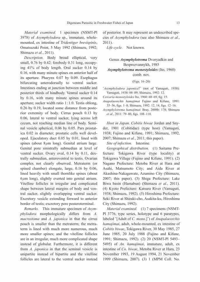
Asymphylotrema monostyloides (Ito, 1960) comb. nov.
(Figs. 16–20)
“Asymphylodora japonica?” (not of Yamaguti, 1938): Yamaguti, 1938: 88–89; Shimazu, 1992: 12.
Cercaria monostyloides Ito, 1960: 68–69, fig. 15.Anapalaeorchis hamajimai Fujino and Kifune, 1991:
35–36, figs. 1–8; Shimazu, 1992: 12, 14, figs. 12–16.Asymphylotrema hamajimai: Bray, 2008b: 178; Shimazu
et al., 2011: 79–80, figs. 108–110.
Host in Japan. Cobitis biwae Jordan and Sny-der, 1901 (Cobitidae) (type host) (Yamaguti, 1938; Fujino and Kifune, 1991; Shimazu, 1992, 2007; Shimazu et al., 2011; this paper).
Site of infection. Intestine.Geographical distribution. (1) Saitama Pre-
fecture: Tokigawa River (type locality) at Tokigawa Village (Fujino and Kifune, 1991). (2) Nagano Prefecture: Metoba River at Hara and Asahi, Matsumoto City; and Aida River at Akashina-Nakagawate, Azumino City (Shimazu, 2007; this paper). (3) Shiga Prefecture: Lake Biwa basin (Hamabun) (Shimazu et al., 2011). (4) Kyoto Prefecture: Katsura River (Yamaguti, 1938; Shimazu, 1992). (5) Hiroshima Prefecture: Seki River at Shiraki-cho, Asakita-ku, Hiroshima City (Shimazu, 1992).
Material examined. (1) 7 specimens (NSMT-Pl 3776, type series, holotype and 6 paratypes, labeled “[Adult of C. mono.]”) of Anapalaeorchis hamajimai, adult, whole-mounted, ex intestine of Cobitis biwae, Tokigawa River, 30 May 1985, 27 June 1985, 20 July 1988 (Fujino and Kifune, 1991; Shimazu, 1992). (2) 20 (NSMT-Pl 5493–5495) of An. hamajimai, immature, adult, ex intestine of Co. biwae, Metoba River at Hara, 23 November 1993, 19 August 1994, 21 November 1999 (Shimazu, 2007). (3) 1 (MPM Coll. No.

14 Takeshi Shimazu
21163), adult, ex intestine of Co. biwae, Aida River, 8 November 2012. (4) 7 (LBM 1-55, 3-40) of Asymphylotrema hamajimai, adult, whole-mounted, ex intestine of Co. biwae, Hamabun, 24 October 1997, 1 November 2000 (Shimazu et al., 2011). (5) 14 (Yamaguti’s Collection, MPM Coll. No. 22034, labeled “Asymphylodora japon-ica?”) of An. hamajimai, immature, adult, whole-mounted, ex intestine of Co. biwae, Katsura River, 25 May 1936 (Yamaguti, 1938; Shimazu, 1992). (6) 5 (NSMT-Pl 3682) of An. hamajimai, adult, whole-mounted, ex intestine of Co. biwae, Seki River, 25 July 1991 (Shimazu, 1992).
Description. Based on type series (NSMT-Pl 3776), after Shimazu (1992), slightly modified from the present study, supplemented by exami-
nation of other specimens (Figs. 16–18). Similar to the foregoing Asymphylodora in morphology, except for two tandem testes and 3-lobed ovary. Body elliptical, very small, 0.70–0.99 by 0.27–0.34 (holotype 0.70 by 0.28); forebody 0.22–0.37 long, occupying 32–37% of body length. Tegu-mental spines wedge-shaped, covering whole body but median ventral field of forebody, becoming smaller and thinner posteriorly and larger and thicker again around excretory pore. Oral sucker 0.09–0.13 in diameter, its lumen not spinose; minute spines present in at least three semicircular rows around anterior half of its aperture. Pharynx 0.05–0.07 in diameter. Esoph-agus bifurcating dorsal to ventral sucker. Intes-tines extending into testicular zoon. Ventral
Figs. 16–18. Asymphylotrema monostyloides, adults found in intestine of Cobitis biwae. — 16, holotype (NSMT-Pl 3776), entire body, ventral view; 17, paratype (NSMT-Pl 3776), terminal genitalia, ventral view; 18, speci-men (NSMT-Pl 5474), ovarian complex, dorsal view. Scale bars: 0.3 mm in Fig. 16; 0.1 mm in Figs. 17–18.

Digeneans Parasitic in Freshwater Fishes of Japan 15
sucker 0.13–0.19 in diameter; minute spines present in at least four circular rows around its aperture; sucker width ratio 1 : 1.2–1.5. Testes two, tandem or almost so, slightly overlapping each other, in third quarter of hindbody; anterior testis usually spherical, 0.08–0.14 by 0.08–0.11; posterior one usually elongate, 0.12–0.16 by 0.07–0.13. Common sperm duct short. Cirrus pouch 0.14–0.16 by 0.04–0.06, just posterior to ventral sucker, rarely extending slightly beyond median line of body. Seminal vesicle bipartite, thin-walled, 0.05–0.07 by 0.03–0.04. Pars pros-tatica 0.01–0.02 in diameter. Ejaculatory duct 0.06–0.09 by 0.01–0.02, lined heavily with spines (8–9 μm long) in posterior two-thirds, eversible. Genital atrium small. Genital pore sinistrally lateral, at level a little anterior to pos-terior border of ventral sucker. Ovary 3-lobed, 0.11–0.14 by 0.07–0.16, dextrally submedian,
anterolateral to anterior testis. Laurer’s canal fairly long, containing sperm. Seminal receptacle 0.05–0.06 by 0.02–0.03, empty or containing a small number of sperm. Uterus occupying all available space in hindbody, extending forward slightly beyond bifurcal level; metraterm about half as long as cirrus pouch, dilated in anterior half to form chamber lined thinly with spines (8–11 μm long). Eggs ovate, symmetrical, yellow, 27–30 by 16–19 μm, fully embryonated. Vitelline follicles six to seven on each side of body, dis-tributed lateral and ventral to intestines between ventral sucker and midlevel of ovary [not ante-rior testis]. Excretory vesicle reaching to middle of posterior [not anterior] testis; excretory pore posteroterminal.
Remarks. Fujino and Kifune (1991) described a new genus and species, Anapalaeor-chis hamajimai, based on the type series. Bray
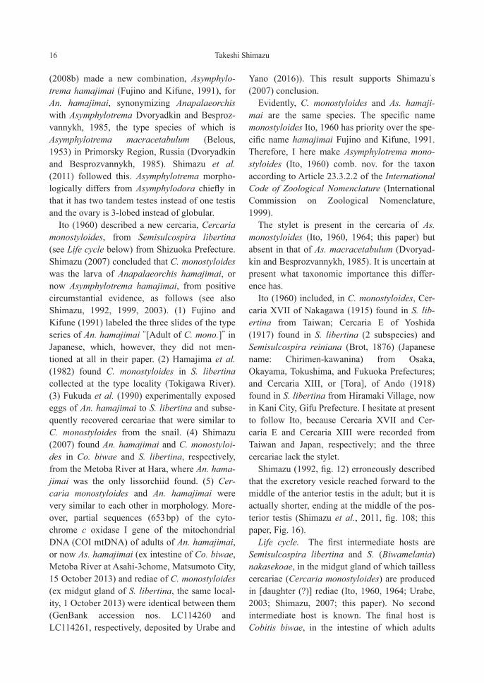
Figs. 19–20. Asymphylotrema monostyloides, life cycle, [daughter (?)] redia (NSMT-Pl 5496) and cercaria (MPM Coll. No. 21164) found in Semisulcospira libertina. — 19, redia; 20, cercaria, penetration glands omit-ted. Scale bars: 0.3 mm in Fig. 19; 0.1 mm in Fig. 20.
16 Takeshi Shimazu
(2008b) made a new combination, Asymphylo-trema hamajimai (Fujino and Kifune, 1991), for An. hamajimai, synonymizing Anapalaeorchis with Asymphylotrema Dvoryadkin and Besproz-vannykh, 1985, the type species of which is Asymphylotrema macracetabulum (Belous, 1953) in Primorsky Region, Russia (Dvoryadkin and Besprozvannykh, 1985). Shimazu et al. (2011) followed this. Asymphylotrema morpho-logically differs from Asymphylodora chiefly in that it has two tandem testes instead of one testis and the ovary is 3-lobed instead of globular.
Ito (1960) described a new cercaria, Cercaria monostyloides, from Semisulcospira libertina (see Life cycle below) from Shizuoka Prefecture. Shimazu (2007) concluded that C. monostyloides was the larva of Anapalaeorchis hamajimai, or now Asymphylotrema hamajimai, from positive circumstantial evidence, as follows (see also Shimazu, 1992, 1999, 2003). (1) Fujino and Kifune (1991) labeled the three slides of the type series of An. hamajimai “[Adult of C. mono.]” in Japanese, which, however, they did not men-tioned at all in their paper. (2) Hamajima et al. (1982) found C. monostyloides in S. libertina collected at the type locality (Tokigawa River). (3) Fukuda et al. (1990) experimentally exposed eggs of An. hamajimai to S. libertina and subse-quently recovered cercariae that were similar to C. monostyloides from the snail. (4) Shimazu (2007) found An. hamajimai and C. monostyloi-des in Co. biwae and S. libertina, respectively, from the Metoba River at Hara, where An. hama-jimai was the only lissorchiid found. (5) Cer-caria monostyloides and An. hamajimai were very similar to each other in morphology. More-over, partial sequences (653 bp) of the cyto-chrome c oxidase I gene of the mitochondrial DNA (COI mtDNA) of adults of An. hamajimai, or now As. hamajimai (ex intestine of Co. biwae, Metoba River at Asahi-3chome, Matsumoto City, 15 October 2013) and rediae of C. monostyloides (ex midgut gland of S. libertina, the same local-ity, 1 October 2013) were identical between them (GenBank accession nos. LC114260 and LC114261, respectively, deposited by Urabe and
Yano (2016)). This result supports Shimazu’s (2007) conclusion.
Evidently, C. monostyloides and As. hamaji-mai are the same species. The specific name monostyloides Ito, 1960 has priority over the spe-cific name hamajimai Fujino and Kifune, 1991. Therefore, I here make Asymphylotrema mono-styloides (Ito, 1960) comb. nov. for the taxon according to Article 23.3.2.2 of the International Code of Zoological Nomenclature (International Commission on Zoological Nomenclature, 1999).
The stylet is present in the cercaria of As. monostyloides (Ito, 1960, 1964; this paper) but absent in that of As. macracetabulum (Dvoryad-kin and Besprozvannykh, 1985). It is uncertain at present what taxonomic importance this differ-ence has.
Ito (1960) included, in C. monostyloides, Cer-caria XVII of Nakagawa (1915) found in S. lib-ertina from Taiwan; Cercaria E of Yoshida (1917) found in S. libertina (2 subspecies) and Semisulcospira reiniana (Brot, 1876) (Japanese name: Chirimen-kawanina) from Osaka, Okayama, Tokushima, and Fukuoka Prefectures; and Cercaria XIII, or [Tora], of Ando (1918) found in S. libertina from Hiramaki Village, now in Kani City, Gifu Prefecture. I hesitate at present to follow Ito, because Cercaria XVII and Cer-caria E and Cercaria XIII were recorded from Taiwan and Japan, respectively; and the three cercariae lack the stylet.
Shimazu (1992, fig. 12) erroneously described that the excretory vesicle reached forward to the middle of the anterior testis in the adult; but it is actually shorter, ending at the middle of the pos-terior testis (Shimazu et al., 2011, fig. 108; this paper, Fig. 16).
Life cycle. The first intermediate hosts are Semisulcospira libertina and S. (Biwamelania) nakasekoae, in the midgut gland of which tailless cercariae (Cercaria monostyloides) are produced in [daughter (?)] rediae (Ito, 1960, 1964; Urabe, 2003; Shimazu, 2007; this paper). No second intermediate host is known. The final host is Cobitis biwae, in the intestine of which adults
Digeneans Parasitic in Freshwater Fishes of Japan 17
live (Fujino and Kifune, 1991; Shimazu, 1992, 2007; Shimazu et al., 2011; this paper).
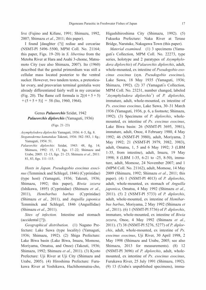
I found [daughter (?)] rediae and cercariae (NSMT-Pl 5496–5500, MPM Coll. No. 21164; this paper, Figs. 19–20) in S. libertina from the Metoba River at Hara and Asahi 3-chome, Matsu-moto City (see also Shimazu, 2007). Ito (1960) described that the genital primordium was still a cellular mass located posterior to the ventral sucker. However, two tandem testes, a pretesticu-lar ovary, and preovarian terminal genitalia were already differentiated fairly well in my cercariae (Fig. 20). The flame cell formula is 2[(4+5+5)+(5+5+5)] = 58 (Ito, 1960, 1964).
Genus Palaeorchis Szidat, 1942 Palaeorchis diplorchis (Yamaguti, 1936)
(Figs. 21–23)
Asymphylodora diplorchis Yamaguti, 1936: 4–5, fig. 8.Steganoderma kamatukae Takeuti, 1936: 582–583, 1 fig.;
Yamaguti, 1954: 51.Palaeorchis diplorchis: Szidat, 1943: 48, fig. 14;
Shimazu, 1992: 15, 17, figs. 17–22; Shimazu and Urabe, 2005: 12–13, figs. 21–23; Shimazu et al., 2011: 81, 83, figs. 111–115.
Hosts in Japan. Pseudogobio esocinus esoci-nus (Temminck and Schlegel, 1846) (Cyprinidae) (type host) (Yamaguti, 1936; Takeuti, 1936; Shimazu, 1992; this paper), Biwia zezera (Ishikawa, 1895) (Cyprinidae) (Shimazu et al., 2011), Hemibarbus barbus (Cyprinidae) (Shimazu et al., 2011), and Anguilla japonica Temminck and Schlegel, 1846 (Anguillidae) (Shimazu et al., 2011).
Sites of infection. Intestine and stomach (accidental [?]).
Geographical distribution. (1) Nagano Pre-fecture: Lake Suwa (type locality) (Yamaguti, 1936; Shimazu, 1992). (2) Shiga Prefecture: Lake Biwa basin (Lake Biwa, Imazu, Momose, Moriyama, Omatsu, and Onoe) (Takeuti, 1936; Shimazu, 1992; Shimazu et al., 2011). (3) Kyoto Prefecture: Uji River at Uji City (Shimazu and Urabe, 2005). (4) Hiroshima Prefecture: Furu-kawa River at Yoshikawa, Hachihonmatsu-cho,
Higashihiroshima City (Shimazu, 1992). (5) Fukuoka Prefecture: Naka River at Terase Bridge, Narutake, Nakagawa Town (this paper).
Material examined. (1) 3 specimens (Yama-guti’s Collection, MPM Coll. No. 22273, type series, holotype and 2 paratypes of Asymphylo-dora diplorchis) of Palaeorchis diplorchis, adult, whole-mounted, ex intestine of Pseudogobio eso-cinus esocinus (syn. Pseudogobio esocinus), Lake Suwa, 18 May 1935 (Yamaguti, 1936; Shimazu, 1992). (2) 37 (Yamaguti’s Collection, MPM Coll. No. 23231, number changed, labeled “Asymphylodora diplorchis”) of P. diplorchis, immature, adult, whole-mounted, ex intestine of Ps. esocinus esocinus, Lake Suwa, 30–31 March 1936 (Yamaguti, 1936, p. 6, a footnote; Shimazu, 1992). (3) Specimens of P. diplorchis, whole-mounted, ex intestine of Ps. esocinus esocinus, Lake Biwa basin: 26 (NSMT-Pl 3695, 3981), immature, adult, Onoe, 4 February 1980, 4 May 1992; 46 (NSMT-Pl 3980), adult, Moriyama, 2 May 1992; 21 (NSMT-Pl 3979, 3982, 3983), adult, Omatsu, 1, 5 and 6 May 1992; 3 (LBM 1-35, from intestine), adult, Imazu, 19 May 1998; 8 (LBM 1-35, 6-21 to -25, 8-50), imma-ture, adult, Momose, 24 November 2007; and 1 (MPM Coll. No. 21162), adult, Momose, 14 May 2009 (Shimazu, 1992; Shimazu et al., 2011; this paper). (4) 1 (NSMT-Pl 4013) of P. diplorchis, adult, whole-mounted, ex stomach of Anguilla japonica, Omatsu, 4 May 1992 (Shimazu et al., 2011). (5) 2 (NSMT-Pl 5733) of P. diplorchis, adult, whole-mounted, ex intestine of Hemibar-bus barbus, Moriyama, 2 May 1992 (Shimazu et al., 2011). (6) 1 (NSMT-Pl 5736) of P. diplorchis, immature, whole-mounted, ex intestine of Biwia zezera, Onoe, 4 May 1992 (Shimazu et al., 2011). (7) 38 (NSMT-Pl 5276, 5277) of P. diplor-chis, adult, whole-mounted, ex intestine of Ps. esocinus esocinus, Uji River, 30 April 1998, 2 May 1998 (Shimazu and Urabe, 2005; see also Shimazu, 2013 for measurements). (8) 12 (NSMT-Pl 3696) of P. diplorchis, adult, whole-mounted, ex intestine of Ps. esocinus esocinus, Furukawa River, 25 July 1991 (Shimazu, 1992). (9) 13 (Urabe’s unpublished specimens), imma-
18 Takeshi Shimazu
ture, adult, ex intestine of Ps. esocinus esocinus, Naka River, 25 November 2003.
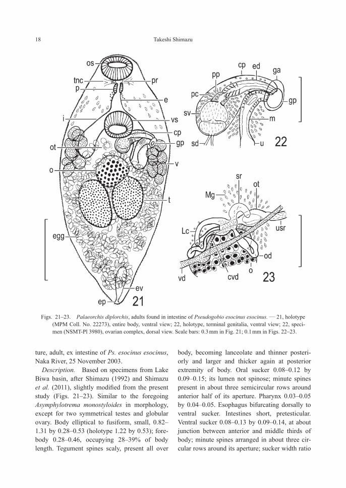
Description. Based on specimens from Lake Biwa basin, after Shimazu (1992) and Shimazu et al. (2011), slightly modified from the present study (Figs. 21–23). Similar to the foregoing Asymphylotrema monostyloides in morphology, except for two symmetrical testes and globular ovary. Body elliptical to fusiform, small, 0.82–1.31 by 0.28–0.53 (holotype 1.22 by 0.53); fore-body 0.28–0.46, occupying 28–39% of body length. Tegument spines scaly, present all over
body, becoming lanceolate and thinner posteri-orly and larger and thicker again at posterior extremity of body. Oral sucker 0.08–0.12 by 0.09–0.15; its lumen not spinose; minute spines present in about three semicircular rows around anterior half of its aperture. Pharynx 0.03–0.05 by 0.04–0.05. Esophagus bifurcating dorsally to ventral sucker. Intestines short, pretesticular. Ventral sucker 0.08–0.13 by 0.09–0.14, at about junction between anterior and middle thirds of body; minute spines arranged in about three cir-cular rows around its aperture; sucker width ratio
Figs. 21–23. Palaeorchis diplorchis, adults found in intestine of Pseudogobio esocinus esocinus. — 21, holotype (MPM Coll. No. 22273), entire body, ventral view; 22, holotype, terminal genitalia, ventral view; 22, speci-men (NSMT-Pl 3980), ovarian complex, dorsal view. Scale bars: 0.3 mm in Fig. 21; 0.1 mm in Figs. 22–23.

Digeneans Parasitic in Freshwater Fishes of Japan 19
1 : 0.9–1.0. Testes two, elliptical, symmetrical to slightly diagonal, contiguous or separated by uterus, submedian on either side of body, in about middle third of hindbody; right testis 0.19–0.30 by 0.09–0.14, left one 0.14–0.27 by 0.10–0.16. Common sperm duct absent. Cirrus pouch 0.13–0.26 by 0.06–0.09, reaching to median line of body or slightly beyond it. Seminal vesicle bipartite, thin-walled, 0.07–0.19 by 0.05–0.09. Pars prostatica 0.02–0.03 in diameter. Ejacula-tory duct 0.07–0.14 by 0.02–0.03, lined heavily with spines (14–24 μm long) in proximal half, eversible. Genital atrium fairly deep. Genital pore sinistrally lateral or sublateral, slightly pos-terior to level of ventral sucker. Ovary irregular in shape (pyriform, subglobular, triangular, weakly 2-lobed, or weakly 3-lobed), 0.11–0.16 by 0.08–0.15, median or dextrally submedian, immediately pretesticular. Seminal receptacle kidney- to retort-shaped, 0.03–0.06 by 0.01, empty or containing a small number of sperm (and oocytes). Uterus much coiled in all avail-able space in hindbody, proximally extending to midlevel of ventral sucker and acting as uterine seminal receptacle; metraterm fairly long, 0.06–0.13 long, dilated in middle three-fifths to form oblong chamber lined thinly with spines (9–17 μm long). Eggs oviform to pyriform, dark brown, 35–42 by 19–24 μm, fully embryonated. Vitelline follicles nine, large, distributed from intestinal shoulder to slightly beyond intestinal end on either side of body. Excretory vesicle short, posterior to testes in fully matured speci-mens, but reaching to testes in immature speci-mens; excretory pore posteroterminal.
Remarks. Yamaguti (10 September 1936) described a new species, Asymphylodora diplor-chis, based on the type series found in the intes-tine of Pseudogobio esocinus esocinus from Lake Suwa. Takeuti (15 October 1936) described a new genus and species, Steganoderma kamatu-kae, based on adult specimens (type specimens not designated) found in the intestine of Ps. eso-cinus esocinus from Lake Biwa. Szidat (1943) established a new genus, Palaeorchis, with A. diplorchis as the type species; but he was
unaware of St. kamatukae at that time. Yamaguti (1954) synonymized St. kamatukae with P. dipl-orchis. Shimazu (1992) agreed to this treatment. Takeuti’s original material was not available, but it was probably lost (see the Materials and Meth-ods in Shimazu, 2013).
The testes are slightly oblique in the holotype (Yamaguti, 1936; Shimazu, 1992; this paper, Fig. 21), but they are almost symmetrical to almost oblique in the other specimens (see Shimazu, 1992; Shimazu and Urabe, 2005; Shimazu et al., 2011). The ovary varies widely in shape (pyri-form, subglobular, triangular, or weakly 2- to 3-lobed) (see Shimazu, 1992; Shimazu and Urabe, 2005; Shimazu et al., 2011). The excre-tory vesicle is small and posterior to the testes in fully matured adult specimens with well-devel-oped uterine loops in the post-testicular region of the body, but it sometimes reaches to the testes in immature specimens without developed uterine loops in this region (Shimazu et al., 2011). Apparently, Palaeorchis closely resembles Asmphylotrema, except for that the excretory vesicle is posterior to the testes instead of reach-ing to them. It is desirable that further critical studies be made of these two genera.
An adult specimen (NSMT-Pl 4013) of P. dipl-orchis was found in the stomach of Anguilla japonica from Omatsu (Shimazu et al., 2011). Since this host fish had been raised on small fish from Lake Biwa in a fish preserve (Shimazu, 2015), it is considered that this infection was accidental from food fish, such as Ps. esocinus esocinus.
Life cycle. Not known. In Japan, another cer-cariaeum, Cercariaeum incognitum Faust, 1924 (syn. Cercariaeum C of Kobayashi, 1922), is known from Semisulcospira libertina (Kobayashi, 1922; Faust, 1924; Ito, 1964; Makita et al., 1996; Urabe, 2003). It is possible that this cercaria is the larva of P. diplorchis, because it has shorter intestines ending at the level of the ventral sucker.

20 Takeshi Shimazu
Key to the genera and species of the family Lissorchiidae in this paper
1.1. Testis one ........................................................................................................................................ 21.2. Testes two ....................................................................................................................................... 32.1. Seminal vesicle unipartite; cirrus pouch larger than metraterm; vitelline follicles lateral to ventral
sucker ..................................................................................................... Asymphylodora innominata2.2. Seminal vesicle unipartite; cirrus pouch smaller than metraterm; vitelline follicles lateral to ventral
sucker ................................................................................................................... Asymphylodora sp.2.3. Seminal vesicle bipartite; cirrus pouch larger than metraterm; vitelline follicles posterior to
ventral sucker ............................................................................................. Asymphylodora japonica3.1. Testes symmetrical; ovary subglobular to weakly 3-lobed; excretory vesicle posterior to testes
......................................................................................................................... Palaeorchis diplorchis3.2. Testes tandem; ovary 3-lobed; excretory vesicle reaching to posterior testis
........................................................................................................... Asymphylotrema monostyloides
Acknowledgments
I am grateful to Dr. Misako Urabe and Ms. Ayaka Yano (Department of Ecosystem Studies, School of Environmental Science, The Univer-sity of Shiga Prefecture, Hikone) for making a molecular analysis of Asymphylotrema monosty-loides and Dr. Thomas H. Cribb (School of Bio-logical Sciences, The University of Queensland, Brisbane, Australia) for reviewing a draft of the manuscript.
References
Abe, H. 1930. [Cercariae of five species and their rediae parasitic in a snail], pp. 966–970, pl. 1. In Ueno, N., K. Ishii and H. Abe: [On cercariae parasitic in freshwater snails in the neighborhood of Kumamoto.] Kumamoto Igakkai Zasshi, 6: 965–976, pls. 1–2. (In Japanese.)
Ando, A. 1918. [On cercariae parasitic in Melania liber-tina from an endemic area of the lung fluke in Gifu Prefecture (addition of six new cercariae).] Chuo Igak-kai Zasshi, 25: 610–627, 1 pl. (In Japanese.)
Besprozvannykh, V. V. 2005. Life cycles of the trema-todes Parasymphilodora japonica [sic] (Yamaguti, 1938) and P. markewitschi (Kulakowskaja, 1947) (Monorcidae) [sic] in the conditions of Primorye Land. Parazitologiya, 39: 137–145. (In Russian with English summary.)
Bray, R. A. 2008a. Superfamily Monorchioidea Odhner, 1911. In Bray, R. A., D. I. Gibson and A. Jones (eds.): Keys to the Trematoda, 3: 143–144. CAB International and The Natural History Museum, Wallingford.
Bray, R. A. 2008b. Family Lissorchiidae Magath, 1917. In
Bray, R. A., D. I. Gibson and A. Jones (eds.): Keys to the Trematoda, 3: 177–186. CAB International and The Natural History Museum, Wallingford.
Dvoryadkin, V. A. and V. V. Besprozvannykh 1985. Sys-tematic position and life cycle of Asymphylotrema mac-racetabulum comb. nov. (Trematoda, Monorchidae [sic]). Parazitologiya, 19: 394–398. (In Russian with English summary.)
Faust, E. C. 1924. Notes on larval flukes from China. II. Studies on some larval flukes from the central and south coast provinces of China. American Journal of Hygiene, 4: 241–301.
Fujino, T. and T. Kifune 1991. Anapalaeorchis hamajimai gen. et sp. n. (Trematoda: Monorchiidae) from the loach, Cobitis biwae, in Japan. Journal of the Helmin-thological Society of Washington, 58: 35–38.
Fukuda, K., F. Hamajima, T. Oguma and K. Hashizume 1990. Infection of Semisulcospira libertina with larvae of a monorchid [sic] trematode. Japanese Journal of Parasitology, 39 (1, Supplement, Abstracts): 154. (In Japanese with English title.)
Hamajima, F., K. Fukuda, A. Komiyama and H. Nakajima 1982. A trematode from digestive tract of Cobitis biwae. Japanese Journal of Parasitology, 31 (1, Supple-ment, Abstracts): 23–24. (In Japanese with English title.)
International Commission on Zoological Nomenclature, 1999. International Code of Zoological Nomenclature. Fourth edition. xxix+306 pp. International Trust for Zoological Nomenclature, London.
Ishii, K. 1930. [Cercariae of nine species and their rediae parasitic in Melania libertina], pp. 970–975, pl. 2. In Ueno, N., K. Ishii and H. Abe: [On cercariae parasitic in freshwater snails in the neighborhood of Kuma-moto.] Kumamoto Igakkai Zasshi, 6: 965–976, pls. 1–2. (In Japanese.)

Digeneans Parasitic in Freshwater Fishes of Japan 21
Ito, J. 1960. Contributions to the morphology of cercariae obtained from a snail, Semisulcospira libertina in Japan. Japanese Journal of Medical Science & Biology, 13: 59–72.
Ito, J. 1964. A monograph of cercariae in Japan and adja-cent territories. In Morishita, K., Y. Komiya and H. Matsubayashi (eds.): Progress of Medical Parasitology in Japan, 1: 395–550. Meguro Parasitological Museum, Tokyo.
Ito, J., H. Mochizuki and M. Noguchi 1959. Studies on the cercariae parasitic in Semisulcospira libertina in Shizuoka Prefecture. Japanese Journal of Parasitology, 8: 913–922. (In Japanese with English summary.)
Kobayashi, H. 1918. [Studies on cercariae from Korea, I.] Chosen Igakkai Zasshi, (21): 19–80, 1 pl. (In Japa-nese.)
Kobayashi, H. 1922. [A review of cercariae from Japan.] Dobutsugaku Zasshi, 34: 252–270. (In Japanese.)
Komiya, Y. 1965. Metacercariae in Japan and adjacent territories. In Morishita, K., Y. Komiya and H. Matsu-bayashi (eds.): Progress of Medical Parasitology in Japan, 2: 1–328. Meguro Parasitological Museum, Tokyo.
Kurokawa, K. 1935. [Studies of trematodes using Bulimus (Parafossarulus) striatus (Pilsbry) as the intermediate host, with special reference to metacercariae found in it.] Tokyo Iji Shinshi, (2937): 1795–1800. (In Japa-nese.)
Kuyama, S. 1938. Die jahreszeitliche Veränderung sowie di Korrelativen Verhältnisse von in Entwicklungssta-dium befindlichen Trematoden. Okayama Igakkai Zasshi, 50: 327–437. (In Japanese with German sum-mary.)
Looss, A. 1894. Die Distomem unserer Fische und Frösche. Neue Untersuchungen über Bau und Entwick-elung des Distomenkörpers. Bibliotheca Zoologica, 16: 1–296, pls. 1–9.
Makita, Y., M. Urabe and M. Nagoshi, 1996. Infection of larval trematodes in freshwater snails Semisulcospira from Nabari River system in Nara and Mie Prefectures. I. Observed cercariae and their host species and preva-lence. Japanese Journal of Parasitology, 45: 309–315. (In Japanese with English abstract.)
Nagano, K. 1930. [On the intermediate host of Asymphy-lodora tincae in Japan.] Proceedings of the Second General Meeting and Abstracts of the Japanese Society of Parasitology, pp. 24–25. (In Japanese.)
Nakagawa, K. 1915. [On the cercariae parasitic in fresh-water snails in Shinchiku Province, Taiwan.] Taiwan Igakkai Zasshi, (148): 107–120. (In Japanese.)
Nakamura, S., M. Urabe and M. Nagoshi 2000. Seasonal change of prevalence and distribution of parasites in freshwater fishes at Higashi-yoshino, Nara Prefecture. Biology of Inland Waters, (15): 12–19. (In Japanese with English abstract.)
Našincová, V. and T. Scholz 1994. The life cycle of Asymphylodora tincae (Modeer 1790) (Trematoda: Monorchiidae): a unique development in monorchiid trematodes. Parasitology Research, 80: 192–197.
Okumura, T. 1919. [On aggregated cercariae found in Melania libertina.] Saikingaku Zasshi, (284): 359–360. (In Japanese.)
Ozaki, Y. 1925. On a new genus of fish trematodes, Genarchopsis, and a new species of Asymphylodora. Japanese Journal of Zoology, 1: 101–108.
Shimazu, T. 1992. Trematodes of the genera Asymphylo-dora, Anapalaeorchis and Palaeorchis (Digenea: Lis-sorchiidae) from freshwater fishes of Japan. Journal of Nagano Prefectural College, (47): 1–19.
Shimazu, T. 1995. A revised checklist and bibliography of the platyhelminth parasites reported by Dr. Yoshimasa Ozaki, 1923–1966, and their specimens deposited in the Meguro Parasitological Museum, Tokyo. Journal of Nagano Prefectural College, (50): 33–50.
Shimazu, T. 1999. [Turbellarians and trematodes of fresh-water animals in Japan.] In Otsuru, M., S. Kamegai and S. Hayashi (chief eds.), Progress of Medical Para-sitology in Japan, 6: 65–86. Meguro Parasitological Museum, Tokyo. (In Japanese.)
Shimazu, T. 2003. Turbellarians and trematodes of fresh-water animals in Japan. In Otsuru, M., S. Kamegai and S. Hayashi (chief eds.), Progress of Medical Para-sitology in Japan, 7: 63–86. Meguro Parasitological Museum, Tokyo.
Shimazu, T. 2007. Digeneans (Trematoda) of freshwater fishes from Nagano Prefecture, central Japan. Bulletin of the National Museum of Nature and Science, Series A (Zoology), 33: 1–30.
Shimazu, T. 2008. Digeneans (Trematoda) found in fresh-water fishes of Wakayama, Tokushima, and Kochi Pre-fectures, Japan. Bulletin of the National Museum of Nature and Science, Series A (Zoology), 34: 41–61.
Shimazu, T. 2013. Digeneans parasitic in freshwater fishes (Osteichthyes) of Japan. I. Aporocotylidae, Bive-siculidae and Haploporidae. Bulletin of the National Museum of Nature and Science, Series A (Zoology), 39: 167–184.
Shimazu, T. 2015. Digeneans parasitic in freshwater fishes (Osteichthyes) of Japan. IV. Derogenidae. Bulle-tin of the National Museum of Nature and Science, Series A (Zoology), 41: 77–103.
Shimazu, T. and M. Urabe 2005. Digeneans found in freshwater fishes of the Uji River at Uji, Kyoto Prefec-ture, and the Takami River at Higashiyoshino, Nara Prefecture, Japan. Journal of Nagano Prefectural Col-lege, (60): 1–14.
Shimazu, T., M. Urabe and M. J. Grygier 2011. Digene-ans (Trematoda) parasitic in freshwater fishes (Osteich-thyes) of the Lake Biwa basin in Shiga Prefecture, cen-tral Honshu, Japan. National Museum of Nature and

22 Takeshi Shimazu
Science Monographs, (43): 1–105.Szidat, L. 1943. Die Fischtrematoden der Gattung Asym-
phylodora Looss 1899 und Verwandte. Zeitschrift für Parasitenkunde, 13: 25–61.
Takeuti, E. 1936. A new trematode, Steganoderma kama-tukae from Pseudogobio esocinus (Temminck & Schle-gel). Zoological Magazine (Dobutsugaku Zasshi), 48: 581–583. (In Japanese with English summary.)
Tang, C.-C. [Z.-Z.] 1962. Studies on the development of Asymphylodora macrostoma Ozaki, 1925 and A. japon-ica Yamaguti, 1928 in their intermediate hosts, with a consideration of the systematic of the group. Fujian Shifan Xueyuan Xuebao, (2): 161–183. (In Chinese with English abstract.)
Urabe, M. 2003. Trematode fauna of prosobranch snails of the genus Semisulcospira in Lake Biwa and the con-nected drainage system. Parasitology International, 52: 21–34.
Wang, S.-X. 1991. [Family Monorchiidae.] In Wu, B.-H. (chief ed.): Fauna of Zhejiang. Trematoda, pp. 212-216. Zhejiang Science and Technology Publishing House, Hangzhou. (In Chinese.)
Wang, W.-J. and J.-P. Pan 1984. Studies on the asymphy-lodorasis of fry with discussion on the specific life his-tory of genus. In Institute of Hydrobiology Academia
Sinica (ed.), Parasitic Organisms of Freshwater Fish of China, pp. 149–158. Agricultural Publishing House, Beijing. (In Chinese.)
Yamaguti, S. 1934. Studies on the helminth fauna of Japan. Part 2. Trematodes of fishes, I. Japanese Journal of Zoology, 5: 249–541.
Yamaguti, S. 1936. Studies on the Helminth Fauna of Japan. Part 15. Trematodes of Fishes, II. 6 pp. Author’s publication, Kyoto.
Yamaguti, S. 1938. Studies on the Helminth Fauna of Japan. Part 21. Trematodes of Fishes, IV. 139 pp., 1 pl. Author’s publication, Kyoto.
Yamaguti, S. 1942. Studies on the helminth fauna of Japan. Part 39. Trematodes of fishes mainly from Naha. Transactions of the Biogeographical Society of Japan, 3: 329–398, pl. 24.
Yamaguti, S. 1953 [issued 1954]. Systema Helminthum. Part I. Digenetic trematodes of fishes. 405 pp. Author’s publication, Tokyo.
Yamaguti, S. 1971. Synopsis of Digenetic Trematodes of Vertebrates. I: 1–1074; II: 349 pls. Keigaku Publishing Company, Tokyo.
Yoshida, S. 1917. [On the cercariae in Melania.] Dobutsugaku Zasshi, 29: 103–119, pl. 2. (In Japanese.)