Embed Size (px)
Citation preview

Dr Mahesh Shrivastava
Nepal Medical College Teaching Hospital Kathmandu, Nepal
9/29/2015 1
Treatment of Fracture Dislocation of Talus by Primary Tibiotalar
Arthrodesis( Blair Fusion) --------------------------------------------------------------------------------------------------------------------
2015 Annual SIGN Conference September 16-19 , 2015
Richland , Washington, USA
“Difficult Fractures around the World “
Asia


9/29/2015 5
Nepal Medical College Teaching Hospital

Learning outcomes • Introduction •Describe the anatomy and different types of blood supply
• Discuss the mechanism of injury
• Discuss the Hawkins classification
• Discuss the surgical procedure of Blair fusion , clinical and functional results • Conclusion
Introduction • The fractures of the talus are relatively uncommon injuries
• The fractures of the talus rank second in frequency of all tarsal bone injuries ( after calcaneal fractues)
• The fractures can be associated with significant complications

Types of Fractures
• The classification of these fractures is based on their anatomic location within the talus – Head – Body – Neck
• Each type has unique features that affect both diagnosis and treatment.

• Surface 60% cartilage • No muscular or tendinous insertions • Supported solely by the joint capsules
,ligaments , and synovial tissues
Anatomy
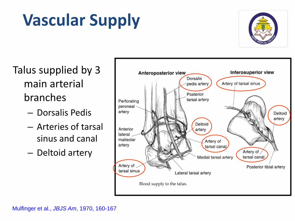
Vascular Supply
Talus supplied by 3 main arterial branches – Dorsalis Pedis – Arteries of tarsal
sinus and canal – Deltoid artery
Mulfinger et al., JBJS Am, 1970, 160-167

Imaging
• AP, lat, obliques of foot and ankle
• Canale view
Canale and Kelly, JBJS Am; 1978, 143-156

Incidence
• 2 % of all fractures • 6-8% of foot fractures • High complication rates
– avascular necrosis – post-traumatic arthritis – malunion

Mechanism of Injury
• Hyperdorsiflexion of the foot on the leg
• Neck of talus impinges against anterior distal tibia, causing neck fracture
• If force continues: – talar body dislocates posteromedial – often around deltoid ligament

Injury Mechanism
• Previously called “aviator’s astragalus” as reported by Anderson in 1919
• Usually due to motor vehicle accident or falls from height
• Approximately 50 % of patients have multiple traumatic injuries

Hawkins Classification (1970)
Group I – Nondisplaced vertical fracture of the talar neck
Group II – Displaced fracture of the talar neck with subluxation
or dislocation of the subtalar joint
Group III – Displaced fracture of the talar neck with dislocation of
the body of the talus from both the subtalar and tibiotalar joints
Hawkins, JBJS Am, 1970, 991-1002
•Reviewed 58 cases of # of talar neck and classified into 3 types, later modified by Canale and Kelly

Canale’s Modification
Type IV – Hawkins III with subluxation or dislocation
of talar head
Canale, JBJS Am; 1978, 143-156

Hawkins I
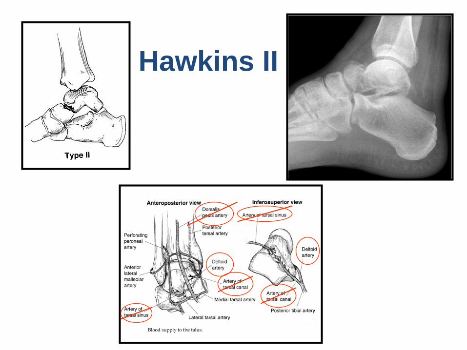
Hawkins II

Hawkins III

Type IV
Canale, JBJS Am; 1978, 143-156

9/29/2015 21
To report the end results of primary Blair fusion of lately presented Hawkins’ grade III fractures of the neck of the talus with long term ( 2 years) follow-up in fourteen cases.
Aims and Objectives

Methodology Type of Study Prospective study
Place Nepal Medical College Teaching Hospital
Total No. of cases 14 ( Jan. 2000- Dec.2008)
Age at operation 27 yrs ( Avg.)
Sex All male patients
Mode of injury RTA – 8 Fall from tree- 3 Fall from height- 3
Type of fracture All Hawkins’ grade III fractures of neck of the talus
Follow- up period 2 years on average

Materials and Methods
The Study included total of 14 patients
23
•All the patients presented to us quite late ( avg. 2 weeks)
• AP and lateral views and oblique x-rays done •All the patients had sustained type III fractures of the neck of the talus.
•No open fracture

Sex

Joint involved
• Right ankle : 8 • Left ankle : 6

Mode of injury
• RTA : 8 •Fall from tree : 3 •Fall from height : 3
Blair Fusion
In 1943, Blair described a technique of ankle fusion to treat osteonecrosis of the talus • Excision of the avascular talar body •Placement of the sliding corticocancellous graft from anterior distal tibia
•Into the residual , viable head and neck of the talus and secured with two cortical screws
•Morris et al in 1971 modified the procedure slightly

9/29/2015 28
Operative procedure
Fig1A: Approach to the ankle Fig. 1-B: Excision of the body of the talus
1-C: The sliding bone graft Fig. 1- D : The graft in its final position
1-A 1-B 1-C 1-D 1 A 1 B 1C 1D

Blair Fusion
Primary Blair fusion was done in all the patients with type III fracture of the neck of the talus with late presentation (2 weeks average) •Considering the difficulties of ORIF in severely displaced fractures with late presentation
• Complications of late surgical procedures
• Considering the poor economical status of the patients ( to avoid more surgery)

Results
• All the fourteen patients were followed and evaluated after surgery
• Mean age of the patients was 27 years .
•Roughly 4 months required for bony union
• Clinical and functional results were evaluated

9/29/2015 32
Clinical results
•Result was graded with Denis et al criteria of 2 years follow up. •Radiological bony fusion was obtained in thirteen patients in 12-14 weeks of time with one pseudoarthrodesis which healed after 10 months.
. Our result
Good 10
Fair 3
Poor 1
Denis’ result
Good 5
Fair 1
Poor 1
Criteria - Denis et al
Asymptomatic, was able to carry out full activities.
Occasional discomfort or restriction of activity.
Pain causing restriction or requiring analgesics.
Denis M. David , Tullos Hugh S.: Blair Tibiotalar Arthrodesis for Injuries to the Talus .J. Bone and Joint Surg. 62- A/1 : 103-107, Jan. 1980

9/29/2015 33
Functional results: • 20 degrees of tibiopedal motion is necessary for normal gait • Functional results correlated more clearly with tibiopedal
motion than anatomocal restoration. • Tibiopedal motion is arc of motion between maximum
dorsiflexion and maximum plantar flexion , the angle being those subtended by the long axis of the tibia and of the foot in the lateral projection.
Tibiopedal motion Range Result Good Fair Poor
16- 20 degrees 11- 15 degrees < 10 degrees
10
3
1
Waugh T. Joe King Visiting Lectureship , Baylor College of Medicine . Houston, Texas , Dec. 1977.:

9/29/2015 34
Grade III- fracture of the neck of the talus
Case- 1

Immediately after Blair fusion

3 months – after Blair fusion

9/29/2015 37
Morris modification of Blair procedure

Discussion •There has been very little mention in the literature of Blair fusion as a treatment of fractures of AVN of talus.
•In 1971- Morris et al- reported on 10 cases, with excellent results in seven, and good results in three. JBJS 1971 53 A : 1289- 1297
• In 1980 , David Dennis et al reported on seven patients with good results in five patients , fair in one and poor in one JBJS 1980 62 A : 103-107

•So far very few results have been published specially about primary Blair fusion for grade III fracture of the neck of the talus.
•In 2010 ,A. Bhattacharya et at reported primary modified Blair Arthrodesis for type III fracture s of the neck of the talus with good results in four cases and poor in one The foot and Ankle Online Journal Volume 3, No. 10, October 2010
• We performed primary Blair fusion in all those patients who presented to us quite late with type III fractures of the neck of the talus with good results in ten cases, fair in three cases and poor in one.

Blair fusion provided some distinct advantages:-
1. Normal appearance of the foot was retained.
2. Normal alignment of the foot to the ankle and the leg was retained.
3. No shortening was produced.
4. Solid fusion allowed motion at talonavicular and anterior subtalar joints , thus maintaining physiological relationship between foot and ankle.

9/29/2015 41
1. Displaced talar fractures with late presentation are challenging to treat.
2. Fourteen cases of grade III fractures of the neck of the talus are presented.
3. Results were uniformly good to fair in this small series of cases except in one case.
4. From our limited experience, we recommend primary Blair fusion as possible solution especially in poor groups of patients for whom secondary procedures will not be possible.
Take-home messages