Embed Size (px)
Citation preview

Virginia Commonwealth University Virginia Commonwealth University
VCU Scholars Compass VCU Scholars Compass
Theses and Dissertations Graduate School
2010
Differential roles of the two major endocannabinoid hydrolyzing Differential roles of the two major endocannabinoid hydrolyzing
enzymes in cannabinoid receptor tolerance and somatic enzymes in cannabinoid receptor tolerance and somatic
withdrawal withdrawal
Joel Schlosburg Virginia Commonwealth University
Follow this and additional works at: https://scholarscompass.vcu.edu/etd
Part of the Medical Pharmacology Commons
© The Author
Downloaded from Downloaded from https://scholarscompass.vcu.edu/etd/2107
This Dissertation is brought to you for free and open access by the Graduate School at VCU Scholars Compass. It has been accepted for inclusion in Theses and Dissertations by an authorized administrator of VCU Scholars Compass. For more information, please contact [email protected].

Joel E. Schlosburg 2010
All Rights Reserved

i
DIFFERENTIAL ROLES OF THE TWO MAJOR ENDOCANNABINOID
HYDROLYZING ENZYMES IN CANNABINOID RECEPTOR TOLERANCE AND
SOMATIC WITHDRAWAL
A dissertation submitted in partial fulfillment of the requirements for the degree of Doctor of
Philosophy at Virginia Commonwealth University.
By
Joel E. Schlosburg
Bachelors of Science, Chemical Engineering, Rensselaer Polytechnic Institute, 2005
Bachelors of Science, Psychology, Rensselaer Polytechnic Institute, 2005
Director: Dr. Aron Lichtman, Ph.D.
Professor, Department of Pharmacology & Toxicology
Virginia Commonwealth University
Richmond, Virginia May 2010

ACKNOWLEDGEMENTS
While it was certainly not conventional for a chemical engineer to suddenly decide to
become a drug abuse researcher, my experience and training during my time here at VCU was
everything I hoped for…and none of the dangerous and unpleasant chemical plants I would’ve enjoyed otherwise.
First and foremost, I have to thank my advisor, Dr. Aron Lichtman. I honestly don’t know why I somehow ended up in his laboratory over any other, if only by mixture of prodding and random choice, and yet I don’t know how I could’ve been quite as successful as I was without
him. He knew exactly when to guide my sometimes wild and random exuberance for doing everything, knew when to introduce new folds into my work to keep things challenging and
hectic, and occasionally gave me an insanely long leash that few might, trusting me to come back with something worthwhile. He taught me not how to work hard, but work efficiently, and to always understand your outcomes on both a numerical and conceptual level.
Thanks are also in order to the original members of my graduate committee: Dr. Guy Cabral & Dr. Hamid Akbarali. Special thanks to Dr. Dan Conrad & Dr. Imad Damaj for stepping in on
short notice and unusual circumstances to become integral influences in my work. It’s been a crazy and evolving group, but they have made sure to keep me focused on getting to this point even when I was busy skipping a few steps, and bringing their “A game” to make sure I got a
thorough humbling just when I thought I had my act together. Additional acknowledgement must be extended to Dr. Laura Sim-Selley & Dr. Dana Selley,
who literally had the confidence in me to give me the keys to the castle, and allowed me free reign of their lab any time I needed. Their guidance and assistance, and insistence that I get my hands dirty whenever possible (quite literally) has made me a far greater and complete scientist
with an incredible breadth of skills to apply towards my future endeavors. It’s not a very well-kept secret that I got into this to teach and mentor, and if research is
accomplished it’s a pleasant consequence. So this goes to Brittany, Divya, Roberta, Dolores, Ashley, Darby, and all those I had a chance to teach and mentor in some way while I was here. I also must acknowledge all those Lichtman lab members who have influenced and broadened my
appreciation of the work I do: Drs. Andy Thorpe, Sree Pattipati, Floride Niyuhire, Steve Varvel, Laura Wise, John Harloe, Troy DeLong, & Rehab Abdullah. To Carlotta and Kelly, you guys
kept me on my toes all the time, and otherwise always entertained with endless random conversation passing the time through what could sometimes be endless monotony. To Scott, well…ours is a bond that few can explain and few states legally condone. But seriously, you’re
presence always kept my time interesting, and added some unusual twists to my day. To Sean, I wish you all the luck. You are taking on what can only be considered a project
that I could never get the courage to take a risk on, and the rewards are potentially fantastic. To Lamont, you’re my boy, and I cannot thank you enough for always trying to keep me (just if only

occasionally) focused on life outside the lab, and keeping me surrounded by other people. This place probably wouldn’t be a very bearable place without having you around. To Steve, I don’t
know whether I’ve been teaching you a thing or two or the other way around all this time, but we seem to work well together on a rather scary level. Lord knows I cannot wait until the day
comes when we meet again at some random conference and instantly captivate a room with the most inane conversation those stiffs have ever seen. Just try to keep it even a little clean.
To my folks, this is obviously just another testament to the rather extreme and extraordinary
efforts you went to make sure I was always able to be on top and go as far as I could. The teaching me algebra on a magnetic desk at age 5, fighting for every bus line that didn’t exist
before, hybrid classes that were never really conceived…you guys will enjoy your retirement with a pair of kids with graduate degrees and highly successful careers for your efforts, despite all the bumps along the way. To my brother, it’s not always been the most congenial
relationship, but we always did things together when it really mattered, and hopefully that’ll always be the way things are. To Mandy, I cannot imagine a more caring and nurturing sister-in-
law…makes me wonder how different life would’ve been growing up with a few more girls around. To Jen, thanks for always being the female sensibility and outside influence I needed when my life got a little too focused in lab.
To my rugrat nephew Ethan, you keep me inspired to do great things and be a great success for you to look toward some day. With the wonderful people around you, you’ll be ready for all
you own success one day, a good start might be getting to the point you can remotely read and understand this paper in the least bit. Good luck with that! And to the future little niece to be named later (Riley), I hope to be successful enough to be around much more by your preschool
years, finger crossed. Finally, this is in memoriam to a couple of gigantic influences in my life for very different
reasons. To Dr. Billy Martin, you steered me to VCU, to cannabinoids, to Aron, and to a rather unusual project from little more than a random observation. It was an honor to unofficially be your last mentee, and your shadow looms large over my launch into the world of research. Your
honesty and practical view the options in front of me brought incredible clarity to a confused kid with too many options and time in front of him. Lastly, to my grandmother (Nana), who passed
just after my acceptance letter here arrived, you were the driving force for my greatness. Your pride and constant showing off of all my little accomplishments to all you knew, your need to make sure I always looked my best and put my best foot forward, and always taking my side
whether I was right or not; it all gave me a reason to go for the next great accomplishment, and to do so in a way that impressed, and do so the way I wanted. I’m still going for it, and I’m still
not done…

TABLE OF CONTENTS
List of Tables ....................................................................................................................................vi
List of Figures ...................................................................................................................................vii
List of Abbreviations ........................................................................................................................ix
Abstract .............................................................................................................................................xii
Introduction .......................................................................................................................................1
The discovery of cannabinoid ligands and receptors .............................................................2
Actions of cannabinoid receptors...........................................................................................3
The endocannabinoid system: ligands and regulatory pathways ...........................................5
Anandamide and fatty acid amide hydrolase inactivation .....................................................9
2-arachidonoylglycerol and monoacylglycerol lipase inactivation .......................................12
Tolerance and receptor adaptations following repeated cannabinoid administration............14
Physical withdrawal resulting from repeated exposure to cannabinoids ...............................16
Rational and Hypothesis ...................................................................................................................18
Methods..............................................................................................................................................22
Chapter 1: Endocannabinoid elevations in attenuation of THC physical withdrawal and
potential for endocannabinoid induction of withdrawal symptoms
1.1 Rimonabant precipitates similar somatic withdrawal signs in FAAH (-/-)
and (+/+) given repeated injections of THC...................................................................36
1.2 Comparison of rimonabant precipitated withdrawal in high-dose THC versus
high-dose AEA ...............................................................................................................40
1.3 Acute administration of the FAAH inhibitor, URB597, reduces the severity of
rimonabant-precipitated withdrawal in THC-dependent mice .......................................43
1.4 Rimonabant-precipitated withdrawal potential during repeated FAAH inhibition ........45
1.5 Acute administration of the MAGL inhibitor, JZL184, reduces the severity of
rimonabant-precipitated withdrawal in THC-dependent mice .......................................48
i
1.6 Precipitated withdrawal potential during repeated MAGL inhibition alone, and
in combination with FAAH inhibition ...........................................................................50
1.7 Rotarod motor coordination tests ....................................................................................56
1.8 Discussion: Substitution during cannabinoid withdrawal...............................................58
Chapter 2: Prolonged endocannabinoid elevations in the induction of cannabinoid
behavioral tolerance and cross-tolerance
2.1 Repeated JZL184 produces tolerance to cannabinoid-mediated effects .........................64
2.2 Analgesic tolerance following prolonged MAGL inhibition versus FAAH
inhibition.........................................................................................................................67
2.3 Chronic MAGL blockade causes behavioral tolerance to exogenous
cannabinoids ...................................................................................................................70
2.4 Discussion: Tolerance following prolonged endocannabinoid elevation .......................76
Chapter 3: Enhanced endocannabinoid availability and functional receptor adaptation
corresponding with behavioral responses
3.1 Endocannabinoid quantification during THC withdrawal ..............................................82
3.2 Prolonged MAGL, but not FAAH, inhibitors lead to accumulation of 2-AG in
brain ................................................................................................................................84
3.3 Genetic inactivation of MAGL enhances 2-AG in brain ................................................87
3.4 Brain CB1 receptors are impaired by chronic MAGL, but not FAAH, blockade ..........87
3.5 Regional analysis of brain CB1 receptor adaptation and eCB accumulation ..................92
3.6 Discussion: Endocannabinoid accumulation and cannabinoid receptor
adapations .......................................................................................................................98
Chapter 4: General discussion and conclusions .................................................................................105
List of References ..............................................................................................................................109
Appendix ............................................................................................................................................124
Vita.....................................................................................................................................................129

i
LIST OF TABLES
Table 1. Brain endocannabinoid quantification following rimonabant-precipitated THC withdrawal ......83
2. Summary of CB1 receptor activation and binding curve alterations due to prolonged
FAAH & MAGL inhibition ...........................................................................................................95

ii
LIST OF FIGURES
Figure 1. Synthetic and degradative pathways of the major endocannabinoids .........................................6
2. Effect of FAAH genotype on high-dose precipitated THC withdrawal ......................................37
3. Effect of FAAH genotype on low-dose precipitated THC withdrawal ........................................39
4. FAAH genotype comparison of rimonabant dose-response to elicit THC withdrawal ................41
5. Precipitated withdrawal intensity of high-dose THC versus anandamide ....................................42 6. Acute URB597 treatment on precipitated withdrawal of both high- and low- dose THC ...........44
7. URB597 treatment of high-dose THC withdrawal in FAAH (+/+) & (-/-) mice..........................46
8. Precipitated withdrawal signs in mice treated with repeated FAAH inhibitors ............................47
9. JZL184 treatment of high-dose THC withdrawal in FAAH (+/+) & (-/-) mice ...........................49
10. Precipitated withdrawal following repeated JZL184 in FAAH (+/+) & (-/-) mice ....................51 11. Precipitated withdrawal following repeated JZL184 in combination with PF3845 ...................52
12. Evaluation of precipitated cannabinoid withdrawal in MAGL (-/-) mice ..................................54
13. Precipitated withdrawal following repeated JZL195 administration ..........................................55
14. Rotarod performance in THC-, URB597-, and JZL184- treated mice .......................................57
15. Cannabinoid behavioral tolerance following repeated JZL184 ..................................................65 16. Hypothermic tolerance to repeated JZL184 under cold challenge conditions ............................68
17. Tail immersion antinociceptive timeline for prolonged FAAH and MAGL inhibition..............69
18. Antinociceptive hypersensitivity in mice with genetically inactivated MAGL..........................71

iii
19. THC behavioral cross-tolerance following repeated JZL184 treatment .....................................73
20. WIN behavioral cross-tolerance due to prolonged MAGL inhibition or inactivation ................75
21. WIN behavioral response following repeated PF3845 administration .......................................77
22. Brain endocannabinoid levels following repeated JZL184 treatment ........................................85
23. Brain endocannabinoid levels following repeated PF3845 treatment ........................................86 24. Brain endocannabinoid levels in mice with genetically inactivated MAGL ..............................88
25. CB1 receptor activation & number in brains treated with repeated JZL184 ...............................89
26. CB1 receptor activation & number in brains following 30-day oral JZL184 .............................91
27. CB1 receptor activation & number in brains of mice with genetically inactivated MAGL .......93
28. CB1 receptor activation & number in brains treated with repeated PF3845 ...............................94
29. Regional brain CB1 receptor activation and endocannabinoid levels following
repeated JZL184 .........................................................................................................................96
30. Regional brain anandamide levels following repeated JZL184..................................................99

iv
LIST OF ABBREVIATIONS
2-AG 2-arachidonoyl glycerol
[3H]CP55,940 Tritium labeled 2-[(1S,2R,5S)-5-hydroxy-2-(3-hydroxypropyl) cyclohexyl]-5-(2-
methyloctan-2-yl)phenol
[35S]GTPγS guanosine 5'-O-[gamma-thio]triphosphate
%MPE maximal percent effect
AA arachidonic acid
ABHD α/β hydrolase
AEA anandamide
AMYG amygdala
ANOVA analysis of variance
Bmax maximal specific binding sites
BSA bovine serum albumin
cAMP cyclic adenosine monophosphate
CB1 cannabinoid receptor, subtype 1
CB2 cannabinoid receptor, subtype 2
CBLM cerebellum
CG CTX cingulate cortex
CPU caudate putamen
DAGL diacylglycerol lipase

v
DSE depolarization-induced suppression of excitation
DSI depolarization-induced suppression of inhibition
DSM-IV Diagnostic and Statistical Manual of Mental Disorders
Emax maximal effect
EC50 half maximal (50%) effective concentration
eCB endocannabinoid
FAA fatty acid amides
FAAH fatty acid amide hydrolase
G-protein guanine nucleotide binding protein
Gi cAMP inhibitory G-protein
Gs cAMP stimulatory G-protein
GABA γ-animobutyric acid
GDP guanosine diphosphate
GP globus pallidus
GPCR G-protein coupled receptor
GRK G-protein coupled receptor kinase
GTP guanosine triphosphate
GTPase guanosine triphosphate hydrolase
HIPP hippocampus
HYPO hypothalamus
i.p. intraperitoneal
JZL184 4-nitrophenyl-4-(dibenzo[d][1,3]dioxol-5-yl(hydroxy)methyl) piperidine-1-
carboxylate

vi
JZL195 4-nitrophenyl 4-(3-phenoxybenzyl) piperazine-1-carboxylate
KD equilibrium dissociation constant
LC-MS-MS liquid chromatography tandem mass spectrometry
MAPK mitogen activated protein kinase
MAGL monoacylglyceride lipase
NAPE-PLD N-acyl phosphatidylethanolamine phospholipase D
PAG periaqueductal gray
PDEMD metal-dependent phosphodiesterase
PF-3845 N-(pyridin-3-yl)-4-(3-(5-(trifluoromethyl)pyridin-2-yloxy)benzyl) piperdine-1-
carboxamide
PLC phospholipase C
POA preoptic area of the hypothalamus
Rim rimonabant
SAMHSA Substance Abuse and Mental Health Services Administration
s.c. subcutaneous
SCID severe combined immunodeficiency
SN substantia nigra
SR1 SR141716A (rimonabant)
SR2 SR144528
SS CTX somatosensory cortex
THC Δ9-tetrahydrocannabinol
TPN22 tyrosine phosphatase (type N22)
Tris tris(hydroxymethyl)aminomethane

vii
TRPV1 transient receptor potential vanilloid 1
URB597 [3-(3-carbamoylphenyl)phenyl] N-cyclohexylcarbamate
WIN55,212 (R)-(+)-[2,3-Dihydro-5-methyl-3-(4-morpholinylmethyl) pyrrolo[1,2,3-de)-1,4-
benzoxazin-6-yl]-1-napthalenylmethanone

xiii
ABSTRACT
DIFFERENTIAL ROLES OF THE TWO MAJOR ENDOCANNABINOID HYDROLYZING ENZYMES IN CANNABINOID RECEPTOR TOLERANCE AND SOMATIC WITHDRAWAL
By Joel E. Schlosburg, Ph.D.
A dissertation submitted in partial fulfillment of the requirements for the degree of Doctor of
Philosophy at Virginia Commonwealth University.
Virginia Commonwealth University, 2010.
Major Director: Dr. Aron Lichtman, Professor, Department of Pharmaco logy & Toxicology
While there is currently active debate over possible therapeutic applications of marijuana and
cannabis-based compounds, consistently their primary drawbacks have been the psychoactive
properties, dependence, and abuse potential. Pro longed administration of ∆9-
tetrahydrocannabinol (THC), the primary psychoactive constituent in marijuana, demonstrates
both tolerance and physical withdrawal in both preclinical and clinical studies. Repeated THC
administration also produces CB1 receptor adaptations in the form of reduced activation of
receptors, along with a downregulation of membrane surface receptors, in many brain regions
involved in THC-associated behaviors. The increased need for drug to maintain therapeutic
effects, and a withdrawal syndrome following discontinuation of use, are common risk factors in
drugs of abuse. Recently, compounds have been developed that prolong the availability of the
major naturally occurring endogenous cannabinoids, anandamide (AEA) and 2-
arachidonoylglycerol (2-AG), through inhibition of their catabolic breakdown by fatty acid
amide hydrolase (FAAH) and monoacylglycerol lipase (MAGL), respectively. The overall
objectives of this research are to elucidate the physiologic roles of these two endogenous ligands

xiv
and to determine if either can produce beneficial therapeutic effects without negative cannabis-
like CNS effects. Therefore, we tested the impact of acute and prolonged blockade of FAAH
and MAGL on a variety of cannabinoid-mediated behaviors and on precipitated cannabinoid
withdrawal. Despite that acute blockade of FAAH and MAGL produce similar efficacy in
reducing nociceptive responses, and both can reduce THC-induced somatic withdrawal,
sustained blockade of these enzymes leads to remarkably different adaptations in CB1 receptor
functioning. Namely, prolonged elevations in brain 2-AG leads to marked antinociceptive
tolerance, cross-tolerance to exogenous cannabinoid agonists, and physical dependence. In
contrast, sustained elevations in brain anandamide continues to dampen pain responses without
apparent signs of physical withdrawal, loss of CB1 receptor activation as measured by
[35S]GTPγS, or receptor downregulation as measured by [3H]CP,55940. These results suggest
that chronic 2-AG elicits greater compensatory changes in CB1 receptor functions than
anandamide. With similar efficacy in most therapeutic endpoints tested, and evidence of reduced
impact on long-term function of the endocannabinoid system, these results distinguish FAAH as
a more promising therapeutic target to treat pain and other conditions than MAGL.

1
INTRODUCTION
At the present time, the legalization of marijuana (cannabis sativa) use and growth may be
closer to reality for the first time since almost 75 years ago (Mechoulam, 1986). A majority of
states now allow some legal provisional use of commonly medically accepted therapeutic uses
(i.e. nausea and glaucoma), with many states expanding the allowances to prescribe solely to the
discretion of the doctor. The state of California is currently considering a ballot initiative that
would officially decriminalize private individual use and growth of marijuana plants, regardless
of intended purpose (Bogdanoski, 2010). While this may represent a new reality of drug culture
in the United States, before the federal marijuana prohibition in 1937, cannabis extracts were of
common use in medical tinctures and elixirs for treatment of a wide variety of psychological and
physiological disorders. Recent studies have identified definitive evidence of use of cannabis
materials that dates back to at least 700 B.C. (Mechoulam et al., 1991). The primary drawbacks
for medical uses of cannabis-based treatments are the untoward psychoactive and cognitive
effects, as well as concerns over abuse potential. In this thesis, I examine and discuss the
potential for using the endogenous bioactive ligands that cannabis constituents mimic, and
selectively target the regulatory system that controls their availability within the body, as a
possible alternative to cannabis use. We will try and explore possible ways in which these
endogenous ligands can substitute for cannabis in a number of therapeutic uses, as well as the
possibility of reducing impairing effects and abuse potential. Most notably, we will examine two
aspects of cannabis dependence as described in the Diagnostic and Statistical Manual of

2
Mental Disorders (DSM-IV), tolerance and physical withdrawal (American Psychiatric
Association and American Psychiatric Association. Task Force on DSM-IV, 2000).
Tolerance is defined as the reduced efficacy of a chemical substance to produce bioactive
effects similar to that of initial exposure, leading to the need for use increasing drug to produce
the same outcomes as initial use. Withdrawal can have both psychological (i.e. craving and
irritability) and physical components (i.e. chills, cramping, nausea), which results from sudden
discontinuation or the application of an antagonist of the substance. Most often, the physical
symptoms (combined with craving) enhance the likelihood of drug users continuing use while
attempting to quit (American Psychiatric Association and American Psychiatric Association.
Task Force on DSM-IV, 2000).
The discovery of cannabinoid ligands and receptors
The discoveries of what makes marijuana a biologically active compound, with the wide
variety of physiological functions that it influences, are comparatively recent amongst drugs with
such long histories of worldwide use. The first constituents of marijuana were isolated by Roger
Adams in 1940; however these compounds were not compounds with psychoactive properties
(Adams et al., 1940a; Adams et al., 1940b). In 1964, Raphael Mechoulam reported on the first
studies in which chemical compounds extracted from the cannabis plant were isolated and found
to have biological activity attributed to the plant. While discovering several active compounds
of similar lipid structural class, he isolated Δ9-tetrahydrocannabinol (THC) as the primary
psychoactive compound responsible for marijuana’s activity (Gaoni and Mechoulam, 1964).
The discovery that the active compounds in marijuana were lipid-based led to active debate over
whether activity at cellular level was targeting a specific receptor system, or merely

3
nonselectively altering membrane composition (Martin et al., 1988). It was finally discovered in
1988 that cannabis-based compounds require components of Gαi proteins to signal in cell
cultures (Howlett et al., 1986), and later that cannabimimetic compounds stereoselectively bound
to specific sites in the brain, suggesting a mode of action via a G-protein coupled receptor
(GPCR) (Devane et al., 1988). Selective radiolabeled ligands and advances in biological tools
allowed for the eventual cloning of two distinct cannabinoid receptors that THC binds to, now
known as CB1 (Matsuda et al., 1990) and CB2 (Munro et al., 1993). Not only were CB1
receptors heavily concentrated in numerous key areas of the brain, but also it was found to be the
most abundant GPCR found in brain.
Actions of cannabinoid receptors
CB1 receptors belong to the G-protein coupled receptor superfamily and activate primarily
Gi/o, resulting in inhibition of adenylyl cyclase, activation of A-type and inwardly rectifying
potassium channels, inhibition of N- and P/Q-type calcium channels and stimulation of MAP
kinase (Howlett et al., 2002). The functional consequences of these cellular signals are a
reduction in fusion of synaptic vesicles to the outer membrane, and suppression of both
excitatory and inhibitory signals in neurons, depending on the other receptor systems present in
the synaptic milieu (Katona et al., 1999; Kreitzer and Regehr, 2001). CB1 receptors are
primarily distributed throughout the nervous system, both centrally and peripherally. The CB2
receptor is commonly found on immune cells, and recent evidence had demonstrated the
presence of CB2 receptors in microglia and brainstem neurons (Cabral and Marciano-Cabral,
2005; Van Sickle et al., 2005). While their enhanced expression during induction of neuro-
inflammation suggests potential neuroprotective function, the function of central CB2 receptors is

4
not yet fully known. Knockout mice have been developed that lack functional CB1, CB2, or both
receptors to further aid in studying the contributions of the activation of each subtype (Buckley
et al., 2000; Zimmer et al., 1999). In addition, selective agonists and antagonists are also
available for each receptor (Rinaldi-Carmona et al., 1994; Rinaldi-Carmona et al., 1998).
Using both CB1 (-/-) mice and antagonists, several physiological changes are attributable
to CB1 receptor activation, including a group of effects highly correlated to CB1 receptor binding
and activation know as the “tetrad”. This battery of four tests, or subsets thereof, is often
employed to screen for cannabimimetic activity, and include: spontaneous locomotor
suppression, analgesia to noxious thermal stimuli, catalepsy, and hypothermia (Compton et al.,
1993). Studies show that all are sensitive to CB1 blockade or inactivation, with spontaneous
activity the only response still seen at higher doses (Varvel et al., 2005). CB1 receptor activation
can also be attributed to several common features of marijuana: increased feeding (Beardsley et
al., 1986; Chambers et al., 2007), reduced emesis and nausea (Darmani, 2001a; Darmani,
2001b), a wide range of analgesia or reductions in pain hypersensitivity (Lichtman and Martin,
1991; Martin et al., 1999), impairments in several aspects of memory (Lichtman and Martin,
1996; Niyuhire et al., 2007), and reduced pressure in the aqueous humor in the eye (Chien et al.,
2003; Green and Pederson, 1973). Interestingly, CB1 (-/-) mice were valuable in demonstrating
that cannabinoid receptor activation plays a role in the rewarding properties of other common
drugs of abuse, as these mice fail to demonstrate elevated dopamine release in nucleus
accumbens or substantial intake by ethanol or morphine (Hungund et al., 2003; Mascia et al.,
1999).
Activation of CB2 receptors, being primarily on immune cells, has been shown to play a
role in reducing inflammatory edema (Berdyshev et al., 1998; Puffenbarger et al., 2000),

5
inflammatory pain and pain from nerve injury (Ibrahim et al., 2005; Sanson et al., 2006), as well
as reductions in hypersensitivity reactions to allergenic stimuli (Jonsson et al., 2006; Maekawa et
al., 2006). While both CB1 and CB2 receptor subtypes share approximately 48% homology and
downstream cellular signaling pathways, their respective distribution likely accounts for the
majority of differential physiological response. However, it should be noted that CB1 is heavily
present throughout the body, with wide distribution outside neurons, and may also have some
role in functions outside neural control such as has been shown with fat deposition (Herling et
al., 2008; Ravinet Trillou et al., 2004).
The endocannabinoid system: ligands and regulatory pathways
A group of endogenous ligands, derived from phospholipid precursors and act on
cannabinoid receptors, have been identified. They are collectively referred to as
endocannabinoids (eCBs). Among these include nonselective agonists such as noladin ether and
arachidonoyl dopamine, and the endogenous CB1 antagonist virodhamine (Gomez-Ruiz et al.,
2007). There is also in vitro evidence that a class of peptide derivatives of α-hemoglobin
(hemopressins) may also be able to bind to CB1 receptors and alter activation, though not
through G-protein activity (Gomes et al., 2009; Heimann et al., 2007). By far, the most studied
and well-characterized ligands are anandamide (Devane et al., 1992) and 2-arachidonoylglycerol
(Mechoulam et al., 1995; Stella et al., 1997). The available signaling pool of both ligands is
tightly regulated by both a series of synthetic, as well as degradative, enzymes (summarized in
Figure 1).
AEA was initially thought to be derived primarily from N-arachidonoyl-
phosphatidylethanolamine (NAPE), and cleaved via a NAPE-specific phospholipase D (NAPE-

6
Figure 1 – Schematic of the synthetic and degradative pathways proposed for the two major
endogenous cannabinoids. Both are synthesized through one of several lipases from
phospholipids contained within the cell membrane to generate ligands for the CB1 and CB2
receptors. The respective degradative enzymes catalyze the active ligands to arachidonic acid,
which is inactive at cannabinoid receptors.

7
PLD). However, the observation that NAPE-PLD knockout mice possess wild-type levels of
AEA invalidated this theory (Leung et al., 2006). An alternative enzyme pathway proposed to be
responsible for AEA biosynthesis includes α/β-hydrolase 4 (ABH4) cleavage to a lipid
intermediate that is further hydrolyzed to anandamide by a mellalo-dependent phosphodiesterase
(Simon and Cravatt, 2006). Subsequent studies in mice lacking the proposed phosphodiesterase,
GDE1, also demonstrated similar brain AEA levels. Deleting both GDE1 and NAPE-PLD
simultaneously did not significantly alter bulk tissue AEA levels, suggesting at least a third
pathway is responsible for AEA synthesis (Simon and Cravatt, 2010). A third pathway was
proposed in which phospholipase C (PLC)-catalyzed cleavage of NAPE generates
phosphoanandamide which is subsequently dephosphorylated by a phosphatase (Liu et al., 2006).
Given that all these pathways have been shown to functionally generate anandamide in
succession, and the differential distribution and cellular condition in which these enzymes are
activated, it is theorized that these pathways may all play a partial role and are tissue specific
(Liu et al., 2008). A clearer regulatory mechanism is known for AEA degradation, which is
rapidly and predominantly hydrolyzed to arachidonic acid and ethanolamine by fatty acid amide
hydrolase (FAAH), and inactivation/inhibition of FAAH greatly increases levels of AEA in a
variety of tissues (Cravatt et al., 2001; Fegley et al., 2005). FAAH also degrades several other
fatty acid amides with known physiological functions, such as: oleamide (sleep),
palmitoylethanolamide (PEA; anti- inflammatory), and oleoylethanolamide (OEA; satiety)
(Cravatt et al., 2001).
2-AG is synthesized by the cleavage of diacylglycerol (DAG) by DAG lipase. Recent
studies of mice with deletions of the two functional DAGL isotypes, α and β, demonstrated that a
majority of the 2-AG content in brain is regulated by DAGLα, as well as all the CB1 receptor-

8
mediated actions attributable to 2-AG in brain studied so far (Gao et al., 2010; Tanimura et al.,
2010). 2-AG is also rapidly degraded, primarily by the enzyme monoacylglycerol lipase
(MAGL). A recent proteomic analysis of the enzymes that hydrolyze 2-AG in brain showed that
3 serine hydrolases made up the majority of degradative activity. MAGL was predominantly
responsible for 2-AG regulation, hydrolyzing approximately 85% of the brain’s 2-AG content.
Novel hydrolases discovered to be involved to lesser degrees in 2-AG hydrolysis included α/β-
hydrolase 6 and 12 (ABHD6/ABHD12), which accounted for 4% and 9% of hydrolysis,
respectively. Further study showed these enzymes have characteristics suggesting differential
cellular localization, and new evidence points to differential distribution of these enzymes among
neuronal and glial cells. FAAH, while displaying 2-AG hydrolysis activity in isolated testing,
displayed negligible contribution to the overall hydrolysis of 2-AG in whole brain (Blankman et
al., 2007).
While the advantages of having two distinct ligands in the brain with overlapping receptor
targets are unclear, there is growing evidence that their functions and localization are as equally
segregated as their regulatory mechanisms. Levels of available pools of 2-AG are almost 1000-
fold higher than that of AEA, though dialysis of extracellular synaptic spaces in the nucleus
accumbens reveals the differences in the pool that potentially serves to signal only about 3-fold
higher (Alvarez-Jaimes et al., 2009). Cellular localization of FAAH appears to be predominantly
postsynaptic, located at sites associated with calcium regulation, while MAGL is found in axon
terminal localized postsynaptically (Gulyas et al., 2004).

9
Anandamide and fatty acid amide hydrolase inactivation
The primary methods of exploring the function of the endogenous cannabinoid system
include: phenotypic changes in CB1 receptor (-/-) mice, the use of inhibitors of FAAH that
elevate AEA levels in brain and several peripheral tissues (Ahn et al., 2009; Boger et al., 2005;
Fegley et al., 2005), and mice that have FAAH genetically inactivated (Cravatt et al., 2001). CB1
receptor antagonist studies have also been used to provide evidence of endocannabinoid
function; however the inverse agonist properties of available antagonists confound the potential
interpretations (Landsman et al., 1997). FAAH inhibition and genetic deletion most directly
examines the physiologic role and therapeutic potential of AEA activity at cannabinoid
receptors, selectively elevating AEA without altering 2-AG levels. Given the other bioactive
fatty acid amides regulated by FAAH, the possibility exists that mediators other than AEA may
provide therapeutic benefits. Given that AEA is the only regulated fatty acid amide that binds
CB1 receptors, FAAH inhibitor effects mediated by AEA should be reversible by CB1
inactivation.
Inhibition by URB597 produces elevations in AEA above vehicle of about 4- fold for up to 3
h, with inhibition lasting for around 12 h. Second generation inhibitors such as PF3845 are able
to elevate AEA from 10- to 15- fold above vehicle, comparable to that seen in FAAH (-/-) mice,
with inhibition of FAAH remaining for up to 36 h. Earlier studies performed on the actions of
exogenous AEA showed immediate cannabimimetic effects using central and intravenous routes
of administration, however these effects were short in duration (Smith et al., 1994). This is
likely due to the rapid metabolism of exogenous AEA, often in a matter of less than 10 minutes
(Willoughby et al., 1997). Administration of AEA exogenously to animals treated with URB597
or FAAH (-/-) mice demonstrate cannabinoid-mediated tetrad behavioral effects (Cravatt et al.,

10
2001; Fegley et al., 2005), displaying the potential for AEA to act in a manner similar to THC in
the absence of rapid degradation. When examining these effects upon CB1 antagonist, or
selective deletion of non-neuronal FAAH, it is clear that all the tetrad behaviors are mediated by
central CB1 receptor activation, with some exception for hypomotility (Cravatt et al., 2001;
Cravatt et al., 2004).
Inhibitors of FAAH have proven to possess therapeutic potential in a wide variety of
applications (for review see Piomelli et al., 2006). The FAAH inhibitor URB597 shows
anxiolytic- like activity in the elevated zero maze, as well as reducing vocalizations during
isolation (Kathuria et al., 2003). URB597 also displays antidepressant activity in forced swim
and tail suspension testing (Gobbi et al., 2005). While these findings have proven difficult to
replicate fully, it appears that the efficacy of the anxiolytic- like and antidepressive- like activity
of FAAH inhibition is enhanced during conditions of exceptional stress and aversiveness (Naidu
et al., 2007). Given that endocannabinoids are produced under conditions of cellular stress “on
demand” suggests that any anxiolytic actions of FAAH inhibition may only show psychoactive
effects during periods of extreme distress (Haller et al., 2009).
Most applications for FAAH inhibitors have focused on the analgesic and anti-
hypersensitive pain modulation properties. FAAH (-/-) mice show hypoalgesic phenotypes to a
variety of painful thermal and chemical noxious stimuli (Lichtman et al., 2004). Both reversible
and irreversible inhibitors of FAAH display similar analgesia in diverse pain tests (Chang et al.,
2006; Lichtman et al., 2004; Suplita et al., 2005). In addition to acute pain models, FAAH
inhibition is effective in reducing allergenic itch response at similar potency (Schlosburg et al.,
2009). FAAH inhibition shows ever greater efficacy at reversing sensitivity and hyperalgesia
due to chronic inflammation (Ahn et al., 2009; Cravatt et al., 2004; Jayamanne et al., 2006;

11
Jhaveri et al., 2008) and nerve injury (Chang et al., 2006; Jayamanne et al., 2006; Jhaveri et al.,
2006; Kinsey et al., 2009).
In addition to reducing inflammatory pain, FAAH inhibition is able to reduce inflammatory
edema (Cravatt et al., 2004; Holt et al., 2005; Wise et al., 2008), an effect that is mostly
attributable to the actions of peripheral FAAH expression outside nervous tissue (Cravatt et al.,
2004). FAAH inhibition also reduces inflammatory markers in visceral models of colitis and
gastrointestinal inflammation (Massa et al., 2004; Storr et al., 2008). These anti- inflammatory
actions are correlated with evidence of reduced cytokine release following immunological insults
by inflammatory mediators such as lipopolysaccharides, which also are able to induce production
of AEA (Liu et al., 2003; Maccarrone et al., 2002; Roche et al., 2008; Tham et al., 2007).
However, in the case of inflammatory and anti-edema effects, there is increasing evidence that
several bioactive FAAH-regulated fatty acid amides (AEA included) are targeting alternative
receptor systems other than cannabinoid receptors (Chang et al., 2006; Costa et al., 2008;
D'Agostino et al., 2007; Lo Verme et al., 2005; Sagar et al., 2008).
In addition to the potential beneficial therapeutic applications in which FAAH inhibition
appears to demonstrate efficacy, FAAH inhibitors have been demonstrated to elicit minimal
cannabinoid-mediated psychoactive effects and possess low potential for drug abuse. Initial
studies of URB597 show that it does not induce a place preference with repeated associations,
and does not display substitution in rats trained to discriminate THC (Gobbi et al., 2005).
Combinations of inhibitors and exogenous AEA reveal that the inhibitors alone are unable to
induce increase in dopamine release from the shell of the nucleus accumbens, a common
hallmark of abuse potential, but can in the presence of exogenous AEA (Solinas et al., 2006).
The combination of exogenous AEA with FAAH inhibition also allows for discrimination by rats

12
trained to identify THC-like effects, an action not found with FAAH inhibitors alone (Solinas et
al., 2007). Studies of self-administration in squirrel monkeys, the only model to show self-
administration of THC so far (Justinova et al., 2003), show that URB597 is not self-administered
or alter drug-seeking behavior in mice trained to press for THC or cocaine, though did potentiate
AEA self-administration. Also unlike THC, FAAH inhibitors do not reinstate extinguished drug
use to THC, cocaine, or even AEA (Justinova et al., 2008).
2-arachidonoylglycerol and monoacylglycerol lipase inactivation
Only recently have the proper tools become available to manipulate 2-AG in the CNS, and
investigate the physiological functions of this second eCB. URB602 was the first reported
inhibitor of MAGL able to elevate 2-AG levels at higher doses, though only using highly
localized injections in the brain. While the ability to enhance 2-AG levels was low, and not
particularly selective against FAAH (Vandevoorde et al., 2007), initial work with this compound
provided the capability to demonstrate that both AEA and 2-AG are responsible for the
phenomena of cannabinoid stress- induced analgesia in the periaqueductal gray (Hohmann et al.,
2005). Further publications have used URB602 systemically to e licit rather questionable
findings attributed to enhanced 2-AG levels (Comelli et al., 2007), and at least one lead
compound (URB754) was found to be completely inactive upon replication (Saario et al., 2006),
later attributed to a toxic and nonselective contaminant of synthesis (Tarzia et al., 2007). Other
nonselective serine hydrolase inhibitors, such as N-arachidonyl maleimide, demonstrated
enhancement of 2-AG in producing CB1 receptor-mediated behaviors and receptor activation,
though not definitively via MAGL inhibition (Burston et al., 2008). The nonselective inhibition
13
of a variety of serine hydrolases by these drugs, especially nonselective towards FAAH, made it
difficult to determine what contribution MAGL inhibition specifically played in the result found.
In 2008, our group in collaboration with the Cravatt group, reported on the first inhib itor
selective and potent enough to acutely elevate 2-AG levels 8-fold when given systemically,
without elevations in AEA. JZL184 was capable of cannabinoid-mediated enhancement in
numerous acute pain tests, hypomotility, and hypothermia (Long et al., 2009a). JZL184 also
produced anti-allodynic effects in mice with peripheral nerve injury, an effect mediated by CB1
receptors. FAAH effects in these same models are dependent on both CB1 and CB2 receptors
(Kinsey et al., 2009). The hypomotility and hypothermic effects seen following JZL184
represent potential differential physiological roles for MAGL, as these effects have never been
reported in FAAH (-/-) mice or mice treated with FAAH inhibitors. Subsequent studies have
demonstrated that JZL184 can enhance cannabinoid-mediated neuronal plasticity in the form of
depolarization-induced suppression of excitation (DSE) and inhibition (DSI). Both result in
cannabinoid-receptor hyperpolarization of a repetitively depolarized neuron, which depending on
the nature of the neuronal cell type, suppresses subsequent vesicular release of excitatory
glutamate or inhibitory GABA. These effects are not mimicked by FAAH inhibitors (Pan et al.,
2009; Straiker et al., 2009). Current efforts are underway to determine comparative efficacy of
JZL184 in the numerous models already established to be modulated by FAAH inhibition.
An intriguing twist to the segregated roles of MAGL and FAAH inhibition behavioral
responses was a second series of studies employing simultaneous FAAH/MAGL inhibition.
Using JZL184 in combination with FAAH (-/-) mice, JZL184 in combination with PF3845, or a
newly described dual-endocannabinoid enzyme inhibitor JZL195, mice showed pronounced
thermal analgesia and even a catalepsy- like response. These effects were absent using isolated

14
inhibition of either enzyme alone. Also, using mice trained to discriminate THC, JZL184
partially substituted when given as a challenge treatment alone to a wild-type mouse, but fully
substituted for THC in FAAH (-/-) mice. JZL195 also demonstrated the capability to produce
full substitution. This study suggests that the simultaneous elevation of both AEA and 2-AG
together in brain may provide the combined CB1 receptor activity to produce psychoactive
effects similar to that of exogenous agonist such as THC (Long et al., 2009b).
Tolerance and receptor adaptations following repeated cannabinoid administration
The presence of cannabis tolerance and dependence following repeated use has long been a
controversial issue, though generally accepted with greater evidence and controlled studies
(Jones et al., 1976; Jones et al., 1981). Before the receptor was ever cloned, cellular adenylyl
cyclase inhibition underwent tolerance during continuous exposure to THC in media, as well as
cross-tolerance to other cannabinoid drugs (Dill and Howlett, 1988). Later studies have
implemented the tetrad behavioral endpoints to measure levels of cross tolerance of THC
towards itself, synthetic cannabinoid agonist, and exogenous anandamide. Similarly, repeated
high-dose AEA and synthetic agonist can produce THC cross-tolerance (Fan et al., 1994; Fride,
1995; Pertwee et al., 1993; Welch, 1997; Wiley et al., 2005). An explanation of how exogenous
AEA produces tolerance despite a very short duration of receptor activation remains unclear.
Conversely, similar tolerance is noted by repeated administration of the CB1 inverse agonist
rimonabant, both behaviorally and in stimulating the cAMP/PKA signaling pathway (Rubino et
al., 2000).
With these behavioral changes following repeated exposure, CB1 receptor desensitization
and downregulation are commonly reported. THC produces loss of membrane CB1 receptor

15
pools (Rodriguez de Fonseca et al., 1994), and increasing reductions in receptor-mediated
activation of G-protein signaling in a dose- and time- dependent manner (Breivogel et al., 1999;
McKinney et al., 2008). Desensitization and downregulation is dependent on G-protein-coupled
receptor kinases and beta-arrestin in a similar fashion as other GPCR proteins (Rubino et al.,
2006), though the specific target site of these proteins on CB1 receptors for desensitization or
downregulation appear to be distinct (Jin et al., 1999). There are regional changes in receptor
desensitization and receptor loss observed following both THC and synthetic cannabinoid
treatment, with striatal regions being consistently the least sensitive to adaptation (Sim et al.,
1996; Sim-Selley and Martin, 2002), which may be correlated to selective elevations in mRNA
for CB1 in striatum during repeated exposure (Romero et al., 1997). Synthetic cannabinoid
agonists generally produce comparable desensitization and receptor loss in most regions (Sim-
Selley and Martin, 2002); however exogenous AEA produced isolated desensitization in a
previous study without any receptor loss or cAMP accumulation (Rubino et al., 2000). Later
studies show FAAH (-/-) mice have similar receptor number in brain compared to wild-type
controls, and normal responses to acute THC administration (Cravatt et al., 2001; Lichtman et
al., 2002). Our group has now recently shown exogenous AEA produces tolerance to AEA and
THC tetrad behaviors in FAAH (-/-) mice, however regional measures of receptor G-protein
activation were minimally affected compared to equipotent doses of THC, further indicating a
reduced impact on CB1 receptor function by FAAH inhibition and AEA elevation (Falenski et
al., 2010). Currently, no studies have examined the role of acute or repeated exposure to MAGL
inhibition of exogenous 2-AG.

16
Physical withdrawal resulting from repeated exposure to cannabinoids
Cannabis is by far the most commonly used illicit drug in the United States, representing
73% of all illicit drug use and more than half of these individuals use marijuana exclusively. Of
the over 14 million people who use marijuana in the United States, almost 4 million are
classified as being dependent or abusing (Substance Abuse and Mental Health Services
Administration: Office of Applied Studies, 2008). While it is common public perception that
marijuana poses reduced physical dependency risk compared to other drugs of abuse, repeated
marijuana smoking has been demonstrated to produce a distinct abstinence syndrome in clinical
settings (Budney et al., 2003; Haney et al., 1999b; Jones et al., 1976). The symptoms of this
syndrome include anxiety, irritability, stomach pains, disrupted sleep, and general physical
discomfort. Marijuana withdrawal has been compared to that of tobacco, and is reported to
increase craving and desire to resume use (Budney et al., 2008; Vandrey et al., 2008). A similar
abstinence syndrome has also been shown upon cessation of repeated oral THC, the primary
psychoactive component of marijuana, in human studies (Haney et al., 1999a). Any abstinence
syndrome may increase the desire to continue drug use and represents a complication in treat ing
dependence.
Despite representing more than half of all classified drug abusers and an average 1 million
people receiving treatment each year for marijuana dependence, there are currently no approved
pharmacological treatments available for cannabis dependence. THC is also the most reliable
and effective pharmacological agent identified that reduces cannabis withdrawal signs in both
preclinical (Beardsley et al., 1986; Lichtman et al., 2001; Wilson et al., 2006) and clinical
(Budney et al., 2007; Haney et al., 2004) studies. In fact, treatments employed for tobacco
cessation and other drugs of abuse, such as bupropion and divalproex, actually worsened

17
marijuana withdrawal symptoms (Haney et al., 2001; Haney et al., 2004). Thus, there is a need
to examine marijuana withdrawal treatment as a unique and separate area of research.
There is only one preclinical study that established clear withdrawal from THC by
spontaneous cessation, measuring decreases in primate response to obtain food during abstinence
from THC (Beardsley et al., 1986). However, rodent models of precipitated cannabinoid
withdrawal have been well characterized since the introduction of the selective CB1 receptor
antagonist, rimonabant (Aceto et al., 1995; Tsou et al., 1995). Mice exposed to either repeated
marijuana smoke or injections of THC display similar physical withdrawal symptoms (Wilson et
al., 2006), with the most common signs being paw tremors and head twitches (Cook et al., 1998;
Hutcheson et al., 1998). These withdrawal behaviors have been correlated with increased
adenylyl cyclase activity in cerebellum (Tzavara et al., 2000), in marked contrast to acute
cannabinoid actions that inhibit adenylyl cyclase activity. This effect also produces cross-
tolerance to adenosine- and GABA- mediated cerebellar adenylyl cyclase inhibition (Selley et
al., 2004). Previous attempts at continuous infusion of exogenous AEA were conducted prior to
the availability of FAAH inhibition, with minimal results in precipitated withdrawal (Aceto et
al., 1998). However, no studies have examined cannabinoid withdrawal utilizing the recent
development of selective inhibitors of endocannabinoid catabolic enzymes. With cannabinoid
substitution being the currently most effective treatment of cannabis withdrawal, the endogenous
cannabinoid system becomes the next likely focus for therapeutic targets (Clapper et al., 2009).

18
Rationale and Hypothesis
Cannabinoid withdrawal
In the present series of studies, we employed FAAH (-/-) mice, MAGL (-/-) mice, FAAH
inhibitors, and MAGL inhibitors to examine the role of endocannabinoid elevations in
modulating established CB1-mediated responses. The first subset of studies tests whether
increasing endogenous cannabinoid levels can acutely ameliorate cannabinoid withdrawal
responses. Given that cannabinoid receptor agonists administered during withdrawal can
ameliorate withdrawal symptoms, we hypothesize that elevations in endocannabinoids can
similarly attenuate withdrawal responses during antagonist precipitated withdrawal. First, we
examined whether FAAH (-/-) mice would display a decrease in the severity of THC withdrawal
responses. Next, we investigated whether acute administration of either URB597 or JZL184
would suppress the somatic signs of THC withdrawal. Finally, the liability of both FAAH and
MAGL inhibition, including several combinations of simultaneous inhibition, were evaluated for
potential to produce physical withdrawal themselves. We hypothesized that mice that have been
subjected to prolonged FAAH inhibition will show no signs of precipitated physical withdrawal.
This is based on aforementioned studies affirming minimal drug abuse potential and alterations
of the cannabinoid receptor system following prolonged FAAH inhibition. MAGL inhibition
may have a greater potential of physical dependence than FAAH, as 2-AG is a full agonist
present in order of magnitudes greater than anandamide in brain. Simultaneous prolonged
inhibition of both MAGL and FAAH is most likely to produce cannabinoid precipitated

19
withdrawal, as dual inhibition acutely has shown numerous characteristics similar to THC not
seen under conditions with either enzyme inhibited alone, notably catalepsy and THC
substitution in discriminative stimulus testing.
Additionally, overall motor suppressant effects of both FAAH and MAGL inhibitors were
examined to determine any undesirable side-effects that would have implications for therapeutic
use. Again, FAAH is hypothesized to show minimal effects in this paradigm, similar to previous
tests in spontaneous activity. With the enhanced THC-like signaling and locomotor suppression
of 2-AG elevations, MAGL inhibition might inhibit motor performance, similar to THC.
Cannabinoid tolerance and cross-tolerance
Given the normal response to THC and receptor levels in FAAH (-/-) mice, it seems
unlikely that repeated FAAH inhibition produces tolerance or cross-tolerance. JZL184 produces
abundant availability of high-efficacy 2-AG for prolonged periods following repeated MAGL
inhibition, which acutely produces a wider array of cannabinoid-mediated behavioral changes,
especially in tetrad testing. Based on this, we hypothesize that we will see profound tolerance to
the acute behavioral effects of JZL184, and subsequent cross-tolerance to other exogenous
cannabinoid agonists. Given the enhanced THC-like effects, dual MAGL and FAAH inhibition
should produce enhanced acute effects as previously reported, but should produce at least
equivalent (if not greater) tolerance and cross-tolerance.
Endocannabinoid and receptor adaptations
Based on previous studies, we would anticipate acute FAAH and MAGL inhibition to
significantly increase AEA and 2-AG in brain, respectively. With the impairment of the

20
degradative mechanisms in place to prevent accumulation of the upstream endocannabinoids, we
would expect levels of AEA and 2-AG to further increase following prolonged inhibition, to
levels equivalent to those seen in knockout animals. These elevations should be relatively
similar across most brain regions, including those examined that are rich in cannabinoid
receptors, as the inhibitors should distribute evenly across the brain.
Receptor adaptations should parallel the tolerance studies closely, with a loss of both G-
protein stimulated activation and receptor binding sites. The magnitude of the loss for both is
hypothesized to be similar for both measures, as cannabinoid receptors readily internalize and
downregulate upon repeated agonist exposure. FAAH inhibition should produce minimal
receptor alterations, in agreement with previous FAAH (-/-) studies. MAGL inhibition, given the
predominant and established role of 2-AG in cannabinoid synaptic plasticity, is likely to produce
downregulation of receptors leading to loss of overall receptor maximal efficacy. Striatal areas
should be minimally affected, due to its insensitivity to THC agonist exposure. While ligand
availability and receptor availability can both impact functional losses and tolerance, we
hypothesize the changes in receptor activation will be the overriding correlate to cannabinoid
tolerance and plasticity.
Overall, these studies should show the potential that endocannabinoids have in reducing
cannabis withdrawal and treating cannabis abstinence. These studies were also designed to
elucidate the ability for repeated endocannabinoid elevations to maintain their therapeutic
efficacy in cannabinoid receptor mediated outcomes, without producing their own potential for
physical withdrawal. While the established literature indicates FAAH inhibition produces
potential therapeutic effects with minimal cannabimimetic activity and minimal functional

21
consequences to the endogenous cannabinoid system, we hope to explore the relative changes in
activity following inhibition of both FAAH and MAGL side-by-side.

22
Methods
Subjects
The subjects were adult male C57BL/6J mice that were purchased from the Jackson
Laboratory (Bar Harbor, ME). Also serving as subjects were adult, male and female FAAH (-/-)
and (+/+) mice that were obtained from the Center Transgenic Colony at Virginia
Commonwealth University (Richmond, VA) backcrossed onto a C57BL/6J (at least 13
generations) background. Mice homozygous for a gene-trap at the Mgll locus (MAGL -/- mice)
are viable, born at the expected Mendelian frequency and display normal cage behavior
compared with wild-type (MAGL +/+) and heterozygous (MAGL +/-) littermates. All MAGL
mutant mice used in this study were on a mixed 129SvEv/C57Bl background, with housing and
experiments performed with littermate controls at Scripps Research Institute.
Mice were kept on a 12-hour light/dark cycle, with all experiments performed during the
light cycle. Mice were housed 4-6 per cage in a temperature (20–22°C) and humidity controlled
environment, in a Association for Assessment and Accreditation of Laboratory Animal Care-
approved facility, with food and water available ad libitum except during testing. All
experiments were approved by the Institutional Animal Care and Use Committee at Virginia
Commonwealth University, Medical College of Wisconsin, or Scripps Research Institute in
accordance with the Guide for the Care and Use of Laboratory Animals. Mice were temporarily
individually housed during all tolerance studies, starting 2 days prior to repeated injections, and

23
through all behavioral testing. After testing was complete, all mice were humanely sacrificed via
CO2 asphyxia followed by rapid cervical dislocation, unless tissue was collected as described
below.
Drugs
JZL184 and JZL195 were synthesized as described previously (Long et al., 2009a; Long et
al., 2009b), as was PF3845 (Ahn et al., 2009). WIN55,212 and URB597 were purchased from
Cayman Chemical (Ann Arbor, MI). Rimonabant, Δ9-THC, AEA, and CP55,940 were obtained
from the Drug Supply Program of the National Institute on Drug Abuse (Rockville, MD).
[35S]GTPγS (1250 Ci/mmol) was obtained from PerkinElmer Life and Analytical Sciences
(Waltham, MA). [3H]SR141716A (44.0 Ci/mmol) was purchased from Amersham Pharmacia
(Piscataway, NJ). Scintillation fluid (ScinitSafe Econo 1) was purchased from Thermo Fisher
Scientific (Waltham, MA) and Whatman GF/B glass fiber filters (Whatman, Clifton, NJ) were
obtained through Fisher Scientific (Pittsburgh, PA). GDP, GTPγS, adenosine deaminase, bovine
serum albumin (BSA), and all other chemicals unless stated otherwise were purchased from
Sigma-Aldrich (St. Louis, MO).
URB597 was dissolved in a vehicle containing Tween 80/DMSO/saline in a ratio of 1:2:7.
In initial experiments, JZL184 was dissolved in a vehicle of PEG 200/Tween 80 in a ratio of 4:1
for THC withdrawal studies, and was injected at a volume of 4 μL/g body mass to limit vehicle
effects. All other drugs, including all subsequent JZL184 experiments, were dissolved in a
vehicle consisting of ethanol, Alkamuls-620 (Sanofi-Aventis, Bridgewater, NJ), and saline in a
ratio of 1:1:18, sonicated as necessary, and injected intraperitoneally in a volume of 10 μl/g body
mass. URB597 was administered 1 h before testing to coincide with previous findings of peak

24
anandamide elevations at this time point (Fegley et al., 2005). Similarly, JZL184 and PF3845
were administered 2 h before testing to coincide with peak levels of 2-AG elevations following
systemic administration (Long et al., 2009a).
Rimonabant-Precipitated Withdrawal
In THC withdrawal, mice were given subcutaneous injections of THC to induce dependence
under either a high or low dosing regimen. In the high-dose regimen, mice were given two daily
injections of THC (50 mg/kg, s.c.) for five and a half days, with each injection separated by
approximately 10-12 h. This paradigm was also used to compare high-dose AEA administered
exogenously (50 mg/kg, s.c.), as other testing showed THC and AEA roughly equipotent in acute
tetrad responses (Falenski et al., 2010). In the low-dose regimen, each mouse was given a single,
daily injection of THC (10 mg/kg, s.c.) for six days. In both conditions, the mice were given an
i.p. injection of rimonabant 30 min after THC. All mice were then monitored and scored as
described below for one hour following rimonabant injection.
For acute treatments, drugs were given coinciding with peak endocannabinoid elevations at
the time of rimonabant injection, at times described above. For evaluation of withdrawal
potential of the respective enzyme inhibitors themselves, URB597 was given twice-daily (10
mg/kg, i.p.), while JZL184 (40 mg/kg, i.p.) and PF3845 (10 mg/kg, i.p.) were given once-daily
for 6 days, the last injections given at times as described above.
Behavioral Evaluation of Somatic Withdrawal Signs
Animals were pretreated with test drugs at times described above. All animals were placed
into white (for contrast) acrylic chambers (20 cm x 20 cm x 20 cm), with a clear acrylic front

25
panel and a mirrored back panel, for a 30 min period for acclimation to the test chamber. The
chambers were enclosed in sound-attenuating cabinets, designed and custom built at Virginia
Commonwealth University, that contained an indirect filtered LED light source and fans for air
circulation and white noise. At the 30 min time point, the animals were briefly removed from
the chambers, were given an i.p. injection of rimonabant, and were immediately returned to the
chambers for a 1 h observation period. The chambers were wiped clean with water just before
the mice were returned for the observation period. Behavior was recorded through the clear front
panel using a series of Fire- i™ digital cameras (Unibrain, San Ramon, CA) and the videos were
processed and saved using ANY-maze™ video tracking software (Stoelting Co., Wood Dale,
IL). Chambers were fully sanitized at the end of each testing day using ammonia-based
cleansers and soap, then left to air dry at least two days to dissipate any odors.
The videos were subsequently placed in randomized order in a separate ANY-maze™
protocol for a trained observer to score using a keyboard-based behavioral tracking system,
blinded to treatment group. ANY-maze™ software was used to track key presses assigned to
somatic withdrawal behaviors for both time pressed and/or number of occurrences. Videos were
scored using time sampling, examining periods of 5 min intervals, and then moving 5 min ahead
on the video starting at minute 5 post-rimonabant injection (i.e. 5-10 min, 15-20 min, etc). At
the end of the hour video, each animal had a similar sampled 30 min period observed and scored
from their recordings.
While several behavioral endpoints were observed that have been previously described in the
literature as common in mice going through cannabinoid withdrawal (i.e. ptosis, retropulsion,
piloerection, etc.), behaviors scored and presented are the most common, quantifiable, and with
the highest inter-rater reliability (Cook et al., 1998). The primary behavior observed was front
26
paw tremors that included a range of behavior from single-paw twitches to full fluttering/shaking
of both paw simultaneously. These motions of the paws are not typical of normal behavior.
Also recorded were head twitches, which generally manifest as rotational shakes of the head,
similar to what is described as “wet dog shakes” in ra ts. The third behavior that was quantified
was hind leg scratching that involved any repetitive scratching motion of the head or torso by
either hind leg. All behaviors were counted as new incidences if either separated by at least 1 s,
and/or interceded by another distinct behavior (i.e. crawling, climbing, grooming).
Rotarod Motor Coordination Testing
Mice were trained for at least three days before testing to remain on a rotating 1¼” rotarod
(IITC Life Sciences, Woodland Hills, CA) until able to stay on a rotarod maintained at 16 RPM.
On drug test days, the rotarod was set to accelerate from 1 RPM to 16 RPM over the course of 60
s. The data shown reflect the RPM speed at which the animal fell off, 16 RPM representing
animals that remained on the rotarod during testing.
On test days, a baseline test was given prior to drug administration. THC (40 mg/kg) was
administered at a dose that demonstrated significant motor impairment in preliminary testing,
and then was tested for CB1 receptor specificity by treating animals with rimonabant (3 mg/kg)
10 min before THC administration. For the enzyme inhibitor tests, URB597 (10 mg/kg) and
JZL184 (16 mg/kg) were given at the same doses as those used in withdrawal experiments. All
drugs were tested at time points before, during, and after times observed during the withdrawal
tests.

27
Cumulative dose-responses
In order to reduce the number of animals used and reduce individual animal variability,
behavioral dose-responses were evaluated using a cumulative dosing regimen. Previous study
shows slow elimination of the drugs, so that cumulative dosing closely parallels brain levels seen
after bolus dosing (Falenski et al., 2010). For evaluation of responses to exogenous cannabinoid
agonists, baseline behavioral endpoints were measured, and then each animal administered the
first dose intraperitoneally. After 30 minutes, the mouse was evaluated for drug effects, then
immediately injected with a dose necessary to achieve the next cumulative dose (i.e. to go from 1
mg/kg to 3 mg/kg, the mouse received a 2 mg/kg injection). This process was repeated through
the entire dose-response, with the entire procedure taking less than 4 h from start to finish.
Behavioral assessment of cannabinoid activity
During tolerance studies, mice were injected with either vehicle daily for six days, vehicle for
five days and drug (JZL184 40 mg/kg or PF3845 10 mg/kg, i.p.) on the sixth day (single groups),
or drug daily (JZL184 40 mg/kg or PF3845 10 mg/kg, i.p.) for six days (repeated groups). These
doses have been previously established to show complete inhibition of the enzyme, maximal
behavioral efficacy acutely, and are active for at least 24 h (Ahn et al., 2009; Long et al., 2009a).
The six-day dosing is meant to parallel previous studies demonstrating precipitated withdrawal
and tolerance in both THC and AEA (Falenski et al., 2010).
Catalepsy was evaluated using the bar test, in which the front paws of each subject were
placed on a rod (0.75 cm diameter) that was elevated 4.5 cm above the surface. Mice were timed
if they remained motionless with their paws on the bar (with the exception of respiratory

28
movements), and the time motionless from 3 attempts to place on the bar were totaled with a
cutoff of 60 s. Hyperreflexive popping and jumping away from the bar was scored, along with
attempts to bite and chew on the bar upon presentation. In the tail immersion test, each mouse
was placed head first into a small bag fabricated from absorbent under pads (VWR Scientific
Products; 4 cm diameter, 11 cm length) with the tail out of the bag. The experimenter gently held
the mouse and immersed approximately 1 cm of the tip of the tail into a water bath maintained at
either 52.0° (MAGL tolerance and cross-tolerance) or 56.0° C (MAGL/FAAH timeline). The
latency for the animal to withdraw its tail from the water within a 10 s cutoff time was scored. In
the hot plate test, each mouse was placed within an open-topped polycarbonate cylinder (7.5 cm
inner diameter) on a hot plate (IITC Inc., Woodland Hills, CA) that was maintained at 56.0° C,
and the latency to jump or lick/shake a hind paw within a 60 s observation period was scored.
Rectal temperature was determined by inserting a thermocouple probe 2.0 cm into the rectum
and temperature was obtained from a telethermometer.
Before any injections, baseline tail nociceptive latencies and rectal temperatures were
assessed for all tests. Mice were then evaluated either at times described in figures, or every 30
minutes according to procedures described for cumulative dose-responses above.
Brain preparation during THC withdrawal
To quantify AEA and 2-AG levels in brain during THC withdrawal, both FAAH (+/+) and (-
/-) mice were administered twice-daily injections of either vehicle or THC (50 mg/kg, s.c.) for
five and a half days, and withdrawal was precipitated by rimonabant (10 mg/kg, i.p.) as
described above. Thirty minutes into the withdrawal period, mice were decapitated and brains
were extracted. Brains were removed, not including brainstem or olfactory bulb, and the brain

29
was further dissected by separation of the cerebellum from the forebrain/midbrain. Activity of
cAMP-dependent pathways in cerebellum has been identified in previous studies as a potential
direct contributor to precipitated cannabinoid somatic withdrawal in mice (Tzavara et al., 2000),
while many midbrain and cortical regions have shown greatest plasticity in response to chronic
THC administration (Gonzalez et al., 2004; Sim-Selley and Martin, 2002). Both sections were
snap frozen in liquid nitrogen, and then stored at -80C until the time of processing.
Brain preparation following enzyme inhibition/inactivation
Whole brains were removed from decapitated mice after 2 h or 26 h following final
injections of either JZL184 or PF3845. Brain sections were snap frozen in liquid nitrogen and
stored at -80°C until extraction. For the regional dissections for eCB quantification, the sections
were landmarked and removed as follows:
The cingulate cortex was dissected from the dorsal surface of the brain from approximately
the genu of the corpus callosum (~Bregma +1.15) to the midpoint of the hippocampus (~ Bregma
-2 mm), both of which are visible after removal of the cortical tissue. Forceps were used to
dissect the sample by aligning one jaw of the dissecting forceps with the longitudinal fissure and
the other approximately 1-1.5 mm laterally, then pinching. The sample includes both anterior
and posterior (retrosplenial) cingulate cortices, and probably a small portion of adjacent
motor/parietal cortex.
The striatum was removed after the cingulate cortex by carefully resecting the remaining
cortex from the surface of the brain. This procedure exposes the striata bilaterally. The striatum
is visible as an almond shaped, striated structure on each exposed surface. The forceps are
aligned on each side of the structure, and tissue is gently pinched to remove the striatum while
30
leaving the underlying cortex intact. The sample includes dorsal (caudate-putamen) and ventral
(nucleus accumbens) striatum, as well as the adjacent rostral extent of the globus pallidus.
The hippocampus is visible on the dorsal surface of the brain after resection of the cortex.
The rostral extent is identified visually, and then pinched with forceps to free from the brain.
Forceps are used to gently hold the free end of the hippocampus and the tissue is gently peeled
from the underlying brain structures. The ventral aspect of the tissue is then pinched to free the
hippocampus from the brain. The sample contains the isolated hippocampal complex throughout
its rostral-caudal extent.
The PAG was dissected from an approximately 2 mm section collected using the superior
and inferior colliculi as anterior and posterior landmarks, respectively (Bregma -3 to Bregma -5).
Cortex and hippocampus were discarded, and then the colliculi were trimmed. Tissue ventral to
the PAG was removed at the midpoint of the section, and the lateralmost aspect of the sample
was removed. The sample included the PAG throughout its rostral-caudal extent, as well as
surrounding reticulum and adjacent colliculi.
The cerebellum was removed by gently pulling the structure away from the brainstem and
severing the cerebellar peduncles. The sample includes the entire cerebellar cortex, as well the
deep nuclei. Spinal cord was taken from the point of decapitation to the hip joint, and forced out
of the spinal column via syringe using forced saline pressure.
Extraction & quantification of endocannabinoids by LC/MS
On the day of processing, tissues were weighed and homogenized with 1.4 ml chloroform:
methanol (2:1 v/v containing 0.0348 mg PMFS/ml) after the addition of internal standards to
each sample (2 pmol AEA –d8 and 1 nmol 2-AG-d8). Homogenates were then mixed with 0.3

31
ml of 0.73% w/v NaCl, vortexed, and then centrifuged for 10 min at 4000 rpm (4 C). The
aqueous phase plus debris were collected and extracted two more times with 0.8 ml chloroform.
The organic phases from the three extractions were pooled and the organic solvents were
evaporated under nitrogen gas. Dried samples were reconstituted with 0.1 ml chloroform and
mixed with 1 ml ice cold acetone. The mixtures were then centrifuged for 5 min at 3000 rpm
and 4C to precipitate the proteins. The upper layer of each sample was collected and
evaporated under nitrogen. Dried samples were reconstituted with 0.1 ml methanol and placed in
autosample vials for analysis.
LC/MS/MS was used to quantify AEA and 2-AG. The mobile phase consisted of (10:90)
water: methanol with 0.1% ammonium acetate and 0.1% formic acid. The column used was a
Discovery HS C18, 4.6* 15 cm, 3 micron (Supelco, USA). The mass spectrometer was run in
Electrospray Ionization, in positive mode. Ions were analyzed in multiple reaction monitoring
mode, and the following transitions were monitored: (348>62) and (348>91) for AEA; (356>62)
for AEA-d8; (379>287) and (279>269) for 2-AG; and (387>96) for 2AG-d8. A calibration
curve was constructed for each assay based on linear regression using the peak area ratios of the
calibrators. The extracted standard curves ranged from 0.03 pmol to 40 pmol for AEA and from
0.05nmol to 64 nmol for 2-AG.
Agonist-stimulated [35S]GTPγS binding
Mice were sacrificed by decapitation, and the whole brain, minus olfactory bulbs, was
removed. Tissues were stored at -80°C until use. Samples were placed in 5 ml of cold
membrane buffer (50 mM Tris-HCl, 3 mM MgCl2, 1 mM EGTA, pH 7.4) and homogenized.
Homogenized samples were centrifuged at 50,000g at 4°C for 10 min. The supernatant was

32
removed and samples were resuspended in 5 ml of assay buffer A (50 mM Tris-HCl, 3 mM
MgCl2, 0.2 mM EGTA, 100 mM NaCl, pH 7.4). Protein concentration was determined by the
Bradford method (Bradford, 1976). To reduce basal activation by adenosine receptors,
preincubation for 15 min at 30°C with adenosine deaminase (3 mU/ml) in assay buffer was
performed before addition to the final buffer mixture. Concentration-effect curves were
generated by incubating 5 μg of membrane protein in assay buffer B (assay buffer A plus 1.25 g/l
BSA), with 3 nM to 3 μM CP55,940, 30 μM GDP, and 0.1 nM [35S]GTPγS in 0.5-ml total
volume. Solutions were vortexed and allowed to incubate for 2 h at 30°C. Basal binding was
measured in the absence of agonist, and nonspecific binding was measured in the presence of 20
μM unlabeled GTPγS. The reaction was terminated by vacuum filtration though Whatman GF/B
glass fiber filters, followed by three washes with 4°C Tris buffer (50 mM Tris-HCl, pH 7.4).
Bound radioactivity was determined by liquid scintillation spectrophotometry at 95% efficiency
after 10-h extraction in ScintiSafe Econo 1 scintillation fluid.
[3H]-SR141716A binding
Membranes were prepared as described above. Membrane proteins (10 μg) were incubated with
0.1-2.5 nM [3H]-SR141716A in 50 mM Tris-HCl, 3 mM MgCl2 , 0.2 mM EGTA, 100 mM
NaCl, 1.25 g/L BSA, pH 7.4 in the presence or absence of 5 μM unlabeled rimonabant (to
determine non-specific binding) for 90 min at 30°C. The reaction was terminated by vacuum
filtration though Whatman GF/B glass fiber filter that was pre-soaked in Tris buffer containing 5
g/L BSA (Tris-BSA), followed by three washes with 4°C Tris-BSA. Bound radioactivity was
determined by liquid scintillation spectrophotometry at 45% efficiency after extraction in
ScinitSafe Econo 1 scintillation fluid.

33
Agonist-stimulated [35S]GTPS autoradiography
Mice were sacrificed by decapitation, and brains were removed and frozen in isopentane at -
30°C and stored at -80°C. Assays were conducted as previously published (Sim et al., 1995).
Briefly, coronal sections (20 µm) were cut on a cryostat at -20°C, thaw-mounted onto gelatin-
subbed slides, and stored desiccated at 4°C overnight. Slides were then stored desiccated at -
80°C until use. For assay, slides were brought to room temperature, then equilibrated in 50 mM
Tris-HCl buffer (pH 7.4) with 3 mM MgCl2, 0.2 mM EGTA, and 100 mM NaCl (Assay Buffer)
for 10 min at 25°C. Next, slides were transferred to Assay Buffer + 0.5% BSA, with 2 mM GDP
and 10 mU/ml adenosine deaminase for 15 min at 25°C. Slides were then incubated in Assay
Buffer + 0.5% BSA containing 0.04 nM [35S]GTPS in the presence or absence (basal) of
maximally effective concentrations of appropriate drug (3 M CP55,940) and/or vehicle for 2
hrs at 25°C. After final incubation, slides were rinsed twice in 50 mM Tris buffer (pH 7.4) at
4°C, then in deionized water. Slides were then dried, and exposed to Kodak BioMax MR film
with [14C] standards for 24-36 hrs. Films were digitized at 8-bits per pixel with a Sony XC-77
video camera. Regions of interest were selected using anatomical landmarks and measured using
NIH ImageJ software.
Data presentation & analysis
All data are reported as mean ± SEM. The somatic withdrawal behaviors were the scored
observations of a 30 min sample observation period from the 1 h recording. Noncontinuous
behaviors, such as head twitches and paw tremors, are presented as number of incidences
observed. The continuous behavior of hind leg scratching is presented as total time observed
scratching. Endocannabinoid levels are reported as mole per gram tissue. With 2-AG being far

34
more abundant in bulk tissue, AEA reported as pM/g and 2-AG as nM/g. Rotarod data are
expressed as the average RPM value at which the animal fell off the apparatus. Experiments
with only two treatment groups were analyzed for statistical significance using the Student’s t
test. Experiments with more than two groups were analyzed using one- and two- way analysis of
variance (ANOVA; treatment or genotype factors). Time courses and cumulative dose-
responses, such as rotarod testing and cross-tolerance experiments, were analyzed using repeated
measures ANOVA. Significant ANOVAs were followed by either Tukey’s post-hoc tests for
multiple comparisons, or Dunnett’s post hoc test was used for dose-responses and comparisons
to controls. Resulting p values of less than 0.05 were considered significant.
[35S]GTPγS binding experiments were performed in triplicate, and all data points are reported as
mean ± SEM of four experiments. Nonspecific binding was first subtracted from all binding
data. Stimulated binding was determined as agonist-stimulated binding minus basal binding, and
values are reported as percentage stimulation above basal. All receptor binding experiments
were performed in duplicate and reported as mean ± SEM of four experiments. Nonspecific
binding was first subtracted from total binding, yielding specific binding data. Nonlinear
regression analyses of agonist concentration-effect curves were performed with Prism 5.0 using a
sigmoidal dose-response model or specific binding of single site model (GraphPad Software Inc.,
San Diego, CA). Values reported from regressions as mean ± SEM for interpolated results.
ANOVA was run using Prism for comparing multiple regression values, with confidence
intervals used to determine post-hoc significance following significant ANOVA F-value.
Regional G-protein stimulation autoradiography data are reported as mean ± SEM of triplicate
sections from 7-8 brains/group. Net [35S]GTPS binding is defined as (agonist-stimulated

35
[35S]GTPS binding basal [35S]GTPS binding). Analysis was performed in GraphPad Prism
Version 5 using Student’s t-test between the two treatments for each individual region analyzed.

36
Chapter 1: Endocannabinoid elevations in attenuation of THC physical withdrawal and
potential for endocannabinoid induction of withdrawal symptoms
1.1 Rimonabant precipitates similar somatic withdrawal signs in FAAH (-/-) and (+/+) mice
given repeated injections of THC.
Toward determining the potential for increased AEA availability substituting for THC
during withdrawal, the purpose the first series of experiments was to determine whether THC
dependence would be reduced in FAAH (-/-) mice compared to FAAH (+/+) mice. In the first
experiment, FAAH (-/-) and (+/+) mice were treated in the high THC dosing regimen or given
vehicle for 5.5 days. On the sixth day, the vehicle-treated mice were given an acute injection of
vehicle or rimonabant (10 mg/kg), while all the THC-treated mice were given an acute injection
of rimonabant (10 mg/kg). Previous research from our laboratory indicated that mice treated
repeatedly with THC and challenged with vehicle do not exhibit any withdrawal symptoms
(Lichtman et al., 2001; Wilson et al., 2006). Front paw tremors/fluttering were the primary
somatic sign observed. As seen in Figure 2a, a significant main effect of treatment on the
number of paw tremors was observed [F(2,30) = 54.0, p < 0.001] in which rimonabant
precipitated increases in paw flutters only in groups that received repeated THC compared to the
other groups (p < 0.001). However, there was no significant effect for either genotype (p = 0.44)
or interaction between genotype and treatment (p = 0.55). Figure 2b shows similar results for
head twitches, with a significant effect of treatment [F(2,30) = 47.0, p < 0.001], but no
significant differences for the main effect of genotype or the genotype by treatment interaction.

37
Figure 2 – FAAH (-/-) and
(+/+) mice show similar
somatic withdrawal signs
following a high THC (50
mg/kg twice daily for 5.5.
days) dosing regimen.
Rimonabant precipitated
significant increases in paw
tremors (a) and head twitches
(b) in mice treated repeatedly
with THC, regardless of
genotype. No genotype
differences were found. (c)
Rimonabant elicited a
increase in scratching
behavior in mice treated
repeatedly with vehicle,
regardless of genotype. n = 6
mice per group. Comparisons
collapsed across genotype: *p
< 0.05 versus vehicle controls,
***p < 0.001 versus all other
treatments.

38
Rimonabant precipitated significantly more head twitches in THC-dependent mice than in
each of the other groups (p < 0.001). However, mice treated repeatedly with vehicle and
challenged with rimonabant showed a small, but significant, increase in head twitching compared
to vehicle control mice (p < 0.05). A significant treatment effect was found for hind leg
scratching behavior [F(2,30) = 14.9, p < 0.001; Figure 2c], and again there was no influence of
genotype. Rimonabant increased scratching in mice given repeated vehicle injections compared
to the other two groups (p < 0.001).
Although FAAH (-/-) mice did not display significant decreases in withdrawal behavior, it
is possible that ceiling effects caused by the high THC dosing regimen obscured subtle genotype
differences influenced by elevated AEA. Thus, a follow-up experiment was conducted using a
mild THC dosing regimen to examine whether severity of rimonabant precipitated withdrawal is
altered in FAAH (-/-) mice. Rimonabant precipitated paw tremors [F(2, 26) = 97.5, p < 0.001;
Figure 3a] and head twitching [F(2, 26) = 5.9, p < 0.05; Figure 3b] in mice treated repeatedly
with low-dose THC compared to the other two groups of mice, though at a lower magnitude for
both endpoints than seen in high-dose THC treated mice. However, there was no effect of
genotype on either of these withdrawal responses. As seen in Figure 3c, vehicle-treated mice
receiving rimonabant alone, regardless of genotype, spent significantly more time scratching
than the other two treatment groups [F(2, 26) = 34.9, p < 0.001]. Because we and others have
found that rimonabant induces scratching behavior in drug naïve mice (Darmani and Pandya,
2000; Wilson et al., 2006), this behavior is not considered a withdrawal response and is not
reported in subsequent experiments.
In the next experiment, we examined whether rimonabant would be more potent in precipitating
withdrawal in FAAH (-/-) mice than in FAAH (+/+) mice. Both genotypes were

39
Figure 3 - FAAH (-/-) and
(+/+) mice show similar
somatic withdrawal signs
following a low THC (10
mg/kg, once daily for 6
days) dosing regimen.
Rimonabant precipitated a
significant increase in paw
tremors (a) and head
twitches (b) in mice treated
repeatedly with THC,
regardless of genotype. No
significant genotype
differences were found. (c)
Rimonabant only elevated
scratching in mice receiving
repeated injections of
vehicle. n = 6 mice per
group. Comparisons
collapsed across genotype:
**p < 0.01 versus vehicle
control, ***p < 0.001 versus
all other treatment groups.

40
subjected to the high THC dosing regimen (i.e., 50 mg/kg twice a day for 5.5 days) and the dose-
response relationship of rimonabant in precipitating paw tremors and head shakes was
determined. Rimonabant elicited a significant dose-responsive effect on paw tremors [F(3, 37) =
38.4, p < 0.001; Figure 4a], with both 3 and 10 mg/kg of rimonabant precipitating tremors
significantly above those of mice given an acute injection of vehicle (p < 0.001). The ED50
values in FAAH (+/+) and (-/-) mice were 2.9 mg/kg (95% C.I. 2.1 to 4.0 mg/kg) and 2.9 mg/kg
(95% C.I. 1.7 to 4.7 mg/kg), respectively. The observation that rimonabant was equipotent in
precipitating withdrawal in both genotypes further demonstrates that de letion of FAAH does not
affect withdrawal responses in THC-dependent mice. Rimonabant challenge also precipitated a
significant increase in head twitching [F(3, 37) = 12.4, p < 0.001; Figure 4b]. Each dose of
rimonabant increased this effect compared to vehicle (p < 0.01).
1.2 Comparison of rimonabant precipitated withdrawal in high-dose THC versus high-dose AEA
Rimonabant (Rim) precipitated a similar magnitude of withdrawal responses once again in
FAAH (-/-) and (+/+) mice treated subchronically with high-dose THC. A two-way ANOVA,
with genotype and treatment (subchronic vehicle-challenge vehicle, subchronic vehicle-
challenge rimonabant, subchronic THC- challenge rimonabant) as between subject factors,
revealed main effects of treatment for both head twitches [F(2, 29) = 48, p < 0.001; Figure 5b]
and paw flutters [F(2, 29) = 65, p < 0.001; Figure 5a]. Post hoc analyses revealed that
rimonabant challenge elicited significant increases in both head shakes and paw tremors in mice
treated with subchronic THC, but not subchronic vehicle. Interestingly rimonabant challenge
precipitated paw flutters in FAAH (-/-) mice treated subchronically with AEA (Figure 5a; t10 =
5.3, p = 0.001), but produced no changes in the number of head twitches (Figure 5b).

41
Figure 4 - Rimonabant (1, 3, and 10 mg/kg) dose-dependently increased the incidence of paw
tremors (a) in mice treated with a high THC dosing regimen. Rimonabant was equipotent in
eliciting paw tremors between FAAH (+/+) mice and FAAH (-/-) mice. (b) Rimonabant also
precipitated a significant increase in head twitching compared to vehicle in THC-dependent
mice. n = 6 mice per group. Comparisons collapsed across genotype: **p < 0.01, ***p < 0.001
versus vehicle control.

42
Figure 5 - Evaluation of
rimonabant precipitated
withdrawal in FAAH (-/-)
mice treated subchronically
with THC (50 mg/kg twice
daily for 6 days) or AEA
(50 mg/kg twice daily for 6
days), and FAAH (+/+)
mice treated subchronically
with THC. (a) Rimonabant
precipitated significant
increases in paw tremors in
mice treated repeatedly with
THC, regardless of
genotype. The magnitude
of this effect was
considerably less for AEA
treated mice than in mice treated repeatedly with THC. (b) Rimonabant precipitated significant
increases in head twitches in mice treated repeatedly with THC, regardless of genotype, but did
not elicit increases in AEA-treated mice. ***p < 0.001 versus corresponding subchronic vehicle-
vehicle challenge group of the same genotype. ###p < 0.001 versus corresponding subchronic
vehicle-rimonabant challenge group of the same genotype. n = 6 mice/condition.

43
Additionally, the magnitude of the rimonabant precipitated paw flutters following
subchronic AEA administration was smaller than following subchronic THC (AEA mean + SEM
= 74 ± 13; THC mean + SEM = 222 ± 41).
1.3 Acute administration of the FAAH inhibitor, URB597, reduces the severity of rimonabant-
precipitated withdrawal in THC-dependent mice
FAAH (-/-) mice possess constitutively elevated levels of AEA that would have occurred
presumably across the development of dependence as well as during rimonabant challenge.
Thus, in the next experiment we investigated whether acute blockade of FAAH using the
irreversible FAAH inhibitor, URB597, would reduce the severity of rimonabant-precipitated
withdrawal responses in THC treated mice. URB597 was given at a high dose (10 mg/kg) prior
to rimonabant precipitation, so as to allow FAAH inhibition to be complete, and AEA to
accumulate, over the time of withdrawal. A comparative study was done looking at URB597’s
potential effectiveness in both high and low dosing regimens.
URB597 altered the overall expression of fluttering behavior, as denoted by a significant main
effect of acute treatment [F(1, 36) = 10.9, p < 0.01], as well as a significant interaction between
URB597 and THC treatments [F(2, 36) = 4.1, p < 0.05]. Split post-hoc analysis of URB597
treatment for each THC regimen shows that URB597 significantly reduced paw fluttering in both
the high and low THC dosing paradigms (p < .05; Figure 6a). While there was a main effect of
head twitching based on THC dosing [F(2, 36) = 7.9, p < 0.01], signifying that high-dose THC
elicited greater head shaking than the other groups (p < .05; Figure 6b), there appeared no effect
of URB597 treatment on head shaking intensity.

44
Figure 6 - Assessment of pretreatment by the irreversible FAAH inhibitor, URB597 (10 mg/kg),
on the incidence of rimonabant-precipitated withdrawal behavior in mice that were treated
subchronically with either low (10 mg/kg daily) or high THC (50 mg/kg twice-daily) dosing
regimens. (a) URB597 reduced the incidence of rimonabant-precipitated paw tremors in both
low and high THC dosing conditions. (b) URB597 did not appear to alter the expression of
head twitches from THC withdrawal. n = 8 mice per group. *p < 0.05 versus respective vehicle
control group.

45
The experiment was repeated, and conducted in both FAAH (+/+) and (-/-) mice to
determine the specificity of any URB597 effects to its actions on FAAH activity. As seen in
Figure 7a, URB597 reduced paw tremors during THC withdrawal by approximately 40% in
FAAH (+/+) mice, but had no effect in FAAH (-/-) animals, as reflected by an interaction
between URB597 treatment and FAAH genotype [F(1, 27) = 4.4, p < 0.05]. ANOVA revealed a
main effect of URB597 on head twitches during THC withdrawal [F(1, 27) = 4.5, p < 0.05].
However, there was no main effect of FAAH genotype and no interaction between FAAH
genotype and URB597 treatment. Planned comparisons did not reveal s ignificant differences
between URB597 and vehicle for each respective genotype (Figure 7b).
1.4 Rimonabant-precipitated withdrawal potential following repeated FAAH inhibition
The purpose of this experiment was to examine whether repeated administration of FAAH
inhibitors produces a cannabimimetic physical dependence. Mice were treated with URB597 (10
mg/kg) or vehicle twice daily for 5.5 days and were challenged with rimonabant 1 h after their
final injection. No irregular behaviors were observed or noted during the recording or scoring of
the videos, and all the same somatic signs tracked during THC withdrawal were quantified
(Figure 8). Rimonabant produced no significant differences between mice that were given
repeated injections of URB597 and vehicle on paw tremors [t(14) = 0.8, p = 0.44] as well as head
twitching [t(14) = 0.4, p = 0.68].
Upon availability, we also tested the longer-acting second generation FAAH inhibitor PF3845.
Its inhibition of FAAH persists for 24 h, is far more selective amongst serine hydrolases, and the
elevations in AEA are far more comparable to those of a FAAH (-/-) mouse. Once-daily PF3845
(10 mg/kg) elicited minimal signs following rimonabant precipitation, exhibiting even fewer

46
Figure 7 - Assessment of the irreversible FAAH inhibitor, URB597 (10 mg/kg), on the incidence
of rimonabant-precipitated withdrawal behavior in FAAH (+/+) and (-/-) mice that were treated
subchronically with a high THC dosing regimen. (a) URB597 reduced the incidence of
rimonabant-precipitated paw tremors in FAAH (+/+) THC-dependent mice, but was without
effect in FAAH (-/-) mice. (b) URB597 reduced the incidence of rimonabant-precipitated head
twitches, regardless of genotype. n = 8 mice per group. **p < 0.01 versus FAAH (+/+) vehicle
control group.

47
Figure 8 – Evaluation of rimonabant-precipitated withdrawal signs following repeated
high-dosing of FAAH inhibitors. The first generation, short-acting inhibitor URB597 was given
at 10 mg/kg twice daily, while the more potent and longer-acting inhibitor PF3845 was given
daily at a dose of 10 mg/kg. Neither FAAH inhibitor given subchronically elicited paw fluttering
(a) or head twitching (b) symptoms compared to vehicle. The vehicle group shown is
representative of collapsed data across separate URB597/PF3845 experiments, while statistical
comparisons were made across specific control groups to particular experiment. n = 8-16 mice
per condition.

48
incidences than URB597, and comparable to vehicle treatment for fluttering [t(14) = 1.1, p =
0.30] and head twitching [t(14) = 0.9, p = 0.39].
1.5 Acute administration of the MAGL inhibitor, JZL184, reduces the severity of rimonabant-
precipitated withdrawal in THC-dependent mice
The first selective MAGL inhibitor reported, JZL184, shows a partial set of CB1 receptor
mediated behavioral effects in the cannabimimetic tetrad test (hypomotility, hypothermia, and
analgesia) (Long et al., 2009a). To examine if acute elevation of 2-AG levels can reduce somatic
signs of rimonabant precipitated-withdrawal in THC-dependent mice, vehicle or JZL184 (16
mg/kg in PEG vehicle) was administered 2 h before rimonabant injection. The high THC dosing
regimen was used. Testing was performed in both FAAH (+/+) and (-/-) mice to examine the
specificity of drug effects to FAAH, and to ascertain whether simultaneous elevation of AEA
and 2-AG levels causes differential responses.
As seen in Figure 9a, JZL184 reduced the incidence of paw tremor activity during THC
withdrawal by approximately 50% [F(1, 21) = 33.3, p < 0.001]. Unlike the reduction seen in
URB597, JZL184 was equally effective in reducing tremors in both FAAH (+/+) and (-/-) mice.
Also in contrast to URB597, there were no effects of JZL184 treatment on the occurrence of
head twitching during withdrawal (Figure 9b). However, it should be noted that the signal of
rimonabant precipitated head shakes in THC-dependent mice was considerably lower than the
signal in the previous experiments (see Figures 2b, 4b, and 6b). This reduced signal of head
twitches may have been the consequence of the PEG-based vehicle used for JZL184.

49
Figure 9 - Assessment of the irreversible MAGL inhibitor JZL184 (16 mg/kg in PEG vehicle),
on the incidence of rimonabant-precipitated withdrawal behavior in FAAH (+/+) and (-/-) mice
that were treated subchronically with a high THC dosing regimen. (a) JZL184 reduced the
incidence of rimonabant-precipitated paw tremors in both FAAH (+/+) and (-/-) mice. (b) No
significant effect was found on head twitches. n = 6-7 mice per group. **p < 0.01 versus
respective genotype vehicle control.

50
1.6 Precipitated withdrawal potential during repeated MAGL inhibition alone, and in
combination with FAAH inhibition
Since acute MAGL inhibition was equally effective in attenuating THC withdrawal as seen
with FAAH inhibition, repeated MAGL inhibition was tested to determine if it has any potential
for physical dependence. As the PEG-based vehicle seemed to produce bioactivity on its own,
such as suppressing open field behaviors independent of drug effects, the traditional 1:1:18
vehicle was used to test repeated daily JZL184 (40 mg/kg). In order to examine additional
withdrawal potential due to simultaneous elevations in AEA and 2-AG, both FAAH (+/+) and (-
/-) mice were given repeated JZL184. In contrast to the absent withdrawal response in FAAH (-
/-) mice and repeated FAAH inhibitors, rimonabant precipitated significant paw fluttering [F(4,
74) = 12.8, p < 0.001] in mice treated subchronically with JZL184, with fluttering significantly
increased compared to vehicle in both FAAH genotypes (p < 0.001; Figure 10a). The magnitude
of withdrawal was comparable to the magnitude seen in mice treated with the low-dose THC
regimen (10 mg/kg). There was also a main effect of treatment for head twitching [F(4, 74) =
3.5, p < 0.05], however only THC elicited significant increases compared to vehicle treatment (p
< 0.05; Figure 10b).
To control for the possibility of developmental adaptations in FAAH (-/-) mice, follow-up
experiments were performed using pharmacological means of dual MAGL-FAAH inhibition.
The first experiment tested combined administration of JZL184 (40 mg/kg) and PF3845 (10
mg/kg). The precipitated withdrawal magnitude of PF3845 and JZL184 were both similar to
previous tests described above, however the combination of JZL184 and PF3845 together [F(3,
28) = 10.0, p < 0.001; Figure 11a] did not alter the magnitude of paw fluttering compared to

51
Figure 10 - Prolonged elevation of 2-AG, by prolonged MAGL inhibition, leads to signs of
cannabinoid physical dependence. (a) Mice treated with 6-day JZL184 (40 mg/kg) show
significant paw fluttering withdrawal behavior upon precipitation with the cannabinoid
antagonist rimonabant (10 mg/kg) equally in both FAAH (+/+) and (-/-) mice. The level of
fluttering is comparable to that of a 6-day moderate dose of THC (10 mg/kg). (b) JZL184 did
not elicit head twitch behavior in a manner that THC does. n = 8-15 per group, ***p < 0.001
versus respective vehicle controls.

52
Figure 11 – Cannabinoid precipitated withdrawal elicited by prolonged MAGL inhibition is not
altered by simultaneous inhibition of FAAH. (a) Mice treated with 6-day JZL184 (40 mg/kg)
show significant paw fluttering withdrawal behavior upon precipitation with the cannabinoid
antagonist rimonabant (10 mg/kg), which is absent following 6-day FAAH inhibition via PF3845
(10 mg/kg), nor enhanced by co-administration of both enzyme inhibitors simultaneously. (b)
None of the combination of enzyme inhibitors elicited head twitching behavior. n = 8 per group,
***p < 0.001 versus respective vehicle controls.

53
JZL184 alone. While there was an overall treatment effect on head twitches [F(3, 28) = 3.5, p <
0.05; Figure 11b], no individual group was significantly elevated from vehicle.
The next experiment examined the withdrawal potential of the dual enzyme inhibitor
JZL195, which has been shown to elevate 2-AG and AEA simultaneously to the equivalent of
JZL184 and PF3845, respectively. Due to the minimal experience with repeated treatment and
time course data of JZL195, two dosing regimens were tested. The first was a high-dose daily
regimen (40 mg/kg), and another that gave an equipotent dose to that of JZL184 twice-daily (20
mg/kg). As seen in Figure 12, the daily high dose of JZL195 failed to elicit paw fluttering
significantly elevated from vehicle, while the twice-daily JZL195 produced paw fluttering
equivalent to that JZL184 [F(3, 27) = 14.5, p < 0.001]. No treatment effect was observed for
head twitches (p > 0.05).
In our final evaluation of withdrawal behaviors elicited by prolonged elevations of
endogenous cannabinoids, we tested the recently developed MAGL (-/-) mice for rimonabant-
precipitated withdrawal. Due to limited availability, and their SV129 background stra in, MAGL
heterozygous mice treated with repeated JZL184 were used as positive controls and to test for
strain difference. As seen in Figure 13, MAGL (-/-) treated with rimonabant failed to produce
enhanced fluttering behavior compared to (+/+) controls. However, multiple observers noted
many common signs of cannabinoid withdrawal in the MAGL (-/-) mice that are not typically
quantifiable, such as hunching and ptosis. In contrast, JZL184 treated mice produced almost
identical fluttering results in the SV129 mice as seen in previous tests with C57 mice [F(3, 18) =
6.2, p < 0.01]. Similarly, no head twitching was found to be increased amongst any group (p >
0.05).

54
Figure 12 – The dual MAGL/FAAH inhibitor JZL195, though apparently shorter-acting in vivo
than JZL184, produces similar withdrawal effects to that of MAGL inhibition alone. (a) While a
high once-daily dose of JZL195 (40 mg/kg) fails to elicit precipitated paw tremoring, twice-daily
lower doses (20 mg/kg) elicited similar flutters to that of once-daily JZL184. (b) No
combination of enzyme inhibition elicited head twitch behavior above that of vehicle. n = 6 per
treatment group. ***p < 0.001 versus vehicle control group.

55
Figure 13 – Cannabinoid precipitated withdrawal elicited by prolonged pharmacological MAGL
inhibition, but not exhibited in mice with MAGL genetically inactivated. (a) Mice treated with
6-day JZL184 (40 mg/kg) show significant paw fluttering withdrawal behavior upon
precipitation with the cannabinoid antagonist rimonabant (10 mg/kg), which is absent in naïve
mice regardless of MAGL genotype. (b) Neither JZL184 treatment nor MAGL genotype
influenced head twitching behavior in rimonabant-treated mice. n = 6 per group, **p < 0.01
versus all other groups.

56
1.7 Rotarod motor coordination tests
While URB597 does not appear to affect locomotor activity (Piomelli et al., 2006), JZL184
has been reported to suppress spontaneous activity (Long et al., 2009a); however, neither
compound has been examined in the rotarod test, an assay used to assess motor coordination. In
order to evaluate whether URB597 or JZL184 elicits motor deficits that may interfere with the
expression of somatic withdrawal signs, both endocannabinoid modulators were evaluated in this
assay. In an initial experiment, we examined the effects of THC (40 mg/kg) vs. rimonabant (3
mg/kg) on performance in the rotarod test. As shown in Figure 14a, THC significantly impaired
performance in the rotarod test, with a significant interaction between THC treatment and time
[F(6, 60) = 2.4, p < 0.05]. THC reduced performance from baseline beginning at 30 min post-
injection and continued to impair performance up to 6 h (p < 0.05). Rimonabant significantly
blocked THC-induced rotarod impairment for up to 2 hours (p < 0.05).
The final experiment examined whether URB597 (10 mg/kg) or JZL184 (16 mg/kg) would
impair performance in the rotarod test (Figure 14b). URB597 showed no significant impairment
of rotarod performance compared to baseline or vehicle-treated control mice for up to 2 h post-
treatment, which includes the time period that was used for observation in the withdrawal tests,
as well as the peak of AEA enhancement (Fegley et al., 2005). JZL184, which elevates 2-AG
brain levels up to 8 hours post- injection (Long et al., 2009a), also showed no evidence of motor
impairment compared to baseline or vehicle controls for a 24 h period following treatment.

57
Figure 14 - THC, but neither URB597 nor JZL184, impaired motor performance in the rotarod
task. (A) THC (40 mg/kg) impairs rotarod performance, expressed as RPM at which point mice
fell off the rod. Rimonabant (3 mg/kg) pretreatment blocked THC-induced rotarod impairment.
(B) Neither URB597 (10 mg/kg) nor JZL184 (16 mg/kg in PEG vehicle) adversely affected
motor coordination in the rotarod test. Doses of each inhibitor used in rotarod test were found
effective in suppressing somatic withdrawal signs. n = 6-12 mice per group. *p < 0.05, **p <
0.01, ***p < 0.001 versus both baseline time point and rimonabant control group.

58
1.8 Discussion: Substitution during cannabinoid withdrawal
In the present studies, we investigated the role of endocannabinoid degradative enzymes in
THC dependence. Specifically, we examined the impact of increasing AEA or 2-AG levels on
somatic withdrawal signs precipitated by the CB1 receptor antagonist rimonabant in THC
dependent mice. Curiously, FAAH (-/-) mice showed no alterations in withdrawal responses
across a variety of conditions. Despite constant elevation of AEA above that of wild-type mice,
the FAAH (-/-) responses suggest that constitutive absence of this enzyme, including across the
development of dependence and rimonabant challenge, does not affect withdrawal responses.
Strikingly, the FAAH inhibitor URB597 and the MAGL inhibitor JZL184 ameliorated
withdrawal responses in THC-dependent mice when administered acutely. In FAAH (-/-) mice,
URB597 no longer reduced precipitated paw tremors, while JZL184 maintained its efficacy.
This pattern of findings is consistent with the notion that these drugs produce their effects
through the inhibition their distinct ascribed enzymes. Unlike direct-acting cannabinoid agonists
that possess dependence liability, repeated administration of URB597 or the longer-acting
second generation inhibitor PF3845, alone did not lead to signs of precipitated cannabinoid
withdrawal after repeated administration. These findings indicate that increasing endogenous
cannabinoid signaling may represent a novel strategy to treat cannabis dependence.
The characteristic pattern of behavior associated with rimonabant-precipitated somatic
withdrawal signs in THC-dependent mice reported here is similar to that previously
characterized in the literature (Cook et al., 1998; Lichtman et al., 2001). Tremors in the front
paws continue to be the most consistent, quantifiable, and consistently dose-responsive. Paw
tremors was dose-responsive to both the dose of THC that was subchronically administered and
dose of rimonabant used to precipitate withdrawal, making it the most principal behavior in
59
defining THC dependence in mice. Rimonabant elicited head twitches in non-dependent mice,
but this effect was augmented in THC dependent mice. On the other hand, scratching behavior
appears to be an intrinsic effect of rimonabant. Previous research has demonstrated that
rimonabant induces scratching in a dose-responsive manner, and is blocked by cannabinoid
agonists (Janoyan et al., 2002). In fact, our research has shown that the endocannabinoid system
may play a modulatory role in scratching behavior (Schlosburg et al., 2009).
Given the observation that URB597 reduced rimonabant precipitated withdrawal signs in
THC dependent mice, the lack of a FAAH (-/-) phenotype in this withdrawal model was
somewhat surprising. Though one might expect that enhanced endocannabinoid signaling might
provide a protective mechanism against cannabinoid withdrawal, especially as AEA is discretely
produced on-demand under conditions of stress (Hohmann et al., 2005), it is also possible that
elevated endocannabinoids during the development of dependence may have enhanced the
severity of precipitated withdrawal. Despite having consistently elevated AEA levels nearly 10-
fold above that of the wild-type animals, FAAH-deficient mice have previously been
demonstrated to display similar responses to acute THC in a battery of cannabinoid sensitive
behaviors as wild type animals (Cravatt et al., 2001). FAAH (-/-) and (+/+) mice also have
identical levels of CB1 receptors in brain and possess similar binding affinities to [3H]CP-55,940,
suggesting no abnormalities in receptor number or function (Lichtman et al., 2002). However,
the possibility exists that other compensatory actions occurred due to genetic deletion of this
gene across ontogeny. At any rate, genetic deletion of FAAH does not appear to influence
rimonabant-precipitated withdrawal responses in THC dependent mice across a variety of
different conditions.

60
The pharmacological inhibition of FAAH via URB597 was timed to elevate AEA levels
during the period withdrawal is precipitated. The short-term elevation of AEA during this period
significantly attenuated the severity of the withdrawal behavior, as seen primarily in the reduced
amount of paw tremors (Figure 6A & 7A). This reduction in paw tremors by URB597 was
completely absent in FAAH (-/-) mice, showing that the effect seen was specific to the actions of
URB597 on FAAH activity. Conversely, it appears that the mechanism by which URB597
altered expression of head twitching was FAAH independent. Although FAAH blockade leads
to increased levels of AEA, this enzyme also regulates the catabolism of noncannabinoid fatty
acid amides, including N-palmitoyl ethanolamine (PEA), N-oleoyl ethanolamine (OEA),
oleamide, and the N-acyl taurines (Cravatt et al., 2001; Saghatelian et al., 2006). Thus, it is
unclear whether the beneficial effects of URB597 in reducing THC withdrawal responses is
related to elevated levels of AEA and/or the other substrates of FAAH. Regardless, the present
results suggest that URB597 or other FAAH inhibitors may be a promising pharmacotherapeutic
approach to alleviate THC withdrawal responses, both mild and severe.
With recent advances allowing systemic examination of MAGL inhibition and consequential
2-AG elevations, we examined whether elevating 2-AG could similarly reduce somatic THC
withdrawal symptoms. While less is known about the behavioral consequences of 2-AG
inhibition, the concentration of this endocannabinoid is more than one hundred-fold greater than
that of AEA in the brain. However, it is possible much of the 2-AG in the body does not play a
role in cannabinoid signaling (Bisogno et al., 1999). With the use of JZL184 to inhibit MAGL
function, and subsequent elevations of 2-AG, we found that there was a clear attenuation of paw
tremor incidence after acute inhibition. One might predict an increased efficacy of JZL184 in
reducing withdrawal symptoms in FAAH (-/-) mice, since both major endogenous cannabinoids,

61
AEA & 2-AG, are elevated above that of wild-type mice simultaneously. However, this was not
the case. The observation that JZL184 was equally efficacious in FAAH (+/+) and (-/-) mice
indicates that the mechanism of JZL184 was independent of FAAH activity. However, given the
limits with knockout animals, full characterization of dual inhibition of both enzymes on
cannabinoid withdrawal is warranted in future studies.
The lack of rimonabant-precipitated cannabinoid withdrawal signs by repeated URB597 or
PF3845 injection is an important observation for the clinical development of these drugs and has
important implications for the development of other FAAH inhibitors. The present findings
examining endocannabinoid attenuation of withdrawal adds to a growing body of literature
demonstrating that URB597 lacks the rewarding properties that are typical of exogenous
cannabinoids. In FAAH (-/-) mice, AEA is equipotent at eliciting full tetrad behavioral effects as
THC (Falenski et al., 2010). However even under a worst case scenario, hitting FAAH (-/-) with
repeated high doses of AEA (ED84 = 50 mg/kg twice daily) that produce effective cross-tolerance
to THC, AEA was only capable of eliciting a fraction of the withdrawal response as that of THC.
This could be due to a faster elimination, faster disassociation from receptors in the brain, or
perhaps differential receptor activation of downstream pathways leading to physical dependence.
While there are no indications of direct correlation, previous studies of AEA activation of CB1
receptors indicates maximal efficacy in vitro equal to or higher than that of THC.
The ability of prolonged MAGL inhibition to elicit precipitated cannabinoid withdrawal
represents a potential drawback of using these inhibitors in treatment of cannabis abuse
disorders. Cannabis withdrawal is not recognized in the current Diagnostic and Statistical
Manual of Mental Disorders (DSM-IV), but is currently being debated for inclusion in the next
edition (Crowley, 2006). Without widespread medical consensus as to the severity (or even

62
existence) of such a condition, possible treatment options must present minimal risk in
contributing to any further dependence problems. It should be noted that the severity of
withdrawal is comparable in magnitude to that of the lowest level of consistent withdrawal
quantifiable using THC. This may indicate that while the risk of physical dependence may exist,
it is minimal compared to exogenous cannabinoid agonists, and which is the only current
effective treatment proven in preclinical and clinical settings. Also promising is the observation
that though simultaneous inhibition of FAAH and MAGL produces enhanced acute cannabinoid-
mediated activity (Long et al., 2009b), there appears to be little enhanced magnitude of
precipitated withdrawal compared to MAGL inhibition alone. This observation was replicated
using a wide variety of genetic and pharmacological tools.
The absence of precipitated withdrawal in MAGL (-/-) further underscores the potential for
compensatory mechanisms in mice with genetic deletions, as well as adaptive behaviors in mice
with developmental and chronic losses in biological functions. An important control was the
eliciting of precipitated withdrawal in JZL184-treated heterozygous mice. The magnitude of
withdrawal demonstrates that strain differences between the SV129 and C57 mice are unlikely to
account for the absent MAGL phenotype. However, further study once mice are backcrossed
onto the C57 background will be necessary to properly compare between studies. In addition,
MAGL (-/-) mice treated with repeated JZL184 should be planned for future studies in order to
confirm the withdrawal signs elicited by JZL184 are a consequence of MAGL inhibition.
The lack of effects of URB597 in the rotarod test complement the results of other studies
showing that genetic deletion or pharmacological inhibition of FAAH does not elicit any
apparent untoward motor effects. In contrast, THC elicited motor incoordination that persisted
for up to six hours. This impairment was reversed by rimonabant, demonstrating a CB1 receptor
63
mechanism of action. While mice receiving JZL184 display a decrease in spontaneous activity,
and exhibit a flattened posture reminiscent of mice receiving THC, these mice were able to
perform normally in the rotarod test throughout the full time course of demonstrated 2-AG
elevations (Long et al., 2009a). These findings suggest that endocannabinoid elevation, through
blockade of enzymatic degradation, is not sufficient to cause dependence or loss of motor
coordination typical of high doses of THC and other exogenous cannabinoid receptor agonists.
In summary, acute administration of the selective FAAH inhibitor URB597, or the selective
MAGL inhibitor JZL184, significantly reduced somatic withdrawal symptoms precipitated by
the CB1 receptor antagonist rimonabant in THC-dependent mice. These findings suggest that
inhibitors of endocannabinoid metabolizing enzymes may offer an effective pharmacotherapy to
treat cannabis withdrawal. Neither FAAH nor MAGL inhibition impaired gross motor function,
and repeated FAAH inhibition did not lead to cannabinoid physical dependence. MAGL elicited
a moderate precipitated withdrawal profile, though minimal compared to the intense THC
withdrawal it was able to attenuate. Collectively, these data suggest that endocannabinoid
modulation represents a promising avenue of treatment for a challenging, yet still controversial
syndrome.

64
Chapter 2: Prolonged endocannabinoid elevations in the induction of cannabinoid
behavioral tolerance and cross-tolerance
2.1 Repeated JZL184 produces tolerance to cannabinoid-mediated effects
The first step in examining the potential for tolerance to develop from prolonged elevations
in 2-AG levels in brain, was to examine the pharmacological effects of JZL184 when given
acutely (Long et al., 2009). We ran a subset of the cannabinoid tetrad tests that show sensitivity
to JZL184, and given the acute synergistic effects in combination with FAAH (Long et al, 2009),
were run simultaneously in FAAH (+/+) and (-/-) mice. With all the behavioral measures, we
saw JZL184 acute effects following the general time course of the drug activity and returning to
values similar to vehicle by 24 h.
As shown in Figure 15a, latency for tail withdrawal from a heated water bath increased for
mice acutely treated with JZL184 in both genotypes. The effect of dual FAAH-MAGL
inhibition synergy was replicated as signified by a significant interaction between treatment and
genotype [F(2, 39) = 3.3, p < 0.05] based on the increased level of analgesia produced compared
to MAGL inhibition in FAAH (+/+) mice. There were also significant interaction terms for the
timeline based on both treatment and genotype [F(8, 156) = 4.0, p < 0.001]. When performing
post-hoc analysis split by both genotype and time, the acute JZL184 groups were elevated in
both genotypes above vehicle starting at 2 h post- injection, and remained so for at least 8 h, with
FAAH (-/-) showing significant increases out to 24 h. The repeated JZL184 treatment groups,

65
Figure 15 – While
FAAH inhibition
enhances acute
behavioral effects of
MAGL inhibition,
prolonged MAGL
inhibition induces
tolerance regardless of
FAAH inhibition. Time-
course comparison of tail
immersion
antinociception (a),
hyperreflexive
“popcorning” behavior
(b), and biting behavior
upon presentation of a
bar (c). Mice were
treated with either vehicle (circles), 1-day JZL184 (40 mg/kg; closed squares), or 6-day JZL184
(open squares and dashed line) administration. While the acute effects of JZL184 are enhanced
in FAAH -/- animals compared to wild-type littermates similar to Long et al (2009), these
behaviors undergo complete tolerance following repeated JZL184 administration. n = 8 per
group. **p < 0.01, ***p < 0.001 denote significant differences between single and repeated
JZL184 groups.

66
regardless of genotype, showed complete tolerance to these effects, and at no point were tail
immersion latencies increased above those of vehicle-treated mice.
Figure 15b shows the percent of subjects that were scored as hyperreflexive or “popcorning”
when presented with the catalepsy bar. This behavior was originally reported as a common
effect of JZL184 treatment, and is also common amongst mice treated with moderate to high
dose cannabinoid agonists. As with the tail immersion data, we see an interaction between time
and treatment [F(6, 117) = 10.2, p < 0.001], and a main effect of genotype [F(1, 39) = 6.9, p <
0.05], but there was no interaction of treatment and genotype. Upon post-hoc analysis of
hyperreflexia split by time, the acute JZL184 groups showed an increased percentage of
incidences, as initially does the repeated JZL184 groups. However, by 4 h the repeated JZL184
groups returned to near vehicle levels, and the acute groups remain actively hyperreflexive for 8
h. Again, the diminished magnitude and time-course of these effects indicate some form of
tolerance regardless of FAAH genotype.
While it is unclear the exact nature of the biting behavior upon presentation of the catalepsy
bar, it has been noted as a behavior selectively enhanced by dual FAAH-MAGL inhibition. As
seen in Figure 15c, only FAAH (-/-) mice showed an enhanced likelihood of biting behavior, as
indicate by the three-way interaction term of genotype on time and treatment [F(6, 117) = 2.5, p
< 0.05]. Upon split post-hoc analysis, at no time did any JZL184 treatment differ from vehicle in
FAAH (+/+) mice, while FAAH (-/-) mice showed enhanced biting in acutely treated JZL184
mice compared to both vehicle and repeated JZL184 mice. This is a behavior only enhanced
under dual inhibition conditions, and yet still undergoes complete tolerance following 6 days of
prolonged inhibition.

67
While JZL184 in saline-based vehicle does not produce hypothermia, we have found that
challenging JZL184-treated mice with hypothermic drugs or environments can elicit a thermal
dysregulation, mediated by the CB1 receptor (see Fig. A1). Upon placing animals in a 4°C
environment, vehicle treated animals show minimal loss of body temperature over time (Figure
16). However, JZL184 treated mice lose several degrees of body heat, that returns to normal
upon returning to ambient room temperatures, as indicated by a significant interaction term
between time and treatment [F(18, 120) = 5.1, p < 0.001]. Examining treatment effects over
time, acutely exposed JZL184 mice show greater loss in body temperature starting at 3 h, and
maintain this temperature loss until an hour following removal from the cold environment.
Meanwhile, rimonabant pretreatment prevents JZL184 hypothermia, and repeated JZL184
treated mice also fail to produce significant hypothermia, again demonstrating tolerance
following 6-day treatment.
2.2 Analgesic tolerance following prolonged MAGL inhibition versus FAAH inhibition
FAAH inhibition does not produce nearly the range of cannabinoid receptor-mediated effects
reported in JZL184-treated mice (i.e. hypothermia, hyperreflexia, locomotor suppression, etc.).
However, either FAAH or MAGL inhibition elicits hypoalgesic phenotypes. Thus, a direct
comparison was made by examining the time-course of analgesic responses to 56°C tail
immersion. Both JZL184 (40 mg/kg) and PF3845 (10 mg/kg) were daily for six days, with
testing of analgesic responses tested at the 2 h post- injection point on days 1, 3, and 6. Both
vehicle groups, and acute drug groups that received vehicle until day 6, were used as controls
and for comparisons for tolerance. The time courses for the change in latency from baseline are
shown in Figure 17a-b, with the values of all groups on day 6 highlighted in Figure 17c.
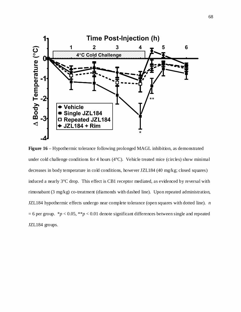
68
Figure 16 – Hypothermic tolerance following prolonged MAGL inhibition, as demonstrated
under cold challenge conditions for 4 hours (4°C). Vehicle treated mice (circles) show minimal
decreases in body temperature in cold conditions, however JZL184 (40 mg/kg; closed squares)
induced a nearly 3°C drop. This effect is CB1 receptor mediated, as evidenced by reversal with
rimonabant (3 mg/kg) co-treatment (diamonds with dashed line). Upon repeated administration,
JZL184 hypothermic effects undergo near complete tolerance (open squares with dotted line). n
= 6 per group. *p < 0.05, **p < 0.01 denote significant differences between single and repeated
JZL184 groups.

69
Figure 17 – In contrast to the tolerance exhibited following prolonged MAGL inhibition,
prolonged FAAH inhibition maintains efficacy in tail immersion antinociception. Time line
comparison of changes in tail immersion latency from baseline over 1, 3, and 6 days following
either JZL184 (a) or PF3845 (b) treatment. (c) Direct comparison of change in latency on day 6.
n = 6 mice per treatment. ***p < 0.001 denotes significant difference from vehicle, or between
single and repeated drug treatment groups.

70
Latencies remained consistent through repeated testing and vehicle injections, while both
drugs showed similar immediate effects, as indicated by day 1 in the repeated groups and day 6
of the single-exposure groups. There was a significant interaction term between treatment and
testing day [F(8, 70) = 19.5, p < 0.001], and split analysis by testing day shows a diminishing
effect of JZL184 over repeated treatment, which returns to vehicle latencies by day 6.
Meanwhile, PF3845 maintains similar analgesia across repeated treatment days to the same
levels as initial exposure, with both repeated and single PF3845 groups having significantly
greater latencies than vehicle (p < 0.001).
It was at this time that the newly-generated MAGL (-/-) mice became available for
evaluation. We tested the responses of MAGL (+/+), (+/-), and (-/-) mice while still on the
SV129 background on which they were created. As shown in Figure 18, MAGL (-/-) mice show
a hyperalgesic phenotype compared to (+/+) controls in the 52°C tail immersion test (p < 0.05;
Figure 18a), a temperature at which our previous studies were conducted, and a temperature at
which FAAH (-/-) latencies match those of wild-type controls. Hyperalgesic responses were also
noted in the 56°C hot plate test (p < 0.05; Figure 18b), a temperature at which FAAH (-/-) show
a hypoalgesic phenotype. These results contrast to those found in FAAH (-/-) mice, which
display a normal or hypoalgesic phenotype in similar tests, though those tests were run on mice
with a C57 background strain.
2.3 Chronic MAGL blockade leads to behavioral cross-tolerance to exogenous cannabinoids
To further explore potential alterations in the endocannabinoid system, we compared the
behavioral effects of cannabinoid receptor agonists in animals with chronic disruptions in MAGL

71
Figure 18 – Baseline pain sensitivity of mice with genetically inactivated MAGL activity. In
both the nociceptive 52°C water bath tail immersion (a) and 56°C hot plate (b) assays, latencies
were lower in MAGL(-/-) mice compared to wild-type controls. n = 6-8 per group. *p < 0.05
versus MAGL (+/+) group.

72
versus controls. It has previously been shown that FAAH (-/-) mice exhibit wild-type responses
in antinociceptive, hypothermic, and cataleptic assays when treated with THC, indicating normal
CB1 function in these animals. However, MAGL (-/-) mice and repeated enzyme inhibitor
administration has not been tested under similar conditions. JZL184 (40 mg/kg), PF3845 (10
mg/kg), MAGL (-/-), and FAAH (-/-) mice were all employed to profile the potential for
prolonged elevations in endocannabinoids to produce cross-tolerance to exogenous cannabinoid
activation.
The first experiment looked at the effect of repeated JZL184 in FAAH (+/+) and (-/-) mice
in response to cumulative dose-responses of THC. All tests were run 26 h following the final
JZL184 injection, based on the previous above studies indicating that the majority of acute
behavioral alterations are returned to baseline levels by 24 h. In all measures, THC increased the
magnitude of effect with increasing dose, with the responses between FAAH genotype nearly
identical. While by the highest dose tested (100 mg/kg), almost all measures were at or
approaching maximal effect in vehicle treated animals, interaction terms for treatment by THC
dose indicate repeated JZL184 treated mice showed significantly reduced response to the
antinociceptive [F(6, 84) = 35.9, p < 0.001; Figure 19a] and hypothermic [F(6, 84) = 19.5, p <
0.001; Figure 19b] characteristics of THC. The potency of THC was not only shifted, but the
maximal efficacy of THC was severely depressed in these endpoints to a point of minimal
response even at extraordinary doses. Interestingly, the cataleptic properties of THC were not
significantly altered by repeated JZL184 pretreatment [ANOVA(THC dose x JZL184), p > 0.05;
Figure 19c]. The extent of cross-tolerance observed was not altered in any way by FAAH
genotype.

73
Figure 19 - Prolonged
JZL184 administration
produces behavioral
cross-tolerance to the
partial cannabinoid
receptor agonist THC.
Cumulative dose-
response comparison
of the behavioral
effects of THC in mice
pretreated with either
repeated vehicle
(circles with solid line)
or 6-day JZL184 (40
mg/kg; open squares
with dashed line). All tests were run in both FAAH (+/+) mice (left column) and FAAH (-/-)
mice (right column). Mice were tested 26 h after their final pretreatment for tail immersion
antinociception (a), hypothermia (b), and catalepsy (c) in response to escalating doses of THC.
Repeated JZL184 produced significant decreases in response to the antinociceptive and
hypothermic effect of THC, but not catalepsy. FAAH genotype did not alter the response to
THC in vehicle treated mice, and did not alter the degree of cross-tolerance to THC following
repeated administration. n = 8 per group, *p < 0.05, **p < 0.01, ***p < 0.001 denote significant
differences from vehicle controls.

74
The extent of cross-tolerance to THC was nearly complete, possibly due to its partial agonist
properties at CB1 receptors, and 2-AG being a full agonist demonstrated by in vitro testing.
Further tests were conducted against the synthetic full agonist WIN55,212-2 (WIN), which is the
most commonly used and best characterized synthetic agonist in comparison to THC, including
comparable regional CB1 receptor downregulation (Sim-Selley and Martin, 2002). MAGL (-/-)
mice were tested first for comparison of dose-responses to WIN versus MAGL (+/+) and (+/-)
controls. As seen with the hyperalgesia tests shown earlier, the MAGL (+/+) and (+/-) groups
showed similar response to WIN, which showed maximal responses in all measures in a manner
about 10-fold more potent than seen with THC. There were significant statistical interaction
terms for genotype and WIN dose observed for the antinociceptive [F(12, 120) = 3.6, p < 0.001;
Figure 20a] and hypothermic [F(12, 120) = 4.6, p < 0.001; Figure 20b] measures. Upon
examining individual doses, the MAGL (-/-) mice showed significantly reduced responses to
WIN at 3 to 10 mg/kg for tail immersion, and 10 to 30 mg/kg for hypothermia. The tail
immersion data converges again at the highest dose, though any differences in response are
obscured by the test cutoff for maximal responses, something that was unable to be achieved by
THC. However, as seen with JZL184 in THC, the cataleptic response to WIN was not altered by
MAGL genotype (Figure 20c).
Due to the differences in background strain that could represent a potential confound to the
MAGL (-/-) results, tests were replicated with repeated JZL184 treated mice. Response and
potency of WIN was roughly equal to that seen in the MAGL (-/-) test, and the results confirmed
those seen in both previous experiments. Repeated JZL184 mice showed reduced response to
WIN-induce antinociception [F(6, 84) = 25.1, p < 0.001; Figure 20d] and hypothermia [F(12,
120) = 40.8, p < 0.001; Figure 20e]. As seen with THC, there was a shift in potency as well as

75
Figure 20 - Repeated JZL184 administration, or genetic deletion of MAGL, produces behavioral
cross-tolerance to a cannabinoid receptor agonist. (a, b, c) Cumulative dose-response
comparison of WIN55,212-2 behavioral effects in MAGL (+/+; circles), (+/-; triangles), and (-/-;
open squares) mice. Mice were tested for tail antinociception (a), hypothermia (b), and
catalepsy (c) in response to increasing doses of WIN. (c, d, e) Cumulative dose-response
comparison of the behavioral effects of WIN55,212-2 in mice pretreated with either repeated
vehicle (circles with solid line) or 6-day JZL184 (40 mg/kg; open squares with dashed line).
Mice were tested 26 h after their final pretreatment for tail antinociception (c), hypothermia (d),
and catalepsy (e) in response to WIN. Repeated JZL184 and MAGL (-/-) mice exhibited
significant decreases in response to the antinociceptive and hypothermic effect of WIN, but not
catalepsy. n = 8 per group in the JZL184 study, and n = 6 per group in the MAGL genotypic
study. *p < 0.05, **p < 0.01, ***p < 0.001 versus vehicle pretreatment or MAGL (+/+) controls.

76
an overall depression of maximal effect, though not of the same magnitude. Catalepsy continued
to be unresponsive to change by prolonged MAGL inhibition.
As a final cross-tolerance comparison in response to elevated endocannabinoids, repeated
PF3845 was tested against increasing doses of WIN. As shown in Figure 21, dose-responses of
antinociception, hypothermia, and catalepsy are all overlapping between PF3845 and vehicle
pretreated groups. These tests indicate that prolonged elevation of 2-AG, but not AEA, produce
cross-tolerance to exogenous cannabinoid receptor activation.
2.4 Discussion: Tolerance following prolonged endocannabinoid elevation
The MAGL inhibitor JZL184 produced a near full profile of behavioral effects in
cannabimimetic tetrad studies reported in the original article (Long et al., 2009a). However,
since then, we have learned that many of these effects are the result of interactions between the
drug and the polyethylene glycol-based vehicle in which it was tested. In accordance with these
findings, we transitioned to using traditional ethanol:emulphor:saline vehicle. While JZL184 is
less soluble and the behavioral profile is less pronounced, similar MAGL inhibition and 2-AG
elevations can be achieved with slightly higher concentrations. Upon testing JZL184 tetrad
behavior in the new vehicle, tail flick antinociception latencies remained enhanced, and
hyperreflexive popcorning maintained its prevalence.
In our effort to replicate the findings of enhanced behavioral effects by simultaneous inhibition
of FAAH and MAGL, JZL184 was also tested in FAAH (-/-) mice. What we observed was a
replication of the original findings of the Cravatt lab (Long et al., 2009b), with FAAH (-/-) mice
showing longer latencies to noxious thermal heat, and a longer timeline of significant

77
Figure 21 – Prolonged inhibition of FAAH via repeated PF3845 administration fails to produce
behavioral cross-tolerance to a cannabinoid receptor agonist. (a, b, c) Cumulative dose-response
comparison of WIN55,212-2 behavioral effects in mice treated with either vehicle (circles) or
PF3845 (10 mg/kg daily; open squares). Mice were tested for tail antinociception (a),
hypothermia (b), and catalepsy (c) in response to increasing doses of WIN. n = 8 per group.

78
efficacy compared to wild-type controls. We also observed a greater percentage of mice
presenting as hyperreflexive for a longer period, and a new behavior in the form of mice
gnawing and biting at the catalepsy bar upon presentation. This was observed almost exclusively
in mice with both FAAH and MAGL inhibited. This behavior is unlikely a measure of
aggression, as mice did not display aggressive behavior toward caged littermates upon return to
home cages, though the biting seems to be generalized to most objects placed in front of them,
which may indicate increase anxiety- like behavior. Follow-up with studies of feeding behavior,
and anxiety tests such as plus maze and light-dark box, might further clarify potential causes for
such behavior.
Regardless of FAAH phenotype, all JZL184-elicited behaviors showed reduced magnitude
and duration following six days of treatment. The analgesic tolerance resulted in essentially
complete absence of effect. When examining the hypothermic cold challenge paradigm, which
is not responsive at all to FAAH inhibition (see Fig. A1), repeatedly-treated JZL184 mice
showed no significant drop in body temperature compared to vehicle, again demonstrating
complete tolerance. Since our original test conditions for tail immersion were designed to be
mild enough as to not elicit FAAH analgesia at baseline (52°C; Lichtman et al., 2004), we
repeated our tests comparing MAGL and FAAH inhibitors head-to-head over repeated treatment
to see which target shows higher efficacy, and whether either maintain their analgesic activity.
The FAAH inhibitor PF3845 showed slightly lower increases in withdrawal latency upon initial
exposure compared to JZL184. However, JZL184 lost the entirety of its analgesic efficacy by
six days, while PF3845 maintained its original effectiveness throughout. FAAH inhibitors also
maintained their efficacy to reduce mechanical and cold allodynia in a nerve injury model of
pain (Kinsey et al., 2009), as well as increased anti- inflammatory properties with repeated

79
treatment. In both these models, repeated MAGL inhibition also underwent complete tolerance
(see Fig. A2). This maintenance of beneficial outcomes in pain and inflammation testing is a
very promising trait for FAAH inhibitors as potential clin ical analgesics, as even the most widely
effective analgesics in use today (i.e. acetaminophen, opioids) undergo some level of tolerance.
Upon examining the baseline pain sensitivity of MAGL (-/-) mice, we see a hypersensitivity
to noxious heat in the spinally-dominant tail immersion assay, and the supraspinally-dominant
hot plate assay. This is in stark contrast to previous study of FAAH (-/-) mice, which show
phenotypic hypoalgesia (Lichtman et al., 2004). This further underscores the adaptations
occurring following prolonged MAGL inhibition, and apparent lack of adaption in the presence
of chronic FAAH inhibition.
Given the complete tolerance to a wide variety of cannabinoid-like behaviors following
repeated JZL184, we expected to see effective cross-tolerance to exogenous cannabinoid
agonists under the same conditions. Mice showed identical baseline behavior given a 26 h
washout period following the last injection of JZL184, suggesting JZL184 behavioral activity
has subsided. We were surprised to see not only profound shifts in THC’s antinociceptive and
hypothermic potency in these mice, but also a substantial depression of THC efficacy as well,
almost to the point of inactivity. As seen with the tolerance data, this effect appears to be
entirely driven by prolonged MAGL inhibition, as no differences were noted in FAAH (-/-) mice.
The absence of catalepsy tolerance represents a noticeable difference between tolerance by an
endogenous ligand and an exogenous agonist. THC would typically produce tolerance to all
tetrad effects, including catalepsy, though when given systemically is expected to hit all
receptors in the brain equally. These catalepsy data may suggest that expression levels of
MAGL in various brain regions may dictate the level of tolerance seen for various behaviors.
80
Since THC cross-tolerance to JZL184 was approaching full insensitivity, we chose to
examine the efficacy of the test drug as a factor in the level of tolerance observed. Typically, it
is harder to suppress activity of a full receptor agonist compared to a partial agonist. Follow-up
testing of WIN in JZL184-treated and MAGL (-/-) mice provided measures of the maximal
tolerance possible by MAGL in the face of maximal possible cannabino id receptor activation.
While the level of cross-tolerance was still clear following prolonged MAGL inhibition, in both
tests it appeared that the behavioral effects were surmountable with increasing doses of WIN.
This fits with expected models of receptor activation from in vitro studies, and suggesting 2-AG
produces complete cross-tolerance to partial agonists, and shifts in potency of other full agonists.
Also fitting with this model, elevated partial agonism by AEA during prolonged FAAH
inhibition was incapable of producing any signs of cross-tolerance to WIN challenge.
Finally, to demonstrate the functional consequences the phenomena of tolerance and cross-
tolerance have on neuronal communication and plasticity, electrophysiological studies were
conducted in mice treated with repeated JZL184 and PF3845 (see Fig. A4). While previous
studies have shown JZL184 acutely enhances cannabinoid-mediated DSI, we’ve demonstrated
that in a pair of brain regions studied (hippocampus and cingulate cortex), repeated JZL184
treated mice show diminished DSI, a functional corollary to tolerance. Furthermore, exogenous
application of cannabinoid agonist normally suppresses neuronal activity, which is also
diminished by repeated JZL184 administration, a functional consequence of reduced CB1
receptor activity. Neither of these effects was seen in repeated PF3845 treated mice.
These data are potential complications for MAGL inhibition as a therapeutic target, as not
only does the analgesic activity of MAGL inhibition diminish with repeated dosing, but so does
the behavioral potency and neuronal function of at least one entire class of compounds. Given

81
the extensive cross-talk of cannabinoid and opioid systems, especially with pain-related
endpoints, testing for extrinsic cross-tolerance to morphine analgesia would need to be evaluated
to determine the extent of functional losses. Yet to be examined as alternative therapeutic
options are either intermittent long-term dosing that takes into account the extraordinary length
of JZL184 activity in brain, or loser dosing of JZL184 that might not produce the level of
elevations in 2-AG or as prolonged an increase as seen with the high-dose regimen used here.
Meanwhile, FAAH inhibitors continue to present no signs of significant consequences on the
normal functioning of cannabinoid receptor action.

82
Chapter 3: Enhanced endocannabinoid availability and functional receptor adaptation
corresponding with behavioral responses
3.1 Endocannabinoid quantification during THC withdrawal
To examine any possible alterations in endogenous cannabinoid levels during THC withdrawal,
AEA and 2-AG were quantified from brains of FAAH (+/+) and (-/-) mice, treated with either
repeated vehicle or THC (50mg/kg), and precipitated with Rimonabant (10 mg/kg). The two
brain tissue sections examined were cerebellum and combined forebrain/midbrain, the results
summarized in Table 1. The cerebellum displayed no significant differences in AEA with regard
to treatment, however FAAH (-/-) mice showed approximate 10-fold AEA elevations [F(1, 28) =
242.4, p < 0.001] regardless of treatment. FAAH (-/-) mice also showed significant decreases in
cerebellum 2-AG content regardless of treatment [F(1, 28) = 5.5, p < 0.05]. In the
forebrain/midbrain, in addition to the elevations in AEA in FAAH (-/-) mice [F(1, 29) = 313.9, p
< 0.001], there was an interaction between genotype and treatment [F(1, 29) = 5.6, p < 0.05].
FAAH (-/-) mice showed reduced AEA levels in midbrain/forebrain tissue during THC
withdrawal when compared to FAAH (-/-) mice receiving rimonabant alone (p < .05). There
were no differences in 2-AG levels in forebrain/midbrain tissues between any groups.

83
Table 1. Endocannabinoid Quantification Following Rimonabant Precipitation
AEA
(pм/g tissue)
2-AG
(nм/g tissue)
Cerebellum
FAAH (+/+) , Veh/Rim 2.7 ± 0.4 14.1 ± 0.7
FAAH (+/+) , THC/Rim 3.0 ± 0.6 12.4 ± 1.1
FAAH (-/-) , Veh/Rim 29.7 ± 2.0 11.8 ± 0.7
FAAH (-/-) , THC/Rim 32.2 ± 2.9 10.3 ± 1.1
Forebrain/Midbrain
FAAH (+/+) , Veh/Rim 3.4 ± 0.5 12.6 ± 1.0
FAAH (+/+) , THC/Rim 2.7 ± 0.4 11.3 ± 0.6
FAAH (-/-) , Veh/Rim 27.6 ± 1.6 10.1 ± 0.8
FAAH (-/-) , THC/Rim 21.1 ± 1.8* 12.1 ± 1.5
*p < .05 vs. FAAH (-/-) Veh/Rim group

84
3.2 Prolonged MAGL, but not FAAH, inhibitors lead to accumulation of 2-AG in brain
The elevation of whole-brain 2-AG levels following pharmacological MAGL inhibition by
JZL184 has been previously established (Long et al., 2009). We further compared the AEA and
2-AG levels following 6 days of JZL184 (40 mg/kg daily), and found a similar accumulation of
eCBs following prolonged inhibition. At the peak 2 h time point (Figure 22a), we see a modest
increase in AEA level only in the repeated JZL184 group, which is gone by 26 h (Figure 22c).
This is likely a nonselective effect of JZL184, which has been shown previously to inhibit FAAH
transiently at higher doses by upward of 60% when given acutely. At the 2 h peak, JZL184
given repeatedly produces 2-AG levels around 15-fold above vehicle-treated levels, and
represent a 79% increase above that observed with JZL184 given just once. This increase
diminishes post- injection, as repeated JZL84 enhancement over single exposure is down to 39%
by 26 h. When testing the peak 2 h effects in FAAH -/- mice, we find JZL184 does not alter the
already elevated AEA levels, and shows similar patterns of 2-AG elevation to FAAH (+/+) mice
(Figure 22b).
When examining the endocannabinoid accumulation in brain following daily dosing of
PF3845 (10 mg/kg), we see AEA elevations acutely 2 h post- injection of more than 15-fold
above that of vehicle (Figure 23a). There does not appear to be any substantial difference in the
level of AEA accumulation following repeated dosing compared to acute exposure. Also,
PF3845 shows clear selectivity, as 2-AG levels are unaltered by either single or repeated dosing
(Figure 23b).

85
Figure 22 - Whole-brain AEA & 2-AG levels following either single or repeated JZL184
treatment. (a) 2 h after treatment JZL184 (40 mg/kg) selectively increases 2-AG about 10-fold
after acute administration, and demonstrates an accumulation of 2-AG with repeated JZL184
administration (left column). FAAH (-/-) mice show similar 2-AG elevations, with typical 10-
fold AEA elevations regardless of treatment. (b) 26 h following final administration, 2-AG
levels are still elevated in JZL184 treated mice, with diminished accumulation effects. (c)
PF3845 elevates AEA levels almost 20-fold, with similar elevations following either single or
repeated administration, and without any elevations in 2-AG levels. n = 6 per group. *p < 0.05,
***p < 0.001 versus vehicle controls, ##p < .01 versus single JZL184 treatment.

86
Figure 23 - Whole-brain AEA & 2-AG levels following either single or repeated PF3845
treatment. PF3845 elevates AEA levels almost 20-fold 2 h post-injection, with similar elevations
following either single or repeated administration, and without any elevations in 2-AG levels. n
= 6 per group. ***p < 0.001 versus vehicle controls.

87
3.3 Genetic inactivation of MAGL enhances 2-AG in brain
The created MAGL (-/-) mice show reduced protein expression and almost 88% impairment
in hydrolysis rates of 2-AG (see Fig. A3). The resulting decrease rate of hydrolysis allows for
the accumulation of several glycerol-containing lipids, though 2-AG is by far the most
prominent, with nearly 15-fold increases observed in MAGL (-/-) mice, a level comparable to
that seen in repeated JZL184-treated mice (Figure 24b). The resulting accumulation is also
apparent when examining the free fatty acid degradation product, whose levels decrease in
MAGL (-/-) animals by nearly 70% compared to (+/+) littermates, a decrease not seen in MAGL
(+/-) mice (Fig. 24c). Importantly, NAE levels, including that of the other major
endocannabinoid AEA, are not significantly altered based on MAGL genotype (Figure 24a).
3.4 Brain CB1 receptors are impaired by chronic MAGL, but not FAAH, blockade
The loss of most all acute behavioral responses to JZL184 with repeated treatment, and
occurrence of cannabinoid cross-tolerance in mice with genetic or prolonged pharmacological
disruption of MAGL suggested that CB1 receptors might be downregulated and/or desensitized
in these animals. To test this possibility, we examined CB1 receptor expression and function
through specific binding of [3H]-SR141716A and CB1 agonist CP55,940-stimulated [35S]-GTPγS
binding, respectively, in whole-brain homogenates from mice repeatedly treated with JZL184.
Prolonged MAGL disruption led to decreases in CB1 receptor binding curves compared to
vehicle, as well as a suppressed magnitude of [35S]-GTPγS binding (Fig. 25a and 25b).
Nonlinear sigmoidal regression on the activation curves revealed a significantly lower maximal
efficacy of agonist stimulation (Emax), with no apparent change in the potency as measured by
EC50. The non- linear regression of the binding data revealed a significantly reduced maximal

88
Figure 24 – Lipid profile of mice with genetically inactivated MAGL activity. Without any
alteration in AEA levels in whole brain (a), MAGL (-/-) mice have 15-fold elevations in 2-AG
whole brain content (b). MAGL (+/-) mice, despite only 50% 2-AG hydrolysis activity, show
normal wild-type levels of 2-AG. (c) With the increase in 2-AG, a decrease in 2-AG’s
metabolite, arachidonic acid, is also seen in MAGL (-/-) mice. ***p < 0.001 versus vehicle
MAGL (+/+) controls.

89
Figure 25 - Loss of
receptor activation and
membrane receptor
levels following 6-day
MAGL inhibition by
JZL184. Comparison
of the membrane
receptor GTPγS
binding (a) stimulated
by the cannabinoid
agonist CP55,940, and
the specific CB1
receptor binding (b) by
the tritiated antagonist
3H-SR141716A, from
homogenate whole
brains following
pretreatment with either vehicle (circles), or 6-day JZL184 (open squares with dashed line).
Repeated JZL184 decreases both the overall level of receptor signaling as well as the number of
surface receptors available. n = 4 tissue samples per group, run in separate experiments with
each point run in triplicate for GTPγS and duplicate for receptor binding.
90
binding (Bmax) without alteration of the KD (Table 2). The magnitude of overall loss in receptor
efficacy closely matched that of loss in receptor binding. These results are typical of receptor
loss without fundamental changes to the receptor function that may alter ligand affinity.
As a generous donation by the Cravatt lab at the Scripps Research Institute, we were given
access to whole-brain samples from control mice that were part of a cancer study. The mice
were severely compromised immune deficient (SCID) mice that were littermates of mice
implanted with tumorigenic cells, and were treated with 30 days of either vehicle or JZL184 (40
mg/kg) by oral gavage. The [35S]-GTPγS curves are shown in Figure 26a, displaying a
substantial reduction in Emax of 53% [Vehicle: 117.3 ± 6.3, JZL184: 55.8 ± 6.2] without any
significant shift in EC50 [Vehicle: 0.06 ± 0.03, JZL184: 0.12 ± 0.06]. In contrast, the loss of
receptor binding in 30-day JZL184 mice was comparable in magnitude to that seen by 6-day
JZL184 treatment (Figure 26b), representing a 33% loss of maximal binding [Vehicle: 3.87 ±
0.30, JZL184: 2.88 ± 0.38], and no shifts in the KD [Vehicle: 0.69 ± 0.12, JZL184: 0.71 ± 0.23].
These decreases in receptor activation in the absence of further receptor loss are typically
indicative of desensitization; however there could also be strain differences.
The questions arising from the previous data were confirmed against MAGL (-/-) mice, which
have genetic inactivation of this enzyme and 2-AG elevations from development, and represent
yet a third different strain of mice against which these receptor adaptations were tested. The
MAGL (-/-) mice showed far greater depressions in maximal receptor activation of 44%, with
MAGL (+/-) mice showing a slight lower and nonsignificant loss of 21% (Figure 27a). A similar
pattern was seen in the receptor binding, with MAGL (-/-) showing 49% decrease in total
binding sites compared to (+/+) controls, and (+/-) mice did not significantly differ from wild-

91
Figure 26 - Loss of
receptor activation and
membrane receptor
levels following 30-day
oral JZL184
administration.
Comparison of the
membrane receptor
GTPγS binding (a)
stimulated by the
cannabinoid agonist
CP55,940, and the
specific CB1 receptor
binding (b) by the
tritiated antagonist 3H-
SR141716A, from
homogenate whole
brains following pretreatment with either vehicle (circles), or 30-day oral gavage or JZL184
(open squares with dashed line) in SCID mice. 30-day JZL184 decreased receptor activation and
the number of surface receptors available, the magnitude of the reduction greater than seen with
6-day JZL184 treatment. n = 4 tissue samples per group, run in separate experiments with each
point run in triplicate for GTPγS and duplicate for receptor binding.

92
type controls (Figure 27b). As with prior tests, the EC50 and KD values indicate no alterations in
receptor affinity and function. The pattern of loss of both activation capacity and binding sites
parallels that seen in the 6-day JZL184 brains, but to a greater magnitude (Table 2).
In contrast, prolonged blockade of FAAH by PF-3845 did not impact CB1 receptor
expression or function (Figure 28 & Table 2). These findings are consistent with previous work
showing no loss of CB1 receptor number or function in FAAH-/- mice.
3.5 Regional analysis of brain CB1 receptor adaptation and eCB accumulation
The observation that prolonged MAGL blockade caused profound cross-tolerance to the
antinociceptive and hypothermic, but not cataleptic effects of cannabinoid receptor agonists
could indicate that CB1 receptors were differentially affected in specific brain regions. To test
this hypothesis, we performed an extensive regional analysis of CP55,940-stimulated
[35S]GTPS binding in mice treated using the 6-day either vehicle or JZL184 (40 mg/kg) dosing
regimen. Representative autoradiograms illustrate CP55,940-stimulated [35S]GTPS binding in
both groups at several coronal levels, including caudate putamen, hippocampus, periaqueductal
gray (PAG), and cerebellum (Figure 29a). Visual inspection shows chronic JZL184 treatment
led to a heterogeneous reduction in CP55,940-stimulated [35S]GTPS binding. Notable brain
regions showing significant CB1 desensitization include the cingulate cortex, hippocampus,
somatosensory cortex, and PAG (Fig. 29b). In contrast, chronic JZL184 treatment did not elicit
desensitization in the striatum or globus pallidus, two brain regions associated with catalepsy
(Pertwee and Wickens, 1991; Wickens and Pertwee, 1993).

93
Figure 27 - Loss of
receptor activation and
membrane receptor levels
in mice with genetically
inactivated MAGL activity.
(a, b) Comparison of the
GTPγS binding (a) and
specific CB1 receptor
binding (b) from
homogenate whole brains
of MAGL (+/+; circles),
MAGL (+/-; triangles), or
MAGL (-/-; open squares
with dashed line) mice.
MAGL (-/-) mice show
significantly reduced
overall receptor activation and membrane receptor numbers compared to wild-type controls, with
MAGL (+/-) showing partial, but still significant reductions. n = 4 tissue samples per group, run
in separate experiments with each point run in triplicate for GTPγS and duplicate for receptor
binding.

94
Figure 28 – Prolonged FAAH inhibition by repeated PF3845 does not alter CB1 receptor
function. GTPγS binding (a) stimulated by the cannabinoid agonist CP55,940, and specific CB1
receptor binding (b) by the tritiated antagonist 3H-SR141716A, was assayed from homogenate
whole brains following pretreatment with either vehicle (circles), or 6-day PF3845 (open squares
with dashed line). No significant changes in either measure were noted based on PF3845
treatment. n = 4 tissue samples per group, run in separate experiments with each point run in
triplicate for GTPγS and duplicate for receptor binding.

95
Table 2. CB1 receptor affinity, activation, and binding following prolonged eCB elevations. Best-fit
values (mean ± SEM) for the G-protein activation and receptor binding curves. **p < 0.01, ***p < 0.001
decrease versus respective control group.
Treatment EC50 (μM) Emax (% stimulation) % Loss (vs. control)
MAGL Genotype MAGL +/+ MAGL +/- MAGL -/-
0.05 ± .02 0.03 ± .01 0.02 ± .01
183.3 ± 11.1 157.0 ± 6.5 107.5 ± 5.7
21% 44%***
JZL184 Treatment Vehicle Repeated JZL184
0.10 ± 0.02 0.09 ± 0.02
78.3 ± 3.4 58.1 ± 3.1
8%
25%** PF3845 Treatment
Vehicle Repeated PF3845
0.03 ± 0.02 0.05 ± 0.02
93.8 ± 8.7
125.8 ± 10.9
N/A
Treatment KD (nM) Bmax (pmol/mg) % Loss (vs. control)
MAGL Genotype MAGL +/+ MAGL +/- MAGL -/-
0.74 ± 0.12 0.48 ± 0.06 0.76 ± 0.11
2.79 ± 0.22 1.94 ± 0.09 1.42 ± 0.10
30% 49%***
JZL184 Treatment Vehicle Repeated JZL184
0.74 ± 0.33 0.96 ± 0.49
3.41 ± 0.28 2.74 ± 0.16
3%
26%** PF3845 Treatment
Vehicle Repeated PF3845
0.91 ± 0.29 0.96 ± 0.16
2.93 ± 0.50 3.60 ± 0.33
N/A

96
Figure 29 - Regional changes in cannabinoid agonist-stimulated [35S]GTPγS binding and 2-AG
levels following prolonged inhibition of MAGL. (a) Representative autoradiograms showing
CP55,940-stimulated [35S] GTPγS binding in coronal brain sections following either repeated
vehicle (left column) or JZL184 (right column) treatment. Pseudocolor images indicate levels of
receptor-mediated G-protein activity and highlight significant decreases in CB1 receptor
activation in the cingulate cortex (row 1), hippocampus (row 2) and periaqueductal gray (row 3),
while no differences are apparent in the caudate-putamen (row 1) or cerebellum (row 4). (b)
Densitometric analysis of CP55,940-stimulated [35S]GTPγS binding in selected regions,
including: cingulate cortex (CG CTX), caudate putamen (CPU), globus pallidus (GP), preoptic
area of the hypothalamus (POA), hippocampus (HIPP), amygdala (AMYG), hypothalamus
(HYPO), somatosensory cortex (SS CTX), substantia nigra (SN), periaqueductal gray (PAG), &
cerebellum (CBLM). (c) Regional 2-AG levels following JZL184 treatment, many

97
corresponding to regions quantified for CB1 receptor activation. Increases, while varying
somewhat in intensity, were less regionally dependent than CB1 receptor activation. n = 8 brains
per group, run in triplicate slices for each targeted region. *p < 0.05, **p < 0.01, ***p < 0.001
versus vehicle treatment for specific region. ##p < 0.01, ###p < 0.001 versus single JZL184
treatment for specific region.

98
To confirm that the differential behavioral adaptations are due to CB1 receptor activation,
and not regional differences in eCB signaling pools, we examined gross regional changes in
endocannabinoids to match key regions examined in [35S]GTPS binding. Vehicle, single
JZL184, and repeated JZL1284 groups were sacrificed 26 h after their final injection to match
the timeline of the autoradiographic study. Levels of AEA were not substantially altered across
regions by any treatment, and directional changes varied by region (Figure 30). While the
elevations in 2-AG vary between rostral and caudal regions, similar relative accumulation effects
as those in whole-brain are seen following repeated JZL184 in each region. Each region showed
significant elevation in 2-AG following acute JZL184 (Figure 29c), which was further enhanced
by repeated administration. 2-AG content is not predictive of CB1 receptor dysfunction, as areas
such as neocortex and striatum have identical 2-AG profiles, but divergent levels of receptor
inactivation. Given that many of the areas associated with motor coordination and function (i.e.
globus palladus, caudate putamen, and cerebellum) do not show desensitization following
repeated JZL184, evidence points to CB1 receptor deficits as the overriding correlate to
behavioral tolerance rather than potential regional differences in 2-AG elevation.
3.6 Discussion: Endocannabinoid accumulation and cannabinoid receptor adaptations
Given that all the behaviors observed involved either direct alteration of the regulatory function
of endocannabinoid ligands, or repeated administration of exogenous cannabinoid agonists that
may alter normal endocannabinoid activity, it is important to understand the changing
endogenous ligand availability in order to predict possible changes in endocannabinoid function.
We found that inhibitors of endocannabinoid degradation reduced antagonist-precipitated
behaviors in mice undergoing THC withdrawal; however no phenotypic differences

99
Figure 30 - Regional AEA levels following JZL184 treatment, many corresponding to regions
quantified for CB1 receptor activation. Changes in AEA content were bidirectional, with
direction of changes regionally-dependent. No effects of repeated JZL184 were observed when
compared to acute JZL184 effects. n = 8 brains per group, run in triplicate slices for each
targeted region. *p < 0.05, **p < 0.01 versus vehicle treatment for specific region.

100
were observed in FAAH (-/-) mice. Follow-up studies shown into AEA and 2-AG content in the
brains of FAAH (-/-) mice showed lower 2-AG content in the cerebellum compared to wild-type
mice, which may be a compensatory mechanism for consistent AEA elevations. There was also
a downregulation of AEA content in midbrain/forebrain regions following chronic THC, which
may be a feedback response on AEA synthesis due to repeated CB1 receptor activation.
Reductions in AEA content of midbrain/forebrain regions have been previously reported in rats
during THC withdrawal, and similarly no alterations were seen in cerebellum (Gonzalez et al.,
2004). Cerebellum also presents less CB1 receptor desensitization and downregulation than
midbrain areas, such as thalamus and hippocampus, following chronic THC (Sim-Selley and
Martin, 2002). Either of the reduced endocannabinoid responses observed in FAAH (-/-) mice
could account for normalized sensitivity to THC withdrawal. Given the possibility that AEA is
not increased under cannabinoid withdrawal conditions, but rather subject to specific decreases,
may provide justification for why FAAH inhibition may be valuable for the stabilization of
endocannabinoid function during THC withdrawal.
When testing the endogenous cannabinoid content in whole brain following enzyme
inhibitor administration, we were addressing two main questions of repeated inhibition: 1) does
FAAH inhibition or MAGL inhibition maintain selectivity for their respective endocannabinoid
target when continuously inactivated over a period of several days, and 2) can repeated inhibition
enhance ligand availability above that of just a single exposure, or are the levels seen following
initial inhibition representative of a stable plateau for endocannabinoids levels? While PF3845
represents a highly selective inhibitor, even compared to URB597, JZL184 always been reported
to possess an ability to inhibit FAAH acutely (Long et al., 2009a; Long et al., 2009b). However,
the ability to inhibit FAAH is far less potent than its MAGL activity, and levels of enzyme

101
inhibition never reached an extent capable of producing AEA elevations in brain when given
acutely. Meanwhile, though approximately 10-fold increases in AEA and 2-AG via treatment of
their respective enzyme inhibitors seems substantial, no studies to date have determined if these
levels continue to stay enhanced, much less elevate further with repeated exposure. Further
elevations would be the result of continued enhancement of ligand availability, likely a
consequence of additional synthesis under conditions of diminished degradative mechanisms.
Conversely, newly established stable levels would be the result of cellular adaptations that allow
for negative feedback mechanisms that stabilize extraphysiologic elevations in endogenous
cannabinoids. These feedback mechanisms could include reduced synthetic mechanisms of
endocannabinoids, induction of increased synthesis of the degradative enzymes, or even
degradation by alternative lower-affinity hydrolases as levels reach higher concentrations.
What we observed when examining repeated JZL184 treated mice was a cumulative effect
of inhibition. Transient enhancements of AEA levels were observed a few hours following
injection only in those receiving repeated JZL184, indicating a slight impact of partial FAAH
inhibition over a period of several days. While this increase is comparatively minimal and brief
compared to elevations seen by FAAH inhibitors and FAAH knockout mice, it does raise
questions as to any contribution AEA is playing in any observed effects in mice treated with
repeated JZL184. Given its relatively minor elevation and absence by 26 h post- injection, it
likely plays minimal role in the long-term adaptations observed. Since MAGL (-/-) show no
elevations in AEA, this confirms that these observations are likely nonselective actions of
JZL184, and studies of repeated JZL184 in MAGL (-/-) mice will confirm the contribution of
simultaneous AEA and 2-AG enhancement on drug action. Importantly, most behavioral and

102
receptor studies performed in MAGL (-/-) so far follow the pattern of findings seen in mice
treated with repeated JZL184.
FAAH (-/-) mice show typical stable 10-fold increases in AEA, and repeated FAAH
inhibition by PF3845 produces similar AEA elevations when comparing initial exposure to
prolonged inhibition. Interestingly, these chronic elevations do not appear to impact the
available pool of precursors for 2-AG, as 2-AG levels in FAAH (-/-) mice and repeated PF3845
treated animals are unaltered compared to respective controls.
It appears that with prolonged inhibition of MAGL, a continual enhancement and further
accumulation occurs for 2-AG above that of a single exposure. With the high abundance and
rapid kinetics of 2-AG formation, it is not surprising that increased time with MAGL inhibited
produces even greater elevations. After six days, 2-AG levels reach up to 70% higher than that
of a single exposure to JZL184, with significant elevations remaining above those of acute
exposure for over 24 h. This may suggest we may not have obtained the peak physiological 2-
AG concentrations possible using pharmacological inhibition. However, this theory is not
confirmed by the results from the MAGL transgenic mice. The 2-AG levels following repeated
JZL184 inhibition closely match those of the MAGL knockouts, suggesting that the
approximately 15-fold increases are about the maximal attainable, which would fit with the fact
that at least two alternate enzymes that are still actively degrading 2-AG in brain tissue in these
mice. More interesting is the fact that heterozygous mice show now major differences compared
to wild-type controls. The initial findings using JZL184 showed that doses that could inhibit
~50% of MAGL brain hydrolysis were capable of modest increases in 2-AG on the order of 3-
fold. However, the same inhibition of hydrolysis fails to produce similar magnitudes of 2-AG

103
content in heterozygous MAGL mice, potentially suggesting some forms of compensatory
developmental compensation of reduced 2-AG hydrolysis.
With the enhancement of 2-AG availability in brain tissue with repeated JZL184
administration, this would provide evidence for enhanced drug activity after six days of
injections. However, the profound tolerance seen points to evidence of receptor level
adaptations in these neuronal circuits. Upon examining whole-brain CB1 receptors, we see that
MAGL inhibition produces profound losses in the maximal stimulation of CB1 receptor pools by
an exogenous agonist. The stable measure of potency across all tests suggests that the membrane
preparations properly removed any 2-AG from the samples, and its presence did not alter the
measurement of CB1 function or binding. Loss of maximal activation is typically indicative of
desensitization or receptors, in which GRK phosphorylation and β-arrestin binding prevents
typical activation in the presence of ligand. However, given that the magnitude of loss in
receptor activation almost exactly equals the loss of receptor binding sites in almost all cases, it
appears that prolonged 2-AG elevation promotes substantial receptor internalization and
degradation. Microscopy studies of GFP-tagged CB1 receptors in drug-treated cells could
confirm this hypothesis, and is of current focus within the Cravatt lab.
Regional analysis of the receptor activation following repeated JZL184 shows that regions
typically associated with high levels of cannabinoid receptor plasticity to exogenous agonist,
such as hippocampus and cortical regions, show extensive loss of stimulated function. In
addition, a region with established function in cannabinoid-mediated analgesia, the
periaqueductal gray, showed the largest relative decrease in receptor activation. Meanwhile
areas associated with catalepsy, such as globus pallidus and caudate putamen, showed no
apparent changes in receptor activity. It should be noted that these regions also show

104
comparative resistance to functional losses following treatment with exogenous cannabinoid
agonist, though significant functional loss is typically noted (Sim-Selley and Martin, 2002; Sim-
Selley et al., 2006). When examining the 2-AG content available in these respective regions, we
see the regional variations in intrinsic activity and susceptibility to internalization of the
cannabinoid receptors is independent of 2-AG availability. Similar elevations are seen across all
brain regions examined, and nearly identical 2-AG levels are quantified in regions that show
functional losses compared to those which do not show any loss in activity. This suggests that
while increased 2-AG likely the catalyst for inducing the loss of receptor activity, it is other
factors such as receptor density, cellular regulatory mechanisms, and turnover rate of receptor
pools themselves that dictate the level of plasticity in each region. These changes in receptor
pools appear to, in turn, alter the resultant behavioral outcomes.

105
Chapter 4: General Discussion and Conclusions
The purpose of the preceding studies was to further elucidate the potential of elevating
endogenous cannabinoids, via inhibition of the degradative enzymes FAAH and MAGL, to elicit
cannabimimetic activity. We hypothesized that elevating endogenous cannabinoids would
demonstrate potential therapeutic benefits, such as reducing cannabinoid withdrawal or as an
analgesic, through actions at cannabinoid receptors. Both endpoints showed responses following
inhibition of either FAAH or MAGL, often with similar efficacy. In addition, we aimed to
evaluate the comparative consequences of acute and prolonged inhibition of either major
degradative enzyme on potential negative effects associated with exogenous cannabinoids and
marijuana. These consequences included impairment of motor coordination, tolerance to drug
actions, and potential for physical dependence as evidenced by precipitated somatic withdrawal.
Our hypothesis that FAAH inhibitors would show minimal impact on cannabinoid function,
similar to previously reported in FAAH (-/-) mice, held for all the endpoints we tested. No
withdrawal, tolerance, or receptor adaptations were observed following FAAH inhibitor
treatment.
We also confirmed our hypothesis about the potential for endocannabinoid system
adaptations observed by MAGL inhibition. However, it is important to place these adaptations
in perspective, as we found no impairment to motor coordination or alterations in catalepsy
response. We also found that withdrawal intensity was equivalent to that of a THC regimen that

106
is minimally required to see reliable withdrawal behaviors. Receptor adaptations and tolerance
were actually greater than expected following prolonged MAGL inhibition, but was more
regionally and behavior specific than that seen with exogenous cannabinoid agonists.
We further investigated the cannabimimetic activity of enzymatic inhibition of FAAH and
MAGL by examining the selectivity of ligand elevations, as well as enhanced accumulation of
endogenous cannabinoid content in brain by prolonged inhibition. With repeated inhibitor
administration, there is equal possibility of either enhanced ligand availability or a new stable
elevated plateau being established. Equally important was evaluation of adaptations in the
function and number of cannabinoid receptors following prolonged elevations in endogenous
cannabinoids. In addition to tolerance to any beneficial effects of elevated endocannabinoids,
desensitization and downregulation of receptors can also impact normal signaling functions
under physiologic levels of endocannabinoids.
Most importantly, these studies allowed the first direct comparisons of elevated AEA and 2-
AG to elicit the same cannabimimetic endpoints, and compare their functional changes to the
endogenous cannabinoid system following prolonged elevation. Collectively, our findings show
that elevations in either AEA or 2-AG can exhibit therapeutic cannabimimetic activity.
However, 2-AG plays a dominant role in the plasticity of cannabinoid receptor function, and also
elicits undesirable adaptations within the endogenous cannabinoid system. Remarkably, AEA
activates cannabinoid receptors following FAAH inhibition in a manner seemingly devoid of
functional loss of cannabinoid activity or function.
In perfect congruence with previous studies of FAAH (-/-) mice, repeated treatment with
PF3845 does not alter CB1 receptor function or number compared to vehicle control. This adds
to a growing literature with examples of how FAAH inhibition exerts cannabimimetic activity

107
with inexplicably little consequence to long-term function of the cannabinoid system. Unlike
exogenous agonists, URB597 fails to elicit conditioned place preferences or aversions (Gobbi et
al., 2005), and also fails to generalize in rats trained to discriminate the drug effects of THC
(Gobbi et al., 2005; Solinas et al., 2007). In addition, URB597 does not increase dopamine
release in the shell of the nucleus accumbens (Solinas et al., 2006), a common hallmark of
almost all substances of abuse. Moreover, it has recently been shown that monkeys previously
trained to administer other drugs of abuse, including THC, will not self-administer URB597.
Finally, URB597 also lacks the ability to prime reinstatement, and fails to increase self-
administration, in monkeys receiving either THC or cocaine (Justinova et al., 2008). The
aforementioned study suggests that not only do FAAH inhibitors lack rewarding properties, but
they also do not enhance the dependence liability of common drugs of abuse. It is important to
follow up on these reports by testing the second-generation FAAH inhibitors such as PF3845,
which not only elevate AEA to higher levels than that of URB597, but also for a far longer
period of time. While our data and findings from FAAH (-/-) mice (Falenski et al., 2010)
suggest these differences may not result in discrepancies from current findings, with improved
duration and selectivity it is important to further underscore the minimal consequences of FAAH
inhibition under any conditions.
The pattern of findings presented here, and in previous studies, suggests MAGL inhibition
may show much of the potential for rewarding effects and hallmarks of drugs of abuse that
FAAH inhibition does not. Already, we’ve established that MAGL inhibitors do produce some
subjective generalization to mice trained to determine THC discriminative stimuli. We’ve also
shown cannabinoid precipitated withdrawal, behavioral and functional tolerance, and
cannabinoid receptor cross-tolerance following prolonged MAGL inhibition. While many of

108
these effects are comparatively low versus moderate to high doses of THC, MAGL inhibitors
showed little advantage in efficacy above FAAH inhibitors in those measures tested in these
studies included within, and therefore provides little evidence for MAGL as the more
advantageous target.
Prolonged MAGL inhibition by JZL184 also induced nearly complete tolerance to all of its
currently known acute measures of cannabinoid activity. Most importantly, this included loss of
analgesic and anti-allodynic (see Fig. A2) efficacy. Simultaneous inhibition of FAAH and
MAGL together does produce significant elevations in both AEA and 2-AG, producing enhanced
acute cannabinoid behavioral effects. However, similar tolerance was seen following prolonged
inhibition of both enzymes as was observed during prolonged MAGL inhibition alone. In
addition to JZL184 tolerance, prolonged MAGL inhibition also produced cross-tolerance to
exogenous cannabinoid agonists and reductions in cannabinoid membrane receptors. This
inactivation of the cannabinoid system following repeated MAGL inhibitor administration
presents a potential limitation in the therapeutic advantages of these compounds over exogenous
cannabinoid agonists. In contrast, FAAH inhibitors maintained their analgesic and anti-
inflammatory efficacy, with no apparent impact or long-term hindrance on cannabinoid receptor
system function. Taken together, these results suggest that FAAH inhibition represents a
promising target for enhancing cannabinoid signaling without the many negative side-effects
associated with cannabinoid agonists and marijuana.

109
List of References

110
List of References
Aceto MD, Scates SM, Lowe JA, Martin BR. (1995) Cannabinoid precipitated withdrawal by the selective cannabinoid receptor antagonist, SR 141716A. Eur J Pharmacol 282:R1-2.
Aceto MD, Scates SM, Razdan RK, Martin BR. (1998) Anandamide, an endogenous
cannabinoid, has a very low physical dependence potential. J Pharmacol Exp Ther 287:598-605.
Adams R, Hunt M, Clark JH. (1940a) Structure of cannabidiol, a product isolated from the marihuana extract of minnesota wild hemp. I. J Am Chem Soc 62:196-200.
Adams R, Pease DC, Clark JH, Baker BR. (1940b) Structure of cannabinol. I. preparation of an
isomer, 3-hydroxy-1-n-amyl-6,6,9-trimethyl-6-dibenzopyran. J Am Chem Soc 62:2197-2200.
Ahn K, Johnson DS, Mileni M, Beidler D, Long JZ, McKinney MK, Weerapana E, Sadagopan N, Liimatta M, Smith SE, Lazerwith S, Stiff C, Kamtekar S, Bhattacharya K, Zhang Y, Swaney S, Van Becelaere K, Stevens RC, Cravatt BF. (2009) Discovery and charac terization of a highly
selective FAAH inhibitor that reduces inflammatory pain. Chem Biol 16:411-420.
Alvarez-Jaimes L, Stouffer DG, Parsons LH. (2009) Chronic ethanol treatment potentiates ethanol- induced increases in interstitial nucleus accumbens endocannabinoid levels in rats. J
Neurochem 111:37-48.
American Psychiatric Association: Task Force on DSM-IV. (2000) Diagnostic and statistical manual of mental disorders.
Beardsley PM, Balster RL, Harris LS. (1986) Dependence on tetrahydrocannabinol in rhesus
monkeys. J Pharmacol Exp Ther 239:311-319.
Berdyshev E, Boichot E, Corbel M, Germain N, Lagente V. (1998) Effects of cannabinoid receptor ligands on LPS-induced pulmonary inflammation in mice. Life Sci 63:PL125-9.
Bisogno T, Berrendero F, Ambrosino G, Cebeira M, Ramos JA, Fernandez-Ruiz JJ, Di Marzo V.
(1999) Brain regional distribution of endocannabinoids: Implications for their biosynthesis and biological function. Biochem Biophys Res Commun 256:377-380.
Blankman JL, Simon GM, Cravatt BF. (2007) A comprehensive profile of brain enzymes that hydrolyze the endocannabinoid 2-arachidonoylglycerol. Chem Biol 14:1347-1356.
Bogdanoski T. (2010) Accommodating the medical use of marijuana: Surveying the differing
legal approaches in australia, the united states and canada. Journal of Law and Medicine 17:508-531.

111
Boger DL, Miyauchi H, Du W, Hardouin C, Fecik RA, Cheng H, Hwang I, Hedrick MP, Leung D, Acevedo O, Guimaraes CR, Jorgensen WL, Cravatt BF. (2005) Discovery of a potent,
selective, and efficacious class of reversible alpha-ketoheterocycle inhibitors of fatty acid amide hydrolase effective as analgesics. J Med Chem 48:1849-1856.
Bradford MM. (1976) A rapid and sensitive method for the quantitation of microgram quantities
of protein utilizing the principle of protein-dye binding. Anal Biochem 72:248-254.
Breivogel CS, Childers SR, Deadwyler SA, Hampson RE, Vogt LJ, Sim-Selley LJ. (1999) Chronic delta9-tetrahydrocannabinol treatment produces a time-dependent loss of cannabinoid
receptors and cannabinoid receptor-activated G proteins in rat brain. J Neurochem 73:2447-2459.
Buckley NE, McCoy KL, Mezey E, Bonner T, Zimmer A, Felder CC, Glass M, Zimmer A. (2000) Immunomodulation by cannabinoids is absent in mice deficient for the cannabinoid CB(2) receptor. Eur J Pharmacol 396:141-149.
Budney AJ, Moore BA, Vandrey RG, Hughes JR. (2003) The time course and significance of
cannabis withdrawal. J Abnorm Psychol 112:393-402.
Budney AJ, Vandrey RG, Hughes JR, Moore BA, Bahrenburg B. (2007) Oral delta-9-tetrahydrocannabinol suppresses cannabis withdrawal symptoms. Drug Alcohol Depend 86:22-
29.
Budney AJ, Vandrey RG, Hughes JR, Thostenson JD, Bursac Z. (2008) Comparison of cannabis and tobacco withdrawal: Severity and contribution to relapse. J Subst Abuse Treat 35:362-368.
Burston JJ, Sim-Selley LJ, Harloe JP, Mahadevan A, Razdan RK, Selley DE, Wiley JL. (2008)
N-arachidonyl maleimide potentiates the pharmacological and biochemical effects of the endocannabinoid 2-arachidonylglycerol through inhibition of monoacylglycerol lipase. J Pharmacol Exp Ther 327:546-553.
Cabral GA and Marciano-Cabral F. (2005) Cannabinoid receptors in microglia of the central
nervous system: Immune functional relevance. J Leukoc Biol 78:1192-1197.
Chambers AP, Vemuri VK, Peng Y, Wood JT, Olszewska T, Pittman QJ, Makriyannis A, Sharkey KA. (2007) A neutral CB1 receptor antagonist reduces weight gain in rat. Am J Physiol
Regul Integr Comp Physiol 293:R2185-93.
Chang L, Luo L, Palmer JA, Sutton S, Wilson SJ, Barbier AJ, Breitenbucher JG, Chaplan SR, Webb M. (2006) Inhibition of fatty acid amide hydrolase produces analgesia by multiple
mechanisms. Br J Pharmacol 148:102-113.
Chien FY, Wang RF, Mittag TW, Podos SM. (2003) Effect of WIN 55212-2, a cannabinoid receptor agonist, on aqueous humor dynamics in monkeys. Arch Ophthalmol 121:87-90.

112
Clapper JR, Mangieri R, Piomelli D. (2009) The endocannabinoid system as a target for the treatment of cannabis dependence. Neuropharmacology 56:235-243.
Comelli F, Giagnoni G, Bettoni I, Colleoni M, Costa B. (2007) The inhibition of
monoacylglycerol lipase by URB602 showed an anti- inflammatory and anti-nociceptive effect in a murine model of acute inflammation. Br J Pharmacol 152:787-794.
Compton DR, Rice KC, De Costa BR, Razdan RK, Melvin LS, Johnson MR, Martin BR. (1993)
Cannabinoid structure-activity relationships: Correlation of receptor binding and in vivo activities. J Pharmacol Exp Ther 265:218-226.
Cook SA, Lowe JA, Martin BR. (1998) CB1 receptor antagonist precipitates withdrawal in mice
exposed to Delta9-tetrahydrocannabinol. J Pharmacol Exp Ther 285:1150-1156.
Costa B, Comelli F, Bettoni I, Colleoni M, Giagnoni G. (2008) The endogenous fatty acid amide, palmitoylethanolamide, has anti-allodynic and anti-hyperalgesic effects in a murine model of neuropathic pain: Involvement of CB(1), TRPV1 and PPARgamma receptors and neurotrophic
factors. Pain .
Cravatt BF, Demarest K, Patricelli MP, Bracey MH, Giang DK, Martin BR, Lichtman AH. (2001) Supersensitivity to anandamide and enhanced endogenous cannabinoid signaling in mice
lacking fatty acid amide hydrolase. Proc Natl Acad Sci U S A 98:9371-9376.
Cravatt BF, Saghatelian A, Hawkins EG, Clement AB, Bracey MH, Lichtman AH. (2004) Functional disassociation of the central and peripheral fatty acid amide signaling systems. Proc
Natl Acad Sci U S A 101:10821-10826.
Crowley TJ. (2006) Adolescents and substance-related disorders: Research agenda to guide decisions on diagnostic and statistical manual of mental disorders, fifth edition (DSM-V). Addiction 101 Suppl 1:115-124.
D'Agostino G, La Rana G, Russo R, Sasso O, Iacono A, Esposito E, Raso GM, Cuzzocrea S, Lo
Verme J, Piomelli D, Meli R, Calignano A. (2007) Acute intracerebroventricular administration of palmitoylethanolamide, an endogenous peroxisome proliferator-activated receptor-alpha
agonist, modulates carrageenan- induced paw edema in mice. J Pharmacol Exp Ther 322:1137-1143.
Darmani NA. (2001a) Delta(9)-tetrahydrocannabinol and synthetic cannabinoids prevent emesis
produced by the cannabinoid CB(1) receptor antagonist/inverse agonist SR 141716A. Neuropsychopharmacology 24:198-203.
Darmani NA. (2001b) Delta-9-tetrahydrocannabinol differentially suppresses cisplatin- induced emesis and indices of motor function via cannabinoid CB(1) receptors in the least shrew.
Pharmacol Biochem Behav 69:239-249.

113
Darmani NA and Pandya DK. (2000) Involvement of other neurotransmitters in behaviors induced by the cannabinoid CB1 receptor antagonist SR 141716A in naive mice. J Neural
Transm 107:931-945.
Devane WA, Dysarz FA,3rd, Johnson MR, Melvin LS, Howlett AC. (1988) Determination and characterization of a cannabinoid receptor in rat brain. Mol Pharmacol 34:605-613.
Devane WA, Hanus L, Breuer A, Pertwee RG, Stevenson LA, Griffin G, Gibson D,
Mandelbaum A, Etinger A, Mechoulam R. (1992) Isolation and structure of a brain constituent that binds to the cannabinoid receptor. Science 258:1946-1949.
Dill JA and Howlett AC. (1988) Regulation of adenylate cyclase by chronic exposure to
cannabimimetic drugs. J Pharmacol Exp Ther 244:1157-1163.
Falenski KW, Thorpe AJ, Schlosburg JE, Cravatt BF, Abdullah RA, Smith TH, Selley DE, Lichtman AH, Sim-Selley LJ. (2010) FAAH(-/-) mice display differential tolerance, dependence, and cannabinoid receptor adaptation after delta(9)-tetrahydrocannabinol and anandamide
administration. Neuropsychopharmacology .
Fan F, Compton DR, Ward S, Melvin L, Martin BR. (1994) Development of cross-tolerance between delta 9-tetrahydrocannabinol, CP 55,940 and WIN 55,212. J Pharmacol Exp Ther
271:1383-1390.
Fegley D, Gaetani S, Duranti A, Tontini A, Mor M, Tarzia G, Piomelli D. (2005) Characterization of the fatty acid amide hydrolase inhibitor cyclohexyl carbamic acid 3'-
carbamoyl-biphenyl-3-yl ester (URB597): Effects on anandamide and oleoylethanolamide deactivation. J Pharmacol Exp Ther 313:352-358.
Fride E. (1995) Anandamides: Tolerance and cross-tolerance to delta 9-tetrahydrocannabinol. Brain Res 697:83-90.
Gao Y, Vasilyev DV, Goncalves MB, Howell FV, Hobbs C, Reisenberg M, Shen R, Zhang MY,
Strassle BW, Lu P, Mark L, Piesla MJ, Deng K, Kouranova EV, Ring RH, Whiteside GT, Bates B, Walsh FS, Williams G, Pangalos MN, Samad TA, Doherty P. (2010) Loss of retrograde
endocannabinoid signaling and reduced adult neurogenesis in diacylglycerol lipase knock-out mice. J Neurosci 30:2017-2024.
Gaoni Y and Mechoulam R. (1964) Isolation, structure, and partial synthesis of an active
constituent of hashish. J Am Chem Soc 86:1646-1647.
Gobbi G, Bambico FR, Mangieri R, Bortolato M, Campolongo P, Solinas M, Cassano T, Morgese MG, Debonnel G, Duranti A, Tontini A, Tarzia G, Mor M, Trezza V, Goldberg SR, Cuomo V, Piomelli D. (2005) Antidepressant- like activity and modulation of brain
monoaminergic transmission by blockade of anandamide hydrolysis. Proc Natl Acad Sci U S A 102:18620-18625.

114
Gomes I, Grushko JS, Golebiewska U, Hoogendoorn S, Gupta A, Heimann AS, Ferro ES, Scarlata S, Fricker LD, Devi LA. (2009) Novel endogenous peptide agonists of cannabinoid
receptors. FASEB J 23:3020-3029.
Gomez-Ruiz M, Hernandez M, de Miguel R, Ramos JA. (2007) An overview on the biochemistry of the cannabinoid system. Mol Neurobiol 36:3-14.
Gonzalez S, Fernandez-Ruiz J, Di Marzo V, Hernandez M, Arevalo C, Nicanor C, Cascio MG,
Ambrosio E, Ramos JA. (2004) Behavioral and molecular changes elicited by acute administration of SR141716 to Delta9-tetrahydrocannabinol- tolerant rats: An experimental
model of cannabinoid abstinence. Drug Alcohol Depend 74:159-170.
Green K and Pederson JE. (1973) Effect of 1 -tetrahydrocannabinol on aqueous dynamics and ciliary body permeability in the rabbit. Exp Eye Res 15:499-507.
Gulyas AI, Cravatt BF, Bracey MH, Dinh TP, Piomelli D, Boscia F, Freund TF. (2004) Segregation of two endocannabinoid-hydrolyzing enzymes into pre- and postsynaptic
compartments in the rat hippocampus, cerebellum and amygdala. Eur J Neurosci 20:441-458.
Haller J, Barna I, Barsvari B, Gyimesi Pelczer K, Yasar S, Panlilio LV, Goldberg S. (2009) Interactions between environmental aversiveness and the anxiolytic effects of enhanced
cannabinoid signaling by FAAH inhibition in rats. Psychopharmacology (Berl) 204:607-616.
Haney M, Hart CL, Vosburg SK, Nasser J, Bennett A, Zubaran C, Foltin RW. (2004) Marijuana withdrawal in humans: Effects of oral THC or divalproex. Neuropsychopharmacology 29:158-
170.
Haney M, Ward AS, Comer SD, Foltin RW, Fischman MW. (1999a) Abstinence symptoms following oral THC administration to humans. Psychopharmacology (Berl) 141:385-394.
Haney M, Ward AS, Comer SD, Foltin RW, Fischman MW. (1999b) Abstinence symptoms following smoked marijuana in humans. Psychopharmacology (Berl) 141:395-404.
Haney M, Ward AS, Comer SD, Hart CL, Foltin RW, Fischman MW. (2001) Bupropion SR worsens mood during marijuana withdrawal in humans. Psychopharmacology (Berl) 155:171-179.
Heimann AS, Gomes I, Dale CS, Pagano RL, Gupta A, de Souza LL, Luchessi AD, Castro LM,
Giorgi R, Rioli V, Ferro ES, Devi LA. (2007) Hemopressin is an inverse agonist of CB1 cannabinoid receptors. Proc Natl Acad Sci U S A 104:20588-20593.
Herling AW, Kilp S, Juretschke HP, Neumann-Haefelin C, Gerl M, Kramer W. (2008) Reversal
of visceral adiposity in candy-diet fed female wistar rats by the CB1 receptor antagonist rimonabant. Int J Obes (Lond) 32:1363-1372.
115
Hohmann AG, Suplita RL, Bolton NM, Neely MH, Fegley D, Mangieri R, Krey JF, Walker JM, Holmes PV, Crystal JD, Duranti A, Tontini A, Mor M, Tarzia G, Piomelli D. (2005) An
endocannabinoid mechanism for stress- induced analgesia. Nature 435:1108-1112.
Holt S, Comelli F, Costa B, Fowler CJ. (2005) Inhibitors of fatty acid amide hydrolase reduce carrageenan- induced hind paw inflammation in pentobarbital-treated mice: Comparison with
indomethacin and possible involvement of cannabinoid receptors. Br J Pharmacol 146:467-476.
Howlett AC, Barth F, Bonner TI, Cabral G, Casellas P, Devane WA, Felder CC, Herkenham M, Mackie K, Martin BR, Mechoulam R, Pertwee RG. (2002) International union of pharmacology.
XXVII. classification of cannabinoid receptors. Pharmacol Rev 54:161-202.
Howlett AC, Qualy JM, Khachatrian LL. (1986) Involvement of gi in the inhibition of adenylate cyclase by cannabimimetic drugs. Mol Pharmacol 29:307-313.
Hungund BL, Szakall I, Adam A, Basavarajappa BS, Vadasz C. (2003) Cannabinoid CB1 receptor knockout mice exhibit markedly reduced voluntary alcohol consumption and lack
alcohol- induced dopamine release in the nucleus accumbens. J Neurochem 84:698-704.
Hutcheson DM, Tzavara ET, Smadja C, Valjent E, Roques BP, Hanoune J, Maldonado R. (1998) Behavioural and biochemical evidence for signs of abstinence in mice chronically treated with
delta-9-tetrahydrocannabinol. Br J Pharmacol 125:1567-1577.
Ibrahim MM, Porreca F, Lai J, Albrecht PJ, Rice FL, Khodorova A, Davar G, Makriyannis A, Vanderah TW, Mata HP, Malan TP,Jr. (2005) CB2 cannabinoid receptor activation produces
antinociception by stimulating peripheral release of endogenous opioids. Proc Natl Acad Sci U S A 102:3093-3098.
Janoyan JJ, Crim JL, Darmani NA. (2002) Reversal of SR 141716A-induced head-twitch and ear-scratch responses in mice by delta 9-THC and other cannabinoids. Pharmacol Biochem
Behav 71:155-162.
Jayamanne A, Greenwood R, Mitchell VA, Aslan S, Piomelli D, Vaughan CW. (2006) Actions of the FAAH inhibitor URB597 in neuropathic and inflammatory chronic pain models. Br J
Pharmacol 147:281-288.
Jhaveri MD, Richardson D, Kendall DA, Barrett DA, Chapman V. (2006) Analgesic effects of fatty acid amide hydrolase inhibition in a rat model of neuropathic pain. J Neurosci 26:13318-
13327.
Jhaveri MD, Richardson D, Robinson I, Garle MJ, Patel A, Sun Y, Sagar DR, Bennett AJ, Alexander SP, Kendall DA, Barrett DA, Chapman V. (2008) Inhibition of fatty acid amide hydrolase and cyclooxygenase-2 increases levels of endocannabinoid related molecules and
produces analgesia via peroxisome proliferator-activated receptor-alpha in a model of inflammatory pain. Neuropharmacology 55:85-93.
116
Jin W, Brown S, Roche JP, Hsieh C, Celver JP, Kovoor A, Chavkin C, Mackie K. (1999) Distinct domains of the CB1 cannabinoid receptor mediate desensitization and internalization. J
Neurosci 19:3773-3780.
Jones RT, Benowitz N, Bachman J. (1976) Clinical studies of cannabis tolerance and dependence. Ann N Y Acad Sci 282:221-239.
Jones RT, Benowitz NL, Herning RI. (1981) Clinical relevance of cannabis tolerance and
dependence. J Clin Pharmacol 21:143S-152S.
Jonsson KO, Persson E, Fowler CJ. (2006) The cannabinoid CB2 receptor selective agonist JWH133 reduces mast cell oedema in response to compound 48/80 in vivo but not the release of
beta-hexosaminidase from skin slices in vitro. Life Sci 78:598-606.
Justinova Z, Mangieri RA, Bortolato M, Chefer SI, Mukhin AG, Clapper JR, King AR, Redhi GH, Yasar S, Piomelli D, Goldberg SR. (2008) Fatty acid amide hydrolase inhibition heightens anandamide signaling without producing reinforcing effects in primates. Biol Psychiatry 64:930-
937.
Justinova Z, Tanda G, Redhi GH, Goldberg SR. (2003) Self-administration of delta9-tetrahydrocannabinol (THC) by drug naive squirrel monkeys. Psychopharmacology (Berl)
169:135-140.
Kathuria S, Gaetani S, Fegley D, Valino F, Duranti A, Tontini A, Mor M, Tarzia G, La Rana G, Calignano A, Giustino A, Tattoli M, Palmery M, Cuomo V, Piomelli D. (2003) Modulation of
anxiety through blockade of anandamide hydrolysis. Nat Med 9:76-81.
Katona I, Sperlagh B, Sik A, Kafalvi A, Vizi ES, Mackie K, Freund TF. (1999) Presynaptically located CB1 cannabinoid receptors regulate GABA release from axon terminals of specific hippocampal interneurons. J Neurosci 19:4544-4558.
Kinsey SG, Long JZ, O'Neal ST, Abdullah RA, Poklis JL, Boger DL, Cravatt BF, Lichtman AH.
(2009) Blockade of endocannabinoid-degrading enzymes attenuates neuropathic pain. J Pharmacol Exp Ther 330:902-910.
Kreitzer AC and Regehr WG. (2001) Retrograde inhibition of presynaptic calcium influx by
endogenous cannabinoids at excitatory synapses onto purkinje cells. Neuron 29:717-727.
Landsman RS, Burkey TH, Consroe P, Roeske WR, Yamamura HI. (1997) SR141716A is an inverse agonist at the human cannabinoid CB1 receptor. Eur J Pharmacol 334:R1-2.
Leung D, Saghatelian A, Simon GM, Cravatt BF. (2006) Inactivation of N-acyl
phosphatidylethanolamine phospholipase D reveals multiple mechanisms for the biosynthesis of endocannabinoids. Biochemistry 45:4720-4726.

117
Lichtman AH, Fisher J, Martin BR. (2001) Precipitated cannabinoid withdrawal is reversed by delta(9)-tetrahydrocannabinol or clonidine. Pharmacol Biochem Behav 69:181-188.
Lichtman AH, Hawkins EG, Griffin G, Cravatt BF. (2002) Pharmacological activity of fatty acid
amides is regulated, but not mediated, by fatty acid amide hydrolase in vivo. J Pharmacol Exp Ther 302:73-79.
Lichtman AH, Leung D, Shelton CC, Saghatelian A, Hardouin C, Boger DL, Cravatt BF. (2004)
Reversible inhibitors of fatty acid amide hydrolase that promote analgesia: Evidence for an unprecedented combination of potency and selectivity. J Pharmacol Exp Ther 311:441-448.
Lichtman AH and Martin BR. (1996) Delta 9-tetrahydrocannabinol impairs spatial memory
through a cannabinoid receptor mechanism. Psychopharmacology (Berl) 126:125-131.
Lichtman AH and Martin BR. (1991) Spinal and supraspinal components of cannabinoid-induced antinociception. J Pharmacol Exp Ther 258:517-523.
Lichtman AH, Shelton CC, Advani T, Cravatt BF. (2004) Mice lacking fatty acid amide
hydrolase exhibit a cannabinoid receptor-mediated phenotypic hypoalgesia. Pain 109:319-327.
Liu J, Batkai S, Pacher P, Harvey-White J, Wagner JA, Cravatt BF, Gao B, Kunos G. (2003) Lipopolysaccharide induces anandamide synthesis in macrophages via CD14/MAPK/phosphoinositide 3-kinase/NF-kappaB independently of platelet-activating factor.
J Biol Chem 278:45034-45039.
Liu J, Wang L, Harvey-White J, Huang BX, Kim HY, Luquet S, Palmiter RD, Krystal G, Rai R, Mahadevan A, Razdan RK, Kunos G. (2008) Multiple pathways involved in the biosynthesis of
anandamide. Neuropharmacology 54:1-7.
Liu J, Wang L, Harvey-White J, Osei-Hyiaman D, Razdan R, Gong Q, Chan AC, Zhou Z, Huang BX, Kim HY, Kunos G. (2006) A biosynthetic pathway for anandamide. Proc Natl Acad Sci U S A 103:13345-13350.
Lo Verme J, Fu J, Astarita G, La Rana G, Russo R, Calignano A, Piomelli D. (2005) The nuclear receptor peroxisome proliferator-activated receptor-alpha mediates the anti- inflammatory actions of palmitoylethanolamide. Mol Pharmacol 67:15-19.
Long JZ, Li W, Booker L, Burston JJ, Kinsey SG, Schlosburg JE, Pavon FJ, Serrano AM, Selley
DE, Parsons LH, Lichtman AH, Cravatt BF. (2009a) Selective blockade of 2-arachidonoylglycerol hydrolysis produces cannabinoid behavioral effects. Nat Chem Biol 5:37-
44.
Long JZ, Nomura DK, Vann RE, Walentiny DM, Booker L, Jin X, Burston JJ, Sim-Selley LJ, Lichtman AH, Wiley JL, Cravatt BF. (2009b) Dual blockade of FAAH and MAGL identifies behavioral processes regulated by endocannabinoid crosstalk in vivo. Proc Natl Acad Sci U S A
106:20270-20275.

118
Maccarrone M, Bari M, Battista N, Finazzi-Agro A. (2002) Endocannabinoid degradation, endotoxic shock and inflammation. Curr Drug Targets Inflamm Allergy 1:53-63.
Maekawa T, Nojima H, Kuraishi Y, Aisaka K. (2006) The cannabinoid CB2 receptor inverse
agonist JTE-907 suppresses spontaneous itch-associated responses of NC mice, a model of atopic dermatitis. Eur J Pharmacol 542:179-183.
Martin B, Bloom A, Howlett A, Welch S. (1988) Cannabinoid action in the central nervous
system. NIDA Res Monogr 90:275-283.
Martin WJ, Loo CM, Basbaum AI. (1999) Spinal cannabinoids are anti-allodynic in rats with persistent inflammation. Pain 82:199-205.
Mascia MS, Obinu MC, Ledent C, Parmentier M, Bohme GA, Imperato A, Fratta W. (1999)
Lack of morphine-induced dopamine release in the nucleus accumbens of cannabinoid CB(1) receptor knockout mice. Eur J Pharmacol 383:R1-2.
Massa F, Marsicano G, Hermann H, Cannich A, Monory K, Cravatt BF, Ferri GL, Sibaev A,
Storr M, Lutz B. (2004) The endogenous cannabinoid system protects against colonic inflammation. J Clin Invest 113:1202-1209.
Matsuda LA, Lolait SJ, Brownstein MJ, Young AC, Bonner TI. (1990) Structure of a cannabinoid receptor and functional expression of the cloned cDNA. Nature 346:561-564.
McKinney DL, Cassidy MP, Collier LM, Martin BR, Wiley JL, Selley DE, Sim-Selley LJ.
(2008) Dose-related differences in the regional pattern of cannabinoid receptor adaptation and in vivo tolerance development to delta9-tetrahydrocannabinol. J Pharmacol Exp Ther 324:664-673.
Mechoulam R, Ben-Shabat S, Hanus L, Ligumsky M, Kaminski NE, Schatz AR, Gopher A,
Almog S, Martin BR, Compton DR. (1995) Identification of an endogenous 2-monoglyceride, present in canine gut, that binds to cannabinoid receptors. Biochem Pharmacol 50:83-90.
Mechoulam R, Devane WA, Breuer A, Zahalka J. (1991) A random walk through a cannabis
field. Pharmacol Biochem Behav 40:461-464.
Mechoulam R. (1986) Cannabinoids as therapeutic agents, CRC Press, Boca Raton, Fla.
Munro S, Thomas KL, Abu-Shaar M. (1993) Molecular characterization of a peripheral receptor for cannabinoids. Nature 365:61-65.
Naidu PS, Varvel SA, Ahn K, Cravatt BF, Martin BR, Lichtman AH. (2007) Evaluation of fatty
acid amide hydrolase inhibition in murine models of emotionality. Psychopharmacology (Berl) 192:61-70.
Niyuhire F, Varvel SA, Martin BR, Lichtman AH. (2007) Exposure to marijuana smoke impairs memory retrieval in mice. J Pharmacol Exp Ther 322:1067-1075.
119
Pan B, Wang W, Long JZ, Sun D, Hillard CJ, Cravatt BF, Liu QS. (2009) Blockade of 2-arachidonoylglycerol hydrolysis by selective monoacylglycerol lipase inhibitor 4-nitrophenyl 4-
(dibenzo[d][1,3]dioxol-5-yl(hydroxy)methyl)piperidine-1-carboxylate (JZL184) enhances retrograde endocannabinoid signaling. J Pharmacol Exp Ther 331:591-597.
Pertwee RG, Stevenson LA, Griffin G. (1993) Cross-tolerance between delta-9-
tetrahydrocannabinol and the cannabimimetic agents, CP 55,940, WIN 55,212-2 and anandamide. Br J Pharmacol 110:1483-1490.
Pertwee RG and Wickens AP. (1991) Enhancement by chlordiazepoxide of catalepsy induced in
rats by intravenous or intrapallidal injections of enantiomeric cannabinoids. Neuropharmacology 30:237-244.
Piomelli D, Tarzia G, Duranti A, Tontini A, Mor M, Compton TR, Dasse O, Monaghan EP, Parrott JA, Putman D. (2006) Pharmacological profile of the selective FAAH inhibitor KDS-
4103 (URB597). CNS Drug Rev 12:21-38.
Puffenbarger RA, Boothe AC, Cabral GA. (2000) Cannabinoids inhibit LPS- inducible cytokine mRNA expression in rat microglial cells. Glia 29:58-69.
Ravinet Trillou C, Delgorge C, Menet C, Arnone M, Soubrie P. (2004) CB1 cannabinoid
receptor knockout in mice leads to leanness, resistance to diet- induced obesity and enhanced leptin sensitivity. Int J Obes Relat Metab Disord 28:640-648.
Rinaldi-Carmona M, Barth F, Heaulme M, Shire D, Calandra B, Congy C, Martinez S, Maruani
J, Neliat G, Caput D. (1994) SR141716A, a potent and selective antagonist of the brain cannabinoid receptor. FEBS Lett 350:240-244.
Rinaldi-Carmona M, Barth F, Millan J, Derocq JM, Casellas P, Congy C, Oustric D, Sarran M, Bouaboula M, Calandra B, Portier M, Shire D, Breliere JC, Le Fur GL. (1998) SR 144528, the
first potent and selective antagonist of the CB2 cannabinoid receptor. J Pharmacol Exp Ther 284:644-650.
Roche M, Kelly JP, O'Driscoll M, Finn DP. (2008) Augmentation of endogenous cannabinoid
tone modulates lipopolysaccharide- induced alterations in circulating cytokine levels in rats. Immunology 125:263-271.
Rodriguez de Fonseca F, Gorriti MA, Fernandez-Ruiz JJ, Palomo T, Ramos JA. (1994)
Downregulation of rat brain cannabinoid binding sites after chronic delta 9-tetrahydrocannabinol treatment. Pharmacol Biochem Behav 47:33-40.
Romero J, Garcia-Palomero E, Castro JG, Garcia-Gil L, Ramos JA, Fernandez-Ruiz JJ. (1997) Effects of chronic exposure to delta9-tetrahydrocannabinol on cannabinoid receptor binding and
mRNA levels in several rat brain regions. Brain Res Mol Brain Res 46:100-108.

120
Rubino T, Vigano D, Costa B, Colleoni M, Parolaro D. (2000) Loss of cannabinoid-stimulated guanosine 5'-O-(3-[(35)S]thiotriphosphate) binding without receptor down-regulation in brain
regions of anandamide-tolerant rats. J Neurochem 75:2478-2484.
Rubino T, Vigano D, Premoli F, Castiglioni C, Bianchessi S, Zippel R, Parolaro D. (2006) Changes in the expression of G protein-coupled receptor kinases and beta-arrestins in mouse
brain during cannabinoid tolerance: A role for RAS-ERK cascade. Mol Neurobiol 33:199-213.
Rubino T, Vigano D, Zagato E, Sala M, Parolaro D. (2000) In vivo characterization of the specific cannabinoid receptor antagonist, SR141716A: Behavioral and cellular responses after
acute and chronic treatments. Synapse 35:8-14.
Saario SM, Palomaki V, Lehtonen M, Nevalainen T, Jarvinen T, Laitinen JT. (2006) URB754 has no effect on the hydrolysis or signaling capacity of 2-AG in the rat brain. Chem Biol 13:811-814.
Sagar DR, Kendall DA, Chapman V. (2008) Inhibition of fatty acid amide hydrolase produces
PPAR-alpha-mediated analgesia in a rat model of inflammatory pain. Br J Pharmacol .
Saghatelian A, McKinney MK, Bandell M, Patapoutian A, Cravatt BF. (2006) A FAAH-regulated class of N-acyl taurines that activates TRP ion channels. Biochemistry 45:9007-9015.
Sanson M, Bueno L, Fioramonti J. (2006) Involvement of cannabinoid receptors in inflammatory
hypersensitivity to colonic distension in rats. Neurogastroenterol Motil 18:949-956.
Schlosburg JE, Boger DL, Lichtman AH. (2009) Endocannabinoid modulation of scratching response in an acute allergenic model: A new prospective neural therapeutic target for pruritus. J
Pharmacol Exp Ther .
Selley DE, Cassidy MP, Martin BR, Sim-Selley LJ. (2004) Long-term administration of Delta9-tetrahydrocannabinol desensitizes CB1-, adenosine A1-, and GABAB-mediated inhibition of adenylyl cyclase in mouse cerebellum. Mol Pharmacol 66:1275-1284.
Sim LJ, Hampson RE, Deadwyler SA, Childers SR. (1996) Effects of chronic treatment with delta9-tetrahydrocannabinol on cannabinoid-stimulated [35S]GTPgammaS autoradiography in rat brain. J Neurosci 16:8057-8066.
Simon GM and Cravatt BF. (2010) Characterization of mice lacking candidate N-acyl
ethanolamine biosynthetic enzymes provides evidence for multiple pathways that contribute to endocannabinoid production in vivo. Mol Biosyst .
Simon GM and Cravatt BF. (2006) Endocannabinoid biosynthesis proceeding through
glycerophospho-N-acyl ethanolamine and a role for alpha/beta-hydrolase 4 in this pathway. J Biol Chem 281:26465-26472.

121
Sim-Selley LJ and Martin BR. (2002) Effect of chronic administration of R-(+)-[2,3-dihydro-5-methyl-3-[(morpholinyl)methyl]pyrrolo[1,2,3-de]-1,4-b enzoxazinyl]-(1-
naphthalenyl)methanone mesylate (WIN55,212-2) or delta(9)-tetrahydrocannabinol on cannabinoid receptor adaptation in mice. J Pharmacol Exp Ther 303:36-44.
Sim-Selley LJ, Schechter NS, Rorrer WK, Dalton GD, Hernandez J, Martin BR, Selley DE.
(2006) Prolonged recovery rate of CB1 receptor adaptation after cessation of long-term cannabinoid administration. Mol Pharmacol 70:986-996.
Smith PB, Compton DR, Welch SP, Razdan RK, Mechoulam R, Martin BR. (1994) The
pharmacological activity of anandamide, a putative endogenous cannabinoid, in mice. J Pharmacol Exp Ther 270:219-227.
Solinas M, Justinova Z, Goldberg SR, Tanda G. (2006) Anandamide administration alone and after inhibition of fatty acid amide hydrolase (FAAH) increases dopamine levels in the nucleus
accumbens shell in rats. J Neurochem 98:408-419.
Solinas M, Tanda G, Justinova Z, Wertheim CE, Yasar S, Piomelli D, Vadivel SK, Makriyannis A, Goldberg SR. (2007) The endogenous cannabinoid anandamide produces delta-9-
tetrahydrocannabinol- like discriminative and neurochemical effects that are enhanced by inhibition of fatty acid amide hydrolase but not by inhibition of anandamide transport. J Pharmacol Exp Ther 321:370-380.
Stella N, Schweitzer P, Piomelli D. (1997) A second endogenous cannabinoid that modulates long-term potentiation. Nature 388:773-778.
Storr MA, Keenan CM, Emmerdinger D, Zhang H, Yuce B, Sibaev A, Massa F, Buckley NE, Lutz B, Goke B, Brand S, Patel KD, Sharkey KA. (2008) Targeting endocannabinoid
degradation protects against experimental colitis in mice: Involvement of CB1 and CB2 receptors. J Mol Med 86:925-936.
Straiker A, Hu SS, Long JZ, Arnold A, Wager-Miller J, Cravatt BF, Mackie K. (2009)
Monoacylglycerol lipase limits the duration of endocannabinoid-mediated depolarization-induced suppression of excitation in autaptic hippocampal neurons. Mol Pharmacol 76:1220-1227.
Substance Abuse and Mental Health Services Administration: Office of Applied Studies. (2008)
Results from the 2007 national survey on drug use and health : National findings, Dept. of Health and Human Services, Substance Abuse and Mental Health Services Administration, Office of
Applied Studies, Rockville, MD.
Suplita RL,2nd, Farthing JN, Gutierrez T, Hohmann AG. (2005) Inhibition of fatty-acid amide hydrolase enhances cannabinoid stress-induced analgesia: Sites of action in the dorsolateral
periaqueductal gray and rostral ventromedial medulla. Neuropharmacology 49:1201-1209.

122
Tanimura A, Yamazaki M, Hashimotodani Y, Uchigashima M, Kawata S, Abe M, Kita Y, Hashimoto K, Shimizu T, Watanabe M, Sakimura K, Kano M. (2010) The endocannabinoid 2-
arachidonoylglycerol produced by diacylglycerol lipase alpha mediates retrograde suppression of synaptic transmission. Neuron 65:320-327.
Tarzia G, Antonietti F, Duranti A, Tontini A, Mor M, Rivara S, Traldi P, Astarita G, King A,
Clapper JR, Piomelli D. (2007) Identification of a bioactive impurity in a commercial sample of 6-methyl-2-p-tolylaminobenzo[d][1,3]oxazin-4-one (URB754). Ann Chim 97:887-894.
Tham CS, Whitaker J, Luo L, Webb M. (2007) Inhibition of microglial fatty acid amide
hydrolase modulates LPS stimulated release of inflammatory mediators. FEBS Lett 581:2899-2904.
Tsou K, Patrick SL, Walker JM. (1995) Physical withdrawal in rats tolerant to delta 9-tetrahydrocannabinol precipitated by a cannabinoid receptor antagonist. Eur J Pharmacol
280:R13-5.
Tzavara ET, Valjent E, Firmo C, Mas M, Beslot F, Defer N, Roques BP, Hanoune J, Maldonado R. (2000) Cannabinoid withdrawal is dependent upon PKA activation in the cerebellum. Eur J
Neurosci 12:1038-1046.
Van Sickle MD, Duncan M, Kingsley PJ, Mouihate A, Urbani P, Mackie K, Stella N, Makriyannis A, Piomelli D, Davison JS, Marnett LJ, Di Marzo V, Pittman QJ, Patel KD,
Sharkey KA. (2005) Identification and functional characterization of brainstem cannabinoid CB2 receptors. Science 310:329-332.
Vandevoorde S, Jonsson KO, Labar G, Persson E, Lambert DM, Fowler CJ. (2007) Lack of selectivity of URB602 for 2-oleoylglycerol compared to anandamide hydrolysis in vitro. Br J
Pharmacol 150:186-191.
Vandrey RG, Budney AJ, Hughes JR, Liguori A. (2008) A within-subject comparison of withdrawal symptoms during abstinence from cannabis, tobacco, and both substances. Drug
Alcohol Depend 92:48-54.
Varvel SA, Bridgen DT, Tao Q, Thomas BF, Martin BR, Lichtman AH. (2005) Delta9-tetrahydrocannbinol accounts for the antinociceptive, hypothermic, and cataleptic effects of marijuana in mice. J Pharmacol Exp Ther 314:329-337.
Welch SP. (1997) Characterization of anandamide-induced tolerance: Comparison to delta 9-THC-induced interactions with dynorphinergic systems. Drug Alcohol Depend 45:39-45.
Wickens AP and Pertwee RG. (1993) Delta 9-tetrahydrocannabinol and anandamide enhance the ability of muscimol to induce catalepsy in the globus pallidus of rats. Eur J Pharmacol 250:205-
208.

123
Wiley JL, Smith FL, Razdan RK, Dewey WL. (2005) Task specificity of cross-tolerance between Delta9-tetrahydrocannabinol and anandamide analogs in mice. Eur J Pharmacol
510:59-68.
Willoughby KA, Moore SF, Martin BR, Ellis EF. (1997) The biodisposition and metabolism of anandamide in mice. J Pharmacol Exp Ther 282:243-247.
Wilson DM, Varvel SA, Harloe JP, Martin BR, Lichtman AH. (2006) SR 141716 (rimonabant)
precipitates withdrawal in marijuana-dependent mice. Pharmacol Biochem Behav 85:105-113.
Wise LE, Cannavacciulo R, Cravatt BF, Martin BF, Lichtman AH. (2008) Evaluation of fatty acid amides in the carrageenan- induced paw edema model. Neuropharmacology 54:181-188.
Zimmer A, Zimmer AM, Hohmann AG, Herkenham M, Bonner TI. (1999) Increased mortality,
hypoactivity, and hypoalgesia in cannabinoid CB1 receptor knockout mice. Proc Natl Acad Sci U S A 96:5780-5785.

124
Appendix

125
Figure A1. MAGL inhibitors produce hypothermia, as demonstrated under cold challenge
conditions for 4 hours (4°C). Vehicle treated mice (triangles) show minimal decreases in body
temperature in cold conditions, however JZL184 (40 mg/kg; diamonds) induced profound drops
in body temperature. This effect is CB1 receptor mediated, as evidenced by reversal with
rimonabant (3 mg/kg) co-treatment (squares). This effect is not apparent in vehicle-treated
FAAH (-/-) mice (right panel), nor the FAAH inhibitor URB597 (circles). Data collected by Jon
Long as a part of the Cravatt group at Scripps Research Institute.

126
Figure A2. Tolerance to anti-
allodynic and anti-edema
effects of JZL184 versus
PF3845 following repeated
administration. Comparison of
pain-related endpoints
following 1-day drug treatment
(JZL184 40 mg/kg or PF3845
10 mg/kg; closed squares) or 6-
day drug treatment (open
squares and dashed line)
administration. (a-d) Tolerance to either JZL184’s (a, c) or PF3845’s (b, d) anti-allodynic effects
over time on mice that received chronic constriction nerve injury, which manifests allodynic
effects to von Frey mechanical stimulation (a-b) and cold allodynia (c-d) when challenged with
acetone. Single treatment groups (closed squares) received vehicle treatment until day 6, while
repeated JZL184 group (open squares and dashed line) received drug every day tested. (e)
Measurement of increased paw thickness 6 h after local paw injection of carageenan. Mice
treated with single injections of JZL184 and PF3845 both show decreases in edema, with
repeated JZL184 showing tolerance, while PF3845 effects are enhanced with repeated
treatement. n = 8 per group in all studies, with the same mice tested in (a, c), as well as (b, d).
*p < .05, **p < .01, ***p < .001 denote significant differences between single and repeated drug
(JZL184 or PF3845) groups, or difference from vehicle controls in panel (e). Data collected by
Dr. Steve Kinsey and Sudeshna Ghosh in the Lichtman lab.

127
Figure A3. Further characterization of endocannabinoid metabolism in mice with chronic
disruptions of MAGL or FAAH. (a) Activity-based protein profiling of MAGL+/+, +/-, and -/-
soluble brain proteomes with or without JZL184 (5 µM) pre-treatment. (b) 2-AG hydrolytic
activities of MAGL+/+, +/-, and -/- soluble brain homogenates with or without JZL184 pre-
treatment (1 µM); n = 4 mice per group. (c) Brain levels of arachidonic acid (AA) in mice
treated acutely or chronically with JZL184 (acute dosing regime: 40 mg/kg, i.p.; chronic dosing
regime: six days, one dose per day, evaluated 2 hr after final dose) or PF-3845 (acute dosing
regime: 10 mg/kg, i.p.; chronic dosing regime: six days, one dose per day, evaluated 2 hr after
final dose); n = 5-6 mice per group. Data are presented as means ± s.e.m. *p < 0.05, **p < 0.01,
***p < 0.001 versus vehicle-treated or wild-type littermate control mice (Dunnett’s test). Data
collected by Jacqueline Blankman as part of the Cravatt group at Scripps Research Institute.

128
Figure A4. Effects
of repeated in vivo
administration of
JZL184 and PF-
3845 on DSI and
CP55,940-induced
depression of
IPSCs in the
hippocampus and
cingulate cortex.
(A, B) Repeated
JZL184 treatment
attenuated DSI in
hippocampal CA1 pyramidal neurons (A) and layer V pyramidal neurons of the cingulate cortex
(B), whereas repeated PF-3845 treatment did not have significant effect (n = 11-15). The lines
superimposed are the single exponential fitting curves of the decay of DSI. (C, D) Bath
application of CP55,940 (3 µM) induced significantly less depression of IPSCs in the
hippocampus (C) and cingulate cortex (D) in JZL184-treated mice than that in vehicle-treated
mice (n = 6 for each group), whereas CP55,940-induced depression of IPSCs in both brain
regions in PF-3845-treated mice were not significantly different from that vehicle-treated mice (n
= 6-7). Data collected by Bin Pan as a part of the Liu group at Medical College of Wisconson.

129
Vita
Joel Schlosburg was born on January 7 th , 1983 in Washington, D.C. and currently resides in Damascus, MD. He is a U.S. cit izen, and began his stud ies at VCU in the Fall of 2005, graduating in Spring 2010.
EDUCATION
Virginia Commonwealth University, Richmond, VA
Ph.D. in Pharmacology & Toxicology (in progress ) 2005-2010
P.I. Dr. Aron Lichtman
Research Concentrations: Cannabinoids and somatic signs (withdrawal, pruritus) / FAAH mechanisms in pain modulation
Rensselaer Polytechnic Institute, Troy, NY
B.S . in Ps ychology (cum laude) 2001-2005 P.I. Dr. Larry Reid
Thesis: “Est rad io l In ject ions Enhance Female Rats ’ In take o f Fat and Sugar and Induce W eight
Gain”
Rensselaer Polytechnic Institute, Troy, NY
B.S . in Chemical Engineering (cum laude) 2001-2005
Areas of Concentration: Process Design, Controller Design
AWARDS
Lauren Woods Award (1s t
Place - Best Graduating Researcher), Department o f Pharmacology & Toxico logy , VCU
2010
Research Retreat Pos ter Competi tion (T-1s t
), Department o f
Pharmacology & Toxico logy , VCU
2009
Ruth L. Kirs chs tein National Research S ervice Awar d for In divi dual
Predoctoral Fel lows (F31), NIDA, “Endocannab ino id Modulat ion o f
Pruritus”
2009-Pres ent
Anthony Ambros e Award (Bes t 3rd
-Year S tudent), Department o f
Pharmacology & Toxico logy , VCU
2008
NIDA Ins ti tutional Training Grant Trainee, Department o f Pharmacology & Toxico logy , VCU
2008 -2009
International Cannabinoi d Res earch S ociety S tudent Travel Awar d 2007 , 2008 , 2009
Graduate Fel lows hip, Department o f Pharmacology & Toxico logy , VCU 2005-2010

130
Lewis S . Coonley Prize (Excel lence in Process Design) , Department o f
Chemical Engineering , RPI
2005
MEMBERS HIPS AND TITLES
Virg in ia Academy of Sciences 2009-2010
The American Associat ion fo r the Advancement o f Science 2008-Pres ent
American Society fo r Pharmacology and Experimental Therapeut ics 2008-Pres ent
In ternat ional Cannabino id Res earch Society 2007-Pres ent
Omega Chi Eps ilon (Chem. Eng . Nat ional Honor Society ), RPI Charter Pres iden t
2004-2005
American Ins t itute o f Chemical Engineers 2002-Pres ent
TEACHING EXPERIENCE / S ERVICE
Virginia Commonwealth University, Richmond, VA
2009
S tudent Evaluat or & Judge, VCU Brain Day / Virg inia Academy of S ciences As a part o f the out reach to school-age ch ild ren , both VCU’s Department o f Psychology & the
Virg in ia Jun ior Academy of Sciences hosted events where young students invest igate an issue concern ing a part icu lar area o f in terest , o r res u lts o f a s mall experiment o f their des ign . I was a
vo lunteer judge to evaluate posters o r p rov ide feedback and quest ions fo r p resenters in the areas
o f pharmacology or behav ioral s ciences .
Pharmacology & Toxicology Research Retreat Entertainment Director 2008-2009
As a part of the annual departmental research retreat, the students present entertainment to promote
cooperation and enhance camaraderie amongst the faculty and new students. I was charged with planning the
games played, collecting information and implementing the presentation of the events, and hosting the
evening’s activities.
Student Coordinator, Questers Program 2007 Questers is a h igh s chool weekend educat ional p rogram for s tuden ts in terested i n various fields
o f advanced s cience. A long with an appoin ted facu lty repres entat ive, it was my duty to o rgan ize
the pharmacology program and experiments performed . I also o rgan ized the o ther student volunteers , and ran the lectu re / experiment on “Pain and Analges ia”.
Rensselaer Polytechnic University, Troy, NY 2005
Teaching Assis tant/Project Developer – for Dr. B Wayne Bequette in
“Chemical Process Dynamics and Control”
Met with students fo r s upp lemental ins t ruct ion , p roctored tests , and managed various open -sess ion classes fo r during final p ro jects . A ls o worked to develop a po tent ial new final p ro ject fo r
cours e based on a real-life chemical p rocess (food ext ruder).

131
Junior Museum, Troy, NY 2003-2004
Educational Aid
I worked at th is scient ific museum for young ch ild ren as an aid to the educat ional p lanner, do ing any tas ks requ ired to be completed . Educat ional dut ies included act iv ity p lann ing , exh ib it
research , and interact ive teach ing act iv it ies and demonst rat ions with v is ito rs .
Walter Reed Army Medical Center, Washington, DC
2002-2005
Educational an d Training Technician Performed ord inary dut ies as a member o f the School-Age Serv ices staff, while also respons ib le
fo r p lann ing summer educat ional p rograms fo r bo th the you th & teen d iv is ions and all major scient ific experiments/demonst rat ions .
PUBLICATIONS
Schlosburg JE, Blankman JL, Pan B, Nguyen PT, Ramesh D, Kins ey SG, Booker L, Burs ton JJ, Abdullah RA, Long JZ, Nomura DK, Ghos h S, W is e LE, Selley DE, Sim- Selley LJ, Liu
QS, Cravatt BF, & Lich tman AH. (2010) Sustained inact ivat ion o f monoacy lg lycero l lipase
p roduces funct ional an tagonis m of the b rain endocannabino id system. Submit ted to Nature Neuroscience March 2010.
Falens ki KW , Thorpe AJ, Sch losburg JE, Cravatt BF, Abdull ah RA, Smith TH, Selley DE,
Lich tman AH, Sim-Selley LJ. (2010) FAAH -/ - Mice Dis p lay Different ial To lerance, Dependence and Cannabino id Receptor Adaptat ion Fo llowing Δ
9-Tet rahydrocannabino l and
Anandamide Admin is t rat ion . Neuropsychopharm (Epub March 31, 2010).
Schlosburg JE, Carlson BLA, Ramesh D, Abdullah RA, Long JZ, Cravat t BF, Lich tman AH. (2009) Inh ib ito rs o f endocannabino id metabo lizing enzymes reduce p recip itated withdrawal
responses in THC dependent mice. AAPS J 11 (2):342-52
Schlosburg JE, Kins ey SG, Lich tman AH. (2009) Target ing fatty acid amide hydro las e (FAAH) to t reat pain and in flammat ion . AAPS J 11 (1):39-44
Schlosburg JE, Boger DL, Cravatt BF, Lich tman AH. (2009) Endocannabino id modulat ion o f
scratch ing respons e in an acute allergen ic model: A new prospect ive neural therapeut ic target fo r p ruritus. J Pharmacol Exp Ther 329 (1):314-23
Long JZ, Li W , Booker L, Burston JJ, Kinsey SG, Sch los burg JE, Pavon FJ, Serrano AM,
Selley DE, Parsons LH, Lich tman AH, Cravat t BF. (2009) Select ive b lockade of 2 -arach idonoylg lycero l hydro lys is p roduces cannab ino id behav ioral effects . Nat Chem Biol
5 :37-44.
MANUS CRIPTS IN PREPARATION
Schlosburg JE, Abdullah RA, Conrad DH, Lich tman AH. (2010) The cannab ino id recep tor antagonis t/ inverse agonist rimonabant p roduces s cratch ing v ia act ion on cent ral CB 1 receptor
act iv ity .
Schlosburg JE, Ramesh D, Kinsey SG, Booker L, Abdullah RA, Blankman JL, Long JZ,
Selley DE, Sim-Selley LJ, Cravatt BF, & Lich tman AH. (2010) Enhanced cannab imimet ic behav iors resu lt ing from s imultaneous inh ib it ion o f bo th FAAH & MAGL undergo to lerance
fo llowing pro longed inh ib it ion .

132
Rogosch T, Sinn ing C, Podlews ki A , W atzer B, Sch losburg JE, Lich tman AH, Cas cio MG,
Bis ogno T, Di Marzo V, Nüs ing R, Imming P. (2010) A novel poss ib le mechanis m of act ion o f d ipyrone (metamizo l).
ABSTRACTS
Schlosburg JE, Burs ton JJ, Ramesh D, Kins ey S G, Booker L, Abdullah RA, Long JZ, Selley
DE, Cravatt BF & Lich tman AH. CB1 Agonist -l ike CNS Effects Following Pro longed Inhib it ion o f Monoacylglycero l Lipase ( MAGL) Undergo Tolerance . 2009 Gordon Research
Conference: Cannabino id Funct ion in the CNS, Bid deford , ME.
Schlosburg JE, Burs ton JJ, Kinsey SG, Booker L, Abdullah RA, Long JZ, Selley DE, Cravatt
BF & Lich tman AH. Behaviora l and Funct ional Adapta tion o f the Endocannabinoid System
Following Repeated Monoacylglycero l Lipase ( MAGL) Inhibi tion . Internat ional Cannabino id
Res earch Society 2009: 19th Annual Sympos ium of the Cannabino ids , St . Charles , IL.
Schlosburg JE & Lich tman AH. Endocannabinoid Modulat ion o f Scratch ing Response in an
Acute Allergenic Model . Caro lina Cannabino id Collaborat ive 2008, W ill iams burg , VA.
Schlosburg JE & Lich tman AH. Endocannabinoid Modulat ion o f Pruri tus: Further
Investigat ions Into Itch . In ternat ional Cannabino id Res earch Society 2008: 18th Annual
Sympos ium of the Cannabino ids , Aviemore, Scot land .
Schlosburg JE, Carlson BLA, & Lich tman AH. The FAAH inh ibi tor URB597 amel iora tes
cannabino id withdrawal in mice. Experimental Bio logy 2008, San Diego , CA.
Also presented at VCU W at ts Research Day
Schlosburg JE. Attenuation o f Somat ic Precipi tated THC Withdrawal Symptoms by FAAH
Inhib it ion .
Caro lina Cannabino id Collaborat ive 2007, W rightsv ille Beach , NC.
Schlosburg JE & Lich tman AH. Cannabinoid Modulat ion o f Pruri tus: Po ten tial Agents
Against Itch . In ternat ional Cannabino id Res earch Society 2007: 17th Annual Sympos ium of
the Cannabino ids , St . Sauveur, PQ.
Also presented at VCU W at ts Research Day
Thorpe AJ, Sch los burg JE, Cravatt BF, Mart in BR, Sim-Selley LJ, & Lich tman AH. FAAH ( -
/ -) Mice Exhib it Normal CB1 Receptor Function Following Acute or Repeated Admin istra tion
o f Cannabinoids , Internat ional Cannabino id Res earch Society 2006: 16t h
Annual Sympos ium of the Cannabino ids , Budapest .
Reid , LD, Bos well, KJ, Klein , LA, Caffalette, CA, Sch los burg , JE, St it t , KT, Reid , ML
Further Studies o f Estradio l and In take o f Palatable Ingesta . Abst racts o f the In ternat ional Behav ioral Neuroscience Society , Volume 14, June 2005, San ta Fe.
INVITED S EMINARS
“Elevat ing Endogenous Cannabino ids : Ins igh ts in to the Endocannabino id System, and Novel
Therapeut ic Applicat ions ”. Scripps Research Inst itu te, Commit tee on the Neurob io logy of
Addict ive Disorders, October 23rd
, 2009.
“Inh ib it ion o f Endocannabino id Degradat ion : Camparing Two Enzymat ic Targets fo r THC -
like Therapeu t ics”. NIDA, Int ramural Res earch Program, October 8th
, 2009.
“Tale o f Two Enzymes : The Poten t ial and Pit falls o f Endocannabino ids as Therapeu t ics”. Medical Univers ity o f South Caro lina, Department o f Neuros ciences , September 17
th, 2009.

133