Embed Size (px)
Citation preview

The Journal of Neuroscience, December 1995, 15(12): 8268-8280
Differential Paired Pulse Depression of Non-NMDA and NMDA Currents in Pyramidal Cells of the Rat Frontal Cortex
Youngnam Kang
Department of Physiology, Faculty of Medicine, Kyoto University, Kyoto 606, Japan
Excitatory postsynaptic currents (EPSCs) were induced in layer II-V pyramidal cells in the frontal cortex of the young rat (postnatal day 14-21) by stimulation of layers ll/lll in the presence of bicuculline using the whole-cell patch-clamp technique. EPSCs usually consisted of fast and slow com- ponents that were sensitive to CNQX and APV, respective- ly. In response to paired stimuli of identical strength, paired pulse depression (PPD) was seen for these EPSCs. The PPD of fast EPSCs was most pronounced at an inter- stimulus interval (ISI) of 200-300 msec and ceased to occur at ISIS greater than 3-5 set, while the PPD of slow EPSCs became most pronounced at an ISI of 500-1000 msec and ceased to occur at ISIS greater than 10 sec. The PPD of fast EPSCs was attenuated by (-)-baclofen (l-5 PM) and re- moved by 2-hydroxy-saclofen (0.2-0.4 mM). By contrast, the PPD of slow EPSCs consisted of early and late com- ponents that were attenuated by (-)-baclofen and musca- rine (l-5 PM), respectively. The late PPD of slow EPSCs induced in the presence of baclofen was removed by pi- renzepine (l-3 PM). Thus, fast and slow components of glu- tamatergic EPSCs displayed two distinct PPDs. These re- sults suggest that a part of the glutamatergic afferents like- ly arising from layer ll/lll pyramidal cells may terminate pre- dominantly on NMDA receptors in pyramidal cells of the frontal cortex and receive distinct presynaptic inhibition through at least the muscarinic receptors.
[Key words: paired pulse depression, non-NMDA and NMDA currents, GABA, receptors, muscarinic receptors, pyramidal cells, rat frontal cortex]
The paired pulse depression (PPD), in which the second re- sponse was smaller than the first in response to two stimuli of identical strength, was first reported in the monosynaptic inhib- itory postsynaptic current (Davies et al., 1990). Since baclofen, a GABA, receptor agonist, attenuated the first response to the same size as that of the second response, and since saclofen or CGP 35384, a GABA, receptor antagonist, removed depression of the second response, the mechanism of PPD has been attrib- uted to presynaptic inhibition arising from the activity of GA- BA, autoreceptors on the presynaptic terminals of GABAergic
Received Apr. 25, 1995; revised Aug. 14, 1995; accepted Aug. 16, 1995.
This work was supported by Grants-in-Aid for General Scientific Research (C) 06680810 and 07680891 to Y.K. from the Japanese Ministry of Education, Science and Culture. I thank Prof. H. Ohmori and Drs. T. Araki and T. Hirano for critical reading of the manuscript.
Correspondence should be addressed to Youngnam Kang, Department of Physiology, Faculty of Medicine, Kyoto University, Sakyo-ku, Kyoto 606, Ja- pan.
Copyright 0 1995 Society for Neuroscience 0270.6474/95/158268-13$05.00/O
fibers. It has also been reported that in dentate granule cells of the hippocampus, the degree of PPD of IPSC, was significantly greater than that of IPSC, (Mott et al., 1993). This difference was attributed to the existence of separate GABA, and GABA, synapses (Otis and Mody, 1992; Sugita et al., 1992; Kang et al., 1994) or different affinities of GABA, and GABA, receptors for GABA (Bowery et al., 1987).
On the other hand, little is known about the PPD of excitatory postsynaptic events. There have been only a few studies that have demonstrated the PPD of EPSCs (Forsythe and Clements, 1990; Mori et al., 1994). It has been reported that in synaptic connections between cultured single hippocampal neurons, the PPD of glutamatergic EPSCs is mediated by the activity of pre- synaptic glutamate autoreceptors (Forsythe and Clements, 1990). However, muscarine, baclofen, and NPY have also been reported to play roles in the presynaptic inhibition of glutamatergic EPSCs evoked in CA1 pyramidal cells (Colmers et al., 1988; Sheridan and Sutor, 1990; Mack et al., 1993). Therefore, if the afferents containing ACh, GABA, and NPY were activated si- multaneously with the glutamatergic afferents, not only the au- toreceptor, but also the heterogeneous receptor on the preter- minal would play a crucial role in inducing PPD of glutama- tergic EPSCs. However, no study has demonstrated that the PPD of glutamatergic EPSCs is dependent on the activity of musca- rinic, GABA, or NPY receptors, but is independent of the ac- tivity of autoreceptors. If the PPD of glutamatergic EPSCs was induced in pyramidal cells of rat frontal cortex, it would be of value to examine whether this PPD is mediated by the activity of autoreceptors or heterogeneous receptors, such as muscarinic, GABA, or NPY receptors. It would also be important to ex- amine if there are any differences in the PPD between non- NMDA and NMDA EPSCs. Differential. PPDs of non-NMDA and NMDA EPSCs would indicate a segregation of NMDA in- puts from non-NMDA inputs. These questions were addressed in the present study.
The present study demonstrated that glutamatergic EPSCs dis- played the PPD and that there was a significant difference in the time course, but not in the degree, of the PPD between non- NMDA and NMDA EPSCs. It was also found that the PPD of non-NMDA EPSCs was mediated by the activity of GABA, receptors, whereas that of NMDA EPSCs was mediated by the activity of both GABA, and muscarinic receptors.
Materials and Methods Preparation. Sprague-Dawley rats (14-21 d postnatal) were deeply anesthetized with ether and decapitated. The brains were removed quickly and blocked with a razor blade to contain the frontal agranular cortex. Using a Microslicer (Dosaka, DSK-2000), slices of 1.50-200 p,rn thickness were cut coronally. The slices were incubated in oxygenated Ringer’s solution for 1 hr at room temperature. The standard Ringer’s

The Journal of Neuroscience, December 1995, 75(12) 8289
Bicuculline 20 PM A
100 msec
B 0 Bit. (20 PM) + CNQX (20 FM)
+40
C
t30 -30
D
t40
-50
E
Bit. (20 PM) + CNQX (20 FM) + APV (50 PM)
Bit. (20 FM) + Wash out
2oo’ - 6
z 100 0 9 0 u1 0 r. *\ .I
ii ..O.O
k -lOO- oooo
-2001 -60 -60 -40 -20 0 20 40
Holding potential (mv)
Figure 1. Fast and slow EPSCs. A, EPSCs induced at various holding potentials in a layer V pyramidal neurons in the presence of bicuculline (20 FM). B, CNQX (20 pM) abolished fast EPSCs evoked at hyperpolarized holding potentials (e.g., -60 mV), leaving slow EPSCs evoked at more depolarized holding potentials (e.g., +40 mV). C, APV (50 FM) abolished slow EPSCs, leaving almost no EPSC. D, Washout of CNQX and APV restored fast and slow EPSCs in the presence of bicuculline (20 PM). E, I-V relationship for fast and slow EPSCs measured at the time indicated with open (A) and solid circles (B), respectively. Note the J-shaped Z-V relationship for slow EPSCs.
solution had the following composition (in mu): 124 NaCl, 1.8 KCl, 2.5 CaCl,, 1.3 MgCI,, 26 NaHCO,, 1.2 KH,PO,, 10 glucose; and was continuously bubbled with a mixture of 95% O,-5% CO,. After incu- bation, a single slice was transferred into a recording chamber (volume about 0.7 ml) placed on the fixed stage of an upright microscope (Ni- kon, Optiphoto-2) and was submerged in the superfusing medium at room temperature. Individual neurons were viewed with a water-im- mersion Nomarski optics (Nikon, 40X; working distance, 1.6 mm). Whole-cell recordings were obtained from layers II-V cortical neurons that were pyramidal in shape and had stem parts of ascending apical dendrites.
Whole-cell recording. Patch pipettes were made from thin-walled bo- rosilicated glass capillaries (Sutter; 1.5 mm o.d.) by pulling them in three steps (Sutter, P-87). The pipette had a DC resistance of 4-6 MR when filled with the standard pipette solution, which had the following ionic composition (in mM): 145 CsCl, 3 MgCl,, 2 ATP-Nq,, 10 HEPES, 10 EGTA; pH 7.2 adjusted with CsOH. Slices were usually bathed in the normal Ringer’s solution described above.
Whole-cell currents were recorded with a patch-clamp amplifier (List, EPC-7). Membrane potentials were not corrected for the liquid-junction potential (3 mV). The sealing resistance was usually more than 10 GR. The series resistance was usually less than 15 MQ and the recordings in which the series resistance was more than 20 Ma were not included in the analysis. The series resistance was compensated by 50% when the amplitude of EPSCs was larger than 0.2 nA. Whole-cell current records were low-pass filtered at 10 kHz (3.pole Bessel filter), digitized at a sampling rate of 10 KHz (TEAC, RD-I 30TE), and stored on DAT- tape for later off-line analysis.
Stimulation and drugs. A monopolar tungsten stimulating electrode
(l-5 Ma) was used to induce EPSCs in pyramidal cells. Paired stimuli (PS; 0.1-0.2 msec duration) of identical strength of less than 5 pA, separated by various interstimulus intervals, were applied at a frequency of 0.04-0.05 Hz to induce the first and second responses. The amplitude of the second response was measured from the tail of the response to the first stimulation alone. On the plane of cortical slices, the stimulating electrode was usually positioned at a site vertically above the tip of the recording pipettes by 100-200 pm within layers II/III. EPSCs were induced in the presence of bicuculline in order to avoid the contami- nation of EPSCs by IPSCs. CNQX (20 FM), APV (50 PM), bicuculline (20 FM), (-)-baclofen (l-5 PM), 2-hydroxy-saclofen (0.2-0.4 mM), muscarine (l-5 FM), and pirenzepine (l-3 PM) were administered via the perfusion medium. 6-cyano-7-nitroquinoxaline-2,3-dione (CNQX), DL-2-amino-5-phosphonovalerate (APV), and 2-hydroxy-saclofen were obtained from Tocris Neuramine. Bicuculline, muscarine, and pirenze- pine were obtained from Sigma. Baclofen was a gift from Ciba-Geigy. Data given in the text are presented as means + SD, and statistical significance was assessed using the two-tailed t test.
Results
Fast and slow components of EPSCs in pyramidal cells Whole-cell patch-clamp recordings were obtained from a total of 82 pyramidal cells of rat frontal cortex. In the presence of bicuculline (20 FM), fast and slow components of EPSCs were usually induced in pyramidal cells by stimulation of layers II/III in the vicinity of recorded neuron as seen in Figure 1A. After adding CNQX in the extracellular medium containing bicucul-

8270 Kang . Differential Control of Non-NMDA and NMDA Inputs
A
B
0 100 msec
100
0 aa8
A0
I
-lOO-
l o -200-•. 0
-3oo- 0 0
-400 I . I . I .
-80 -60 -40 -20 0 20 40
Holding potential (mv)
-loo! - I - 1 ’ I ’ ! - I - I
-80 -60 -40 -20 0 20 40
Holding potential (mv)
D
‘.OI 0.8
1 -T
0 ‘E 0.6
i?
P 0.4
P
0.2
-60
I -30
- l-l!
30
Holding potential (mv)
Figure 2. Differential PPD between fast and slow EPSCs. A, PPD of EPSCs induced by PS at 300 msec ISI at various holding potentials. Note a prominent PPD of fast EPSCs at hyperpolarized holding potentials (e.g., -70 mV). B, I-V relationships of the early components of the first and second responses measured at the time indicated with open and solid circles, respectively. C, I-V relationships of the late components of the first and second responses measured at the time indicated with open and solid squares, respectively. D, Relationship between holding potential and the mean PPD ratio of the early (solid column) and late (open column) components obtained from five cells in response to 300 msec ISI. Error bars represent SD values. Note a larger degree of PPD of the early component than that of the late component, regardless of the holding potential. There was no significant difference (p > 0.2) in the PPD ratio among early components at the three different holding potentials, but the PPD ratio of late components at -30 and +30 mV was slightly larger than that at -60 mV (p < 0.05). This may partly be due to the voltage-dependent involvement of NMDA currents in the late component,
line, fast EPSCs were abolished, but slow EPSCs remained (Fig. 1B). The slow EPSCs displayed the J-shaped current-voltage relationship characteristic to NMDA currents (solid circles in Fig. 1E). These slow EPSCs were completely and reversibly abolished by APV (Fig. lC,D). The fast and slow EPSCs were, therefore, regarded as non-NMDA and NMDA currents, respec- tively. Thus, in pyramidal cells of the rat frontal cortex, EPSCs evoked by microstimulation of layer II/III were predominantly composed of non-NMDA and NMDA currents. The effects of paired stimuli (PS) were examined, in these EPSCs.
Paired pulse depression of fast and slow EPSCs
In response to PS of identical strength with an interstimulus interval (ISI) of 300 msec applied at various holding potentials, the second response was usually smaller than the first (Fig. 2A). The amplitudes of fast EPSCs (early components) of the first and second responses were measured at the timings indicated by open and solid circles in Figure 2, A and B, respectively, and plotted against holding potentials. The Z-V relationship of the first early component was slightly inwardly rectified although that of the second component displayed a different pattern (open
and solid circles, Fig. 2B). The amplitudes of EPSCs of the first and second responses measured at 50 msec after the stimulus onset (late components), indicated by open and solid squares in Figure 2, A and C, respectively, were also plotted against hold- ing potentials. Since the Z-V relationships.of both the first and second responses at this timing were similar to that of NMDA currents (Fig. 2C), the late component of EPSCs measured at this timing appeared to consist largely of slow EPSCs. The PPD ratio, which is a relative amplitude of the second to the first response, was calculated for the early and late components and was plotted against three different holding potentials of -60, -30, and +30 mV as indicated with solid and open columns, respectively, in Figure 20. Regardless of the holding potential, at an IS1 of 300 msec, the PPD ratio of the early component was invariably smaller than that of the late component in the five pyramidal cells tested. Although fast and slow EPSCs were not isolated pharmacologically, this observation raised the ques- tion of whether there are any differences in the PPD between fast and slow EPSCs. Therefore, the PPDs of fast and slow EPSCs were examined separately in response to PS with various ISIS.

The Journal of Neuroscience, December 1995, 15(12) 8271
A Bit. (20 PM) B Bit. (ZO PM) + CNQX (20 @A)
200 msec
S-f “W 200 msec
1sec
400 msec 2sec
c 1.0 D 1.0 0.9 0.9
.g t 0.6 2 5 0.8
f 0.7
K
0.7
0.6 0.6
0.5 0.5 .Ol .I 1 10 0 2 4 6 8 10
lnterstlmulus interval (set) lnterstlmulus Interval (set)
Figure 3. Temporal profile of PPDs of fast and slow EPSCs. A, Superimposed traces of the first and second responses of fast EPSCs induced at various ISIS at ~70 mV in a pyramidal neuron in the presence of bicuculline. Note differences in the time scale in respective traces in A. B, Superimposed traces of the first and second responses of slow EPSCs induced at various ISIS at -30 mV in a pyramidal neuron in the presence of bicuculline (20 PM) and CNQX (20 PM). Note differences in the time scale in respective traces in B. Also note differences in the time scale between A and B. C, plottings of mean PPDs of fast EPSCs (open circles) and slow EPSCs (solid circles) against ISIS with a logarithmic scale, obtained from five and six neurons, respectively. Error bars represent SEM values. D, plottings of mean PPDs of fast EPSCs (open circles) and slow EPSCs (solid circles) against ISIS with a normal scale. Error bars represent SEM values. Note a difference in the early phase of the temporal profile of the PPDs between fast and slow EPSCs in C and also a difference in the late phase of the PPDs in D.
Dijterent time courses of PPDs of fast and slow EPSCs
In response to PS with various ISIS, the temporal profile of the PPD of fast EPSCs was examined in the presence of bicuculline at a holding potential of ~70 mV, where slow EPSCs were very small and the peak amplitude of fast EPSCs was never affected by APV (see Figs. IA-C, lOA,,A,.; Hestrin et al., 1990). The depression of the second response was maximal at ISIS of 200- 300 msec and ceased to occur at ISIS greater than 3-5 sec. As seen in Figure 3A, the peak amplitude of the first responses was fairly constant, but that of the second response was smaller than that of the first and varied depending on the ISI. There was no appreciable difference in the latency between the first and sec- ond responses (e.g., Figs. 3A, top trace; 4AJ3). The latency of the second response varied very slightly from 2.6 to 2.8 msec. Usually, the range of changes in the latency of the second re- sponse was less than 0.3 msec (n = 5) in spite of large changes in the depression of the second response. The PPD ratio of fast EPSCs was plotted against the logarithm of the IS1 (open circles, Fig. 3C). The PPD ratio started to decline at an IS1 of 50 msec and reached the minimum value (0.59 2 0.03, n = 5) at an IS1 of 200 msec. Thereafter, the peak amplitude of the second re- sponse gradually returned to a size comparable to that of the first response at ISIS greater than 3-5 sec. A similar time course
of the PPD was observed in five neurons tested (open circles, Fig. 3C,D).
The time course of the PPD of slow EPSCs, pharmacologi- cally isolated from fast EPSCs in the presence of bicuculline and CNQX, was examined at a holding potential of -30 mV. In response to PS with varying ISI, various PPDs of slow EPSCs were seen (Fig. 3B). The PPD ratio of.slow EPSCs was plotted against the logarithm of ISIS (solid circle, Fig. 3C). The PPD ratio started to decrease at an IS1 of about 100 msec and reached the minimum value (0.57 ? 0.08, n = 6) at an IS1 of 500 msec (Fig. 3C). Thereafter, the amplitude of the second response grad- ually returned to a size comparable to that of the first response at ISIS of about 10 sec. A similar time course of the PPD of slow EPSCs was observed in a total of six neurons. At 200 msec ISI, the PPD ratio of slow EPSCs (0.72 2 0.06, IZ = 6) was significantly larger (p < 0.01) than that of fast EPSCs (0.59 -C 0.03, n = 5) whereas at 3 set ISI, the PPD ratio of slow EPSCs (0.63 2 0.12, n = 6) was significantly smaller 0, < 0.01) than that of fast EPSCs (0.89 ? 0.06, IZ = 5). At 500 msec ISI, the difference in the PPD ratio between fast and slow EPSCs was less significant (p = 0.03), although the PPD ratio of slow EPSCs (0.57 -C 0.08, n = 6) was smaller than that of fast EPSCs (0.69 & 0.07, n = 5). Thus, both the onset and offset time in
8272 Kang . Differential Control of Non-NMDA and NMDA Inputs
A Bit. (20 pM) + APV (50 pM) B Bit. (20 FM) + APV (50 PM) C Bit. (20 PM) + CNQX (20 pM)
l
200 msec
b 10 mV
40 msec
400 msec
I b
I - 1OmV
100 msec
100 pA I
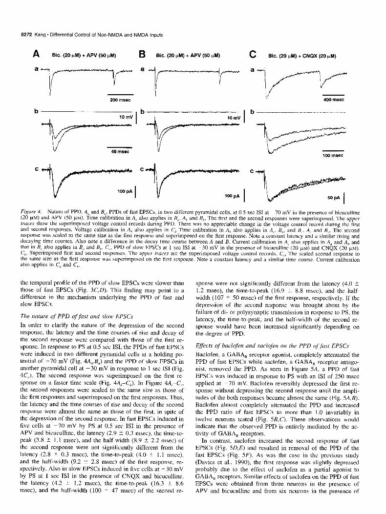
Figure 4. Nature of PPD. A, and B,, PPDs of fast EPSCs, in two different pyramidal cells, at 0.5 set IS1 at -70 mV in the presence of bicuculline (20 pM) and APV (50 FM). Time calibration in A, also applies in B,. A, and B,, The first and the second responses were superimposed. The upper truces show the superimposed voltage control records during PPD. There was no appreciable change in the voltage control record during the first and second responses. Voltage calibration in A, also applies in C,, Time calibration in A,, also applies in A,, B,,, and B,. A, and B,, The second response was scaled to the same size as the first response and superimposed on the first response. Note a constant latency and a similar rising and decaying time courses. Also note a difference in the decay time course between A and B. Current calibration in A, also applies in A, and A,, and that in B, also applies in B, and B,. C,, PPD of slow EPSCs at 1 set IS1 at -30 mV in the presence of bicuculline (20 FM) and CNQX (20 FM).
C,,, Superimposed first and second responses. The upper truces are the superimposed voltage control records. C,, The scaled second response to the same size as the first response was superimposed on the first resnonse. Note a constant latency and a similar time course. Current calibration
- _ _
also applies in C, and C,.
the temporal profile of the PPD of slow EPSCs were slower than those of fast EPSCs (Fig. 3C,D). This finding may point to a difference in the mechanism underlying the PPD of fast and slow EPSCs.
The nature of PPD of fast and slow EPSCs
In order to clarify the nature of the depression of the second response, the latency and the time courses of rise and decay of the second response were compared with those of the first re- sponse. In response to PS at 0.5 set ISI, the PPDs of fast EPSCs were induced in two different pyramidal cells at a holding po- tential of -70 mV (Fig. 4A,,B,) and the PPD of slow EPSCs in another pyramidal cell at -30 mV in response to 1 set IS1 (Fig. 4CJ. The second response was superimposed on the first re- sponse on a faster time scale (Fig. 4A,C,,). In Figure 4A,-C,, the second responses were scaled to the same size as those of the first responses and superimposed on the first responses. Thus, the latency and the time courses of rise and decay of the second response were almost the same as those of the first, in spite of the depression of the second response. In fast EPSCs induced in five cells at -70 mV by PS at 0.5 set IS1 in the presence of APV and bicuculline, the latency (2.9 -+ 0.3 msec), the time-to- peak (3.8 -C 1.1 msec), and the half-width (8.9 -C 2.2 msec) of the second response were not significantly different from the latency (2.8 ? 0.3 msec), the time-to-peak (4.0 -’ 1.1 msec), and the half-width (9.2 ? 2.8 msec) of the first response, re- spectively. Also in slow EPSCs induced in five cells at -30 mV by PS at 1 set IS1 in the presence of CNQX and bicuculline, the latency (4.2 2 1.2 msec), the time-to-peak (16.3 2 8.6 msec), and the half-width (100 -t 47 msec) of the second re-
sponse were not significantly different from the latency (4.0 + 1.2 msec), the time-to-peak (16.9 -C 8.8 msec), and the half- width (107 rt 50 msec) of the first response, respectively. I f the depression of the second response was brought about by the failure of di- or polysynaptic transmission in response to PS, the latency, the time-to-peak, and the half-width of the second re- sponse would have been increased significantly depending on the degree of PPD.
Effects of baclofen and saclofen on the PPD of fast EPSCs
Baclofen, a GABA, receptor agonist, completely attenuated the PPD of fast EPSCs while saclofen, a GABA, receptor antago- nist, removed the PPD. As seen in Figure 5A, a PPD of fast EPSCs was induced in response to PS with an IS1 of 250 msec applied at -70 mV. Baclofen reversibly depressed the first re- sponse without depressing the second response until the ampli- tudes of the both responses became almost the same (Fig. 5A,B). Baclofen almost completely attenuated the PPD and increased the PPD ratio of fast EPSCs to more than 1.0 invariably in twelve neurons tested (Fig. 5B,C). These observations would indicate that the observed PPD is entirely mediated by the ac- tivity of GABA, receptors.
In contrast, saclofen increased the second response of fast EPSCs (Fig. 5D,E) and resulted in removal of the PPD of the fast EPSCs (Fig. 5F). As was the case in the previous study (Davies et al., 1990) the first response was slightly depressed probably due to the effect of saclofen as a partial agonist to GABA, receptors. Similar effects of saclofen on the PPD of fast EPSCs were obtained from three neurons in the presence of APV and bicuculline and from six neurons in the presence of

The Journal of Neuroscience, December 1995, 75(12) 8273
A Bit. (20 PM)
Control
w
hl 100 pA
Baclofen (2 @) 100 msec
Wash out
D Bit. (20 phi) t APV (50 PM)
V 100 msec
Saclofen (0.2 mM)
---k--3(
-
% - 3 200-
g E 150 -
u) 100 - 2
L z 50 -
L
07 I con bat wash
E
Control Saclofen
Figure 5. Effects of (-)-baclofen and saclofen on the PPD of fast EPSCs. A, PPDs of fast EPSCs at 250 msec IS1 at -70 mV in the presence of bicuculline. Each trace is the average of five trials for each condition. Baclofen (2 FM) reduced the first response without reducing the second response. B, Open and solid columns represent the mean amplitude (*SD, 12 = 5) of the first and second responses of fast EPSCs, respectively, for each condition. Baclofen significantly (*, p < 0.001, two-tailed t test) reduced the amplitude of the first response. C, the mean PPD ratio (*SD, n = 5) for each condition for the same cell. Note that baclofen significantly (*, p < 0.001) increased the PPD ratio. D, An averaged (n = 6) control record of the PPD of fast EPSCs at 200 msec ISI at -70 mV in the presence of bicuculline (20 pM) and APV (50 PM). Saclofen (0.2 mM) increased the second response without marked effects on the first response. E, Open and solid columns represent the mean amplitude (?SD, n = 6) of the first and second responses of fast EPSCs for each condition, respectively. Saclofen significantly (*, p < 0.01) increased the amplitude of the second response. F, The mean PPD ratio (+SD, n = 6) for each condition for the same cell. Note that saclofen significantly (*, p < 0.01) increased the PPD ratio.
C 1.4
l-
1.2 c
1.0 .o
E 0.8
f 0.6
0.4
0.2
0.0 L)3
con bat wash
F 1.2
0 0.8 F I I !
0.6
ii!
n 0.4
0.2
1 .o *
0.0 !ii
Control Saclofen
bicuculline alone. Although saclofen appeared to be less potent than baclofen, saclofen increased the PPD ratio of fast EPSCs evoked at 200 msec IS1 from 0.59 -C 0.07 to 0.86 ? 0.12 in the nine neurons. It was difficult to wash out saclofen, presumably because the effective concentration (0.2-0.4 mM) of saclofen was about 100 times higher than that of other drugs affecting the PPD in the present study. Since the PPD of fast EPSCs was completely attenuated by a GABA, agonist and largely removed by a GABA, antagonist, the PPD of fast EPSCs may have been mediated by the activation of GABA, receptors, which were probably located on the presynaptic terminal likely arising from glutamatergic neurons in layer II/III. The properties of the PPD of fast EPSCs were very similar to those of IPSCs in rat hip- pocampus reported previously (Davies et al., 1990).
Effects of baclofen on the PPDs of fast EPSCs evoked at various ISIS were also examined to see if there are any baclofen- insensitive components in the time course of the PPD of fast EPSCs. As shown in Figure 6A,, the respective traces of the PPD response of fast EPSCs induced in response to PS with various ISIS in the presence of bicuculline and APV, were su- perimposed to show the PPD time course. A bath application of baclofen decreased the first response without any marked reduc- tions in the second responses, as shown in Figure 6A,. In Figure
6B, the mean PPD ratio of fast EPSCs obtained before (open circles) and during (solid circles) application of baclofen in 4 cells were plotted against various ISIS. Baclofen almost com- pletely attenuated the PPD at all ISIS examined. Thus, the whole time course of the PPD of fast EPSCs, obtained in the presence of APV and bicuculline, appeared to be baclofen sensitive.
Esfects of baclofen and muscarine on slow EPSCs
The effects of baclofen on slow EPSCs were different from those on fast EPSCs. As seen in Figure 7A, PPDs of slow EPSCs at an IS1 of 0.2 set were induced ‘at -30 mV in the presence of CNQX and bicuculline. In contrast to fast EPSCs, not only the first response, but also the second response started to decrease after an application of baclofen. Consequently, both the first and second responses were decreased in amplitude significantly 0, < 0.01, two tailed t test) by baclofen although this resulted in a marked attenuation of the PPD of slow EPSCs (Fig. 7C). Ba- clofen increased the PPD ratio of slow EPSCs evoked at 0.2 set IS1 from 0.65 2 0.09 to 0.86 5 0.11 in four neurons tested. If the observed PPD of slow EPSCs had been mediated entirely by the activity of GABA, receptors, the second response would not have been decreased until the amplitude of the first response reached the same level as that of the second response, similar

8274 Kang - Differential Control of Non-NMDA and NMDA Inputs
Figure 6. Effects of baclofen on A PPDs of fast EPSCs at various ISIS. A,, The first and second fast EPSCs ob-
Bit. (20 $4) + APV (50 FM)
tained in response to PS at various ISIS were superimposed. Fast EPSCs were induced in the presence of bicuculline (20 PM) and APV (50 PM). A,>, A bath application of baclofen (5 FM) in the media containing bicuculline (20 )LM) and APV (50 )LM) decreased the am- plitude of the first response with no marked changes in the second re-
400 msec
Bit. (20 FM) + APV (50 PM) + BBC. (5 g)
sponses. B, The mean PPD ratio of fast EPSCs obtained in the presence of bi- cuculline (20 FM) and -APV (50 FM)
from four neurons before (open circle) and during applying baclofen (solid circle). Error bars represent SD values.
to the case with fast EPSCs. Therefore, the simultaneous reduc- tion of the first and second responses may indicate an involve- ment of an additional component of the PPD of slow EPSCs. Alternatively, during the PPD of slow EPSCs, not all of the GABA, receptors may have been activated so that further de-
pression of the second response could occur when baclofen was exogenously applied, although this was not the case for the PPD of fast EPSCs.
On the other hand, the PPD of slow EPSCs evoked at an IS1 of 0.5 set was largely attenuated by muscarine. As shown in
Control in Bit. (20 PM) + CNQX (20 PM) A 4. . ,
. Baclofen (5 fl)
con bat wash
E
con bat wash 100 nlsec
D Control in Bit. (20 PM) + CNQX (20 PM)
, Muscarine (3 PM) I
100
60
60
Wash out
200 msec con must wash con must wash
Figure 7. Effects of baclofen and muscarine on PPDs of slow EPSCs. A, Baclofen (5 FM) reduced not only the first but also the second response of slow EPSCs. Each trace is the averaged record (IZ = 5) of the PPD of slow EPSCs at 200 msec ISI at -30 mV in the presence of bicuculline (20 pM) and CNQX (20 FM). B, Open and solid columns represent the mean amplitude (*SD, II = 5) of the first and second responses of slow EPSCs, respectively, for each condition. Baclofen significantly (*, p < 0.001, two-tailed t test) reduced the amplitude of the first response. The second response was also significantly (*, p < 0.01) reduced by baclofen. C, The mean PPD ratio (?SD, n = 5) for each condition for the same cell. Note that baclofen significantly (*, p < 0.01) increased the PPD ratio. D, Muscarine (3 FM) reduced the first response without marked effects on the second response. Each trace is the averaged record (n = 5) of the PPD of slow EPSCs at 500 msec IS1 at -30 mV in the presence of bicuculline (20 FM) and CNQX (20 PM). E, Open and solid columns represent the mean amplitude (&SD, n = 5) of the first and second responses of slow EPSCs, respectively, for each condition. Muscarine significantly (*. p < 0.001) reduced the first response without significant (p > 0.05) effects on the second response. F, The mean PPD ratio (+SD, n = 5) for each condition for the same cell. Note that muscarine significantly (*, p < 0.001) increased the PPD ratio.

The Journal of Neuroscience, December 1995, 75(12) 8275
A Bit. (20 pful) + CNQX (20 phi)
Control
a
. Muscarine (5 FM)
Muscarine (5 yM) + Baclofen (5 pM) 0.6 -
C ;I 50 pA
0.51 .1 1 10
400 msec ISI (set)
B
0.8 - .g
z
f 0.7-
Figure 8. Early and late components of PPD of slow EPSCs. A,, The respective averaged (n = 4) traces of the PPD at ISIS of 0.2 (arrowhead), 0.5 (double arrowhead) and 1 set at -30 mV in the presence of bicuculline (20 FM) and CNQX (20 PM) were superimposed. A,, Muscarine (5 FM) reduced the first response without reducing the second response at 0.5 and 1 set ISIS markedly, resulted in a marked attenuation of PPD at 0.5 and 1 set ISI. In contrast, the second response at 0.2 set IS1 was reduced by muscarine simultaneously with the first response, resulted in no marked attenuation of PPD at 0.2 set ISI. A,, An addition of baclofen (5 PM) in the presence of muscarine further reduced the first response without reducing the second response at 0.2 set ISI, resulted in a marked attenuation of PPD at 0.2 set ISI. In contrast, the second response at 0.5 and 1 set ISIS were reduced as much as the first response. B, Plottings of PPD ratio against ISIS with a logarithmic scale. The mean PPDs obtained from four cells in the control condition, during muscarine and during muscarine and baclofen were represented by open circles, solid circles, and open squares, respectively. Error bars represent SD values. Note that the PPD of slow EPSCs is composed of early baclofen-sensitive and late muscarine- sensitive PPD. Also note that the time course of muscarine-sensitive PPD is slower than that of baclofen-sensitive PPD.
Figure 70, a PPD of slow EPSCs was evoked at an IS1 of 0.5 set at a holding potential of -30 mV in the presence of bicu- culline and CNQX. A bath application of muscarine reversibly decreased the first response of slow EPSCs without marked ef- fects on the second response (Fig. 7D,E). In contrast to the effect of baclofen on the PPD of slow EPSCs at 0.2 set ISI, muscarine did not decrease the second response at 0.5 set IS1 significantly (p > 0.05, two-tailed t test), resulted in a marked attenuation of the PPD ratio (Fig. 7&F). Muschrine (5 pM) increased the PPD ratio evoked at 0.5 set IS1 from 0.56 -C 0.12 to 0.85 ? 0.10 in 10 cells tested. However, about 15% of the PPD evoked at 0.5 set IS1 still remained in the presence of muscarine (5 FM). This would indicate that either the effect of muscarine on the PPD ratio was not saturated at the concentration used, or an additional component of the PPD of slow EPSCs that is independent of the activity of muscarinic receptors was present.
Early and late components of the PPD of slow EPSCs
Taking the effect of baclofen and muscarine on slow EPSCs into consideration, it can be predicted that the PPD of slow EPSCs may be composed of baclofen- and muscarine-sensitive com- ponents. Therefore, the effects of baclofen and muscarine were examined in the same neuron. As seen in Figure 8A,, respective traces obtained in response to PS at various ISIS were super- imposed in order to show changes in the respective second re- sponses. Regardless of the constant amplitude of the first slow EPSCs, the amplitude of the second response varied in response to PS at ISIS of 0.2 (arrowhead in A,), 0.5 (double arrowhead in A,), 1 and 5 sec. As seen in Figure 8A,, a bath application of
muscarine largely decreased the first response without marked reduction in the second response of slow EPSCs evoked at ISIS longer than 0.5 set (double arrowhead in A,). However, the sec- ond response evoked at an IS1 of 0.2 set (arrowhead in A,) was decreased simultaneously with the first response, leaving the PPD ratio at 0.2 set IS1 almost unchanged (Fig. 8A,). Conse- quently, only the late phase of the PPDs seen at ISIS longer than 0.5 set was attenuated markedly by muscarine (Fig. 8B, compare open and solid circles). The simultaneous reduction of the first and second responses at 0.2 set IS1 is similar to that shown in Figure 7, A and B. The unchanged PPD at 0.2 set IS1 resulting from the simultaneous reduction would indicate the presence of muscarine-insensitive component in the early phase of the PPD. This muscarine-insensitive component of the PPD at 0.2 set IS1 was largely sensitive to baclofen, as expected from the obser- vations shown in Figure 7A-C. As shown in Figure 8, A, and B (compare the solid circle and open square), in the presence of muscarine, the addition of baclofen reduced the first response without reducing the second response evoked at 0.2 set ISI, resulted in a marked attenuation of the early phase of the PPD (compare Fig. 8A,,A,; see also Fig. 8s). In the absence of mus- carine, baclofen did not markedly attenuate PPDs at ISIS longer than 0.5 set, although the second response at these ISIS was also reduced simultaneously with the first response (figure not shown). Thus, the PPD of slow EPSCs appeared to consist of two distinct components; the baclofen-sensitive early component and the muscarine-sensitive late component (Fig. 8B). This may be the reason why the time course of the PPD of slow EPSCs was slower than that of fast EPSCs (see Fig. 3C,D).

8276 Kang * Differential Control of Non-NMDA and NMDA Inputs
Figlrre 9. Effects of piretxepine on the late PPD of slow EPSCs. A, In the preSence of baClOfcn (5 FM), CNQX
(20 KM) and bicuculline (20 kM), ef- fects. of pirenr.epine were examined on the PPD of slow EPSCs at 2 set ISI. Each trace is the averaged record (n = 5) for control (a), during pirenzepine (b), and washout of pirenzepine (c,). Pi- renzepine (3 F,M) increased the second response without marked effect on the first response. B, Olx~n and solid cd
WWJ represent the mean amplitude (*SD, IZ = 5) of the first and second responses of slow EPSCs, respectively, for each condition. PirenLepine signif- icantly (“:, ,> < 0.01, two-tailed t test) increased the amplitude of the second response. C, The mean PPD ratio (&SD, IZ = 5) for each condition for the same cell. Note that piren/epine significantly (*, p < 0.01) increased the PPD ratio.
Bit. (20 FM) + CNQX (20 PM) t Baclofen (5 PM)
I:
Pirenzepine (3 PM)
“- Wash out
1 set
Baclofen (5 FM) and muscarine (5 PM) decreased the ampli- tude of slow EPSCs by 49.5 i- 6.4% (11 = 5) and 50.4 t 3.4% (n = 7), respectively (e.g., Figs. 7-l I). However, simultaneous application of baclofen and muscarine decreased the amplitude of slow EPSCs by 60-70% (12 = 3; e.g., Fig. 8). It appears that the effects of baclofen and muscarine were not linearly sum- mated, suggesting that in part, the depression caused by baclofen and muscarine may mutually occlude.
In the presence of baclofen (5 FM), CNQX, and bicucullinc, 32% PPD of slow EPSCs could still be produced in a pyramidal cell in response to PS at an ISI of 2 set (Fig. 9A). Under these conditions. pirenzepine almost completely removed the depres- sion of the second response without marked effects on the first response, as seen in Figure 9, A and B. The PPD ratio was significantly 01 < 0.01, two-tailed t test) increased by applying pirenzepine (Fig. 9C). The PPD ratio of slow EPSCs at l-2 set ISIS in the presence of baclofen was increased from 0.67 -t 0. I2 to 0.96 t 0.17 by pirenzepine in six pyramidal cells tested.
EIljects of nmrcarine on ,fixst EPSCs
Muscarine (l-5 FM) easily attenuated the PPD of slow EPSCs in IO out of eleven pyramidal neurons tested, but did not affect fast EPSCs markedly in six neurons tested. The effects of mus- carine were examined on fast and slow EPSCs simultaneously in one and the same neuron for comparison. As seen in Figure IO, PPDs of fast and slow EPSCs were observed in response to PS at 0.5 set ISI at a holding potential of -60 (Fig. IOA,,) and -30 mV (Fig. IOB,,), respectively. A bath application of mus- carine decreased the peak amplitude of the first response by 28.5% when examined at -30 mV (Fig. IOH,), but decreased that of the first one by only 6% when examined at -60 mV (Fig. IOAJ. This would indicate that slow EPSCs were more sensitive to muscarine than fast EPSCs were. This notion was further confirmed by the following observation. Adding APV in addition to bicuculline and muscarine almost completely abol- ished slow EPSCs, leaving only the fast EPSCs (Fig. IOAc,Bc) without any appreciable changes in the peak amplitude of the
Control Pirenzepine Washout
Control Pirenzepine Washout
first and second fast EPSCs at -60 mV (compare Fig. lOA,,,A,). The degree of PPD of fast EPSCs was not affected appreciably by APV. As seen in Figure IOB,, fast EPSCs evoked at -30 mV returned to the base line almost completely at the time indicated with an upward arrow. Therefore, EPSCs evoked at -30 mV and measured at this timing, as indicated with horizontal arrows in Figure IO, B, and B,,, appeared to consist largely of APV- sensitive slow components. Consequently, it follows that the APV-sensitive slow component was depressed by about 32%, whereas the peak amplitude of APV-insensitive fast EPSCs was depressed by only 6%. Thus, slow EPSCs were much more sen- sitive to muscarine than fast EPSCs were. Although the first and second fast EPSCs were slightly depressed by muscarine by 5.4 +- 6.4% and 7.4 -f 7.5%, respectively (Fig. IOC,,), the PPD ratio of fast EPSCs at 0.5 set ISI was not affected significantly (17 > 0.1) in six cells tested both in the presence of bicuculline alone (n = 3) and in the presence of APV and bicuculline (n = 3), as seen in Figure IOC,,. Similarly, the PPD ratio of fast EPSCs at 0.2 set IS1 was not affected significantly 01 > 0. I) by muscarine in 4 cells tested in the presence of bicuculline alone (n = 2) and in the presence of APV and bicuculline (n = 2). Therefore, it is not likely that the PPD of fast EPSCs involves the activation of muscarinic receptors. By contrast, baclofen largely depressed these muscarine-insensitive fast EPSCs and attenuated the PPD completely (Fig. IOA,). On the other hand, it was also examined whether or not glutamatergic fibers are sensitive to glutamate, and it was found that PPDs of fast and slow EPSCs were not appreciably attenuated by glutamate (5-10 PM; n = 6).
Paired pulse ,ficcilitution and depression
As shown in Figure 8, the PPD of slow EPSCs usually consisted of early baclof’en-sensitive and late muscarine-sensitive compo- nents. However, in 8 of 39 pyramidal neurons, the early phase of the PPD of slow EPSCs was less prominent than the late phase of the PPD. As seen in Figure I I, A,, and A,, the PPD at 0.2 set IS1 was not prominent in a pyramidal neuron, though the PPD was seen at ISIS larger than 0.5 sec. A bath application

The Journal of Neuroscience, December 1995, 15(12) 8277
A H.P.=dOmV
a Bit. (20 +4)
b Bit. (so @A) + MUSC. (5 $4)
C MC. (20 PM) + MUSC. (5 KM) + APV (50 vM)
d Bit. (20 PM) + APV (50 pM) + Bat. (5 PM)
H. P. = -30 mV a BIG. (20 pM)
b Blc. (20 PM) + MISC. (5 ,A)
n
C Blc. (20 pM) + MISC. (5 rM) + APV (50 pM)
a
b 1.01
0.6
200 m*ec
Figure 10. Effects of muscarine on PPD of fast EPSCs. A, and B,, PPDs of fast and slow EPSCs induced in one and the same pyramidal cell at 0.5 set IS1 at -60 and -30 mV, respectively, in the presence of bicuculline (20 PM). Horizontal arrows in A, indicate the peak levels of the first and second responses in A, obtained during applying muscarine. A, and B,, A bath application of muscarine (5 FM) largely reduced the first response at -30 mV but not at -60 mV. A, and B,, An addition of APV (50 pM) in the media containing bicuculline (20 PM) and muscarine (5 FM)
abolished slow EPSCs, leaving fast EPSCs alone. An upward urrowhead in B, indicates the timing when fast EPSCs returned to the base line almost completely. EPSCs measured at this timing (indicated with horizontal arrowheads) in B, and B,, are largely composed of slow EPSCs. In spite of a large attenuation of slow EPSCs at -30 mV by muscarine (compare the level of the horizontal arrowheads in B, and B,,), the peak amplitude of the fast EPSCs at -60 mV remained almost constant. A,, After washing out of muscarine, baclofen (5 FM) attenuated PPD of fast EPSCs by depressing the first response. C,, Open and solid columns represent the mean amplitude (+SD, IZ = 5) of the first and second responses of fast EPSCs at -60 mV, respectively, for each condition. There were slight differences (p < 0.05) in the amplitudes of the first and second responses before and during application of muscarine. C,, The mean PPD ratio of fast EPSCs at -60 to -70 mV for each condition from six cells, either in the presence of APV and bicuculline (n = 3) or in the presence of bicuculline alone (n = 3). Note no significant 0, > 0.1) difference in the PPD ratio of fast EPSCs between each condition.
of baclofen decreased the first. and second responses, but in- creased the PPD ratio at ISIS shorter than about 3 set, disclosing the presence of a paired pulse facilitation (PPF) at ISIS shorter than about 0.5-l set (Fig. 1 lA,A,). The PPF may have masked the early phase of the PPD of slow EPSCs. Similar effects of baclofen on the less prominent PPD of slow EPSCs at short ISIS were observed in three neurons, although the degree of PPF varied.
In another pyramidal neuron, fast EPSCs displayed a promi- nent PPD at 0.5 set ISI, whereas the PPD of slow EPSCs was not prominent at the same IS1 in the same neuron. As seen in Figure 1 lB,, a prominent PPD of fast EPSCs was evoked by applying PS with 0.5 set IS1 at -70 mV. In the same neuron, when PS were applied at -30 mV in the presence of CNQX, the second response of slow EPSCs was not smaller, but in con- trast, was slightly larger than the first response (Fig. llB,,). Even in such cases, muscarine depressed the first response with- out decreasing the second response (Fig. 1 lB,,>, as was the case in the PPD of slow EPSCs shown in Figures 7, 8, and 10. On the other hand, pirenzepine enhanced the second response with- out marked changes in the first response (Fig. 1 lB,,), as was the case in the PPD of slow EPSCs shown in Figure 9. Therefore,
it was considered that the PPD at 0.5 set IS1 was present, but masked by the PPF of slow EPSCs. A similar PPF at 0.5 set IS1 was observed by applying either muscarine or pirenzepine in a total of five pyramidal neurons. Thus, the early phase of the PPD of slow EPSCs was occasionally masked by the PPE This implies that facilitation and depression are not mutually exclusive and may be controlled independently. Since fast and slow EPSCs often behaved independently in response to PS, it appears that fast and slow EPSCs are separately modified.
Discussion
Differential PPD between fast and slow EPSCs In the present study, the PPD of EPSCs was induced in layer II-V pyramidal cells of the rat frontal cortex in response to PS applied at layer II/III in the vicinity (100-200 pm) of the re- corded neuron. Fast and slow EPSCs were clearly distinguished in terms of the PPD. First, the time course of the PPD of slow EPSCs was slower than that of fast EPSCs. Second, the PPD of fast EPSCs was completely attenuated by baclofen and removed by saclofen, whereas the PPD of slow EPSCs was largely atten- uated by baclofen and muscarine, and removed by pirenzepine in the presence of baclofen. The PPD of slow EPSCs consisted

8278 Kang * Differential Control of Non-NMDA and NMDA Inputs
A a Control in Bit. + CNQX
b Baclofen 5pM
a Bit., -70 mV
Lc
i,
MO msec b Bit. + CNQX, -30 mV
1 Control
3 Wash out
4 Pirenzepine 1.4pM
Figure II. PPD and PPF of slow EPSCs. A,, The respective traces of the PPD at ISIS of 0.2, 0.5, and 1 set at -30 mV in the presence of bicuculline (20 PM) and CNQX (20 FM) were superimposed. Note that PPD was not prominent at 0.2 set ISI. A,,, Baclofen (5 FM) reduced the first and second responses at respective ISIS markedly, but resulted in a PPF at 0.2 set ISI. A,, Plottings of PPD ratio against ISIS with a logarithmic scale. The PPDs obtained in the control condition (partly shown in A,) and during baclofen (partly shown in Ah) were represented by open and solid circles, respectively. Error bars at 0.2 and 0.5 set ISIS represent SD values (n = 6 trials). Each open and solid circle represents the mean value of three to six trials. Note that baclofen attenuated the early phase of the PPD and revealed PPF at ISIS shorter than 1 set, leaving the late phase of the PPD (ISIS > 3 set) almost unchanged. B,, a PPD of fast EPSCs at 0.5 set IS1 at -70 mV induced in a layer V pyramidal cell in the presence of bicuculline. B,, not PPD but a slight PPF induced at -30 mV in the same cell as in Ba during applying CNQX (20 FM) in addition to bicuculline (20 FM). Under these conditions, muscarine (3 PM) reduced the first response without marked effect on the second response (2). Washout of muscarine restored the first response (3). Pirenzepine (1.4 FM) increased the second response without marked effect on the first response (4). Note a different behavior between fast and slow EPSCs in response to PS in the same neuron.
of early baclofen-sensitive and late muscarine-sensitive compo- nents. Thirdly, even when PPF or no PPD was seen in either fast or slow EPSCs, the other displayed a prominent PPD in the same neurons. These observations suggest that a part of gluta- matergic fibers likely arising from layer II/III terminate predom- inantly onto NMDA receptors in single pyramidal cells, and re- ceive distinct presynaptic control through at least the muscarinic receptors.
With respect to the selective activation of NMDA receptors, it has been reported that EPSPs that were almost selectively mediated by the activity of NMDA receptors were induced in layer II/III pyramidal cells in rat frontal cortex (Thomson, 1986). Recently, it has also been reported that NMDA currents were almost selectively induced in layer V pyramidal cells by caged transmitter activated locally on apical dendrites using scanning laser photostimulation (Dalva and Katz, 1994), although the dominance of NMDA responses may change with development (Agmon and O’Dowd, 1992).
The time course of baclofen and muscarine-sensitive PPD of slow EPSCs was slower than that of baclofen-sensitive PPD of fast EPSCs. The time course of muscarinic EPSPs has been re- ported to be several tens of seconds in CA1 pyramidal neurons (Benardo and Prince, 1982; Cole and Nicoll, 1984; Madison et al., 1987) whereas that of GABA, IPSPs has been reported to be about one second (Alger and Nicoll, 1982; Connors et al., 1988; McCormick, 1989; Kang et al., 1994). Thus, the activation of postsynaptic muscarinic receptors occurs over a much longer
time course than that of postsynaptic GABA, receptors. The difference in the time course of the PPD between fast and slow EPSCs observed in the present study may reflect a difference in the temporal profile of activity of GABA, and muscarinic re- ceptors, which are probably located on the presynaptic terminals.
Origin qf the PPD of fast and slow EPSCs
The presynaptic mechanism of the PPD of monosynaptic IPSPs or IPSCs is well established (Davies et al., 1990; Forsythe and Clements, 1990; Davies and Collingridge, 1993; Fukuda et al., 1993: Mott et al., 1993). It has also been reported that polysyn- aptic IPSCs displayed a similar PPD (Mott et al., 1993). In the present study, if EPSCs had been induced polysynaptically, the PPD might have been brought ahout by the postsynaptic inhi- bition of excitatory interneurons in the polysynaptic pathway. If some excitatory interneurons, which possessed GABA, and/or muscarinic receptors, had mediated glutamatergic EPSCs, it is possible for the second response to have been reduced by the postsynaptic inhibition of these interneurons through the acti- vation of postsynaptic GABA, and/or muscarinic receptors by the first stimulation. If this was the case, the latency and the time-to-peak of the second response would have increased with an increase in the degree of PPD, because the generation of action potential in excitatory interneurons by the second stim- ulus could have been delayed by the increasing inhibition. How- ever, the latency and the time course of the second fast and slow EPSCs remained almost unchanged in spite of the depression of

The Journal of Neuroscience, December 1995, 15(12) 8279
the second responses as described in the Results (e.g., Figs. 3, 4). In addition, the time course of the PPD of fast and slow EPSCs was much longer than that of GABA, IPSPs (<I set; Alger and Nicoll, 1982; Connors et al., 1988; Tseng and Ha- berly, 1988; McCormick, 1989; Kang et al., 1994). These ob- servations would clearly indicate that the PPD is distinct from postsynaptic inhibition in the di- or polysynaptic pathways. On the other hand, if the PPD of EPSCs was caused by a reduction of driving potential due to an inadequate space clamp or by a reduction of the numbers of available receptors resulting from the residual occupation of receptors during the decay time course of the first response, then the reduction of the second response would have occurred in response to only those PS with ISIS shorter than the duration of the first EPSCs. However, the PPD was much less prominent at those shorter ISIS than at longer ISIS and the time course of the PPD was much longer than the duration of EPSCs (Fig. 3). Therefore, it is unlikely that the PPD is caused by reduction of either the driving potential or the num- bers of available receptors. Nevertheless, this possibility can not be discarded entirely at short ISIS (CO.2 set; Mori et al., 1994).
Involvement afpolysynaptic EPSCs
Fast and slow EPSCs may involve di- or polysynaptic compo- nents. Slow EPSCs were induced in the presence of CNQX. As it has been reported that di-synaptic IPSPs induced in pyramidal cells of rat frontal cortex were completely abolished by applying 20 p,M CNQX (Kawaguchi, 1992) di- or polysynaptic transmis- sion would not be likely to occur in the presence of CNQX. Even so, it might be possible that a bath application of bicucul- line may have enhanced isolated NMDA components to a degree where NMDA components alone mediated disynaptic transmis- sion. As is evident from the presence of a long tail on the EPSC recorded at -60 mV in the presence of bicuculline (Figs. lA, lOA,+,.), bicuculline might have enhanced di- or polysynaptic transmission. However, CNQX abolished this tail current almost completely (Fig. IB). Usually, PPD of slow EPSCs was exam- ined after confirming that no appreciable EPSCs were induced at -70 mV in the presence of bicuculline and CNQX. Therefore, polysynaptic transmission enhanced by bicuculline may have been suppressed largely by CNQX, and slow EPSCs may be induced monosynaptically. In contrast, for fast EPSCs, an in- volvement of polysynaptic components can not be discarded en- tirely. However, as shown in Figure 10, A,, and A,., APV abol- ished large and long tail currents without any appreciable changes in the peak amplitude of fast EPSCs and in the degree of PPD. This suggests that the PPD of fast EPSCs is independent of the presence of late polysynaptic components, although they may have been enhanced largely by NMDA components in the presence of bicuculline. This notion was further supported by the observation that fast “EPSCs,” which were contaminated with depolarizing IPSCs evoked in 145 mM CsCI-loaded cells at -70 mV in the absence of bicuculline, were reduced in am- plitude but prolonged in the decay time course by an application of bicuculline without any significant changes in the PPD ratio of fast EPSCs (data not shown).
Effects of baclofen and muscarine on fast and slow EPSCs
Baclofen completely attenuated the PPD of fast EPSCs without reducing the second responses (n = 12). This suggests that the PPD of fast EPSCs was mediated entirely by the activity of GABA, receptors. However, effects of baclofen on the PPD of slow EPSCs were different from those on fast EPSCs. Baclofen
largely attenuated the PPD of slow EPSCs in response to PS at 0.2 set ISI, by decreasing not only the first but also the second response (Fig. 7A-C). However, in the presence of muscarine, baclofen reduced the first response alone without reducing the second response appreciably at 0.2 set IS1 (Fig. 8A,,,A,). Fur- thermore, muscarine simultaneously decreased the first and sec- ond responses of slow EPSCs evoked at 0.2 set IS1 (see Fig. 8A,,A,J with only a slight attenuation of the PPD (Fig. 8s). A simultaneous reduction of the first and second response at a cer- tain IS1 by baclofen (or muscarine) may indicate the presence of an additional component of PPD that is insensitive to baclofen (or muscarine).
A similar simultaneous reduction of the first and second IPSCs by baclofen leading to a marked attenuation of the PPD has been reported in synaptic connections between single cul- tured hippocampal neurons (Wilcox and Dichter, 1994). How- ever, GABA, antagonists were not able to remove the PPD of IPSCs in the same study. Since GABAergic neurons are known to contain various transmitters, such as ACh, CCK and NPY in addition to GABA (e.g., Kaneko et al., 1992), those transmitters may have been released simultaneously with GABA. If the ac- tion of these unknown transmitters causes a PPD that is also sensitive to baclofen, it would be impossible for GABA, antag- onists to remove the PPD as has been observed in the previous studies (Yoon and Rothman, 1991; Wilcox and Dichter, 1994), although a bath application of baclofen should be able to atten- uate the PPD.
References
Agmon A, O’Dowd D (1992) NMDA receptor-mediated currents are prominent in the thalamocortical synaptic response before maturation of inhibition. J Neurophysiol 68345-349.
Alaer BE. Nicoll RA (1982) Feed-forward dendritic inhibition in rat hippocampal pyramidal ceils studied in vitro. J Physiol (Lond) 328: 105-123.
Asanuma H, Rosen I (1973) Spread of mono- and polysynaptic con- nections within cats motor cortex. Exp Brain Res 16:507-520.
Benardo LS, Prince DA (1982) Ionic mechanisms of cholinergic ex- citation in mammalian hippocampal pyramidal cells. Brain Res 249: 333-344.
Bowery NG, Hudson AL, Price GW (1987) GABA, and GABA, re- ceptor site distribution in the rat central nervous system. Neurosci- ence 20:365-383.
Cole AE, Nicoll RA (1984) Characterization of a slow cholinergic postsynaptic potential recorded in vitro from rat hippocampal pyra- midal cells. J Physiol (Lond) 352:173-188.
Colmers WE Lukowiak K, Pittman Q (1988) Neuropeptide Y action in the rat hippocampal slice: site and mechanism of presynaptic in- hibition. J Neurosci 8:3827-3837.
Connors SW, Malenka RC, Silva LR (1988) Two inhibitory postsyn- aptic potentials, and GABA, and GABA, receptor-mediated re- sponses in neocortex of rat. J Physiol (Lond) 406:443468.
Dalva MB, Katz LC (1994) Functional NMDA receptors are prefer- entially located on apical dendrites of pyramidal neurons in ferret visual cortex. Sot Neurosci Abstr 20:349.13.
Davies CH, Collingridge GL (1993) The physiological regulation of synaptic inhibition by GABA, autoreceptors in rat hippocampus. J Physiol (Lond) 472:2455265.
Davies CH, Davies SN, Collingridge GL (1990) Paired-pulse depres- sion of monosynaptic GABA-mediated inhibitory postsynaptic re- sponses in rat hippocampus. J Physiol (Lond) 4245 13-53 I.
Forsythe ID, Clements JD (1990) Presynaptic glutamate receptors de- press excitatory monosynaptic transmission between mouse hippo- campal neurons. J Physiol (Lond) 429:1-16.
Fukuda A, Mody I, Prince DA (1993) Differential ontogenesis of pre- synaptic and postsynaptic GABA, inhibition in rat somatosensory cortex. J Neurophysiol 70:448452.
Harrison NL (I 990) On the presynaptic action of baclofen at inhibitory

8280 Kang - Differential Control of Non-NMDA and NMDA Inputs
synapses between cultured rat hippocampal neurons. J Physiol (Lond) 422: 433446.
Hestrin S, Nicoll RA, Perkel DJ, Sah P (1990) Analysis of excitatory synaptic action in pyramidal cells using whole-cell recording from rat hippocampal slices J Physiol (Lond) 422:203-225.
Kaneko T, Nakaya Y, Mizuno N (1992) Paucity of glutaminase-im- munoreactive nonpyramidal neurons in the rat cerebral cortex. J Comp Neural 322:181-190.
Kang Y, Kaneko T, Ohishi H, Endo K, Araki T (1994) Spatiotempo- rally differential inhibition of pyramidal cells in the cat motor cortex. J Neurophysiol 71:280-293.
Kawaguchi Y (1992) Receptor subtypes involved in callosally-induced postsynaptic potentials in rat frontal agranular cortex in vitro. Exp Brain Res 88:3-40.
Mack BJ, Smith PA, Colmers W F (1993) 4-aminopiridine (4.AP) does not affect presynaptic inhibition by muscarine in rat hippocampal CA1 in vitro. Sot Neurosci Abstr 19:625.1.
Madison DV, Lancaster B, Nicoll RA (1987) Voltage clamp analysis of cholinergic action in the hippocampus. J Neurosci 7:733-741.
McCormick DA (1989) GABA as an inhibitory neurotransmitter in human cerebral cortex. J Neurophysiol 62: 1018-1027.
Mori A, Takahashi T, Miyashita Y, Kasai H (1994) Two distinct glu- tamatergic synaptic inputs to striatal medium spiny neurons of neo- natal ra& and paired-p&e depression. J Physiol IL&d) 476:2 17-228.
Mott DD. Xie CW. Wilson WA. Swarzwelder HS. Lewis DV (1993) GABA, autoreceptors mediate activity-dependent disinhibitioi ani
enhance signal transmission in the dentate gyrus. J Neurophysiol 69: 674-69 1.
Scholz KP, Miller RJ (1991) GABA, receptor-mediated inhibition of Ca*+ currents and synaptic transmission in cultured rat hippocampal neurons. J Physiol (Lond) 444:669-686.
Sheridan RD, Sutor B (1990) Presynaptic M, muscarinic cholinocep- tors mediate inhibition of excitatoiy synaptic transmission in the hi& DocamDus in vitro. Neurosci Lett 108:273-278.
Sugita $&Johnson SW, North RA (1992) Synaptic inputs GABA, and GABA, originate from discrete afferent neurons. Neurosci Lett 29: 243-248.
Thomson AM (1986) A magnesium-sensitive post-synaptic poten- tial in rat cerebral cortex resembles neuronal resDonses to N-methylaspartate. J Physiol (Lond) 370:531-549.
Tseng GE Haberlv LB (1988) Characterization of svnaoticallv medi- ” . <. <
ated fast and slow inhibitory processes in piriform cortex in an in vitro slice preparation. J Neurophysiol 59: 1352-l 376.
Verhage M, McMahon HT, Ghijsen WEJM, Boomsma F, Scholten G, Wiegant VM, Nicholls DG (1991) Differential release of amino acids, neuropeptides, and catecholamines from isolated nerve termi- nals. Neuron 6:5 17-524.
Wilcox KS, Dichter MA (1994) Paired pulse depression in cultured hippocampal neurons is due to a presynaptic mechanism independent of GABA, autoreceptor activation. J Neurosci 14:1775-1788.
Yoon KW, Rothman SM (1991) The modulation of rat hippocampal synaptic conductances by baclofen and gamma-aminobutyric acid. J Physiol (Lond) 4421377-390.