Embed Size (px)
Citation preview

PRECLINICAL STUDIES
Differential nucleobase protection against 5-fluorouracil toxicityfor squamous and columnar cells: implication for tissue functionand oncogenesis
John P. Vanden Heuvel1,2 & Jerry T. Thompson1& Prajakta Albrecht2 &
Donald Mandetta3 & Harry Kamerow3& John P. Ford3,4
Received: 14 April 2015 /Accepted: 2 June 2015 /Published online: 1 July 2015# The Author(s) 2015. This article is published with open access at Springerlink.com
Summary Purpose The goal of these studies was to test iflocal excess of a normal nucleobase substrate prevents thetoxicity of protracted 5FU exposure used in human cancertreatment. Methods Messenger RNA expression studies wereperformed of 5FU activating enzymes in human colon cancercells lines (CaCo-2, HT-29), primary human gingival cells(HEGP), and normal esophageal and gastric clinical tissuesamples. Excess nucleobase was then used in vitro to protectcells from 5FU toxicity. Results Pyrimidine salvage pathwayspredominate in squamous cells of the gingiva (HEGP) andesophageal tissue. Excess salvage nucleobase uracil but notadenine prevented 5FU toxicity in HEGP cells. Pyrimidinede novo synthesis predominates in columnar Caco-2, HT-29and gastric tissue. Excess nucleobase adenine but not uracilprevented 5FU toxicity to Caco-2 and HT-29 cells.Conclusion The directed application of the normal nucleobaseuracil to the squamous cells of the oral mucosa and palms andsoles together with the delivery of the normal nucleobase ad-enine to the columnar cells of the GI tract may enable the safedelivery of higher 5FU dose intensity. These results also sug-gest a feature of tissue function where squamous cells grow
largely by recycling overlying tissue cell components. Colum-nar cells use absorbed surface nutrients for de novo growth. Adisruption of this tissue function can result in growth derivedfrom an underlying nutrient source. That change would alsocause the loss of the region of cell turnover at the tissue sur-face. Subsequent cell proliferation with limiting nutrient avail-ability could promote oncogenesis in such initiated tissue.
Keywords 5-fluorouracil . Cancer chemotherapy .
Pyrimidine salvage . Toxicity prevention . Preclinical studies .
Oncogenesis
Introduction
5-Fluorouracil (5-FU) and its pro-drugs are a component ofthe standard therapy for a variety of malignancies. However,the toxicity, in particular to the gastrointestinal tract and skinof hands and feet, limits the overall utility of these drugs [1]. 5-FU rapidly enters the cell using the same facilitated transportmechanisms as uracil, including the nucleobase transportersSLC29A2 [2] and SLC22A7 [3]. 5-FU is converted intracellu-larly to several active metabolites by three routes [4]: the 5-phosphoribosyl 1-pyrophosphate (PRPP)-mediated conversionof 5-FU to 5-fluoro-uridine-monophosphate (FUMP) byorotate phosphoribosyl transferase (OPRT)/Uridinemonophosphate synthetase (UMPS); sequential conversion of5-FU to FUMP by uridine phosphorylase (UPP1) and uridinekinase (UCK2), and; the sequential conversion of 5-FU to 5-fluoro-deoxy-uridine-monophosphate (FdUMP) by thymidinephosphorylase (TYMP) and thymidine kinase (TK1). The an-titumor activity results from inhibition of thymidylate synthase(TS) by FdUMP, as well as from incorporation of 5-FU metab-olites into RNA and DNA. Only a small part of the 5-FU doseis activated via these routes, as 80–90 % of the administered
* John P. [email protected]
1 Department of Veterinary and Biomedical Sciences and Center forMolecular Toxicology and Carcinogenesis, Penn State University,325 Life Sciences Building, University Park, PA 16802, USA
2 INDIGO Biosciences, Inc., 1981 Pine Hall Road, StateCollege, PA 16801, USA
3 Mount Nittany Medical Center, 1850 East Park Avenue, StateCollege, PA 16803, USA
4 Asymmetric Therapeutics LLC, 141 Main St., PO Box J,Unadilla, NY 13849, USA
Invest New Drugs (2015) 33:1003–1011DOI 10.1007/s10637-015-0259-x

dose in humans is degraded to 5, 6 dihydrofluorouracil (DHFU)by dihydropyrimidine dehydrogenase (DPYD).
The toxicity and efficacy of 5-FU is highly variable and isdependent on the expression and polymorphisms of the detox-ification and activation enzymes. For example, the Gly213Alapolymorphism in UMPS/OPRT is predictive of GI toxicityfollowing 5-FU chemotherapy [5], a finding that may be rel-evant for the present study. DPYD mutations are associatedwith altered 5-FU toxicity (6). The inter-individual differencesin expression of 5-FU metabolism enzymes in addition tofunctional protein differences further complicates the predic-tion of the likelihood of toxicity to 5-FU [6]. In vitro, theexpression of enzymes such as DPYD, TYMS, UCK2, andUMPS influence sensitivity to 5-FU [7]. The use of oral 5-FUresults in irregular absorption with marked intra- and inter-individual differences due to the variable activity of DPYD,especially in the GI tract.
As a result of these concerns, oral 5-FU pro-drugs havebeen developed [1, 8]. Three commonly utilized 5-FUprodrugs are capecitabine (CAP), UFT (ftorafur (FTO) plusuracil), and S-1 (FTO plus 5-chloro- 2,4-dihydroxypyridineand potassium oxonate) [1]. CAP is converted into 5-FU ina three-stage process. In an alternate effort to improve thetherapeutic index of the 5-FU, UFT, a mixture of the FTOand uracil (U) in molar proportions of 1:4, has been devel-oped. Uracil, a natural substrate for DPYD, competes with 5-FU for degradation [9]. Finally, a third alternative to improvethe therapeutic index of 5-FU is S-1, a combination of a FTOand 5-chloro-2,4-dihydroxypyridine (CDHP) and potassiumoxonate (OXO). CDHP is a potent and reversible inhibitorof DPYD and OXO is a competitive inhibitor of UMPS.UFT and S-1 expose the patient systemically to drug compo-nents, which may alter the therapeutic index of 5FU. Systemicuracil in UFT may compete for the activation of 5-FU in thetumor by UPP-1 and decrease anti-tumor activity. OXO in S-1may also inhibit UMPS in the normal GI tract and decrease thesynthesis of UMP and thus increase the toxicity of 5-FU.
We attempted to prevent the toxicity of 5-FU by a strategyof applying a non-competitive inhibitor, a normal substrate ofthe target enzyme of 5-FU, locally and in excess to the tissuecell-type subject to toxicity. Using cell culture model systems,we demonstrate that adenine, but not uracil, is effective andnon-toxic in preventing 5-FU toxicity in the colon cancer celllines (Caco-2 and HT-29). Uracil, but not adenine, is effectiveat preventing 5-FU toxicity and is itself non-toxic to primarygingival cells (HGEP). Differences in gene expression of py-rimidine metabolic enzymes between the tissue types mayexplain the differential sensitivity to nucleobase protectionof 5-FU toxicity.
To sustain growth squamous cells of the oropharynyx relypredominantly on salvage of cell constituents from the over-lying cells closer to the tissue surface. GI glandular cells relypredominantly on de novo pyrimidine synthesis from nutrients
in the GI contents. This gene expression pattern differenceparadoxically suggests a fundamental common feature of on-cogenesis for both squamous and columnar cells of the GItract. For normal cells of both tissues there may be a normalnutrient-driven growth away from the zone of replication atthe epithelial/mesenchymal interface and towards the surface.When the normal growth directed away from the zone ofreplication is inadequate to meet the nutrient requirement ofthe tissue, both squamous and glandular cells evoke a nutri-tional response through neovascularization of the underlyingmesenchymal layer. The consequence for both squamous andcolumnar cells is postulated to be growth towards, rather thanaway from, the zone of replication. A competition for nutri-ents and survival could develop at the epithelial/mesenchymaljunction and leads to dysplasia and if sustained oncogenesis.Taken together, these studies show differential protection of 5-FU toxicity by nucleobases and also suggest a fundamentalcommon characteristic of GI epithelial tissue function.
Material and methods
Cell culture Caco-2, obtained from American Type CultureCollection (ATCC, Manassas, VA), were grown in DMEMsupplemented with 5 ml penicillin (100 UI/ml), streptomycin(100 μg/ml), 5 ml amphotericin B (250 μg/ml) and 5 % FBS.Normal human gingival progenitor cells, cryopreserved at P2(HGEP), were cultured as instructed by the supplier, (Zen-Bio,Research Triangle Park, NC) using the supplied media andantibiotics. Cells were seeded into 96-well tissue culture platesand treated as outlined in the figure legends. Cell viability wasassessed using CellTiter-Glo® Luminescent Cell Viability As-say following the supplied protocol (Promega Corp., MadisonWI). For experiments where delivery of nucleosides was byliposomes, Trans-IT TKO (Mirus, Madison WI) was usedfollowing the protocol provided for delivery of siRNA.
Tissue samples After obtaining informed consent, 5 pairedbiopsy specimens were obtained during routine upper endos-copy at the Mount NittanyMedical Center from the squamouscells lining the esophagus, above the gastroesophageal junc-tion, as well as from the columnar cells lining the gastricmucosa, below the gastroesophageal junction. The projectwas presented to and approved by the Institutional ReviewBoard at Mount Nittany Medical Center. One portion of thebiopsy specimens was analyzed in part by microscopy to con-firm the predicted histology. No sample revealed significantpathology. The remaining tissue was snap-frozen on dry iceand subsequently stored at −80 °C until analysis.
Gene expression analysis Total RNA was isolated from thetissues using TriReagent (Sigma, St. Louis, MO) according tothe manufacturer’s instructions; real time quantitative PCR
1004 Invest New Drugs (2015) 33:1003–1011
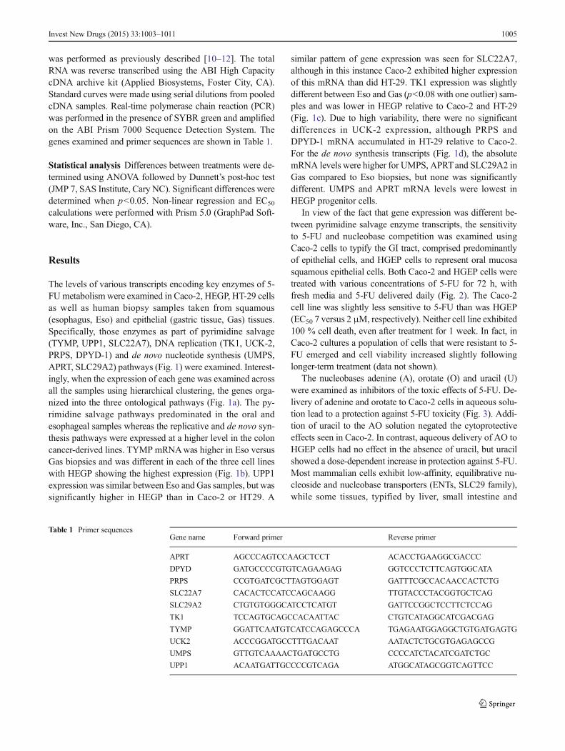
was performed as previously described [10–12]. The totalRNA was reverse transcribed using the ABI High CapacitycDNA archive kit (Applied Biosystems, Foster City, CA).Standard curves were made using serial dilutions from pooledcDNA samples. Real-time polymerase chain reaction (PCR)was performed in the presence of SYBR green and amplifiedon the ABI Prism 7000 Sequence Detection System. Thegenes examined and primer sequences are shown in Table 1.
Statistical analysis Differences between treatments were de-termined using ANOVA followed by Dunnett’s post-hoc test(JMP 7, SAS Institute, Cary NC). Significant differences weredetermined when p<0.05. Non-linear regression and EC50
calculations were performed with Prism 5.0 (GraphPad Soft-ware, Inc., San Diego, CA).
Results
The levels of various transcripts encoding key enzymes of 5-FUmetabolism were examined in Caco-2, HEGP, HT-29 cellsas well as human biopsy samples taken from squamous(esophagus, Eso) and epithelial (gastric tissue, Gas) tissues.Specifically, those enzymes as part of pyrimidine salvage(TYMP, UPP1, SLC22A7), DNA replication (TK1, UCK-2,PRPS, DPYD-1) and de novo nucleotide synthesis (UMPS,APRT, SLC29A2) pathways (Fig. 1) were examined. Interest-ingly, when the expression of each gene was examined acrossall the samples using hierarchical clustering, the genes orga-nized into the three ontological pathways (Fig. 1a). The py-rimidine salvage pathways predominated in the oral andesophageal samples whereas the replicative and de novo syn-thesis pathways were expressed at a higher level in the coloncancer-derived lines. TYMP mRNAwas higher in Eso versusGas biopsies and was different in each of the three cell lineswith HEGP showing the highest expression (Fig. 1b). UPP1expression was similar between Eso and Gas samples, but wassignificantly higher in HEGP than in Caco-2 or HT29. A
similar pattern of gene expression was seen for SLC22A7,although in this instance Caco-2 exhibited higher expressionof this mRNA than did HT-29. TK1 expression was slightlydifferent between Eso and Gas (p<0.08 with one outlier) sam-ples and was lower in HEGP relative to Caco-2 and HT-29(Fig. 1c). Due to high variability, there were no significantdifferences in UCK-2 expression, although PRPS andDPYD-1 mRNA accumulated in HT-29 relative to Caco-2.For the de novo synthesis transcripts (Fig. 1d), the absolutemRNA levels were higher for UMPS, APRTand SLC29A2 inGas compared to Eso biopsies, but none was significantlydifferent. UMPS and APRT mRNA levels were lowest inHEGP progenitor cells.
In view of the fact that gene expression was different be-tween pyrimidine salvage enzyme transcripts, the sensitivityto 5-FU and nucleobase competition was examined usingCaco-2 cells to typify the GI tract, comprised predominantlyof epithelial cells, and HGEP cells to represent oral mucosasquamous epithelial cells. Both Caco-2 and HGEP cells weretreated with various concentrations of 5-FU for 72 h, withfresh media and 5-FU delivered daily (Fig. 2). The Caco-2cell line was slightly less sensitive to 5-FU than was HGEP(EC50 7 versus 2 μM, respectively). Neither cell line exhibited100 % cell death, even after treatment for 1 week. In fact, inCaco-2 cultures a population of cells that were resistant to 5-FU emerged and cell viability increased slightly followinglonger-term treatment (data not shown).
The nucleobases adenine (A), orotate (O) and uracil (U)were examined as inhibitors of the toxic effects of 5-FU. De-livery of adenine and orotate to Caco-2 cells in aqueous solu-tion lead to a protection against 5-FU toxicity (Fig. 3). Addi-tion of uracil to the AO solution negated the cytoprotectiveeffects seen in Caco-2. In contrast, aqueous delivery of AO toHGEP cells had no effect in the absence of uracil, but uracilshowed a dose-dependent increase in protection against 5-FU.Most mammalian cells exhibit low-affinity, equilibrative nu-cleoside and nucleobase transporters (ENTs, SLC29 family),while some tissues, typified by liver, small intestine and
Table 1 Primer sequencesGene name Forward primer Reverse primer
APRT AGCCCAGTCCAAGCTCCT ACACCTGAAGGCGACCC
DPYD GATGCCCCGTGTCAGAAGAG GGTCCCTCTTCAGTGGCATA
PRPS CCGTGATCGCTTAGTGGAGT GATTTCGCCACAACCACTCTG
SLC22A7 CACACTCCATCCAGCAAGG TTGTACCCTACGGTGCTCAG
SLC29A2 CTGTGTGGGCATCCTCATGT GATTCCGGCTCCTTCTCCAG
TK1 TCCAGTGCAGCCACAATTAC CTGTCATAGGCATCGACGAG
TYMP GGATTCAATGTCATCCAGAGCCCA TGAGAATGGAGGCTGTGATGAGTG
UCK2 ACCCGGATGCCTTTGACAAT AATACTCTGCGTGAGAGCCG
UMPS GTTGTCAAAACTGATGCCTG CCCCATCTACATCGATCTGC
UPP1 ACAATGATTGCCCCGTCAGA ATGGCATAGCGGTCAGTTCC
Invest New Drugs (2015) 33:1003–1011 1005

kidney, also exhibit concentrative, sodium-dependent nucleo-side transport (SLC28 family) [2]. To assess the role of thesetransporters on uptake of the nucleobases adenine, orotate anduracil, liposomes were prepared for delivery into Caco-2 andHGEP cells. The application of the empty liposomes
themselves reduced 5FU toxicity to the cells but the resultswith the application of uracil, adenine and orotate as 5FUprotective agents were similar to those obtained by aqueousincubation with nucleobases (data not presented). Thus, deliv-ery of the nucleobases via SLC transporters is unlikely to be
Fig. 1 Expression of mRNA for 5-FU metabolism enzymes. The basallevel of expression of messenger RNA for 5-FU metabolic enzymes inCaco-2, HT29 and HEGP cells (n=3) as well as esophagus (Eso) andgastric (Gas) biopsies (5 individuals) was assessed by quantitative RT-PCR. Data is expressed relative to β-actin. The Esophogus and Gastric
tissues were compared using paired t-test and asterisk denotes asignificant difference between the samples (p<0.05). For the cell lines,bars with different letters are significantly different from each other(ANOVA, followed by Tukey’s multicomparison test, p<0.05)
Fig. 2 Sensitivity of Caco-2 and HGEP cells to 5-FU toxicity. Cells wereplated in 96-well plates and allowed to adhere overnight. Subsequently, 5-FU was dissolved in water and added to media at various concentrations.
Media was changed every day with reapplication of 5-FU. After 72 h, thecell viability was assessed and is expressed relative to the vehicle control.(Caco-2, n=3; HGEP, n=4)
1006 Invest New Drugs (2015) 33:1003–1011

responsible for the differences observed in cytoprotection ofadenine, orotate and uracil from 5FU toxicity. Because of therelative simplicity of aqueous incubation, the subsequent ex-periments utilized this method of treatment.
The cytoprotective effect of aqueous delivery of adenine,orotate and uracil in Caco-2 cells was examined in more detail(Fig. 4). Adenine at 0.0125 mM through 1.25 mM resulted inprogressively greater cytoprotection against 10 μM 5-FU tox-icity (Fig. 4a). Addition of orotate (0–1.25 mM) did not atten-uate 5-FU toxicity produced by adenine. At higher concentra-tions of adenine (2.5 mM), the addition of orotate at 1.25 mMmay have provided enhanced protection against 5-FU toxicityin Caco-2 cells. Still higher concentrations of orotate causedprogressively greater abrogation of salvage from 5-FU toxic-ity (Fig. 4b). As shown in Fig. 4c, the purine nucleoside ino-sine does not prevent 5-FU toxicity in contrast to the purinenucleobase adenine. A reversal of AO cytoprotection by uracilwas seen in cells exposed to 10 μM 5-FU (Fig. 4d).
Discussion
Differential expression of the enzymes of de novo synthesisversus salvage of pyrimidine nucleosides between anatomicregions may explain the differences in nucleobase specificityin preventing 5-FU toxicity. The expression of pyrimidinesalvage enzymes, including TYMP, UPP1 and SLC22A7, ishighest in HEGP of oral cavity-derived keratinized squamouscell tissue. HEGP cells were slightly more sensitive to 5-FUtoxicity and were protected from 5-FU toxicity by uracil thatcan be salvaged by TYMP1 to uridine but not by adenine andorotate, nucleobases involved in de novo pyrimidine synthe-sis. The esophagus has a simple stratified squamous epithelial
surface without keratin. The esophagus utilizes both salvageand de novo synthesis, as evidenced by the slightly higherexpression of UMPS, APRT and SLC29A2 relative to thegingival cell line that has a keratin layer. The increased pro-liferative enzymes in the distal esophagus are consistent withtissue stress.
The comprehensive analysis of gene expression in humanGI tract and Caco-2 [13] was reanalyzed to examine 5-FUmetabolic enzymes (data not shown) and illustrated thatCaco-2 cells most closely resemble cells of the colonic epithe-lium. For the Caco-2 cells of columnar origin, exposure toadenine showed a significant and dose-dependent protectiveeffect from 5-FU toxicity. The sensitivity of Caco-2 cells toadenine cytoprotection may be dependent on the dominantrole of de novo synthesis, in particular UMPS. The mecha-nism of protection by adenine may be the result of relativedepletion of PRPP by APRT. The Km for PRPP are similar forAPRT (33 μM, [14]) and UMPS (25 μM, [15]), which indi-cates that in the presence of vast excess adenine relative to 5-FU, the utilization of PRPP by APRT will be favored. Inaddition because of pK difference, the Km of UMPS fororotate (5.5 μM) is 30-fold lower than that of 5-FU(190 μM [15]). Thus, with limited PRPP availability endoge-nously produced orotate will be utilized by UMPS over 5-FU.
The failure of orotate as a monotherapy to prevent 5-FUtoxicity probably relates to two facts: orotate with a negativecharge at neutral pH is poorly taken-up by the cells. Becauseof its low Km for UMPS, increased intracellular orotate com-petes successfully for PRPP and can deplete PRPP and cancause “purineless” cell death.
Uracil enhanced 5-FU toxicity in Caco-2 cells. This resultmay reflect the depletion of ribose-1-phosphate by uracil,thereby increasing 5-FU concentration available for activationby UMPS in a cell type significantly dependent on de novosynthesis of pyrimidine nucleosides. Because of its muchhigher Km, uracil competes ineffectively with 5-FU for acti-vation by UMPS. Taken together, these studies have shownthat differences in gene expression of pyrimidine metabolicenzymes may explain the differential sensitivity to nucleobaseprotection of 5-FU toxicity (see Fig. 5, top panels).
Evidence for a clinical role for uracil protection from 5-FUtoxicity already exists. Systemic uracil is a component of UFTthat is in clinical use and UFT is well-tolerated and, in contrastto the administration of 5-FU alone, has a very low incidenceof both 5-FU cutaneous toxicity (“hand-foot” syndrome) andoral stomatitis [9]. Topical uracil has been applied clinicallyand has prevented “Hand-Foot” syndrome from 5-FU treat-ment [16]. Systemic adenine has been used clinically as wellin other contexts. Its unique toxicity, renal stones from themetabolite 2,8-dihydroxyadenine, is prevented by co-administration of allopurinol [17]. The dose of adenine re-quired in the present context is much lower than that cited(100 mg/kg body weight). A clinical test of topical uracil
Fig. 3 Protection of 5-FU toxicity by aqueous delivery of adenine plusorotate. Cells were plated in 96-well plates and allowed to adhereovernight. Subsequently, cells were treated with adenine plus orotate(2.5 mM each, A/O) or A/O plus uracil (A/O/U) at 1.25 and 2.5 mM inwater. 5-FU was dissolved in water and added to media at 10 μM. Mediawas changed every day with reapplication of 5-FU and nucleosides. After72 h, the cell viability was assessed and is expressed relative to the vehiclecontrol. (Caco-2, n=3; HGEP, n=4). Within each cell line, bars withdifferent letters are significantly different from each other (ANOVA,followed by Tukey’smulticomparison test, p<0.05)
Invest New Drugs (2015) 33:1003–1011 1007

applied to the mouth to prevent oral stomatitis and, with thesame rationale, oral slow-release adenine to protect the GImucosa from the effects of 5-FU treatment would bereasonable.
Tissue cell-type differences in pyrimidine metabolism,whether predominantly by salvage or by de novo synthesis,suggests a common aspect of tissue structure (see Fig. 5, bot-tom panels) with implications for 5-FU toxicity as well asoncogenesis. For normal squamous cells in the GI tract, rep-lication occurs at the epithelial-mesenchymal interface andgrowth proceeds towards the surface layer with concomitantand progressive loss and release of cell components. For glan-dular cells there is a normal zone of replication at the base ofthe crypt that involves aWnt (wingless and Int-1) gradient andthe replicative signal originates with the intestinalsubepithelial myofibroblasts near the muscularis mucosa of
the mesenchyme [18]. Growing normally, epithelial glandularcells migrate away from the zone of replication towards thetop of the crypt and the predominant de novo nutrient sourcein the GI lumen. At the top of the crypt the cells are shed.
As the first layer of epithelial cells for both squamous andcolumnar cell types derives nutrition from the mesenchymallayer below, the normal response of tissue to nutrient stress is atransient reversal nutrient source, such as likely occurs inwounding and ontogeny. For both squamous and columnarcells, persistent nutrient stress causes an activation of HIF1-alpha. This transcription factor is synthesized in response toanoxia or lack of glucose and results in a proliferation of newblood vessels in the underlying mesenchymal layer [19]. Withdysplasia, the site of growth and replication persists. Thisresults for both squamous and columnar cells in a tissue wherethe epithelial layer growth pattern is inverted. Of vital
Fig. 4 Dose–response relationships for adenine (A), inosine (I), uracil(U) and orotate (O) for protection of Caco-2 cells from 5-FU toxicity.Cells were plated in 96-well plates and allowed to adhere overnight.Subsequently, cells were treated with adenine (A), inosine (I), uracil (U)and orotate (O), in concentration as shown in parenthesis beneath each bar(in mM). 5-FU was dissolved in water and added to media at 10 μM.
Media was changed every day with reapplication of 5-FU. After 72 h, thecell viability was assessed and is expressed relative to the vehicle control.(Caco-2, n=3). Panel a, asterisks denote significance from respectivecontrol. Panels b-d, bars with different letters are significantly differentfrom each other (ANOVA, followed by Tukey’smulticomparison test,p<0.05)
1008 Invest New Drugs (2015) 33:1003–1011

importance, the region of cell turnover disappears. Thus, anormal tissue, in which the regions of cell replication and cellturnover are separated, becomes a dysplastic tissue where allthe epithelial cells replicate in response to the unattentuatedproliferative signals originating from the mesenchymal layer.The consequence is that all the cells within the epithelial layercompete progressively for nutrients from the underlying vas-culature, as they increase in number. This selection pressure, ifsustained, is predicted to cause progressive phenotypic andgenotypic evolution of the epithelial cells, as has been ob-served for GI glandular cells of the colon [20]. This oncogenicprocess may result in a breach by the epithelial cell of thebasement membrane and EMT (epithelial mesenchymal trans-formation) in the competition for nutrients.
Support for this model of tissue growth, with a reverseddirection of growth in oncogenesis comes from explanations itprovides for several currently puzzling clinical and researchobservations. First, H. Pylori is a bacterial infection that in-volves the glycocalyx or superior aspect of gastric columnarcells [21]. Perhaps, surprisinglyH. Pylori infection can lead tocancer. However, this bacterium uses glucose as a nutrientsource and thus infection by H. Pylori may cause a localnutrient stress to the underlying glandular cells and result ina tissue with a reversed direction of growth and an increasedcancer risk.
Second, the esophagus and the colon are much more likelythan the small bowel to undergo epithelial cell oncogenesis.The present model would suggest that this could result from
Fig. 5 NDGP in normal and dysplastic GI squamous and glandular cellsand 5-FU metabolism in normal cells. Shown in the top panels are thepredominant pathways of 5-FU metabolism. In normal oral squamouscells salvage pathways dominate and uracil competes with 5-FU formetabolism by UPP1/TYMP. In normal GI glandular mucosa, de novosynthesis pathways dominate and excess exogenous adenine relativelydepletes PRPP though metabolism by APRT and makes it rate limitingfor UMPS and favors the lower Km orotate substrate over 5FU. Thelower panels represent the source of nutrients and areas of cellular
replication in both normal and dysplastic tissues. Labels: A, Zone ofcell turnover; B, Zone of replication; C, muscularis mucosa layer; D,Mesenchymal layer; E, Blood vessel. White arrows depict route ofcellular migration. The nutrient gradient is depicted with shading withinthe image, with darker shading representing higher levels of nutrients.The drawings are based on photographs of pathologic slides. Thepanels depicting dysplastic tissues ignore nutrient gradients that wouldoriginate from blood vessels in the tissue either above or below the planeof the photographed, thin pathologic specimen
Invest New Drugs (2015) 33:1003–1011 1009

the fact that more nutrients are available and absorbed by theglandular enterocytes of the small bowel than either the squa-mous cells of the esophagus or the columnar colonic epithelialcells [22].
Third, although both portend increased risk of cancer, com-plete intestinal metaplasia (fully enabled absorptiveenterocytes with villous projections) of the stomach is lesslikely than incomplete metaplasia of the esophagus to leadto epithelial cell cancer [23]. Incomplete metaplasia demon-strates a histologic pattern with goblet cells like that of thedistal ileum not like that of the adjacent stomach. Consistentwith nutrient stress as the cause of the increased cancer riskfrom incomplete metaplasia, normal squamous cells of theesophagus have a doubling time of 21 days, normal columnarcells of the stomach 12 days and normal columnar cells of theileum only 4 to 5 days [24]. In addition the stomach, in con-trast to the esophagus, is normally bathed in a high nutrientcontent mixture that would tend to mitigate nutrient stress[25].
The cellular mechanism that directs cell growth undernutrient stress within the GI tissue is not fully under-stood, although autophagy is believed to play an impor-tant role. Autophagy is currently the process of deliveryof cellular cargo via the autophagasome for lysosomaldegradation. Autophagy thus acts as a cellular survivalmechanism under conditions of stress. Autophagy main-tains cellular integrity by regenerating metabolic precur-sors and clearing subcellular debris [26, 27]. We postu-late autophagy may also have an apparently paradoxic,vital role in maintaining tissue integrity by eitherrestricting cell turnover or catalyzing cell turnover de-pending upon nutrient adequacy. Such a role for autoph-agy is consistent with the activity of coiled-coil myosin-like BCL2-interacting protein (Beclin-1). Beclin-1 (atg-6) activates autophagosome formation. When doubly de-leted in mice, Beclin-1 results in an abnormal ectoder-mal layer and early embryonic lethality [27]. A role forautophagy in maintaining tissue structure is evident inthe requirement for autophagy in order to have propersalivary gland degradation in Drosophila [28].
The control of cell degradation by autophagy in the pres-ence of nutrient adequacy may involve regulation of Beclin-1through abrogation of Bcl-2/Bcl-Xl inhibition by both c-junN-terminal kinase-1 (JNK-1) [29] and death associated proteinkinase (DAPK) autophagic pathways. DAPK phosphorylatesBeclin-1 itself with constitutive activation of autophagy andcell death in contrast to JNK1 phosphorylation of Bcl-2/Bcl-Xl [30]. As cells of GI glandular histology approach the top ofthe crypts, the expression of DAPK increases [30]. Cells ofboth normal GI squamous and glandular histology expressBeclin-1. Beclin-1 expression is increased in podocytes atcrypt tops by exposure to high glucose levels [30]. Beclin-1expression decreases with progressive dysplasia [31].
Evidence for related nutrient-responsive systems that func-tion in the normal region of cell turnover exists. In GI glandu-lar cells, the cell detachment process is likely mediated byEphrin ligand that is sensitive to high glucose levels, in con-trast to Eph receptor in the replication zone at the crypt basethat is sensitive to low glucose states [32]. High glucose levelsinduce bone morphogenic protein-2 (BMP-2) [33]. BMP in-hibits intestinal stem cell activation and promotes intestinalcell differentiation at the top of the colonic crypt [18].
As we did with the directed application of a tissue appro-priate nucleobase to prevent 5FU toxicity, it may be reason-able to test if restoring an adequate nutrient level at the normaltissue zone of turnover would also restore normal direction ofcell growth and tissue autophagic function. The interventionwould consist of the directed application of nutrient mix (like-ly including glucose) to the tissue surface of dysplastic tissues(prior, normal region of cell turnover). Monitoring the resto-ration of normal, upregulated Beclin-1 expression may be ameans to follow the efficacy of the intervention.
Acknowledgments The drawings in Fig. 5 were made by WilliamsonAdams, Walnut Creek, CA (415) 254 3393. We thank Stephen J.Benkovich (Department of Chemistry, Pennsylvania State University)for his insightful comments during preparation of the manuscript.
Conflict of interest J.P.V.H. is a shareholder and the Chief ScientificOfficer of INDIGOBiosciences, Inc. J.P.F. is principal member of Asym-metric Therapeutics, LLC, Unadilla, NY. All other authors: no conflicts.
Open Access This article is distributed under the terms of the CreativeCommons At t r ibut ion 4 .0 In te rna t ional License (h t tp : / /creativecommons.org/licenses/by/4.0/), which permits unrestricted use,distribution, and reproduction in any medium, provided you give appro-priate credit to the original author(s) and the source, provide a link to theCreative Commons license, and indicate if changes were made.
References
1. Malet-Martino M (2002) Clinical studies of three oral prodrugs of5-fluorouracil (capecitabine, UFT, S-1): a review. Oncologist 7:288–323
2. Baldwin SA, Beal PR, Yao SYM, King AE, Cass CE, Young JD(2004) The equilibrative nucleoside transporter family, SLC29.Pflugers Arch 447:735–743
3. Kobayashi Y, Ohshiro N, Sakai R, Ohbayashi M, Kohyama N,Yamamoto T (2005) Transport mechanism and substrate specificityof human organic anion transporter 2 (hOat2 [SLC22A7]). J PharmPharmacol 57:573–578
4. Longley DB, Harkin DP, Johnston PG (2003) 5-fluorouracil: mech-anisms of action and clinical strategies. Nat Rev Cancer 3:330–338
5. Ichikawa W, Takahashi T, Suto K, Sasaki Y, Hirayama R (2006)Orotate phosphoribosyltransferase gene polymorphism predictstoxicity in patients treated with bolus 5-fluorouracil regimen. ClinCancer Res 12:3928–3934
6. Maring JG, Groen HJM, Wachters FM, Uges DRA, de Vries EGE(2005) Genetic factors influencing pyrimidine-antagonist chemo-therapy. Pharmacogenomics J 5:226–243
1010 Invest New Drugs (2015) 33:1003–1011

7. Muhale FA, Wetmore BA, Thomas RS, McLeod HL (2011)Systems pharmacology assessment of the 5-fluorouracil pathway.Pharmacogenomics 12:341–350
8. Jeung H-C, Rha SY, Shin SJ, Lim SJ, Roh JK, Noh SH, Chung HC(2011) Predictive values of 5-fluorouracil pathway genes for S-1treatment in patients with advanced gastric cancer. AnticancerDrugs 22:801–810
9. Hoff PM (2000) The tegafur-based dihydropyrimidine dehydroge-nase inhibitory fluoropyrimidines, UFT/leucovorin (ORZEL) andS-1: a review of their clinical development and therapeutic poten-tial. Invest New Drugs 18:331–342
10. Lee Y, Thompson JT, Vanden Heuvel JP (2009) 9E,11E-conjugatedlinoleic acid increases expression of the endogenousantiinflammatory factor, interleukin-1 receptor antagonist, inRAW 264.7 cells. J Nutr 139:1861–1866
11. Zhang J, Kris-Etherton PM, Thompson JT, Vanden Heuvel JP(2010) Effect of pistachio oil on gene expression of IFN-inducedprotein with tetratricopeptide repeats 2: a biomarker of inflamma-tory response. Mol Nutr Food Res 54(Suppl 1):S83–S92
12. Gillies PJ, Bhatia SK, Belcher LA, Hannon DB, Thompson JT,Vanden Heuvel JP (2012) Regulation of inflammatory and lipidmetabolism genes by eicosapentaenoic acid-rich oil. J Lipid Res53:1679–1689
13. Lorenzi PL, Landowski CP, Brancale A, Song X, Townsend LB,Drach JC, Amidon GL (2006) N-methylpurine DNA glycosylaseand 8-oxoguanine dna glycosylase metabolize the antiviral nucleo-side 2-bromo-5,6-dichloro-1-(beta-D-ribofuranosyl) benzimid-azole. Drug Metab Dispos 34:1070–1077
14. Gutensohn W (1984) Inherited disorders of purine metabolism–underlying molecular mechanisms. Klin Wochenschr 62:953–962
15. Reyes P, Guganig ME (1975) Studies on a pyrimidinephosphoribosyltransferase from murine leukemia P1534J. Partialpurification, substrate specificity, and evidence for its existence as abifunctional complex with orotidine 5-phosphate decarboxylase. JBiol Chem 250:5097–5108
16. Ford JP (2012) US Patent 7,816,366. Compositions and methodsfor treating and preventing dermatoses
17. Bührdel P, Krüger W, Hirschberg K, Wehnert M (1985) Adeninetherapy in Lesch-Nyhan syndrome. Acta Paediatr Hung 26:327–333
18. Kosinski C, Li VSW, Chan ASY, Zhang J, Ho C, Tsui WY, ChanTL, Mifflin RC, Powell DW, Yuen ST, Leung SY, Chen X (2007)Gene expression patterns of human colon tops and basal crypts andBMP antagonists as intestinal stem cell niche factors. Proc NatlAcad Sci U S A 104:15418–15423
19. Ling FC, Khochfar J, Baldus SE, Brabender J, Drebber U,Bollschweiler E, Hoelscher AH, Schneider PM (2009) HIF-1alpha protein expression is associated with the environmental
inflammatory reaction in Barrett’s metaplasia. Dis Esophagus 22:694–699
20. Vermeulen L, Morrissey E, van der Heijden M, Nicholson AM,Sottoriva A, Buczacki S, Kemp R, Tavaré S, Winton DJ (2013)Defining stem cell dynamics in models of intestinal tumor initia-tion. Science 342:995–998
21. RektorschekM,WeeksD, Sachs G,Melchers K (1998) Influence ofpH on metabolism and urease activity of Helicobacter pylori.Gastroenterology 115:628–641
22. Karam SM (1999) Lineage commitment and maturation of epithe-lial cells in the gut. Front Biosci (Landmark Ed) 4:D286–D298
23. Odze RD (2005) Unraveling the mystery of the gastroesophagealjunction: a pathologist’s perspective. Am J Gastroenterol 100:1853–1867
24. Squier C, Brogden K (2011) Human oral mucosa: development,structure and function. Wiley-Blackwell
25. Fenoglio-Preiser CM, Noffsinger AE, Stemmermann GN, LantzPE, Isaacson PG (1989) Gastrointestinal pathology: an atlas andtext. Raven, New York
26. Choi AMK, Ryter SW, Levine B (2013) Autophagy in humanhealth and disease. N Engl J Med 368:651–662
27. Levine B, Kroemer G (2008) Autophagy in the pathogenesis ofdisease. Cell 132:27–42
28. Berry DL, Baehrecke EH (2007) Growth arrest and autophagy arerequired for salivary gland cell degradation in Drosophila. Cell 131:1137–1148
29. Ma T, Zhu J, Chen X, Zha D, Singhal PC, Ding G (2013) Highglucose induces autophagy in podocytes. Exp Cell Res 319:779–789
30. Chakilam S, Gandesiri M, Rau TT, Agaimy A, Vijayalakshmi M,Ivanovska J, Wirtz RM, Schulze-Luehrmann J, Benderska N,Wittkopf N, Chellappan A, Ruemmele P, Vieth M, Rave-FränkM, Christiansen H, Hartmann A, Neufert C, Atreya R, Becker C,Steinberg P, Schneider-Stock R (2013) Death-associated proteinkinase controls STAT3 activity in intestinal epithelial cells. Am JPathol 182:1005–1020
31. Roesly HB, Khan MR, Chen HDR, Hill KA, Narendran N, WattsGS, Chen X, Dvorak K (2012) The decreased expression of Beclin-1 correlates with progression to esophageal adenocarcinoma: therole of deoxycholic acid. Am J Physiol Gastrointest Liver Physiol302:G864–G872
32. Pasquale EB (2008) Eph-ephrin bidirectional signaling in physiol-ogy and disease. Cell 133:38–52
33. Chen NX, Duan D, O’Neill KD, Moe SM (2006) High glucoseincreases the expression of Cbfa1 and BMP-2 and enhances thecalcification of vascular smooth muscle cells. Nephrol DialTransplant 21:3435–3442
Invest New Drugs (2015) 33:1003–1011 1011


![Nucleobase–Guanidiniocarbonyl-Pyrrole Conjugates as Novel ...fulir.irb.hr/3840/1/BanZ_Nucleobase_Molecules-22_2017_2213.pdf · nucleobase cytosine [28,29], while the guanidiniocarbonyl-pyrrole](https://img.dokumen.tips/doc/110x75/5eadaa6c2f808b2f2c0bb939/nucleobaseaguanidiniocarbonyl-pyrrole-conjugates-as-novel-fulirirbhr38401banznucleobasemolecules-2220172213pdf.jpg)