Embed Size (px)
Citation preview

EUKARYOTIC CELL, Sept. 2010, p. 1383–1397 Vol. 9, No. 91535-9778/10/$12.00 doi:10.1128/EC.00042-10Copyright © 2010, American Society for Microbiology. All Rights Reserved.
Differential Filamentation of Candida albicans and Candida dubliniensisIs Governed by Nutrient Regulation of UME6 Expression�†
Leanne O’Connor, Nicole Caplice, David C. Coleman, Derek J. Sullivan, and Gary P. Moran*Microbiology Research Unit, Division of Oral Biosciences, Dublin Dental School and Hospital, Trinity College Dublin,
University of Dublin, Dublin 2, Republic of Ireland
Received 22 February 2010/Accepted 1 July 2010
Candida dubliniensis is closely related to Candida albicans; however, it is responsible for fewer infections inhumans and is less virulent in animal models of infection. C. dubliniensis forms fewer hyphae in vivo, and thismay contribute to its reduced virulence. In this study we show that, unlike C. albicans, C. dubliniensis fails toform hyphae in yeast extract-peptone-dextrose (YPD) medium supplemented with 10% (vol/vol) fetal calfserum (YPDS medium). However, C. dubliniensis filaments in water plus 10% (vol/vol) fetal calf serum (WS),and this filamentation is inhibited by the addition of peptone and glucose. Repression of filamentation in YPDSmedium could be partly overcome by preculture in synthetic Lee’s medium. Unlike C. albicans, inoculation ofC. dubliniensis in YPDS medium did not result in increased UME6 transcription. However, >100-fold inductionof UME6 was observed when C. dubliniensis was inoculated in nutrient-poor WS medium. The addition ofincreasing concentrations of peptone to WS medium had a dose-dependent effect on reducing UME6 expres-sion. Transcript profiling of C. dubliniensis hyphae in WS medium identified a starvation response involvingexpression of genes in the glyoxylate cycle and fatty acid oxidation. In addition, a core, shared transcriptionalresponse with C. albicans could be identified, including expression of virulence-associated genes includingSAP456, SAP7, HWP1, and SOD5. Preculture in nutrient-limiting medium enhanced adherence of C. dublini-ensis, epithelial invasion, and survival following coculture with murine macrophages. In conclusion, C. albicans,unlike C. dubliniensis, appears to form hyphae in liquid medium regardless of nutrient availability, which mayaccount for its increased capacity to cause disease in humans.
Candida dubliniensis is the closest known relative of Candidaalbicans, the predominant fungal pathogen of humans (27, 28).Epidemiological evidence has shown that C. albicans is moreprevalent in the human population as a commensal of the oralcavity and is responsible for more infections (both oral andsystemic) than C. dubliniensis (10, 13, 15). C. albicans is re-sponsible for approximately 60% of cases of candidemia,whereas C. dubliniensis accounts for fewer than 2% of cases(13). Evidence from animal infection models also suggests thatC. dubliniensis is less virulent than C. albicans (26, 29). Fol-lowing oral-intragastric inoculation, C. dubliniensis strains aremore rapidly cleared from the gastrointestinal tract than C.albicans strains and are less able to establish disseminatedinfection (26). Following tail vein inoculation in the systemicmouse model of infection, only a small number of C. dublini-ensis isolates have been shown to establish disseminated infec-tions, and most studies conclude that C. dubliniensis isolatesare generally less virulent than C. albicans isolates (1, 29).
Virulence studies have associated the reduced capacity of C.dubliniensis to establish infection with a reduced ability toundergo the transition from yeasts to hyphae (1, 26). In theoral-intragastric infection model, C. dubliniensis cells in the
stomach and kidney were found to be in the yeast form only,while using the same models C. albicans cells were found to bein both the yeast and hyphal forms (26). Asmundsdottir et al.(1) also noted that C. dubliniensis produced significantly fewerhyphae than C. albicans following dissemination to the liverand kidney in mice. In vitro, C. dubliniensis forms true hyphaeless efficiently than C. albicans in response to serum, pH shiftsin Lee’s medium, and CO2 and in certain defined media suchas RPMI 1640 medium (16, 26). Poor hypha production hasalso been observed in C. dubliniensis in vitro during coculturewith murine macrophages and during infection of reconsti-tuted human oral epithelial tissues (16, 24). This results in aninability of C. dubliniensis to evade macrophage killing andlimited invasion of epithelial surfaces.
Although C. dubliniensis produces true hyphae less effi-ciently than C. albicans, C. dubliniensis can produce abundantpseudohyphae and chlamydospores on certain solid media(27). Recently, Staib and Morschhauser (25) demonstratedthat the propensity for C. dubliniensis to form large numbers ofchlamydospores on these media was due to species-specificdownregulation of the NRG1 repressor. Further studies haveshown that downregulation of the NRG1 transcript is alsorequired for efficient production of true hyphae in C. albicansin response to serum (22). We have shown that under condi-tions where C. dubliniensis fails to filament, for example, fol-lowing phagocytosis by murine macrophages, this species doesnot downregulate NRG1, whereas C. albicans responds tothese conditions by shutting down NRG1 transcription (16).Deletion of the NRG1 gene in C. dubliniensis can partly offsetthe failure of this species to filament in vitro and leads to more
* Corresponding author. Mailing address: Microbiology ResearchUnit, Division of Oral Biosciences, Dublin Dental School and Hospi-tal, Trinity College Dublin, University of Dublin, Dublin 2, Republic ofIreland. Phone: 353 1 612 7245. Fax: 353 1 612 7295. E-mail: [email protected].
† Supplemental material for this article may be found at http://ec.asm.org/.
� Published ahead of print on 16 July 2010.
1383
on Septem
ber 2, 2018 by guesthttp://ec.asm
.org/D
ownloaded from
efficient production of hyphae in response to serum and CO2
and during coculture with murine macrophages (16).In this study, we have examined in detail the environmental
signals required for filamentation in C. dubliniensis. We haveshown that nutrient-rich conditions inhibit efficient hypha for-mation by suppressing UME6 expression in C. dubliniensis.This study also includes the first description of a C. dublinien-sis-specific microarray that we used to generate a transcriptprofile for C. dubliniensis true hyphae. The effects of inducinghypha formation in C. dubliniensis under these conditions onthe ability to infect reconstituted oral epithelial tissues and toevade macrophage killing were also examined.
MATERIALS AND METHODS
Candida strains and culture conditions. All Candida strains were routinelycultured on yeast extract-peptone-dextrose (YPD) agar at 37°C. For liquid cul-ture, cells were grown with shaking (at 200 rpm) in YPD broth at 30°C or 37°C,as indicated in the figure legends (9). Genotypes of strains used in this study arelisted in Table S1 in the supplemental material. Liquid culture was also carriedout at 30°C in the liquid medium of Lee et al. (14) supplemented with 400 mMarginine, 0.001% (wt/vol) biotin, and trace metals (0.2 mM ZnSO, 0.25 mMCuSO, 1 mM FeCl, 1 mM MgCl, and 1 mM CaCl). Where indicated in Fig. 5band 7f, Lee’s medium was buffered to pH 5.0 or pH 7.2 with 0.1 M potassiumphosphate buffer. Supplementation of Lee’s and other media with peptone wascarried out with bacteriological peptone (Oxoid). Peptone supplementation upto 2% (wt/vol) did not significantly alter the pH of Lee’s medium or serum.Hyphal induction was carried out in liquid YPD plus 10% (vol/vol) fetal calfserum (YPDS) or in sterile Milli-Q H2O supplemented with 10% (vol/vol) fetalcalf serum (WS) at 37°C. The proportion of germ tubes or hyphae in each culturewas assessed at intervals by microscopic examination of an aliquot of culture witha Nikon Eclipse 600 microscope (Nikon U.K., Surrey, United Kingdom).
Genetic manipulation of C. dubliniensis. Ectopic expression of C. albicansUME6 (CaUME6) in C. dubliniensis was achieved using plasmid pCaUme6-3,containing UME6 under the control of a doxycycline-inducible promoter (32).The expression cassette was released from pCaUme6-3 by ApaI and PmlI diges-tion and was used to transform strains Wu284 and CDM10 by electroporation, asdescribed previously (16). Plasmid pNRG1 was generated from plasmids pNIM1and pTET42 (18). NRG1 was removed from pTET42 as an SalI/BglII fragmentand ligated to SalI/BglII-digested pNIM1 to generate pNRG1. The expressioncassette was released from pNRG1 by SacII and KpnI digestion and was used totransform Wu284 and CDM10 by electroporation, as described previously (16).Integration of pNIM1 derivatives at the ADH1 locus was confirmed by PCR.
In order to create strains harboring a PECE1-GFP fusion, we used the inte-grating vector pCDRI (16). A derivative of this plasmid was created by insertingyeast enhanced green fluorescent protein (yEGFP) fused to the actin terminatoron a HindIII/MluI fragment to create pGM175. An ECE1 promoter fragmentfrom bases �1 to �921 was amplified from C. albicans SC5314 with primersECEAF (GTACGGGCCCAAGAGTCTCATTCAGATAACG) and ECEXR(GCATCTCGAGTTTAACGAATGGAAAATAGTTG) and cloned upstreamof yEGFP following digestion of both fragments with ApaI and XhoI. Theplasmid was linearized within the CDR1 region and used to transform C. albicansSC5314, C. dubliniensis Wu284, and the nrg1� derivative CDM10 as describedpreviously (16). Ectopic integration in the CDR1 gene was confirmed by South-ern hybridization.
Transcriptional profiling with oligonucleotide microarrays. A set of 5,999open reading frames (ORFs) from the CD36 genome was used to design a C.dubliniensis expression microarray. Two unique 60-mer oligonucleotides specificfor each ORF were designed using the Agilent eArray probe design tool. Each60-mer was printed in quadruplicate on glass slides by Agilent technologies. Toexamine the hyphal transcript profile of C. dubliniensis strain Wu284, the strainwas grown for 18 h in Lee’s medium (pH 4.5) at 30°C with shaking, washed insterile H2O, and inoculated in 200 ml of H2O plus 10% (vol/vol) fetal calf serumto a density of 2 � 106 cells/ml. Samples (50 ml) were removed for RNApreparation at 1, 3, and 5 h postinoculation. To examine the effects of cell densitychanges, nutrient depletion, a shift to 37°C, and a shift to alkaline pH, identical18-h Lee’s medium cultures were washed and inoculated at 2 � 106 cells/ml in (i)fresh Lee’s medium (pH 4.5) at 30°C, (ii) 10% (vol/vol) Lee’s medium (pH 4.5)at 30°C, (iii) Lee’s medium (pH 4.5) at 37°C, and (iv) Lee’s medium (pH 7.2) at30°C. RNA was extracted from these cultures following 3 h of incubation under
each condition. To identify NRG1-regulated genes in C. dubliniensis, RNA wasextracted from Wu284 and its nrg1� derivative, CDM10, following growth to anoptical density at 600 nm (OD600) of 1.0 in YPD broth at 30°C. For RNApreparation, cell pellets were snap-frozen in liquid N2 and disrupted usingMikro-Dismembrator S system (Sartorius Stedim Biotech, Gottingen, Germany).RNA was prepared using TRI-Reagent (Sigma Chemical Co.) according to themanufacturer’s instructions. Poly(A) mRNA was then isolated using a SigmaGenelute mRNA isolation kit. A 200-ng aliquot of mRNA was labeled with Cy5or Cy3 using an Agilent Two-Color Low RNA Input Linear Amplification KitPLUS, according to the manufacturer’s instructions. Hybridization and washingof the arrays were carried out using an Agilent Gene Expression HybridizationKit and Gene Expression Wash Pack according the manufacturer’s instructions.For each condition, four biological replicate experiments were performed, in-cluding two dye swap experiments. Slides were scanned using a GenePix personal4100A scanner (Axon), and data were extracted using GenePix Pro, version 6.1(Axon). Spots were flagged absent if the signal was less than background �1standard deviation in both fluorescent channels. Raw data were exported toGeneSpring GX11, and signals for each replicate spot were background cor-rected and normalized using Loess normalization. Log2 fluorescence ratios weregenerated for each replicate spot and averaged. Oligonucleotides were excludedfrom analysis if �50% of replicates in each condition were flagged absent. Genesdifferentially expressed across all conditions were identified by analysis of vari-ance (ANOVA) with a Student Newman Keuls (SNK) posthoc test in Gene-spring GX11. A total of 7,107 oligonucleotide probes were significantly differ-entially expressed, with a corrected P value (Benjamini-Hochberg false discoveryrate [FDR]) of �0.05. Hierarchical clustering was used to compare gene expres-sion under each condition using the default settings in Genespring GX11. Someindividual samples (sera from 1 h, 3 h, and 5 h) were also analyzed using aone-sample t test in order to identify genes exhibiting significant differentialexpression (2-fold or greater) from preculture cells. All P values were adjustedusing the Benjamini-Hochberg multiple correction test to limit false differentialgene expression, and oligonucleotides with P values of �0.015 were selected foranalysis.
The C. albicans hypha-induced gene set used in this study included the hypha-regulated genes identified by Nantel et al. (17) and by Kadosh and Johnson (12).Additional C. albicans hypha-regulated genes were identified in the data set ofKadosh and Johnson (12) following analysis of the data set with GeneSpringGX11. These additional genes were included if they exhibited significant (�2-fold) regulation (t test, P � 0.01) in the 2-h and 3-h data sets (12).
Real-time PCR analysis of gene expression. Cultures for RNA preparation forquantitative reverse transcription-PCR (QRT-PCR) were set up in identicalfashion to those used for microarray analysis. RNA for QRT-PCR was isolatedusing an RNeasy Mini-kit (Qiagen). Cells were disrupted using a FastPrep beadbeater (Bio101). RNA samples were rendered DNA free by incubation withTurbo-DNA free reagent (Ambion, Austin, TX). cDNA synthesis was carried outas described by Moran et al. (16). Primers used in this study are listed in TableS2 in the supplemental material and were designed using Primer Express soft-ware, version 1.5 (Applied Biosystems, Foster City, CA). These primers yieldedsingle, specific amplimers from genomic DNA and cDNA templates. Primerpairs for UME6 and NRG1 were selected that yielded similar amplificationefficiencies as the TEF1 primer pair against a serial dilution of template DNA.Real-time detection of amplimers was carried out using a Power SYBR greenPCR Master Mix (Applied Biosystems, Foster City, CA) and an ABI 7500sequence detector, with separate reactions performed for each gene. Gene ex-pression levels were normalized against the expression levels of the constitutivelyexpressed TEF1 gene in the same cDNA sample.
Epithelial adhesion and invasion studies. Adherence of Candida strains tomonolayers of the oral epithelial cell line TR146 was determined using the assayof Rotrosen et al. (20). Monolayers of TR146 cells were cultured in six-well tissueculture dishes in complete medium (CM), which consisted of Dulbecco’s mod-ified Eagle’s medium supplemented with 10% fetal bovine serum, penicillin (100units/ml), and streptomycin (100 �g/ml). A suspension of 2 � 102 yeast cells perml was prepared in CM, and 1 ml was added to triplicate wells and incubated at37°C in 5% (vol/vol) CO2 for 30, 60, or 90 min. The same suspension was alsoplated on YPD agar to enumerate CFU in the starting inoculum. Followingincubation, nonadherent cells were removed from the monolayer by washing with10 ml of phosphate-buffered saline (PBS). The monolayer was then overlaid with2 ml of YPD agar and incubated at 37°C overnight. The number of coloniespresent on the monolayers relative to the starting inoculum was determined, andresults are expressed as percentage adherence. Statistical analysis of the data wasperformed using ANOVA in Prism, version 4.0 (GraphPad Software).
Invasion of reconstituted human oral epithelial (RHE) tissue of TR146 cellswas determined using RHE tissues purchased from Skinethic Laboratories (Nice,
1384 O’CONNOR ET AL. EUKARYOT. CELL
on Septem
ber 2, 2018 by guesthttp://ec.asm
.org/D
ownloaded from

France) and used as described previously (23, 26). The release of lactate dehy-drogenase (LDH) from epithelial cells into the cell culture medium was mea-sured to quantify the extent of epithelial cell damage using a CytoTox 96 non-radioactive cytotoxicity assay (Promega Corp., Madison, WI) as described byMoran et al. (16).
Macrophage cell culture and infection with Candida. Infection of the murinemacrophage-like cell line RAW264.7 with Candida isolates was carried out asdescribed by Moran et al. (16). Evaluation of yeast cell proliferation in coculturewith macrophages was assessed after 18 h of incubation using an XTT [2,3-bis(2-methoxy-4-nitro-5-sulfophenyl)-2H-tetrazolium-5-carboxanilide] dye reductionassay (Sigma-Aldrich), also described by Moran et al. (16).
Microarray data accession number. Results from all 32 microarrays have beensubmitted to the Gene Expression Omnibus (GEO) archive under accessionnumber GSE20537.
RESULTS
Effect of nutrient concentration on hypha formation in C.dubliniensis. Previous studies have examined the transcript pro-file of C. albicans hyphae when they are induced in YPDmedium supplemented with 10% (vol/vol) fetal calf serum(YPDS) at 37°C (12, 17). In this study, we wished to comparethe transcript profiles of C. dubliniensis Wu284 hyphae inducedin YPDS medium. However, preliminary experiments demon-strated that C. dubliniensis did not produce sufficient numbersof true hyphae under these conditions over a period of 5 h (Fig.1a). This differential filamentation phenotype was confirmedwith an additional 11 C. dubliniensis isolates and five C. albi-cans isolates (Fig. 1d). On average, 21% (range, 9 to 43%) ofyeast cells in C. dubliniensis YPDS cultures produced germtubes or filaments following 2 h of incubation (Fig. 1d). Incontrast, 80% (range, 61 to 95%) of cells in C. albicans culturesproduced germ tubes or filaments under the same conditions(Fig. 1d). Previous studies by Stokes et al. (26) demonstratedthat water supplemented with 10% fetal calf serum (WS) wasa more potent inducer of C. dubliniensis hyphae. Under theseconditions, C. dubliniensis Wu284 was approximately 90% hy-phal after 3 h of incubation (Fig. 1b). Eleven additional C.dubliniensis isolates exhibited significantly increased rates offilamentation in WS medium compared to filamentation inYPDS medium, whereas the rate of filamentation in six C.albicans isolates was similar in both media (Fig. 1d). Inductionof hypha-specific gene expression was examined by observinginduction of yEGFP expression from the CaECE1 promoter inboth species. C. albicans produced fluorescent hyphae in WSand YPDS media, whereas cells of C. dubliniensis producedfluorescence only in WS medium (Fig. 1c).
These data suggest that efficient filamentation in C. dublini-ensis requires nutrient depletion. We investigated whether theaddition of nutrients present in YPD medium such as glucoseor peptone to C. dubliniensis incubated in WS medium couldinhibit filamentation. The addition of 2% (wt/vol) glucose toWS cultures had no significant effect on the rate of filamenta-tion of C. dubliniensis Wu284 (Fig. 1e). However, a reductionin filamentation was observed upon the addition of 2% (wt/vol)peptone, and a greater effect was observed when WS mediumwas supplemented with both glucose and peptone (Fig. 1e). Asthe addition of a nitrogen source (peptone) determinedwhether filamentation could proceed in C. dubliniensis, wecarried out further experiments to characterize whether differ-ent nitrogen sources could mediate this effect. Addition ofwhole protein (bovine serum albumin [BSA]) to C. dubliniensis
WS cultures elicited similar effects as peptone but did notaffect filamentation by C. albicans. Ammonium sulfate (100mM) had no effect on filamentation of C. albicans but resultedin an �50% reduction in filamentation by C. dubliniensis. Ad-dition of specific amino acids (up to 10 mM methionine, tryp-tophan, histidine, or proline) to WS medium did not affectfilamentation of either species.
Preculture in Lee’s medium at pH 4.5 enhances filamenta-tion in C. dubliniensis. We investigated whether preculture inLee’s medium, a peptone-free synthetic medium, could affectsubsequent filamentation of C. dubliniensis in YPDS medium.Cells precultured in Lee’s medium (pH 4.5) at 30°C showed agreater capacity to form true hyphae than cells precultured inYPD medium (pH 5.6), also at 30°C (Fig. 2a). Following pre-culture in Lee’s medium, approximately 56% of cells wereobserved to produce germ tubes (Fig. 2a). However, buddinggrowth resumed after several hours of incubation, indicatingthat Lee’s medium preculture alone could not maintain hyphalelongation under these conditions (Fig. 2a). We examinedwhether the pH shift, the temperature shift, or the nutrientcomposition of Lee’s medium was responsible for this pheno-type. Preculture in Lee’s medium at 37°C or in Lee’s mediumbuffered to pH 7.2 could inhibit filamentation in strain Wu284,indicating that a pH and temperature shift was required (Fig.2b). However, we also showed that addition of 1% peptone toLee’s medium could inhibit subsequent filamentation byWu284 in YPDS medium, indicating that the medium compo-sition also played a role (Fig. 2b). In C. albicans SC5314, theaddition of peptone (1%) to the Lee’s preculture mediumcould not inhibit filamentation in YPDS medium (Fig. 2b),whereas preculture of C. albicans at 37°C in Lee’s mediumincreased the numbers of pseudohyphae relative to true hy-phae (Fig. 2b).
Lee’s medium preculture enhanced filamentation in 10 of 12additional C. dubliniensis isolates examined, exhibiting an av-erage rate of filamentation of 48% following 2 h of incubationin YPDS medium (Fig. 1d). Analysis of six independent C.albicans isolates showed that Lee’s medium preculture alsoenhanced filamentation in YPDS medium by approximately10% in these isolates relative to cells precultured in YPDmedium (Fig. 1d).
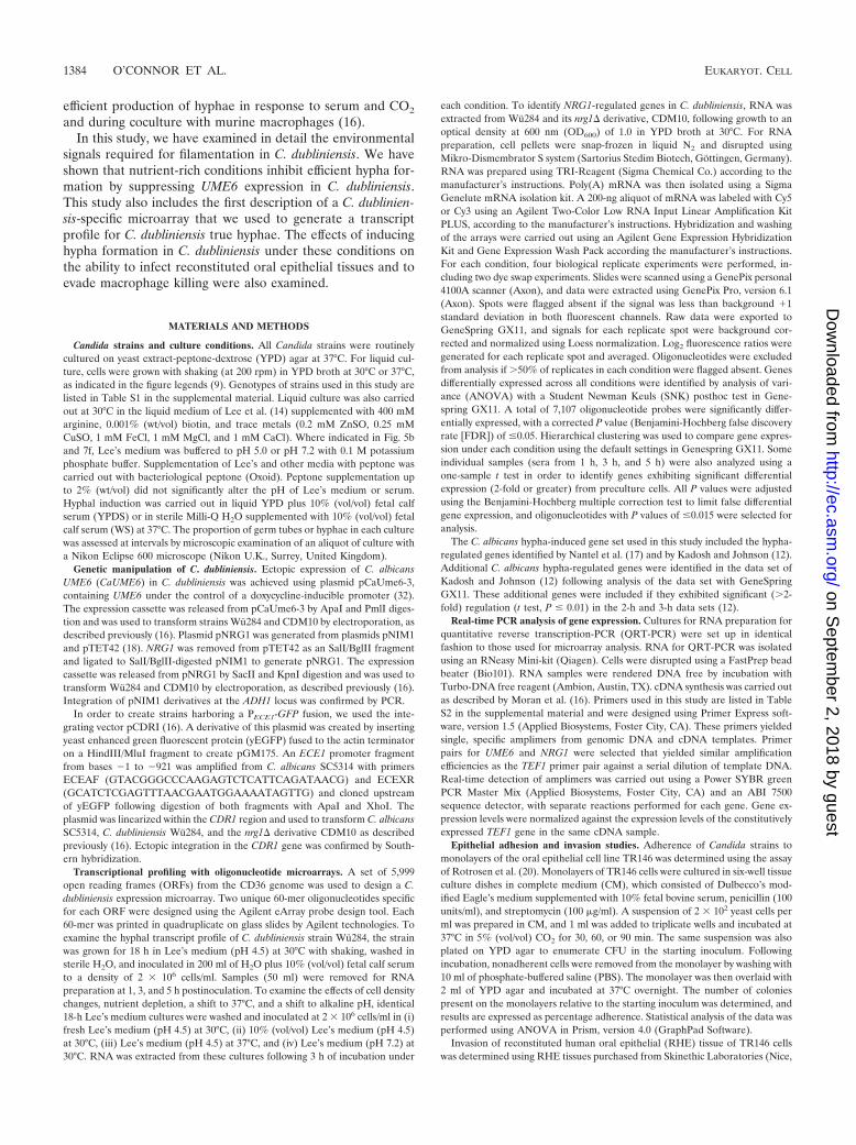
Regulation of UME6 and NRG1 transcription. Previousstudies have shown that in C. albicans, filamentation in YPDSmedium is associated with downregulation of NRG1 transcriptlevels and increased expression of UME6 (16). Examination ofNRG1 transcript levels in C. dubliniensis in YPDS mediumdemonstrated that NRG1 transcript levels increased following1 h of incubation in YPDS medium at 37°C (Fig. 3a). However,inoculation of cells precultured in Lee’s medium resulted in atransient drop in NRG1 transcript levels by approximately 50%following 1 h (Fig. 3a). Inoculation of C. dubliniensis in WSmedium yielded a 70% decrease in NRG1 transcript levels by3 h (Fig. 3a), similar to the decreases observed during filamen-tation of C. albicans in YPDS medium (data not shown). Anal-ysis of UME6 transcript levels in C. dubliniensis in YPDS me-dium revealed no significant change (Fig. 3b). However, whencells were precultured in Lee’s medium (pH 4.5), we observedan �30-fold increase in UME6 expression in YPDS medium(Fig. 3b). In addition, we observed �100-fold induction of
VOL. 9, 2010 NUTRIENTS REGULATE UME6 AND FILAMENTATION IN CANDIDA 1385
on Septem
ber 2, 2018 by guesthttp://ec.asm
.org/D
ownloaded from

UME6 in C. dubliniensis following inoculation in WS medium(Fig. 3b).
Addition of peptone to WS cultures showed that peptonecould decrease UME6 expression in C. dubliniensis in a con-centration-dependent manner, with 2% (wt/vol) peptone re-ducing UME6 expression by approximately 80%. Glucose (2%wt/vol) alone did not significantly decrease UME6 expression
although the combination of glucose and peptone had an ad-ditive effect on UME6 expression.
Overexpression of UME6 enhances filamentation in C. dub-liniensis. We further investigated the roles of NRG1 and UME6in hypha formation in the C. dubliniensis nrg1� mutantCDM10. Previously, we have shown that the nrg1� strain, un-like the wild type, forms hyphae in response to CO2 and fila-
FIG. 1. (a) Hypha formation in YPD medium plus 10% FCS (YPDS) by C. dubliniensis Wu284 (gray lines) and C. albicans SC5314 (black lines)following preculture in YPD medium at 30°C (solid lines) or 37°C (dashed lines). (b) Enhanced filamentation of C. dubliniensis Wu284 in waterplus 10% fetal calf serum (WS) following preculture in YPD medium at 30°C (solid black line), YPD medium at 37°C (dashed black line), or inLee’s medium at pH 4.5 and 30°C (gray line). Error bars correspond to standard deviations in at least three replicate experiments. A sigmoidalcurve was fitted to the data for visualization using Prism, version 4.0 (GraphPad Software, Inc.). (c) Examination of induction of GFP expressionfrom the hypha-specific ECE1 promoter in C. albicans and C. dubliniensis in YPDS and WS media. (d) Average percent hypha formation in C.dubliniensis (12 isolates) and C. albicans (6 isolates) at 37°C in YPD medium plus 10% serum (YPDS), in YPDS following preculture in Lee’smedium at pH 4.5, and in water plus 10% serum (WS). Error bars correspond to the standard error of the mean. (e) Filamentation of C.dubliniensis Wu284 in WS medium supplemented with 2% peptone, 2% glucose, or both peptone and glucose.
1386 O’CONNOR ET AL. EUKARYOT. CELL
on Septem
ber 2, 2018 by guesthttp://ec.asm
.org/D
ownloaded from

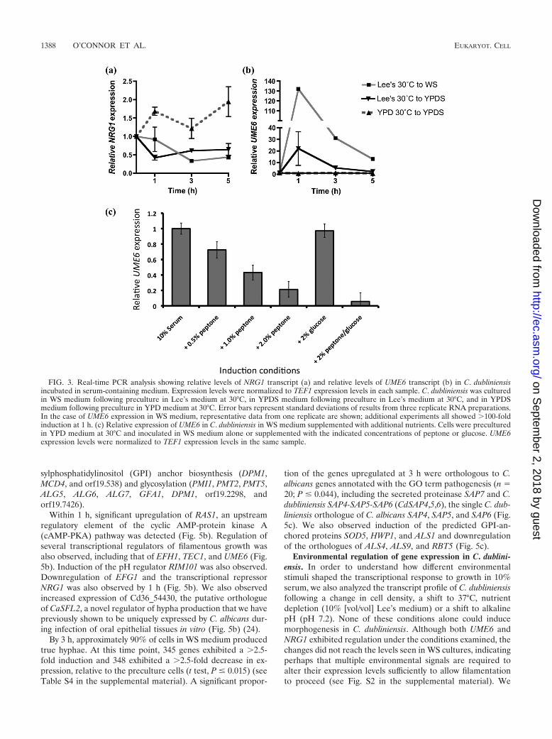
ments more rapidly in response to serum in water (16). In thisstudy, a derivative of CDM10 harboring a PECE1-GFP pro-moter fusion (M10EGFP) formed elongated filaments inYPDS medium; however, these filaments possessed the char-acteristic constrictions of pseudohyphae (Fig. 4a). StrainM10EGFP was weakly fluorescent in YPD and YPDS media(Fig. 4a), whereas in WS medium the same strain emittedstrong fluorescence and formed masses of true hyphae (Fig.4a). We tested whether overexpression of UME6 from a doxy-cycline-inducible promoter could enhance true hypha produc-tion by CDM10 in YPDS medium. Addition of 20 �g/ml doxy-cycline promoted conversion of pseudohyphae to true hyphaein this strain (Fig. 4b). Similarly, introduction of the sameconstruct in the parent isolate Wu284 could promote the for-mation of true hyphae in YPDS medium (Fig. 4b).
We also tested whether constitutive NRG1 expression fromthe doxycycline-inducible promoter could prevent filamenta-tion. Constitutive expression of NRG1 in Wu284 and CDM10could block pseudohypha formation in YPDS medium. How-ever, expression of NRG1 from this promoter was not sufficientto block true hypha formation in WS medium (data notshown).
Transcript profiling of C. dubliniensis in serum. This studyhas shown that under nutrient depleted conditions, C. dublini-ensis can form hyphae as effectively as C. albicans. In order todetermine whether C. dubliniensis hyphae can express the samerange of virulence-associated factors as C. albicans hyphae, we
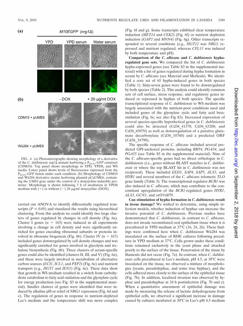
carried out whole-genome transcript profiling of C. dubliniensisduring growth in WS medium. Samples were analyzed at 1 h,3 h, and 5 h postinoculation in WS medium. Within 1 h, weobserved a 2.5-fold or greater change in transcription in 1,095genes relative to preculture cells (t test, P � 0.015) (see TableS3 in the supplemental material). This corresponds to 18% ofthe genome. Analysis of the upregulated genes (n 526) forsignificant shared Gene Ontology (GO) terms identified largegroups of genes associated with transport (102 genes), or-ganelle organization (73), the cell cycle (44), and translation(43) (Fig. 5a). Many of these genes were associated with pro-cesses known to be involved in hyphal development, such asthe assembly of actin cables (TPM2, ARF3, MEA1, ARP9, andYEL1), Spitzenkorper assembly (MLC1), and GTPases withroles in actin organization (RSR1, RAC1, RDI1, and RHO3)(see Fig. S1 in the supplemental material). These data alsohighlighted some processes not previously associated with hy-pha formation, such as downregulation of vacuolar metabo-lism, including vacuolar protein catabolysis (8 of 10 annotatedgenes) (see Fig. S1), suggesting a shutdown in autophagic pro-cesses. However, increased expression of genes with roles invacuolar biogenesis and inheritance was also observed (VAM3,YPT7, YPT72, and YKT6) (see Fig. S1). Reorganization ofmembrane lipid structure was indicated by a significant de-crease in sphingolipid metabolism (9 of 25 annotated genes)(see Fig. S1). Reorganization of the cell surface was indicatedby an increase in expression of genes associated with glyco-
FIG. 2. (a) Filamentation rate of strain Wu284 in YPDS medium at 37°C following preculture in YPD broth at 30°C or following preculturein Lee’s medium at pH 4.5 and 30°C. Error bars correspond to standard deviations in three replicate experiments. (b) Photomicrographs showingtypical morphologies of C. dubliniensis Wu284 and C. albicans SC5314 following 2 h of incubation in YPD medium plus 10% (vol/vol) FCSfollowing preculture in Lee’s medium. Cells were precultured for 24 h in modified Lee’s medium, buffered to pH 5.0 or 7.2 with 0.1 M potassiumphosphate buffer or supplemented with 1% (wt/vol) peptone.
VOL. 9, 2010 NUTRIENTS REGULATE UME6 AND FILAMENTATION IN CANDIDA 1387
on Septem
ber 2, 2018 by guesthttp://ec.asm
.org/D
ownloaded from
sylphosphatidylinositol (GPI) anchor biosynthesis (DPM1,MCD4, and orf19.538) and glycosylation (PMI1, PMT2, PMT5,ALG5, ALG6, ALG7, GFA1, DPM1, orf19.2298, andorf19.7426).
Within 1 h, significant upregulation of RAS1, an upstreamregulatory element of the cyclic AMP-protein kinase A(cAMP-PKA) pathway was detected (Fig. 5b). Regulation ofseveral transcriptional regulators of filamentous growth wasalso observed, including that of EFH1, TEC1, and UME6 (Fig.5b). Induction of the pH regulator RIM101 was also observed.Downregulation of EFG1 and the transcriptional repressorNRG1 was also observed by 1 h (Fig. 5b). We also observedincreased expression of Cd36_54430, the putative orthologueof CaSFL2, a novel regulator of hypha production that we havepreviously shown to be uniquely expressed by C. albicans dur-ing infection of oral epithelial tissues in vitro (Fig. 5b) (24).
By 3 h, approximately 90% of cells in WS medium producedtrue hyphae. At this time point, 345 genes exhibited a �2.5-fold induction and 348 exhibited a �2.5-fold decrease in ex-pression, relative to the preculture cells (t test, P � 0.015) (seeTable S4 in the supplemental material). A significant propor-
tion of the genes upregulated at 3 h were orthologous to C.albicans genes annotated with the GO term pathogenesis (n 20; P � 0.044), including the secreted proteinase SAP7 and C.dubliniensis SAP4-SAP5-SAP6 (CdSAP4,5,6), the single C. dub-liniensis orthologue of C. albicans SAP4, SAP5, and SAP6 (Fig.5c). We also observed induction of the predicted GPI-an-chored proteins SOD5, HWP1, and ALS1 and downregulationof the orthologues of ALS4, ALS9, and RBT5 (Fig. 5c).
Environmental regulation of gene expression in C. dublini-ensis. In order to understand how different environmentalstimuli shaped the transcriptional response to growth in 10%serum, we also analyzed the transcript profile of C. dubliniensisfollowing a change in cell density, a shift to 37°C, nutrientdepletion (10% [vol/vol] Lee’s medium) or a shift to alkalinepH (pH 7.2). None of these conditions alone could inducemorphogenesis in C. dubliniensis. Although both UME6 andNRG1 exhibited regulation under the conditions examined, thechanges did not reach the levels seen in WS cultures, indicatingperhaps that multiple environmental signals are required toalter their expression levels sufficiently to allow filamentationto proceed (see Fig. S2 in the supplemental material). We
FIG. 3. Real-time PCR analysis showing relative levels of NRG1 transcript (a) and relative levels of UME6 transcript (b) in C. dubliniensisincubated in serum-containing medium. Expression levels were normalized to TEF1 expression levels in each sample. C. dubliniensis was culturedin WS medium following preculture in Lee’s medium at 30°C, in YPDS medium following preculture in Lee’s medium at 30°C, and in YPDSmedium following preculture in YPD medium at 30°C. Error bars represent standard deviations of results from three replicate RNA preparations.In the case of UME6 expression in WS medium, representative data from one replicate are shown; additional experiments all showed �100-foldinduction at 1 h. (c) Relative expression of UME6 in C. dubliniensis in WS medium supplemented with additional nutrients. Cells were preculturedin YPD medium at 30°C and inoculated in WS medium alone or supplemented with the indicated concentrations of peptone or glucose. UME6expression levels were normalized to TEF1 expression levels in the same sample.
1388 O’CONNOR ET AL. EUKARYOT. CELL
on Septem
ber 2, 2018 by guesthttp://ec.asm
.org/D
ownloaded from
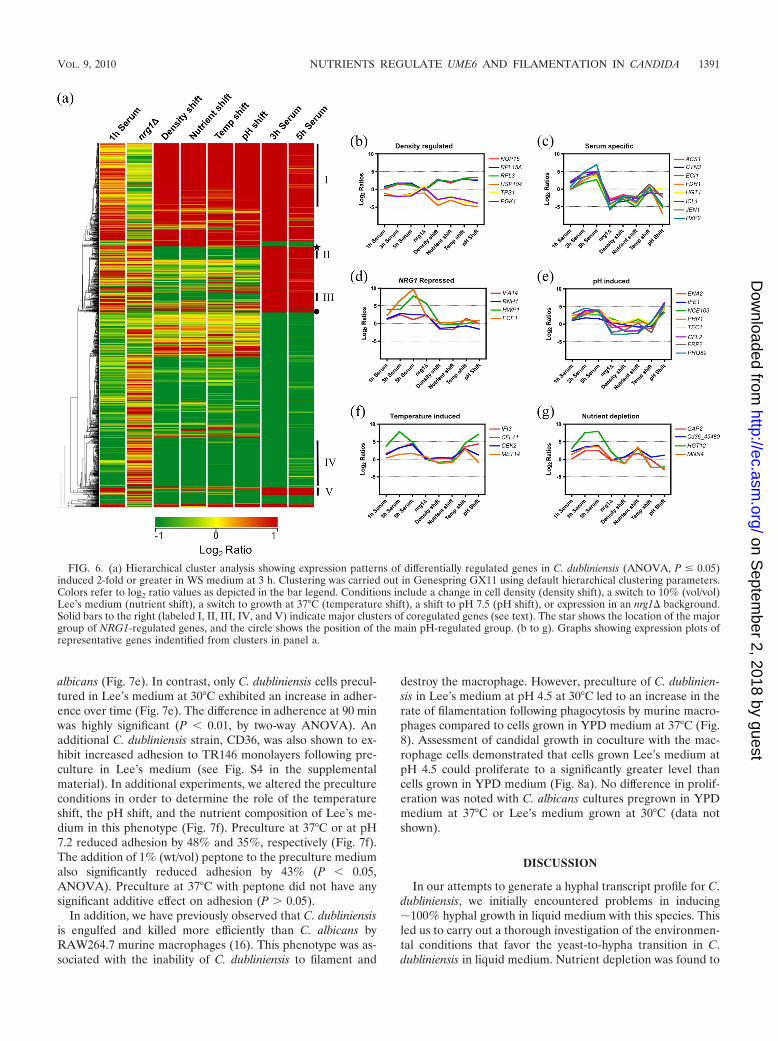
carried out ANOVA to identify differentially regulated tran-scripts (P � 0.05) and visualized the results using hierarchicalclustering. From this analysis we could identify two large clus-ters of genes regulated by changes in cell density (Fig. 6a).Cluster I genes (n 163) were induced in all experimentsinvolving a change in cell density and were significantly en-riched for genes encoding ribosomal subunits or proteins in-volved in ribosome biogenesis (Fig. 6b). Cluster IV (n 167)included genes downregulated by cell density changes and wassignificantly enriched for genes involved in glycolysis and tre-halose biosynthesis (Fig. 6b). Three clusters of serum-specificgenes could also be identified (clusters II, III, and V) (Fig. 6a),and these were largely involved in metabolism of alternativecarbon sources (ECI1, ICL1, and PXP2) (Fig. 6c) and nutrienttransport (e.g., HGT1 and JEN1) (Fig. 6c). These data showthat growth in WS medium resulted in a switch from carbohy-drate catabolism to fatty acid oxidation and the glyoxylate cyclefor energy production (see Fig. S3 in the supplemental mate-rial). Smaller clusters of genes were identified that were in-duced by alkaline pH or relief of NRG1 repression (Fig. 6d ande). The regulation of genes in response to nutrient-depletedLee’s medium and the temperature shift was more complex
(Fig. 6f and g). Some transcripts exhibited clear temperatureinduction (MET14 and CEK2) (Fig. 6f) or nutrient depletioninduction (GAP2 and MNN4) (Fig. 6g). Other transcripts re-sponded to several conditions (e.g., HGT12 was NRG1 re-pressed and nutrient regulated, whereas CFL11 was inducedby both temperature and pH).
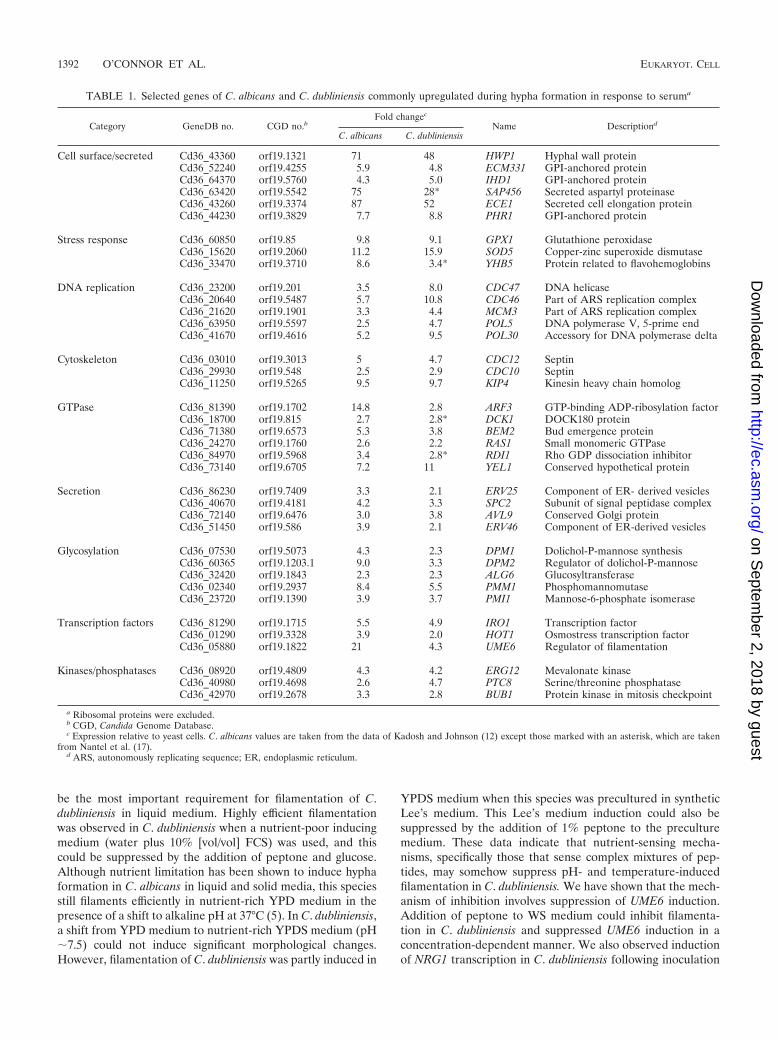
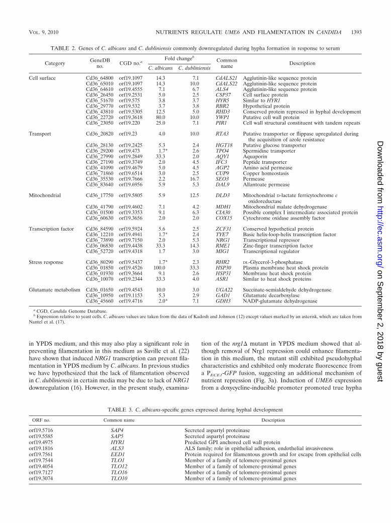
Comparison of the C. albicans and C. dubliniensis hypha-regulated gene sets. We compared the list of C. dubliniensishypha-expressed genes (see Table S3 in the supplemental ma-terial) with a list of genes regulated during hypha formation inserum by C. albicans (see Material and Methods). We identi-fied a core set of 65 hypha-induced genes in both species(Table 1). Sixty-seven genes were found to be downregulatedby both species (Table 2). This analysis could identify commonsets of cell surface, stress response, and regulatory genes in-duced or repressed in hyphae of both species. The specifictranscriptional response of C. dubliniensis to WS medium waslargely associated with the nutrient-poor conditions used andincluded genes of the glyoxylate cycle and fatty acid beta-oxidation (Fig. 6c; see also Fig S3). Increased expression ofseveral species-specific hypothetical genes in C. dubliniensiscould also be detected (Cd36_41370, Cd36_63200, andCd36_65070) as well as downregulation of a putative gluta-mate decarboxylase (Cd36_10760) and a predicted ORF(Cd36_34790).
The specific response of C. albicans included several pre-dicted GPI-anchored proteins, including RBT4, PGA54, andPGA55 (see Table S5 in the supplemental material). Nine ofthe C. albicans-specific genes had no direct orthologue in C.dubliniensis (i.e., genes without BLAST matches in C. dublini-ensis or where the top BLAST hit in C. dubliniensis was notreciprocal). These included EED1, SAP4, SAP5, ALS3, andHYR1 and several members of the C. albicans telomeric TLOgene family (Table 3). The transcriptional regulator BCR1 wasalso induced in C. albicans, which may contribute to the con-comitant upregulation of the BCR1-regulated genes HYR1,ALS3, GCN1, and orf19.6079.
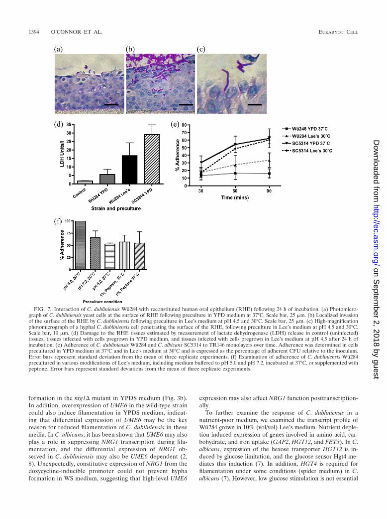
Can stimulation of hypha formation in C. dubliniensis resultin tissue damage? We wished to determine, using simple in-fection models, whether induction of hyphae can increase theinvasive potential of C. dubliniensis. Previous studies havedemonstrated that C. dubliniensis, in contrast to C. albicans,does not invade reconstituted oral epithelium tissue when it isprecultured in YPD medium at 37°C (16, 24, 26). These find-ings were confirmed here when C. dubliniensis Wu284 wasinoculated on the surface of RHE cultures following precul-ture in YPD medium at 37°C. Cells grown under these condi-tions remained exclusively in the yeast phase and attachedpoorly to the surface of the tissue. Penetration of the tissue byfilaments did not occur (Fig. 7a). In contrast, when C. dublini-ensis cells precultured in Lee’s medium, pH 4.5, at 30°C wereinoculated on the tissue, we observed a mixture of morpholo-gies (yeasts, pseudohyphae, and some true hyphae), and thecells adhered more closely to the surface of the epithelial tissue(Fig. 7b). In addition, localized invasion was observed by hy-phae and pseudohyphae at 24 h postinfection (Fig. 7b and c).When a quantitative assessment of epithelial damage wasmade by measuring the release of lactate dehydrogenase fromepithelial cells, we observed a significant increase in damagecaused by cultures incubated at 30°C in Lee’s pH 4.5 medium
FIG. 4. (a) Photomicrographs showing morphology of a derivativeof the C. dubliniensis nrg1� mutant harboring a PECE1-GFP construct(CDM10). Top panel shows morphology in YPD, YPDS, and WSmedia. Lower panel shows levels of fluorescence expressed from thePECE1-GFP fusion under each condition. (b) Morphology of CDM10and Wu284 derivative strains harboring plasmid pCaUME6, contain-ing the UME6 gene under the control of a doxycycline-inducible pro-moter. Morphology is shown following 3 h of incubation in YPDSmedium with (�) or without (�) 20 �g/ml doxycycline (DOX).
VOL. 9, 2010 NUTRIENTS REGULATE UME6 AND FILAMENTATION IN CANDIDA 1389
on Septem
ber 2, 2018 by guesthttp://ec.asm
.org/D
ownloaded from
compared to YPD-grown cultures (Fig. 7d). Increased celldamage was also recorded in RHE infections with C. dublini-ensis strain CD36 following preculture in Lee’s medium (7.0 0.6 LDH U/liter) relative to YPD medium (5.2 0.2 LDHU/liter).
These data suggest that the difference in tissue damage andinvasion elicited by C. dubliniensis cells grown in YPD medium
and Lee’s medium may be due to differences in adherence. Wecarried out a more detailed investigation of the adhesion of C.dubliniensis to TR146 cell monolayers over 90 min. Within 30min of inoculation, 10 to 20% of yeast cells had adhered to themonolayer (Fig. 7e). Adherence of C. albicans SC5314 in-creased by 60 min, and this was independent of precultureconditions and corresponded with germ tube formation by C.
FIG. 5. (a) Graphical representation of the changes in expression in selected Gene Ontology (GO) groups during filamentation in C.dubliniensis. The total number of genes up- or downregulated 2.5-fold in each group is shown at each time point. (b) Microarray expression ofselected regulators of filamentous growth during hypha formation in C. dubliniensis Wu284 in WS medium. Columns for each gene show expressionlevels relative to preculture cells at (left to right) 1, 3, and 5 h postinoculation. (c) Microarray expression of selected virulence-associated genesduring hypha formation in C. dubliniensis Wu284 in WS medium. Columns for each gene show expression levels relative to preculture cells at (leftto right) 1, 3, and 5 h postinoculation. Error bars in panels b and c represent standard deviations from the mean generated in Genespring GX11from two distinct oligonucleotide probes per gene in four biological replicate experiments.
1390 O’CONNOR ET AL. EUKARYOT. CELL
on Septem
ber 2, 2018 by guesthttp://ec.asm
.org/D
ownloaded from
albicans (Fig. 7e). In contrast, only C. dubliniensis cells precul-tured in Lee’s medium at 30°C exhibited an increase in adher-ence over time (Fig. 7e). The difference in adherence at 90 minwas highly significant (P � 0.01, by two-way ANOVA). Anadditional C. dubliniensis strain, CD36, was also shown to ex-hibit increased adhesion to TR146 monolayers following pre-culture in Lee’s medium (see Fig. S4 in the supplementalmaterial). In additional experiments, we altered the precultureconditions in order to determine the role of the temperatureshift, the pH shift, and the nutrient composition of Lee’s me-dium in this phenotype (Fig. 7f). Preculture at 37°C or at pH7.2 reduced adhesion by 48% and 35%, respectively (Fig. 7f).The addition of 1% (wt/vol) peptone to the preculture mediumalso significantly reduced adhesion by 43% (P � 0.05,ANOVA). Preculture at 37°C with peptone did not have anysignificant additive effect on adhesion (P � 0.05).
In addition, we have previously observed that C. dubliniensisis engulfed and killed more efficiently than C. albicans byRAW264.7 murine macrophages (16). This phenotype was as-sociated with the inability of C. dubliniensis to filament and
destroy the macrophage. However, preculture of C. dublinien-sis in Lee’s medium at pH 4.5 at 30°C led to an increase in therate of filamentation following phagocytosis by murine macro-phages compared to cells grown in YPD medium at 37°C (Fig.8). Assessment of candidal growth in coculture with the mac-rophage cells demonstrated that cells grown Lee’s medium atpH 4.5 could proliferate to a significantly greater level thancells grown in YPD medium (Fig. 8a). No difference in prolif-eration was noted with C. albicans cultures pregrown in YPDmedium at 37°C or Lee’s medium grown at 30°C (data notshown).
DISCUSSION
In our attempts to generate a hyphal transcript profile for C.dubliniensis, we initially encountered problems in inducing�100% hyphal growth in liquid medium with this species. Thisled us to carry out a thorough investigation of the environmen-tal conditions that favor the yeast-to-hypha transition in C.dubliniensis in liquid medium. Nutrient depletion was found to
FIG. 6. (a) Hierarchical cluster analysis showing expression patterns of differentially regulated genes in C. dubliniensis (ANOVA, P � 0.05)induced 2-fold or greater in WS medium at 3 h. Clustering was carried out in Genespring GX11 using default hierarchical clustering parameters.Colors refer to log2 ratio values as depicted in the bar legend. Conditions include a change in cell density (density shift), a switch to 10% (vol/vol)Lee’s medium (nutrient shift), a switch to growth at 37°C (temperature shift), a shift to pH 7.5 (pH shift), or expression in an nrg1� background.Solid bars to the right (labeled I, II, III, IV, and V) indicate major clusters of coregulated genes (see text). The star shows the location of the majorgroup of NRG1-regulated genes, and the circle shows the position of the main pH-regulated group. (b to g). Graphs showing expression plots ofrepresentative genes indentified from clusters in panel a.
VOL. 9, 2010 NUTRIENTS REGULATE UME6 AND FILAMENTATION IN CANDIDA 1391
on Septem
ber 2, 2018 by guesthttp://ec.asm
.org/D
ownloaded from
be the most important requirement for filamentation of C.dubliniensis in liquid medium. Highly efficient filamentationwas observed in C. dubliniensis when a nutrient-poor inducingmedium (water plus 10% [vol/vol] FCS) was used, and thiscould be suppressed by the addition of peptone and glucose.Although nutrient limitation has been shown to induce hyphaformation in C. albicans in liquid and solid media, this speciesstill filaments efficiently in nutrient-rich YPD medium in thepresence of a shift to alkaline pH at 37°C (5). In C. dubliniensis,a shift from YPD medium to nutrient-rich YPDS medium (pH�7.5) could not induce significant morphological changes.However, filamentation of C. dubliniensis was partly induced in
YPDS medium when this species was precultured in syntheticLee’s medium. This Lee’s medium induction could also besuppressed by the addition of 1% peptone to the preculturemedium. These data indicate that nutrient-sensing mecha-nisms, specifically those that sense complex mixtures of pep-tides, may somehow suppress pH- and temperature-inducedfilamentation in C. dubliniensis. We have shown that the mech-anism of inhibition involves suppression of UME6 induction.Addition of peptone to WS medium could inhibit filamenta-tion in C. dubliniensis and suppressed UME6 induction in aconcentration-dependent manner. We also observed inductionof NRG1 transcription in C. dubliniensis following inoculation
TABLE 1. Selected genes of C. albicans and C. dubliniensis commonly upregulated during hypha formation in response to seruma
Category GeneDB no. CGD no.bFold changec
Name Descriptiond
C. albicans C. dubliniensis
Cell surface/secreted Cd36_43360 orf19.1321 71 48 HWP1 Hyphal wall proteinCd36_52240 orf19.4255 5.9 4.8 ECM331 GPI-anchored proteinCd36_64370 orf19.5760 4.3 5.0 IHD1 GPI-anchored proteinCd36_63420 orf19.5542 75 28* SAP456 Secreted aspartyl proteinaseCd36_43260 orf19.3374 87 52 ECE1 Secreted cell elongation proteinCd36_44230 orf19.3829 7.7 8.8 PHR1 GPI-anchored protein
Stress response Cd36_60850 orf19.85 9.8 9.1 GPX1 Glutathione peroxidaseCd36_15620 orf19.2060 11.2 15.9 SOD5 Copper-zinc superoxide dismutaseCd36_33470 orf19.3710 8.6 3.4* YHB5 Protein related to flavohemoglobins
DNA replication Cd36_23200 orf19.201 3.5 8.0 CDC47 DNA helicaseCd36_20640 orf19.5487 5.7 10.8 CDC46 Part of ARS replication complexCd36_21620 orf19.1901 3.3 4.4 MCM3 Part of ARS replication complexCd36_63950 orf19.5597 2.5 4.7 POL5 DNA polymerase V, 5-prime endCd36_41670 orf19.4616 5.2 9.5 POL30 Accessory for DNA polymerase delta
Cytoskeleton Cd36_03010 orf19.3013 5 4.7 CDC12 SeptinCd36_29930 orf19.548 2.5 2.9 CDC10 SeptinCd36_11250 orf19.5265 9.5 9.7 KIP4 Kinesin heavy chain homolog
GTPase Cd36_81390 orf19.1702 14.8 2.8 ARF3 GTP-binding ADP-ribosylation factorCd36_18700 orf19.815 2.7 2.8* DCK1 DOCK180 proteinCd36_71380 orf19.6573 5.3 3.8 BEM2 Bud emergence proteinCd36_24270 orf19.1760 2.6 2.2 RAS1 Small monomeric GTPaseCd36_84970 orf19.5968 3.4 2.8* RDI1 Rho GDP dissociation inhibitorCd36_73140 orf19.6705 7.2 11 YEL1 Conserved hypothetical protein
Secretion Cd36_86230 orf19.7409 3.3 2.1 ERV25 Component of ER- derived vesiclesCd36_40670 orf19.4181 4.2 3.3 SPC2 Subunit of signal peptidase complexCd36_72140 orf19.6476 3.0 3.8 AVL9 Conserved Golgi proteinCd36_51450 orf19.586 3.9 2.1 ERV46 Component of ER-derived vesicles
Glycosylation Cd36_07530 orf19.5073 4.3 2.3 DPM1 Dolichol-P-mannose synthesisCd36_60365 orf19.1203.1 9.0 3.3 DPM2 Regulator of dolichol-P-mannoseCd36_32420 orf19.1843 2.3 2.3 ALG6 GlucosyltransferaseCd36_02340 orf19.2937 8.4 5.5 PMM1 PhosphomannomutaseCd36_23720 orf19.1390 3.9 3.7 PMI1 Mannose-6-phosphate isomerase
Transcription factors Cd36_81290 orf19.1715 5.5 4.9 IRO1 Transcription factorCd36_01290 orf19.3328 3.9 2.0 HOT1 Osmostress transcription factorCd36_05880 orf19.1822 21 4.3 UME6 Regulator of filamentation
Kinases/phosphatases Cd36_08920 orf19.4809 4.3 4.2 ERG12 Mevalonate kinaseCd36_40980 orf19.4698 2.6 4.7 PTC8 Serine/threonine phosphataseCd36_42970 orf19.2678 3.3 2.8 BUB1 Protein kinase in mitosis checkpoint
a Ribosomal proteins were excluded.b CGD, Candida Genome Database.c Expression relative to yeast cells. C. albicans values are taken from the data of Kadosh and Johnson (12) except those marked with an asterisk, which are taken
from Nantel et al. (17).d ARS, autonomously replicating sequence; ER, endoplasmic reticulum.
1392 O’CONNOR ET AL. EUKARYOT. CELL
on Septem
ber 2, 2018 by guesthttp://ec.asm
.org/D
ownloaded from
in YPDS medium, and this may also play a significant role inpreventing filamentation in this medium as Saville et al. (22)have shown that induced NRG1 transcription can prevent fila-mentation in YPDS medium by C. albicans. In previous studieswe have hypothesized that the lack of filamentation observedin C. dubliniensis in certain media may be due to lack of NRG1downregulation (16). However, in the present study, examina-
tion of the nrg1� mutant in YPDS medium showed that al-though removal of Nrg1 repression could enhance filamenta-tion in this medium, the mutant still exhibited pseudohyphalcharacteristics and exhibited only moderate fluorescence froma PECE1-GFP fusion, suggesting an additional mechanism ofnutrient repression (Fig. 3a). Induction of UME6 expressionfrom a doxycycline-inducible promoter promoted true hypha
TABLE 2. Genes of C. albicans and C. dubliniensis commonly downregulated during hypha formation in response to serum
Category GeneDBno. CGD no.a
Fold changebCommon
name DescriptionC. albicans C. dubliniensis
Cell surface Cd36_64800 orf19.1097 14.3 7.1 CdALS21 Agglutinin-like sequence proteinCd36_65010 orf19.1097 14.3 10.0 CdALS22 Agglutinin-like sequence proteinCd36_64610 orf19.4555 7.1 6.7 ALS4 Agglutinin-like sequence proteinCd36_26450 orf19.2531 5.0 2.5 CSP37 Cell surface proteinCd36_51670 orf19.575 3.8 3.7 HYR5 Similar to HYR1Cd36_29770 orf19.532 3.7 3.8 RBR2 Hypothetical proteinCd36_43810 orf19.5305 12.5 5.0 RHD3 Conserved protein repressed in hyphal developmentCd36_22720 orf19.3618 80.0 10.0 YWP1 Putative cell wall proteinCd36_23050 orf19.220 25.0 7.1 PIR1 Cell wall structural constituent with tandem repeats
Transport Cd36_20820 orf19.23 4.0 10.0 RTA3 Putative transporter or flippase upregulated duringthe acquisition of azole resistance
Cd36_28130 orf19.2425 5.3 2.4 HGT18 Putative glucose transporterCd36_29200 orf19.473 1.7* 2.6 TPO4 Spermidine transporterCd36_27990 orf19.2849 33.3 2.0 AQY1 AquaporinCd36_27190 orf19.3749 2.0 4.5 IFC3 Peptide transporterCd36_41090 orf19.4679 5.0 4.5 AGP2 Amino acid permeaseCd36_71860 orf19.6514 3.0 2.5 CUP9 Copper homeostasisCd36_35530 orf19.7666 2.2 16.7 SEO3 PermeaseCd36_83640 orf19.6956 5.9 5.3 DAL9 Allantoate permease
Mitochondrial Cd36_17750 orf19.5805 5.9 12.5 DLD3 Mitochondrial D-lactate ferricytochrome coxidoreductase
Cd36_41790 orf19.4602 7.1 4.2 MDH1 Mitochondrial malate dehydrogenaseCd36_01500 orf19.3353 9.1 6.3 CIA30 Possible complex I intermediate associated proteinCd36_60630 orf19.3656 2.0 2.0 COX15 Cytochrome oxidase assembly factor
Transcription factor Cd36_84590 orf19.5924 5.6 2.5 ZCF31 Conserved hypothetical proteinCd36_12210 orf19.4941 1.7* 2.4 TYE7 Basic helix-loop-helix transcription factorCd36_73890 orf19.7150 2.0 5.3 NRG1 Transcriptional repressorCd36_06830 orf19.4438 33.3 14.3 RME1 Zinc-finger transcription factorCd36_52720 orf19.4318 1.7 3.0 MIG1 Transcriptional regulator
Stress response Cd36_80290 orf19.5437 1.7* 2.3 RHR2 DL-Glycerol-3-phosphataseCd36_01850 orf19.4526 100.0 33.3 HSP30 Plasma membrane heat shock proteinCd36_01930 orf19.3664 9.1 2.6 HSP31 Membrane heat shock proteinCd36_10070 orf19.2344 33.3 4.0 ASR1 Similar to heat shock proteins
Glutamate metabolism Cd36_01650 orf19.4543 10.0 3.0 UGA22 Succinate-semialdehyde dehydrogenaseCd36_10950 orf19.1153 5.3 2.9 GAD1 Glutamate decarboxylaseCd36_45660 orf19.4716 2.0* 7.1 GDH3 NADP-glutamate dehydrogenase
a CGD, Candida Genome Database.b Expression relative to yeast cells. C. albicans values are taken from the data of Kadosh and Johnson (12) except values marked by an asterisk, which are taken from
Nantel et al. (17).
TABLE 3. C. albicans-specific genes expressed during hyphal development
ORF no. Common name Description
orf19.5716 SAP4 Secreted aspartyl proteinaseorf19.5585 SAP5 Secreted aspartyl proteinaseorf19.4975 HYR1 Predicted GPI anchored cell wall proteinorf19.1816 ALS3 ALS family; role in epithelial adhesion, endothelial invasivenessorf19.7561 EED1 Protein required for filamentous growth and for escape from epithelial cellsorf19.7544 TLO1 Member of a family of telomere-proximal genesorf19.4054 TLO12 Member of a family of telomere-proximal genesorf19.7127 TLO16 Member of a family of telomere-proximal genesorf19.3074 TLO10 Member of a family of telomere-proximal genes
VOL. 9, 2010 NUTRIENTS REGULATE UME6 AND FILAMENTATION IN CANDIDA 1393
on Septem
ber 2, 2018 by guesthttp://ec.asm
.org/D
ownloaded from
formation in the nrg1� mutant in YPDS medium (Fig. 3b).In addition, overexpression of UME6 in the wild-type straincould also induce filamentation in YPDS medium, indicat-ing that differential expression of UME6 may be the keyreason for reduced filamentation of C. dubliniensis in thesemedia. In C. albicans, it has been shown that UME6 may alsoplay a role in suppressing NRG1 transcription during fila-mentation, and the differential expression of NRG1 ob-served in C. dubliniensis may also be UME6 dependent (2,8). Unexpectedly, constitutive expression of NRG1 from thedoxycycline-inducible promoter could not prevent hyphaformation in WS medium, suggesting that high-level UME6
expression may also affect NRG1 function posttranscription-ally.
To further examine the response of C. dubliniensis in anutrient-poor medium, we examined the transcript profile ofWu284 grown in 10% (vol/vol) Lee’s medium. Nutrient deple-tion induced expression of genes involved in amino acid, car-bohydrate, and iron uptake (GAP2, HGT12, and FET3). In C.albicans, expression of the hexose transporter HGT12 is in-duced by glucose limitation, and the glucose sensor Hgt4 me-diates this induction (7). In addition, HGT4 is required forfilamentation under some conditions (spider medium) in C.albicans (7). However, low glucose stimulation is not essential
FIG. 7. Interaction of C. dubliniensis Wu284 with reconstituted human oral epithelium (RHE) following 24 h of incubation. (a) Photomicro-graph of C. dubliniensis yeast cells at the surface of RHE following preculture in YPD medium at 37°C. Scale bar, 25 �m. (b) Localized invasionof the surface of the RHE by C. dubliniensis following preculture in Lee’s medium at pH 4.5 and 30°C. Scale bar, 25 �m. (c) High-magnificationphotomicrograph of a hyphal C. dubliniensis cell penetrating the surface of the RHE, following preculture in Lee’s medium at pH 4.5 and 30°C.Scale bar, 10 �m. (d) Damage to the RHE tissues estimated by measurement of lactate dehydrogenase (LDH) release in control (uninfected)tissues, tissues infected with cells pregrown in YPD medium, and tissues infected with cells pregrown in Lee’s medium at pH 4.5 after 24 h ofincubation. (e) Adherence of C. dubliniensis Wu284 and C. albicans SC5314 to TR146 monolayers over time. Adherence was determined in cellsprecultured in YPD medium at 37°C and in Lee’s medium at 30°C and is expressed as the percentage of adherent CFU relative to the inoculum.Error bars represent standard deviation from the mean of three replicate experiments. (f) Examination of adherence of C. dubliniensis Wu284precultured in various modifications of Lee’s medium, including medium buffered to pH 5.0 and pH 7.2, incubated at 37°C, or supplemented withpeptone. Error bars represent standard deviations from the mean of three replicate experiments.
1394 O’CONNOR ET AL. EUKARYOT. CELL
on Septem
ber 2, 2018 by guesthttp://ec.asm
.org/D
ownloaded from

for filamentation in C. albicans as HGT4 mutants form fila-ments normally in glucose-rich YPDS medium (7). It is alsounlikely that HGT4 signaling is required for filamentation of C.dubliniensis in WS medium as addition of 2% glucose to WSmedium did not significantly inhibit filamentation in C. dub-liniensis. Repression of filamentation was more apparent whenC. dubliniensis was exposed to a complex mixture of peptides orwhole protein (BSA), indicating that nitrogen-sensing mecha-nisms may be important. Growth in WS medium was associ-ated with induction of several general amino acid permeases(GAP2, GAP4, CAN2, and CAN3), suggesting that this me-dium is amino acid limiting. However, addition of amino acidsto WS medium did not affect filamentation in C. dubliniensis,indicating that cell surface amino acid sensors such as SSY1may not be involved in repression of filamentous growth (6).However, high concentrations of ammonium sulfate (100 mM)could reduce filamentation. Ammonium sulfate has beenshown to inhibit filamentation of C. albicans on solid mediumvia the Mep2 sensor; however, as shown here and previously byBiswas and Morschhauser (4), this effect is not seen in C.albicans in the presence of serum. However, ammonium sul-fate-mediated repression of filamentation may be serum inde-pendent in C. dubliniensis. Interestingly, the most repressiveeffects on filamentation were observed when WS medium wassupplemented with a mixture of both peptone and glucose,suggesting that either a combination of sensing mechanisms orpossibly a more general nutrient-sensing mechanism is in-volved. It has recently been shown in C. albicans that an or-thologue of the general nutrient sensor Tor1 can modulateNRG1 expression in spider medium (3). In addition, it has alsobeen shown that a C. albicans MDS3 mutant can form hyphaeonly in the presence of the Tor1 inhibitor rapamycin (30). Weare currently assessing whether C. dubliniensis Tor1 could playnovel role in nutrient sensing and filamentation.
The transcript profiling data presented here also indicateimportant roles for pH, temperature, and cell density changesin activating the transcription of hypha-specific genes in C.dubliniensis. The transcript profiling data presented here showa key role for the pH response in activating the filamentousgrowth regulators SFL2, UME6, TEC1, and RIM101 (5, 8).UME6 was also found to be NRG1 repressed, whereas TEC1also exhibited induction due to cell density changes. Temper-ature changes also induced EFH1 and CPH1. These data showthat induction of filamentation under the conditions examinedin C. dubliniensis involves multiple environmental signals.
The microarray data presented here highlighted some novelprocesses regulated during filamentation in C. dubliniensis andidentified a strong core transcriptional response shared with C.albicans. The data show rapid induction of genes involved inregulating polarized growth, including genes involved in actinpolymerization, vesicle transport, and septin formation. Thedata also provide evidence for processes not previously de-scribed during the morphological switch. This includes evi-dence for changes in lipid composition, with a shutdown intranscription of genes involved in sphingolipid synthesis and anincrease in fatty acid biosynthesis gene expression. Changes invacuole function are also indicated with an increase in expres-sion of genes involved in vacuolar biogenesis and inheritanceand decreases in expression of vacuolar proteases, suggestingthat the vacuole plays a structural rather than metabolic role inhyphae. Comparison of this transcript profile with previouslypublished studies of gene expression in C. albicans allowed usto identify a core transcriptional response to filamentation inboth species which consists of 132 genes regulated 2-fold orgreater (12, 17). This strongly conserved core response sup-ports the hypothesis that a specific program of transcriptionalchanges may be essential for filamentation to proceed in bothspecies, in addition to posttranscriptional events. Induction of
FIG. 8. Survival of C. dubliniensis Wu284 following coculture with murine RAW264.7 macrophages. (a) Proliferation of viable Candida cellswas assayed using an XTT dye reduction assay following 18 h of coculture at several multiplicities of infection (MOI; Candida CFU tomacrophages). Wu284 cells precultured in Lee’s medium at pH 4.5 exhibited significantly greater proliferation at MOIs of 1:8 and 1:32.Morphology of C. dubliniensis Wu284 cells grown in YPD medium (b) and in Lee’s medium at pH 4.5 (c) following 5 h of incubation with murineRAW264.7 macrophages. Error bars represent standard deviations from the means of three replicate experiments.
VOL. 9, 2010 NUTRIENTS REGULATE UME6 AND FILAMENTATION IN CANDIDA 1395
on Septem
ber 2, 2018 by guesthttp://ec.asm
.org/D
ownloaded from
several secreted and cell wall-associated proteins was specificto C. albicans under the conditions examined, and these in-cluded RBT4, PGA54, and PGA55. Several species-specificgenes were also induced in C. albicans, including HYR1, ALS3,and EED1. C. albicans expresses three SAP genes, SAP4,SAP5, and SAP6 during filamentation, whereas C. dubliniensispossesses only one orthologue of these genes, termedCdSAP456, which is also induced during hyphal growth (11,21). However, secreted aspartyl proteinase (SAP) activity inC. dubliniensis may be supplemented by SAP7 expression,which exhibited an 8-fold increase in expression. C. albicansalso expresses the putative invasin ALS3 (19). However, wedid not identify any compensatory expression of ALS genesin C. dubliniensis although orthologues of C. albicans ALS2,ALS4, and ALS9 all exhibited decreased expression duringhyphal growth.
Overall, transcript profiling revealed that C. dubliniensis hy-phae express a number of genes associated with virulence,suggesting that induction of filamentation in C. dubliniensiscould promote tissue invasion. Recently, Spiering et al. con-cluded that the reduced virulence of C. dubliniensis in the RHEmodel was a result of a failure to initiate filamentation and thespecific transcriptional program associated with this (24). Inthe present study we have shown that induction of UME6expression in C. dubliniensis by preculturing in Lee’s mediumat 30°C could enhance filamentation in the RHE model. Thisresulted in greater attachment of C. dubliniensis cells to thetissue surface and localized invasion of the epithelium. Wehave never previously identified RHE invasion in a wild-typestrain of C. dubliniensis (16, 24, 26). Examination of adhesionof C. dubliniensis to TR146 monolayers demonstrated that thisadherent phenotype could be partly inhibited by the additionof peptone to the preculture medium, as well as by removingthe pH or temperature shift. However, the level of damage tothe RHE tissues was still significantly lower than that routinelyobserved when tissues are infected with C. albicans. There maybe several reasons for this. First, the transition following Lee’spreculture is largely short-lived, and by 24 h most cells havereverted to budding growth. Second, although C. dubliniensiscan be induced to form hyphae, the absence of several C.albicans-specific hypha-associated genes (ALS3, SAP5, HYR1,and EED1) may also attenuate the virulence of this species (19,21, 31). Studies are under way to determine if these geneticdifferences are crucial to the greater pathogenicity of C. albi-cans.
Finally, this study suggests that the ability of C. albicans toform filaments at alkaline pH, irrespective of nutrient avail-ability, may enable it to colonize and infect a wider range ofniches relative to C. dubliniensis. C. dubliniensis may have lostor perhaps failed to acquire this morphological flexibility sincethe divergence of the two species. The genome sequence of C.dubliniensis suggests that, due to gene loss and pseudogeniza-tion, C. dubliniensis may be undergoing niche specialization. Itmay be possible that reduced filamentation is part of this spe-cialization process and that it may even be of benefit to C.dubliniensis in certain niches, particularly where tissue damage,inflammation, and attraction of the of host’s defenses is unfa-vorable.
ACKNOWLEDGMENTS
Plasmids pNIM1 and pTET42 were obtained from the JoachimMorschhauser Institut fur Molekulare Infektionsbiologie, UniversitatWurzburg, and plasmid pCaUme6-3 was generated by Arnold Bito(Department of Cell Biology, University of Salzburg). We thank JanWalker at St. James’s Hospital Dublin for fixation and staining of theRHE tissue sections. We also thank the anonymous reviewers whosehelpful comments greatly enhanced the manuscript.
This work was supported by the Irish Health Research Board (re-search grant RP/2004/235) and by Science Foundation Ireland (Pro-gramme Investigator grant number 04/IN3/B463).
REFERENCES
1. Asmundsdottir, L. R., H. ErlendsdUttir, B. A. Agnarsson, and M. Gottfreds-son. 2009. The importance of strain variation in virulence of Candida dub-liniensis and Candida albicans: results of a blinded histopathological study ofinvasive candidiasis. Clin. Microbiol. Infect. 15:576–585.
2. Banerjee, M., D. S. Thompson, A. Lazzell, P. L. Carlisle, C. Pierce, C.Monteagudo, J. L. Lopez-Ribot, and D. Kadosh. 2008. UME6, a novel fila-ment-specific regulator of Candida albicans hyphal extension and virulence.Mol. Biol. Cell 19:1354–1365.
3. Bastidas, R. J., J. Heitman, and M. E. Cardenas. 2009. The protein kinaseTor1 regulates adhesin gene expression in Candida albicans. PLoS Pathog.5:e1000294.
4. Biswas, K., and J. Morschhauser. 2005. The Mep2p ammonium permeasecontrols nitrogen starvation-induced filamentous growth in Candida albicans.Mol. Microbiol. 56:649–669.
5. Biswas, S., P. Van Dijck, and A. Datta. 2007. Environmental sensing andsignal transduction pathways regulating morphopathogenic determinants ofCandida albicans. Microbiol. Mol. Biol. Rev. 71:348–376.
6. Brega, E., R. Zufferey, and C. B. Mamoun. 2004. Candida albicans Csy1p isa nutrient sensor important for activation of amino acid uptake and hyphalmorphogenesis. Eukaryot. Cell 3:135–143.
7. Brown, V., J. A. Sexton, and M. Johnston. 2006. A glucose sensor in Candidaalbicans. Eukaryot. Cell 5:1726–1737.
8. Carlisle, P. L., M. Banerjee, A. Lazzell, C. Monteagudo, J. L. Lopez-Ribot,and D. Kadosh. 2009. Expression levels of a filament-specific transcriptionalregulator are sufficient to determine Candida albicans morphology and vir-ulence. Proc. Natl. Acad. Sci. U. S. A. 106:599–604.
9. Gallagher, P. J., D. E. Bennett, M. C. Henman, R. J. Russell, S. R. Flint,D. B. Shanley, and D. C. Coleman. 1992. Reduced azole susceptibility ofCandida albicans from HIV-positive patients and a derivative exhibitingcolony morphology variation. J. Gen. Microbiol. 138:1901–1911.
10. Jabra-Rizk, M. A., J. K. Johnson, G. Forrest, K. Mankes, T. F. Meiller, andR. A. Venezia. 2005. Prevalence of Candida dubliniensis fungemia at a largeteaching hospital. Clin. Infect. Dis. 41:1064–1067.
11. Jackson, A. P., J. A. Gamble, T. Yeomans, G. P. Moran, D. Saunders, D.Harris, M. Aslett, J. F. Barrell, G. Butler, F. Citiulo, D. C. Coleman, P. W.de Groot, T. J. Goodwin, M. A. Quail, J. McQuillan, C. A. Munro, A. Pain,R. T. Poulter, M. A. Rajandream, H. Renauld, M. J. Spiering, A. Tivey, N. A.Gow, B. Barrell, D. J. Sullivan, and M. Berriman. 2009. Comparativegenomics of the fungal pathogens Candida dubliniensis and C. albicans.Genome Res. 10:2231–2244.
12. Kadosh, D., and A. D. Johnson. 2005. Induction of the Candida albicansfilamentous growth program by relief of transcriptional repression: a ge-nome-wide analysis. Mol. Biol. Cell 16:2903–2912.
13. Kibbler, C. C., S. Seaton, R. A. Barnes, W. R. Gransden, R. E. Holliman,E. M. Johnson, J. D. Perry, D. J. Sullivan, and J. A. Wilson. 2003. Manage-ment and outcome of blood stream infections due to Candida species inEngland and Wales. J. Hosp. Infect. 54:18–24.
14. Lee, K. L., H. R. Buckley, and C. C. Campbell. 1975. An amino acid liquidsynthetic medium for the development of mycelial and yeast forms of Can-dida albicans. Sabouraudia 13:148–153.
15. Meiller, T. F., M. A. Jabra-Rizk, A. Baqui, J. I. Kelley, V. I. Meeks, W. G.Merz, and W. A. Falkler. 1999. Oral Candida dubliniensis as a clinicallyimportant species in HIV-seropositive patients in the United States. OralSurg. Oral Med. Oral Pathol. Oral Radiol. Endod. 88:573–580.
16. Moran, G. P., D. M. MacCallum, M. J. Spiering, D. C. Coleman, and D. J.Sullivan. 2007. Differential regulation of the transcriptional repressor NRG1accounts for altered host cell interactions in Candida albicans and Candidadubliniensis. Mol. Microbiol. 66:915–929.
17. Nantel, A., D. Dignard, C. Bachewich, D. Harcus, A. Marcil, A. P. Bouin,C. W. Sensen, H. Hogues, M. Van het Hoog, P. Gordon, T. Rigby, F. Benoit,D. C. Tessier, D. Y. Thomas, and M. Whiteway. 2002. Transcription profilingof Candida albicans cells undergoing the yeast-to-hyphal transition. Mol.Biol. Cell 13:3452–3465.
18. Park, Y. N., and J. Morschhauser. 2005. Tetracycline-inducible gene expres-sion and gene deletion in Candida albicans. Eukaryot. Cell 4:1328–1342.
19. Phan, Q. T., C. L. Myers, Y. Fu, D. C. Sheppard, M. R. Yeaman, W. H.Welch, A. S. Ibrahim, J. E. Edwards, Jr., and S. G. Filler. 2007. Als3 is a
1396 O’CONNOR ET AL. EUKARYOT. CELL
on Septem
ber 2, 2018 by guesthttp://ec.asm
.org/D
ownloaded from

Candida albicans invasin that binds to cadherins and induces endocytosis byhost cells. PLoS Biol. 5:e64.
20. Rotrosen, D., J. E. Edwards, Jr., T. R. Gibson, J. C. Moore, A. H. Cohen, andI. Green. 1985. Adherence of Candida to cultured vascular endothelial cells:mechanisms of attachment and endothelial cell penetration. J. Infect. Dis.152:1264–1274.
21. Sanglard, D., B. Hube, M. Monod, F. C. Odds, and N. A. R. Gow. 1997. Atriple deletion of the secreted aspartyl proteinase genes SAP4, SAP5, andSAP6 of Candida albicans causes attenuated virulence. Infect. Immun. 65:3539–3546.
22. Saville, S. P., A. L. Lazzell, C. Monteagudo, and J. L. Lopez-Ribot. 2003.Engineered control of cell morphology in vivo reveals distinct roles for yeastand filamentous forms of Candida albicans during infection. Eukaryot. Cell2:1053–1060.
23. Schaller, M., H. C. Korting, W. Schafer, J. Bastert, W. Chen, and B. Hube.1999. Secreted aspartic proteinase (Sap) activity contributes to tissue dam-age in a model of human oral candidosis. Mol. Microbiol. 34:169–180.
24. Spiering, M. J., G. P. Moran, M. Chauvel, D. M. Maccallum, J. Higgins, K.Hokamp, T. Yeomans, C. D’Enfert, D. C. Coleman, and D. J. Sullivan. 2010.Comparative transcript profiling of Candida albicans and Candida dublini-ensis identifies SFL2, a C. albicans gene required for virulence in a recon-stituted epithelial infection model. Eukaryot. Cell 9:251–265.
25. Staib, P., and J. Morschhauser. 2005. Differential expression of the NRG1repressor controls species-specific regulation of chlamydospore developmentin Candida albicans and Candida dubliniensis. Mol. Microbiol. 55:637–652.
26. Stokes, C., G. P. Moran, M. J. Spiering, G. T. Cole, D. C. Coleman, and D. J.
Sullivan. 2007. Lower filamentation rates of Candida dubliniensis contributeto its lower virulence in comparison with Candida albicans. Fungal Genet.Biol. 44:920–931.
27. Sullivan, D. J., G. P. Moran, E. Pinjon, A. Al-Mosaid, C. Stokes, C. Vaughan,and D. C. Coleman. 2004. Comparison of the epidemiology, drug resistancemechanisms, and virulence of Candida dubliniensis and Candida albicans.FEMS Yeast Res. 4:369–376.
28. Sullivan, D. J., T. J. Westerneng, K. A. Haynes, D. E. Bennett, and D. C.Coleman. 1995. Candida dubliniensis sp. nov.: phenotypic and molecularcharacterization of a novel species associated with oral candidosis in HIV-infected individuals. Microbiology 141:1507–1521.
29. Vilela, M. M., K. Kamei, A. Sano, R. Tanaka, J. Uno, I. Takahashi, J. Ito, K.Yarita, and M. M. 2002. Pathogenicity and virulence of Candida dubliniensis:comparison with C. albicans. Med. Mycol. 40:249–257.
30. Zacchi, L. F., J. Gomez-Raja, and D. A. Davis. 10 May 2010. Mds3 regulatesmorphogenesis in Candida albicans through the TOR pathway. Mol. Cell.Biol. doi.10:1128/MCB.01549-09.
31. Zakikhany, K., J. R. Naglik, A. Schmidt-Westhausen, H. Holland, M.Schaller, and B. Hube. 2007. In vivo transcript profiling of Candida albicansidentifies a gene essential for interepithelial dissemination. Cell. Microbiol.9:2938–2954.
32. Zeidler, U., T. Lettner, C. Lassnig, M. Muller, R. Lajko, H. Hintner, M.Breitenbach, and A. Bito. 2009. UME6 is a crucial downstream target ofother transcriptional regulators of true hyphal development in Candida al-bicans. FEMS Yeast Res. 9:126–142.
VOL. 9, 2010 NUTRIENTS REGULATE UME6 AND FILAMENTATION IN CANDIDA 1397
on Septem
ber 2, 2018 by guesthttp://ec.asm
.org/D
ownloaded from