Embed Size (px)
DESCRIPTION
with notes on symbioses --By: Kott, P. -- Micronesica Vol. 18 No. 1 June, 1982
Citation preview

Didemnid-Algal Symbioses: Host Species in the Western Pacific with Notes on the Symbiosis
PATRICIA KOTT
Queensland Museum, Gregory Terrace Brisbane, Australia 4006
Abstract- Collections from Palau, Philippine and Caroline Islands, Guam and the Great Barrier Reef contain 15 (including 2 that are new) of the 20 didemnid species known to contain algal symbionts. With the exception of the Fijian species (Diplosoma multipapillata) and two species known only from the Atlantic and eastern Pacific (Trididemnum solidum and T. della vallei), all the known species have a wide geographic range in the western Pacific and some extend into the western Indian Ocean. Populations of several of the species suggest a degree of isolation, but not speciation. The known geographic ranges of all but the most conspicuous species are affected by patterns of collecting. A key to identification of species is based on features of both the zooids and the colony and its contained spicules. Species descriptions complement those of Kott (1980).
Introduction
Algae-bearing didemnid ascidians of the Indo-West Pacific region have been reported on by Kott (1980, 1982). Records from Palau and the Philippine Islands referred to in those reports were based on published accounts (Van Name 1918; Tokioka 1942, 1950, 1955, 1967). This account is based principally on more recent collections from these two locations in the western Pacific and from the Great Barrier Reef at Lizard Island and to the north. Small collections from Guam and the Caroline Islands (Truk and Pulusuk) are also included.
Fifteen of the 20 species known to have prokaryotic algal symbionts are present in these collections. Two additional species (Echinoclinum triangulum and Trididemnum strigosum) have been recorded previously from a range of locations in the western Pacific including the Philippines (Kott 1980, 1982). Further collecting in the Indian Ocean will very likely demonstrate an Indo-West Pacific range for all these species with the exception of Diplosoma multipapillata, which is thought to be endemic, having been recorded only from a specialised habitat in Fiji, and Trididemnum solidum and T. della vallei known only from the Atlantic and the eastern Pacific, respectively.
Intraspecific morphological variation in these species is generally not conspicuous, despite the vast distances between their populations. Diversity in the appearance of the colonies most often results from variations in the crowding of spicules in the surface test that shade the contained algal symbionts (e.g., Lissoclinum Micronesica 18(1): 95-127. 1982 (June).

96 Micronesica
voeltzkowi in Kott 1980 and Lissoclinum bistratum in Kott 1982). Olson (1980) has shown a similar increase in the density of spicules in the surface test and a withdrawal of plant cells away from the surface as a response to increased light in the Atlantic species, Trididemnum solidum.
These responses, by which the host ascidian orders the arrangement of its spicules in order to satisfy the needs of the contained algae, clearly demonstrate the symbiotic nature of the ascidian-algal relationship. They are the result of environmental rather than genetic factors. Variability in the distribution and intensity of cartenoid pigments is also apparently the result of environmental factors (cf. L. bistratum at Lizard Island in Kott 1980).
There are also variations in colony size and shape that result from age, substrate, and lobulation (subdivision). Small rounded colonies of many of the species are usually maintained by lobulation, as in Diplosoma virens (Thinh et al. I 981 ). In some populations of certain species, lobulation does not occur (e.g., Lissoclinum bistratum specimens from Mooloolaba in Kott, 1980, and L. voeltzkowi specimens from the Palau Islands, see below) and the colonies form sheets rather than small rounded cushions. This may be genetic and reflect population adaptations rather than environmental factors. The relatively large larvae with a multiplicity of adhesive organs of Diplosoma virens at Lizard Island are also a likely population adaptation reflecting only a degree of isolation, rather than complete reproductive isolation and speciation.
Thus, morphological variations resulting from genetic isolation are rare and the present collections tend to support the proposition that a remarkable pattern of larval recruitment links the islands and reefs of the tropical Indo-West Pacific to maintain flow between populations of these widely ranging species (Kott 1980).
A description of the morphology of each species is available in Kott (1980) which supplements this account of the more recently collected material set out here. The material is deposited in the Queensland Museum (QM) with duplicate specimens in the collection of the Marine Laboratory, University of Guam.
Identification of Ascidian Specimens
The spicules and cloacal systems comprise the most easily observed and reliable taxonomic characters. However, observation of branchial sac and gonads will also be necessary to obtain definitive identification of most genera and species. It should be stressed 'that the distinction between the genera Didemnum and Trididemnum is based exclusively on the number of rows of branchial stigmata. These are often difficult to determine in these small zooids and microscopic examination of the cleared, relaxed pharynx is essential to distinguish between Didemnum viride and Trididemnum nubilum which otherwise have very similar colonies, zooids and spicules.
Colour photographs of many of the species in situ have been reproduced with this report. These demonstrate clearly that many of the species have a close superficial resemblance. Further, many grow in similar and often the same habitat, sometimes in

Vol. 18. June 1982 97
close assoc1at10n. Species identification based on the colour, shape, or other macroscopic features of specimens is unreliable. Dissection of colonies and microscopic examination of spicules and zooids must be undertaken in order to establish species identity.
Dissection of a didemnid colony is most readily achieved by cutting a thin vertical wedge or slice from a common cloacal aperture to the outer border of the colony. The organisation of the common cloaca, the relationships of the zooids to it and to the surface of the colony, and the distribution of the spicules may then be readily observed and zooids may be removed without mutilation.
KEY TO ALGAE-BEARING DIDEMNIDS OF THE INDO-WEST PACIFIC
I. Calcareous spicules absent from the test (Diplosoma) . . . . . . . . . . . . . . . . 2. 1 '. Calcareous spicules present in the test . . . . . . . . . . . . . . . . . . . . . . . . . . . . . . 4.
2. Colonies sheeting, not lobulated; retractor muscle from posterior end of thorax . . . . . . . . . . . . . . . . . . . . . . . . . . . . . . . . Diplosoma simi/is
2'. Colonies oval or rounded, lobulated; retractor muscle from part way along esophagus . . . . . . . . . . . . . . . . . . . . . . . . . . . . . . . . . . . . . . 3.
3. Stigmata 6/row; retractor muscle moderate length; cloacal canals at thoracic level complicated; green . . . . . . . . . . . . . . . . . . . . . . . . . . . . Diplosoma virens
3'. Stigmata 5/row; retractor muscle very short; cloacal canals at thoracic level not complicated; blue. . . . . . . . . . . . . . . . . . . . . . . . . . . . Diplosoma multipapillata
4. Stigmata in 3 rows (Trididemnum) . . . . . . . . . . . . . . . . . . . . . . . . 5.
4' . Stigmata in 4 rows . . . . . . . . . . . . . . . . . . . . . . . . . . . . . . . . . . . . . . 11.
5. Atrial siphon present . . . . . . . . . . . . . . . . . . . . . . . . . . . . . . . . . . . . . . . . . . . . 6.
5'. Atrial siphon absent . . . . . . . . . . . . . . . . . . . . . . . . . . . . . . . . . . . . . . . . . . . . 7.
6. Spicules up to 0.04 mm diameter; colonies with single systems .. .. . . . . . . . . . . . . . . . . . . . . . . . . . . . . . . . . . . . . . . Trididemnum clinides
6'. Spicules up to 0.08 mm diameter; colonies with more than one system . . . . . . . . . . . . . . . . . . . . . . Trididemnum paraclinides n. sp.
7. Endostylar pigment cap present; plant cells in common cloacal cavity . . 8.
7' . Endostylar pigment cap absent; plant cells embedded . . . . . . . . . . . . . . . . 9.
8. Vas deferens with 9 1/ 2 coils; colonies sheeting ................. . . . . . . . . . . . . . . . . . . . . . . . . . . . . . . . . . . . . . Trididemnum paracyclops
8'. Vas deferens with 51/ 2 coils; colonies oval . . .. Trididemnum cyclops
9. Spicules spherical (with fiat-ended rays) . .. .. . .... .. Trididemnum miniatum
9'. Spicules stellate (with pointed rays) . . . . . . . . . . . . . . . . . . . . . . . . . . . . . . . . 10.

98 Micronesica
10. Spicules up to 0.05 mm, sparse in middle layers of test ......... . . . . . . . . . . . . . . . . . . . . . . . . . . . . . . . . . . . . . Trididemnum nubilum
I 0'. Spicules up to 0.08, dense throughout. ..... Trididemnum strigosum
1!. Vas deferens spirals around the male follicle (Didemnum) . . . . . . . . . . . . 12.
11 '. Vas deferens runs straight . . . . . . . . . . . . . . . . . . . . . . . . . . . . . . . . . . . . . . . . 14.
12. Colonies flask- or dome-shaped; mucus secretion copious; plant cells confined to common cloaca . . . . . . . . . . . . . . Didemnum molle
12' . Colonies not flask- or dome-shaped; mucus secretion not apparent; plant cells embedded in test . . . . . . . . . . . . . . . . . . . . . . . . . . . . 13.
13. Colonies sheeting; spicules stellate with 7 to 11 conical rays in optical section . . . . . . . . . . . . . . . . . . . . . . . . . . . . . . . . . . . . . . . . . . . . . . . . Didemnum viride
I 3'. Colonies lobulated; spicules stellate with 5 to 7 almost cylindrical rays in optical section ...... . .. . . ........ ... . .. ........ . . . Didemnum etiolum n. sp.
14. Spicules flattened with rays of unequal length ................. . . . . . . . . . . . . . . . . . . . . . . . . . . . . . . . . . . . . Echinoclinum triangulum
14'. Spicules stellate or spherical with rays of equal length (Lissoclinum) . . . . . . . . . . . . . . . . . . . . . . . . . . . . . . . . . . . . . . . . . . . . . . . . . . . . 15.
15. Spicules form capsule around zooid; colonies slimy, delicate ............. . . . . . . . . . . . . . . . . . . . . . . . . . . . . . . . . . . . . . . . . . . . . . Lissoclinum punctatum
15'. Spicules do not form capsule around zooid; colonies neither slimy nor delicate . . . . . . . . . . . . . . . . . . . . . . . . . . . . . . . . . . . . . . . . . . . . . . . . . . . . . . . . . . . . 16.
16. Spicules absent from basal test and from thickened surface ridges . . . . . . . . . . . . . . . . . . . . . . . . . . . . . . . . . . . . . . Lissoclinum pattella
16'. Spicules present in basal test; no thickened spicule-free surface ridges; variable density of spicules in surface test . . . . . . . . . . . . . . . . 17.
17. Spicules spherical. . . . . . . . . . . . . . . . . . . . . . . . . . . . . . . . Lissoclinum bistratum
17'. Spicules stellate
Systematic Account
Did.emnum molle (Herdman) Figs. 1, 2a-f, 19a
Diplosomoides molle Herdman, 1886: 310.
Didemnum molle: Kott, 1980: 2 and synonymy.
Lissoclinum voeltzkowi
NEW RECORDS: Fiji (Mumbualau, on seagrass, 2m). Philippines (Calatagan,


Vol. 18. June 1982 101
when pigment is dense above the spicules. Pigment cells can fill the interstices between the bladder cells of the superficial layer of test and are forced into irregular fusiform or stellate shapes that form a three-dimensional trabecular pattern. Thus there is a wide range in density and distribution of pigment and spicules in the surface test that causes the colour variations in this species. The single cloacal aperture is usually in the center of the upper surface, although the larger colonies are usually assymmetrical. Colonies have been observed dividing across the cloacal aperture (M. Cowan, pers. comm.)
The colonies are prolific and well developed larvae are found free in the common cloaca and are readily released from the colony before fixation. In the metamorphosing larva the posterior half of the larval trunk, with the plant cells that surround it, rolls inwards around the base of the tail and is depressed into the larval trunk. The posterior end of the posterior haemocoelic cavity becomes conical and protrudes from the center of this cavity. At this stage the larva is very much reduced in length anteroposteriorly. Anteriorly the trunk is rounded, with adhesive organs and ectodermal ampullae protruding from it. Posteriorly the tail protrudes from the center of a concavity that is filled with plant cells. The larval test over the posterior end of the larva appears to be differentiated into hair-like strands that entangle the plant cells in the same way as the hairs of the diplosomid rastrum (Kott 1980). These hairs hold the plant cells in place in the posterior concavity. At the anterior end the adhesive organs protrude to make the first contact with the substrate. It is the ectodermal ampullae which subsequently reinforce the fixation. Withdrawal of the tail into the posterior haemocoelic cavity is sometimes before, but usually immediately after, the adhesive organs make their first contact with the substrate. After the tail is withdrawn, the larval test extends upwards over the posterior concavity and its contained plant cells, leaving an opening to the exterior that becomes the common cloacal aperture in the center of the upper surface of the colony.
Dark brown colonies with pigment in the outer layer of test and few spicules produce juveniles in which the basal surface (formerly the anterior end of the larva) becomes enveloped in brown pigment cells interrupted only at 4 points where the ectodermal ampullae formerly protruded. This localized saucer of pigment cells has not been observed in juveniles from grey to white adult colonies.
Didenmum viride (Herdman) Figs. 3- 5
Leptoclinum viride Herdman, 1906: 340
Didemnum viride: Kott, 1980: 4 and synonymy.
NEW RECORDS: Philippines (Apo Reef, 10m, QM GH488; Calatagan, 16m, QM GH483). Palau (QM GH498, 501).

I
): !It'"
I'.:
102 Micronesica
RANGE: The new records extend the known range of this species from the Indian Ocean (Malagasy and Sri Lanka) into the western Pacific.
DESCRIPTION: The colonies form investing sheets, 2 to 12cm in maximum dimension and from 0.3 to 1.0 em thick. The zooids are restricted to the upper part of the colony and variations in thickness are a result of variations in
Fig. 3. Spicules of Didemnum viride (QM GH483, 488).
Fig. 4. Thorax of D. viride (same specimen).
Fig. 5. The whole zooid of D. viride (same specimen), showing the position of the Vshaped mass of pigment cells in the inner curve of the gut loop and a clump of plant cells outside the gut loop.

Vol. 18. June 1982 103
thickness of the basal test. Zooids are arranged in double rows around circular areas of test and appear as dark points in the living colony and as light points in the narcotized, preserved material. Living colonies are white to grey, depending on the density of the plant cells embedded in the surface layer of test. Colonies often have a "marbled" appearance resulting from variations in density of spicule distribution. The test is tough and is usually packed with spicules. Spicules are absent only from the thin superficial layer of test.
The spicules are 0.03 to 0.04 mm and occasionally to 0.06 mm in diameter with 9 to 11 conical, pointed or rounded rays that break up fairly readily. The plant cells are usually found embedded in the superficial test. Beneath this they are found in reduced density mixed with spicules in the upper layer of test, and lining the cloacal canals. They are invariably absent from the basal test. In some colonies they are also absent from the superficial layer of test. Plant cells are 0.008- 0.010 mm in diameter.
The common cloacal canals are thoracic and rather restricted. In the largest colonies there are also some narrow posterior abdominal canals and these are also lined with plant cells. There are relatively large oval masses of dark green pigment cells in the basal test.
The zooids are very small, less than I mm in length. The branchial siphon is long and muscular and sometimes as long as the pharynx. The branchial lobes around the aperture are relatively shallow. These small branchial lobes are not outlined by spicules in the surface test. There are 4 rows of 6 rather rounded stigmata. The short retractor muscle is free from halfway down the long esophageal neck. There is a lateral organ opposite the third row of stigmata on each side of the endostyle. The atrial aperture is an incised opening across the middorsal surface of the thorax. The gut loop is flexed and there is a V -shaped mass of dark greenish pigment cells on the ventral side of the inner curve of the gut loop. There appears also to be a clump of plant cells close against this part of the abdomen. There are 7t coils of the vas deferens around the single male follicle.
A single embryo was found in the test of the specimen from Apo Reef (QM GH488). The trunk is 0.52 mm long and is almost spherical. The tail is wound only half way around the trunk. The embryo is surrounded with a coating of plant cells that are absent only from a circular area over the sense organs and an elongate area down the center of the anterior end of the trunk where the 3 adhesive organs are exposed.
REMARKS: The colony is strongly reminiscent of Trididemnum nubilum and the species are distinguished only by the zooids. The coating of plant cells around the embryo is also similar to that found in Trididemnum miniatum, T. cyclops and T. clinides.
. I'
104 Micronesica
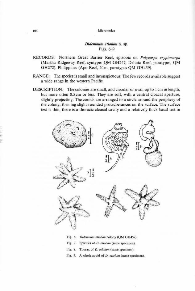
Didemnum etiolum n. sp. Figs. 6-9
RECORDS: Northern Great Barrier Reef, epizooic on Polycarpa cryptocarpa (Martha Ridgeway Reef, syntypes QM GH247; Deltaic Reef, paratypes, QM GH272). Philippines (Apo Reef, 20m, paratypes QM GH459).
RANGE: The species is small and inconspicuous. The few records available suggest a wide range in the western Pacific.
DESCRIPTION: The colonies are small, and circular or oval, up to 1 em in length, but more often 0.5 em or less. They are soft, with a central cloacal aperture, slightly projecting. The zooids are arranged in a circle around the periphery of the colony, forming slight rounded protruberances on the surface. The surface test is thin, there is a thoracic cloacal cavity and a relatively thick basal test in
Fig. 6. Didemnum etiolum colony (QM GH459).
Fig. 7. Spicules of D. etiolum (same specimen).
Fig. 8. Thorax of D. etiolum (same specimen).
Fig. 9. A whole zooid of D. etiolum (same specimen).

Vol. 18. June 1982 105
which the abdomina are embedded. Spicules are found throughout, although they are most crowded in the basal test. Plant cells (0.008-0.01 rom in diameter) are found embedded in the test. These become less dense basally. In some of the colonies there are dark pigment cells (0.001-0.008 rom) in the body wall, especially over the abdomen and in the surface layer of test. The distinctive spicules are stellate, generally about 0.045 mm in diameter with 5 to 7 long almost cylindrical rays in optical section.
The zoo ids are only about 0.5 rom long. The branchial siphon is of moderate length and the branchial lobes are distinct but not deeply separated. The atrial aperture is a deep transverse incision across the dorsal border of the thorax. There are 4 rows of 5 rectangular stigmata. A very fine retractor muscle separates from the posterior end of the thorax. There is a large oval lateral organ opposite the last row of stigmata on each side of the endostyle. The gut loop is flexed upwards. No mature testis follicles were observed.
REMARKS: These small soft colonies superficially resemble those of Trididemnum clinides. The species is distinguished from Didemnum viride by its smaller plant symbionts, by its small and soft colonies, by its spicules, and by its very fine retractor muscle from the I?Osterior end of the thorax. Although mature male follicles are not present, the species appears to be closely related to Didemnum viride and is accordingly assigned to the genus Didemnum.
Trididemnum nubilum Kott Figs. 10-12
Trididemnum nubilum Kott, 1980: 9 and synonymy; 1981: 188.
NEW RECORDS: Philippines (Apo Reef, 20m, QM GH539). Great Barrier Reef (Lizard Island, on Acropora rubble on sandy reef fiat, QM GH150).
RANGE: The species is known from the Philippines, Fiji and the Great Barrier Reef (Lizard Island).
DESCRIPTION: The colonies are firm, fiat (3 rom thick) and irregular in outline. Although previous records are of small colonies, up to 2 em in maximum dimension, the colony from Apo Reef forms a larger investing sheet. The superficial layer of test has embedded plant cells and some scattered spicules. The superficial test varies in thickness to some extent and the plant cells are not found always in the surface, causing a marbled appearance. The thoracic cloacal cavity is very shallow and restricted. Spicules are present throughout the test in moderate density. They are 0.03- 0.04 mm in diameter with 9- 11 short conical rays. The plant cells (0.006-0.012 mm in diameter) are present in the upper layer of test, and lining the cloacal canals.
The zoo ids are about 0. 7 mm long. The branchial siphon is of moderate


Vol. 18. June 1982 107
The gut loop is only slightly flexed upwards. There are 8t coils of the vas deferens around a rather flattened male follicle. There is a V -shaped mass of dark greenish black cells in the loop of the gut (as in D. vir ide) and oval masses of these cells in the basal test.
REMARKS: Colonies of this species bear a close resemblance to colonies of Didemnum viride. The spicules of the latter species have a greater size range, up to 0.06 mm and slightly longer rays, but they are of similar form to those of the present species and do not represent a reliable means of distinguishing the species. The double rows of zooids are more conspicuous from the surface of the colonies in D. viride, but the plant cells are the same size and there are similar abdominal masses of dark pigment cells in the basal test. Despite the presence of 4 rows of stigmata, the branchial sac in D. viride is smaller and relatively narrower than is the case in T. nubilum. The species are also distinguished by the long branchial siphon of D. viride, and by the retractor muscle, which in the present species is free from the proximal end of the esophageal neck, rather than half way down it as in D. viride.
T . clinides and related species are distinguished from T. nubilum by their atrial siphons and more delicate retractor muscles free from the posterior end of the thorax.
Trididemnum paraclinides n. sp. Figs. 13- 15
Trididemnum clinides: Kott, 1981: 186 (part, specimens from Mumbualau and Dravuni).
NEW RECORDS: Palau (Nemelis, 1m, holotype, QM GH575).
RANGE: The species bas been recorded from Fiji (Mumbualau, paratype QM GH 144; Dravuni, paratype QM GH 91) in high energy parts of the reef. The Palau specimen was taken in shallow lagoonal waters protected from wave action.
DESCRIPTION: Colonies are firm, fiat surfaced, irregular sheets up to 2 em in greatest extent. Living colonies are green to blackish slate. In preservative they are clear green. The colour appears to persist for reasonably long periods in formalin and is contained in the plant cells that are embedded in the smooth superficial layer of test. There are no spicules in this layer which covers the surface and borders of the colony. Spicules are present in a single layer beneath the plant cells (immediately above the thoracic cloacal cavity) and throughout the basal layer of test. Spicules are large (0.03- 0.08 mm) and stellate, with 9 to 11 short conical rays. Plant cells are 0.008-0.015 mm in diameter. They are present in greatest density in the superficial test and lining the quite extensive thoracic

108 Micronesica
Fig. 13. Spicules of Trididemnum paraclinides (QM GH575).
Fig. 14. Thorax ofT. paraclinides (same specimen).
Fig. 15. A whole zooid ofT. paraclinides (same specimen).
common cloacal cavity. They are not present in the basal test beneath the common cloacal cavity. Minute dark pigment cells are scattered through the test and are present in oval accumulations in the basal test.
Zooids are only about 1 mm in total length. The thorax is slightly larger than the abdomen. The thorax is muscular and the branchial sac is never exposed. The atrial aperture is on a muscular siphon directed laterally or posteriorly. The branchial aperture has 6 large, deeply divided lobes. There is a very short, delicate retractor muscle from the posterior end of the thorax. There are 3 rows of 6 oval stigmata. The gut loop is relatively short and is only slightly flexed upwards. There are 8t coils of the vas deferens around the almost hemispherical male follicle.
The larval trunk is 0.7mm long. There are 3 adhesive organs and the usual 4 pairs of ectodermal ampullae. The larval test has embedded plant cells, leaving only the sense organs and adhesive organs at the anterior end of the larva exposed.
REMARKS: The zooids of this species are almost indistinguishable from those of T. clinides. Their only distinguishing characters are the larger number of vas deferens coils (8t) in the present species and the embedded plant cells and the spicules of T. paraclinides which are both larger than those of T. clinides. The colonies provide principal distinction between the two species. Trididemnum paraclinides has more extensive, firm, flat topped colonies, whereas those of T.

Vol. 18. June 1982 109
clinides are small, soft, rounded cushions with single systems of zooids. Further the conspicuous superficial layer of test, without spicules, conferring a clear greenish jelly-like appearance to the outside of the colony, is quite distinctive. The species is distinguished from T. nubilum by its more extensive cloacal cavities, larger spicules, absence of spicules from the superficial layer of test, and atrial siphon.
It is this species that most closely resembles the eastern Pacific and Atlantic Ocean species Trididemnum solidum. The zooids are similar; there is a similarly directed atrial siphon; the plant cells are embedded in the superficial layer of test and the spicules are of the same size and form in both species. T. solidum appears to be distinguished only by its larger numbers of vas deferens coils and stigmata.
Trididemnum clinides Kott Fig. 16
Trididemnum c/inides Kott, 1977: 617; 1980: 5 and synonymy; I 981: 186 (part, not specimens from Mumbualau and Dravuni).
NEW RECORDS: Philippines (Tambuli Beach, 2m, QM GH396). Guam (Pago Bay, underside of rocks, intertidal, QM GH696).
RA NGE: The species has a wide range in the West Pacific, having been recorded from Heron Island at the southern end of the Great Barrier Reef and from the Philippines, Eniwetok and Fiji. It has not been recorded from the Indian Ocean, although it is small and inconspicuous and may have been overlooked there.
DESCRIPTION: The sma ll, rounded , soft colonies of this species, with embedded plant cells and patches of spicules over the zooids, are quite distinctive. In preservative they are invariably brownish green. The plant cells are 0.006-0.008 mm in diameter and are very bright green and are found throughout the test. Cyanophyceae are embedded in the test of specimens from Guam, as reported for Philippine specimens (T. viride: Tokioka, 1967, zooids with atrial siphons < T. clinides, see Kott 1980).
The atrial spiphon , that is often funnel-shaped or frilled when extended (see Kott, 1980, fig. 5) is quite distinctive.
REMARKS: Trididemnum paraclinides has been confused with the present species on the basis of the very similar zooids, branchial lobes, retractor muscle, thoracic cloacal cavity and embedded plant cells. In T. clinides, the small colony and characteristic distribution of the spicules in the surface test appears to represent a reliable distinction that is confirmed by size differences in the spicules and embedded symbionts and by a difference in the number of vas deferens coils.
T. clinides bas been taken in very shallow (1 - 2m) water, jnshore, on

110 Micronesica
seagrass in association with small colonies of Didemnum cuculliferum (Kott 1982) at both Tambuli Beach, Philippines and Green Island in the Great Barrier Reef.
Fig. 16. Test inclusions of Trididemnum cfinides (QM GH696) including spicules, Proch/oron, and cyanophytes. Scale=O.Ol mm.

Vol. 18. June 1982 111
Trididemnum miniatum Kott
Trididemnum miniatum Kott, 1977: 617; 1980: 7 and synonymy.
NEW RECORDS: Northern Great Barrier Reef (Deltaic Reef, off Polycarpa cryptocarpa, QM GH269).
DESCRIPTION: The small, bright green colonies, with embedded plant cells and zooids less than 1 mm long are characteristic of the species. The algal cells are 0.007-0.01 mm in this species.
Trididemnum cyclops Michaelsen
Fig. 19b, c
Trididemnum cyclops Michaelsen, 1921: 19; Kott, 1980: 10 and synonymy; 1981: 188
Trididemnum symbioticum Peres, 1962: 40.
NEW RECORDS: Palau (Urukthapel, Ascidian Lake, 1m, QM GH580; Kamori Island, 10m, QM GH581). Caroline Islands (Truk, lagoon, QM GH812).
RANGE: Indo-West Pacific. The species is found on weed or hard substrates in protected lagoonal and reef front habitats that are not too exposed. It is taken from the low tide mark down to at least 12m.
DESCRIPTION: These small colonies from Palau are characteristic of the species. The spicules are less crowded in the basal test than in other parts of the colony. The plant cells are 0.008- 0.012 mm in 5 colonies each from a different location in the range (Viti Levu, Great Astrolabe Reef, Heron Island, Lizard Island, and Kamori Island.) Thinh (1979) found plant cells in his specimens to be 0.012-0.019 mm.
Trididemnum paracyclops Kott Fig. 19d
Trididemnwn paracyclops Kott, 1980: 12 and synonymy.
NEW RECORDS: Philippines (Apo Reef, 1 m on coral rubble, QM GH503). Palau (QM GH507; Nemelis, 1 m on coral, QM GH582). Guam (Double Reef, 7 m, QM GH824).
RANGE: The species is also recorded from the whole extent of the Great Barrier Reef and from Fiji. It occurs on hard substrates and in shallow water, on reef flat and front where it extends down into the interstices of the reef frame and invests the flat surfaces of rubble.

112 Micronesica
DESCRIPTION: The branchial lobes of some of the zooids are especially well defined, with the most ventral lobes longer than the dorsal ones. There are dense clumps of spicules associated with these lobes that are seen as white patches on the surface, marking the branchial openings of each zooid. Spicules are absent from the basal test, although there may be a single layer against the substrate. Black pigment is present in the test and on the abdominal walls. The endostylar pigment cap is distinct. The plant cells are 0.010--0.015 m.m.
The single colony from Guam has a characteristic black border. The zooids of this colony have an unusually long oesophageal neck. The free part of the retractor muscle is from the pyloric rather than the thoracic end of this neck.
The usual 9! coils of the vas deferens are present around the male follicle. Larvae are of the usual form and the trunk is 0.9 mm long (QM GH582).
REMARKS: The colonies in this collection are of moderate size, but do not form the large investing sheets that have been previously noted. The black pigment that has been observed around the border of the spreading sheets (Kott 1980) has not been recorded for these specimens.
Lissoclinum bistratum (Sluiter) Fig. l9e, f; 20a
Didemnum bistratum Sluiter, 1905: 18.
Lissoclinum bistratum: Kott, 1980: 16 and synonymy; 1981: 189.
NEW RECORDS: Caroline Islands (Pulusuk, reef flat, QM GH 814). Philippines (Apo Reef, 1 , QM GH504, 505; Tambuli Beach, QM GH41 7). Smiths Reef, off Moreton Island, 5 m, QM GH370). Singapore.
RANGE: Moreton I. constitutes the most southerly record for this common species. It ranges widely in the Indo-West Pacific, from the Gulf of Aden to the Tokara Islands, Palau Islands, and Fiji.
Lissoclinum voe/tzkowi (Michaelsen) Fig. 20a-c
Didemnum voeltzkowi Michaelsen, 1920: 54.
Lissoclinum voeltzkowi: Kott, 1980: 13 and synonymy; 1981: 190.
NEW RECORDS: Caroline Islands (Pulusuk, reef flat, QM GH815). Philippines (Calatagan, Batangas encrusting Enhalus, I m, QM GH520; Apo Reef, 3--20m, QM GH452, 505). Palau (Ngell Channel, I m, QM GH583, 502; Kamori Island, from Enhalus, intertidal QM GH586). Guam (Double Reef, 7 m, QM GH827).

Vol. 18. June 1982 113
RANGE: The species is common in shallow protected lagoonal habitats on sand, rubble, brown algae, Enhalus and Halimeda from the northern part of the Great Barrier Reef (north from Green Island) and from Fiji, Philippines, and Malagasy. It is one of the species with a range confined to lower latitudes in the Indo-West Pacific.
DESCRIPTION: The newly recorded collections contain characteristic colonies, with some variation in the density of spicules in the surface test.
Some of the colonies from Palau (QM GH502) form larger sheets than those previously known, with maximum dimension up to 9 em. The surface test in these colonies has patches and flecks of dark pigment and spicules. The pointed, spicule-filled papillae occur at one side of the slit-like branchial apertures, especially around the borders of the colony, as previously described (Kott 1980). The spicules are only very dense beneath the superficial layer of test and in the test around the thoraces. They are sparse at abdominal level and absent altogether from the upper layer of basal test (below the posterior abdominal canal). These colonies differ from those previously described in that lobulation has not occurred to limit the size of the colony.
Some of the intertidal specimens from Kamori Island are irregular plates up to 3 em and are within the size range previously recorded. Spicules are not dense in the surface test and the green plant cells show through very clearly. Spicules are also very sparse in the basal layer of test and, where that spreads out beyond the border of the colony, there is a translucent area with only sparse, scattered spicules.
Lissoclinum patella (Gottschaldt) Fig. 19g
Didemnoides patella Gottschaldt, 1898: 653.
Lissoclinum pattelum: Kott, 1980: 18, and synonymy; 1981: 189.
NEW RECORDS: Philippines (Calatagan, l m, near base of Enhalus and on silt, QM GH514; Apo Reef, QM GH518; Tambuli Beach, QM GH420). Palau (QM G 12679). Guam (Double Reef, 7 m, QM GH826). Northern Great Barrier Reef (Tijou Reef, QM GH289).
RANGE: The species is recorded from the east Indian Ocean and western Pacific. The most southerly records are at Cockburn Sound (Western Australia) and Heron Island. It is also recorded from Fiji. It occurs on hard substrates down to at least 10m and is sometimes found over-growing living corals.

114 Micronesica
Lissoclinum punctatum Kott Fig. 19h
Lissoclinum punctatum Kott, 1977: 620; 1980: 20 and synonymy; 1981: 190.
NEW RECORDS: Palau (Kamori Island, on Lithothamnion, intertidal, QM GH585). Singapore.
RANGE: This delicate species with soft test that readily disintegrates into mucus when removed from the substrate, is recorded only from the Great Barrier Reef (Heron Island to Lizard Island) and from Fiji and Palau. It is found on firm or hard substrates, and is usually cryptic, growing in the interstices of algal mats and coral rubble.
Diplosoma virens (Hartmeyer) Figs. 17a-e; 20e- g
Leptoclinum virens Hartmeyer, 1909: 1455 (nom. nov. for Diplosoma viride Herdman, 1906: 341).
Diplosoma virens: Kott, 1980: 22 and synonymy; 1981: 193.
NEW RECORDS: Caroline Islands (Pulusuk, reef flat, QM GH816). Philippines (Apo Reef, lagoon on mangrove roots, QM GH447; Apo Reef, reef flat, encrusting coral rubble, QM GH469, 480). Palau (Koror-Babeldaup bridge, intertidal mud fiat, QM GH579). Northern Great Barrier Reef (Lizard Island, reef between Palfrey and Lizard Islands, I m, QM GH589, 510; on Acropora rubble, Eagle Island, reef flat, 1m, QM GH12, 591; Sue Island western reef flat, Acropora association, QM GH592).
RANGE: The species has a wide recorded range in the Indo-West Pacific, from Sri Lanka to Fiji, and the Marshall Is. It has been recorded from Heron I. at the southern end of the Great Barrier Reef, but is more often encountered at low latitudes. It is usually found in very shallow often intertidal habitats on weed or hard substrates or on mud. The specimens from Palau were taken from the lagoonal edge of mangrove flats, on aerial roots, submerged branches and driftwood.
DESCRIPTION: The colonies from all locations are irregularly shaped mats up to 5 em, but usually only 1.0-1.5 em in greatest diemnsion. They are al~ays flat on the upper surface, with a distinct rounded border. The basal test varies in thickness and colonies are 2- 8 mm thick. The test is very tough. There are the usual extensive posterior abdominal cloacal cavities, and a complex arrangement of canals at zooid level. Vascular ampullae can be seen in the test around the borders of the colonies and in the basal test. Zooids are fairly losely spaced

Vol. 18. June 1982
Fig. 17. Larvae of Diplosoma virens (QM GH590) from a Lizard Island population. (a) early larva showing adhesive apparatus constricted from front of larval trunk; (b-e) larva at stage of differentiation of rastrum; (d) anterior part of larval tunk showing adhesive organ replication; (e) fully developed larva. Scale=O.l mm.
115
e

116 M icronesica
around the perimeter of the colony but are less crowded internally.
The zooids are slightly longer than I mm, exluding gonads posterior to the gut loop. The branchial siphon is rather long, with a distinct circular sphincter muscle. There are 6 oval s tigmata in each row and the usual wide atrial opening exposes the branchial sac. A long slender retractor muscle separates from the proximal half of the relatively long oesophageal neck . The stomach is short and rounded, occupying only about ha lf the length of the proximal limb of the horizontal gut loop.
Embryos are genera lly present in the thick basal test, altho11gh they are never numerous. They are especially robust at the stage when the rastrum has differentiated , but before it has developed its two horns. The trunk is up to 1.5 mm long (from the anterior end of the larva to the base of the tail). The tai l is wound about half way around the larvae. More mature embryos with the rastrum very developed have an a lmost spherical larval trunk about 1.0 mm long. Before metamorphosis, the posterior end of the trunk narrows and is subsequently overgrown by the larval test.
Larvae from Philippine specimens usually have 3 med ian adhesive organs and 2- 3 pairs of ectodermal ampullae (although there is a single specimen with 4). In immature embryos the anterior end of the larval trunk with the ad hesive organs is constricted off from the main part of the trunk. In a lithe larvae found in the large numbers of colonies from Lizard Island, a greater number of adhesive organs than is usual for this genus is present. These result from subdivision of the 3 p rimary organs. In most cases there are 7- 8 papillae, although a single larva with on ly 4 has been observed. These numerous adhesive organs a re arranged in a single median line anteriorly. The rastrum is present in mature embryos (Kott 1980).
REMA RKS: Most of these specimens are large and robust. They resemble colonies from Heron Island (Kott 1980) and from Mumbualau, Fiji (Kott 1981). ln addition to the complex cloacal system and tough test, the solid and thick basal layer is characteristic.
Zooids are distinguished from those of D. simi/is by their long branchial siphons and by the origin of the retractor muscle from the esophageal neck rather than from the base of the thorax near the posterior end of the endostyle as in D. simi/is. Although in both species the retractor muscle varies in length, it is usually longer in D. virens than in D. simi/is. The stomach in D. virens is short and rounded, occupying only about half of the proxima l limb of the gut loop, whereas in D. simi/is it is elongate and occupies the greater part of the proximal limb of the gut loop.
The la rvae of D. virens are more robust than those of D. simi/is. The larvae of both these species become shorter as the rastrum separates from the trunk. Al

Vol. 18. June 1982 117
this stage the larvae of D. virens are seldom less than 0.9 mm long from the anterior point of the trunk to the base of the tail and are more conspicuously spherical than those of D. simi/is which are only about 0. 75 mm long. The t~ils of D. virens larvae are also robust and when wound around the trunk do not extend further than about half way while those of D. simi/is extend almost the whole way around.
The larvae of the specimens of D. virens from Lizard Island are unique in the multiplication of the adhesive organs. These are maintained in a median vertical line unlike those of D. multipapillata Kott, 1980, which form a rosette occupying the whole anterior end of the larva. The larvae of these specimens are also larger than those of other specimens of D. virens. The zooids of the Lizard Island colonies are also slightly larger than those in other populations of the species, but there are no other distinctions in either colonies or zooids. Subdivision of adhesive organs to multiply their number can be observed occasionally in other populations (Apo Reef, QM GH480) and it is likely that there is a propensity for this to occur in this species. Further, there are variations in the number and pattern of division in the Lizard Island populations, even within a single colony, so that although the propensity to subdivide may be genetic, the degree to which this occurs very likely varies with the individual and reflects the genetic diversity of this species.
Thus, although the condition of the Lizard Island larvae suggests a degree of isolation for the populations, it is probably not indicative of reproductive isolation and speciation. Such evidence of isolation is not often observed in the Great Barrier Reef, or even in the Indo-West Pacific. The adaptive advantages of the large number of adhesive organs may be associated with the shallow water habitats where tidal currents move between the line of islands that confine the lagoon of Lizard Island.
D. virens has been recorded from Lizard Island on only one previous occasion (QM G8593) (Kott 1977). These specimens, like colonies from Carter Reef (QM GH22), are juvenile colonies of D. simi/is.
Diplosoma simi/is (Sluiter) Figs. 18; 20d
Leptolinum simile Sluiter, 1909: 77.
Diplosoma simi/is: Kott, 1980: 26 and synonymy; 1981: 191.
NEW RECORDS: Caroline Islands (Truk, lagoon, QM GH813). Philippines (Tambuli Beach, 2m, QM GH414); Palau (Kamori Island, 3m, QM GH576, 577; Arakabesong, 3m, QM GH578). Guam (Double Reef, 7m, QM GH825) Northern Great Barrier Reef (Sue Island, western reef flat, Acropora association, QM GH291; Lizard Island, reef between Palfrey and Lizard Islands, 1m, QM GH589, 588; Carter Reef, QM GH22). Singapore.

118 Micronesica
Fig. 18. Diagrammatic vertical section of part of a colony of Diplosoma simi/is showing algal oclls lin ing the common cloacal canals.
RANGE: The species has a wide range in the West Pacific Ocean, from the Tokara Islands to Heron Island. Its most easterly record is from Hawaii. The species has not yet been recorded from the Indian Ocean. It is found on hard substrates and rubble, lining the interstices of the reefal framework, wrapped around the bases of coral skeletons and binding rubble together in pools in the outer high energy parts of the reef flat, at depths from low water to at least 7 m.
Biogeographic Notes
Recent collecting of this group of organisms has demonstrated a wide tropical range for most of the known species. The only known exception is Diplosoma multipapillata Kott, 1980, from Viti Levu, Fiji (Fig. 20h). The species occupies a very specialised habitat in a high energy location. Although similar habitats may occur in other tropical locations, the Fijian populations appear to be isolated and the species may be endemic. The known range of each species is set out in Table 1.
Table I shows the effects of more exhaustive collecting recently undertaken in shallow subtidal habitats in the Philippines, Palau, Fiji and Heron, Green and Lizard Islands on the Great Barrier Reef. The number of species recorded at each location appears to be directly related to collecting effort.
The most thorough survey for these species has been at Heron Island, one of the


120 Micronesica
explanation for the patchily recorded ctistribution of Didemnum viride, Trididemnum strigosum, T. paraclinides and T. nubilum, all of which form conspicuous colonies, may be that their habitats, in high energy locations or in deeper waters, have not been adequately sampled.
There is no evidence of a barrier between Indian Ocean and Pacific Ocean populations and even species with the most limited latitudinal range have been recorded from the western Indian Ocean. The possibility that in due course the majority of species will be found to have a wide longitudinal Indo-West Pacific range should not be overlooked.
Identity of E astern Pacific and Atlantic Species
Inferences that can be made from studies on the eastern Pacific and Atlantic species of symbionts are restricted because the ascidian host has not been identified. The Caribbean species Trididemnum solidum (Van Name) (> T. cyanophorum Lafargue and Duclaux, 1979) has been confirmed as such a host. This species varies in colour from white or grey to green and dark purple, according to the distribution of plant cells and spicules in the colony (Olson 1980).
The specimen (QM G 12702) from Baja California is probably of the same species as the colonies referred to by Lewin (Lewin 1975; Lewin, Cheng and Lafargue 1980). It appears to be a specimen of Trididemnum della vallei Ritter and Forsyth. It resembles T. opacum (from southern California) and is distinguished from T. solidum by its massive colonies in which the surface is raised into rounded ridges and lobes. The common cloacal apertures occur on these prominences. The zooids are arranged in groups surrounded by deep cloacal cavities that extend the whole length of, but rarely posterior to, the zooids and the variations in the thickness of the basal test cause the variations in thickness of the colonies. The zooids are about 1.2 mm long. They have brown-black pigment in the body wall, especially over the anterior end of the thorax. The atrial siphon is directed posteriorly and there is a short but rather thick retractor muscle from the posterior end of the thorax. The gut loop is flexed upwards and there are I 0-! coils of the vas deferens around the spherical male follicle as in T. solidum. The plant cells are embedded in the surface test, as in T. solidum. The spicules are large (up to 0.06 mm) and stellate with at least II conical pointed rays in optical transverse section, thus distinguishing it from T. opacum from southern California, which has smaller spicules with fewer rays.
Other specimens of which the identity is not known are ?D. virens and ?D. candidum from the West Indies (Lewin, Cheng and Lafargue, 1980).
Specimens of D. virens (Whatley 1977) thought to be from Mexico (Thinh 1978), are more likely to be from Hawaii where D. virens does occur.
Evolution of the Symbiosis
Many of the plant cell symbionts of these didemnids have been shown to be

Vol. 18. June 1982 121
prokaryotic and it is a reasonable assumption that symbionts in closely related hosts are also prokaryotic. The present status of knowledge regarding the nature of these symbionts is set out in Table 2.
A close morphological relationship between these prokaryotes and Cyan~phyta has been demonstrated (Newcomb and Pugh 1975; Thinh and Griffiths 1977; Thorne eta!., 1977). However, Lewin (1976, 1977) has proposed a new division of algae, the ProchJorophyta, distinguished from the Cyanophyta by the presence of chlorophyll b and the absence of bilin pigments. This division accommodates a single genus Prochloron Lewin.
Table 2. Nature of plant symbionts. Related didemnids are grouped.
**Diameter Host Ascidian Plant Symbionts Reference Symbionts
(J.l)
D. similis Prochloron >D. virens: Newcomb & Pugh 1975; Thorne et al. 1977
10-15
D. virens Prochloron Thinh & Griffiths 1977; 7-15 Thinh 1978; Thinh et al. 1981
D. multipapillata ? 8-10
T. cyclops Prochloron Thorne et al. 1977; 12- 19* Thinh 1979* 8- 12
T. paracyclops Prochloron D. Parry pers. comm. 10-15
L. bistratum Prochloron > L. molle: Newcomb & Pugh 1975
10-15
L. voeltzkowi Prochloron Cox in press 10-15 L. patella ? 10-20 L . punctatum ? 15-20
D. molle Prochloron >D. ternatanum: Newcomb & 15-26 Pugh 1975
T. solidum Cyanophyta > T. cyanophorum Lafargue and Duclaux 1979 8- 24
? T. della val/ei Prochloron Lewin 1975
T. paraclinides ? 8- 15 T. clinides ? 6-8
T . nubilum Cyanophyta R. Olson pers. comm. 6-12 T. strigosum ? 4----8 T. miniatum ? 7-10
D. viride ? 8-10 D. etiolum ? 8-10
E. triangulum 13- 15
•• Measurements were made on formalin preserved material in the Queensland Museum unless indicated (*).


Vol. 18. June 1982 123
occurrence of Cyanophyta as symbiont in at least 2 of the species.
ACK:-10WLEDG~1E:-;TS
Material from the Palau Islands was collected by Mr. R. Olson, Dr. C. Birkeland and others during a workshop on these organisms supported by a grant from the National Geographic Society to D r. R. L. Pardy. The collections from the northern part of the Great Barrier Reef were made by Mr. E. Lovell of the Australian Institute of Marine Science. Miss Marti Ellen Cowan collected some of the material from subtidal locations in the Philippines. Small collections were made from Guam by Dr. C. Birke land and from the Caroline Islands by Dr. S. Nelson. At Lizard Island 1 was assisted in the field by Ms. L. Crevola Gillespie and Mr. A. Rozefelds and our visit there was made possible by a grant from the Australian Biological Resources Survey. I am also gratefu l! to Miss 1. Bennett, Professor J . Ryland, Dr. C. Birkeland and M r. D . Parry, who, respectively, took the colour photographs from Norfolk Island, Fiji, Palau a nd Lizard Island, that a re reproduced in Figs. 19 and 20.
Fig. 19a. 19b.
19c.
19d.
19e.
19f.
19g.
19h.
Fig. 20a.
20b.
20c.
20d.
20e.
20f. 20g.
20h.
Small colonies of Didemnum molle in Palau. Colonies of Trididemnum cyclops with spicules in the surface test (3 m, from
a coral reef, in the lagoon of Kamori Island, Palau). Colonies of T. cyclops with few spicules in the surface test ( 1.5 m), Ascidian Lake, Urukthapcl Island, Palau). Typical colonies of Trididemnum paracydops investing hard substratum (at
amanda. Fiji). Typical colonies of Lissoclinum bistratum with reduced spicule density in surface tests on the shaded side of a basalt boulder ( orfolk Island). A population of Lissodinw11 bistrawm on a sandy reef flat with dcn;e surface spicules and with pink and yellow carotenotd pigments (2m, Bird Island at Lizard Island, Great Barrier Reef). Lissoc/inum patella near the low tide mark in a sea grass bed (Kamori Island. Palau). Lissoclinwn ptmctatwn with white spicules encapsulating the zootds seen toward the base of the colonies (Vuda Point , fiji).
Lobulated colonies of Lissoclimun voeltzkowi wi th crowded surface spicules (white) mixed with colonies of Lissoc/immt bistratum <II the top right of the view (intertidal, Fiji). Lobulated colonies of L. voeltzkowi with spicules relative ly sparse in the surface test (on driftwood, Lizard Island, Great Barrie r Reef). Sheet-like colonies of L. l'Oelt=kon·i on En/talus (Kamo ri Island. Palau). Spicules are relatively spa rse in the surface test. A typical colony of Dip/osoma simi/is investing rubble ( I m, lagoon. Kamori Island, Palau). T ypical lobulated colonies of Diplosoma l'irens on an intertidal mangrove branch (Koror. Palau). Close up view of Fig. 20e. At)pically large and robust colonies of D. l'irem wnh 7 8 lar\'al adhesi\e o rgans (2m, lagoon, Lizard Island. Great Barrier Reef). Colonies of Diplosoma muftipapillata under cascades (Malcvu, Fiji).

124 M icronesica
Fig. 19.

Vol. 18. June 1982 125
Fig. 20.

126 Micronesica
References Cited
Cheng, L, and R. A. Lewin. 1979. Symbiotic d idcmnids In Abstracts of Symposia and Contributed Papers. 60th Annual Meeting, Western Society of Natura lists. Pomona, California.
Gottschaldt, R. 1898. Synascidien von Ternate. Abh. Senckenb. Naturf. Geselasch. 24: 641-66. Cox, Y. In press. Engulfment of Prochloron cells by cells of the ascidian Lissoclinum. J. Mar. Bioi. Assoc.
U.K . Hartmeyer, R. 1909. Ascidien (continuation of work by Seeliger). Pages 1281- 1772. In H. G. Bronn,
Klassen und Ordnungen des Tier-reichs. Leipzig, 3, suppl., (89-98). Hekel , H. 1973. Late Oligocene to Recent nannoplankton from the Capricornia Basin (Great Barrier
Reef area). Palaeontological papers Geol. Survey Qd. 33(359): 1- 24. Herdman, W. A. 1886. Report on Tunicata collected during voyage of H. M.S. 'Challenger' during
yea rs 1873- 76 Pt. II . Ascidiae compositae. Z.ool. Chall. Exp. 14(38) 1-425. 1906. Report on the Tunica ta. Ceylon Pearl Oyster Fisheries, suppl. rept., 39: 295-348.
Kott, P. 1977. Algal supporting didemnid ascidians of the Great Barrier Reef. Pages 615-621. In D . L. Taylor (ed.), Proc. Third Interna l. Coral Reef Symp. Miami. I. Biology.
1980 Algal-bearing d idemnid ascidians in the Indo-West-Pacific Mem. Qd. Mus. 20(1): 1-47.
198 1. The ascidians of the reef fiats of Fiji. Proc. Linn. Soc. N.S.W. 105(3): 147-212. Lafargue, F., and G. Duclaux. 1979. Premier example en Atlantique tropical d'une association
symbio tique entre une ascidie Didemnidae et une cyanophycee chroococale: Trididemnum cyanophorum nov. sp. et Synechocystis trididemni nov. sp. Ann. Inst. Oceanogr. Paris.
Lewin, R. A. 1975. A marine Synechocystis (Cyanophyta Cbroococcales) epizoic on ascidians. Phycologia 14(3): 153-160.
1976. Prochlorophyta as a proposed new division of algae. Nature 261: 697-698. 1977. Proch/oron, type genus of the Prochlorophyta . Phycologia 16(2): 217.
Lewin, R. A., L. Cheng and F. Lafargue. 1980. Prochlorophytes in the Caribbean. Bull . Mar. Sci. 30(3): 744-745.
Michaelsen, W. 1920. Die krikobranchen Ascidien des westlichen Indischen Ozeans: Didemniden. Jb. hamb. Wiss. Anst. 37: 1- 74.
1921. Ascidien vom westlichen lndischen Ozean aus dem Reichmuseum zu Stockholm. Ark. Zoo!. 13(23): 1- 25.
Monniot, F., and E. Buge. 93- 106.
197 1. Les spicules d'ascidies fossiles et actuelles. Annals Paleont. 57(i):
Newcomb, E. H., and T. D. Pugh. 1975. Blue-green algae associated with ascidians of the Great Ba rrier Reef. Nature 253: 533-4.
Olson, R. R. 1980. Sun. adaptation of a colonia l ascidian with procaryotic symbion t. Am. Soc. Zoologists Abstract , December 1980.
Peres, J . M. 1962. Sur une co llection d'ascidies de Ia cote Israelienne de Ia Mer Rouge et de Ia peninsule du Sinai. Contributions to knowledge of the Red Sea 24. Bull. Res. Stat. Haifa 30: 39- 47.
Raven, P. H. 1970. A multiple origin for plastids and mitochondria. Science 169: 641- 646. Sluiter, C. P. 1905. Tuniciers recueillis en 1904 par M. Ch. Gravier dans le Golfe de Tadjourah
(Somalic franyaise). Bull. Mus. Hist. Nat. Paris II : 100- 103. 1909. Die tun icaten der Siboga Expendition Pt. 2. Die merosomen Ascidien, Siboga Exped.
568: 1- 112. Thinh, L. V. 1978. Photosynthetic lamellae of Prochloron (Prochlorophyta) associated with the
ascid ian Diplosoma virens (Hartmeyer) in tbe vicini ty of Townsvi lle. Aust. J . Bot. 26: 6 17- 20. 1979. Prochloron (Prochlo rophyta) associated with the ascidian Trididemnwn cyclops
Michaelsen. Phycologia 18(1) 77-82. Th.inh, L. V., and D. J. Griffiths. 1977. Studies of the relationship between the ascidian Dip/osoma
virens and its associated microscopic a lgae. Aust. J. mar. Freshw. Res. 28: -673- 81. Thinh, L. V., D . J. G riffiths and Yinam Ngan. 1981. Study of the relationships between the ascidian

Vol. 18. June 1982 127
Diplosoma virens and its associated microalgae. IJ. Aspects of the ecology of the ascidian. Aust. J. mar. Freshw. Res. 32(5): 795- 804.
Thorne, S. W., E. H. Newcomb and C. B. Osmond. 1977. Identification of chlorophyll bin extracts of prokaryotic algae by fluorescence spectroscopy. Proc. Nat!. Acad. Sci. USA 74(2): 575-578.
Tokioka, T. 1942. Ascidians found on the mangrove trees in lwayama Bay, Palao. Palao trop. bioi. Stn. Stud. 2(3): 497- 507.
1950. Ascidians from the Palao Is. I. Pubis. Seto mar. biol. Lab. 1(3): 115- 50. 1955. Ascidians from the Palao Is. II. Pubis. Seto mar. bioi. Lab. 5(1): 43-57. 1967. Pacific Tunicata of the United States National Museum. Bull. U.S. Natn. Mus. 25 1:
1- 242. Van Name, W. G. 1918. Ascidians from the Philippine and the adjacent waters. Washin!,>ton
Smithsonian lnst. Bull. U.S. Natn. Mus. 100(1): 49- 174. Whatley, J. M. 1977. The fine structure of Prochloron. New Phytol. 79: 309-313.