Embed Size (px)
Citation preview

DICOM as a format forneuro-imaging with fMRI
David Clunie, MBBS, FRACRCTO - RadPharm
(Princeton Radiology Pharmaceutical Research)
overview
background & history encoding mechanisms existing MR image storage object new multi-frame MR object spatial registration & fiducials time-based waveforms services, beyond storage

background & history
1985 ACR-NEMA 1993 DICOM (Digital Imaging and
COmmunication in Medicine) network services modality, workstation, printer, PACS 1995 interchange media file format ubiquitous in radiology, cardiology

encoding mechanisms
defined in part 5 list of tag-value pairs (like TIFF)
– binary tag– binary or string value, depending on VR
value representation (VR)– specifies the data type– integers, floats, strings, names, dates– individual values or bulk data

tag-value pair
group element length value
tag
04 00 B6 00 00 00
VR
U L02 00 00 00
*Little Endian Explicit Value Representation Transfer Syntax

dataset
list of concatenated tag-value pairs encoded in ascending tag order tags cannot repeat end of dataset is implicitly defined
– end of file– end of message (on network)
obsolete: group lengths allows for private attributes for extension

data dictionary
where tags are defined (part 6) names of tags
– e.g., (0008,0020) Study Date value representation
– e.g., (0008,0020) VR = DA value multiplicity
– e.g., (0008,0020) VM = 1

dataset excerpt...(0028,0002) Samples per Pixel VR=<US> VL=<0x0002> [0x0001] (0028,0004) Photometric Interpretation VR=<CS> VL=<0x000c> <MONOCHROME2 > (0028,0008) Number of Frames VR=<IS> VL=<0x0004> <124 > (0028,0010) Rows VR=<US> VL=<0x0002> [0x0100] (0028,0011) Columns VR=<US> VL=<0x0002> [0x0100] (0028,0100) Bits Allocated VR=<US> VL=<0x0002> [0x0010] (0028,0101) Bits Stored VR=<US> VL=<0x0002> [0x0010] (0028,0102) High Bit VR=<US> VL=<0x0002> [0x000f] ...
...00000560 .. .. .. .. .. .. 28 00 02 00 55 53 02 00 01 00 |..........US....|00000570 28 00 04 00 43 53 0c 00 4d 4f 4e 4f 43 48 52 4f |(...CS..MONOCHRO|00000580 4d 45 32 20 28 00 08 00 49 53 04 00 31 32 34 20 |ME2 (...IS..124 |00000590 28 00 10 00 55 53 02 00 00 01 28 00 11 00 55 53 |(...US....(...US|000005a0 02 00 00 01 28 00 00 01 55 53 02 00 10 00 28 00 |....(...US....(.|000005b0 01 01 55 53 02 00 10 00 28 00 02 01 55 53 02 00 |..US....(...US..|...
*names are shown for clarity - they are not actually encoded

implementation - toolkit
impractical to parse/create binary tag-value pairformats by hand
experience with consumer formats– toolkits & libraries for TIFF, JPEG, PNG, ZIP, XML
DICOM toolkits & libraries– free, open-source & commercial– widely re-used in many commercial & free applications– all common languages & platforms– encoding, parsing, network services– validation tools

toolkit - object parsing
Java example
AttributeList list = new AttributeList();
list.read(new DicomInputStream(new FileInputStream(dicomFileName)));
SourceImage volume = SourceImage(list);BufferedImage[] frames = volume.getBufferedImages();
double[] spacing =list.get(TagFromName.PixelSpacing).getDoubleValues();

toolkit - object creation
Java example
AttributeList list = new AttributeList();{
Attribute a =new UnsignedShortAttribute(TagFromName.Rows);
a.addValue(256);list.put(a);
}...list.write(outFile,TransferSyntax.ExplicitVRLittleEndian);

toolkit - higher level support
Java example
AttributeList list;
GeometryOfVolume geometry = new GeometryOfVolume(list);
double[] location = new double[3]; // in 3d space
geometry.lookupImageCoordinate(location,col,row,frame);

toolkit - validation tool
vital tool for creators of images correctness of encoding correctness of object
% dciodvfy XH1323D5Error - Media Storage SOP Instance UID different from SOP
Instance UIDError - Value invalid for this VR - (0x0029,0x2920) LO ?
LO [0] = <$%$1$> - Character invalid for this VR = '' (0xd)DXImageForPresentationWarning - Optional Type 1C Conditional
Element=<PlanarConfiguration> Module=<ImagePixel>

nesting: sequences
some descriptions require repeating regularstructures
special VR: SQ - Sequence of Items each Item is an entire dataset allows for unlimited nesting depth may be fixed length or delimited

sequence excerpt
…(0028,9110) Pixel Measures Sequence VR=<SQ> VL=<0xffffffff>(fffe,e000) Item VL=<0xffffffff>(0018,0050) Slice Thickness VR=<DS> VL=<0x0008> <1.20000 > (0028,0030) Pixel Spacing VR=<DS> VL=<0x0012> <0.937500\0.937500 > (fffe,e00d) Item Delimitation Item(fffe,e0dd) Sequence Delimitation Item…
*Names are shown for clarity - they are not actually encoded*Sequence and Item VL of 0xffffffff means delimited*Note the backslash ‘\’ delimiter between string values*Note that string values are padded to even lengths

private attributes for extensions
odd group numbers are all private (gggg,00cc) is a private creator string (gggg,ccxx) is the block defined for that creator
(0019,0010) “David’s Stuff”(0019,0011) “Harry’s Stuff”(0019,1001) 1st of david’s private attributes(0019,1002) 2nd of david’s private attributes…(0019,1101) 1st of harry’s private attributes(0019,1102) 2nd of harry’s private attributes…

information objects
unconstrained list of attributes insufficient forinteroperability
modality-specific objects information object definition (IOD) modules (mandatory/optional) attributes (mandatory/optional) information model defined in Part 3

information model
Patient
Study
Series
Instance

composite IOD modules
Patient
Study
Series
Instance
General Patient
General StudyPatient Study
General SeriesGeneral EquipmentFrame of Reference
General ImageImage PlaneImage PixelSOP Common

MR image IOD
Patient
Study
Series
Instance
General Patient
General StudyPatient Study
General SeriesGeneral EquipmentFrame of Reference
MR ImageGeneral ImageImage PlaneImage PixelSOP Common

image IODs*Computed Radiography (CR) Image
*Computed Tomography (CT) Image
Enhanced Computed Tomography (CT) Image
*Magnetic Resonance (MR) Image
Enhanced MR Image
MR Spectroscopy
Raw Data
*Nuclear Medicine (NM) Image
*Ultrasound (US) Image
Ultrasound (US) Multi-frame image
*Secondary Capture Image
Multi-frame Single Bit Secondary Capture Image
Multi-frame Grayscale Byte Secondary Capture Image
Multi-frame Grayscale Word Secondary Capture Image
Multi-frame True Color Secondary Capture Image
X-Ray Angiographic (XA) Image
X-Ray RF Image
Positron Emission Tomography (PET) Image
Hardcopy Grayscale Image
Hardcopy Color Image
Digital X-Ray (DX) Image
Digital Mammography X-Ray Image
Digital Intra-oral X-Ray Image
Visible Light (VL) Endoscopic Image
Visible Light (VL) Microscopic Image
Visible Light (VL) Slide-Coordinates Microscopic Image
Visible Light (VL) Photographic Image
Video Endoscopic Image
Video Microscopic Image
Video Photographic Image

non-image IODsRadio-Therapy (RT) Image
Radio-Therapy (RT) Dose
Radio-Therapy (RT) Structure Set
Radio-Therapy (RT) Plan Information
Radio-Therapy (RT) Beams Treatment Record
Radio-Therapy (RT) Brachy Treatment Record
Radio-Therapy (RT) Treatment Summary Record
Basic Voice Audio Waveform
12-Lead Electrocardiogram Waveform
General Electrocardiogram Waveform
Ambulatory Electrocardiogram Waveform
Hemodynamic Information Waveform
Basic Cardiac Electrophysiology Waveform
Spatial Registration
Spatial Fiducials
Basic Text Structured Report
Enhanced Structured Report
Comprehensive Structured Report
Key Object Selection Document
Mammography CAD
Chest CAD
Procedure Log
Grayscale Softcopy Presentation State
Stored Print
*Standalone Overlay
*Standalone Curve
*Basic Study Descriptor
*Standalone Modality LUT
*Standalone VOI LUT
Standalone PET Curve

composite IODs
all image and non-image composite IODs sharethe same basic information model
can use common architecture to store, exchangeand query objects (e.g., in PACS archive)
as much commonality factored out as possible infrastructure is readily extensible to new
modalities as well as private extensions

example moduleIMAGE PLANE MODULE ATTRIBUTES
Attribute Name Tag Type Attribute Description
Pixel Spacing (0028,0030) 1 Physical distance in the patient between
the center of each pixel, specified by a
numeric pair - adjacent row spacing
(delimiter) adjacent column spacing in mm.
Image Orientation (Patient) (0020,0037) 1 The direction cosines of the first row and
the first column with respect to the patient.
See C.7.6.2.1.1 for further explanation.
Image Position (Patient) (0020,0032) 1 The x, y, and z coordinates of the upper
left hand corner (center of the first voxel
transmitted) of the image, in mm. See
C.7.6.2.1.1 for further explanation.
Slice Thickness (0018,0050) 2 Nominal slice thickness, in mm.
Slice Location (0020,1041) 3 Relative position of exposure expressed in
mm. C.7.6.2.1.2 for further explanation.
*Type 1 required, 2 required, may be zero length, 3 optional

persistent objects
instances of composite IODs are– persistent– immutable– uniquely identified– may be referenced
SOP Instance UID– globally unique– e.g., 1.3.6.1.4.1.5962.1.1.5017.1.2.2791

unique identifiers
at each level of information model (other thanpatient)
Study Instance UID Series Instance UID SOP Instance UID
locates every instance within model new object creators MUST create new UIDs

meta-information header
dataset is defined for network transmission network negotiation of:
– encoding (transfer syntax)– type of object (SOP class)
no negotiation possible on media, sointerchange file format defines– meta-information header

meta information header
More FilesA File contains one SOP Instance
DICOM Data Set
A File contains one SOP Instance
DICOM Data Set
DICOM DICOM DICOM
DICOM Part 5 Encoding DICOM Part 5 Encoding
Information
DICOM
File Meta
Information
DICOM
File Meta
SOP Instance
A File-set contains DICOM Formated Files
...
...SOP Instance SOP Inst.

types of content in images
management & identification– e.g., patient names, study date, UIDs
descriptive– e.g., study & series description
common technique– e.g., pixel spacing, slice thickness, 3D position and
orientation, timing & gating parameters modality-specific technique
– e.g., echo time, pulse sequence

3D position & orientation
frame of reference module– UID of coordinate system– shared between objects (e.g. slices)
coordinate system– patient (not gantry) relative– right-handed Cartesian– Left Posterior Head +ve– arbitrary (but consistent) origin

3D position & orientation
Image Plane Module Image Position (Patient) attribute
– coordinate (XYZ) mm offset from origin– top left hand corner (TLHC)– center of voxel
Image Orientation (Patient) attribute– unit vector of row (XYZ)– unit vector of column (XYZ)

3D position & orientation
300mm FOV axial slice at isocenter Image Position (Patient)
– -150.0\-150.0\0.0 Image Orientation (Patient)
– 1.0\0.0\0.0 (i.e., row left)– 0.0\1.0\0.0 (i.e., column posterior)
vector is necessary– acquisition may be oblique in 1 or more axes– especially if graphically prescribed

describing orientation
given row and column vectors, for each– find largest absolute value
• e.g., 0.72\0.69\0.00, largest is X (Left-Right)– determine sign
• e.g., 0.72\0.69\0.00, X is +ve, therefore Left
to further qualify oblique– find next largest absolute value
• e.g., 0.72\0.69\0.00, next is Y (Pos-Ant) describe “LP”
more complex example• 0.9994\-0.0078\-0.0340\0.0000\0.9744\-0.2250• L(AF)\PF, i.e., oblique axial (lumbar disk)

use of position & orientation
arrange slices in same acquisition– if parallel, same orientation vectors– sort position along normal to orientation
cross-reference location in slices in differentacquisitions but same frame of reference
spatial registration between frames of reference– encode as affine transform in spatial registration IOD
specify location of fiducials– for landmark based registration

extracting volumes
pre-DICOM– vendors stored one slice per image file
DICOM inherited this legacy images within a series
– are they all the same volume ?– are they parallel ?– are they contiguous ?– are they sorted ?– are they acquired at the same time ?
DICOM requires no specific series semantics

extracting volumes
given a series of images, partition– rows, columns same– pixel spacing (therefore FOV) same– image orientation same (parallel)– reconstruction interval same (position along
normal to orientation)– timing same (can be a challenge)– pulse sequence & parameters same

new approach - new IOD
enhanced multi-frame MR image address issues with decade old IOD
– allow multiple slices in single object– potential transfer performance gains– communicate dimension navigation information known
by creating application, rather than try to derive itretrospectively
– encode new technique attributes– increase consistency: mandatory attributes

Dataset (attributes+pixels)
C-Store response (acknowledgement)
C-Store request
UIDs
Store, parse, check
Association
DB

Dataset (attributes+pixels)
C-Store response (acknowledgement)
C-Store request
UIDs
Store, parse, check
Association
DB DB

Dataset (attributes+pixels)
C-Store response (acknowledgement)
C-Store request
UIDs
Store, parse, check
Association
DB DB DB

Dataset (attributes+pixels)
C-Store response (acknowledgement)
C-Store request
UIDs
Store, parse, check
Association
DB DB DB
DB

1
2
3
Multi Frame
Single Frame
0
5
10
15
20
25
Time in seconds
1=DICOM, 2=DICOM, 3=HTTP
CTA - 548x512x512 (275MB) File read/transfer/save (GB Ethernet)
Multi Frame 11.14111111 14.86703704 13.07333333
Single Frame 16.905 17.97 23.42666667
1 2 3

compression
lossless for fMRI applications DICOM offers range of ISO schemes
– JPEG lossless– JPEG-LS– JPEG 2000 (2D and multidimensional)
all such image-aware coders significantlyoutperform naïve dictionary coders like zip

0
0.5
1
1.5
2
2.5
3
3.5
4
Compression
Ratio
Slices in 3rd dimension
Lossless JPEG 2000 Compression (Alexis Tzannes, Aware, 2003)
127x256x8 7.9MB 2.073490814 2.415902141 2.430769231 2.438271605 2.445820433
449x512x16 224MB 2.955145119 3.572567783 3.595505618 3.607085346 3.624595469
620x512x16 310MB 2.583333333 2.952380952 2.980769231 3.069306931 3.1
single 20 40 80 all


enhanced MR IOD features multi-frame pixel data comprehensive, mandatory, coded attributes shared and per-frame functional groups
– compact & makes explicit what doesn’t change dimensions
– a priori hints as to how the frames are organized stacks temporal positions concatenations
– reasonable size chunks, view in batches as acquired– address single file pixel data size limits (2^32-2 or 4GB)

Per-frame attributes
Pixel data
Shared attributes
multi-frame functional groups

Per-frame attributes
Pixel data
Shared attributes
concatenations

application support
more technique-specific attributes– majority of them mandatory
more technique-specific terms– categorizing acquisition types– describing acquisition parameters
less dependence on private attributes better organization of data

mandatory attributes
Terms(Enumerated)
Attributes(Mandatory)
SOP Class
228 (47)38 (9)86 (18)4 (2)
103 (94)44 (2)41 (39)18 (0)
EnhancedOriginalEnhancedOriginal
MRCT

MR Image Type Value 3
original MR IOD• MPR, PROJECTION IMAGE, T1 MAP, T2 MAP, DIFFUSION MAP,
DENSITY MAP, PHASE MAP, VELOCITY MAP, IMAGE ADDITION,PHASE SUBTRACT, MODULUS SUBTRACT, OTHER
enhanced MR IOD (image “flavor”)– common to CT and MR
• ANGIO, FLUOROSCOPY, LOCALIZER, MOTION, PERFUSION,PRE_CONTRAST, POST_CONTRAST, REST, STRESS, VOLUME
– MR-specific• ANGIO_TIME, METABOLITE_MAP, CINE, DIFFUSION,
FLOW_ENCODED, FLUID_ATTENUATED, FMRI, MAX_IP,MIN_IP, M_MODE, METABOLITE_MAP, MULTIECHO,PROTON_DENSITY, REALTIME, STIR, TAGGING,TEMPERATURE, T1, T2, T2_STAR, TOF, VELOCITY

MR Image Type Value 4
original MR IOD– none
enhanced MR IOD (derived pixel contrast)– common to CT and MR
• ADDITION, DIVISION, MASKED, MAXIMUM, MEAN,MINIMUM, MTT, MULTIPLICATION, RCBF, RCBV,RESAMPLED, STD_DEVIATION, SUBTRACTION,T_TEST, TTP, Z_SCORE
– MR-specific• ADC, DIFFUSION, DIFFUSION_ANISO,
DIFFUSION_ATTNTD, METABOLITE_MAP, NEI,R_COEFFICIENT, RHO, SCM, SNR_MAP, T1_MAP,T2_STAR_MAP, T2_MAP, TCS, TEMPERATURE,VELOCITY

organization of data
shared and per-frame functional groups– each functional group contains attributes that likely
vary as a group, e.g. pixel measures, plane orientation,velocity encoding, etc.
dimensions– specify intended order of traversal, such as space, then
time (e.g., for cardiac cine loops) stacks
– groups of spatially-related slices, repeatable temporal position index

5
StackID
In-Stack Position
1
3
4
1 2 3 4
2
5

Space
5
In-Stack Position
Stack ID = 1
43
21
Start with a dimension ofspace.
A set of contiguous slicesthrough the heart.
Dimensions

TemporalPositionIndex
2
1
TriggerDelayTime
48 ms
0 ms
Space
Time
5
In-Stack Position
Stack ID = 1
43
21
5
In-Stack Position
Stack ID = 1
43
21
Add dimension of time(delay time from R-wave).
Sets of contiguous slicesthroughout cardiac cycle.

TemporalPositionIndex
2
1
TriggerDelayTime
48 ms
0 ms
Space (1)
Time (2)
1 \ 5 \ 2Dimension
IndexValues
Dimension Index Pointers:1. Stack ID2. In-Stack Position3. Temporal Position Index
5
In-Stack Position
Stack ID = 1
43
21
5
In-Stack Position
Stack ID = 1
43
21

TemporalPositionIndex
2
1
TriggerDelayTime
48 ms
0 ms
Space (1)
Time (2)
1 \ 5 \ 2Dimension
IndexValues
Dimension Index Pointers:1. Stack ID2. In-Stack Position3. Temporal Position Index
5 1\5\1
In-Stack Position
Stack ID = 1
4 1\4\13 1\3\1
2 1\2\11 1\1\1
5 1\5\2
In-Stack Position
Stack ID = 1
4 1\4\23 1\3\2
2 1\2\21 1\1\2

TemporalPositionIndex
2
1
TriggerDelayTime
48 ms
0 ms
Space (2)
Time (1)
2 \ 1 \ 5Dimension
IndexValues
Dimension Index Pointers:1. Temporal Position Index 2. Stack ID3. In-Stack Position
5 1\1\5
In-Stack Position
Stack ID = 1
4 1\1\43 1\1\3
2 1\1\21 1\1\1
5 2\1\5
In-Stack Position
Stack ID = 1
4 2\1\43 2\1\3
2 2\1\21 2\1\1

TemporalPositionIndex
2
1
TriggerDelayTime
48 ms
0 ms
Space (2)
Time (1)
2 \ 1 \ 5Dimension
IndexValues
Dimension Index Pointers:1. Trigger Delay Time 2. Stack ID3. In-Stack Position
5 1\1\5
In-Stack Position
Stack ID = 1
4 1\1\43 1\1\3
2 1\1\21 1\1\1
5 2\1\5
In-Stack Position
Stack ID = 1
4 2\1\43 2\1\3
2 2\1\21 2\1\1

organization of data goal is to reduce the work that the receiving application
has to do to “figure out”– how the data is organized– why it is organized that way
without preventing use of the data in unanticipated ways– e.g., 3D recon on a dataset not intended as a volume
two levels– the detailed shared & per-frame attributes– the overall dimensions, stacks and temporal positions

color information

color information
*there is no transparency, color “replaces” grayscale*the “underlying” grayscale data can still be windowed
*blending (fusion) is defined by a separate presentation object

real world value mapping
E.g., mapping stored pixel values to velocity (cm/s)
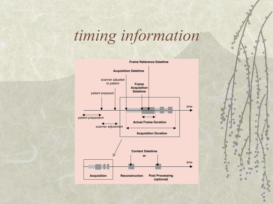
timing information
time
patient prepared
patient preparation
scanner adjusted
to patient
scanner adjustment
Acquisition Datetime
Acquisition Duration
Actual Frame Duration
Frame Reference Datetime
FrameAcquisition
Datetime
time
Acquisition Post Processing(optional)
Content Datetime
Reconstruction
or

cardiac timing information

spectroscopy
storage ofspectroscopy data
metabolite maps

but when (new MR IOD) ?
Modality PACS

time-based waveforms
introduced for cardiac imaging ECG, hemodynamic waveforms synchronization with images
– relative to temporal frame of reference same as images in terms of
– model (patient/study/series)– encoding (IOD/module/attributes)
different bulk data payload– samples instead of pixels– channels and multiplex groups

spatial registration
growing need to store/interchange– multi-modality registration and fusion
spatial registration object– same information model/encoding– payload is affine transform between frames of reference– well-known frames of reference for common atlases
separate fiducials object– for landmark based registration, etc.
separate color blending object– to specify superimposition of one (registered) volume
on another, with color transparency

structured reports storage of information with reference to images
– human-readable reports– quantitative analysis and measurements
structured report object(s)– same information model/encoding– payload is tree of tag-value pairs
specialized objects for– key instance selection (key image note)– mammography and chest CAD– procedure log
templates for general objects for– quantitative cardiac and ultrasound analysis, etc.

DICOM services
not primarily a “file format” goal is integration of devices “behind the scenes” to everyday user network services
– storage– query and retrieval– workflow management
• storage commitment• worklist and procedure step• instance availability notification
– print & media creation management

application integration
crude– ability to read/write DICOM “files”– folder/file storage hierarchy exposed– no use of management capabilities
elegant– ability to query & retrieve & send studies from/to a
PACS (clinical or research)– details of organization are hidden– user sees patients & studies, not files– management is reliable, not error prone

processing integration
processing workflow as a series of transformations intermediate forms as image objects
– encoded as conventional modality IODs– secondary capture IODs with extended attributes– private IODs– new standard IODs
addition of coded descriptions of type of image– e.g., temporally & spatially re-sampled & registered
can be managed using worklist & procedure step

conclusions about DICOM
ubiquitous in the clinical environment the only standard modality vendors will support well-defined, robust and extensible new objects regularly added to address state of the art
requirements (e.g. enhanced MR IOD) no more or less complex than any other standard necessary
to encode the information simplicity through the use of readily available free &
commercial libraries, toolkits & platforms for mostoperating systems & languages

conclusions about DICOM broad base of experienced developers readily available support groups and training (e.g.
news:comp.protocols.dicom) offers integration opportunities well beyond a mere file
format open development process in which all may participate the ND object being developed by the DICOM 3D
working group may be of particular interest

potential gaps in DICOM
encode/describe intermediate forms ? floating point pixel data ? vector data ? temporal transformation/resampling ? waveforms for stimuli/responses ? waveforms for EEG & MEG ? fMRI technique-specific attributes ?
others ?