Embed Size (px)
Citation preview

Q11
Q10
Q1
Q3
Q2
Osteoarthritis and Cartilage xxx (2015) 1e9
123456789101112131415161718192021222324252627282930313233343536373839404142434445464748495051525354
55
YJOCA3642_proof ■ 24 December 2015 ■ 1/9
5657585960616263646566676869707172737475
Dickkopf-3 is upregulated in osteoarthritis and has achondroprotective role
S.J.B. Snelling y *, R.K. Davidson z, T.E. Swingler z, L. Le z, M.J. Barter x, K.L. Culley k,A. Price y, A.J. Carr y, I.M. Clark zy Nuffield Department of Orthopaedics, Rheumatology and Musculoskeletal Sciences, University of Oxford, Oxford, UKz School of Biological Sciences, University of East Anglia, Norwich, UKx Institute of Cellular Medicine, University of Newcastle, Newcastle, UKk Hospital for Special Surgery and Weill Cornell Medical College, New York, NY, USA
7677
7879808182838485868788a r t i c l e i n f o
Article history:Received 3 September 2015Accepted 24 November 2015
Keywords:CartilageWntDickkopfTGFbOsteoarthritis
* Address correspondence and reprint requestsDepartment of Orthopaedics, Rheumatology and MusResearch Centre, University of Oxford, Nuffield OrthopHeadington, Oxford OX3 7LD, UK. Tel: 44-01865-2234
E-mail address: [email protected] (S
http://dx.doi.org/10.1016/j.joca.2015.11.0211063-4584/© 2015 Published by Elsevier Ltd on beha
89909192939495969798
Please cite this article in press as: Snelling SJand Cartilage (2015), http://dx.doi.org/10.10
s u m m a r y
Objective: Dickkopf-3 (Dkk3) is a non-canonical member of the Dkk family of Wnt antagonists and itsupregulation has been reported in microarray analysis of cartilage from mouse models of osteoarthritis(OA). In this study we assessed Dkk3 expression in human OA cartilage to ascertain its potential role inchondrocyte signaling and cartilage maintenance.Methods: Dkk3 expression was analysed in human adult OA cartilage and synovial tissues and duringchondrogenesis of ATDC5 and human mesenchymal stem cells. The role of Dkk3 in cartilage maintenancewas analysed by incubation of bovine and human cartilage explants with interleukin-1b (IL1b) andoncostatin-M (OSM). Dkk3 expression was measured in cartilage following murine hip avulsion.Whether Dkk3 influenced Wnt, TGFb and activin cell signaling was assessed in primary human chon-drocytes and SW1353 chondrosarcoma cells using RT-qPCR and luminescence assays.Results: Increased gene and protein levels of Dkk3 were detected in human OA cartilage, synovial tissueand synovial fluid. DKK3 expression was decreased during chondrogenesis of both ATDC5 cells andhumans MSCs. Dkk3 inhibited IL1b and OSM-mediated proteoglycan loss from human and bovinecartilage explants and collagen loss from bovine cartilage explants. Cartilage DKK3 expression wasdecreased following hip avulsion injury. TGFb signaling was enhanced by Dkk3 and Wnt3a and activinsignaling were inhibited.Conclusions: We provide evidence that Dkk3 is upregulated in OA and may have a protective effect oncartilage integrity by preventing proteoglycan loss and helping to restore OA-relevant signaling pathwayactivity. Targeting Dkk3 may be a novel approach in the treatment of OA.
© 2015 Published by Elsevier Ltd on behalf of Osteoarthritis Research Society International.
99100
101 102103104105106107108109Introduction
Osteoarthritis (OA) is characterized by loss of articular cartilage,joint pain and instability. The mechanisms regulating diseasepathogenesis remain elusive with a combination of genetic, in-flammatory, mechanical and metabolic factors implicated1e3.
to: S.J.B. Snelling, Nuffieldculoskeletal Sciences, Botnaraedic Centre, Windmill Road,23..J.B. Snelling).
lf of Osteoarthritis Research Societ
110111112113114115116
B, et al., Dickkopf-3 is upregul16/j.joca.2015.11.021
Chondrocytes from OA cartilage exhibit a disrupted phenotype,hallmarks of which include; altered synthesis of extracellular ma-trix (ECM) and ECM-degrading enzymes, altered cell signaling ac-tivity and increased proliferation4. Dysregulation of cell signalingpathways likely contributes to OA pathogenesis by reducing thechondrocyte's ability to maintain cartilage integrity, leading to orexacerbating the phenotypic shift associated with OA. TheWnt andTGFb signaling pathways have been strongly implicated in OApathogenesis5,6.
Dickkopf-3 (Dkk3) is a structurally and functionally divergentmember of the Dkk family of Wnt antagonists. Dkk3 activates orinhibitsWnt signaling in a tissue-dependentmanner and its impacton cartilage Wnt signaling is unknown7e9. Dkk3 is a tumour
y International.
117118119
ated in osteoarthritis and has a chondroprotective role, Osteoarthritis

Q4
S.J.B. Snelling et al. / Osteoarthritis and Cartilage xxx (2015) 1e92
1234567891011121314151617181920212223242526272829303132333435363738394041424344454647484950515253545556575859606162636465
66676869707172737475767778798081828384858687888990919293949596979899
100101102103104105106107108109110111112113114115116117118119120121122123124125126127128129130
YJOCA3642_proof ■ 24 December 2015 ■ 2/9
suppressor that inhibits proliferation of cancer cells and is down-regulated in several types of human cancer8e10. It can modulateinflammatory cell activity, maintain tissue organisation via TGFbsignaling and can protect against myocardial infarction-inducedfibrosis11e14.
The function of Dkk3 in other tissues suggests it could be animportant mediator of chondrocyte homeostasis and maintenanceof cartilage integrity. Several studies using animal models of OAhave reported increased Dkk3 in diseased cartilage15e17. HoweverDkk3 expression has not been well characterized in human OAtissue nor has its role in chondrocyte biology been explored. Ouraim was to assess whether Dkk3 shows aberrant expression inhuman OA and to establish whether it can regulate chondrocytebehaviour and OA-associated cartilage degradation in vitro.
Materials and methods
Primary tissue
Primary human OA cartilage and synovium were obtained fromage-matched individuals undergoing hip replacement for OA andcontrol cartilage and synovium obtained upon hip replacement forneck-of-femur fracture (NOF); cartilage OA n ¼ 13, NOF n ¼ 12, OAsynovium n ¼ 8; NOF synovium n ¼ 11. Anteromedial OA (AMG)specimens were obtained from patients undergoing uni-compartmental knee replacement (UKR) for OA. Primary humanchondrocytes (HAC) were obtained from macroscopically normalregions of the tibial plateau of OA patients undergoing total kneereplacement (TKR) and collagenase digested following standardprotocols. Explants of cartilage were used for proteoglycan andcollagen release assays (DMMB and hydroxyproline respectively).Synovial fluid was collected from individuals undergoing TKR(n ¼ 3), UKR (n ¼ 3), arthroscopy for cartilage lesions (n ¼ 5),matrix-assisted chondrocyte implantation (MACI, n ¼ 7) or controlpatients (n ¼ 3) with no cartilage lesion but meniscal tears.
Ethical approval (09/H0606/11 and 2005ORTHO7L) was grantedby Oxfordshire Research Ethics Committee and East Norfolk andWaveney Research Governance Committee. Informed consent wasobtained from all patients.
Cell culture
SW1353 chondrosarcoma cells (ATCC) and primary HAC werecultured in DMEM þ 10% (v/v) FCS. ATDC5 cells were cultured inDMEM/F12 (Lonza, UK) containing 5% (v/v) FCS, 2 mM glutamine,10 ug/ml apotransferrin (Sigma) and 30 nM sodium selenite.Confluent ATDC5 cells were stimulated to undergo chondrogenesisby addition of 10 ug/ml insulin (Sigma). Human MSCs (Lonza) wereexpanded in Mesenchymal Stem Cell Growth Medium (Lonza)supplemented with 5 ng/ml fibroblast growth factor-2 (R&D Sys-tems) before high density transwell culture as described18,19.Micromass cultures were established as described20 before treat-ment with 100 ng/ml Wnt3a for 4 days.
Cartilage explant assays
Bovine nasal septum and human articular cartilage weredissected and 2 mm cartilage discs explanted and equilibrated for24 h before treatment with interleukin-1b (IL1b) (0.5 ng/ml),oncostatin-M (OSM) (5 ng/ml) plus Dkk3 (50, 125 and 250 ng/ml).Treatments were refreshed every 2e3 days and collected for GAGand collagen release assays. Remaining cartilage was harvested at14 days for papain digestion and DMMB and hydroxyproline as-says21. Control and IL1/OSM-treated explants were collectedthroughout the time course for RNA extraction (Trizol, Invitrogen,
Please cite this article in press as: Snelling SJB, et al., Dickkopf-3 is upreguland Cartilage (2015), http://dx.doi.org/10.1016/j.joca.2015.11.021
UK), subsequent cDNA synthesis (Superscript, Invitrogen UK) ac-cording to manufacturer's instructions prior to RT-qPCR. Threeintra-experimental replicates were carried out for each treatmentcondition.
Hip avulsion assay
The hip joint from 5 to 6 week old C57BL/6J mice was dislocatedat the femur and the femoral cap avulsed using forceps as previ-ously described22. Hip joint cartilage was cultured for 1e48 h inserum-free medium before RNA extraction using Trizol (Invitrogen,UK). cDNA synthesis using Superscript (Invitrogen, UK) was per-formed prior to RT-qPCR.
Immunohistochemistry
Specimens were fixed in 10% (v/v) formalin for 12 h beforedecalcification in 5 M HNO3, paraffin embedding and cutting into5 mM sections. Following deparaffinisation and antigen retrievalwith 0.2% (v/v) Triton-X 100, sections were blocked and incubatedat 4�C overnight in primary antibody (DKK3, R&D Systems,Abingdon, UK) before visualisation using Vectastain ABC (Vectorlaboratories) with Diaminobenzidine (DAB) and Haematoxylin QS(Vector laboratories).
ELISA
Dkk3 level in synovial fluid was measured using Dkk3 ELISA(R&D Systems, UK) according to manufacturer's instructions.
Cytokine treatments
Cells were serum starved overnight and treated with recombi-nant IL1b (5 ng/ml) and/or OSM (10 ng/ml) for 24 h or pre-treatedfor 1 h with recombinant Dkk3 (250 ng/ml unless otherwise stated)or carrier alone (R&D Systems) before addition of recombinantWnt3a (100 ng/ml, 10 h), activin (20 ng/ml, 6 h) or TGFb1 (4 ng/ml,6 h) (R&D Systems). Three intra-experimental replicates werecarried out per cytokine treatment.
Following cytokine treatment cDNA was synthesized usingMMLV from DNase-treated cell lysates harvested in Cells-to-cDNAlysis buffer (Ambion) according to manufacturer's instructions.
RT-qPCR
Expression of genes was measured by RT-qPCR on a ViiA7(Applied Biosystems). Relative quantification is expressed as 2�DCt ,where DCt is Ct(gene of interest) � Ct(18S rRNA). Samples whichgave a Ct reading þ 1.5Ct greater or less than the median for 18Swere excluded from further analyses.
Luciferase assays
SW1353 chondrosarcoma cells were used for plasmid trans-fections using Lipofectamine 2000 with the Smad-responsive re-porter (CAGA)12-luc, Wnt-responsive 8xTCF/LEF binding site(TOPFlash) and mutant TCF/LEF site control FOPFlash and b-galac-tosidase transfection control plasmid23,24. Cells were treated withWnt3a (100 ng/ml) for 10 h or TGFb (4 ng/ml) or activin (20 ng/ml)for 3 h with and without 1 h Dkk3 pre-incubation before mea-surement of luciferase activity using the Luciferase and Beta-Gloassay systems (Promega).
ated in osteoarthritis and has a chondroprotective role, Osteoarthritis

Fig. 1. Dkk3 levels are altered in OA and during chondrogenesis. (A) DKK3 expression is elevated in OA cartilage and synovium from patients undergoing total hip arthroplasty. OAcartilage ¼ COA, n ¼ 13, NOF control cartilage (CN, n ¼ 11), OA synovium (SOA, n ¼ 8) and NOF control synovium (SN, n ¼ 11). DKK3 gene (B) and protein (C) levels were elevated indamaged compared to undamaged cartilage from individuals with AMG (n ¼ 5), IHC scale bar ¼ 20 mM. (D) Dkk3 protein measured by ELISA of synovial fluid was increased inindividuals undergoing TKR for OA, n ¼ 3. Levels were also measured in individuals with no cartilage lesions (control, n ¼ 3), undergoing arthroplasty for cartilage lesions (lesion,n ¼ 5), MACI (n ¼ 7) following arthroscopy, or UKR (n ¼ 3) for AMG. (A, B) analysed by t test, (D) by ANOVAwith Tukey post-test, three technical replicates per patient with the meanof these used in statistical analysis and represented as a dot (biological replicate) on each graph.
S.J.B. Snelling et al. / Osteoarthritis and Cartilage xxx (2015) 1e9 3
1234567891011121314151617181920212223242526272829303132333435363738394041424344454647484950515253545556575859606162636465
6667686970717273747576777879808182838485868788899091929394
YJOCA3642_proof ■ 24 December 2015 ■ 3/9
siRNA
Cells (HAC and SW1353) were transfected with 2.5 nM of siRNAagainst Dkk3 (Qiagen) or Allstars non-targeting negative control(Qiagen) using Dharmafect (Thermoscientific, UK) according tomanufacturers instructions. Cells were transfected 48 h prior tocytokine treatment.
Fig. 2. Dkk3 is regulated by inflammatory cytokines and injury and during chondrogenesishowed a reduction in DKK3 expression (n ¼ 8 mice). (B) 24 h treatment with IL1b and IL1b/Oreplicates per condition), this was partially inhibited by 10 mM of the p38 MAPK inhibitor Sinduced MMP13 and MMP1 expression was inhibited by Dkk3 (n ¼ 4 patients, four techniATDC5 cells (microarray) and human MSCs (RT-qPCR, n ¼ 2e3 biological replicates) (E & F)biological replicates.
Please cite this article in press as: Snelling SJB, et al., Dickkopf-3 is upreguland Cartilage (2015), http://dx.doi.org/10.1016/j.joca.2015.11.021
Statistical analysis
Analyses were carried out using Graphpad Prism 6.0. Student's ttest was used to test differences between two samples whilstANOVA with either Dunnett's or Tukey post-test was used formultiple samples. Normality was tested using the ShapiroeWilktest. P < 0.05 was considered statistically significant. *�0.05,
s. (A) RT-qPCR of RNA extracted from murine hip cartilage following ex vivo avulsionSM reduced DKK3 expression in primary monolayer HAC (n ¼ 4 patients, four technicalB202190 (SB) (n ¼ 4 patients, four technical replicates per condition) (C). (D) IL1/OSM-cal replicates per condition). DKK3 expression was reduced during chondrogenesis of. (AeD) and (F) ANOVA with Dunnett's post-test. All statistical analysis carried out on
9596979899
100101102103104105106107108109110111112113114115116117118119120121122123124125126127128129130
ated in osteoarthritis and has a chondroprotective role, Osteoarthritis

S.J.B. Snelling et al. / Osteoarthritis and Cartilage xxx (2015) 1e94
1234567891011121314151617181920212223242526272829303132333435363738394041424344454647484950515253545556575859606162636465
66676869707172737475767778798081828384858687888990
YJOCA3642_proof ■ 24 December 2015 ■ 4/9
**�0.01, ***�0.001. Graphs show mean ± 95% confidence intervalsof biological (patient or cell) replicates.
Results
Dkk3 expression is upregulated in OA tissue
Expression of DKK3 mRNA was increased >10-fold (P < 0.0001)in OA cartilage compared to NOF control [Fig. 1(A)]. Analysis ofsynovium from OA patients and NOF controls showed a 3.2-fold(P ¼ 0.0235) increase in DKK3 mRNA in diseased tissue. DKK3mRNA expression [Fig. 1(B)] was 2.1-fold (P ¼ 0.019) higher indamaged cartilage from patients with AMG. Our previous workshows reduced MMP and FRZB mRNA expression in damagedcompared to undamaged cartilage25. Immunohistochemistry inAMG patients also showed significant Dkk3 staining in the super-ficial zone of damaged but not undamaged cartilage [Fig. 1(C)].Dkk3 protein [Fig. 1(D)] in synovial fluid was 2.1-fold higher(P ¼ 0.0002) in patients undergoing TKR for OA compared to con-trol individuals, those with cartilage lesions (4.33-fold, P < 0.0001)or patients undergoing UKR (2.83-fold, P¼ 0.0016). Matrix-inducedautologous chondrocyte implantation (MACI) is performed 4e6weeks following initial assessment of cartilage lesions by arthros-copy. Dkk3 levels at the time of MACI were significantly higher thanat arthroscopy (i.e., lesion) (2.3-fold, P ¼ 0.0029).
Fig. 3. Dkk3 inhibits ex vivo cartilage degradation. (A) Dkk3 reduced IL1/OSM-induced collagthree technical replicates per condition). (B) BNC (n ¼ 4) and (C) human knee (n ¼ 4) cartilagin the presence of Dkk3 compared to IL1/OSM treatment alone, three technical replicates pOSM treatment and increased from day 5 onwards. (A), (B) and (C) ANOVA with Dunnett'sO ¼ IL1/OSM. All statistical analysis carried out on biological replicates (each biological rep
Please cite this article in press as: Snelling SJB, et al., Dickkopf-3 is upreguland Cartilage (2015), http://dx.doi.org/10.1016/j.joca.2015.11.021
DKK3 expression is downregulated following cartilage injury andduring chondrogenesis
The OA phenotype includes reinitiation of development26, thusestablishing Dkk3 regulation in chondrogenesis is important.ATDC5 differentiation is an established model of chondrogenesis.Following chondrogenic differentiation, microarray analysisshowed Dkk3 expression decreased relative to non-induced controlcultures [Fig. 2(A)]. Expression of chondrogenicmarkers Col2a1 andAgc1 (data not shown) were increased across these time points23.Human MSCs in high density transwell cultures also showed asignificant 1.3e21-fold reduction (P < 0.01) in DKK3 expressionthroughout chondrogenic differentiation into cartilage discs[Fig. 2(B)], with increases in COL2A1 and ACAN across the timecourse18.
Joint injury is associated with secondary OA therefore Dkk3regulation during injury or in response to inflammatory mediatorsof injury was investigated. Dkk3 expression in murine cartilage wasdecreased 1.8-fold (P ¼ 0.0005) immediately (1 h) following hipavulsion injury and remained low (3.54-fold reduction, P < 0.0001)48 h after injury [Fig. 2(C)]. Treatment of HAC for 72 h with IL1b orthe combination IL1b/OSM reduced DKK3 expression (2.4-fold,P ¼ 0.0086 and 5.25-fold, P ¼ 0.0009) [Fig. 2(D)], this was partiallyinhibited by inhibition of p38 MAPK activity [Fig. 2(E)]. IL1b/OSMtreatment of HAC inducedMMP13 andMMP1 expression [Fig. 2(F)],
en degradation (hydroxyproline release) from BNC explants (n ¼ 4 biological replicates,e explants showed a reduction in proteoglycan degradation (GAG release, DMMB assay)er condition. DKK3 expression was significantly reduced in BNC (n ¼ 3) at day 1 of IL1/post-test relative to IL1/OSM alone (D) t test relative to untreated timepoint control. I/licate the mean of technical replicates for that sample).
919293949596979899
100101102103104105106107108109110111112113114115116117118119120121122123124125126127128129130
ated in osteoarthritis and has a chondroprotective role, Osteoarthritis
S.J.B. Snelling et al. / Osteoarthritis and Cartilage xxx (2015) 1e9 5
1234567891011121314151617181920212223242526272829303132333435363738394041424344454647484950515253545556575859606162636465
666768
YJOCA3642_proof ■ 24 December 2015 ■ 5/9
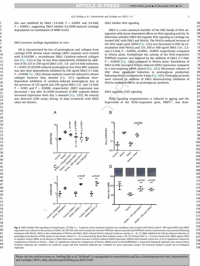
this was inhibited by Dkk3 (1.9-fold, P < 0.0001 and 3.9-fold,P < 0.0001), suggesting Dkk3 inhibits IL1/OSM-induced cartilagedegradation via modulation of MMP levels.
697071727374757677787980818283848586878889
Dkk3 prevents cartilage degradation in vitro
OA is characterized by loss of proteoglycan and collagen fromcartilage ECM. Bovine nasal cartilage (BNC) explants were treatedwith IL1b/OSM ± recombinant Dkk3. Cytokine-induced collagenloss [Fig. 3(A)] at day 14 was dose-dependently inhibited by addi-tion of 50, 125 or 250 ng/ml Dkk3 (2.0-, 3.6- and 5.6-fold reduction,P < 0.001) IL1b/OSM-induced proteoglycan loss from BNC explantswas also dose-dependently inhibited by 250 ng/ml Dkk3 [1.1-fold,P¼ 0.0049, Fig. 3(B)]. Human explants cannot be induced to releasecollagen however they showed [Fig. 3(C)] significant dose-dependent inhibition of cytokine-induced proteoglycan loss inthe presence of 125 ng/ml and 250 ng/ml Dkk3 (1.6- and 1.5-fold,P ¼ 0.003 and P ¼ 0.0008, respectively). DKK3 expression wasdecreased 1 day after IL1/OSM treatment of BNC explants beforeincreased expression from day 3 onwards [Fig. 3(D)]. No toxicitywas detected (LDH assay) during 14 days treatment with Dkk3(data not shown).
Fig. 4. Dkk3 inhibits Wnt signaling in chondrocytes. (A) HAC (n ¼ 4 patients, three technicalexpression was reduced in the presence of Dkk3. (B) SW1353 cells were transfected with thetreatment with Wnt3a, Dkk3 or the combination of Wnt3a and Dkk3. Dkk3 reduced Wnt3a-proteoglycan production of HAC grown in micromass culture (n ¼ 4) as measured by alcian bor negative control siRNA. In the absence of Dkk3 there was a relative increase in Wnt3a-inducomparisons of Wnt3a to Wnt3a þ Dkk3, (C) significance shown for comparisons of Wnt3a_technical replicates per condition for luciferase assays and four technical replicates per creplicates.
Please cite this article in press as: Snelling SJB, et al., Dickkopf-3 is upreguland Cartilage (2015), http://dx.doi.org/10.1016/j.joca.2015.11.021
Dkk3 inhibits Wnt signaling
Dkk3 is a non-canonical member of the Dkk family of Wnt an-tagonists with tissue-dependent effects onWnt signaling activity. Todetermine whether Dkk3 did regulate Wnt signaling in cartilage wetreated HAC with Dkk3 and Wnt3a. The Wnt3a-induced increase ofthe Wnt target gene AXIN2 [Fig. 4(A)] was decreased in HAC by co-incubation with Wnt3a and 125, 250 or 500 ng/ml Dkk3 (1.6-, 2.2-and 2.5-fold, P ¼ 0.0050, <0.0001, <0.0001 respectively) comparedto Wnt3a alone. Furthermore the activity of the Wnt-responsiveTOPFlash reporter was reduced by the addition of Dkk3 (1.7-fold,P ¼ 0.0010) [Fig. 4(B)] compared to Wnt3a alone. Knockdown ofDkk3 in HAC increasedWnt3a-induced AXIN2 expression comparedto a non-targeting siRNA control [Fig. 4(C)]. Micromass cultures ofHAC show significant reduction in proteoglycan productionfollowingWnt3a treatment for 4 days [Fig. 4(D)]. Proteoglycan levelswere restored by addition of Dkk3 demonstrating inhibition ofWnt3a-mediated effects on proteoglycan synthesis.
Dkk3 regulates TGFb signaling
TGFb signaling responsiveness is reduced in ageing and OA.Expression of the TGFb-responsive gene, TIMP327, was dose-
replicates per condition) were treated with Wnt3a with 0e500 ng/ml Dkk3 and AXIN2TOPFlash reporter plasmid and FOPFlash control. Luminescence was assessed followinginduced luciferase activity (n ¼ 8). (C) Dkk3 inhibited the Wnt3a-induced reduction inlue staining, mean ± SD. (D) Primary HAC (n ¼ 4) were treated with siRNA against Dkk3ced AXIN2 expression. ANOVAwith Dunnett's post-test, (A, B, D) significance shown forsiRNAcontrol to Wnt3siRNADkk3. n represents biological replicates (the mean of threeondition for gene expression assays). All statistical analysis carried out on biological
90919293949596979899
100101102103104105106107108109110111112113114115116117118119120121122123124125126127128129130
ated in osteoarthritis and has a chondroprotective role, Osteoarthritis

Q5
S.J.B. Snelling et al. / Osteoarthritis and Cartilage xxx (2015) 1e96
1234567891011121314151617181920212223242526272829303132333435363738394041424344454647484950515253545556575859606162636465
666768697071727374757677787980818283848586878889909192
YJOCA3642_proof ■ 24 December 2015 ■ 6/9
dependently enhanced in HAC treated with TGFb plus 250 and500 ng/ml Dkk3 compared to TGFb alone (2.1- and 2.2-fold,P < 0.001) [Fig. 5(A)]. TGFb-responsive PAI1 [SupplementaryFig. 2(A)] and ADAM12 (data not shown) were also enhancedwhilst MMP13 expression was decreased by TGFb in combinationwith 250 ng/ml Dkk3 [Fig. 5(C)] compared to TGFb alone (2.6-fold,P < 0.001). 250 ng/ml Dkk3 also increased activity of the TGFb-responsive (CAGA)12-luciferase reporter in SW1353 cells relative toTGFb alone (2.8-fold, P < 0.0001) [Fig. 5(B)]. No effect of Dkk3 alonewas seen on TIMP3, PAI1 or ADAM12 gene expression or CAGA-lucinduction. The extent of TGFb induction of TIMP3 [Fig. 5(D)], PAI1[Supplementary Fig. 1(B)] and ADAM12 (data not shown) expres-sion and CAGA-luc [Fig. 5(E)] activity was decreased by Dkk3knockdown. Knockdown of Dkk3 partially repressed the TGFb-induced decrease of MMP13 in primary HAC [Fig. 5(F)]. p38 MAPK-mediated stabilization of Smad4 has been described in Xenopuslaevis28, therefore we inhibited p38 MAPK. The induction of TGFb-induced TIMP3 [Fig. 5(G)] and PAI1 [Supplementary Fig. 2(B)]expression by Dkk3 was abrogated following p38 inhibition in HAC[Fig. 5(G)].
Activin is a member of the TGFb superfamily that also signals viaSmad2/3. To assess whether Dkk3 impacted other Smad2/3-relatedsignaling pathways, HAC and SW1353 were treated withactivin ±Dkk3. Activin-induced TIMP3 expression and (CAGA)12-lucactivity whilst co-incubation with Dkk3 caused a dose-dependentreduction in both of these outputs [Fig. 6(A and B)]. Knockdownof Dkk3 enhanced activin-induced TIMP3 expression and CAGA-luc
Fig. 5. Dkk3 enhances TGFb signaling response. (A) HAC (n ¼ 4) treated with TGFb showedresponsive (CAGA)12-luciferase activity in SW1353 cells (n ¼ 8) was also enhanced by Dkkluciferase activity (D, n ¼ 8) was reduced following knockdown of Dkk3. (E) Inhibition ofinduced enhancement of TIMP3 expression following TGFb treatment (n ¼ 3). (F) Dkk3(n ¼ 4) and siRNA against Dkk3 partially inhibited the TGFb-induced reduction in MMP13 exfor comparison between TGFb alone and TGFb þ Dkk3 (AeC) and for TGFb þ siControl to TGof TGFb þ Dkk3 to TGFb alone for with and without SB202190. n represents biological replictechnical replicates per condition for gene expression assays). All statistical analysis carried
Please cite this article in press as: Snelling SJB, et al., Dickkopf-3 is upreguland Cartilage (2015), http://dx.doi.org/10.1016/j.joca.2015.11.021
activity suggesting endogenous Dkk3 may act to reduce cellularactivin-induced responses [Fig. 6(C and D)]. There was no repres-sion of HAC TIMP3 expression when p38 MAPK activity wasinhibited [Fig. 6(E)]. Activin-induced PAI1 expression followed thesame trends as TIMP3 [Supplementary Fig. 3(AeC)].
Discussion
Altered expression of cytokines and consequent disruption ofcell signaling is associated with OA pathogenesis. Dkk3 is a non-canonical member of the Dkk family of Wnt antagonists that hasnot been explored in cartilage biology despite numerous studiesnoting its increased expression in models of OA. In this study wedemonstrate that Dkk3 is upregulated in adult human OA cartilageand synovial tissue but is decreased during chondrogenesis. Dkk3protects against in vitro cartilage degradation and its expression isregulated by both injury and inflammatory cytokines. Wnt andactivin signaling are both inhibited by Dkk3whilst TGFb signaling isenhanced. The upregulation of Dkk3 in OA may be a protectivemechanism to limit cartilage damage and to regulate aberrant cellsignaling associated with disease.
OA is a complex disease affecting multiple joint tissues, with aunique combination of factors likely to regulate pathogenesiswithin each tissue and across different joint locations. We showthat Dkk3 is upregulated in both hip and knee OA and in both sy-novial tissue and cartilage from diseased joints. Dkk3 upregulationis also reported in OA subchondral bone from patients undergoing
increased TIMP3 expression in the presence Dkk3 compared to TGFb alone. (B) TGFb-3 compared to TGFb alone. TGFb-induced TIMP3 expression (C, n ¼ 4) and (CAGA)12-HAC p38 MAPK activity by treatment with 10 mM SB202190 (SB) abolished the Dkk3-treatment decreased MMP13 expression in HAC compared to TGFb treatment alonepression in HAC (n ¼ 4) (G). (AeF) ANOVA with Dunnett's post-test, significance shownFb þ siDkk3 (DeF). (G) ANOVA plus Tukey post-test, significance shown for comparisonates (the mean of three technical replicates per condition for luciferase assays and fourout on biological replicates.
93949596979899
100101102103104105106107108109110111112113114115116117118119120121122123124125126127128129130
ated in osteoarthritis and has a chondroprotective role, Osteoarthritis

Fig. 6. Dkk3 inhibits activin signaling response. (A) HAC (n ¼ 4) treated with activin showed increased TIMP3 expression in the presence Dkk3 compared to Activin alone. (B)(CAGA)12-luciferase activity in SW1353 cells (n ¼ 8) was also reduced in the presence of Dkk3 compared to activin alone. Activin-induced TIMP3 expression (C, n ¼ 4) and (CAGA)12-luciferase activity (D, n ¼ 4) was increased following knockdown of Dkk3. (E) Inhibition of HAC p38 MAPK activity by treatment with 10 mM SB202190 (SB) abolished the Dkk3(250 ng/ml)-induced reduction in TIMP3 expression following Activin treatment (n ¼ 4). (AeD) ANOVAwith Dunnett's post-test, significance shown for comparison between Activinand Activin þ Dkk3 (A, B) and between Activin_siControl and Activin_siDkk3 (C, D). (E) ANOVA with Tukey post-test, significance shown for comparison between Activin alone andActivin þ Dkk3 in the absence and presence of SB202190. n represents biological replicates (the mean of three technical replicates per condition for luciferase assays and fourtechnical replicates per condition for gene expression assays). All statistical analysis carried out on biological replicates.
S.J.B. Snelling et al. / Osteoarthritis and Cartilage xxx (2015) 1e9 7
1234567891011121314151617181920212223242526272829303132333435363738394041424344454647484950515253545556575859606162636465
66676869707172737475767778798081828384858687888990919293949596979899
100101102103104105106107108109110111112113114115116117118119120121122123124125126127128129130
YJOCA3642_proof ■ 24 December 2015 ■ 7/9
TKR29. This suggests Dkk3 is relevant to whole joint biology in twocommon sites of disease. The increased Dkk3 in synovial fluid ofpatients with tricompartmental OA may implicate Dkk3 as abiomarker distinguishing end-stage disease. Further studies ofDkk3 as a circulating biomarker are warranted.
Dysregulation of Wnt and TGFb family members has beenstrongly implicated in experimental and human OA5,6. An imbal-ance in Wnt signaling leads to OA development in murine models,and Wnt antagonists DKK1 and FRZB have been reported asdownregulated in human OA30e32. Wnts and activin are alsoreleased following cartilage injury33,34. TGFb signaling andresponsiveness decrease with age and OA development whilstincreased activin has been detected in OA tissues34,35. Dkk3 hasboth agonistic and antagonistic effects on the Wnt pathwaydependent on tissue of expression and thus investigation of itsimpact on Wnt signaling in cartilage was investigated in ourstudy7e9. Opposing regulatory roles of Dkk3 on TGFb signaling inXenopus and prostate cancer13,28 have been reported but its func-tion in musculoskeletal tissue has not been studied.
In adult HAC we have shown that Dkk3 antagonized Wntsignaling and protected against Wnt-induced proteoglycan reduc-tion. Dkk3 enhanced TGFb signaling in chondrocytes and interest-ingly was necessary for TGFb-induced reduction of MMP13expression. Dkk3 may mediate protective effects on cartilage
Please cite this article in press as: Snelling SJB, et al., Dickkopf-3 is upreguland Cartilage (2015), http://dx.doi.org/10.1016/j.joca.2015.11.021
partially through upregulation of TGFb signaling and inhibition ofWnt signaling. Surprisingly, Dkk3 inhibited activin signaling incartilage despite both activin and TGFb commonly signalingthrough Smad2/3. Inhibition of p38 MAPK signaling abrogated theeffects of Dkk3 on both TGFb and activin signaling which showsDkk3 action here is p38 MAPK dependent. A previous studydemonstrated Dkk3-dependent Smad4-stabilization by p38 MAPKand this requires further investigation in chondrocytes36. Our datamay indicate that Dkk3 effects on TGFb require p38 MAPK forstabilization of Smad4. The effect of Dkk3 on activin signaling isalso p38MAPK dependent but may operate through a pathway thatdoes not use Smad4. The mechanism by which differential regu-lation of activin and TGFb can occur is currently unknown andbeyond the scope of this study.
Injury to the joint commonly leads to OA development. Tomodel cartilage injury ex vivo the murine hip was avulsed and Dkk3levels found to be decreased within 1 h. Decreased Dkk3 proteinwas also shown in pilot data from an ex vivo porcine explantmodel37 following cutting injury (data not shown). Treatment withIL1b/OSM also led to a reduction in Dkk3 expression that waspartially p38 MAPK dependent. In contrast, previous reports onmurine OA15e17 and our data in human tissue show an increase inDkk3 expression in established disease. Dkk3 may be regulated in atemporal manner during disease pathogenesis. This is supported by
ated in osteoarthritis and has a chondroprotective role, Osteoarthritis

Q6
Q7,8
S.J.B. Snelling et al. / Osteoarthritis and Cartilage xxx (2015) 1e98
1234567891011121314151617181920212223242526272829303132333435363738394041424344454647484950515253545556575859606162636465
66676869707172737475767778798081828384858687888990919293949596979899
100101102103104105106107108109110111112113114115116117118
YJOCA3642_proof ■ 24 December 2015 ■ 8/9
our BNC data that shows an initial decrease in DKK3 expressionfollowed by an increase as cartilage degradation occurs. It is also ofnote that synovial fluid Dkk3 levels were lower at the time ofarthroscopy than 4e6 weeks later when MACI was performed. Thismay indicate that injury to the joint capsule leads to significantDkk3 release from other joint tissues that overcomes any decreasedue to cartilage injury. The sources of Dkk3 in the joint requirefurther investigation. Any initial injury response leading todecreased Dkk3may have been completed at MACI and Dkk3 levelsare consequently increased in the ensuing repair attempt.
Paralleling the potential roles of the Wnt and TGFb pathways inOA pathogenesis, chondrogenesis and articular cartilage develop-ment require TGFb signaling as well as regulation of Wntsignaling5,38. Given the reversion of OA chondrocytes to adevelopmental-like phenotype39 our data showing decreased Dkk3during chondrogenesis, shows a potential role for Dkk3 in chon-drogenesis, and also suggests that the immediate downregulationof Dkk3 in injury may be an early repair response.
Strikingly, Dkk3 protected against IL1b/OSM-stimulated carti-lage degradation. The increase in Dkk3 in OA may be a protectivemechanism to minimize cartilage degradation and the OA-associated shift in chondrocyte phenotype. This is supported bythe reduction in cartilage-degrading MMP13 expression by Dkk3 inthe presence of IL1b/OSM. Microarray analysis of HAC treated withsiRNA against Dkk3 did not reveal pathways of Dkk3 action onunstimulated cells (data not shown), thus future analysis will usecytokine-stimulated. However siRNA treatment did increaseMMP13 expression in TGFb-treated cells suggesting that Dkk3 maylimit cartilage damage partially through reduction of both IL1b/OSM and TGFb-effects on MMP13.
Overall Dkk3 upregulation in disease may be a defence mech-anism to counteract disease-related dysregulation of cell signalingpathways; inhibiting inflammatory cytokine effects on cartilagedegradation and enhancing TGFb signaling whilst maintainingregulation of Wnt signaling in an attempt to counteract disease-associated changes in these pathways. Supplementation withDkk3 at an early stage of disease or post-injury may therefore betherapeutically beneficial.
Further investigation of Dkk3 in murine models of OA isnecessary to ascertain its contribution to cartilage homeostasis anddisease pathogenesis. Although the Dkk3 null mouse40does nothave an overt musculoskeletal phenotype our preliminary analysissuggests increased knee OA in 3- and 6-month old animals, we arecurrently investigating injury-models of OA. Dkk3 gene therapy isin clinical trial for prostate cancer with promising results41, butfurther preclinical evaluation is necessary alongside more detailedinvestigation of the role of Dkk3 in other tissues of the healthy andOA joint.
In summary we have demonstrated that Dkk3 is upregulated inhuman OA and reduces cartilage degradation. These findings mayhave clinical implications as treatment with Dkk3 may preventcartilage degeneration in OA and early intervention with Dkk3-based therapy may slow OA progression.
119120121122123124125126127
Contributors
SJBS and IMC designed the study. SJBS, RKD, TES, MJB, KC and LLcarried out data acquisition. AJC and AP provided patient samplesand assisted with data interpretation. SJBS and IMC carried out dataanalysis and interpretation. All authors helped prepared themanuscript and approved the manuscript for submission.
128129130
Conflict of interestsThe authors have no competing interests to declare.
Please cite this article in press as: Snelling SJB, et al., Dickkopf-3 is upreguland Cartilage (2015), http://dx.doi.org/10.1016/j.joca.2015.11.021
FundingGrants: Alzheimer's Research UK grants 20087 (SJBS), 19222 (SJBS),19424 (MJB) and the NIHR Oxford Musculoskeletal BiomedicalResearch Unit funded this work.
Acknowledgements
We thank Chethan Jayadev and Raj Rout for their assistance incartilage and synovial fluid collection, and Fiona Watt and Anas-tasios Chanalaris for their assistancewith unpublished pilot data onthe porcine-injury model.
Supplementary data
Supplementary data related to this article can be found at http://dx.doi.org/10.1016/j.joca.2015.11.021.
References
1. Wojdasiewicz P, Poniatowski LA, Szukiewicz D. The role ofinflammatory and anti-inflammatory cytokines in the patho-genesis of osteoarthritis. Mediators Inflamm 2014;2014:561459.
2. Andriacchi TP, Favre J. The nature of in vivo mechanical signalsthat influence cartilage health and progression to knee oste-oarthritis. Curr Rheumatol Rep 2014;16:463.
3. Loeser RF. Aging processes and the development of osteoar-thritis. Curr Opin Rheumatol 2013;25:108e13.
4. Goldring MB. The role of the chondrocyte in osteoarthritis.Arthritis Rheum 2000;43:1916e26.
5. Staines KA, Macrae VE, Farquharson C. Cartilage developmentand degeneration: a Wnt Wnt situation. Cell Biochem Funct2012;30:633e42.
6. van der Kraan PM, Goumans MJ, Blaney Davidson E, tenDijke P. Age-dependent alteration of TGF-beta signalling inosteoarthritis. Cell Tissue Res 2012;347:257e65.
7. Nakamura RE, Hunter DD, Yi H, Brunken WJ, Hackam AS.Identification of two novel activities of the Wnt signalingregulator Dickkopf 3 and characterization of its expression inthe mouse retina. BMC Cell Biol 2007;8:52.
8. Ueno K, Hirata H, Majid S, Chen Y, Zaman MS, Tabatabai ZL,et al. Wnt antagonist DICKKOPF-3 (Dkk-3) induces apoptosis inhuman renal cell carcinoma. Mol Carcinog 2011;50:449e57.
9. Yue W, Sun Q, Dacic S, Landreneau RJ, Siegfried JM, Yu J, et al.Downregulation of Dkk3 activates beta-catenin/TCF-4signaling in lung cancer. Carcinogenesis 2008;29:84e92.
10. Wang Z, Ma LJ, Kang Y, Li X, Zhang XJ. Dickkopf-3 (Dkk3) in-duces apoptosis in cisplatin-resistant lung adenocarcinomacells via the Wnt/beta-catenin pathway. Oncol Rep 2015;33:1097e106.
11. Kinoshita R, Watanabe M, Huang P, Li SA, Sakaguchi M,Kumon H, et al. The cysteine-rich core domain of REIC/Dkk-3 iscritical for its effect on monocyte differentiation and tumorregression. Oncol Rep 2015;33:2908e14.
12. Meister M, Papatriantafyllou M, Nordstrom V, Kumar V,Ludwig J, Lui KO, et al. Dickkopf-3, a tissue-derived modulatorof local T-cell responses. Front Immunol 2015;6:78.
13. Romero D, Kawano Y, Bengoa N, Walker MM, Maltry N,Niehrs C, et al. Downregulation of Dickkopf-3 disrupts prostateacinar morphogenesis through TGF-beta/Smad signalling.J Cell Sci 2013;126:1858e67.
14. Zhang Y, Liu Y, Zhu XH, Zhang XD, Jiang DS, Bian ZY, et al.Dickkopf-3 attenuates pressure overload-induced cardiacremodelling. Cardiovasc Res 2014;102:35e45.
ated in osteoarthritis and has a chondroprotective role, Osteoarthritis

Q9
S.J.B. Snelling et al. / Osteoarthritis and Cartilage xxx (2015) 1e9 9
1234567891011121314151617181920212223242526272829303132333435363738394041424344454647484950515253545556
57585960616263646566676869707172737475767778798081828384858687888990919293949596979899
100101102103104105106107108
YJOCA3642_proof ■ 24 December 2015 ■ 9/9
15. Blom AB, Brockbank SM, van Lent PL, van Beuningen HM,Geurts J, Takahashi N, et al. Involvement of the Wnt signalingpathway in experimental and human osteoarthritis: promi-nent role of Wnt-induced signaling protein 1. Arthritis Rheum2009;60:501e12.
16. Meng J, Ma X, Ma D, Xu C. Microarray analysis of differentialgene expression in temporomandibular joint condylar carti-lage after experimentally induced osteoarthritis. OsteoarthritisCartilage 2005;13:1115e25.
17. Loeser RF, Olex AL, McNulty MA, Carlson CS, Callahan MF,Ferguson CM, et al. Microarray analysis reveals age-relateddifferences in gene expression during the development ofosteoarthritis in mice. Arthritis Rheum 2012;64:705e17.
18. Barter MJ, G�omez R, Woods S, Hui W, Smith GR, Shanley DP,et al. Genome-wide microRNA and gene analysis of mesen-chymal stem cell chondrogenesis identifies an essential roleand multiple targets for miR-140-5p. Stem Cells 2015.
19. Murdoch AD, Grady LM, Ablett MP, Katopodi T, Meadows RS,Hardingham TE. Chondrogenic differentiation of human bonemarrow stem cells in transwell cultures: generation ofscaffold-free cartilage. Stem Cells 2007;25:2786e96.
20. Greco KV, Iqbal AJ, Rattazzi L, Nalesso G, Moradi-Bidhendi N,Moore AR, et al. High density micromass cultures of a humanchondrocyte cell line: a reliable assay system to reveal themodulatory functions of pharmacological agents. BiochemPharmacol 2011;82:1919e29.
21. Davidson RK, Jupp O, de Ferrars R, Kay CD, Culley KL, Norton R,et al. Sulforaphane represses matrix-degrading proteases andprotects cartilage from destruction in vitro and in vivo.Arthritis Rheum 2013;65:3130e40.
22. Chong KW, Chanalaris A, Burleigh A, Jin H, Watt FE,Saklatvala J, et al. Fibroblast growth factor 2 drives changes ingene expression following injury to murine cartilage in vitroand in vivo. Arthritis Rheum 2013;65:2346e55.
23. Swingler TE, Wheeler G, Carmont V, Elliott HR, Barter MJ, Abu-Elmagd M, et al. The expression and function of microRNAs inchondrogenesis and osteoarthritis. Arthritis Rheum 2012;64:1909e19.
24. Korinek V, Barker N, Morin PJ, van Wichen D, de Weger R,Kinzler KW, et al. Constitutive transcriptional activation by abeta-catenin-Tcf complex in APC�/� colon carcinoma. Science1997;275:1784e7.
25. Snelling S, Rout R, Davidson R, Clark I, Carr A, Hulley PA, et al.A gene expression study of normal and damaged cartilage inanteromedial gonarthrosis, a phenotype of osteoarthritis.Osteoarthritis Cartilage 2014;22:334e43.
26. Saito T, Kawaguchi H. HIF-2a as a possible therapeutic target ofosteoarthritis. Osteoarthritis Cartilage 2010;18:1552e6.
27. Su S, Dehnade F, Zafarullah M. Regulation of tissue inhibitor ofmetalloproteinases-3 gene expression by transforming growthfactor-beta and dexamethasone in bovine and human articularchondrocytes. DNA Cell Biol 1996;15:1039e48.
Please cite this article in press as: Snelling SJB, et al., Dickkopf-3 is upreguland Cartilage (2015), http://dx.doi.org/10.1016/j.joca.2015.11.021
28. Pinho S, Niehrs C. Dkk3 is required for TGF-beta signalingduring Xenopus mesoderm induction. Differentiation 2007;75:957e67.
29. Chou CH, Lee CH, Lu LS, Song IW, Chuang HP, Kuo SY, et al.Direct assessment of articular cartilage and underlying sub-chondral bone reveals a progressive gene expression change inhuman osteoarthritic knees. Osteoarthritis Cartilage 2013;21:450e61.
30. Zhu M, Tang D, Wu Q, Hao S, Chen M, Xie C, et al. Activation ofbeta-catenin signaling in articular chondrocytes leads toosteoarthritis-like phenotype in adult beta-catenin conditionalactivation mice. J Bone Miner Res 2009;24:12e21.
31. Zhu M, Chen M, Zuscik M, Wu Q, Wang YJ, Rosier RN, et al.Inhibition of beta-catenin signaling in articular chondrocytesresults in articular cartilage destruction. Arthritis Rheum2008;58:2053e64.
32. Leijten JC, Bos SD, Landman EB, Georgi N, Jahr H, Meulenbelt I,et al. GREM1, FRZB and DKK1 mRNA levels correlate withosteoarthritis and are regulated by osteoarthritis-associatedfactors. Arthritis Res Ther 2013;15:R126.
33. Dell'accio F, De Bari C, Eltawil NM, Vanhummelen P, Pitzalis C.Identification of the molecular response of articular cartilageto injury, by microarray screening: Wnt-16 expression andsignaling after injury and in osteoarthritis. Arthritis Rheum2008;58:1410e21.
34. Alexander S, Watt F, Sawaji Y, Hermansson M, Saklatvala J.Activin A is an anticatabolic autocrine cytokine in articularcartilage whose production is controlled by fibroblast growthfactor 2 and NF-kappaB. Arthritis Rheum 2007;56:3715e25.
35. van der Kraan PM. Age-related alterations in TGF betasignaling as a causal factor of cartilage degeneration in oste-oarthritis. Biomed Mater Eng 2014;24:75e80.
36. Hsu RJ, Lin CC, Su YF, Tsai HJ. Dickkopf-3-related gene regu-lates the expression of zebrafish myf5 gene through phos-phorylated p38a-dependent Smad4 activity. J Biol Chem2011;286:6855e64.
37. Gruber J, Vincent TL, Hermansson M, Bolton M, Wait R,Saklatvala J. Induction of interleukin-1 in articular cartilage byexplantation and cutting. Arthritis Rheum 2004;50:2539e46.
38. Shen J, Li S, Chen D. TGF-beta signaling and the developmentof osteoarthritis. Bone Res 2014;2.
39. van der Kraan PM, van den BergWB. Chondrocyte hypertrophyand osteoarthritis: role in initiation and progression of carti-lage degeneration? Osteoarthritis Cartilage 2012;20:223e32.
40. Barrantes Idel B, Montero-Pedrazuela A, Guadano-Ferraz A,Obregon MJ, Martinez de Mena R, Gailus-Durner V, et al.Generation and characterization of dickkopf3 mutant mice.Mol Cell Biol 2006;26:2317e26.
41. Kumon H, Sasaki K, Ariyoshi Y, Sadahira T, Ebara S, Hiraki T,et al. Ad-REIC gene therapy: promising results in a patient withmetastatic CRPC following chemotherapy. Clin Med InsightsOncol 2015;9:31e8.
109110111112
ated in osteoarthritis and has a chondroprotective role, Osteoarthritis