Embed Size (px)
Citation preview

Diagnostic Tests SHUROUQ QADOSE

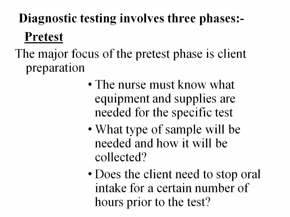
Diagnostic and laboratory Tests:
Are tools that provide information about the client, also run to find the cause of disease or discomfort; tests used to make a diagnosis.
Purpose of diagnostic tests:
• Help to confirm a diagnosis
• Monitor an illness
• Provide valuable information about the client’s response to treatment
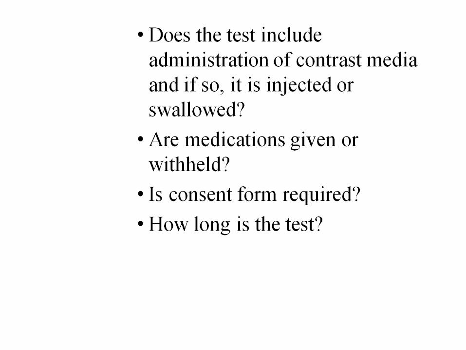
Intratest
It focuses on specimen collection and performing or assisting with certain diagnostic testing.
• The nurses uses of special standard precautions and sterile techniques
• Provide emotional and physical support while monitoring the client as needed (e.g., V /S, pulse oximetry, ECG).
• Correct labeling, storage, and transportation of the specimen to avoid invalid test results.

Post test
The focus of this phase is on nursing care of the client and follow up activities and observations.
• Compare the previous and current test results
• Modify nursing interventions as needed
• Report the results to appropriate health team members.
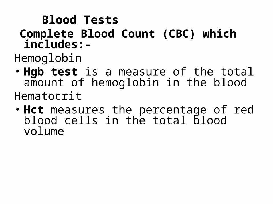
Blood Tests Complete Blood Count (CBC) which
includes:-Hemoglobin• Hgb test is a measure of the total amount of
hemoglobin in the bloodHematocrit• Hct measures the percentage of red blood cells
in the total blood volume
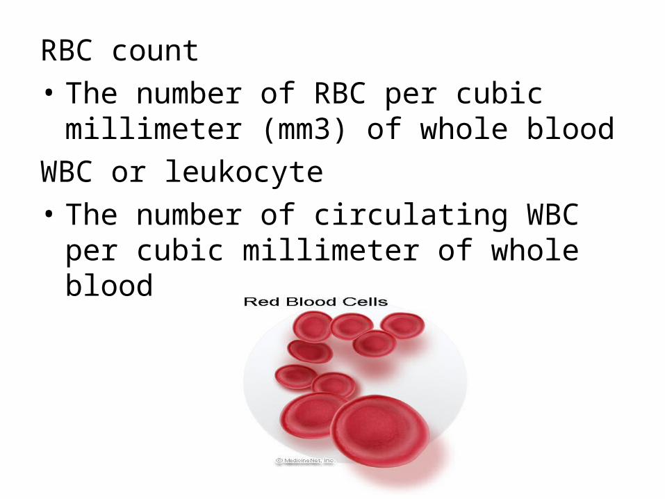
RBC count
• The number of RBC per cubic millimeter (mm3) of whole blood
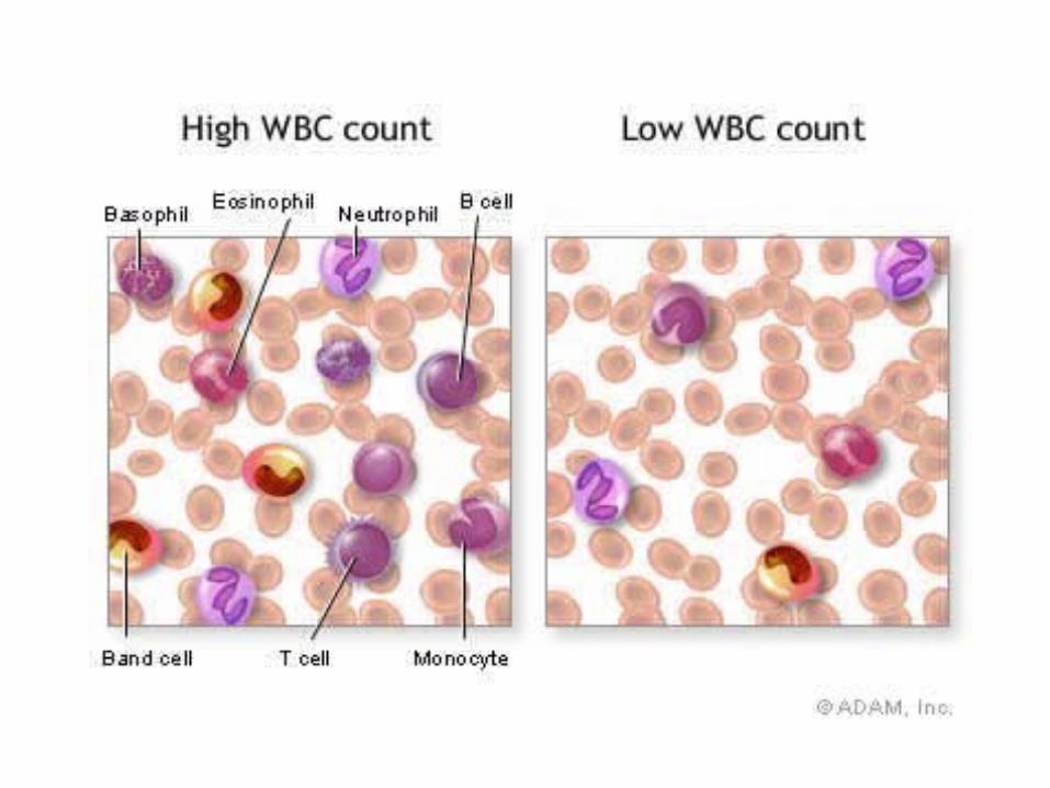
WBC or leukocyte
• The number of circulating WBC per cubic millimeter of whole blood

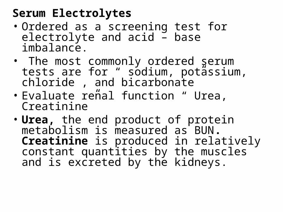
Serum Electrolytes• Ordered as a screening test for electrolyte and
acid – base imbalance.• The most commonly ordered serum tests are
for “ sodium, potassium, chloride , and bicarbonate”
• Evaluate renal function “ Urea, Creatinine”• Urea, the end product of protein metabolism is
measured as BUN. Creatinine is produced in relatively constant quantities by the muscles and is excreted by the kidneys.
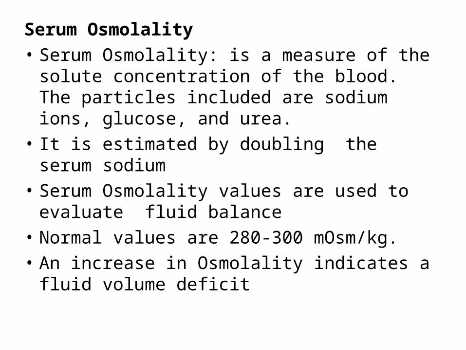
Serum Osmolality
• Serum Osmolality: is a measure of the solute concentration of the blood. The particles included are sodium ions, glucose, and urea.
• It is estimated by doubling the serum sodium
• Serum Osmolality values are used to evaluate fluid balance
• Normal values are 280-300 mOsm/kg.
• An increase in Osmolality indicates a fluid volume deficit

Drug monitoringIs conducted when the client is taking a
medication with narrow therapeutic range such as digoxin.
• This includes monitoring drawing blood samples for peak and trough levels to determine if the blood serum levels of a specific drug are at a therapeutic level and not a sub therapeutic or toxic level.
• Peak level: the highest concentration of the drug in the blood serum
• Trough level: lowest concentration
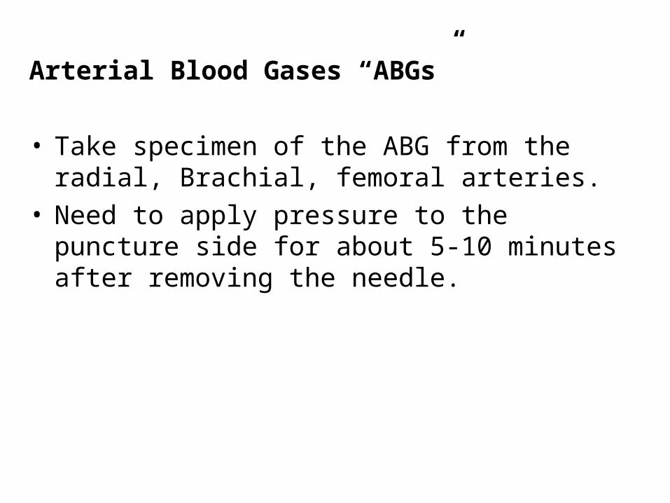
Arterial Blood Gases “ABGs”
• Take specimen of the ABG from the radial, Brachial, femoral arteries.
• Need to apply pressure to the puncture side for about 5-10 minutes after removing the needle.
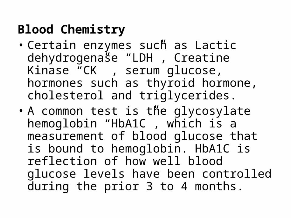
Blood Chemistry• Certain enzymes such as Lactic dehydrogenase
“LDH”, Creatine Kinase “CK” , serum glucose, hormones such as thyroid hormone, cholesterol and triglycerides.
• A common test is the glycosylate hemoglobin “HbA1C”, which is a measurement of blood glucose that is bound to hemoglobin. HbA1C is reflection of how well blood glucose levels have been controlled during the prior 3 to 4 months.

Capillary Blood Glucose
• Measure blood glucose when frequent tests are
required or a veinpuncture cannot be performed.
• Less painful, easily performed
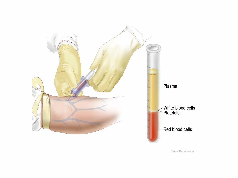
Specimen Collection
Nurses always assume the responsibility for specimen
collection depending on the type of specimen and skill required.
Nursing responsibilities with specimen collection
• Explain the purpose of specimen collection and the procedure for obtaining the specimen.
• Provide client comfort, privacy, and safety.• Use the correct procedure for obtaining the
specimen collection.• Note relevant information on the laboratory
requisition slip such as medication the client is taking that may affect the results.
• Transport the specimen to the laboratory promptly to have more correct results.
• Report abnormal results to the health care provider
Stool Specimens
• Reasons for testing feces include the following:
• To determine the presence of occult blood “hidden blood”. This test is referred to as the guaiac test.
• To analyze for dietary products and digestive secretions. An excessive amount of fat in the stool (steatorrhea) can indicate faulty absorption of fat from the small intestine. Decreased amount of bile in stool indicate obstruction in bile flow from liver and gallbladder to the intestine.

• To detect the presence of ova and parasites. When collecting specimen for parasites, it is important to send the specimen as soon as possible while it is still warm.
• To detect the presence of bacteria or viruses, so the container must be sterile and aseptic technique in collection.
Collecting Stool Specimens
Before obtaining the specimen, the nurse must determine the reason for the collecting the stool specimen and the correct method of obtaining and handling.
Nurse need to give the clients the following instructions:
• Defecate in a clean bedpan or bedside commode.
• Do not contaminate the specimen by urine or menstrual discharge.
• Do not place toilet tissues in the bedpan after defecation.
• Notify the nurse as soon as possible after defecation to send the specimen as soon as possible.

When obtaining the specimen the nurse should:-
• Follow medical aseptic technique meticulously.
• Wear disposable gloves to prevent hand contamination and take care not to contaminate the outside of the specimen container.
• Use one or two clean tongue blades to transfer the specimen to the container.
• The amount of stool to be sent depends on the purpose for which the specimen is collected.
• Label the specimen and the laboratory requisition have the correct information on them.
• Sends the specimen to the laboratory immediately.
• Document all relevant information, date, time of collection and all nursing assessments” color, odor, consistency and amount of feces”.

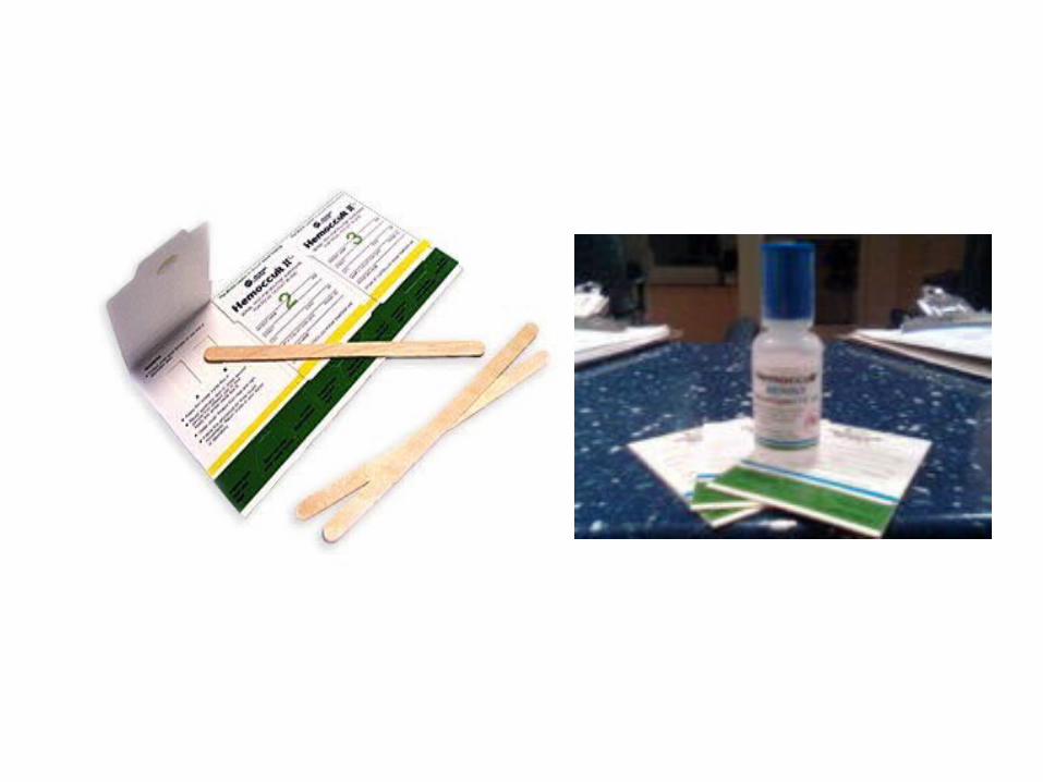
Testing Feces for Occult Blood
• Use of chemical reagent substance to detect the presence of the enzyme peroxidase in the hemoglobin molecule.
• A blue color indicates a guaiac positive result, that is, the presence of occult blood. No color change or any color other than blue is a negative finding, indicating the absence of blood in the stool.
• Certain foods, medications, and vitamin C can produce incorrect test results.
• False positive results can occur if the client has recently ingested (a) red meat (b) raw vegetables or fruits, (c) certain medications that irritate the gastric mucosa and cause bleeding such as aspirin, NSAID, anticoagulants.
• False – negative results can occur if the client has taken more than 250mgper day of vitamin C up to 3 days before the test – even if bleeding is present.

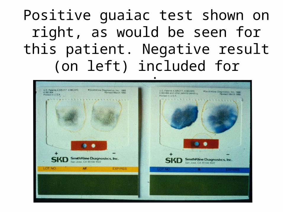
Positive guaiac test shown on right, as would be seen for this patient. Negative result (on left) included for comparison.
Urine Specimens
The nurse is responsible for collecting urine specimens for a number of tests: Clean voided specimens for routine urinalysis, clean – catch or midstream urine specimens for urine culture, timed urine specimen, indwelling catheter specimen.
Clean voided Urine Specimen• Usually done on the first voided specimen in the
morning• At least 10 ml of urine sufficient for routine analysis• Client who is disabled, seriously ill or disoriented may
need to use a bedpan or urinal in bed, these need special instructions:
• The specimen must be free of fecal contamination• Don’t discard the toilet tissue in bedpan• Put the lid tightly on the container to prevent
spillage of the urine.• If the outside of container is contaminated, clean
it with a disinfectant.
The nurse must:-–Make sure that the specimen label with laboratory
requisition carry the correct information and attach them securely to the specimen.

Clean-Catch or Midstream Urine Specimen It is collected when a urine culture is ordered to
identify microorganisms causing urinary tract infection. Care is taken to ensure that the specimen is as free as possible from contamination around the urinary meatus.
• Clean – catch specimens are collected into a sterile specimen container with a lid.
• Clean the urinary meatus with antiseptic solution• Instruct the client to start voiding• Collect 30-60 ml of urine in the container.• Label the specimen and transport it to the laboratory.
Timed Urine Specimen
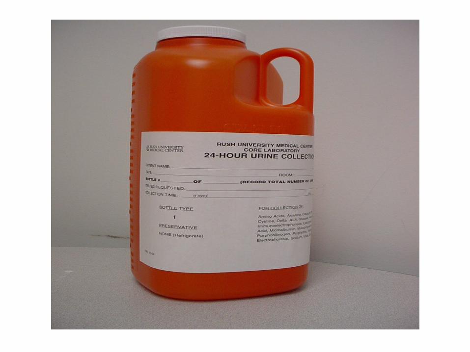
It requires collection of all urine produced and voided over a specific period of time, ranging from 1-2 hours to -24 hours. Each voiding of urine is collected in a small, clean container and then emptied immediately into the large refrigerated bottle or carton.

It is done for the following purposes:
• To assess the ability of the kidney to concentrate and dilute urine.
• To determine disorders of glucose metabolism such as DM.
• To determine levels of specific constitutes such as albumin, creatinine.
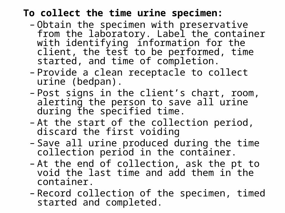
To collect the time urine specimen:– Obtain the specimen with preservative from the
laboratory. Label the container with identifying information for the client, the test to be performed, time started, and time of completion.
– Provide a clean receptacle to collect urine (bedpan).
– Post signs in the client’s chart, room, alerting the person to save all urine during the specified time.
– At the start of the collection period, discard the first voiding
– Save all urine produced during the time collection period in the container.
– At the end of collection, ask the pt to void the last time and add them in the container.
– Record collection of the specimen, timed started and completed.
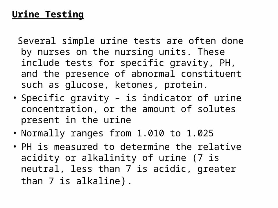
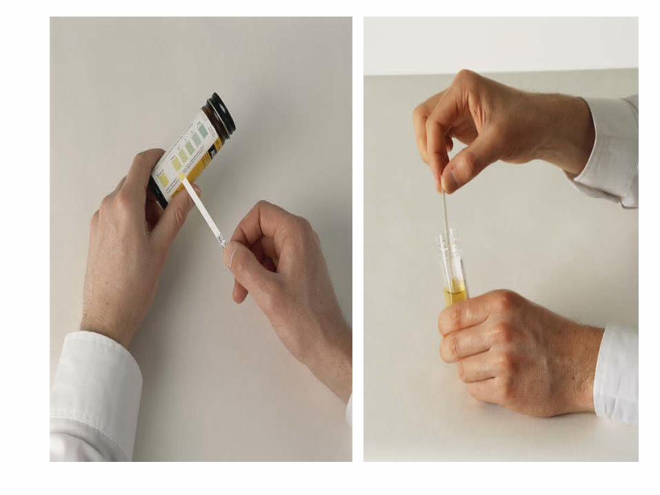
Urine Testing
Several simple urine tests are often done by nurses on the nursing units. These include tests for specific gravity, PH, and the presence of abnormal constituent such as glucose, ketones, protein.
• Specific gravity – is indicator of urine concentration, or the amount of solutes present in the urine
• Normally ranges from 1.010 to 1.025
• PH is measured to determine the relative acidity or alkalinity of urine (7 is neutral, less than 7 is acidic, greater than 7 is alkaline).

• Glucose – urine is tested for glucose to screen clients for diabetes mellitus and to assess clients for abnormal glucose to tolerance during pregnancy.
• Ketones – ketones bodies a product of the breakdown of fatty acids normally are not present in the urine.
• Protein
• Occult blood
• Osmolality – is measure of the solute concentration of urine. Normal values are 500 to 800 mOsm/kg.
Sputum specimen
Sputum is the mucous secretion from the lungs, bronchi, and trachea.
A sputum trap is used when the specimen is obtained by suctioning.
Sputum collection for the following purposes:
• For culture and sensitivity to identify a specific microorganisms
• For cytology to identify the origin, structure, function and pathology of cells.
• For acid-fast bacillus for TB.
• To assess the effectiveness of therapy.
Throat Culture
A throat culture sample is collected from the mucosa of the oropharynx and tonsillar regions using a culture swab.
Obtaining a throat culture is an invasive procedure that requires the application of scientific knowledge and potential problem solving to ensure client safety.
Visualization Procedures
It includes indirect visualization (noninvasive) and direct visualization (invasive) techniques for visualizing body organ and system function.
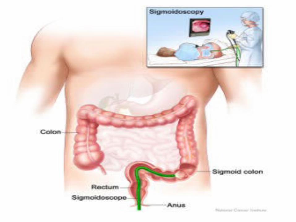
Clients with Gastro intestinal Alteration: Direct visualization techniques include:• Anoscopy: viewing of the anal canal• Proctoscopy: viewing of the rectum• Proctosigmoidoscopy: viewing the rectum and
sigmoid colon• Colonoscopy: viewing of the large intestine.
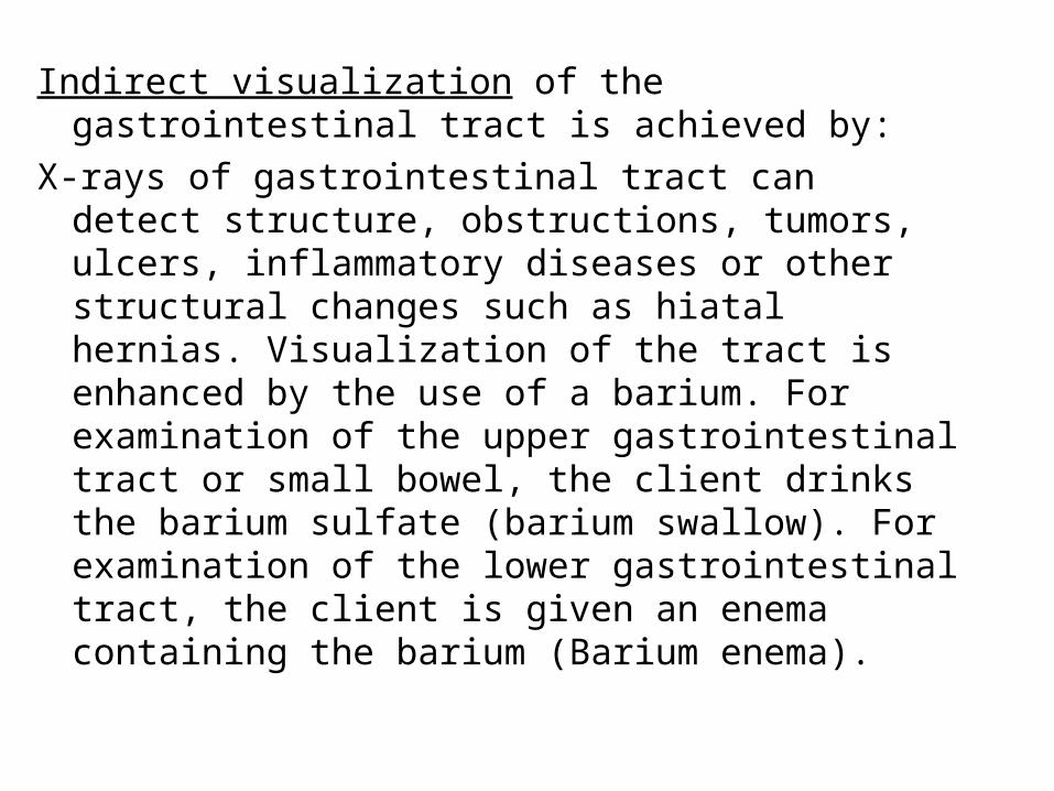
Indirect visualization of the gastrointestinal tract is achieved by:
X-rays of gastrointestinal tract can detect structure, obstructions, tumors, ulcers, inflammatory diseases or other structural changes such as hiatal hernias. Visualization of the tract is enhanced by the use of a barium. For examination of the upper gastrointestinal tract or small bowel, the client drinks the barium sulfate (barium swallow). For examination of the lower gastrointestinal tract, the client is given an enema containing the barium (Barium enema).

Barium Swallow
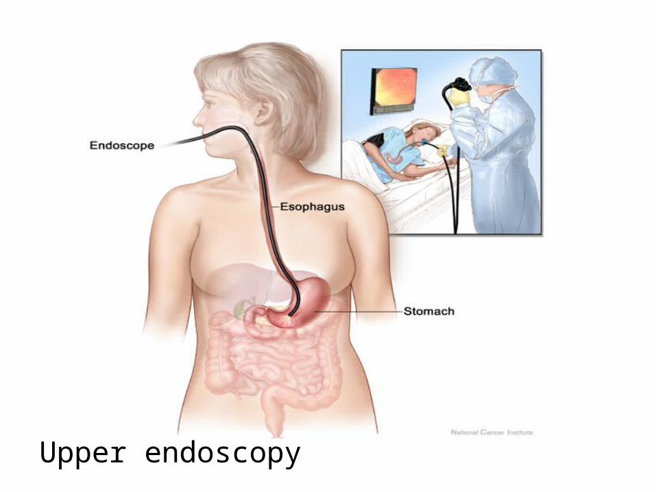
Upper endoscopy
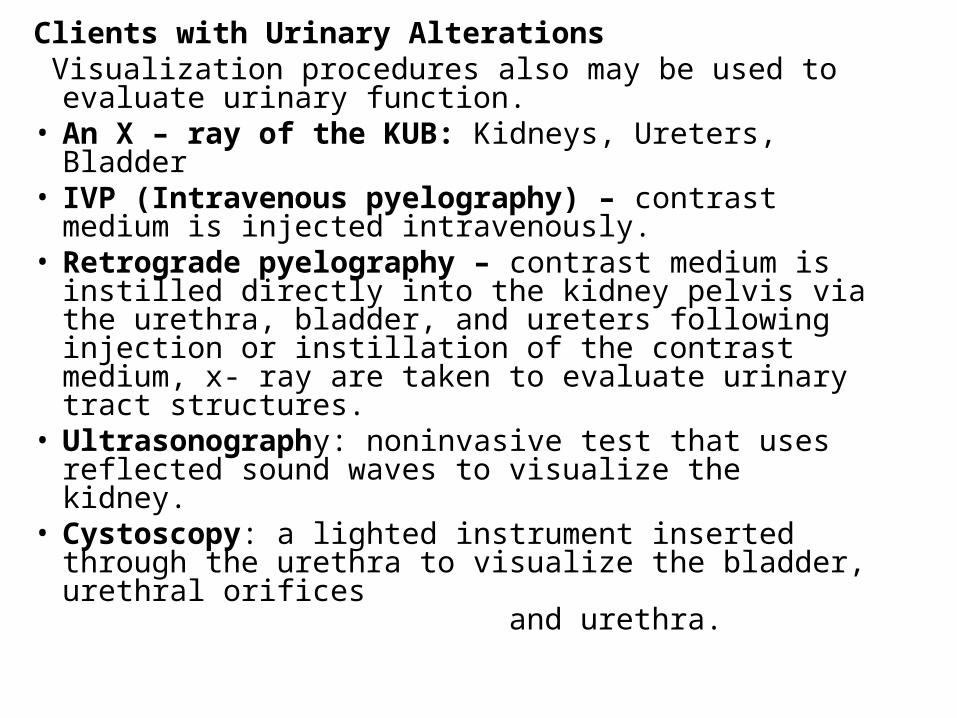
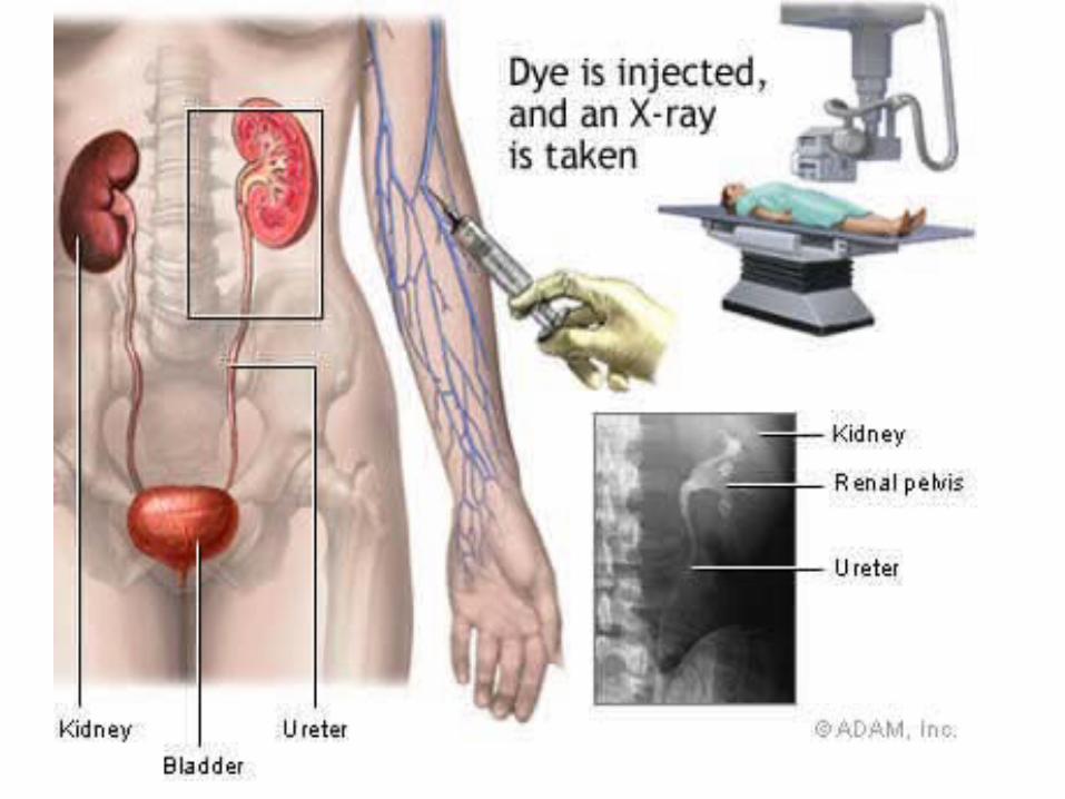
Clients with Urinary Alterations Visualization procedures also may be used to evaluate
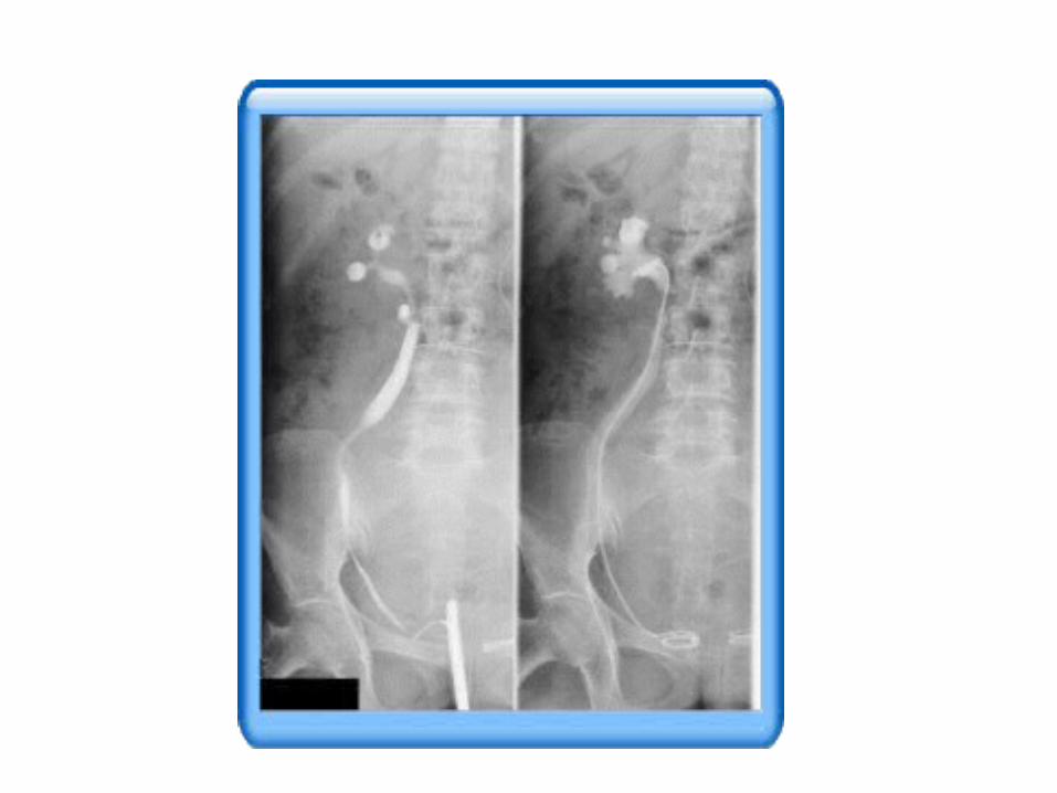
urinary function.• An X – ray of the KUB: Kidneys, Ureters, Bladder• IVP (Intravenous pyelography) – contrast medium
is injected intravenously.• Retrograde pyelography – contrast medium is
instilled directly into the kidney pelvis via the urethra, bladder, and ureters following injection or instillation of the contrast medium, x- ray are taken to evaluate urinary tract structures.
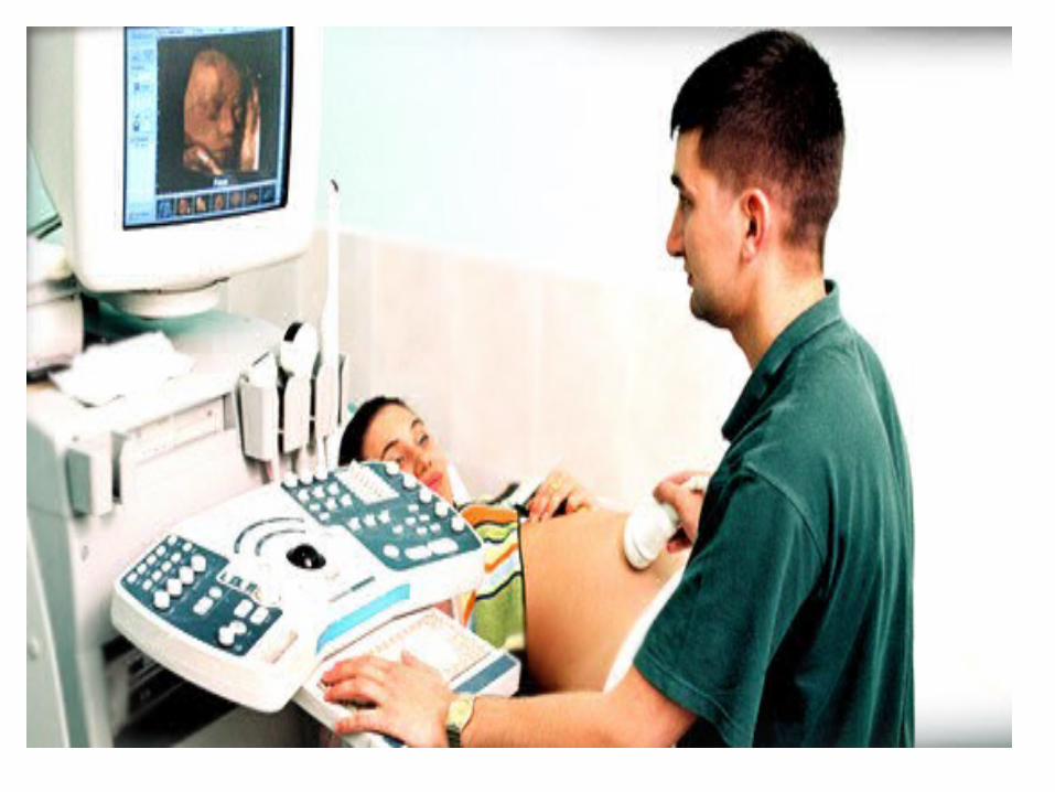
• Ultrasonography: noninvasive test that uses reflected sound waves to visualize the kidney.
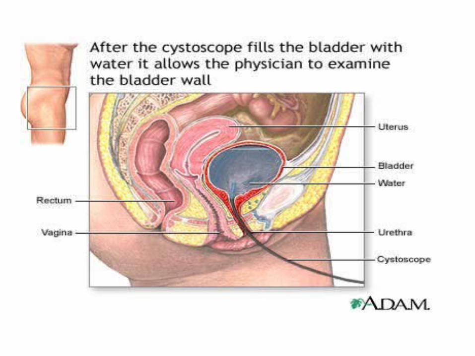
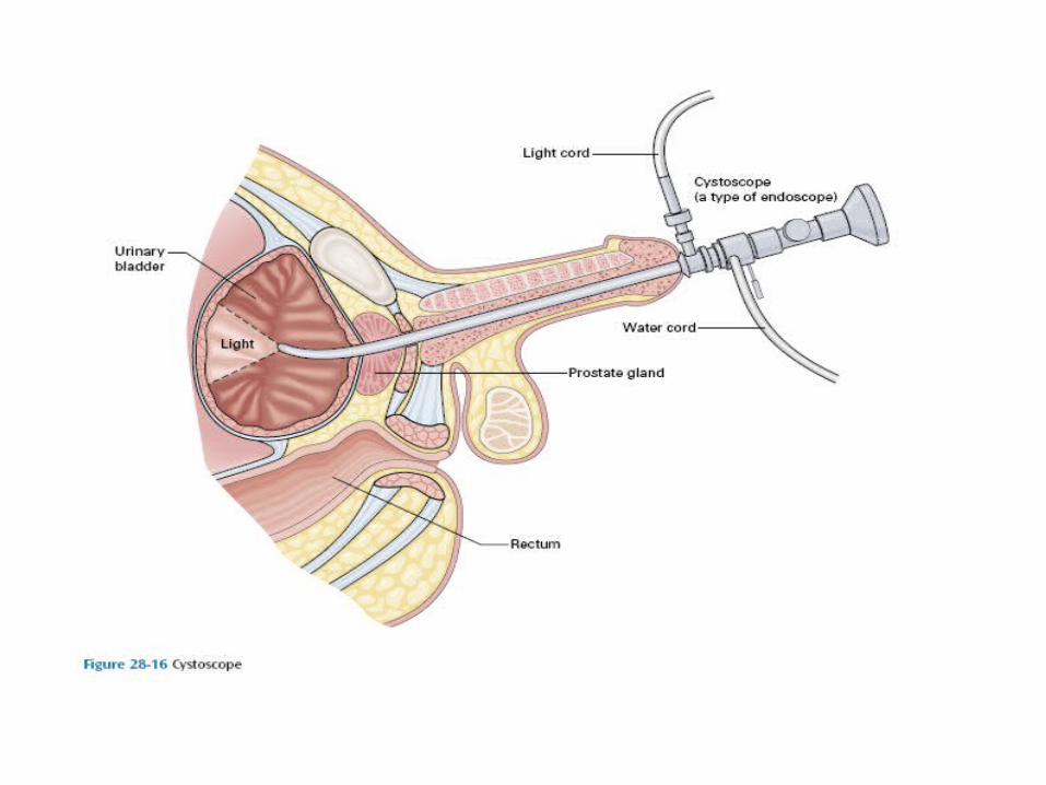
• Cystoscopy: a lighted instrument inserted through the urethra to visualize the bladder, urethral orifices and urethra.


Clients with cardio pulmonary Alterations
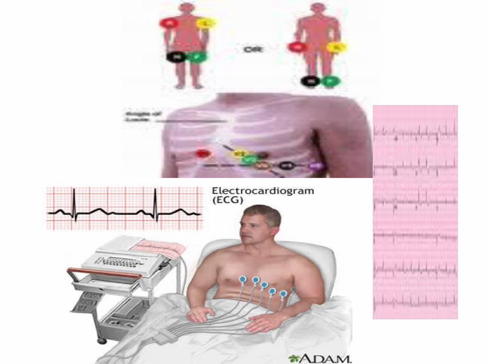
• ECG: Electrocardiography, recording of the heart’s electrical activity.
• Electrocardiogram: Detect arrhythmias and alteration in conduction indicative of myocardial damage, enlargement of the heart, or drug effects.
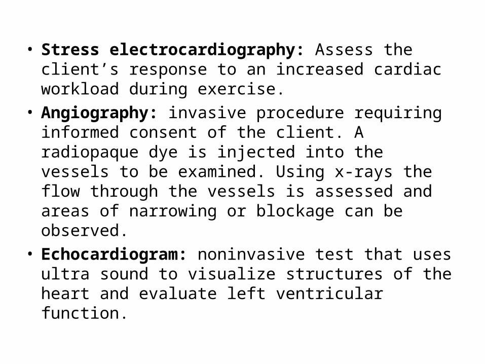
• Stress electrocardiography: Assess the client’s response to an increased cardiac workload during exercise.
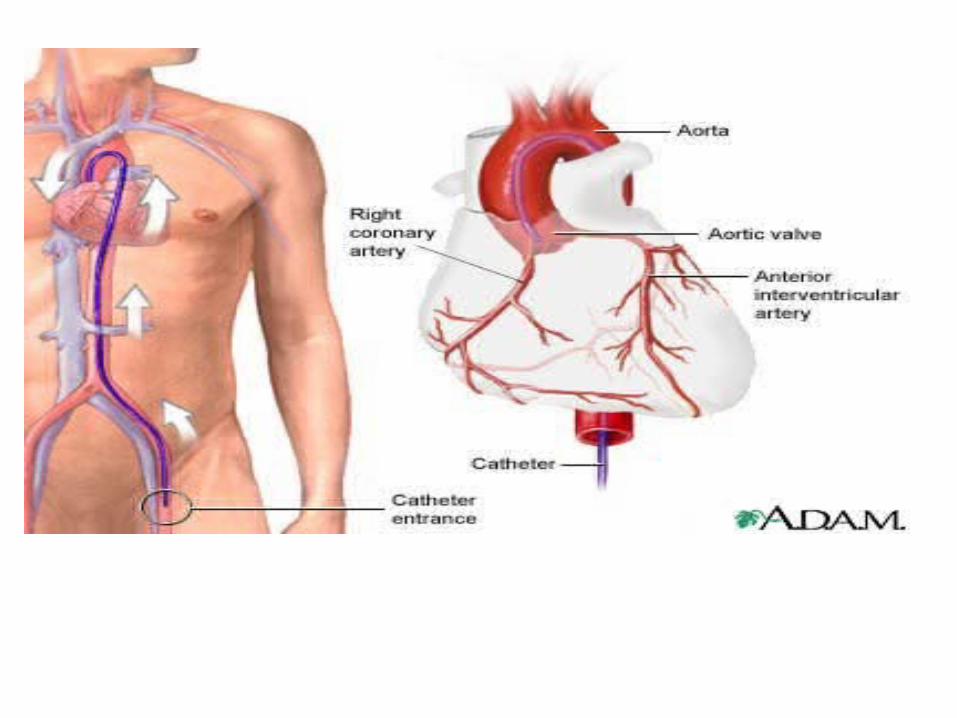
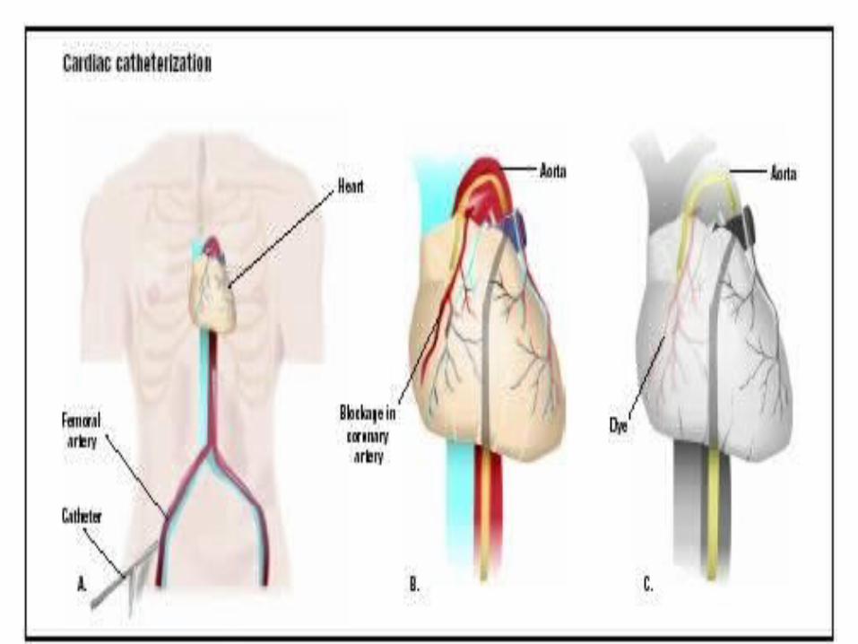
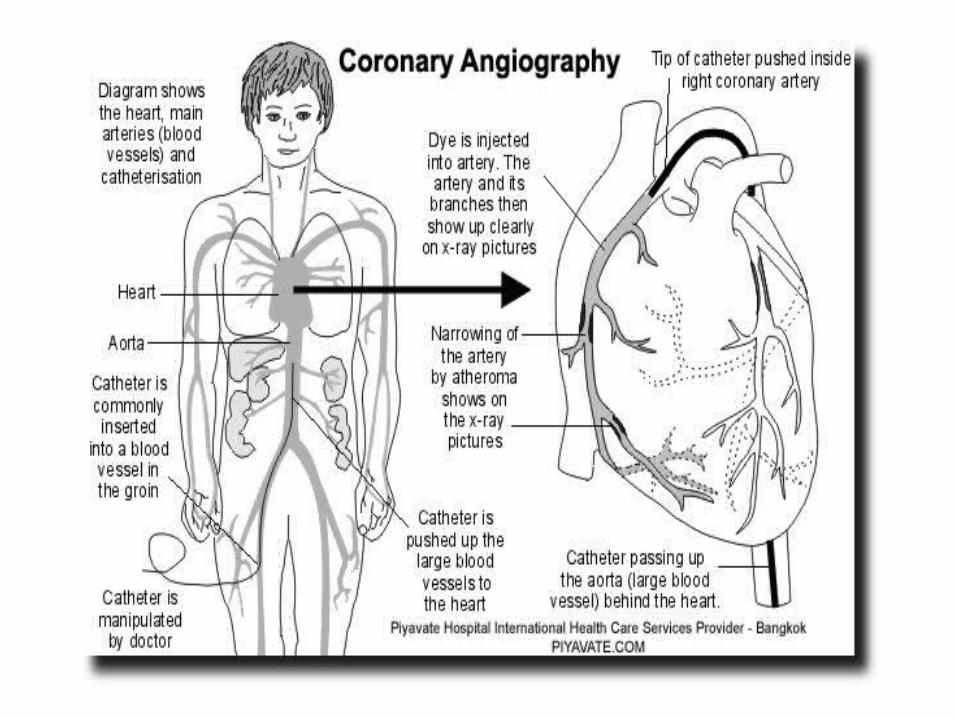
• Angiography: invasive procedure requiring informed consent of the client. A radiopaque dye is injected into the vessels to be examined. Using x-rays the flow through the vessels is assessed and areas of narrowing or blockage can be observed.
• Echocardiogram: noninvasive test that uses ultra sound to visualize structures of the heart and evaluate left ventricular function.
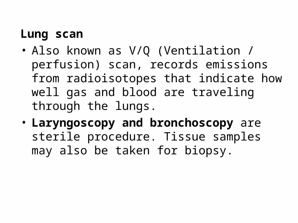
Lung scan
• Also known as V/Q (Ventilation / perfusion) scan, records emissions from radioisotopes that indicate how well gas and blood are traveling through the lungs.
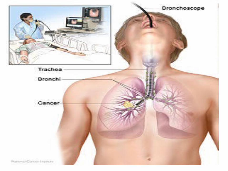
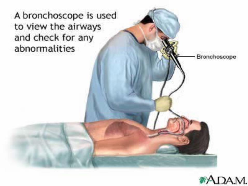
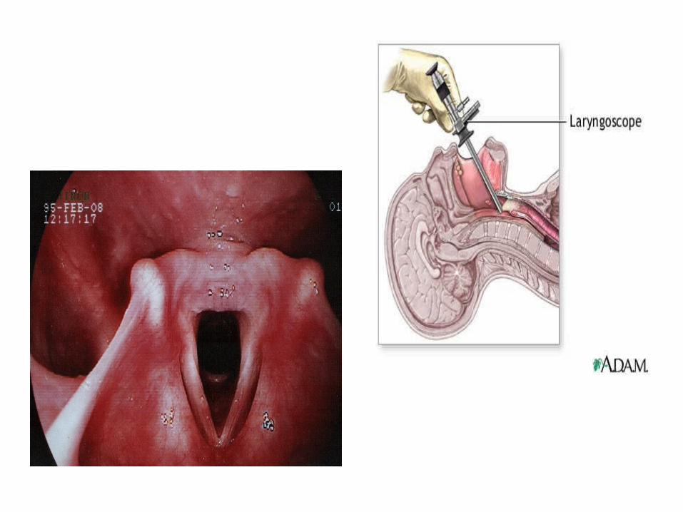
• Laryngoscopy and bronchoscopy are sterile procedure. Tissue samples may also be taken for biopsy.

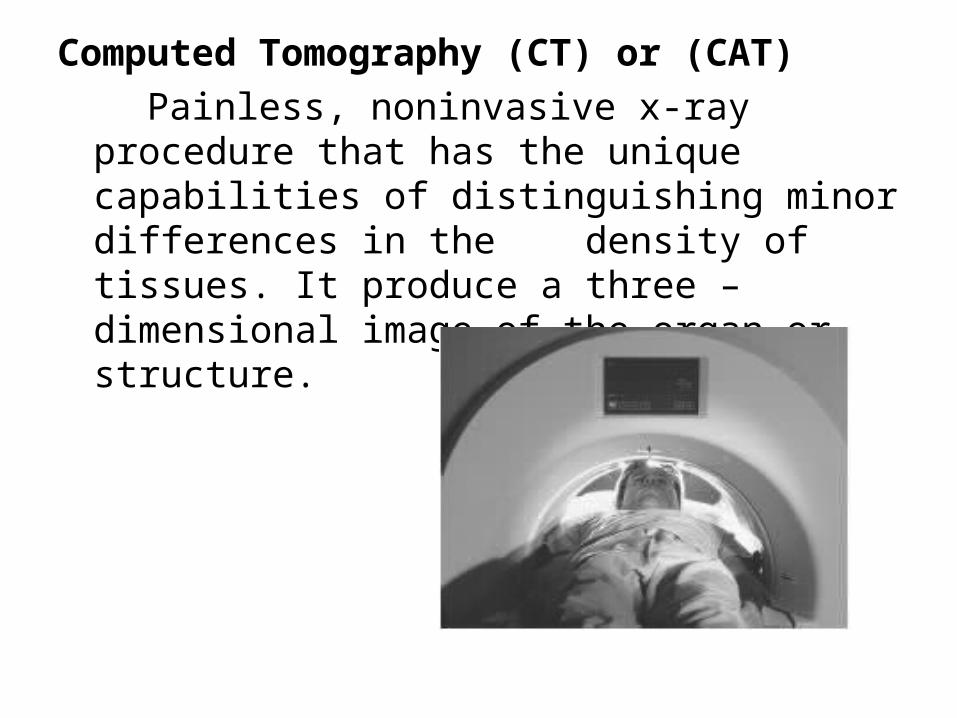
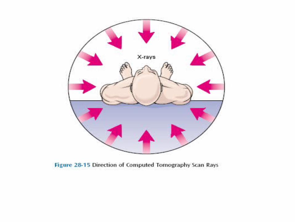
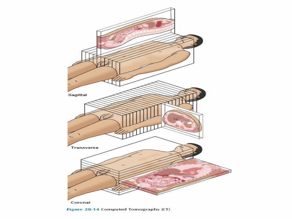
Computed Tomography (CT) or (CAT)
Painless, noninvasive x-ray procedure that has the unique capabilities of distinguishing minor differences in the density of tissues. It produce a three – dimensional image of the organ or structure.
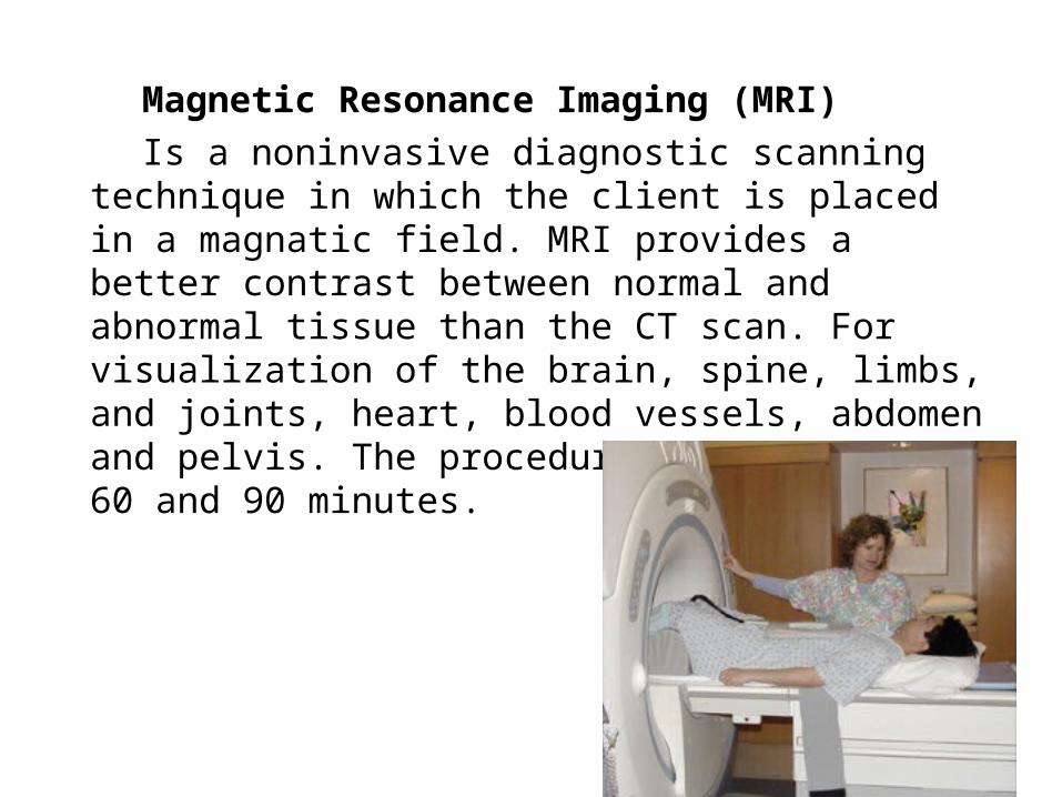
Magnetic Resonance Imaging (MRI)
Is a noninvasive diagnostic scanning technique in which the client is placed in a magnatic field. MRI provides a better contrast between normal and abnormal tissue than the CT scan. For visualization of the brain, spine, limbs, and joints, heart, blood vessels, abdomen and pelvis. The procedure lasts between 60 and 90 minutes.
Aspiration/Biopsy
Aspiration: withdrawal of fluid that has abnormally collected such as pleural cavity, abdominal cavity.
Biopsy: removal and examination of tissues. Usually performed to determine a diagnosis or to detect malignancy. Both aspiration and biopsy it needs sterile techniques.
“Determine if the facility requires a signed informed- consent from the aspiration / biopsy procedures”
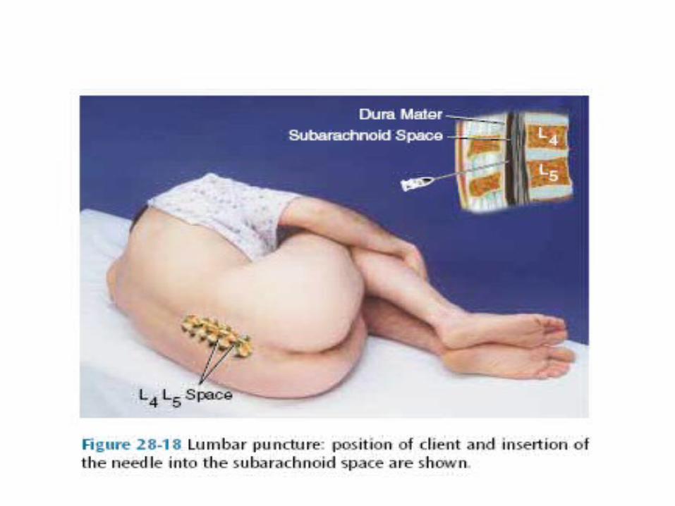
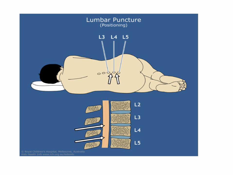
Lumbar Puncture
In lumbar puncture “LP” CSF is withdrawn through a needle inserted into the subarachnoid space of the spinal canal between the third and fourth lumbar vertebrae or between the fourth and fifth lumbar vertebrae.
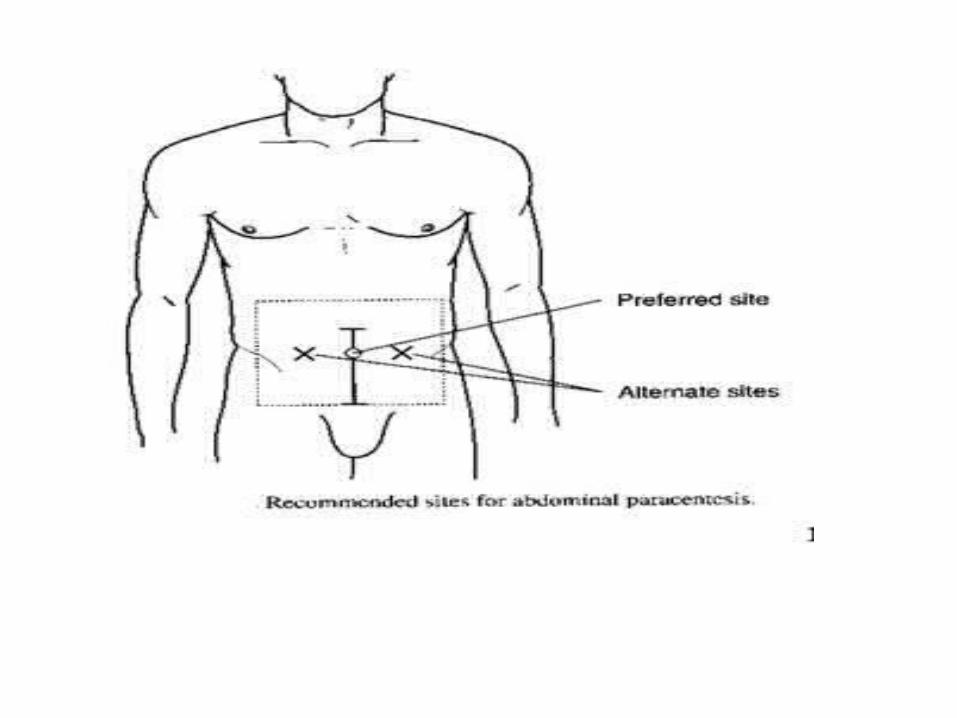
Abdominal Paracentesis
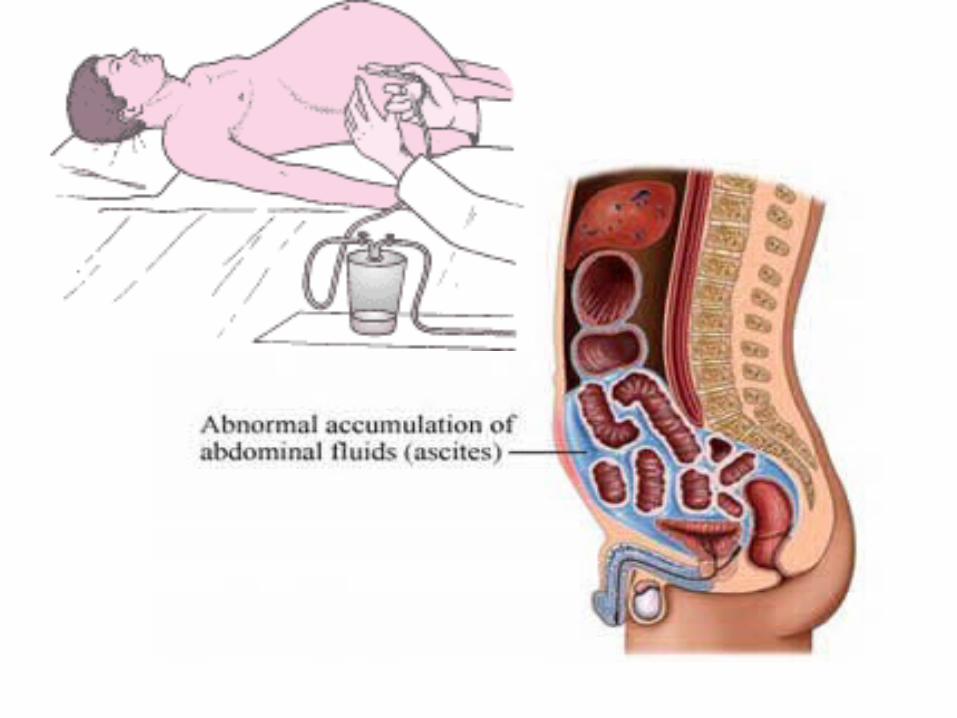
Normally the body creates just enough peritoneal fluid for lubrication. The fluid is continuously formed and absorbed into the lymphatic system.
Ascites: large amount of fluid accumulates in the abdominal cavity.
Abdominal Paracentesis: carried out to obtain a fluid specimen for laboratory study and to relieve pressure on the abdominal organs due to the presence of excess fluid. A common site for abdominal Paracentesis is the midway between the umbilicus and the symphysis pubis on the midline. Normally about 1,500 ml is the maximum amount of fluid drained at one time to avoid Hypovolemic shock.
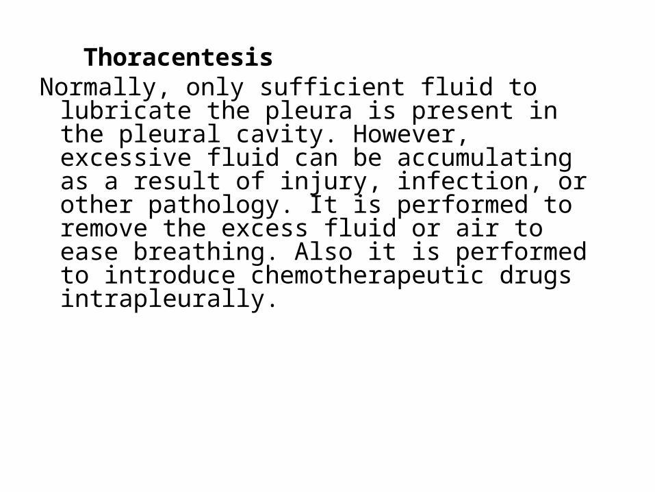
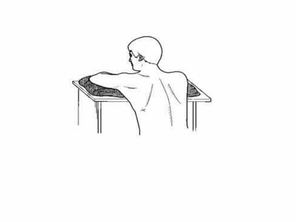
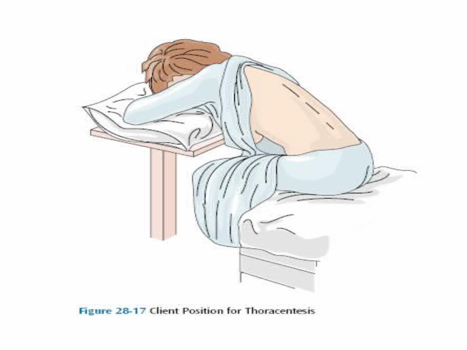
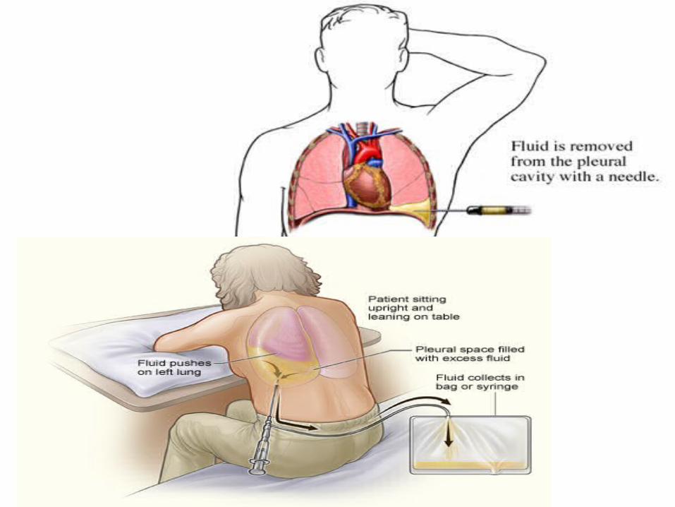
ThoracentesisNormally, only sufficient fluid to lubricate the
pleura is present in the pleural cavity. However, excessive fluid can be accumulating as a result of injury, infection, or other pathology. It is performed to remove the excess fluid or air to ease breathing. Also it is performed to introduce chemotherapeutic drugs intrapleurally.
This is usually a sitting position with the arms above the head, which spreads the ribs and enlarges the intercostals space. Or in which the client leans forward over a pillow.
A site on the lower posterior chest is often used to remove fluid, and a site on the upper anterior chest is used to remove air.
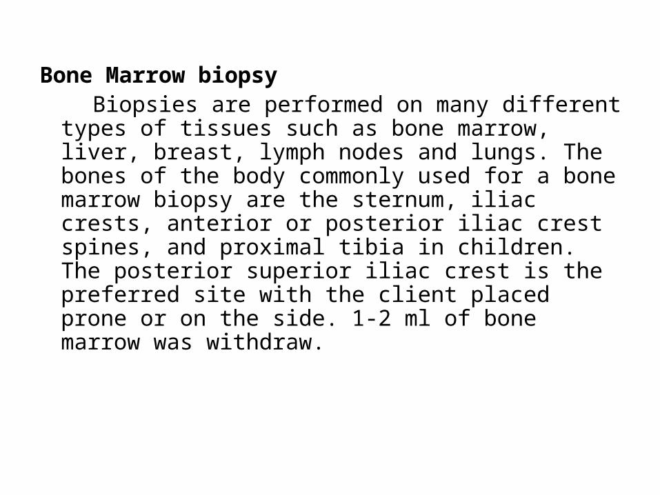
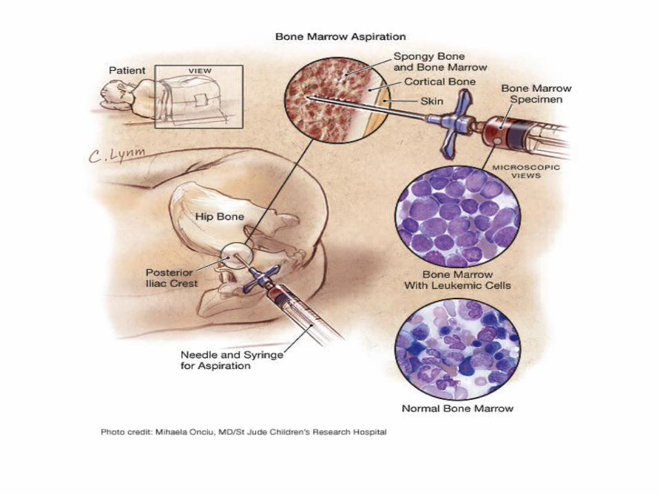
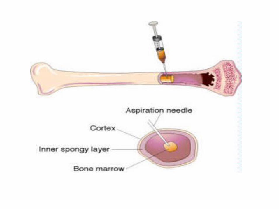
Bone Marrow biopsy Biopsies are performed on many different types of
tissues such as bone marrow, liver, breast, lymph nodes and lungs. The bones of the body commonly used for a bone marrow biopsy are the sternum, iliac crests, anterior or posterior iliac crest spines, and proximal tibia in children. The posterior superior iliac crest is the preferred site with the client placed prone or on the side. 1-2 ml of bone marrow was withdraw.
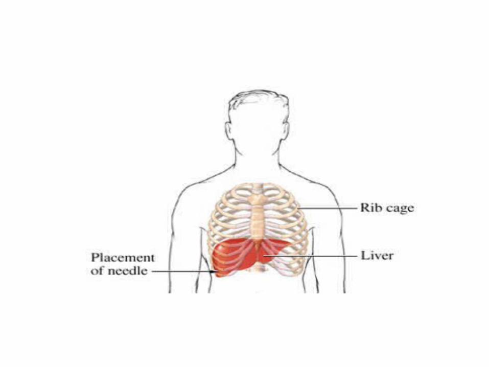
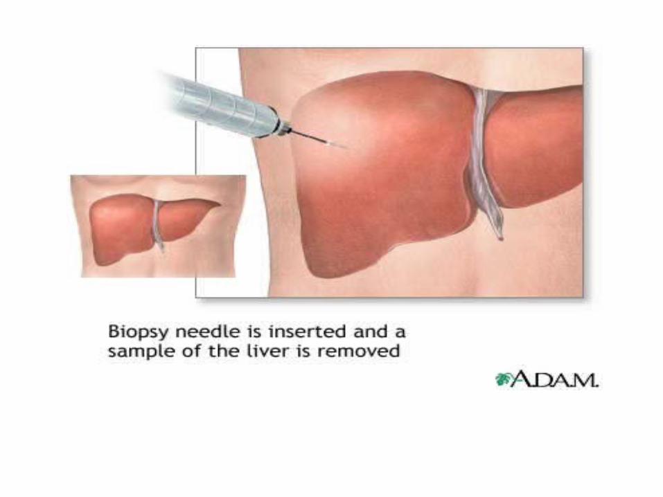
Liver biopsy It is performed at the client’s bedside, in which a
sample of liver tissue is aspirated. A physician inserted a needle in the intercostals space between two of the right lower ribs and into the liver or through the abdomen below the right the right rib cage. The nurse applies pressure to the site to prevent bleeding, often by positioning the client on the biopsy site.