Embed Size (px)
Citation preview
339
Turkish Journal of Trauma & Emergency Surgery
Original Article Klinik Çalışma
Ulus Travma Acil Cerrahi Derg 2010;16 (4):339-343
Diagnostic process and management of diaphragmatic injuries: approach in patients with blunt and penetrating trauma
Diyafram yaralanmalarında tanı süreci ve yönetim: Künt ve penetran travmalı hastalara yaklaşım
Atilla ÇELİK,1 Ediz ALTINLI,1 Neşet KÖKSAL,1 Kasım ÇAĞLAYAN,2 Mehmet Ali UZUN,1 Doğan ERDOĞAN,1 Ömer Faruk ÖZKAN1
12nd Department of General Surgery, Haydarpasa Numune Training and Research Hospital, Istanbul; 2Department of General Surgery,
Kars State Hospital, Kars, Turkey.
1Haydarpaşa Numune Eğitim ve Araştırma Hastanesi, 2. Genel Cerrahi Kliniği, İstanbul; 2Kars Devlet Hastanesi,
Genel Cerrahi Kliniği, Kars.
Correspondence (İletişim): Atilla Çelik, M.D. Haydarpaşa Numune Eğitim ve Araştırma Hastanesi, 2. Genel Cerrahi Kliniği, Haydarpaşa, İstanbul, Turkey.Tel: +90 - 216 - 414 45 02 / 1533 e-mail (e-posta): [email protected]
AMAÇTravmalı hastalarda en sık gözden kaçan yaralanmalar-dan birisi diyafram yırtığıdır. Tespit edilmesi halinde teda-vi için laparatomi veya torakotomi gerekmektedir. Bu ça-lışmanın amacı, ameliyat öncesinde ve ameliyat esnasın-da tespit edilen diyafram yaralanmalı, hastaların tanısal sü-reçlerini, mortalite ve morbidite oranları eşliğinde değer-lendirmektir.
GEREÇ VE YÖNTEMKliniğimize yedi yıllık süre içinde kabul edilen 16 diyaf-ram yaralanmalı hastada cerrahi girişim, eşlik eden yara-lanmalar, hastanede kalış süresi, transfüzyon gereksini-mi, mortalite ve morbidite oranları retrospektif olarak ir-delendi.
BULGULARYedi yılda 16 hasta diyafram yaralanması nedeniyle tedavi edildi. Kadın/erkek oranı 2/14 idi. Hastaların 15’i ameliyat edilirken bir hasta konservatif tedavi edildi.
SONUÇDiyafram yaralanmaları son yıllarda giderek artmaktadır. Yaralanma şiddeti skoru yüksek hastalarda diyafram yara-lanması olasılığı mutlaka düşünülmelidir.
Anahtar Sözcükler: Algoritm; diyafram yaralanması; yaralanma şiddet skoru; yönetim.
BACKGROUNDDiaphragmatic rupture is one of the most commonly missed injuries in trauma cases. Traditionally, laparotomy or thora-cotomy has been the treatment of choice for this condition. We aimed to evaluate the diagnostic process in patients with diaphragmatic injuries (DIs) who were diagnosed with diaphragm rupture during the preoperative or intraopera-tive course together with morbidity and mortality rates.
METHODSSixteen patients with DIs were admitted to our department during the last seven-year period. Surgical procedure, ac-companying injuries, duration of hospital stay, transfusion necessity, and morbidity and mortality rates were analyzed retrospectively.
RESULTSIn seven years, 16 patients were treated and followed-up for DI. Female/male ratio was 2/14. Fifteen patients were operated and one was treated conservatively. The mortality rate was 2/16.
CONCLUSIONDIs are being seen with increasing frequency in recent years. In patients with high Injury Severity Score (ISS), probability of DI should be taken into consideration.Key Words: Algorithm; diaphragm injury; Injury Severity Score; management.
340 Temmuz - July 2010
Ulus Travma Acil Cerrahi Derg
The diaphragm is a dome-shaped musculofibrous septum, which separates the thorax from the abdomi-nal cavity.[1,2] The first description of a traumatic dia-phragmatic rupture was made by Sennertus in 1541, and the first successful surgical repair was performed by Riolfi in 1886.[3,4] Diaphragmatic injuries (DIs) are usually caused by blunt trauma or penetrating injuries.[5] DI occurs in 0.8-5% of hospitalized auto-mobile victims, in approximately 5% of blunt trauma patients who undergo laparotomy, and in 10-15% of patients with penetrating trauma.[6,7] Diaphragmatic rupture as a result of abdominal trauma is one of the most commonly missed injuries.[8] The diagnosis may be delayed due to confusing clinical and radiographic findings. Delayed or missed diagnosis at the time of the initial injury and the life-threatening catastrophic sequelae if left untreated for an extended period com-pound the problem.[9,10] Acute DI is associated with widely ranging mortality of 5.5-51%, with death typi-cally resulting from associated injuries or in-hospital complications, such as adult respiratory distress syn-drome.[11] Complications usually relate to visceral her-niation through the diaphragmatic defect, and include respiratory compromise that is due to impaired pulmo-nary inflation and visceral incarceration with or with-out strangulation or perforation.[12]
The aim of this retrospective study was to review the experience of our department with the manage-ment of DI in order to identify incidence, associated morbidity and mortality, predictors of outcome, and factors contributing to diagnostic delay.
MATERIALS AND METHODSHaydarpasa Numune Training and Research Hos-
pital is an affiliated hospital of the Ministry of Health and one of the largest multidisciplinary training hospi-tals in Turkey. It has a trauma center, and is considered to be the one of the busiest hospitals in Istanbul as well. Our hospital has approximately 40,000 admis-sions per year, and traumatized patients and blunt and penetrating trauma to the body account for approxi-mately 10,000 admissions among those.
A total of 16 patients with a diagnosis of DI ad-mitted to the 2nd Department of General Surgery of Haydarpasa Numune Training Hospital between Janu-ary 2001 and June 2008. We investigated the medical records and radiological examinations of the patients who were diagnosed as DI in our department.
The variables studied in relation to the outcome were incidence, age, sex, causes of injury, side of rup-ture, type of injury, hemodynamic status upon admis-sion, imaging studies performed, method of diagnosis, time to diagnosis, concomitant injuries, Injury Sever-ity Score (ISS), performed and additional surgical intervention, herniated organs, morbidity, mortality,
duration of hospital stay, and reasons for diagnostic delay.
Unpaired Student’s t-test was used for continuous variables and the χ2 test for differences between cat-egorical variables. A p-value of <0.05 was considered statistically significant.
RESULTSPatient DemographicsIn the seven-year period, there were 14 (87%) male
and 2 (13%) female patients, aged 14-52 (mean: 28) years. The causes of injury were: motor vehicle crash (n: 1, 6.2%), penetrating injury (n: 12, 75%), fall from height (n: 1, 6.2%), and gunshot injury (n: 2, 12.5%). The anatomic distributions of DIs consisted of 3 right-sided injuries (19%) and 13 left-sided injuries (81%). One of the two patients with blunt injury had right and the other had left DI. The clinical features of the 16 patients are listed in Table 1.
Diagnostic CourseFive of 16 patients were hemodynamically un-
stable upon admission (patients had systolic blood pressure of <80 mmHg or pulse >120 per minute and/or shock table clinically). All of the unstable patients were admitted to the emergency operating room after fast resuscitation that included proper fluid, electro-lyte and blood transfusion. Stable patients were eval-uated fully, according to the appropriate advanced life support modalities. After the radiological evalua-tion, required surgical interventions were performed. There were 5 unstable patients (3 stab wounds, 2 gun-shot injuries).
Diagnostic modalities included plain chest X-rays (all patients), abdominal ultrasonography (4 patients), computerized tomography (CT) (4 patients), diagnos-tic peritoneal lavage (1 patient), and diagnostic lapa-roscopy (1 patient).
Abdominal ultrasonography and CT of the thorax and abdomen were performed for hemodynamically stable patients.
Among the 16 operated patients, the reasons for surgery were as follows:
- 3 patients due to DI detected on CT scans- 5 patients due to suspicion on X-ray films and
physical examination- 5 patients because of hemodynamic instability - 3 patients for their progression into acute abdomi-
nal findingsThe earliest operation was performed at the 10th
minute of the hospital admission while the latest was on the 11th day.
Cilt - Vol. 16 Sayı - No. 4 341
Diagnostic process and management of diaphragmatic injuries
Associated InjuriesAssociated organ injuries were present in 12 pa-
tients (75%) and included spleen (n: 4, 33%), bowel (n: 2, 16%), stomach (n: 3, 25%), liver (n: 2, 16%), kidney (n: 1, 8%), pancreas (n: 1, 8%), heart (n: 1, 8%), lung (n: 2, 16%), major vessel (n: 1, 8%), omen-tum (n: 1, 8%), and head injury (n: 1, 8%). Four pa-tients had isolated DI. All of the patients with isolated diaphragmatic rupture had penetrating injuries.
The majority of patients had multiple trauma and significant associated injuries, which are shown in Table 2.
SurgeryThe diaphragm was repaired via laparotomy in 11
patients (73%), thoracotomy in 2 patients (13%), lapa-roscopy in 1 patient (6.6%), and both laparotomy and thoracotomy in 1 patient (6.6%). Both interrupted and running techniques with non-absorbable suture were used.
Among the 16 patients with DI, one had right-sid-ed injury. As he was hemodynamically stable, he was conservatively treated without surgical intervention.
OutcomeBeginning from the admission to the hospital, the
earliest operation was performed at the 10th minute, and the latest operation was performed on the 11th day. Among 13 patients operated and discharged, the mean hospital stay was 8.2 (3-22) days. One patient medically treated was discharged on the 4th day. Two patients (12.5%) died due to the injury (2/16). One of
the patients who died had admitted to hospital with penetrating cardiac injury because of gunshot and was hemodynamically unstable. He was operated 10 min-utes after admitting to our hospital. Thoracotomy was performed, but the patient died on the operating table due to non-reversible hypovolemic shock. Totally, 2 units of erythrocyte suspension were administrated during the operation intravenously, and the ISS score was 75. The other mortality had admitted to the hospi-tal due to a fall from height. In the first physical exam-ination, the patient was evaluated as stable. In addition to the DI, the patient had splenic laceration and cra-nial injury. Splenectomy and phrenography were per-formed. This patient was treated in the intensive care unit for 20 days due to diffuse subarachnoid bleeding and died with an ISS score of 41.
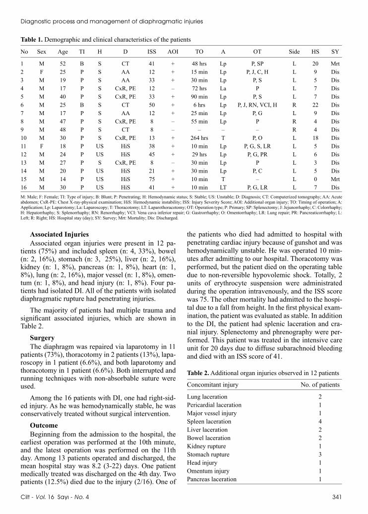
Table 1. Demographic and clinical characteristics of the patients
No Sex Age TI H D ISS AOI TO A OT Side HS SY
1 M 52 B S CT 41 + 48 hrs Lp P, SP L 20 Mrt2 F 25 P S AA 12 + 15 min Lp P, J, C, H L 9 Dis3 M 19 P S AA 33 + 30 min Lp P, S L 5 Dis4 M 17 P S CxR, PE 12 – 72 hrs La P L 7 Dis5 M 40 P S CxR, PE 33 + 90 min Lp P, S L 7 Dis6 M 25 B S CT 50 + 6 hrs Lp P, J, RN, VCI, H R 22 Dis7 M 17 P S AA 12 + 25 min Lp P, G L 9 Dis8 M 47 P S CxR, PE 8 – 55 min Lp P R 4 Dis9 M 48 P S CT 8 – – – – R 4 Dis10 M 30 P S CxR, PE 13 + 264 hrs T P, O L 18 Dis11 F 18 P US HiS 38 + 10 min Lp P, G, S, LR L 5 Dis12 M 24 P US HiS 45 + 29 hrs Lp P, G, PR L 6 Dis13 M 27 P S CxR, PE 8 – 30 min Lp P L 3 Dis14 M 20 P US HiS 21 + 30 min Lp P, C L 5 Dis15 M 14 P US HiS 75 + 10 min T – L 0 Mrt16 M 30 P US HiS 41 + 10 min LT P, G, LR L 7 DisM: Male; F: Female; TI: Type of injury; B: Blunt; P: Penetrating; H: Hemodynamic status; S: Stable; US: Unstable; D: Diagnosis; CT: Computerized tomography; AA: Acute abdomen; CxR-PE: Chest X-ray-physical examination; HiS: Hemodynamic instability; ISS: Injury Severity Score; AOI: Additional organ injury; TO: Timing of operation; A: Application; Lp: Laparotomy; La: Laparoscopy; T: Thoracotomy; LT: Laparothoracotomy; OT: Operation type; P: Primary; SP: Splenectomy; J: Jejunorrhaphy; C: Colorrhaphy; H: Hepatorrhaphy; S: Splenorrhaphy; RN: Renorrhaphy; VCI: Vena cava inferior repair; G: Gastrorrhaphy; O: Omentorrhaphy; LR: Lung repair; PR: Pancreaticorrhaphy; L: Left; R: Right; HS: Hospital stay (day); SY: Survey; Mrt: Mortality; Dis: Discharged.
Table 2. Additional organ injuries observed in 12 patients
Concomitant injury No. of patients
Lung laceration 2Pericardial laceration 1Major vessel injury 1Spleen laceration 4Liver laceration 2Bowel laceration 2Kidney rupture 1Stomach rupture 3Head injury 1Omentum injury 1Pancreas laceration 1
Ulus Travma Acil Cerrahi Derg
342 Temmuz - July 2010
The mean ISS score of the patients who did not survive was 58, while this was 23.8 in surviving pa-tients, and the difference was statistically significant (p<0.005).
The mean ISS score in the patients with additional organ injury was 34.5, while it was 9 in DI only pa-tients, and the difference was statistically significant (p<0.005). We found that additional organ injury pro-longed the hospital stay.
The postoperative complications were empyema,[1] intraabdominal abscess,[2] pneumonia,[3] urinary tract infection,[3] and wound infection.[4]
DISCUSSIONDiaphragmatic injury is not a condition common-
ly faced by emergency care physicians, which may partially explain why the diagnosis and onset of the treatment are not always so easy. Furthermore, in the literature, the clinical series do not include sufficient cases. Our study included 16 patients, 15 of whom were operated. One patient with right-sided DI was treated medically and released on the 4th day of the hospital stay.
The true incidence of DI is unknown because in 7-66% of major trauma victims, the diagnosis is missed. This is particularly true for right-sided DI.[9] Left-sided DIs are considerably more common than right-sided injuries.[13,14] In our study, the left-sided injury rates were higher than stated in the literature (3/16). It may be due to high penetrating injury rates.
Diagnostic methods that have been reported to be useful in the evaluation of DI include plain chest X-ray, upper gastrointestinal contrast study, diagnostic peritoneal lavage, ultrasound, CT, magnetic resonance
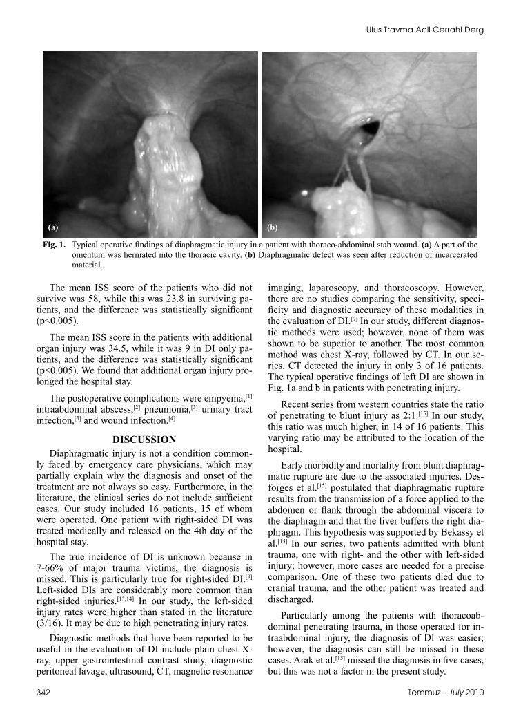
imaging, laparoscopy, and thoracoscopy. However, there are no studies comparing the sensitivity, speci-ficity and diagnostic accuracy of these modalities in the evaluation of DI.[9] In our study, different diagnos-tic methods were used; however, none of them was shown to be superior to another. The most common method was chest X-ray, followed by CT. In our se-ries, CT detected the injury in only 3 of 16 patients. The typical operative findings of left DI are shown in Fig. 1a and b in patients with penetrating injury.
Recent series from western countries state the ratio of penetrating to blunt injury as 2:1.[15] In our study, this ratio was much higher, in 14 of 16 patients. This varying ratio may be attributed to the location of the hospital.
Early morbidity and mortality from blunt diaphrag-matic rupture are due to the associated injuries. Des-forges et al.[15] postulated that diaphragmatic rupture results from the transmission of a force applied to the abdomen or flank through the abdominal viscera to the diaphragm and that the liver buffers the right dia-phragm. This hypothesis was supported by Bekassy et al.[15] In our series, two patients admitted with blunt trauma, one with right- and the other with left-sided injury; however, more cases are needed for a precise comparison. One of these two patients died due to cranial trauma, and the other patient was treated and discharged.
Particularly among the patients with thoracoab-dominal penetrating trauma, in those operated for in-traabdominal injury, the diagnosis of DI was easier; however, the diagnosis can still be missed in these cases. Arak et al.[15] missed the diagnosis in five cases, but this was not a factor in the present study.
Fig. 1. Typical operative findings of diaphragmatic injury in a patient with thoraco-abdominal stab wound. (a) A part of the omentum was herniated into the thoracic cavity. (b) Diaphragmatic defect was seen after reduction of incarcerated material.
(a) (b)
In penetrating traumas, there is no predisposing area of injury and the defect of the diaphragm is usu-ally smaller than in defects caused by blunt traumas; therefore, they are potentially more dangerous in terms of later obstruction and strangulation.[16] Eighty-four percent of DIs due to penetrating injury have a defect of less than 2 cm, but the defects due to blunt trauma are larger, with the majority being over 10 cm.[17] In our study, in one of the blunt trauma patients, the in-jury was as long as 10 cm, and in the other patient, the diaphragm was injured on three different sides. In two of the penetrating injury patients, the lesions were 4 cm, while in the other 11 patients, the lesions were smaller than 2 cm. In the patients treated medically, the lesions detected on CT measured 2 cm.
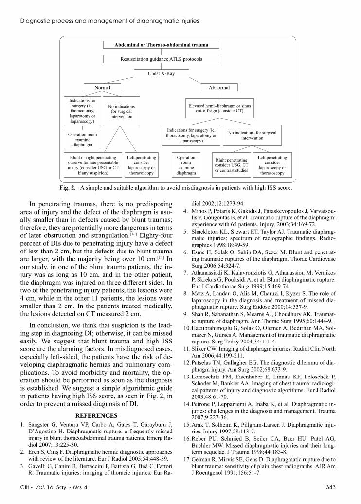
In conclusion, we think that suspicion is the lead-ing step in diagnosing DI; otherwise, it can be missed easily. We suggest that blunt trauma and high ISS score are the alarming factors. In misdiagnosed cases, especially left-sided, the patients have the risk of de-veloping diaphragmatic hernias and pulmonary com-plications. To avoid morbidity and mortality, the op-eration should be performed as soon as the diagnosis is established. We suggest a simple algorithmic guide in patients having high ISS score, as seen in Fig. 2, in order to prevent a missed diagnosis of DI.
REFERENCES1. Sangster G, Ventura VP, Carbo A, Gates T, Garayburu J,
D’Agostino H. Diaphragmatic rupture: a frequently missed injury in blunt thoracoabdominal trauma patients. Emerg Ra-diol 2007;13:225-30.
2. Eren S, Ciriş F. Diaphragmatic hernia: diagnostic approaches with review of the literature. Eur J Radiol 2005;54:448-59.
3. Gavelli G, Canini R, Bertaccini P, Battista G, Bnà C, Fattori R. Traumatic injuries: imaging of thoracic injuries. Eur Ra-
diol 2002;12:1273-94.4. Mihos P, Potaris K, Gakidis J, Paraskevopoulos J, Varvatsou-
lis P, Gougoutas B, et al. Traumatic rupture of the diaphragm: experience with 65 patients. Injury. 2003;34:169-72.
5. Shackleton KL, Stewart ET, Taylor AJ. Traumatic diaphrag-matic injuries: spectrum of radiographic findings. Radio-graphics 1998;18:49-59.
6. Esme H, Solak O, Sahin DA, Sezer M. Blunt and penetrat-ing traumatic ruptures of the diaphragm. Thorac Cardiovasc Surg 2006;54:324-7.
7. Athanassiadi K, Kalavrouziotis G, Athanassiou M, Vernikos P, Skrekas G, Poultsidi A, et al. Blunt diaphragmatic rupture. Eur J Cardiothorac Surg 1999;15:469-74.
8. Matz A, Landau O, Alis M, Charuzi I, Kyzer S. The role of laparoscopy in the diagnosis and treatment of missed dia-phragmatic rupture. Surg Endosc 2000;14:537-9.
9. Shah R, Sabanathan S, Mearns AJ, Choudhury AK. Traumat-ic rupture of diaphragm. Ann Thorac Surg 1995;60:1444-9.
10. Haciibrahimoglu G, Solak O, Olcmen A, Bedirhan MA, Sol-mazer N, Gurses A. Management of traumatic diaphragmatic rupture. Surg Today 2004;34:111-4.
11. Sliker CW. Imaging of diaphragm injuries. Radiol Clin North Am 2006;44:199-211.
12. Patselas TN, Gallagher EG. The diagnostic dilemma of dia-phragm injury. Am Surg 2002;68:633-9.
13. Lomoschitz FM, Eisenhuber E, Linnau KF, Peloschek P, Schoder M, Bankier AA. Imaging of chest trauma: radiologi-cal patterns of injury and diagnostic algorithms. Eur J Radiol 2003;48:61-70.
14. Petrone P, Leppaniemi A, Inaba K, et al. Diaphragmatic in-juries: challenges in the diagnosis and management. Trauma 2007;9:227-36.
15. Arak T, Solheim K, Pillgram-Larsen J. Diaphragmatic inju-ries. Injury 1997;28:113-7.
16. Reber PU, Schmied B, Seiler CA, Baer HU, Patel AG, Büchler MW. Missed diaphragmatic injuries and their long-term sequelae. J Trauma 1998;44:183-8.
17. Gelman R, Mirvis SE, Gens D. Diaphragmatic rupture due to blunt trauma: sensitivity of plain chest radiographs. AJR Am J Roentgenol 1991;156:51-7.
Diagnostic process and management of diaphragmatic injuries
Cilt - Vol. 16 Sayı - No. 4 343
Fig. 2. A simple and suitable algorithm to avoid misdiagnosis in patients with high ISS score.
Abdominal or Thoraco-abdominal trauma
Chest X-Ray
Normal Abnormal
Elevated hemi-diaphragm or sinus cut-off sign (consider CT)
Indications for surgery (ie, thoracotomy, laparotomy or
laparoscopy)
No indications for surgical intervention
Left penetrating consider
laparoscopy or thoracoscopy
Right penetrating consider USG, CT or contrast studies
Indications for surgery (ie,
thoracotomy, laparotomy or laparoscopy)
No indications for surgical intervention
Operation room examine
diaphragm
Blunt or right penetrating observe for late presentable injury (consider USG or CT
if any suspicion)
Left penetrating consider
laparoscopy or thoracoscopy
Operation room
examine diaphragm
Resuscitation guidance ATLS protocols