Embed Size (px)
Citation preview

Diagnostic Microbiologydr. Agus Eka Darwinata, S.Ked., Ph.D
A
E
D

Overview
Pathogen identification is essential for effective antimicrobial and supportive theraphy
Antimicrobial empirical treatment base on:
Microbiologic epidemiology
Patient’s sign and symptoms
A
E
D

Definitive microbiologi diagnostic:
Direct microscopic visualization
Cultivation and identification
Detection of microbial antigens
Detection of microbial DNA or RNA
Detection of host immune response to microorganism
A
E
D

“Patient history and Physical Examination is the most important”
A
E
D

“All laboratory studies must be directed by the patient’s history and physical examination”
Patient
Clinical Procedures (medical history and physical examination)
Laboratory diagnostic test and precedures
A
E
D

Medical Microbiology and Infection at a Glance, Fourth Edition. Stephen H. Gillespie, Kathleen B. Bamford. © 2012 John Wiley & Sons, Ltd. 14 Published 2012 by John Wiley & Sons, Ltd.
4 The laboratory investigation of infection
Clinicalspecimens
Microscopy
EXAMPLES OF SPECIMENS
Urinary tract infection (UTI) – Midstream urineWound – Pus or swabMeningitis – CSF & bloodPyrexia of unknown origin (PUO) – Blood for culture + serologyPneumonia – Sputum, lavage, serology
MICROSCOPY
Light – Direct (stool – parasites) – Gram (CSF – bacteria) – Z–N (sputum – TB) – Giemsa (blood – malaria)Immunofluorescence – Respiratory syncytial virus (RSV) diagnosisElectron microscopy (EM) – Virus detection and identification
EXAMPLES OF TYPINGMETHODS
• Phage• Serology• Bacteriocin• Endonuclease digestion• Multilocus sequence typing• Whole genome sequence
History andexamination
Differentialdiagnosis Culture
Susceptibility
Bacterialgrowth
Zone of inhibition
Filterpaper disc
Identification
Typing
Public healthmeasures
Diagnosis Treatment
37ºC
EXAMPLES OF SEROLOGICAL TECHNIQUES
• Agglutination• Precipitation• Complement fixation• Virus neutralization• Enyzme linked immunosorbent assay (ELISA)• Radioimmunoassay (RIA)• Immunofluorescence
EXAMPLES OFMOLECULAR TECHNIQUES
• DNA hybridization• Nucleic acid amplification testing – Polymerase chain reaction (PCR) – Ligase chain reaction (LCR) – Automated DNA amplification – Real time PCR
TYPES OF MEDIA
• Enrichment• Selective• Solid• Liquid
Atmosphere – Aerobic – CO2 – Anaerobic – Microaerophilic
Cell culture – Virus – Chlamydia
Serology
Nucleic aciddetection method
A
E
D

Direct Visualization of Microorganism
A
E
D

Direct Visualization of Microorganismpathogenic organisms (excluding viruses) can often be directly visualized by microscopic examination of patient specimens.
can provide the first screening step in arriving at a specific identification.
The organisms to be examined do not need to be alive or able to multiply.
Microscopy yields rapid and inexpensive results, allow the clinician to initiate treatment without waiting for the results of a culture.
A
E
D

Gram Stain
The most common and useful staining procedure is the Gram stain, which separates bacteria into two classifications according to their cell wall composition.
More about Gram stain:
h t tps: / /mikrob io log i fkunud.com/gram-biomedik2.html
A
E
D

Intepretation
“Shape and Gram” ex. Gram-negative rod, Gram-positive coccus
A
E
D
Gram stain applications: The Gram stain is important therapeutically because gram-positive and gram-negative bacteria differ in their susceptibility to various antibiotics, be used to guide initial therapy until the microorganism can be definitively identified.
Gram stain limitations: The number of microorganisms required is relatively high. Visualization with the Gram stain requires greater than 10 4 organisms/mL.
A
E
D

Acid-fast stainSta ins such as Z ieh l -Neelsen are used to identify organisms that have waxy material (mycolic acids) in their cell walls.
The most clinically important ac id- fas t bacte r ium is Mycobacterium tuberculosis, which appears pink, often beaded, and slightly curved
Mycobacterium tuberculosis stained with acid-fast stain
A
E
D
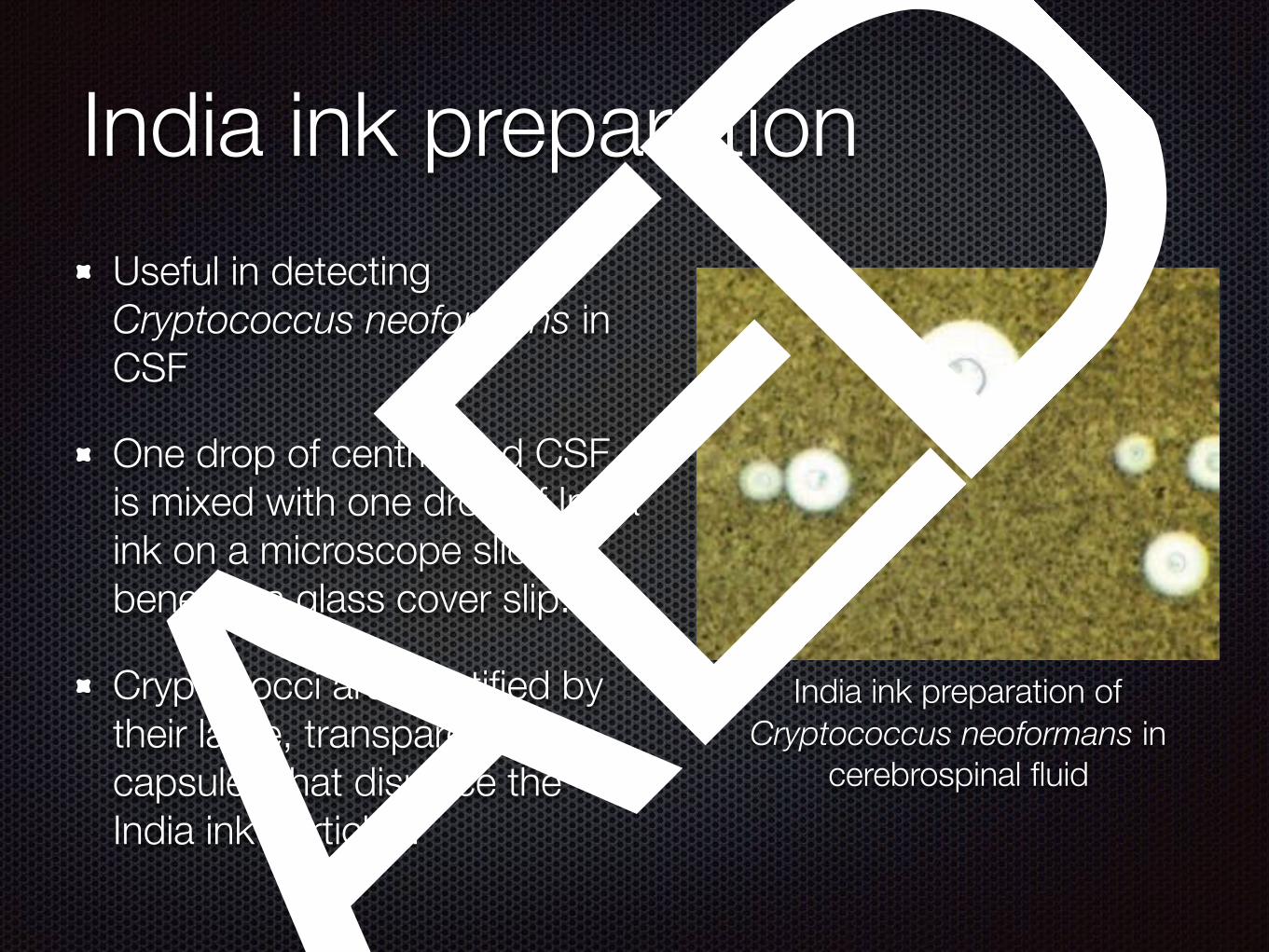
India ink preparationUseful in detecting Cryptococcus neoformans in CSF
One drop of centrifuged CSF is mixed with one drop of India ink on a microscope slide beneath a glass cover slip.
Cryptococci are identified by their large, transparent capsules that displace the India ink particles.
India ink preparation of Cryptococcus neoformans in
cerebrospinal fluid
A
E
D

Potassium hydroxide preparation (KOH)
Treatment with potassium hydroxide (KOH) dissolves host cells and bacteria, sparing fungi
One drop of sputum or skin scraping is treated with 10 percent KOH, and the specimen is examined for fungal forms.
Fungi in unstained nasal sinus exudate
A
E
D

GROWING BACTERIA IN CULTURECulturing is routine for most bacterial and fungal infections.
Microorganisms isolated in culture are identified using such characteristics as colony size, shape, color, Gram stain, hemolytic reactions on solid media, odor, and metabolic properties.
Pure cultures provide samples for antimicrobial susceptibility testing
The success of culturing depends on appropriate collection and transport techniques and on selection of appropriate culture media.
A
E
D
A
E
D

A B
A, Lactose-fermenting, gram-negative rods producing pink colonies on MacConkey (MAC).
B, Nonlactose-fermenting, gram-negative rods producing colorless colonies on MAC.
A
E
D

A, Lactose-fermenting Escherichia/Citrobacter-like organisms growing on MacConkey (MAC). Notice the dry appearance of the colony and the pink precipitate of bile salts extending beyond the periphery of the colonies. B, Close-up of dry, flat Escherichia/Citrobacter-like lactose fermenters growing on MAC
A B
A
E
D

A, Klebsiella/Enterobacter-like lactose fermenters growing on MacConkey (MAC). Notice the pink, heaped, mucoid appearance. B, Close-up of Klebsiella/Enterobacter-like colonies on MAC. Notice the mucoid, heaped appearance and the slightly cream-colored center after 48 hours’ growth.
A B
A
E
D

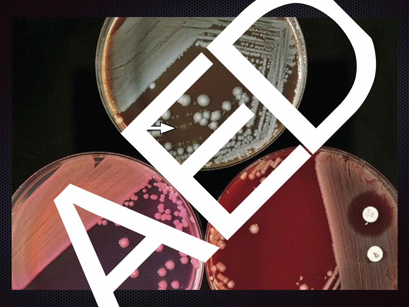
Left, blood agar plate (BAP): small, white colonies are gram-positive cocci. Right, BAP: large, gray, mucoid colonies are enteric gram-negative rods.
A
E
D

Illustration of form or margin to describe colonial morphology
A
E
D

Illustration of elevations to describe colonial
morphology
A
E
D
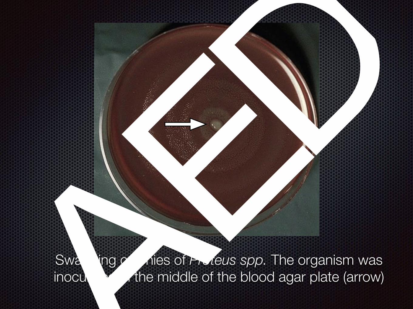
Swarming colonies of Proteus spp. The organism was inoculated in the middle of the blood agar plate (arrow)
A
E
D

“Diphtheroid” colonies with rough edges, dry appearance, and umbonate center growing on blood agar plate, buff color.
A
E
D
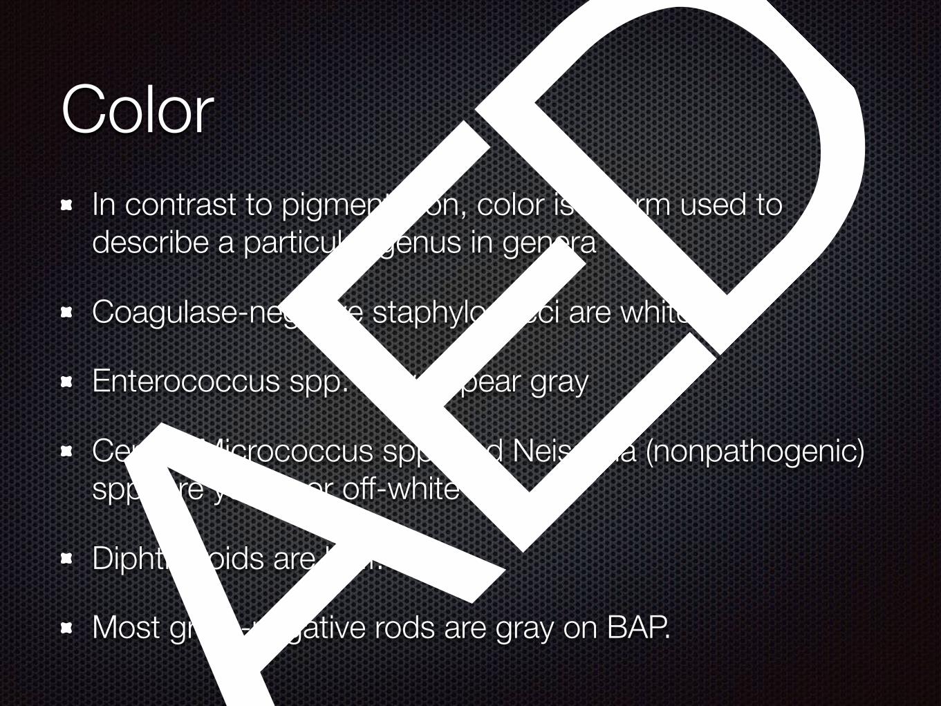
ColorIn contrast to pigmentation, color is a term used to describe a particular genus in genera
Coagulase-negative staphylococci are white
Enterococcus spp. may appear gray
Certain Micrococcus spp. and Neisseria (nonpathogenic) spp. are yellow or off-white
Diphtheroids are buff.
Most gram-negative rods are gray on BAP.
A
E
D

Example of white colonies of coagulase-negative staphylococci on blood agar plate
A
E
D

Example of the yellow colonies characteristic of certain nonpathogenic species of Neisseria organisms
on blood agar plate
A
E
D

PigmentP. aeruginosa—green, sometimes a metallic sheen
Serratia marcescens—brick-red, especially at room temperature
Kluyvera spp.—blue
Chromobacterium violaceum—purple
Prevotella melaninogenica—brown-black (anaerobic) Pigment production for these organisms is variable.
A
E
D

A,Pseudomonas aeruginosa illustrating the metallic sheen and green pigmentation of colonies on blood agar plate (BAP). B, Not all strains of the same organism have the same colonial appearance. This is a mucoid strain of P. aeruginosa on BAP.
A
E
D

Brick-red pigment of Serratia marcescens, which is evident on MacConkey (right). This brick-red pigment should not be confused
with lactose fermentation. The pigment is slightly visible on chocolate (left). Additional incubation at room temperature
enhances the brick-red pigmentation.
A
E
D

Brick-red pigment of Serratia marcescens, which is evident on MacConkey (right). This brick-red pigment should not be confused
with lactose fermentation. The pigment is slightly visible on chocolate (left). Additional incubation at room temperature
enhances the brick-red pigmentation.
A
E
D

OdorS. aureus—old sock (stocking that has been worn c ously for a few days without washing); this odor is evident when growing on mannitol salt agar
P. aeruginosa—fruity or grapelike
P. mirabilis—putrid
Haemophilus spp.—musty basement, “mousy” or “mouse nest” smell
Nocardia spp.—freshly plowed field
A
E
D

Identification of Bacteria
A
E
D

Single-enzyme tests
A
E
D

Automated system
A
E
D

Tests based on the presence of metabolic pathways
Rapid manual biochemical system for bacterial identification. Different appearances of the upper and lower pairs of wells indicate the positive
or negative ability of a bacterium to utilize each substrate.
A
E
D

Serum antigens or antibodies
Latex agglutination test A. Schematic representation
of antigens agglutinating latex beads with bound antibody.
B. Photograph of agglutination reaction.
A
E
D

Principle of enzyme-linked immunosorbent assay (ELISA)
A
E
D

Nucleic acid amplification for diagnosis
PCR: Band patterns appearing in lanes 1-4 are specific for the cytotoxin genes (vvhA) of V. vulnificus
A
E
D

Applications: Nucleic acid–amplification techniques are generally quick, easy, and accurate. A major use of these techniques is for the detection of organisms that cannot be grown in vitro or for which current culture techniques are insensitive.
Limitation: the occurrence of false-positives due to cross-contamination with other microorganisms’ nucleic acid. PCR tests are often costly and require skilled personnel.
A
E
D

SUSCEPTIBILITY TESTINGAfter a pathogen is cultured, its sensitivity to specific antibiotics serves as a guide in choosing antimicrobial therapy
Disk-diffusion method
A
E
D

A
E
D

A
E
D

Terima Kasih
A
E
D

![Theory of latency-insensitive design - Computer-Aided ...luca/research/lipTransactions.pdf · delay-insensitive circuits [19], [20]. A delay-insensitive circuit is designed to operate](https://img.dokumen.tips/doc/110x75/5e77b28d15933b649935c2f3/theory-of-latency-insensitive-design-computer-aided-lucaresearchliptransactionspdf.jpg)