Embed Size (px)
Citation preview

SECTION 1
DIAGNOSTIC APPLICATIONS
www.cambridge.org© in this web service Cambridge University Press
Cambridge University Press978-1-107-60924-2 - Case Studies in Pediatric Emergency and Critical Care UltrasoundEdited by David J. Mclario and John L. KendallExcerptMore information
www.cambridge.org© in this web service Cambridge University Press
Cambridge University Press978-1-107-60924-2 - Case Studies in Pediatric Emergency and Critical Care UltrasoundEdited by David J. Mclario and John L. KendallExcerptMore information
1 Right-upper-quadrant pain in a 10-year-old femaleMarla C. Levine, Eitan Dickman, and Alex C. Arroyo
HISTORY OF PRESENT ILLNESSA 10-year-old, previously healthy female began to complain ofright-upper-quadrant pain approximately 3 weeks prior to herED presentation. According to her mother, the pain seemedto wax and wane, but had increased in frequency and sever-ity, being particularly noticeable after eating. She had episodicnausea, but no vomiting or diarrhea, and denied other symp-toms or inter-current illness.
PHYSICAL EXAMINATIONGENERAL APPEARANCE: Obese female in no obvious distress.
VITAL SIGNS:
Temperature 98.6°F (37.0°C)Pulse 98 beats/minBlood pressure 92/60 mmHgRespirations 18 breaths/minOxygen saturation 100% on room air
HEENT: Normocephalic, atraumatic, PERRL, normal TMs,nose and oropharynx.
NECK: Supple neck with no pain to palpation or notablelymphadenopathy.
CARDIOVASCULAR: Regular rate and rhythm. Capillary refillless than 2 s.
LUNGS: Clear to auscultation bilaterally, no wheezes, rhonchi,or retractions.
ABDOMEN: Soft and non-distended. Tenderness to palpationin the right upper quadrant. Murphy’s sign was not elicited. No
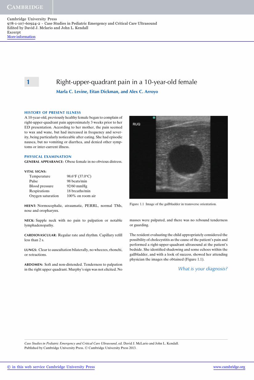
Figure 1.1 Image of the gallbladder in transverse orientation.
masses were palpated, and there was no rebound tendernessor guarding.
The resident evaluating the child appropriately considered thepossibility of cholecystitis as the cause of the patient’s pain andperformed a right-upper-quadrant ultrasound at the patient’sbedside. She identified shadowing and some echoes within thegallbladder, and with a look of success, showed her attendingphysician the images she obtained (Figure 1.1).
What is your diagnosis?
Case Studies in Pediatric Emergency and Critical Care Ultrasound, ed. David J. McLario and John L. Kendall.Published by Cambridge University Press. C© Cambridge University Press 2013.
www.cambridge.org© in this web service Cambridge University Press
Cambridge University Press978-1-107-60924-2 - Case Studies in Pediatric Emergency and Critical Care UltrasoundEdited by David J. Mclario and John L. KendallExcerptMore information
4 Section 1: Diagnostic Applications
ANSWER
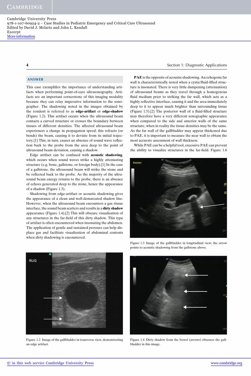
This case exemplifies the importance of understanding arti-facts when performing point-of-care ultrasonography. Arti-facts are an important cornerstone of this imaging modalitybecause they can relay imperative information to the sono-grapher. The shadowing noted in the images obtained bythe resident is referred to as edge-artifact or edge-shadow(Figure 1.2). This artifact occurs when the ultrasound beamcontacts a curved structure or crosses the boundary betweentissues of different densities. The affected ultrasound beamexperiences a change in propagation speed; this refracts (orbends) the beam, causing it to deviate from its initial trajec-tory.[1] This, in turn, causes an absence of sound wave reflec-tion back to the probe from the area deep to the point ofultrasound beam deviation, causing a shadow.
Edge artifact can be confused with acoustic shadowing,which occurs when sound waves strike a highly attenuatingstructure (e.g. bone, gallstone, or foreign body).[1] In the caseof a gallstone, the ultrasound beam will strike the stone andbe reflected back to the probe. As the majority of the ultra-sound beam energy returns to the probe, there is an absenceof echoes generated deep to the stone, hence the appearanceof a shadow (Figure 1.3).
Shadowing from edge-artifact or acoustic shadowing givesthe appearance of a clean and well-demarcated shadow line.However, when the ultrasound beam encounters a gas–tissueinterface, the sound beam scatters and results in a dirty shadowappearance (Figure 1.4).[2] This will obscure visualization ofany structures in the far-field of this dirty shadow. This typeof artifact is often encountered when insonating the abdomen.The application of gentle and sustained pressure can help dis-place gas and facilitate visualization of abdominal contentswhen dirty shadowing is encountered.
Figure 1.2 Image of the gallbladder in transverse view, demonstratingan edge artifact.
PAE is the opposite of acoustic shadowing. An echogenic farwall is characteristically noted when a cystic/fluid-filled struc-ture is insonated. There is very little dampening (attenuation)of ultrasound beams as they travel through a homogenousfluid medium prior to striking the far wall, which acts as ahighly reflective interface, causing it and the area immediatelydeep to it to appear much brighter than surrounding tissue(Figure 1.5).[2] The posterior wall of a fluid-filled structuremay therefore have a very different sonographic appearancewhen compared to the side and anterior walls of the samestructure, when in reality the tissue densities may be the same.As the far wall of the gallbladder may appear thickened dueto PAE, it is important to measure the near wall to obtain themost accurate assessment of wall thickness.
While PAE can be a helpful tool, excessive PAE can preventthe ability to visualize structures in the far-field. Figure 1.6
Figure 1.3 Image of the gallbladder in longitudinal view; the arrowpoints to acoustic shadowing from the gallstone above.
Figure 1.4 Dirty shadow from the bowel (arrows) obscures the gall-bladder in this image.
www.cambridge.org© in this web service Cambridge University Press
Cambridge University Press978-1-107-60924-2 - Case Studies in Pediatric Emergency and Critical Care UltrasoundEdited by David J. Mclario and John L. KendallExcerptMore information
1: Right-upper-quadrant pain in a 10-year-old female 5
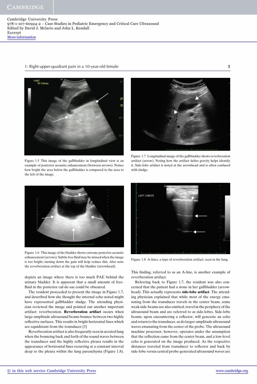
Figure 1.5 This image of the gallbladder in longitudinal view is anexample of posterior acoustic enhancement (between arrows). Noticehow bright the area below the gallbladder is compared to the area tothe left of the image.
Figure 1.6 This image of the bladder shows extreme posterior acousticenhancement (arrows). Subtle free fluid may be missed when the imageis too bright; turning down the gain will help reduce this. Also notethe reverberation artifact at the top of the bladder (arrowhead).
depicts an image where there is too much PAE behind theurinary bladder. It is apparent that a small amount of free-fluid in the posterior cul-de-sac could be obscured.
The resident proceeded to present the image in Figure 1.7,and described how she thought the internal echo noted mighthave represented gallbladder sludge. The attending physi-cian reviewed the image and pointed out another importantartifact: reverberation. Reverberation artifact occurs whenlarge-amplitude ultrasound beams bounce between two highlyreflective surfaces. This results in bright horizontal lines whichare equidistant from the transducer.[5]
Reverberation artifact is also frequently seen in aerated lungwhen the bouncing back and forth of the sound waves betweenthe transducer and the highly reflective pleura results in theappearance of horizontal lines recurring at a constant intervaldeep to the pleura within the lung parenchyma (Figure 1.8).
Figure 1.7 Longitudinal image of the gallbladder shows reverberationartifact (arrow). Noting how the artifact defies gravity helps identifyit. Side-lobe artifact is noted at the arrowhead and is often confusedwith sludge.
Figure 1.8 A-lines, a type of reverberation artifact, seen in the lung.
This finding, referred to as an A-line, is another example ofreverberation artifact.
Referring back to Figure 1.7, the resident was also con-cerned that the patient had a stone in her gallbladder (arrow-head). This actually represents side-lobe artifact. The attend-ing physician explained that while most of the energy ema-nating from the transducer travels in the center beam, someweak side-beams are also emitted, travel in the periphery of theultrasound beam and are referred to as side-lobes. Side-lobebeams, upon encountering a reflector, will generate an echoand return to the transducer, as do larger-amplitude ultrasoundwaves emanating from the center of the probe. The ultrasoundmachine processor, however, operates under the assumptionthat the reflection came from the center beam, and a low-levelecho is generated on the image produced. As the respectivedistances traveled from transducer to reflector and back byside-lobe versus central probe-generated ultrasound waves are
www.cambridge.org© in this web service Cambridge University Press
Cambridge University Press978-1-107-60924-2 - Case Studies in Pediatric Emergency and Critical Care UltrasoundEdited by David J. Mclario and John L. KendallExcerptMore information
6 Section 1: Diagnostic Applications
Figure 1.9 This image represents mirror artifact. The liver is mirroredabove the diaphragm (arrow). Notice how there appears to be thesame tissue above and below the diaphragm.
slightly different, the low-level returning echoes may misrep-resent the precise location of highly reflective tissue interfaces.
Side-lobe artifact is primarily encountered in fluid-filledstructures such as the gallbladder or urinary bladder. This arti-fact can be differentiated from actual sludge in that the artifactextends beyond the wall of the gallbladder and is often noted tobe floating, rather than sinking to the most dependent portionof the organ, as would be expected of a gallstone or sludge.
The resident also mentioned that it appeared as thoughliver tissue was both above and below the diaphragm in theright-upper-quadrant view (Figure 1.9). This artifact is referredto as mirror artifact. Mirror artifact occurs when ultrasoundwaves interact with a highly reflective curved surface, suchas the hemidiaphragm.[2] The ultrasound machine processorperforms distance calculations based on the assumption thatsound waves travel in a straight line, at a determined velo-city. Mirror artifact occurs when a portion of the ultrasoundwaves are delayed in their return to the transducer. The re-reflected waves will re-contact the curved surface (e.g. thediaphragm) and only then return to the transducer. The ultra-sound machine processor will account for this alteration insignal and timing by creating a duplicate image deep to theoriginal image.[2]
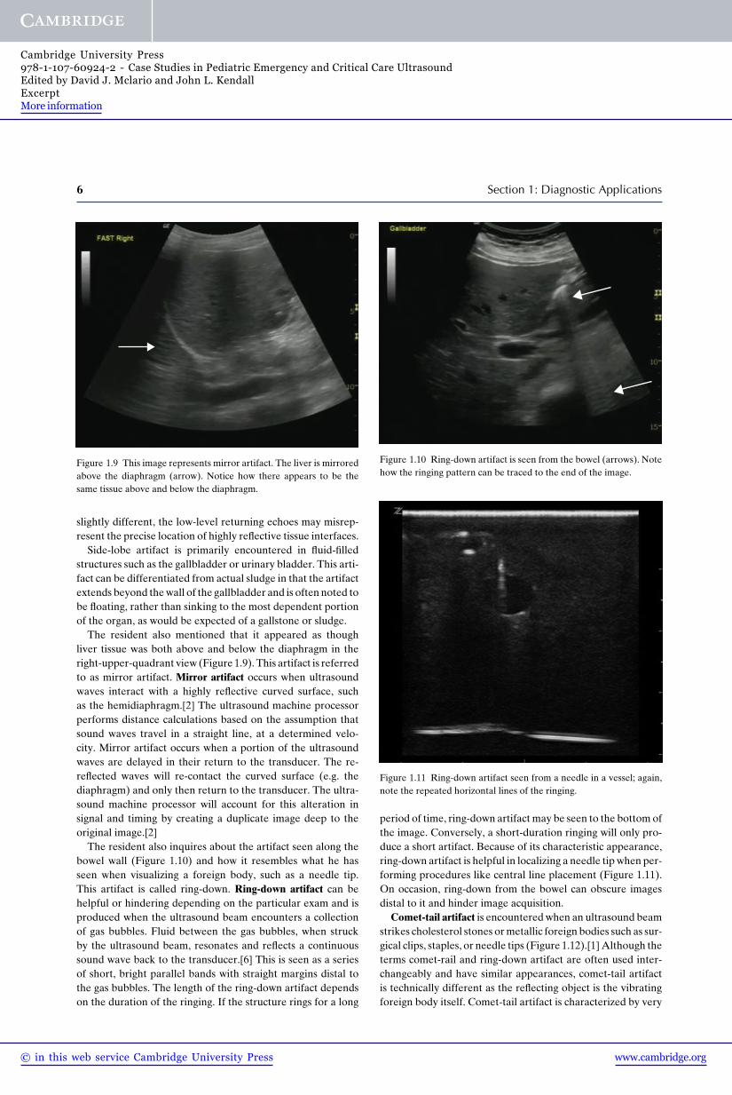
The resident also inquires about the artifact seen along thebowel wall (Figure 1.10) and how it resembles what he hasseen when visualizing a foreign body, such as a needle tip.This artifact is called ring-down. Ring-down artifact can behelpful or hindering depending on the particular exam and isproduced when the ultrasound beam encounters a collectionof gas bubbles. Fluid between the gas bubbles, when struckby the ultrasound beam, resonates and reflects a continuoussound wave back to the transducer.[6] This is seen as a seriesof short, bright parallel bands with straight margins distal tothe gas bubbles. The length of the ring-down artifact dependson the duration of the ringing. If the structure rings for a long
Figure 1.10 Ring-down artifact is seen from the bowel (arrows). Notehow the ringing pattern can be traced to the end of the image.
Figure 1.11 Ring-down artifact seen from a needle in a vessel; again,note the repeated horizontal lines of the ringing.
period of time, ring-down artifact may be seen to the bottom ofthe image. Conversely, a short-duration ringing will only pro-duce a short artifact. Because of its characteristic appearance,ring-down artifact is helpful in localizing a needle tip when per-forming procedures like central line placement (Figure 1.11).On occasion, ring-down from the bowel can obscure imagesdistal to it and hinder image acquisition.
Comet-tail artifact is encountered when an ultrasound beamstrikes cholesterol stones or metallic foreign bodies such as sur-gical clips, staples, or needle tips (Figure 1.12).[1]Although theterms comet-rail and ring-down artifact are often used inter-changeably and have similar appearances, comet-tail artifactis technically different as the reflecting object is the vibratingforeign body itself. Comet-tail artifact is characterized by very
www.cambridge.org© in this web service Cambridge University Press
Cambridge University Press978-1-107-60924-2 - Case Studies in Pediatric Emergency and Critical Care UltrasoundEdited by David J. Mclario and John L. KendallExcerptMore information
1: Right-upper-quadrant pain in a 10-year-old female 7
Figure 1.12 Short comet-tail artifact (arrow) from a cholesterol stonein the wall of a gallbladder.
narrow and closely spaced sound wave reverberation. This arti-fact, visualized deep to the insonated foreign body, not onlyconfirms its presence but assists in localization.
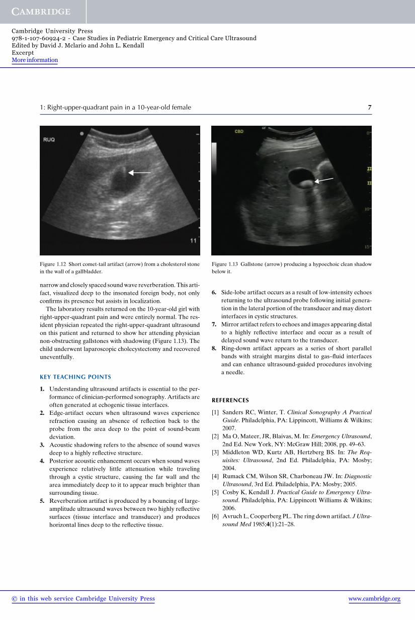
The laboratory results returned on the 10-year-old girl withright-upper-quadrant pain and were entirely normal. The res-ident physician repeated the right-upper-quadrant ultrasoundon this patient and returned to show her attending physiciannon-obstructing gallstones with shadowing (Figure 1.13). Thechild underwent laparoscopic cholecystectomy and recovereduneventfully.
KEY TEACHING POINTS
1. Understanding ultrasound artifacts is essential to the per-formance of clinician-performed sonography. Artifacts areoften generated at echogenic tissue interfaces.
2. Edge-artifact occurs when ultrasound waves experiencerefraction causing an absence of reflection back to theprobe from the area deep to the point of sound-beamdeviation.
3. Acoustic shadowing refers to the absence of sound wavesdeep to a highly reflective structure.
4. Posterior acoustic enhancement occurs when sound wavesexperience relatively little attenuation while travelingthrough a cystic structure, causing the far wall and thearea immediately deep to it to appear much brighter thansurrounding tissue.
5. Reverberation artifact is produced by a bouncing of large-amplitude ultrasound waves between two highly reflectivesurfaces (tissue interface and transducer) and produceshorizontal lines deep to the reflective tissue.
Figure 1.13 Gallstone (arrow) producing a hypoechoic clean shadowbelow it.
6. Side-lobe artifact occurs as a result of low-intensity echoesreturning to the ultrasound probe following initial genera-tion in the lateral portion of the transducer and may distortinterfaces in cystic structures.
7. Mirror artifact refers to echoes and images appearing distalto a highly reflective interface and occur as a result ofdelayed sound wave return to the transducer.
8. Ring-down artifact appears as a series of short parallelbands with straight margins distal to gas–fluid interfacesand can enhance ultrasound-guided procedures involvinga needle.
REFERENCES
[1] Sanders RC, Winter, T. Clinical Sonography A PracticalGuide. Philadelphia, PA: Lippincott, Williams & Wilkins;2007.
[2] Ma O, Mateer, JR, Blaivas, M. In: Emergency Ultrasound,2nd Ed. New York, NY: McGraw Hill; 2008, pp. 49–63.
[3] Middleton WD, Kurtz AB, Hertzberg BS. In: The Req-uisites: Ultrasound, 2nd Ed. Philadelphia, PA: Mosby;2004.
[4] Rumack CM, Wilson SR, Charboneau JW. In: DiagnosticUltrasound, 3rd Ed. Philadelphia, PA: Mosby; 2005.
[5] Cosby K, Kendall J. Practical Guide to Emergency Ultra-sound. Philadelphia, PA: Lippincott Williams & Wilkins;2006.
[6] Avruch L, Cooperberg PL. The ring down artifact. J Ultra-sound Med 1985;4(1):21–28.
www.cambridge.org© in this web service Cambridge University Press
Cambridge University Press978-1-107-60924-2 - Case Studies in Pediatric Emergency and Critical Care UltrasoundEdited by David J. Mclario and John L. KendallExcerptMore information

2 Motor vehicle accident evaluation in a 5-year-old maleAnnie Heffernan Rominger and David J. McLario
HISTORY OF PRESENT ILLNESSA 5-year-old male was transported to the Pediatric ED byEMS following a motor vehicle accident. He was a car-seat-restrained passenger in the rear of a vehicle that sustainedmoderate front-end damage after striking the side of anothervehicle ticketed for failure to yield at an intersection. Theestimated speed at impact was 30 mph. He was not ejected andwas found by EMS within the vehicle, strapped in the non-displaced car seat, awake, and crying. His 17-year-old brother,the driver of the vehicle in which he had been riding, wastransported to the adjacent Adult ED for suspected minorinjuries.
He arrived immobilized on a backboard with a cervical collarin place. According to the EMS report, he was alert duringtransport, with complaints of right shoulder and forearm pain.In view of the mechanism of injury, trauma team activationoccurred in advance of his arrival.
PHYSICAL EXAMINATIONGENERAL APPEARANCE: Patient was crying but spoke clearlywith c-collar applied appropriately.
VITAL SIGNS:
Temperature 97.5°F (36.4°C)Pulse 122 beats/minBlood pressure 105/62 mmHgRespirations 21 breaths/minOxygen saturation 100% on room air
HEENT: Normocephalic, PERRL, tympanic membranes nor-mal, nose atraumatic, oropharynx normal.
NECK: No cervical spine tenderness nor pain with activemotion.
CARDIOVASCULAR: Normal color, heart sounds and pulses,capillary refill 2 s.
LUNGS: Equal breath sounds bilaterally without retractionsor nasal flaring.
ABDOMEN: Non-tender, no lap-belt or other ecchymosis, nobruising.
GENITOURINARY: No blood at the urethral meatus, exam oth-erwise normal.
EXTREMITIES: Right forearm deformity, abrasion rightshoulder.
NEUROLOGICAL: Alert, GCS 15, normal motor and sensoryevaluation.
Following the primary survey, the Emergency Medicine fellowproceeded with a FAST examination (Figure 2.1).
What is your diagnosis?
Case Studies in Pediatric Emergency and Critical Care Ultrasound, ed. David J. McLario and John L. Kendall.Published by Cambridge University Press. C© Cambridge University Press 2013.
www.cambridge.org© in this web service Cambridge University Press
Cambridge University Press978-1-107-60924-2 - Case Studies in Pediatric Emergency and Critical Care UltrasoundEdited by David J. Mclario and John L. KendallExcerptMore information

2: Motor vehicle accident evaluation in a 5-year-old male 9
(a) (b)
(c) (d)
Figure 2.1 The patient’s FAST exam (a) subxiphoid, (b) peri-hepatic, (c) suprapubic, and (d) peri-splenic views.
www.cambridge.org© in this web service Cambridge University Press
Cambridge University Press978-1-107-60924-2 - Case Studies in Pediatric Emergency and Critical Care UltrasoundEdited by David J. Mclario and John L. KendallExcerptMore information
10 Section 1: Diagnostic Applications
ANSWER
The FAST is negative. Hemopericardium is not present andthere is no evidence of IPH. The patient was log-rolled andevaluation of his posterior torso and extremities was nor-mal. He had no TLS-spine tenderness or deformity and wasremoved from the backboard. Trauma labs were normal,including a bedside hematocrit of 43%.
Plain films of the chest, pelvis, and c-spine were negative. Aforearm x-ray revealed displaced fractures of the right radiusand ulna. The orthopedic service was consulted and reducedhis forearm fracture using a Bier Block. He was admitted forovernight observation where serial FAST and clinical abdomi-nal examinations performed by the trauma team were normal.He was discharged the following afternoon and made a com-plete recovery.
FAST – focused assessment with sonography for trauma
In 1971, the use of ultrasound in the evaluation of traumawas first reported in the literature.[1] Twenty-five years later,The American College of Surgeons advocated ultrasound usewithin the ATLS evaluation. Subsequently, FAST has sup-planted deep peritoneal lavage as the primary modality fordetection of trauma-induced IPH.
The FAST examination is based on the presumption thatfree fluid will follow gravity and accumulate in dependentregions of the peritoneal cavity. Also foundational is the factthat blood is sonographically anechoic (black) and contrastswith the more echoic (gray) echo-textures of adjacent solidorgans.[2] The FAST exam is not intended to be a detailedmulti-organ investigation of all possible abdominal pathology,but rather a screening evaluation for the detection of IPH inthe setting of trauma.
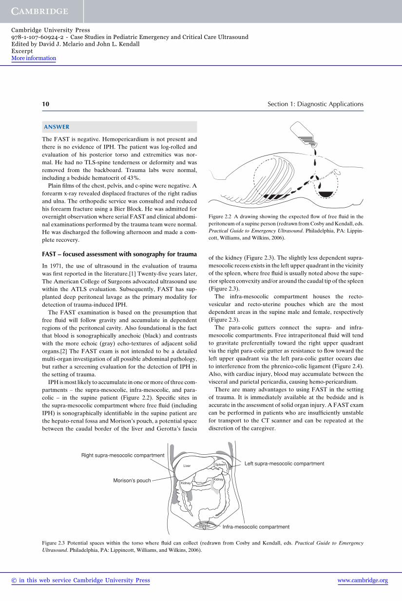
IPH is most likely to accumulate in one or more of three com-partments – the supra-mesocolic, infra-mesocolic, and para-colic – in the supine patient (Figure 2.2). Specific sites inthe supra-mesocolic compartment where free fluid (includingIPH) is sonographically identifiable in the supine patient arethe hepato-renal fossa and Morison’s pouch, a potential spacebetween the caudal border of the liver and Gerotta’s fascia
Morison’s pouch
Liver
BladderBladder
Right supra-mesocolic compartment
Infra-mesocolic compartment
Kidney
Bladder
Kidney
Spleen Left supra-mesocolic compartment
Figure 2.3 Potential spaces within the torso where fluid can collect (redrawn from Cosby and Kendall, eds. Practical Guide to EmergencyUltrasound. Philadelphia, PA: Lippincott, Williams, and Wilkins, 2006).
Figure 2.2 A drawing showing the expected flow of free fluid in theperitoneum of a supine person (redrawn from Cosby and Kendall, eds.Practical Guide to Emergency Ultrasound. Philadelphia, PA: Lippin-cott, Williams, and Wilkins, 2006).
of the kidney (Figure 2.3). The slightly less dependent supra-mesocolic recess exists in the left upper quadrant in the vicinityof the spleen, where free fluid is usually noted above the supe-rior spleen convexity and/or around the caudal tip of the spleen(Figure 2.3).
The infra-mesocolic compartment houses the recto-vesicular and recto-uterine pouches which are the mostdependent areas in the supine male and female, respectively(Figure 2.3).
The para-colic gutters connect the supra- and infra-mesocolic compartments. Free intraperitoneal fluid will tendto gravitate preferentially toward the right upper quadrantvia the right para-colic gutter as resistance to flow toward theleft upper quadrant via the left para-colic gutter occurs dueto interference from the phrenico-colic ligament (Figure 2.4).Also, with cardiac injury, blood may accumulate between thevisceral and parietal pericardia, causing hemo-pericardium.
There are many advantages to using FAST in the settingof trauma. It is immediately available at the bedside and isaccurate in the assessment of solid organ injury. A FAST examcan be performed in patients who are insufficiently unstablefor transport to the CT scanner and can be repeated at thediscretion of the caregiver.
www.cambridge.org© in this web service Cambridge University Press
Cambridge University Press978-1-107-60924-2 - Case Studies in Pediatric Emergency and Critical Care UltrasoundEdited by David J. Mclario and John L. KendallExcerptMore information