Embed Size (px)
Citation preview

1
Diagnostic and Treatment of Joint Diseases of Small Animals
UpdatesClinical Management

2
Introduction
Increase of the frequence of joint diseases in dogs and cats due to:
increase of life expectancymore medium - large breedsmore obese animalsIncrease of animal of predisposed breeds
Difficult to accurately diagnose

3
Structure of Joint
Synovial fluidfeeds the cartilage
+ lubricationArticular Cartilage« shock absorption »
Subchondral plate:« support »
Joint Capsule:ligament-likestructure
+ synovialmembrane

4
The Healthy Cartilage
Matrix: shock absorption80% water12% collagen2% proteoglycansother substances
Chondrocytes:synthesis of proteoglycanssecretion of catabolic enzymes
=> regulation system:balance synthesis / degradation
Sources: McIlwraith, C.W., Trotter, G.W. (1996): Joint disease in the horse., W.B. Saunders, Philadelphia, USA.

5
Structure of Cartilage Matrix

6
Physiology of Cartilage
Not irrigated by blood vessels=> specific nutrition system: « pumping »
DEPRESSIONaspiration of the synovial
fluid
COMPRESSIONrejection of waste in the
synovial fluid
WASTE NUTRIMENTS

7
Inflammatory Joint Diseases
Very numerous:more than 300 are listed in the litterature
Varied causes:immune mediated factorsmicrobial infectionsecondary effect of trauma

8
Inflammatory Joint Diseases
Immune mediated polyarthritis
Picture: Masahiro Okumura..

9
Non-Inflammatory Joint Diseases
Degeneration of joint structures, like cartilage:
OSTEOARTHRITIS
OSTEOARTICULAR DYSPLASIA

10
General Pathogenic Process
In case of joint disease:
Modification of cartilage matrix homeostasis
=> lowering of viscoelasticity
=> increase of frictions
This leads to degenerative disease= osteoarthritis (OA) = degenerative joint disease (DJD)

11
OA Pathogenic Process: Phase 1
Over-pressureon normal cartilage:
luxationunstability of the jointdysplasiaoverweight
OR Normal pressureon un-normal cartilage
tumoral processold cartilagegenetic malformationnutritionnal troubles

12
OA Pathogenic Process: Phase 2Hyperpressure
Increase ofMetalloproteases
Decreaseof synthesis
Release of cartilage fragments
PgE2
Inflammation
Activation of chondrocytes
Destructionof cartilage Matrix
PAIN

13
OA Pathogenic Process: Phase 3Hyperpressure
Increase ofMetalloproteasesDecrease
of synthesis
Release of cartilage fragments
Inflammation
Activation of chondrocytesIL 1 beta
Activation of synoviocytes
Destructionof cartilage Matrix
IL 1 betaIL6TNF alpha
PgE2
Synthesisof proteases
+m
acro
phag
es
PAIN

14
OA Pathogenic Process: Results
Fibrosis of the capsuleOsteophytesBone remodelling
Healthy OA
Biochemicaland MechanicalVicious Circle

15
General Physical ExaminationInterview of the owner:
Sudden / slow onset of the disease ?Date of apparition and evolution, cyclic or permanent ?General mobility of the animal + specific motion of the legs ?
Demeanour
Examination of the Gait
Neurological tests (to exclude neurological etiology)
16
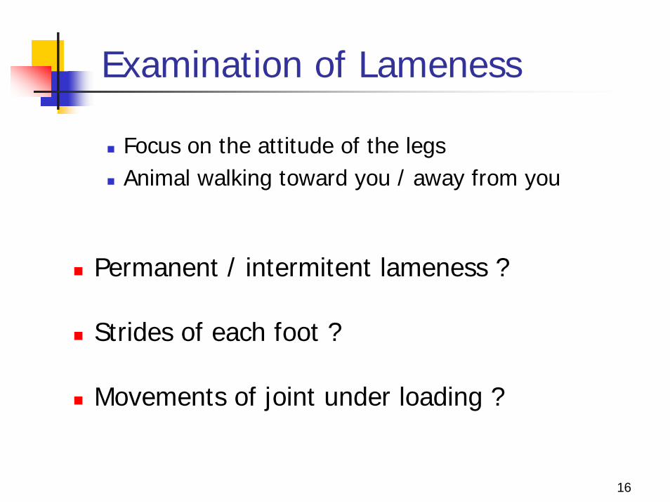
Examination of Lameness
Focus on the attitude of the legsAnimal walking toward you / away from you
Permanent / intermitent lameness ?
Strides of each foot ?
Movements of joint under loading ?

17
Orthopaedic Evaluation (1)
Animal standing still:
Posture ?Muscle atrophy ?Muscular reaction ?Pain along the spine, at the neck or the back ?

18
Orthopaedic Evaluation (2)
Palpation (1):identifiate painful bones, ligaments, tendons
Abnormalities ? Displacements ?Joints with fibrosis ?Pain ?In-stability ?

19
Orthopaedic Evaluation (3)
Palpation (2):
Restriction in the motion of any leg / joint ?Crepitus ?Condition of soft tissues around joint ?Swelling ?

20
Orthopaedic Evaluation (4)
Palpation of the joint of the elbow
Picture: Masahiro Okumura..

21
Orthopaedic Evaluation (5)
Palpation of the joint of the elbow
Picture: Masahiro Okumura..

22
Orthopaedic Evaluation (6)
Palpation of the joint of the hip
Picture: Masahiro Okumura..

23
Orthopaedic Evaluation (7)
Biodynamic tests:
To carry out on a sedated / anesthesied animal
To evaluate stability of the joint
Cranial drawer signOrtolani signBarden sign

24
Orthopaedic Evaluation (8)
Biodynamic tests:
Cranial drawer sign
anterior cruciate ligament rupture of the knee joint
Source: Whittick, W.G. (1990): canine Orthopedics. Lea & Febiger, Philadehphia, USA.

25
Orthopaedic Evaluation (9)
Biodynamic tests:
Ortolani sign
stability of hip joint having dyspasia
Source: Whittick, W.G. (1990): canine Orthopedics. Lea & Febiger, Philadehphia, USA.

26
Diagnostic Imaging
Changes in joint structure
Sources: McIlwraith, C.W., Trotter, G.W. (1996): Joint disease in the horse., W.B. Saunders, Philadelphia, USA.

27
Diagnostic Imaging: X-Ray
Time gap between the begining of the disease and the stage when the lesions are detectable by radiography
All lesions may not be all related to the present disease

28
Diagnostic Imaging: X-RayOA of elbow joint
Fragmented medial coronoid process
Remodeling of the subchondral bone
Source: Whittick, W.G. (1990): canine Orthopedics. Lea & Febiger, Philadehphia, USA.

29
Diagnostic Imaging: X-RayOA of hip joint
Source: Whittick, W.G. (1990): canine Orthopedics. Lea & Febiger, Philadehphia, USA.

30
Diagnostic Imaging: X-RayOA of knee joint
Source: Whittick, W.G. (1990): canine Orthopedics. Lea & Febiger, Philadehphia, USA.

31
Diagnostic Imaging: Arthroscopy
Interest:
direct observation of ligaments, tendons, synovial membrane, articular cartilage
biopsy of synovial membrane
excising, removing fragments

32
Diagnostic Imaging: Arthroscopy
Arthroscopy of the Elbow
Picture: Masahiro Okumura..
33
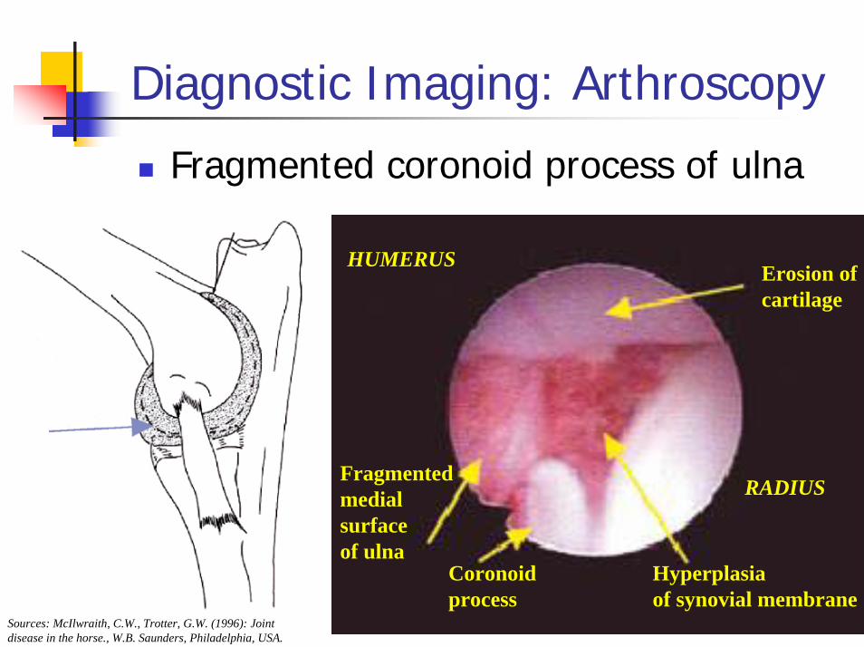
Diagnostic Imaging: Arthroscopy
Fragmented coronoid process of ulna
Erosion ofcartilage
Hyperplasiaof synovial membrane
Coronoidprocess
Fragmentedmedialsurfaceof ulna
HUMERUS
RADIUS
Sources: McIlwraith, C.W., Trotter, G.W. (1996): Joint disease in the horse., W.B. Saunders, Philadelphia, USA.

34
Diagnostic Imaging: Arthroscopy
Fragmented coronoid process of ulna
=> Inflammation of thesynovial membrane
Biceps tendon inthe shoulder joint =>

35
Diagnostic Imaging: Tomography
X-Ray CT:excellent for detecting calcification
X-Ray CT 3DMRI:
can show images of soft tissues like cartilage and tendon
Limits:costdeep sedation / anesthesia requiredresolution of the images obtained

36
Diagnostic Imaging: Tomography
X-Ray CT:Fragmented coronoid process of ulna
Picture: Masahiro Okumura..

37
Diagnostic Imaging: Tomography
X-Ray CT 3D:Fragmented coronoid process of ulna
HUMERUS
ULNA
RADIUSULNA
HUMERUS
Picture: Masahiro Okumura..
Picture: Masahiro Okumura..

38
Diagnostic Imaging: Tomography
Ultrasonography:
useful in detection of lesions of biceps and ligaments
not a practical examination for joints because of a very small acoustic window in this particular area

39
Tests on Synovial Fluid
Interesting to detect and evaluate:infectionsneoplasiainflammation
Puncturing:a perfect aseptia is required22-25G needle2ml syringe

40
Tests on Synovial Fluid
Elements to analyse:amount of fluidappearancecytological analysis
presence of PNN => inflammationPNN morphology: immune disease vs. infection
protein contentbacteriological culture test

41
Tests on Synovial Fluid
Amount of fluidAppearance
Picture: Masahiro Okumura..

42
Treatment Strategy
Conservative therapyremove the cause of the diseasecontrol pain and inflammationprotect the damaged cartilage
=> a Combined Drug Therapy is needed
Surgical treatmentoften last resort
when the control of painbecomes impossible
43
Conservative Therapy
Supportive treatment:
Mild exercice is needed every day:to maintain range of motion of jointto strengthen soft tissues around the jointto enhance the metabolic activity of cartilage
Weight controlto decrease the over-pressure on the cartilage

44
Conservative Therapy
Purposes of drug therapy:
Control of Pain, on short and long term for animal welfare & to support cartilage physiologyControl of InflammationModify the cartilage Matrix Metabolism
Long term treatment => drugs with minimal side effects are required

45
Conservative Therapy: Drugs (1)
Available drugs:NSAIDs: oral & injectableDMOAs: oral & injectable
(Disease-modifying osteoarthritis agents)
Steroids: oral & injectableMorphinics: oral & injectableLocal anesthetics

46
Conservative Therapy: Drugs (2)
All drugs are complementary:
DrugsReduction of
Pain Reduction of
InflammationProtection
of cartilageAdverse effects
NSAIDs ++, short term
++, short term
0/+ +
DMOAs ++, long term
++, long term
+++ 0
Steroids ++, temporary
+++ - - - ++
Morphinics ++ 0 0 + Local
anesthetics +++,
short action0 0 0

47
Conservative Therapy: Drugs (3)Hyperpressure
Increase ofMetalloproteases
Increase ofsynthesis
Release of cartilage fragments
Inflammation
Activation of chondrocytesIL 1 beta
Activation of synoviocytes
Destructionof cartilage Matrix
IL 1 betaIL6TNF alpha
PgE2
Synthesisof proteases
CS
CS
NSAID
NSAID
NSAID
NSAIDCS
CS
CS
CS
PAIN
MorphinicsLocal Anesth.
+macrophages
Steroids
Steroids
CS
CS = Chondroitin Sulfate (DMOA)

48
Conservative Therapy: Drugs (4)
Drug Strategy:
Common case: NSAID (short-medium term)
+ DMOA (from the beginning- long term effect)
If severe inflammation: + Steroids (short term)
If severe pain still present after NSAIDs:+ Local anesthetics+ Morphinics

49
Conservative Therapy: Drugs (5)
Available DMOAs:
Drugs Pro Cons Recommendations
Chondroitine Sulfate
- Anti-inflammatory + chondroprotective effect
- The predominant proteoglycan in the matrix
- Carry-over effect
- Needs 2 weeks to show effects by oral route
- Highly degradated in the digestive tract
- As soon as the begining of the treatment, in
combination with pain killers - Long term management
Hyaluronic acid - Lubrication role when
injected in the joint
- Difficult management of injections on the long
term
- Injection at the begining of the treatment
Glucosamine
- Lubrication role when injected in the joint
- Resistant to digestive enzymes
- More structural than anti-inflammatory effect
- No direct study in small animals
- Idem CS - May be completed with NSAIDs on the long term

50
Surgical Treatment
Arthrodesiscommon procedure
Replacement arthroplastyuse of artificial materials=>use for the hip joint of large dogs only
Excisional arthroplastyEasier, but functions reduced after surgery(except for the hip)

51
Surgical Treatment: Arhrodesis
Articulardisintegration
Picture: Masahiro Okumura..

52
Conclusion
Joint diseases concern welfare and quality of live
Easy and objective methods for diagnosisTreat the underlying diseasesRestore joint functionCombined therapy is needed for treatment, in addition to animal welfare & quality of life