Embed Size (px)
Citation preview
ANALYTICAL BIOCHEMISTRY 19, 4045 (1967)
Diagnosis of Homocystinuria by Gas Chromatography1
MELVIN GREER AND CLYDE M. WILLIAMS
Departments of Medicine and Radiology, University of Florida College of Medicine, Gainesville, Florida
Received September 8, 1966
Homocystinuria (l-3) is an inborn error of methionine metabolism associated with ectopia lentis, mental retardation, arterial and venous thromboses, and skeletal abnormalities similar to those associated with the Marfan syndrome (4). The disease is associated with a deficiency of cystathionine synthase (5)) which results in an elevated urinary excretion of homocystine. Homocystinuria may be relatively common since it has been reported second in incidence only to phenylketonuria in a survey of 2290 mentally defective individuals studied in Northern Ireland (6). It has also been estimated that 5% of cases of nontraumatic ectopia lentis have homocystinuria (7) and it is likely that a similar proportion of patients with the Marfan syndrome have homocystinuria (4). The early detection of patients with homocystinuria is important because of the possibility that dietary restriction of methionine or supplementation with cysteine may prove beneficial in preventing mental retardation and the vascular thromboses.
At the present time, there is no simple method for the determination of urinary homocystine. In our hospital, as in others, urine specimens from suspected cases are screened for sulfur-containing amino acids by the cyanide nitroprusside test (8). When this test is positive, the presence of homocystine must be confirmed by more specific analytical methods. These include: (1) a variation of the method of Spackman, Stein, and Moore (9) using an automatic amino acid analyzer, (W) a somewhat shorter quantitative method involving oxidation of homocystine followed by ion-exchange chromatography of homocysteic acid (lo), and (3) thin- layer chromatography of the dinitrophenyl derivative (11). We recently developed for the rapid determination of urinary homocystine by gas chromatography a simple method based upon the N-trifluoroacetyl methyl ester derivative.
IThis study was aided by grants from the National Institutes of Health (AM- @3391-50 and HDO 1326-M).
40
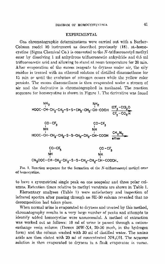
DIAGRAM OF HOMOCYSTINURIA 41
EXPERIMENTAL
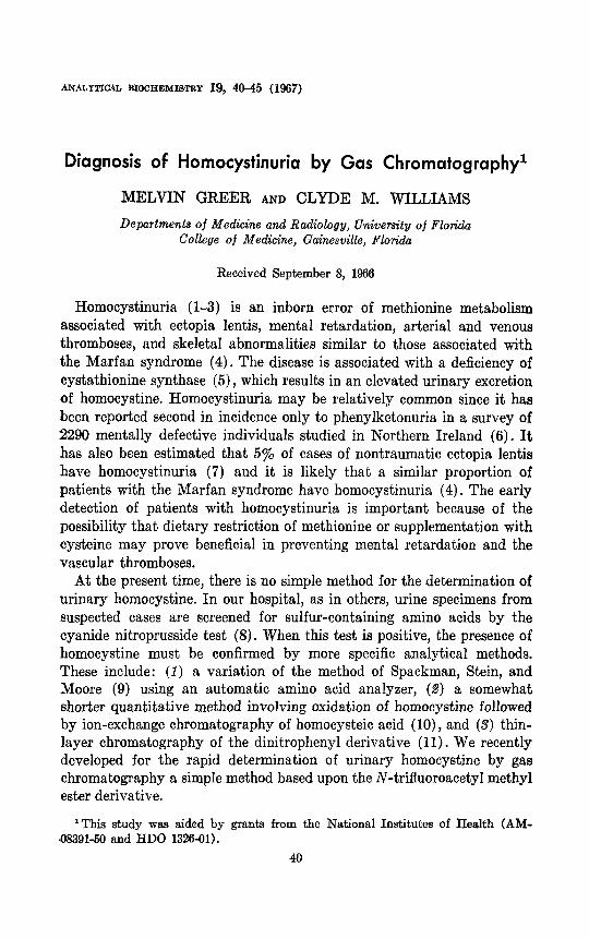
Gas chromatographic determinations were carried out with a Barber- Colman model 10 instrument as described previously (18). m-homo- cystine (Sigma Chemical Co.) is converted to the N-trifluoroacetyl methyl ester by dissolving 1 ml anhydrous trifluoroacetic anhydride and 0.5 ml trifluoroacetic acid and allowing to stand at room temperature for 20 min. After evaporation of the excess reagents to dryness under air, the oily residue is treated with an ethereal solution of distilled diazomethane for 15 min or until the evolution of nitrogen ceases while the yellow color persists. The excess diazomethane is then evaporated under a stream of air and the derivative is chromatographed in methanol. The reaction sequence for homocystine is shown in Figure 1. The derivative was found
NH I 2 y2
HOOC-CH-CH,-CH,-S-S-CH2-CH2-CH-COOH (CF3 -CO,,0
b CF,-CO,H
CO-CF3
tdH
CO-CF3
dH
HOOC-&I-CH2-CH2-S-S-CH,-CH,-C;I-COOH C”2N2)
ether
CO -CF3
tdH
CO-CF3
dH
CH300C-C;I-CHgCH,-S-S-CH,-CH2-Cii-COOCH,
FIG. 1. Reaction sequence for the formation of the N-trifluoroacetyl methyl ester of homocystine.
to have a symmetrical single peak on one nonpolar and three polar col- umns. Retention times relative to methyl veratrate are shown in Table 1.
Elementary analyses (Table 1) were satisfactory and inspection of infrared spectra after passing through an SE-30 column revealed that no decomposition had taken place.
When normal urine is evaporated to dryness and treated by this method, chromat,ography results in a very large number of peaks and attempts to identify added homocystine were unsuccessful. A method of extraction was worked out as follows: 10 ml of urine is passed through a cation- exchange resin column (Dowex 5OW-X4, 20-50 mesh, in the hydrogen form) and the column washed with 30 ml of distilled water. The amino acids are then eluted with 20 ml of concentrated NH,OH. The aqueous solution 6s then evaporated to dryness in a flask evaporator in vczcuo.
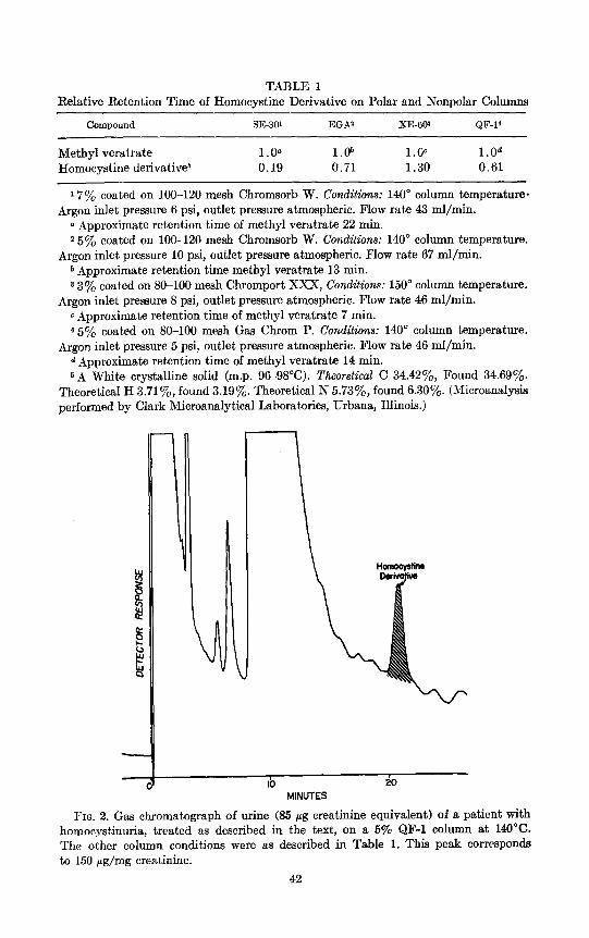
TABLE 1 Relative Retention Time of Homocystine Derivative on Polar and Nonpolar Columns
Compound SE-30’ EGA2 XE-603 &F-l’
Methyl veratrate Homocystine derivative6
1.05 l.ob l.OC l.Od 0.19 0.71 1.30 0.61
17% coated on loo-120 mesh Chromsorb W. Conditions: 140” column temperature. Argon inlet pressure 6 psi, outlet pressure atmospheric. Flow rate 43 ml/min.
0 Approximate retention time of methyl veratrate 22 min. 2 5y0 coated on loo-120 mesh Chromsorb W. Conditions: 140” column temperature.
Argon inlet pressure 10 psi, outlet pressure atmospheric. Flow rate 67 ml/min. b Approximate retention t,ime methyl veratrate 13 min. 8 3% coated on 80-100 mesh Chromport XXX, Conditiolas: 150” column temperature.
Argon inlet pressure 8 psi, outlet pressure atmospheric. Flow rate 46 ml/min. c Approximate retention time of methyl veratrate 7 min. 450J0 coated on 80-100 mesh Gas Chrom P. Conditions: 140” column temperature.
Argon inlet pressure 5 psi, outlet pressure atmospheric. Flow rate 46 ml/min. d Approximate retention time of methyl veratrate 14 min. 6A White crystalline solid (m.p. 96-98°C). Theoretical C 34.42%, Found 34.69’%.
Theoretical H 3.71y,, found 3.19%. Theoretical N 5.73%, found 6.30y0. (Microanalysis performed by Clark Microanalytical Laboratories, Urbana, Illinois.)
-
- IO 20
MINUTES
FIG. 2. Gas chromatograph of urine (85 pg creatinine equivalent) of a patient with homocystinuria, treated as described in the text, on a 5% &F-l column at 140°C. The other column conditions were as described in Table 1. This peak corresponds to 150 ag/mg creatinine.
42
DIAGNOSIS OF HOMOCYSTINURIA 43
Recoveries of homocystine added to urine in amounts ranging from 100 to 500 j.~g were 90-100%.
Homocystine is not detectable in the urine of norma children. In three cases of patients with ectopia lentis, mental retardation, and skeleta1 features suggesting the Marfan syndrome it was detected in abnormal amounts.
Samples of urine were treated as described above and then chromato- graphed. Figure 2 shows the appearance of a chromatograph of a specimen
IO $0
MINUTES
FIG. 3. Gas chromatograph of urine (550 pg creatinine equivalent) of normal control. Treatment and conditions as in Figure 2.
from a patient with homocystinuria. Note the large peak corresponding to the exact retention time of homocystine. The peak was trapped in a Teflon tube and rechromatographed on the other three columns (EGA, XE-60, SE-30). On each column, a single peak with the same width at half-height was found at the exact retention time of the homocystine derivative. Figure 3 shows a chromatograph of a urine specimen from a normal control treated in the same way. In this case no homocystine was detected at all, although six times as much extract (creatinine equivalent) was
44 GRJ3EB AND WILLIAMS
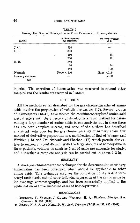
TABLE 2 Urinary Excretion of Homocystine in Three Patients with Homocyatinuria
J. C. D. B.
R. B.
Normala Homocystinuriea
(3)
150 390 130 200 880 20
180 None <l.O
-
- -
68 97
24 100
None <1.5 7-80
injected. The excretion of homocystine was measured in several other samples and the results are recorded in Table 2.
DISCUSSION
All the methods so far described for the gas chromatography of amino acids involve the preparation of volatile derivatives (12). Several groups of investigators (13-17) have studied the N-trifluoroacetylated amino acid methyl esters with the objective of developing a rapid method for deter- mining a large number of amino acids in one analysis, but in these there has not been complete success, and none of the authors has described analytical techniques for the gas chromatography of urinary acids. Our method of derivative preparation is a modification of that of Wagner and Winkler (15) and Cruickshank and Sheehan (17) which permits deriva- tive formation in about 45 min. With the large amounts of homocystine in these patients, volumes as small as 5 ml of urine are adequate for study, and altogether a complete analysis can be carried out in about 2-3 hr.
SUMMARY
A short gas chromatographic technique for the determination of urinary homocystine has been developed which should be applicable to other amino aoids. This technique involves the formation of the N-trifluoro- acetyl amino acid methyl ester following separation of the amino acids by ion-exchange chromatography, and has been successfully applied to the confirmation of three suspected cases of homocystinuria.
REFERENCES
1. GERRITSEN, T., VAUGHN, J. G., AND WAISMAN, H. A., Biochem. Biophys. Res. Commun. 9, 493 (1962).
2. CARSON, N. A. J., AND NEILL, D. W., Arch, Diseases Childhood 37, 505 (1962).
DIAGNOSIS OF HOMOCYSTINURIA 45
3. GERRITSEN, T., .~ND WAISMAN, H. A., in “The Metabolic Basis of Inherited Diseases” (J. B. Stanbury, J. B. Wyngaarden, and D. S. Fredrickson, eds.), 2nd ed., p. 420. McGraw-Hill, New York, 1966.
4. MCKUSICK, V. A., “Heritable Disorders of Connective Tissue,” pp. 150-178. Mosby, St.. Louis, 1966.
5. LASTER, L., MUDD, S. H., FINKELSTEIN, J. D., AND IRREVERRE, R., J. Clin. Invest. 44, 1708 (1965).
6. CARSON, N. A. J., DENT, C. E., FIELD, C. 111. B., AND GAULL, G. E., J. Pediat. 66, 565 (1965).
7. SCHIMKE, R. N., MCKUSICK, V. A., HUANG, T., AND POLL~CK, A. D., J. Am. Med. Assoc. 193, 711 (1965).
8. BRAND, E., HARRIS, M. M., AND BILOON, S., J. Biol. Chem. 86, 315 (1930). 9. SPACKMAN, D. H., STEIN, W. H., AND MOORE, S., Anal. Chem. 30, 1190 (1958).
10. GERRITSEN, T., AND WAISMAN, H. A., Pediatrics 33,413 (1964). 11. SUNDERMAN, I?. W., AND SUNDERMAN, F. W., JR., “Clinical Pathology of Infancy.”
C. C Thomas, Springfield, Ill., 1967. 12. FALES, H. M., AND PISANO, J. J., in “Biomedical Applications of Gas Chromatog-
raphy” (H. A. Szymanski, ed.), pp. 39-88. Plenum Press, New York, 1964. 13. BEYER, E., in “Gas Chromatography” (D. H. Desty, ed.), p. 333. Academic Press,
New York, 1958. 14. SAROFF, H. A., AND KARMEN, A., Anal. Biochem. 1,344 (1960). 15. WAGNER, J., AND WINKLER, G., 2. Anal. Chem. 183, 1 (1961). 16. WEYGAND, F., KOLB, B., PROX, A., TILAK, M. A., AND TOMIDA, I., Z. Physiol. Chem.
322, 38 (1960). 17. CRUICKSHANK, P. A., AND SHEEHAN, J. C., Anal. Chem. 36, 1191 (1964). 18. WILLIAMS, C. M., Anal. Biochem. 11, 224 (1965).


![Three Main Causes of Homocystinuria: of Metabolism ... · most frequent causes are classical homocystinuria [deficiency of cystathionine beta-synthase (CBS)], methylmalonic aciduria](https://img.dokumen.tips/doc/110x75/5e951dcb19bd325819567b57/three-main-causes-of-homocystinuria-of-metabolism-most-frequent-causes-are.jpg)


