Embed Size (px)
Citation preview

Chapter 1
Diagnosis of Gastritis – Review from Early PathologicalEvaluation to Present Day Management
Imre Laszlo Szabo, Kata Cseko, Jozsef Czimmer andGyula Mozsik
Additional information is available at the end of the chapter
http://dx.doi.org/10.5772/52884
1. Introduction
The gastritis is an inflammatory condition of the gastric mucosa characterized by existenceof elementary histological alternations. However these structural changes observed by thepioneer of gastric histology were noted more than a century ago, their etiology and properinterpretation for clinical practice required much longer time.
The ancient Egyptians wrote that the diseases of internal organs are difficult to detect evenin well-preserved bodies, hence they were not able to comprehend outstanding discoverieson the stomach as they did on other organ diseases. The first major discovery in the field ofgastric diseases was the description of gastric cancer by the Persian Avicenna around 1000(quoted by Rugge et al, 2003). At the same time the discoveries of non-neoplastic gastric dis‐eases, especially gastritis, was really elusive for quite a long time due to less macroscopicfeatures and to post-mortem alternations. The inflammation of the inner lining of the stom‐ach was first noted as “gastritis” by a German physician, Georg Ernst Stahl in 1728 (quotedby Bock, 1974). Italian anatomical pathologist Giovanni Battista Morgagni further describedthe signs of gastric inflammation. He gave the first classical description of an erosive or ul‐cerating gastritis. He stated that some of the erosions can become gangrenous, and descri‐bed corrosive gastritis as it was the most well-known gastritis form of that time due highnumber of lye intoxication. French physician, François-Joseph-Victor Broussais gathering in‐formation by autopsy of dead French soldiers between 1808 and 1831, described commonchronic gastritis as he called “Gastritides”, and sometimes got delusive conclusions as gas‐tritis was the cause of ascites and other diseases, like typhoid fever and meningitis (Bock,1974). Jones Handfield and Wilson Fox (1854) described microscopic changes of mucousmembrane in gastric inflammation, which exists in diffuse and segmental forms. Not much
© 2013 Szabo et al.; licensee InTech. This is an open access article distributed under the terms of the CreativeCommons Attribution License (http://creativecommons.org/licenses/by/3.0), which permits unrestricted use,distribution, and reproduction in any medium, provided the original work is properly cited.

later another British physician, William Brinton (1859) emphasized the symptomatic and mi‐croscopic differences of acut, subacute and chronic gastritis in his medical book entitled“Diseases of Stomach”, and described haemorrhagic erosion and follicular ulceration. Mean‐while Baron Carl von Rokitansky besides his major discoveries was the first to note hyper‐trophic gastritis in 1855. The next major footstep was done by Samuel Fenwick in 1870, whonoted the presence of glandular atrophy due to gastric inflammation when classifying gas‐tric lesions and anatomical alternations of the gastric mucosa (Fenwick, 1870). He also dis‐covered that pernicious anaemia is associated with gastric mucosal atrophy. Germansurgeon, Georg Ernst Konjetzny using surgical specimens showed first that both gastric ul‐cer and gastric cancer are either secondary diseases or are associated in their pathogenesis tochronic gastric inflammation. Shields Warren and Willam A. Meissner described intestinalmetaplasia of the stomach. They noted intestinal metaplasia as a feature of chronic gastritis,and found seldom extensive in duodenal ulcer patients, while it was extensive in stomachsremoved due to carcinoma (Warren & Meissner, 1944; Rugge et al, 2003).
2. In vivo diagnosis of gastritis – Introduction of gastroscopy
In vivo diagnosis of gastritis got a huge drive with the development of routine gastroscopy.By the 1950’s, Rudolf Schindler’s part-flexible endoscopes became very common making rig‐id endoscopes to disappear. From 1960’s, the commercial introduction of flexible endo‐scopes gave easy access for gastric biopsy and diagnosis of gastritis (Palmer, 1956). By theuse of biopsy based histology Schindler gave overview of gastritis in his monograph entitled‘Gastritis’ in 1947, he divided inflammation into ‘superficial’, ‘atrophic’ and ‘hypertrophic’gastritis chronica (Schindler, 1947). Cheli and Dobero in 1958 differentiated ‘superficial’, ‘in‐terstitial’ and ‘atrophic gastritis’ in the terminology of gastric inflammatory lesions (Cheli &Dobero, 1956). Up to his time classifications lack topography, but in 1972, Whitehead distin‐guished antral, fonical, corporal and pyloric region inflammation based on classical patho‐morphology. Whitehead divided chronic gastritis into ‘superficial’ and ‘atrophic’, both‘active’ or ‘in-active’ based on the presence of granulocyte infiltration in epithelium and in‐terstitium beside the inflammatory infiltration of lamina propria from lymphocytes andplasmatic cells (Whitehead et al, 1972). He suggested the use of a mild-moderate-severescale to evaluate the atrophy. He also introduced the evaluation of intestinal and pseudo‐pyloric metaplasia into everyday pathological assessment.
Based on recent research data, Robert G. Strickland and Ian R. MacKay proposed the classi‐fication of gastritis based on additional factors just beside just histology and topography(Strickland & Mackay, 1973). They suggested that immunological and etiological datashould be included along with pathomorphological and topographic parameters; gastric pa‐rietal cell antibody and serum level of gastrin have to be seen to get better classification ofchronic gastritis. They used the term ‘Type A gastritis’ for gastric corporal inflammationmostly corresponding to pernicious anaemia, and ‘Type B’ for antral gastritis suspected tobe induced by duodeno-gastric reflux according to some thoughts. In 1975 George B JerzyGlass and Capecomorin S. Pitchumoni added the ‘Type AB’ to the classification. This term
Current Topics in Gastritis - 20124

was aimed to be used for extended gastritis observed in corpus to pre-pyloric region (Glass& Pitchumoni, 1975). Those cases were named ‘AB-plus’ where antibody positivity was alsofounds against parietal cells. In 1980, the classification was further modified by Correa di‐viding chronic gastritis into autoimmune chronic gastritis with pernicious anaemia, ‘hyper‐secretory’ and ‘environmental’ forms. He described the gastritis accompanying ulcer tohypersecretory. All the rest of gastritis was called environmental, which are mostly due todiet and geographic localization (Correa, 1980). Later as more data were known from histo‐logical assessments, he changed his classification for ‘diffuse antral’, ‘diffuse corporal’ and‘multifocal’ gastritis. By seeing his nomenclature, sometimes showing etiology, sometimesreflecting topography, we are able to see the controversy existed between pathologist andclinicians in the field of gastritis at that time. The extensiveness in topography along withhistological and etiologic features were not to be combined in an uniformed nomenclature,even Correa in 1988 returned to his previous version of classification (Correa, 1988). Later,he went to different direction by dividing gastritis into two major categories of ‘atrophic’and ‘non-atrophic’ gastritis.
The next major step was added by Judith I. Wyatt and Michael F. Dixon by the introductionof ‘type C’ gastritis for chemical (drug)-induced inflammation of gastric mucosa (Wyatt &Dixon, 1988). Two years later, examining 316 patients Sobala confirmed that most of refluxgastritis in intact (non-operated) stomach is not due to bile reflux but rather NSAID use. Ac‐cording to their proposition the term ‘type C’ or ‘chemical’ gastritis might be used for condi‐tion caused by both etiology (Sobala et al, 1990).
3. Modern time – Development of the Sydney system
Modern aspects of gastritis classification and knowledge of its biological course and conse‐quences were relatively well-known at the time when Helicobacter pylori (H. pylori) was dis‐covered by Robin Warren and Barry Marshall in 1982 (Warren & Marshall, 1983). Theirdiscovery showed that the commonest form of gastritis is simply an infectious diseasecaused by an otherwise known pathogen. At that time gastroenterologist and pathologisthad limited knowledge on even simple aspects of this chronic bacterial inflammation of gas‐tric mucosa and the classification system used was confusing and differing from county toanother. Very soon considerable amount of data became known about H. pylori, its diseaseassociations and their natural courses by many physicians, microbiologist and basic re‐searchers entering the field. As a consequence in the late 1980's several pre-meeting ofWorking Party (Anthony Axon, Wladimir Bogomoletz, Michael F. Dixon, Steart Goodwin,Jules Haot, Konrad L. Heilmann, Adrian Lee, Barry Marshall, George Misiewicz, AshleyPrice, Penti Sipponen, Enrico Solcia, Manfred Stolte, Robert Strickland, Guido Tytgat) wasset up to review the biology and natural course of chronic gastritis and to propose a newclassification for gastritis by the leadership of George Misiewicz and Guido Tytgat. Theworking party actually consisted of two groups mainly working parallel to another: as apathological group and a clinical group (Sipponen & Price, 2011). Based on new etiologicalfacts and data collected, a new system of classification was presented at the World Congress
Diagnosis of Gastritis – Review from Early Pathological Evaluation to Present Day Managementhttp://dx.doi.org/10.5772/52884
5

of Gastroenterology held in Sydney, Australia in 1990, and subsequently published as sixpapers in the Journal Gastroenterology and Hepatology. The existence of the two Working Par‐ties reflects on the histological and endoscopic division of Sydney System. The histologicaldivision of Sydney System intended to be a practical guideline showing which of the mor‐phological features of gastritis in endoscopic biopsy specimens should be documented(Price, 1991). Type, severity and extent of gastric inflammation linked to possible etiologyshould be detailed according to a chart designed (see Fig. 1). The Sydney System declaredthe routine biopsy sampling protocol, the number of biopsies should be taken, the biopsies’proper localisation (two from antrum and two from corpus, both from anterior and posteri‐or walls) and sample fixation in adequately labelled separate containers (Misiewicz et al,1990; Price & Misiewicz, 1991). Many pathologist think to these last as the most importantconclusions of the system. The system also established a four-level scale for defining severi‐ty (extent) of pathomorphological elements.
Figure 1. Chart designed for the histological division of the original Sydney System as presented to the World Con‐gress of Gastroenterology. Published in Journal of Gastroenterology and Hepatology in 1991. Describes the nomencla‐ture should be used in histological reporting of gastritis. Adopted etiological suffix phrases to topography andmorphological features with grading suffixes to be documented in endoscopic biopsy reporting.
Current Topics in Gastritis - 20126
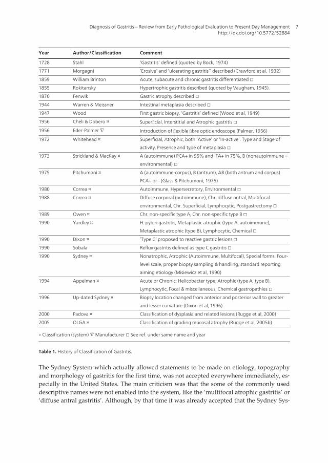
Year Author/Classification Comment
1728 Stahl ‘Gastritis’ defined (quoted by Bock, 1974)
1771 Morgagni ’Erosive’ and ’ulcerating gastritis’’ described (Crawford et al, 1932)
1859 William Brinton Acute, subacute and chronic gastritis differentiated □
1855 Rokitansky Hypertrophic gastritis described (quoted by Vaugham, 1945).
1870 Fenwik Gastric atrophy described □
1944 Warren & Meissner Intestinal metaplasia described □
1947 Wood First gastric biopsy, ‘Gastritis’ defined (Wood et al, 1949)
1956 Cheli & Dobero ¤ Superficial, Interstitial and Atrophic gastritis □
1956 Eder-Palmer ∇ Introduction of flexible fibre optic endoscope (Palmer, 1956)
1972 Whitehead ¤ Superficial, Atrophic, both ‘Active’ or ‘In-active’. Type and Stage of
activity. Presence and type of metaplasia □
1973 Strickland & MacKay ¤ A (autoimmune) PCA+ in 95% and IFA+ in 75%, B (nonautoimmune =
environmental) □
1975 Pitchumoni ¤ A (autoimmune-corpus), B (antrum), AB (both antrum and corpus)
PCA+ or - (Glass & Pitchumoni, 1975)
1980 Correa ¤ Autoimmune, Hypersecretory, Environmental □
1988 Correa ¤ Diffuse corporal (autoimmune), Chr. diffuse antral, Multifocal
environmental, Chr. Superficial, Lymphocytic, Postgastrectomy □
1989 Owen ¤ Chr. non-specific type A, Chr. non-specific type B □
1990 Yardley ¤ H. pylori gastritis, Metaplastic atrophic (type A, autoimmune),
Metaplastic atrophic (type B), Lymphocytic, Chemical □
1990 Dixon ¤ ’Type C’ proposed to reactive gastric lesions □
1990 Sobala Reflux gastritis defined as type C gastritis □
1990 Sydney ¤ Nonatrophic, Atrophic (Autoimmune, Multifocal), Special forms. Four-
level scale, proper biopsy sampling & handling, standard reporting
aiming etiology (Misiewicz et al, 1990)
1994 Appelman ¤ Acute or Chronic; Helicobacter type, Atrophic (type A, type B),
Lymphocytic, Focal & miscellaneous, Chemical gastropathies □
1996 Up-dated Sydney ¤ Biopsy location changed from anterior and posterior wall to greater
and lesser curvature (Dixon et al, 1996)
2000 Padova ¤ Classification of dysplasia and related lesions (Rugge et al, 2000)
2005 OLGA ¤ Classification of grading mucosal atrophy (Rugge et al, 2005b)
◦ Classification (system) ∇ Manufacturer □ See ref. under same name and year
Table 1. History of Classification of Gastritis.
The Sydney System which actually allowed statements to be made on etiology, topographyand morphology of gastritis for the first time, was not accepted everywhere immediately, es‐pecially in the United States. The main criticism was that the some of the commonly useddescriptive names were not enabled into the system, like the ‘multifocal atrophic gastritis’ or‘diffuse antral gastritis’. Although, by that time it was already accepted that the Sydney Sys‐
Diagnosis of Gastritis – Review from Early Pathological Evaluation to Present Day Managementhttp://dx.doi.org/10.5772/52884
7

tem was not designed to be the textbook of gastric pathology, but to be a guide for standardmethology of reporting. Correa and Yardley criticized the system for missing out certaintypes of the gastritis and well as it is not a ‘classification’ (Correa & Yardley, 1992). Conse‐quently, a new system needed to gain wider acceptance.
In 1994, a two-day consensus meeting was held in Houston. After this another consensus re‐port, “Up-dated Sydney System” was published in 1997 (Dixon et al, 1996). Original classifi‐cation of gastritis dividing into acute, chronic and special forms, and grading of chronicinflammation, polymorph activity, atrophy, intestinal metaplasia and H. pylori density intomild, moderate and marked categories were kept. This up-dated system introduced a visualanalogue scale for evaluating the severity of histopathological elements (grading). Itchanged the routine of endoscopic biopsy sampling by the introduction of biopsy samplingfrom the incisura angularis and modified corpus and antrum biopsy locations from the twoopposite walls to lesser and greater curvature of both parts. The Up-dated Sydney Classifi‐cation received different reactions among pathologists. Most of the pathologist agreed withthe need of incisural biopsy, since the most degree of atrophy and intestinal metaplasia isfound in the incisural region. That would reduce the sampling error of missing premalig‐nant lesions and improve the diagnosis of multifocal gastritis. However, later prospectivestudies could not really show its benefit (Stolte & Meining, 2001). Even in our conductedstudy higher number of intestinal metaplasia were found in antral biopsies then in the biop‐sies taken from the incisura angularis (Szabo et al, 2012). After the development of the visualanalogue scale according to the Up-dated Sydney System, the grading of atrophy still con‐tinued to show a considerable inter-observer variability (El-Zimaity et al, 1996). The updat‐ed system categorised chronic gastritis into ‘non-atrophic’ and ‘atrophic’ forms with thelatter divided into autoimmune (diffuse corpus atrophy) and multifocal. Histological report‐ing of gastritis should take into account the topographical pattern (antral or corpus predom‐inant), and the final diagnostic term should ideally combine morphology and etiology tomaximize the clinical value of gastric biopsy diagnosis (Dixon et al, 1997). The up-dated sys‐tem beside its major benefits in further standardizing endoscopic sampling, histological as‐sessment and formality of reporting, still showed weaknesses specially in grading atrophyas pointed out by Johan A. Offerhaus in 1999 (see ref). His proposition was to simplify thegrading system to two grades (low and high).
4. Classification by Appleman
The clearest division of gastritis for clinicians was published by Appleman in 1994. He div‐ided gastric inflammatory diseases to acute and chronic (see Table II). The most commonform of gastritis that was called earlier as chronic diffuse antral gastritis, gastritis chronictype B, gastritis chronica active antralis, gastritis non-specifica or gastritis typus hypersecre‐tions was named as Helicobacter pylori related gastritis. At this time lot of work proved that H.pylori infection causes chronic gastritis in the prepyloric region later leading to atrophy ofglands and development of gastric adenocarcinoma and less frequently of lymphoma (Ap‐pelman, 1994, Kozlowski et al, 2011).
Current Topics in Gastritis - 20128

According to Appelman’s classification the autoimmune gastritis used to be called as gastri‐tis autoimmunogenes, gastritis chronic atrophica typus A, gastritis chronic typus A and gas‐tritis chronic diffusa corporis, was called to autoimmune chronic atrophic gastritis. Appelmanpointed out the presence of autoantibodies against parietal cells and intrinsic factor beingimportant in diagnosis, enterochromaffinlike (ECL) cell hyperplasia and risk of carcinoma.
Appelman’s classification of gastritis continues with the multifocal atrophic gastritis earliercalled as environmental gastritis or type B chronic atrophic gastritis. At that time the causeof this form of gastritis was not clearly known. Beside known environmental factors respon‐sible for geographic differences in its epidemiology, raising circumstantial evidences froman Italian study examining gastric distribution of H. pylori, pointed out the role of H. pyloriin its generation (Rugge et al, 1993). Evidences suggested that H. pylori first infects the an‐trum, and later it involves the body leading to atrophic gastritis.
Appelman seeing similarity of the histological changes of patients with gastroenteric anasto‐mosis and taking nonsteroidal anti-inflammatory (NSAID) medications, called third divi‐sion of gastritis caused by bile reflux or NSAIDs to chemical gastropathies. Due to lessinflammation these histological changes consisting foveolar hyperplasia, decrease of mucinin foveolar cells, superficial oedema, increase of smooth muscle fibres in the lamina propriawere named as ‘gastropathies’. Recognition of this distinction of gastritis greatly helped tosimply classification, although many times elements histological changes usually found inchemical gastropathy can be noticed in other forms of gastritis as well as in other gastric dis‐ease. Finding them singular and unassociated wit other changes like atrophy, intestinal met‐aplasia, presence of bacteria, ulcers, polyps, should raise the possibility of chemical gastritis.
Appelman kept the name of lymphotic gastritis used by his frontiers for the fourth distinctiveform of gastritis (Haot et al, 1988, 1990). In this form of chronic gastritis huge lymphocyticinfiltration of the surface epithelium, superficial pits and lamina propria can be observed.Others used to call this as superficial gastritis, gastritis chronic erosive or gastritis variolifor‐mis. That time in 1990, the histological changes seen in lymphocytic gastritis was already de‐scribed in patients with sprues and gluten-sensitivity. Lymphocytic gastritis tends to form“varioliform gastritis” endoscopically. This includes thick folds and small bumps with cen‐tral depression seen during endoscopy. But lymphocytic gastritis also can form giant foldsleading clinical symptoms (Ménétrier’s disease).
Appelman’s division of gastritis contained a miscellaneous group of gastritis. There aremany gastritis forms that do not differ significantly from similar inflammations found otherorgans, including those that occur in syphilis, mycobacterial and cytomegalovirus, humanimmunodeficiency virus infections, histoplasmosis, candidiasis, cryptosporidiosis and otheropportunistic fungi. There is a family of granulomatous reactions or granulomatous gastritis.Some of these are part of a systemic or focal gut granulomatous disease, such as sarcoidosisor Crohn’s disease, and some have been described as part of a systemic vasculitis syndromeor Whipple’s disease. There are still others which are not associated with any other diseasesand designated as ‘isolated granulomatous gastritis’. Allergic gastritis is usually part of a gas‐trointestinal allergic disease. Appelman also categorized the recently described collagenousgastritis into this miscellaneous group.
Diagnosis of Gastritis – Review from Early Pathological Evaluation to Present Day Managementhttp://dx.doi.org/10.5772/52884
9

Acute Acute infectious gastritis (including Hp)
Erosive (caused mostly by NSAID or alcohol)
Necrotising and haemorrhagic (caused mostly by ischaemia)
Chronic Helicobacter pylori type
Atrophic Type A: autoimmune, diffuse
Type B: non-autoimmune, multifocal, enviromental
Lymphocytic Including varioliform, ’sprue-like’ and Ménétrier-like
Chemical¤ Bile reflux
NSAIDs
others (caused by other damaging agents and physical trauma)
Miscellaneous Granulomatous (part of Crohn’s, Whipple’s, vasculitis, sarcoidosis or
isolated granulomatous gastritis)
Allergic
Specific infectious (HIV, mycobacterial, syphilis, Cytomegalovirus,
histoplasmosis, cryptosporidiosis
Collagenous
◦ Gastropathies
Table 2. Appleman’s classification of gastritis (1994)
5. Precancerous lesions
Warren and Meissner describing intestinal metaplasia and recognising the clinical-patholog‐ical pattern of gastritis, described the bases of etiopathogenic relationship between gastriccancer and chronic gastritis (Warren & Meissner, 1944; Rugge et al, 2003). In 1980, Morson etal. (see ref.) defined gastric precancerous conditions as atrophic gastritis, gastric ulcer, perni‐cious anaemia, gastric stumps, gastric polyps, and Ménétrier's disease. They emphasizedthat epithelial dysplasia being a precancerous lesion is common in these conditions; dyspla‐sia should be graded as mild, moderate and severe; and underlined the problems of differ‐entiating inflammatory or regenerative changes from mild dysplasia, and intramucosalcarcinoma from severe dysplasia (Morson et al, 1980). Japanese pathologists by studying se‐rial sections of gastric mucosa obtained from gastric cancer patients described several bor‐der line lesions with histological and cytological changes. The premalignant significance ofthese was questioned for quite a long time; finally, the long-term follow-up studies closedthis debate (Rugge et al, 1994, 1997). The high inter-observer inconsistency in histological as‐sessment of premalignant lesions and new result supporting their neoplastic intraglandularnature obtained from genotyping studies highlighted the need of a broad consensus to re-
Current Topics in Gastritis - 201210

define precancerous lesions uniformly. International group of pathologists met in Padova,Italy in April, 1998 on an international consensus conference. The conference reached anagreement on the definitions of the spectrum of gastric premalignant lesions and on com‐mon glossary for pathologist and clinicians, and applied strict diagnostic criteria (Rugge etal, 2000) (see Table III).
Negative for dysplasia 1.0 Normal
1.1 Reactive foveolar hyperplasia
1.2 Intestinal metaplasia 1.2.1 Complete type
1.2.2 Incomplete type
Indefinite for dysplasia 2.1 Foveolar hyperproliferation
2.2 Hyperproliferative intestinal metaplasia
Non-invasive neoplasma
(flat or elevated)
3.1 Low-grade
3.2 High-grade 3.2.1 Including suspicious for carcinoma without invasion
(intraglandular)
3.2.2 Including carcinoma without invasion (intraglandular)
Suspicious for invasive carcinoma
Invasive carcinoma
Table 3. Padova Classification of gastric dysplasia and related lesions (2000)
6. Evaluation of atrophy
The Sydney System and Up-dated Sydney System attempted to incorporate etiologic, topo‐graphic, and morphologic criteria into a clinically relevant scheme to reach a broad consen‐sus in classification of gastritis. One of the most controversial issues at the HoustonWorkshop was the concept of atrophy. It was pointed out that "normal" was not preciselydefined; the loss of appropriate glands occurs with distinct patterns and has different func‐tional significance in antrum and corpus; the relationship between atrophy and intestinalmetaplasia remained incompletely understood; and the topographic patterns of distributionand its evolution made the atrophic gastritis to the most controversial topic of gastritis (Gen‐ta, 1996). Later long-term follow-up studies have confirmed that the extent of gastric mucos‐al atrophy parallels gastric cancer risk (Meining et al, 2002; Sipponen et al, 1985, 1994, 1997;Stolte et al, 2000). At the same time Sydney System did not present a reporting terminologyfor chronic gastritis understandable and providing prognostic and therapeutic informationfor clinicians. Whereas, hepatitis staging had already improved useful, simple terminologyfor interdisciplinary communication representing disease progression and cancer risk.
Diagnosis of Gastritis – Review from Early Pathological Evaluation to Present Day Managementhttp://dx.doi.org/10.5772/52884
11

Inspired by these facts, international group of gastroenterologists and pathologists named asOperative Link on Gastritis Assessment (OLGA) developed an improved histological stagingsystem for gastric atrophy (Rugge & Genta, 2005a, 2005b). OLGA system uses the gastric biop‐sy sampling protocol defined by Sydney System and the visual analogue system recommend‐ed by the Up-dated Sydney System. The gastritis staging is defined from combined extent ofatrophy scored histologically with the topography of atrophy identified through biopsy map‐ping (see Fig. 2). Long-term follow-up studies proved that gastritis OLGA staging conveys rele‐vant information on clinic-pathological outcome of gastritis and therefore H. pylori negativepatients with low OLGA stages could be confidently excluded from secondary preventive sur‐veillance invasive procedures (Rugge et al, 2010). Whereas patients with high OLGA stages(Stages III and IV) should be considered definitely candidates for endoscopic surveillance. Sig‐nificant correlation was shown between OLGA stages and pepsinogen serology (marker of gas‐tric atrophy). The ratio of pepsinogen I and II gives adequate information on the severity ofatrophy, but its measurement fails to differentiate between neoplastic and non-neoplastic dis‐ease among patients with high stages of gastric mucosal atrophy (Rugge et al, 2010).
Similar to the OLGA system another system, called the Baylor system was also introduced.The Baylor system follows the Baylor biopsy protocol (which uses Sydney System biopsysites with two additional distal corporal biopsies) and scores the atrophy of antrum and cor‐pus independently (Graham et al, 2006). Antral atrophy stage is an average score, but cor‐pus atrophy stage is independent of antral atrophy, independent of individual reading ineach biopsy but dependent on location. As corpus atrophy starts at the incisura and extendsin continuity proximally and towards the greater curve, atrophy in a distal biopsy is earlyand atrophy in the most proximal location is advanced. The comparison of the two atrophygrading systems is still controversial. Although there were studies performed showing thesuperiority of Baylor system over OLGA in indentifying cancer risk (El-Zimaity et al, 2008),the evaluation of gastric atrophy by OLGA is more widely used, further developed andmore studied.
Rugge et al. developed the OLGIM system for more precise evaluation of cancer risk. Thissystem basically incorporates the OLGA frame, but replaces the atrophy score with an as‐sessment of intestinal metaplasia (IM) alone. Examining a series of more than 4500 biopsies(2007-2009) showed that OLGIM staging is less sensitive than OLGA staging in the identifi‐cation of patients at high risk of gastric cancer (Rugge et al, 2011). However, replacement ofatrophic gastritis by intestinal metaplasia in the staging of gastritis considerably increasesinter-observer agreement. The correlation with the severity of gastritis remains at least asstrong. Therefore, the OLGIM may be preferred over the OLGA for the prediction of gastriccancer risk in patients with premalignant lesions (Capelle et al, 2010).
Even though above precursor lesions were commonly known and found in everyday prac‐tice, there were no international recommendation to guide the clinicians in management ofpatients with such lesions. This resulted wide heterogeneity of surveillance practice and fail‐ure in diagnosing patients with early, curable stage cancer. The European Society of Gastro‐intestinal Endoscopy (ESGE), the European Helicobacter Study Group (EHSG), theEuropean Society of Pathology (ESP) and the Sociedade Portuguesa de Endoscopia Digesti‐
Current Topics in Gastritis - 201212

va (SPED) have therefore combined efforts to develop evidence-based guideline on the man‐agement of patients with precancerous conditions and lesions in stomach (termed MAPS). Panel ofEuropean gastroenterologist pathologist and other researchers met in Barcelona, Spain in2010, agreed on methodology, set up key questions for literature search and drafted prelimi‐nary statements. The panel divided into several subgroups searched for evidence on a cer‐tain question. Finally representatives of European national societies reviewed the evidencegathered and formed statements. Later, online sessions were held for voting and furthercomments; finally a second meeting held in Porto, Portugal finalized the guideline. Theguideline details diagnostic assessment, treatment and follow-up of individuals with atro‐phic gastritis or intestinal metaplasia or dysplasia of gastric mucosa (Dinis-Ribeiro et al,2012) (see Fig. 3).
Figure 2. Gastritis Staging by OLGA system (Rugge & Genta, 2005a, 2005b, 2007), published in Gut in 2007. Atrophyis score in a four-tiered scale (0-3) in each compartment. The atrophy stage defined from the combination of atrophicchanges assessed in gastric antral and corporal biopsies.
The recommendations contain that conventional white light endoscopy cannot accuratelydifferentiate between and diagnose pre-neoplastic gastric conditions/lesions. Thus, magnifi‐cation chromoendoscopy or narrow-band imaging (NBI) endoscopy with or without magni‐fication may be offered in these cases as it improves diagnosis of such lesions. In addition, atleast four biopsies of the proximal and distal stomach, on the lesser and greater curvature,are needed for adequate assessment of premalignant gastric conditions. Systems for histopa‐thological staging (e.g. OLGA or OLGIM assessment) may be useful for identifying sub‐groups of patients with different risks of progression to gastric cancer namely those withextensive lesions (i. e., atrophy and/or intestinal metaplasia in both antrum and corpus). Al‐though only low potential applicability was reported by participants for this indicator, lowserum pepsinogen levels can also predict this phenotype and, in such patients, H. pylori se‐rology may also be useful for further detection of high risk individuals. Beyond a family his‐tory of gastric cancer, neither age, gender, H. pylori virulence factors, or host geneticvariations change these clinical recommendations. Patients with extensive atrophy and/orextensive intestinal metaplasia should be offered endoscopic surveillance every 3 years. Pa‐tients with mild to moderate atrophy/intestinal metaplasia only in antrum do not need fol‐
Diagnosis of Gastritis – Review from Early Pathological Evaluation to Present Day Managementhttp://dx.doi.org/10.5772/52884
13

low-up. If H. pylori infection is present, eradication should be offered to prevent high gradedysplasia or carcinoma. Patients with dysplasia without a visible endoscopic lesion shouldbe closely followed up, either immediately and 6 to 12 months thereafter, or within 12months, respectively, for those with high grade or low grade dysplasia. Those with dyspla‐sia or cancer within an endoscopically visible lesion should undergo staging and resection(see Fig. 3.) (Dinis-Ribeiro et al, 2012).
Figure 3. Summary of management for patients with atrophic gastritis, gastric intestinal metaplasia and gastric epi‐thelial dysplasia. Published in Endoscopy, 2012 (Dinis-Ribeiro et al, 2012).
This review critically offers and emphasizes the necessity of an international consensusmeeting, which will establish a more uniform classification of gastritis respecting the widermultidisciplinary aspects (morphology, clinical picture, endoscopic view, immunology, bac‐teriology, molecular pharmacology, general medicine, oncology and causative factors aswell as social/environmental circumstances of the people) in this field.
7. Conclusion
During the about last 150 years the knowledge on “gastritides” has enlarged enormously.The discovered new forms of gastritis, the new etiopathogenic evidences have continuously
Current Topics in Gastritis - 201214

modified our views on gastritis classification. Recently, good agreement has been establish‐ed among pathologist and clinicians to standardise the methodology of biopsy sampling,histological assessment and reporting leading to reproducible and clinically useful diagno‐sis. Recent recommendations for the management of bleeding, H. pylori infected or cancerrisk patients help clinicians to endorse up-to-date therapy and follow-up. Presently there arestill many unanswered questions regarding lot of segment of various forms of gastritis.Pathologist still need to issue descriptive histological report of ‘chronic non-specific gastri‐tis’ to clinicians due to either lack of clinical information or knowledge of identifying gastricinflammations distinctive from known categories. For reducing the number of these casesfurther communication and consensus (as well as further consensus meetings) will be need‐ed between pathologists and gastroenterologists. The growing information from researchand clinical studies might show further new directions and require modification of classifi‐cation. It is possible that at some day the presently known different types of gastritis will beknown as various stages of the same disease, or partition of a present form could happendue to discovered futural diverse etiologic causes.
Author details
Imre Laszlo Szabo, Kata Cseko, Jozsef Czimmer and Gyula Mozsik
First Department of Medicine, University of Pécs, Hungary
References
[1] Appelman H.D. (1994). Gastritis: terminology, etiology, and clinicopathological cor‐relations: another biased view. Hum Pathol, Vol.25, pp.1006-19.
[2] Brinton W. (1857). On the Pathology, Symptoms and Treatment of Ulcer of the Stom‐ach, London, J. Churchill.
[3] Bock O.A. (1974). The relationship between chronic gastritis, gastric ulceration andcarcinoma of the stomach. A historical review. S Afr Med J, Vol.48: pp.2063-6.
[4] Capelle L.G., de Vries A.C., Haringsma J., et al. (2010). The staging of gastritis withthe OLGA system by using intestinal metaplasia as an accurate alternative for atro‐phic gastritis. Gastrointest Endosc, Vol.71: pp.1150-8.
[5] Cheli R., Dodero M. (1956). Sulle alterazioni ghiandolari fundiche nelle gastriti croni‐che. Ricerche bioetiche e correlazioni anatomo-secretorie. Min Gastroenterol, Vol.4:pp.1–6.
[6] Correa P. (1980). The epidemiology and pathogenesis of chronic gastritis; three etio‐logic entities. Front Gastrointestinal Res, Vol. 6: pp.98-108.
Diagnosis of Gastritis – Review from Early Pathological Evaluation to Present Day Managementhttp://dx.doi.org/10.5772/52884
15

[7] Correa P. (1988). Chronic gastritis: a clinico-pathological classification. Am J Gastroen‐terol, Vol.83: pp.504-9.
[8] Correa P., Yardley J.H. (1992). Grading and classification of chronic gastritis: oneAmerican response to the Sydney System. Gastroenterology, Vol.102, pp.355-9.
[9] Crawford Q., Crawford A. (1832). Ulceration of the Brain. The Medico-Chirurgical Re‐view, and Journal of Practical Medicine, Vol.16, pp.601.
[10] Dinis-Ribeiro M., Areia M., de Vries A.C., et. al. (2012). European Society of Gastroin‐testinal Endoscopy; European Helicobacter Study Group; European Society of Path‐ology; Sociedade Portuguesa de Endoscopia Digestiva. Management of precancerousconditions and lesions in the stomach (MAPS): guideline from the European Societyof Gastrointestinal Endoscopy (ESGE), European Helicobacter Study Group (EHSG),European Society of Pathology (ESP), and the Sociedade Portuguesa de EndoscopiaDigestiva (SPED). Endoscopy, Vol.44: pp.74-94.
[11] Dixon M.F. (1990). Progress in the pathology of gastritis and duodentitis. In: Gastroin‐testinal Pathology, Williams G.T. (Ed.), Springer-Verlag, Berlin, Germany, pp.1-27.
[12] Dixon M.F., Genta R.M., Yardley J.H., Correa P, Participants on the InternationalWorkshop on the Histopathology of Gastritis. (1996). Classification and grading ofgastritis. The updated Sydney System. Am J Surg Pathol, Vol.20, pp.1161-81.
[13] Dixon M.F., Genta R.M., Yardley J.H., Correa P. (1997). Histological classification ofgastritis and Helicobacter pylori infection: an agreement at last? The InternationalWorkshop on the Histopathology of Gastritis. Helicobacter, Vol.2 Suppl 1: pp.17-24.
[14] El-Zimaity H.M., Graham D.Y., al-Assi M.T., et al. (1996). Interobserver variation inthe histopathological assessment of Helicobacter pylori gastritis. Hum Pathol, Vol.27:pp.35-41.
[15] El-Zimaity H. (2008). Gastritis and gastric atrophy. Curr Opin Gastroenterol, Vol.24:pp.682-6.
[16] Fenwick S. (1870). On atrophy of the stomach. Lancet, Vol. ii: pp.78-80.
[17] Genta R.M. (1996). Recognizing atrophy: another step toward a classification of gas‐tritis. Am J Pathol, Vol.20 Suppl 1: pp.23-30.
[18] Glass G.B.J., Pitchumoni C.S. (1975). Atrophic gastritis. Hum Pathol, Vol.6: pp.219-50.
[19] Graham DY, Nurgalieva ZZ, El-Zimaity HM, et al.. (2006). Noninvasive versus histo‐logic detection of gastric atrophy in a Hispanic population in North America. ClinGastroenterol Hepatol, Vol.4: pp.306-14.
[20] Haot J., Hamichi L., Wallez L., Mainguet P. (1988). Lymphocytic gastritis: a newly de‐scribed entity: a retrospective endoscopic and histological study. Gut, Vol.29: pp.1258-64.
Current Topics in Gastritis - 201216

[21] Haot J., Jouret A., Willette M., Gossuin A., Mainguet P. (1990). Lymphocytic gastri‐tis--prospective study of its relationship with varioliform gastritis. Gut, Vol.31: pp.282-5.
[22] Kozlowski W., Jochymski C, Markiewicz T. (2011). Chronic gastritis, In: Gastritis andGastric Cancer – New Insights in Gastroprotection, Diagnosis and Treatments, Tonino P.(Ed.), 76-92, In-Tech, Rijeka, Croatia.
[23] Marshall B.J., Warren J.R. (1984). Unidentified curved bacilli in the stomach of pa‐tients with gastritis and peptic ulceration. Lancet, Vol.1: pp.1311-5.
[24] Meining A., Riedl B., Stolte M. (2002). Features of gastritis predisposing to gastric ad‐enoma and early gastric cancer. J Clin Pathol, Vol.55: pp.770-3.
[25] Misiewicz J.J., Tytgat G.N.J., Goodwin C.S., et al. (1990). The Sydney System: a newclassification of gastritis. World Congresses of Gastroenterology, 1990 August 26–31.Sydney, pp.1-10.
[26] Morson B.C., Sobin L.H., Grundmann E., Johansen A., Nagayo T., Serck-Hanssen A.(1980). Precancerous conditions and epithelial dysplasia in the stomach. J Clin Pathol,Vol.33: pp.711–21.
[27] Offerhaus GJ, Price AB, Haot J, et al. (1999). Observer agreement on the grading ofgastric atrophy. Histopathology, Vol.34: pp.320-5.
[28] Owen D.A: (1989). Stomach. In: Diagnostic surgical Pathology and Its Clinical Implica‐tions, Stenberg S.S. (Ed.), New York, NY, Raven Press, pp. 939-48.
[29] Palmer E.D. (1956). Clinical benmefit of routine combined oesophagogastroscopywith the help of the two trabnsoesophagoscopic gastroscope. Bull Am Gastrosc Soc,Vol.4: pp.7-11.
[30] Price A.B., Misiewicz J.J. (1991). Sydney classification for gastritis. Lancet, Vol.337:pp.174.
[31] Price A.B. (1991). The Sydney System: Histological division. J Gastoenterol Hepatol,Vol.6, pp.209-22.
[32] Rugge M., Di Mario F., Cassaro M., et al. (1993). Pathology of the gastric antrum andbody associated with Helicobacter pylori infection in non-ulcerous patients: is the bac‐terium a promoter of intestinal metaplasia? Histopathology, Vol.22: pp.9-15.
[33] Rugge M., Farinati F., Baffa R., Sonego F., Di Mario F., Leandro G., Valiante F. (1994).Gastric epithelial dysplasia in the natural history of gastric cancer: a multicenter pro‐spective follow-up study. Interdisciplinary Group on Gastric Epithelial Dysplasia.Gastroenterology, Vol.107: pp.1288-96.
[34] Rugge M., Cassaro M., Farinati F., Di Mario F. (1997). Diagnosis of gastric carcinomain Japan and western countries. Lancet, Vol.350: pp.448.
Diagnosis of Gastritis – Review from Early Pathological Evaluation to Present Day Managementhttp://dx.doi.org/10.5772/52884
17

[35] Rugge M., Correa P., Dixon M.F., et al. (2000). Gastric dysplasia: the Padova interna‐tional classification. Am J Surg Pathol, Vol.24: pp.167–76.
[36] Rugge M., Russo V.M., Guido M. (2003). Review article: what have we learnt fromgastric biopsy? Aliment Pharmacol Ther, Vol.17 Suppl 2: pp.68-74.
[37] Rugge M., Genta R.M. (2005a). Staging and grading of chronic gastritis. Hum Pathol,Vol.36: pp.228-33.
[38] Rugge M., Genta R.M., OLGA Group. (2005b). Staging gastritis: an international pro‐posal. Gastroenterology, Vol.129: pp.1807-8.
[39] Rugge M., Meggio A., Pennelli G., et al. (2007). Gastritis staging in clinical practice:the OLGA staging system. Gut,Vol.56: pp.631-6.
[40] Rugge M., de Boni M., Pennelli G., et al. (2010). Gastritis OLGA-staging and gastriccancer risk: a twelve-year clinico-pathological follow-up study. Aliment PharmacolTher, Vol.31: pp.1104-11.
[41] Rugge M., Fassan M., Pizzi M., et al. (2011). Operative link for gastritis assessment vsoperative link on intestinal metaplasia assessment. World J Gastroenterol, Vol.17: pp.4596-601.
[42] Schindler R. (1947). Gastritis. London: William Heinmann (Medical Books).
[43] Sipponen P., Kekki M., Haapakoski J., Ihamäki T., Siurala M. (1985). Gastric cancerrisk in chronic atrophic gastritis: statistical calculations of cross-sectional data. Int JCancer, Vol.35: pp.173-7.
[44] Sipponen P, Price A.B. (2011). The Sydney System for classification of gastritis 20years ago. J Gastroenterol Hepatol, Vol.26 Suppl 1: pp.31-4.
[45] Sipponen P., Riihelä M., Hyvärinen H., Seppälä K. (1994). Chronic nonatropic ('su‐perficial') gastritis increases the risk of gastric carcinoma. A case-control study. ScandJ Gastroenterol, Vol.29: pp.336-40.
[46] Sipponen P., Stolte M. (1997). Clinical impact of routine biopsies of the gastric an‐trum and body. Endoscopy, Vol.29: pp.671-8.
[47] Sobala G.M., King R.F., Axon A.T., Dixon M.F. (1990). Reflux gastritis in the intactstomach. J Clin Pathol, Vol.43: pp.303-6.
[48] Stolte M., Meining A. (2000). Helicobacter pylori gastritis of the gastric carcinoma phe‐notype: is histology capable of identifying high-risk gastritis? J Gastroenterol, Vol.35Suppl 12: pp.98-101.
[49] Stolte M., Meining A. (2001). The updated Sydney system: classification and gradingof gastritis as the basis of diagnosis and treatment. Can J Gastroenterol, Vol.15: pp.591-8.
[50] Strickland R.G., Mackay I.R. (1973). A reappraisal of the nature and significance ofchronic atrophic gastritis. Am J Dig Dis, Vol.18: pp.426-40.
Current Topics in Gastritis - 201218

[51] Szabo I., Illes A., Godi S., et al. (2012). Gastritis staging in clinical practice by OLGA(Operative Link for Gastritis Assessment) system - Evaluation of gastric mucosalatrophy and metaplasia. Z Gastroenterol, Vol.50, A72.
[52] Vaughan W. (1945). Antral Gastritis: Roentgenologic and Gastroscopic Findings. Ra‐diology, Vol.44, pp. 531-42.
[53] Warren J.R., Marshall B. (1983). Unidentified curved bacilli on gastric epithelium inactive chronic gastritis. Lancet, Vol.321, pp.1273-4.
[54] Warren S., Meissner W.A. (1944). Chronic Gastritis and Carcinoma of the Stomach.Gastroenterology, Vol.3: pp.251-6.
[55] Whitehead R., Truelove S.C., Gear M.W. (1972). The histological diagnosis of chronicgastritis in fibreoptic gastroscope biopsy specimens. J Clin Pathol, Vol.25: pp.1–11.
[56] Wood I.J., Doig R.K., Motteram R. et al. (1949). Gastric biopsy; report on 55 biopsiesusing a new flexible gastric biopsy tube. Lancet, Vol.1: pp.18-21.
[57] Wyatt J.I., Dixon M.F. (1988). Chronic gastritis--a pathogenetic approach. J Pathol,Vol.154: pp.113-24.
[58] Yadley J.H. (1990). Pathology of chronic gastritis and duodenitis. In: GastrointestinalPathology, Ch: 3, Goldman H., Appelman H.D., Kaufman N. (Eds.), Williams & Wil‐kins, Baltimore, MD, pp.69-121.
Diagnosis of Gastritis – Review from Early Pathological Evaluation to Present Day Managementhttp://dx.doi.org/10.5772/52884
19


![WJG 20th Anniversary Special Issues (6): Helicobacter pylori Role …€¦ · Annibale et al[18] described the presence of H. pylori-related gastritis as the unique pathological finding](https://img.dokumen.tips/doc/110x75/5fa81508ad1f810c5b5bc7d7/wjg-20th-anniversary-special-issues-6-helicobacter-pylori-role-annibale-et-al18.jpg)