Embed Size (px)
Citation preview

EDITED BY
Howard L. Weiner and James M. Stankiewicz
Multiple SclerosisDIAGNOSIS AND THERAPY
EDITED BY
Howard L. Weiner, MD, Harvard Medical School, and Partners Multiple Sclerosis Center,
Brigham and Women’s Hospital, Boston, MA, USA
James M. Stankiewicz, MD, Harvard Medical School, and Partners Multiple Sclerosis Center,
Brigham and Women’s Hospital, Boston, MA, USA
Multiple sclerosis: a complex disease requiring sophisticated management
Multiple sclerosis poses labyrinthine challenges. There is no blood test to rely
on for diagnosis; clinical acumen is essential. Yet an effective diagnosis only
takes you part of the way: treatment offers further enigmas. The MS treatment
landscape is complicated, and will become even more so with time.
Multiple Sclerosis: Diagnosis and Therapy is the map you need to navigate
this maze. Written and edited by leaders in the field, it guides you towards
effective and positive choices for your patients. The diagnosis section
provides state-of-the-art thinking about pathogenesis. With clear coverage
of biomarkers, genetics, and imaging, it presents a coherent framework for
making the correct diagnosis. The management section comprehensively
covers current and future treatments to steer you through the many
options for
• Symptom management
• Cognitive dysfunction
• Depression and other mental health issues
‘Top Tips’ throughout provide the practical guidance you need for the best
management of your patients.
Multiple Sclerosis: Diagnosis and Therapy should be on the bookshelf of anyone
who treats patients with multiple sclerosis.
Multiple SclerosisDIAGNOSIS AND THERAPY
Cover image: © ktsdesign - fotolia.com
Mu
ltiple Sclerosis
Wein
er and
Stankiew
icz


Multiple Sclerosis

Multiple SclerosisDiagnosis and Therapy
ED I T E D B Y
Howard L. Weiner, MDRobert L. Kroc Professor of Neurology
Harvard Medical School
Director, Partners Multiple Sclerosis Center
Co-Director, Center for Neurologic Diseases
Brigham and Women’s Hospital
Boston, MA, USA
James M. Stankiewicz, MDAssistant Professor of Neurology
Harvard Medical School
Director of Education, Partners Multiple Sclerosis Center
Neurology Clerkship Director
Brigham and Women’s Hospital
Boston, MA, USA

This edition first published 2012, � 2012 by John Wiley & Sons Ltd
Wiley-Blackwell is an imprint of JohnWiley & Sons, formed by themerger ofWiley’s global Scientific,
Technical and Medical business with Blackwell Publishing.
Registered office: John Wiley & Sons, Ltd, The Atrium, Southern Gate, Chichester, West Sussex,
PO19 8SQ, UK
Editorial offices: 9600 Garsington Road, Oxford, OX4 2DQ, UK
The Atrium, Southern Gate, Chichester, West Sussex, PO19 8SQ, UK
111 River Street, Hoboken, NJ 07030-5774, USA
For details of our global editorial offices, for customer services and for information about how
to apply for permission to reuse the copyright material in this book please see our website at
www.wiley.com/ wiley-blackwe ll
The right of the author to be identified as the author of this work has been asserted in accordance
with the UK Copyright, Designs and Patents Act 1988.
All rights reserved. No part of this publication may be reproduced, stored in a retrieval system,
or transmitted, in any form or by any means, electronic, mechanical, photocopying, recording
or otherwise, except as permitted by the UK Copyright, Designs and Patents Act 1988,
without the prior permission of the publisher.
Designations used by companies to distinguish their products are often claimed as trademarks.
All brand names and product names used in this book are trade names, service marks, trademarks
or registered trademarks of their respective owners. The publisher is not associated with any product
or vendor mentioned in this book. This publication is designed to provide accurate and authoritative
information in regard to the subject matter covered. It is sold on the understanding that the publisher
is not engaged in rendering professional services. If professional advice or other expert assistance is
required, the services of a competent professional should be sought.
The contents of this work are intended to further general scientific research, understanding, and
discussion only and are not intended and should not be relied upon as recommending or promoting a
specificmethod, diagnosis, or treatment by physicians for any particular patient. The publisher and the
author make no representations or warranties with respect to the accuracy or completeness of
the contents of this work and specifically disclaim all warranties, including without limitation any
implied warranties of fitness for a particular purpose. In view of ongoing research, equipment
modifications, changes in governmental regulations, and the constant flow of information relating to
the use of medicines, equipment, and devices, the reader is urged to review and evaluate the
information provided in the package insert or instructions for eachmedicine, equipment, or device for,
among other things, any changes in the instructions or indication of usage and for added warnings
and precautions. Readers should consult with a specialist where appropriate. The fact that an
organization or Website is referred to in this work as a citation and/or a potential source of further
information does not mean that the author or the publisher endorses the information the organization
or Website may provide or recommendations it may make. Further, readers should be aware that
Internet Websites listed in this work may have changed or disappeared between when this work was
written and when it is read. No warranty may be created or extended by any promotional statements
for this work. Neither the publisher nor the author shall be liable for any damages arising herefrom.
Library of Congress Cataloging-in-Publication Data
Multiple sclerosis : diagnosis and therapy / edited by Howard L. Weiner, James M. Stankiewicz.
p. ; cm.
Includes bibliographical references and index.
ISBN-13: 978-0-470-65463-7 (hardcover : alk. paper)
ISBN-10: 0-470-65463-5
I. Weiner, Howard L. II. Stankiewicz, James M.
[DNLM: 1. Multiple Sclerosis–etiology. 2. Multiple Sclerosis–therapy. WL 360]
LC classification not assigned
616.8’34–dc23 2011031415
A catalogue record for this book is available from the British Library.
Wiley also publishes its books in a variety of electronic formats. Some content that appears
in print may not be available in electronic books.
Set in 9.5/13pt Meridien by Thomson Digital, Noida, India
1 2012

Contents
List of Contributors, vii
Preface, ix
Part I Pathology and Diagnosis, 1
1 Disease Pathogenesis, 3
Roopali Gandhi and Howard L. Weiner
2 Biomarkers, 26
Manuel Comabella and Samia J. Khoury
3 Epidemiology and Genetics, 56
Philip L. De Jager
4 Diagnosis, 77
James M. Stankiewicz, Varun Chaubal, and Guy J. Buckle
5 Pediatric Multiple Sclerosis and Acute Disseminated
Encephalomyelitis, 101
Tanuja Chitnis
6 Magnetic Resonance Imaging in Multiple Sclerosis, 136
Mohit Neema, Antonia Ceccarelli, Jonathan S. Jackson,
and Rohit Bakshi
7 Predicting Clinical Course, 163
Brian Healy and Maria Liguori
Part II Management, 181
8 Medication Treatment in Multiple Sclerosis, 183
James M. Stankiewicz and Samia J. Khoury
9 Symptom Management, 213
Lynn Stazzone and Brandon Brown
10 Cognitive Dysfunction in Multiple Sclerosis, 239
Bonnie I. Glanz and Maria K. Houtchens
v

11 Depression and Other Psychosocial Issues in Multiple Sclerosis, 263
David J. Rintell
12 Future Therapeutic Approaches, 283
Howard L. Weiner and Laura Edwards
Index, 301
vi Contents
List of Contributors
Rohit Bakshi, MD FAANLaboratory for Neuroimaging Research
Partners Multiple Sclerosis Center
Brigham and Women’s Hospital,
Department of Neurology
Harvard Medical School
Boston, MA, USA
Brandon Brown, PharmDNovartis Pharmaceuticals
West Roxbury, MA, USA
Guy J. Buckle, MD MPHPartners Multiple Sclerosis Center
Brigham and Women’s Hospital
Department of Neurology
Harvard Medical School
Boston, MA, USA
Antonia Ceccarelli, MDLaboratory for Neuroimaging Research
Partners Multiple Sclerosis Center
Brigham and Women’s Hospital,
Department of Neurology
Harvard Medical School
Boston, MA, USA
Varun Chaubal, MDPartners Multiple Sclerosis Center
Brigham and Women’s Hospital
Department of Neurology
Harvard Medical School
Boston, MA, USA
Tanuja Chitnis, MDAssistant Professor of Neurology
Harvard Medical School;
Director, Partners Pediatric Multiple
Sclerosis Center
Department of Pediatric Neurology
Massachusetts General Hospital for Children
Boston, MA, USA
Manuel Comabella, MDCentre d’Esclerosi M�ultiple de Catalunya,
CEM-Cat
Unitat de Neuroimmunologia Clinica
Hospital Universitari Vall d’Hebron (HUVH)
Barcelona, Spain
Philip L. De Jager, MD, PhDProgram in Translational NeuroPsychiatric
Genomics
Institute for the Neurosciences
Department of Neurology
Brigham and Women’s Hospital
and Harvard Medical School, Boston
Program in Medical & Population Genetics
Broad Institute of Harvard University
and Massachusetts Institute of Technology
Cambridge, MA, USA
Laura Edwards, PhDPartners Multiple Sclerosis Center
Brigham and Women’s Hospital
Department of Neurology
Harvard Medical School
Boston, MA, USA
Roopali Gandhi, PhDPartners Multiple Sclerosis Center
Brigham and Women’s Hospital
Department of Neurology
Harvard Medical School
Boston, MA, USA
Bonnie I. Glanz, PhDPartners Multiple Sclerosis Center
Brigham and Women’s Hospital
Department of Neurology
Harvard Medical School
Boston, MA, USA
vii
Brian Healy, PhDPartners Multiple Sclerosis Center
Brigham and Women’s Hospital
Department of Neurology
Harvard Medical School
Boston, MA, USA
Maria K. Houtchens, MD, MsciDirector, Women’s Health Program
Partners Multiple Sclerosis Center
Brigham and Women’s Hospital
Department of Neurology
Harvard Medical School
Boston, MA, USA
Jonathan S. Jackson, PhDLaboratory for Neuroimaging Research
Partners Multiple Sclerosis Center
Brigham and Women’s Hospital,
Department of Neurology
Harvard Medical School
Boston, MA, USA
Samia J. Khoury, MD, FAANJack, Sadie and David Breakstone Professor of
Neurology
Harvard Medical School
Co-Director, Partners Multiple Sclerosis Center
Brigham and Women’s Hospital
Boston, MA, USA
Maria Liguori, MD, PhDNational Research Council
Institute of Neurological Sciences
Mangone, Italy
Laboratory for Neuroimaging Research
Partners Multiple Sclerosis Center
Brigham and Women’s Hospital, and
Department of Neurology
Harvard Medical School
Boston, MA, USA
Mohit Neema, MDLaboratory for Neuroimaging Research
Partners Multiple Sclerosis Center
Brigham and Women’s Hospital
Department of Neurology
Harvard Medical School
Boston, MA, USA
David J. Rintell, EdDClinical Instructor in Psychiatry
Partners Multiple Sclerosis Center
Brigham and Women’s Hospital
Boston, MA, USA
Lynn Stazzone, RN, MSN, NPPartners Multiple Sclerosis Center
Brigham and Women’s Hospital
Department of Neurology
Harvard Medical School
Boston, MA, USA
viii List of Contributors

Preface
Until it was shown that immunosuppressive therapy could affect the course
ofmultiple sclerosis (MS) in the early 1980s, thediseasewas considered to be
untreatable. Today a patient receiving a diagnosis ofMS has reason to hope.
Great strides have been made in our understandings of MS in the last three
decades and several drugs have now been approved by the FDA for the
treatment of this disease. Because we now have treatments to offer, a
diagnosis of MS can be made more frequently and often at earlier stages
of the disease. A number of genetic loci involved in susceptibility to the
disease have been identified. Immunologic discoveries continue, sometimes
driven by treatments that are shown to confer protection from the disease.
Although the T cell remains at center stage, the B cell now shares some of
the limelight with other components of the immune system, such as den-
dritic cells andmicroglia.We are now able to profile the immune system for
signatures that are characteristic of different stages of the disease. This ability
will ultimately help us to administer a more individualized treatment,
and increase our chances of success. We now have the first orally approved
medication with others on the way.
Despite these advances, many challenges remain. MS is still the most
commonnon-traumatic cause of disability in the young.More sophisticated
imaging techniques have revealed that injury occurs early in the disease and
that even tissue with a normal appearance can be damaged. MS can affect
not only white matter, but gray matter. We now better appreciate how
MS affects children, often causing cognitive and psychiatric challenges.
Sometimes, notwithstanding our best efforts, the symptoms of MS remain
and we have nomedicine that can halt the progressive phase of the disease.
This book endeavors to define our current understanding of MS in terms
of diagnosis and treatment, as well as its underlying pathophysiology.
We continue to be deluged with clinical and research findings that expand
our conception of the disease, and have done our best to provide an up-to-
date, informative, and as engaging as possible view ofMS in the current era.
We hope it will also serve as a practical guide that can be used to help
clinicians to provide the best possible care to patients.
ix


PART I
Pathology and Diagnosis

CHAPTER 1
Disease PathogenesisRoopali Gandhi and Howard L. WeinerPartners Multiple Sclerosis Center, Center for Neurologic Diseases, Brigham and Women’s Hospital
and Department of Neurology, Harvard Medical School, Boston, MA, USA
Introduction
Multiple sclerosis (MS) is a chronic inflammatory disease of the central
nervous system (CNS) that primarily affects young adults [1]. The role of
immune system inMS is indisputable. The primary function of the immune
system is to protect the body against myriad ever-evolving pathogens and it
broadly falls into two categories the “innate immune system” and “adaptive
immune system.” The important difference in the innate and adaptive arms
of immunity is that the adaptive immune system is highly specific toward an
antigen. The immune-mediated inflammation of MS was initially recog-
nized in 1948 by Elvin Kabat who observed the presence of oligoclonal
immunoglobulins in the cerebrospinal fluid fromMS patients. In following
years, great strides have been made in understanding the role of both
adaptive and innate immune system in Experimental Autoimmune
Encephalomyelitis (EAE, an animal model of MS) MS but it is not
known the degree to which the adaptive and innate immune systems
interact in MS.
In most instances, MS begins as a relapsing remitting disease that in
many patients becomes secondary progressive. Approximately 10% of
patients begin with a primary progressive form of the disease. Although
primary progressive MS differs clinically and in treatment response from
relapsing MS [2], it is somehow related as there are families in which one
member has relapsing MS and another the primary progressive form. Not
all patients enter the secondary progressive stage and, in addition to these,
there are benign and malignant forms of MS. This heterogeneity of the
clinical course may relate to changes that occur in the adaptive and innate
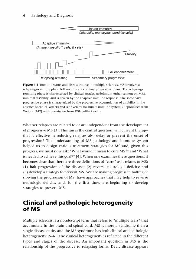
immune system over the course of the illness (Figure 1.1). The progressive
forms of the disease are the most disabling and are likely similar in terms of
pathogenic mechanisms. Epidemiologic studies have raised the question
Multiple Sclerosis: Diagnosis and Therapy, First Edition. Edited by Howard L. Weiner and
James M. Stankiewicz. � 2012 John Wiley & Sons, Ltd.
Published 2012 by John Wiley and Sons, Ltd.
3
whether relapses are related to or are independent from the development
of progressive MS [3]. This raises the central question: will current therapy
that is effective in reducing relapses also delay or prevent the onset of
progression? The understanding of MS pathology and immune system
helped us to design various treatment strategies for MS and, given this
progress, we must now ask: “What would it mean to cure MS?” and “What
is needed to achieve this goal?” [4]. When one examines these questions, it
becomes clear that there are three definitions of “cure” as it relates to MS:
(1) halt progression of the disease; (2) reverse neurologic deficits; and
(3) develop a strategy to prevent MS. We are making progress in halting or
slowing the progression of MS, have approaches that may help to reverse
neurologic deficits, and, for the first time, are beginning to develop
strategies to prevent MS.
Clinical and pathologic heterogeneityof MS
Multiple sclerosis is a nondescript term that refers to “multiple scars” that
accumulate in the brain and spinal cord. MS is more a syndrome than a
single disease entity and the MS syndrome has both clinical and pathologic
heterogeneity [5–6]. The clinical heterogeneity is reflected in the different
types and stages of the disease. An important question in MS is the
relationship of the progressive to relapsing forms. Devic disease appears
Innate Immunity
(Microglia, monocytes, dendritic cells)
Adaptive immunity
(Antigen-specific T cells, B cells)
Disability
GD enhancement
Secondary progressiveRelapsing-remitting
Figure 1.1 Immune status and disease course in multiple sclerosis. MS involves a
relapsing-remitting phase followed by a secondary progressive phase. The relapsing-
remitting phase is characterized by clinical attacks, gadolinium enhancement on MRI,
minimal disability, and is driven by the adaptive immune response. The secondary
progressive phase is characterized by the progressive accumulation of disability in the
absence of clinical attacks and is driven by the innate immune system. (Reproduced from
Weiner [147] with permission from Wiley–Blackwell.)
4 Pathology and Diagnosis
to be an MS variant associated with antibodies to the aquaporin recep-
tor [7–8]. There are rare malignant forms including Marburg’s variant,
tumefactive MS and Balo’s concentric sclerosis. An unanswered question
relates to why benign forms of MS exist [9–10]. Although some cases of MS
are defined as benign, and progress with prolonged follow up [11] there are
clearly benign formsof the disease. Bydefinitionpatientswith benignMSdo
not enter the progressive phase. The ability to identify benign or malignant
MS early in the course of the illness is very important for treatment
strategies. We compared brain parenchymal fraction (BPF) over a 2-year
period in benign vs early relapsing-remitting MS matched for age and the
EDSS and found that patientswith benignMShad a smaller loss of BPF [12].
As it impinges on the EDSS, the majority of disability inMS relates to spinal
cord dysfunction. The relationship between spinal cord changes and brain
MRI changes is not well known, but changes in the medulla oblongata
which reflect spinal cord can be visualized on brain MRI and may correlate
with entering the progressive phase [13]. In addition, an HLA-DR2 dose
effect may be associated with a more severe form of the disease [14].
What triggers MS?
The etiology of MS is still debatable but the current data suggests that
environmental factors in genetically susceptible background can predispose
an individual to MS. Family studies assessing the risk of relatives suggests
that first-degree relatives are 10–25 times at greater risk of developing MS
than the general population[15–17]. The strongest genetic effect is corre-
lated with HLA haplotypes. For instance HLA�1501, HLA-DRB1�0301, HLA-DRB1�0405, HLA-DRB1�1303, HLA-DRB1�03, HLA-DRB1�01, HLA-DRB1�10,HLA-DRB1�11, HLA-DRB1�14 and HLA-DRB1�08 have been shown to have
either positive or negative association with MS [15]. Ethnicity and sex
are other contributors in susceptibility toMS. The white population is more
susceptible to disease than theAfricanAmerican population andwomenare
at higher risk of developingMS thanmen [18], which is not associated with
any MS-related gene present on the X chromosome but is more correlated
with female physiology and hormones [19]. Other potential environmental
risk factors are infections, vaccination, climate, and diet. Infections are
considered the most common risk factor for MS as many infections and
antibodies generated in response to these infections are present in sera or the
cerebrospinal fluid (CSF) of MS patients at higher titers than controls.
Epstein–Barr virus (EBV) is of great interest as >99% of MS patients and
approximately 94% of age-matched controls are infected with EBV and
increased antibody titers to EBV nuclear antigen 1 (EBNA-1) antigen are
reported in MS [20–21]. Other infectious agents linked to MS etiology are
Disease Pathogenesis 5
herpes virus 6, retroviruses and Chlamydia pneumonia [22]. Evidence for
association of Chlamydia pneumonia with MS is debatable, as contradictory
presence of this virus has been reported by different groups [23–26].
Decreased sunlight exposure, vitamin D level, and vitamin intake are
also associatedwithMS incidence or protection [27–28]. In addition, studies
using different cohorts of MS patients have shown a strong association
between smoking and MS [29–30]. The etiology of MS is discussed in detail
in the Chapter 3.
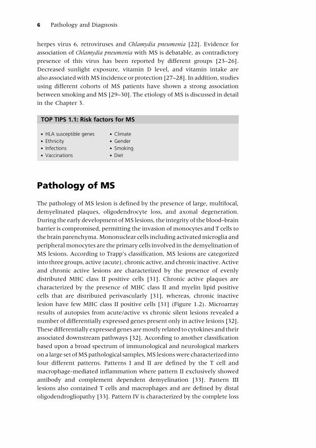
TOP TIPS 1.1: Risk factors for MS
. HLA susceptible genes . Climate
. Ethnicity . Gender
. Infections . Smoking
. Vaccinations . Diet
Pathology of MS
The pathology of MS lesion is defined by the presence of large, multifocal,
demyelinated plaques, oligodendrocyte loss, and axonal degeneration.
During the early development ofMS lesions, the integrity of the blood–brain
barrier is compromised, permitting the invasion of monocytes and T cells to
the brain parenchyma.Mononuclear cells including activatedmicroglia and
peripheralmonocytes are the primary cells involved in the demyelination of
MS lesions. According to Trapp’s classification, MS lesions are categorized
into three groups, active (acute), chronic active, and chronic inactive.Active
and chronic active lesions are characterized by the presence of evenly
distributed MHC class II positive cells [31]. Chronic active plaques are
characterized by the presence of MHC class II and myelin lipid positive
cells that are distributed perivascularly [31], whereas, chronic inactive
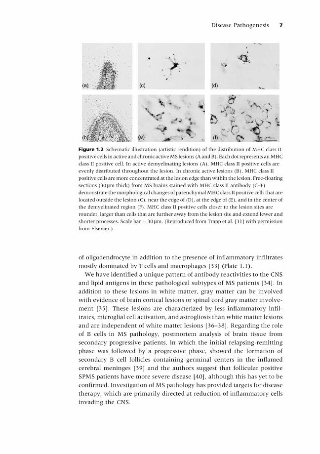
lesion have few MHC class II positive cells [31] (Figure 1.2). Microarray
results of autopsies from acute/active vs chronic silent lesions revealed a
number of differentially expressed genes present only in active lesions [32].
Thesedifferentially expressed genes aremostly related to cytokines and their
associated downstream pathways [32]. According to another classification
based upon a broad spectrum of immunological and neurological markers
on a large set ofMSpathological samples,MS lesionswere characterized into
four different patterns. Patterns I and II are defined by the T cell and
macrophage-mediated inflammation where pattern II exclusively showed
antibody and complement dependent demyelination [33]. Pattern III
lesions also contained T cells and macrophages and are defined by distal
oligodendrogliopathy [33]. Pattern IV is characterized by the complete loss
6 Pathology and Diagnosis
of oligodendrocyte in addition to the presence of inflammatory infiltrates
mostly dominated by T cells and macrophages [33] (Plate 1.1).
We have identified a unique pattern of antibody reactivities to the CNS
and lipid antigens in these pathological subtypes of MS patients [34]. In
addition to these lesions in white matter, gray matter can be involved
with evidence of brain cortical lesions or spinal cord gray matter involve-
ment [35]. These lesions are characterized by less inflammatory infil-
trates, microglial cell activation, and astrogliosis than white matter lesions
and are independent of white matter lesions [36–38]. Regarding the role
of B cells in MS pathology, postmortem analysis of brain tissue from
secondary progressive patients, in which the initial relapsing-remitting
phase was followed by a progressive phase, showed the formation of
secondary B cell follicles containing germinal centers in the inflamed
cerebral meninges [39] and the authors suggest that follicular positive
SPMS patients have more severe disease [40], although this has yet to be
confirmed. Investigation of MS pathology has provided targets for disease
therapy, which are primarily directed at reduction of inflammatory cells
invading the CNS.
Figure 1.2 Schematic illustration (artistic rendition) of the distribution of MHC class II
positive cells in active andchronic activeMS lesions (AandB).Eachdot represents anMHC
class II positive cell. In active demyelinating lesions (A), MHC class II positive cells are
evenly distributed throughout the lesion. In chronic active lesions (B), MHC class II
positive cells aremore concentrated at the lesion edge thanwithin the lesion. Free-floating
sections (30mm thick) from MS brains stained with MHC class II antibody (C–F)
demonstrate themorphological changes of parenchymalMHCclass II positive cells that are
located outside the lesion (C), near the edge of (D), at the edge of (E), and in the center of
the demyelinated region (F). MHC class II positive cells closer to the lesion sites are
rounder, larger than cells that are further away from the lesion site and extend fewer and
shorter processes. Scale bar¼ 30mm. (Reproduced from Trapp et al. [31] with permission
from Elsevier.)
Disease Pathogenesis 7
Initiation of disease
(Th1/Th17) T cells
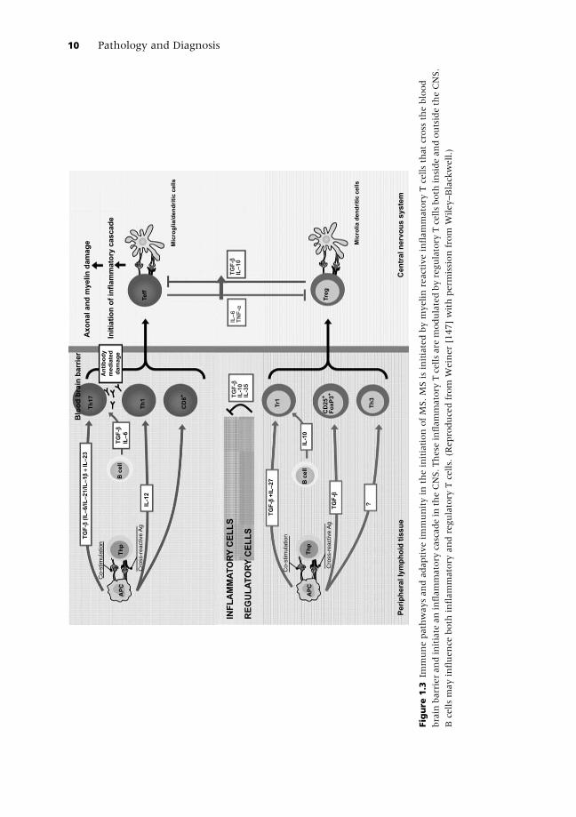
Cd4þ Pathogenic T cells: Upon antigenic stimulation, na€ıve CD4þ T cells
activate, expand and differentiate into distinct subsets of T cells which are
characterized by the production of different cytokines upon activation [41].
It is generally believed that acute MS lesions are initiated by a myelin
reactive CD4þ T cell that is stimulated in the periphery and enters the brain
and spinal cord (Figure 1.3). These CD4þ T cells have previously been felt to
be IFN-g secreting Th1 cells as IFN-g was found to be present at the site of
inflammation [42–45] and adoptive transfer of Th1 cells were able to
transfer the disease [46]. However, it was found later that IFN-g deficient
mice are not resistant but highly susceptible to organ-specific autoimmune
diseases [47]. It is now recognized that Th17 cells play a crucial role in
autoimmunity in the experimental allergic encephalomyelitis (EAE)
model [48] and increased numbers of Th17 cells have also been identified
inMS [49]. Both types of pathogenic cell (Th1 and Th17]most probably play
a role in MS and could account for the immunologic and clinical hetero-
geneity of thedisease [50]. Immunohistochemical examinationsof thebrain
demonstrate Th1/Th17 immune responses [51]. Th1vsTh17 responses have
beenassociatedwithdifferent types of EAE [50]. TGF-b, a central cytokine inthe induction of regulatory T cells, induces Th17 cells when combined with
IL-6 [52]. Anti-IL-6 therapy is being investigated for the treatment of
autoimmunity.
Cd8þ Pathogenic T cells: These cells are another subset of T cells thatmostly
provide defense against viral infections using cytotoxic weapons. Although
CD8þ T cells have not been at the forefront of thinking in MS, CD8þ T cells
are found in MS lesions at a higher frequency and CD8þ T cells reactive to
myelin antigenshavebeen reported inMS. It is likely thatCD8þT cells play a
role inMSandalso contribute to diseaseheterogeneity [53–54]. CD8þT cells
are also well poised to contribute directly to demyelination and axonal loss
during inflammation by expression of various cytotoxic molecules (e.g.
perforin and granzymeB) aswell as death receptor ligands (e.g. FasL, TNF-a,TNF-related molecules). CD8þ T cells isolated from brain lesions show
evidence of antigen-driven clonal expansion [55]. T cell lines generated
from CD8þ T cell clones isolated from MS patients and healthy controls
could mediate MHC class I restricted lysis of oligodendrocytes [56]. CD8þ
T cells could also target other CNS resident cells including microglia,
astrocytes, and neurons [57] suggesting a pathogenic potential of these
cells in MS biology. The same group also observed the close proximity of
granzyme B expressing CD8þ T cells to injured axons in MS lesions, which
8 Pathology and Diagnosis

furthermore emphasizes their role in the direct cytotoxicity of axons [57].
The importance of CD4þ and CD8þ T cells in EAEwas compared in CD4 and
CD8 knockout mice in a MOG-DBA/1 model. CD8-/- mice had reduced
demyelination and CNS inflammation compared to wild type animals.
CD4-/- animals, however, were refractory to EAE induction, suggesting a
pathogenic role forCD8þT cells [58]. Furthermore,CD8þTcells are also able
to contribute toward the secretion of IL-17 [59] and IFN-g [57], which, as
discussed above, are the important cytokines involved in disease pathology.
These observations suggest that both CD4þ and CD8þ T cells are capable of
playing pathogenic roles and their relative contribution might be respon-
sible for disease heterogeneity.
B cells and antibodiesB cells are another essential component of an adaptive immune system,
which mediates immunity against pathogens by the secretion of antigen-
specific antibodies and by acting as an antigen presenting the cells required
for T cell differentiation. Like T cells, B cells are also efficient in the
production of various cytokines including IL-1, IL-4, IL-6, IL-10, IL-12,
IL-23 and IL-16 [60–61]. Antibodies secreted by B cells or immune com-
plexes can also activate other antigen-presenting cells like dendritic cells
(DCs) and macrophages through the Fc receptor (Figure 1.3). Although
autoantibodies (antibodies against self-antigens) have been reported inMS,
there is no evidence that there are high affinity pathogenic antibodies inMS
as in other antibody mediated autoimmune diseases such as myasthenia
gravis [62]. Antibodies to myelin components, however, may participate in
myelin loss [63]. A classic finding in MS is increased locally produced IgG
and oligoclonal bands in the CSF, the pathogenic significance of which
remains unknown. Treatment with rituximab, a monoclonal antibody that
deletes B cells, dramatically reduces inflammatory disease activity as mea-
sured by MRI without affecting immunoglobulin levels, demonstrating a
clear role for B cells in relapsing forms of MS [64]. The almost immediate
response to rituximab suggests that B cells are either affecting T cell
regulation via their antigen presentation function or by directly participat-
ing in lesion formation. B cells may have both anti-inflammatory and pro-
inflammatory functions [60–65].
Regulation/remission of disease
Regulatory cells
Cd4þ and CD8þ regulatory T cells: It is now clear that the adaptive immune
system consists of a network of regulatory T cells (Tregs) [66]. Regulatory
Disease Pathogenesis 9
Th
17
Co-s
tim
ula
tion
TG
F-β
(IL
–6/IL
–21/IL
–1
β +
IL–23
Blo
od
bra
in b
arr
ier
Init
iati
on
of
infl
am
ma
tory
ca
sc
ad
e
Ax
on
al a
nd
my
eli
n d
am
ag
e
An
tib
od
y
med
iate
d
dam
ag
e
Th
1C
ross-r
eactive A
g
AP
C
AP
C
Th
p
IL-1
2
B c
ell
Teff
TG
F-β
IL–6
CD
8+
Mic
rog
lia/d
en
dri
tic c
ells
INF
LA
MM
AT
OR
Y C
EL
LS
RE
GU
LA
TO
RY
CE
LL
S
TG
F-β
IL-1
0IL
-35
TG
F-β
IL–10
IL–6
TN
F-α
Th
p
TG
F-β
+IL
–27
Tr1
IL-1
0B
cell
Co-s
tim
ula
tion
TG
F-β
Tre
g
Mic
rolia d
en
dri
tic c
ells
CD
25
+
Fo
xP
3+
Cro
ss-r
eactive A
g
Pe
rip
he
ral
lym
ph
oid
tis
su
e
?
Ce
ntr
al n
erv
ou
s s
ys
tem
Th
3
Figure
1.3
Immunepathwaysandadaptiveim
munityin
theinitiationofMS.MSisinitiatedbymyelinreactiveinflammatory
Tcellsthatcross
theblood
brain
barrierandinitiate
aninflammatory
cascadein
theCNS.These
inflammatory
Tcellsare
modulatedbyregulatory
Tcellsboth
insideandoutsidetheCNS.
Bcellsmayinfluence
both
inflammatory
andregulatory
Tcells.(R
eproducedfrom
Weiner[147]withperm
issionfrom
Wiley–Blackwell.)
10 Pathology and Diagnosis
T cells mediate active suppression of self-antigen specific T cell responses
and in the maintenance of peripheral tolerance [67–68]. Regulatory T cells
can broadly be classified as natural Tregs and induced Tregs. Foxp3 is the
major transcription factor for Tregs. CD25 marks natural Tregs and TGF-binduces Treg differentiation. Th3 cells are induced Tregs that secrete
TGF-b [69] and Tr1 cells are induced Treg cells that secrete IL–10 [70].
Defects in regulatory T cell percentages[71–72] and function have been
described in MS [73–75] and a major goal of MS immunotherapy is to
induce regulatory cells in a physiologic and nontoxic fashion [76–77]. Th2
cells which are recognized by secretion of IL-4, IL-5, and IL-13, may also
have regulatory T cell function as patients with parasitic infections that
induce Th2 type responses have a milder form of MS [78]. Experimental
data suggests that regulatory cells may not be effective if there is ongoing
CNS inflammation [79]. We have taken the approach of using the mucosal
immune system to induce regulatory cells and have found that oral anti-
CD3monoclonal antibody [80], and ligands that bind the aryl hydrocarbon
receptor induce TGF-b dependent regulatory T cells that suppress EAE and
provide a novel avenue for treatingMS [81–82]. The regulatory function of
CD8þ T cells is mostly ascribed to a population of T cells lacking expression
of CD28 on their cell surface. These cells induce regulatory effect in aMOG-
induced EAE model via induction of tolerogenic dendritic cells which in
turn induces CD4þ and CD8þ regulatory T cell subpopulations [83–85].
Another interesting regulatory population in CD8þ T cell subset is
CD8þCD122þ T cells which mediate suppressive effects via IL-10
[85–86]. The human counterpart of this population is recognized as
CD8þCXCR3þ [87]. Depletion of CD8þCD122þ T cells increased the dura-
tion of disease symptoms. Conversely, transfer of this population amelio-
rated the disease in the MOG EAE model on a C57BL/6 background,
suggesting a protective role of this population [88]. In addition, we have
described the existence of a novel LAPþCD8þ T cell population that
exhibited regulatory properties in EAE mice in a TGF-b and IFN-g depen-
dent manner [89].
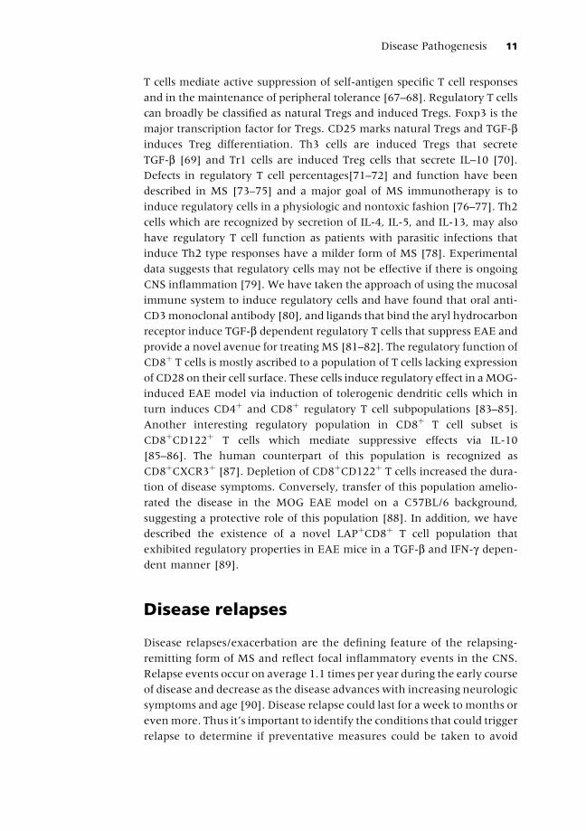
Disease relapses
Disease relapses/exacerbation are the defining feature of the relapsing-
remitting form of MS and reflect focal inflammatory events in the CNS.
Relapse events occur on average 1.1 times per year during the early course
of disease and decrease as the disease advances with increasing neurologic
symptoms and age [90]. Disease relapse could last for a week to months or
evenmore. Thus it’s important to identify the conditions that could trigger
relapse to determine if preventative measures could be taken to avoid
Disease Pathogenesis 11
relapse. A strong correlation was found between upper respiratory tract
infections and MS relapses [91–92]. This study confirmed that two-thirds
of the attacks occur during a period of risk (the interval 1week before
and 5 weeks after the initiation of URI symptoms) and attack rates were
2.92 per year at risk compared to 1.16 per year when not at risk [92].
Another longitudinal study with 73 patients also confirmed these results,
showing an increased attacks rate (rate ratio 2.1) during the period of risk
that was associated with an increase in the number of gadolinium-
enhancing regions suggesting that systemic infections result in more
sustained damage than other disease exacerbations [93]. No specific
virus was identified among these studies. Viral infection is associated
with activation of autoreactive T cells through molecular mimicry (T cell
reactive to viral antigen cross-react with self-antigen) [94], epitope
spreading (release of sequestered antigen secondary to tissue destruction
Gra
y M
atte
r
NA
WM
Wh
ite M
atte
r
Teff
1
CD8+1
Direct damage
Axonal
and
myelin
damage
Cytokines
Primary epitope
Molecular mimicry
Epitope spreading2
Teff
2
Teff
2
Viral super-antigens
3
Secondary epitopeAg release
Figure 1.4 Disease relapse. (1) Cells reactive to a viral antigen can cross react to myelin
self-antigen (molecular mimicry) and initiate a self-reactive immune response.
(2) Inflammatory T cells that enter the CNS initiate a complex immunologic cascade
consisting of epitope spreadingwhich triggers new attacks through activation ofmore self-
reactive T cells (epitope spreading). (3) Nonspecific activation of autoreactive T cells
through cytokines released during an immune response against viral infection (viral super
antigens). Dashed arrows indicate activation of T cells and dotted arrows suggest
inflammation mediated by T cells through cytokine secretion and by direct damage of
myelin sheath. (Reproduced fromWeiner [147] with permission fromWiley–Blackwell.)
12 Pathology and Diagnosis
mediated by viral antigen) [95–96], and viral superantigens (nonspecific
stimulation of autoreactive T cells) (Figure 1.4) [97]. Similarly, inflam-
matory cytokines like TNF-a and IFN-g also increase during disease
relapses [98]. Thus treatments targeting or controlling these cytokine
responses should help to reduce relapse rates. Blocking TNF-a using
antibodies or soluble receptors could decrease disease severity in murine
EAE but has a worsening effect inMS patients [99–101]. Other factors that
contribute toward disease relapse include a stressful life event [102–103],
pregnancy [104], and high-dose cranial radiation [105–106]. Based upon
studies describing important factors in the initiation, relapse, and pro-
gression of the disease it appears that lifestyle changes (including stress
management, diet, exercise, smoking, alcohol consumption) in combi-
nation with anti-inflammatory therapy can modify the disease activity
and should be suggested to MS patients.
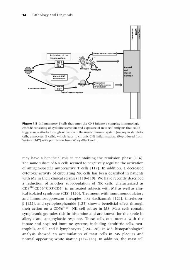
Disease progression
Activation of the innate immune systemThe innate immune system consists of dendritic cells, monocytes, micro-
glia, natural killer (NK), and mast cells. It is increasingly recognized
that the innate immune system plays an important role in the immu-
nopathogenesis of MS. Although the secondary progressive phase of
MS may be related to neuorodegenerative changes in the CNS, it is
now clear that the peripheral innate immune system changes when
patients transition from the relapsing-remitting to the progressive stage.
We found increased expression of osteopontin and costimulatory
(CD40) [107] molecules and decreased expression of IL-27 (unpub-
lished) in dendritic cells isolated from relapsing MS. Coversely, we
observed abnormalities in the expression of CD80 and secretion of
IL-12 and IL-18 in the dendritic cells from progressive patients [108–110].
Chronic microglial activation also occurs in MS [111] and this activation
contributes to MS and EAE pathology via secretion of various pro-
inflammatory cytokines and through antigen presentation [112]. Per-
sistent activation of microglial cells has also been observed in the chronic
phase of relapsing-remitting EAE and a correlation has been found
between activated microglia and the loss of neuronal synapses [113].
Natural killer (NK) cells, another component of innate immune cells,
are present in demyelinating lesions of patients with MS [114] and are
thought to play a protective role through the production of various
neurotrophic factors [115] and cytokines. An increase in IL-5 and IL-13
secreting “NK2” subpopulation was observed in MS patients in remission
compared to patients in relapse, suggesting that the NK2 subpopulation
Disease Pathogenesis 13
may have a beneficial role in maintaining the remission phase [116].
The same subset of NK cells seemed to negatively regulate the activation
of antigen-specific autoreactive T cells [117]. In addition, a decreased
cytotoxic activity of circulating NK cells has been described in patients
with MS in their clinical relapses [118–119]. We have recently described
a reduction of another subpopulation of NK cells, characterized as
CD8dimCD56þCD3-CD4-, in untreated subjects with MS as well as clin-
ical isolated syndrome (CIS) [120]. Treatment with immunomodulatory
and immunosuppressant therapies, like daclizumab [121], interferon-
b [122], and cyclophosphamide [123] show a beneficial effect through
their action on a CD56bright NK cell subset in MS. Mast cells contain
cytoplasmic granules rich in histamine and are known for their role in
allergic and anaphylactic response. These cells can interact with the
innate and acquired immune systems, including dendritric cells, neu-
trophils, and T and B lymphocytes [124–126]. In MS, histopathological
analysis showed an accumulation of mast cells in MS plaques and
normal appearing white matter [127–128]. In addition, the mast cell
Gra
y M
atte
r
NA
WM
Wh
ite M
atte
rActivation of the
innate system
Danger signals + cytokines
CCL–2
MIP–1a
IFN-γ
MonocyteMicroglia
Astrocytes
Chronic CNS
inflammation
Axonal
and
myelin
damage
Macrophage
Blood brain barrier
Central nervous system
PhagocytosisProteolytic enzymesNO productionMIP-1a
IFN-γ
Figure 1.5 Inflammatory T cells that enter the CNS initiate a complex immunologic
cascade consisting of cytokine secretion and exposure of new self-antigens that could
triggers new attacks through activation of the innate immune system (microglia, dendritic
cells, astrocytes, B cells), which leads to chronic CNS inflammation. (Reproduced from
Weiner [147] with permission from Wiley–Blackwell.)
14 Pathology and Diagnosis
specific enzyme tryptase is elevated in the CSF of MS patients [129]
along with other mast-cell-specific genes in MS plaques.
In summary, the immunopathogenesis of MS integrates both limbs of the
immune system and links them to different disease stages and processes.
Thus, the adaptive immune system drives acute inflammatory events
(attacks,gadoliniumenhancementonMRI)whereasinnateimmunitydrives
progressive aspects ofMS. Amajor question is whether aggressive and early
anti-inflammatory treatmentwill prevent the secondary progressive formof
the disease. There is some evidence that this is occurring; studies are begin-
ning to show that treatment with interferons delays the onset of the pro-
gressive stage [130]. Of note, there is a form of EAE driven by the innate,
rather than the adaptive, immune system [131]. There are no specific
therapies designed to affect the innate immune system in MS, and efforts
to investigate the innate immune system in MS and characterize it are now
being explored to determine how the innate immune system relates to the
disease stage and response to therapy. Furthermore, like the adaptive
immune system, there are different classes of innate immune responses,
e.g. protective and tolerogenic vs pathogenic and pro-inflammatory.
TOP TIPS 1.2: Immune cell involvement in MS pathogenesis
. Decreased percentages of CD4þ regulatory T cells
. Increased frequency of Th1 and Th17 CD4þ T cells
. CD8þ T cells
. B cells
. Activated dendritic cells
. Natural killer cells
. Microglial cells and monocytes
Neurodegeneration in MSAxonal andmyelin loss are prominent pathologic features ofMS [132] and
can be directly caused by immune cells (e.g. cytotoxic CD8 cells damaging
neurons or macrophages stripping myelin from the axon [133]); or can
result from release of toxic intermediates (e.g. glutamate, nitric oxide).
These intermediates can trigger immune cascades that further enhance
inflammatory-mediatedCNSdamage. Thus, glutamate andnitric oxide can
lead to enhanced expression of CCL2 on astrocytes which, in turn, leads to
infiltration of CD11b cells and additional tissue damage [134]. AMPA
antagonists have been shown to have an ameliorating affect in acute
EAE models [135–136] and we have found that a carbon-based fullerene
linked to an NMDA receptor with anti-excitotoxic properties slows
Disease Pathogenesis 15
progression and prevents axonal damage in the spinal cord in a model of
chronic progressive EAE [134]. Although the compound is not an immune
compound, it reduces the infiltration of CD11b cells into the CNS. Another
important component of neurodegeneration relates to changes in sodium
channels, suggesting that thesecouldbepotential therapeutic targets [137].
TOP TIPS 1.3: Potential therapeutic pathways for the treatment ofMS
. Decease Th1/Th17 cells . Affect innate immunity
. Induce regulatory T cells . Provide neuroprotection
. Prevent lymphocyte trafficking . Promote remyelin
. Deplete B cells
Conclusion
In summary,MS represents an immune cell mediated neurologic syndrome
rather than a single disease entity that has both clinical and pathologic
heterogeneity [5–6]. Amajor tool to address the pathological heterogeneity
of MS and devise appropriate treatment strategies is to develop reliable
biomarkers. MRI has served as the primary biomarker for MS [138] and
although conventional imaging does not link strongly to clinical outcomes,
every FDA-approved MS drug has shown efficacy on MRI outcomes.
Advances in magnetic resonance imaging are beginning to better define
MS and its heterogeneity. We have also developed a Magnetic Resonance
Disease Severity Scale (MRDSS) which combines multiple measures to
provide an index of disease severity and progression as measured by
MRI [139]. The additionof spinal cord imaging andgraymatter involvement
to theMRDSS should enhance its value as a biomarker. In addition, we and
others have shown immune measures that are associated with disease
activity and MRI activity [140–142]. RNA profiling is beginning to identify
gene expression patterns associated with different forms of MS and disease
progression [143–144]. In addition, we have demonstrated unique serum
immune signatures linked to different stages and pathologic processes inMS
that could provide a new avenue to understand disease heterogeneity, to
monitor MS, and to characterize immunopathogenic mechanisms and
therapeutic targets in the disease.
A complex disease such as MS will require treatment(s) that can affect
multiple pathways, including (1) suppression of Th1/Th17 responses,
(2) induction of Tregs, (3) altering the traffic of cells into the CNS,
(4) protecting axons and myelin from degeneration initiated by inflam-
mation that affects the innate immune system. If multiple drugs are
required to achieve this effect, wemust be certain that one treatment does
not interfere with another. For example, it has been reported that statins
16 Pathology and Diagnosis
may interfere with the action of interferons [145]. Because of disease
heterogeneity, there will be responders and nonresponders to each
“effective” therapy and the earlier that treatment is initiated, the more
likely it is to be effective. Inherent in the concept of curing MS by halting
progression is the ability to demonstrate that progression has been halted
in a group of patients and to identify those factors associated with
preventing the onset of progressive disease. We have thus initiated the
CLIMB natural history study in which more than 2000 patients with MS
will be followed over a period of time with clinical evaluation, MRI
studies, and immune and genetic markers, to identify the factors that are
associated with the various stages of the disease and disease progres-
sion [146]. We believe that the identification of such factors may lead to
the stratification of MS patients into smaller subclinical groups with
defined common mechanisms of initiation of disease, inflammation,
and demyelination during the disease progression that could help in
designing/selecting subtype-specific treatment.
References
1 McFarland HF, Martin R. Multiple sclerosis: a complicated picture of autoimmunity.
Nat Immunol 2007 Sep; 8(9): 913–19.
2 Miller DH, Leary SM. Primary-progressivemultiple sclerosis. Lancet Neurol 2007Oct;
6(10): 903–12.
3 Confavreux C, Vukusic S, Moreau T, et al. Relapses and progression of disability in
multiple sclerosis. N Engl J Med 2000 Nov; 343(20): 1430–8.
4 WeinerH. CuringMSHowScience is Solving theMysteries ofMultiple Sclerosis. New
York: Crown Publishers; 2004.
5 Lassmann H, Bruck W, Lucchinetti CF. The immunopathology of multiple sclerosis:
an overview. Brain Pathol 2007 Apr; 17(2): 210–18.
6 Breij EC, Brink BP, Veerhuis R, et al. Homogeneity of active demyelinating lesions in
established multiple sclerosis. Ann Neurol 2008 Jan; 63(1): 16–25.
7 Hinson SR, Roemer SF, Lucchinetti CF, et al. Aquaporin-4-binding autoantibodies in
patients with neuromyelitis optica impair glutamate transport by down-regulating
EAAT2. J Exp Med 2008 Oct; 205(11): 2473–81.
8 Misu T, Fujihara K, Kakita A, et al. Loss of aquaporin 4 in lesions of neuromyelitis
optica: distinction from multiple sclerosis. Brain 2007 May; 130(Pt 5): 1224–34.
9 Ramsaransing GS, De Keyser J. Benign course in multiple sclerosis: a review. Acta
Neurol Scand 2006 Jun; 113(6): 359–69.
10 Pittock SJ, McClelland RL, Mayr WT, et al. Clinical implications of benign multiple
sclerosis: a 20-year population-based follow-up study. Ann Neurol 2004 Aug; 56(2):
303–6.
11 Hawkins SA, McDonnell GV. Benign multiple sclerosis? Clinical course, long term
follow up, and assessment of prognostic factors. J Neurol Neurosurg Psychiat 1999
Aug; 67(2): 148–52.
12 Gauthier S, Berger AM, Liptak Z, et al. Benign MS is characterized by a lower rate of
brain atrophy as compared to early MS. Arch Neurol 2008.
Disease Pathogenesis 17
13 Liptak Z, Berger AM, Sampat MP, et al. Medulla oblongata volume: a biomarker of
spinal cord damage and disability in multiple sclerosis. Am J Neuroradiol 2008 Sep;
29(8): 1465–70.
14 Barcellos LF,Oksenberg JR, BegovichAB, et al. HLA-DR2dose effect on susceptibility
to multiple sclerosis and influence on disease course. Am J Hum Genet 2003 Mar;
72(3): 710–6.
15 Trapp BD, Nave KA. Multiple sclerosis: an immune or neurodegenerative disorder?
Annu Rev Neurosci 2008; 31: 247–69.
16 Sadovnick AD, Baird PA, Ward RH. Multiple sclerosis: updated risks for relatives.
Am J Med Genet 1988 Mar; 29(3): 533–41.
17 Ebers GC, Sadovnick AD, Dyment DA, Yee IM, Willer CJ, Risch N. Parent-of-origin
effect inmultiple sclerosis: observations inhalf-siblings. Lancet 2004May; 363(9423):
1773–4.
18 WallinMT, PageWF, Kurtzke JF.Multiple sclerosis in US veterans of the Vietnam era
and later military service: race, sex, and geography. Ann Neurol 2004 Jan; 55(1):
65–71.
19 Whitacre CC. Sex differences in autoimmune disease. Nat Immunol 2001 Sep; 2(9):
777–80.
20 Sundstrom P, Juto P, Wadell G, Hallmans G, Svenningsson A, Nystrom L, et al. An
altered immune response to Epstein-Barr virus in multiple sclerosis: a prospective
study. Neurology 2004 Jun; 62(12): 2277–82.
21 Levin LI, Munger KL, Rubertone MV, Peck CA, Lennette ET, Spiegelman D, et al.
Temporal relationship between elevation of epstein-barr virus antibody titers and
initial onset of neurological symptoms in multiple sclerosis. JAMA 2005 May;
293(20): 2496–500.
22 Marrie RA. Environmental risk factors in multiple sclerosis aetiology. Lancet Neurol
2004 Dec; 3(12): 709–18.
23 Layh-Schmitt G,BendlC,HildtU,Dong-Si T, Juttler E, Schnitzler P, et al. Evidence for
infection with Chlamydia pneumoniae in a subgroup of patients with multiple
sclerosis. Ann Neurol 2000 May; 47(5): 652–5.
24 Munger KL, Peeling RW,HernanMA, Chasan-Taber L, OlekMJ, Hankinson SE, et al.
Infection with Chlamydia pneumoniae and risk of multiple sclerosis. Epidemiology
2003 Mar; 14(2): 141–7.
25 Sriram S, Stratton CW, Yao S, Tharp A, Ding L, Bannan JD, et al. Chlamydia
pneumoniae infection of the central nervous system inmultiple sclerosis. AnnNeurol
1999 Jul; 46(1): 6–14.
26 Numazaki K, Chibar S. Failure to detect Chlamydia pneumoniae in the central
nervous system of patients with MS. Neurology 2001 Aug; 57(4): 746.
27 Munger KL, Zhang SM, O’Reilly E, Hernan MA, Olek MJ, Willett WC, et al.
Vitamin D intake and incidence of multiple sclerosis. Neurology 2004 Jan; 62(1):
60–5.
28 Munger KL, Levin LI, Hollis BW, Howard NS, Ascherio A. Serum25-hydroxyvitamin
D levels and risk of multiple sclerosis. JAMA 2006 Dec; 296(23): 2832–8.
29 Riise T, Nortvedt MW, Ascherio A. Smoking is a risk factor for multiple sclerosis.
Neurology 2003 Oct; 61(8): 1122–4.
30 Hedstrom AK, Sundqvist E, Baarnhielm M, Nordin N, Hillert J, Kockum I, et al.
Smoking and two human leukocyte antigen genes interact to increase the risk for
multiple sclerosis. Brain 2011 Mar; 134(Pt 3): 653–64.
31 Trapp BD, Bo L, Mork S, Chang A. Pathogenesis of tissue injury in MS lesions.
J Neuroimmunol 1999 Jul; 98(1): 49–56.
18 Pathology and Diagnosis