Embed Size (px)
Citation preview

1
Diagnosis and Management of Acute Kidney Injury
Ashita Tolwani, M.D., M.S.
Professor of Medicine
University of Alabama at Birmingham
2017
Disclosures
Consultant for Baxter
Patent on 0.5% citrate anticoagulant solution for CRRT
AKI Outline
Epidemiology
Definition
Pathophysiology and differential diagnosis
Overview of prevention and management
Epidemiology of AKI

2
Acute Kidney Injury: Why Do We Care?
AKI is common (KDIGO definition) 21% of all hospital admissions
>50% of ICU patients
AKI is associated with increased risk of CKD, ESKD, CV disease, and death
Dialysis‐requiring AKI ICU patients have the worst outcomes 11% of ICU patients with AKI require dialysis and 10‐30% survivors remain
dialysis dependent at time of hospital discharge
AKI can be preventable, treatable, and reversible
Healthcare workers are not well informed about AKI and its consequences
Mehta RL et al. Lancet 2015Pannu et al. CJASN 2013Cerda, et al. CJASN 2015
Worldwide, 2,000,000 people will die this year
of AKI!

3
Definition of AKI
Definition
More than 30 different definitions exist with a variety of quoted incidence rates, risk factors, and morbidity and mortality rates
A staging system is needed to stratify patients so that both accurate identification and prognostication are possible
www.ADQI.net
Using RIFLE, Patients with AKI Have Poorer Outcomes
Source: Ricci Z. Kidney Int. 73: 538-546, 2008
Analysis of 71,000 pts/13 studies to validate RIFLE Criteria
Mild AKI have poor outcomes
Mortality Risk in Hospitalized Patients
↑SCr↑SCr> 0.3 > 0.5 > 1.0 > 2.0
mg/dL
Chertow et al, JASN 16: 3365-3370, 2005Chertow et al, JASN 16: 3365-3370, 2005

4
R (I)
I (II)
F (III)
Increased SCr x1.5OR > 0.3 mg/dL
UO < .3ml/kg/hx 24 hr or Anuria x 12 hrs
UO < .5ml/kg/hx 12 hr
UO < .5ml/kg/hx 6 hr
Increased SCr x2
Increase SCr x3or SCr 4mg/dl
(Acute rise of 0.5 mg/dl)
HighSensitivity
HighSpecificity
RRT Started
Modifications proposed by AKINAmsterdam, 2005
I (II)
Criterion must be reached within 48hr
AKIN Criteria (Rifle V2.0)
KDOQI Commentary AJKD 2013
KDIGO AKI Guidelines: Definition of AKI
Problems with Serum Creatinine
Creatinine is influenced by age, muscle mass, gender, and ethnicity
Creatinine does not reflect the presence or absence of structural injury and thus provides no guidance on AKI etiology or the likelihood of response to various targeted therapies
The rise is serum creatinine is delayed by 2‐3 days after the injury has occurred
Fluid therapy may dilute serum creatinine and therefore delay diagnosis
Inter‐laboratory variation in measuring creatinine, and bilirubin and other compounds interfere with the colorimetric modified Jaffe assay hence affect serum creatinine levels
Serum Creatinine and GFR in AKI
Muscle mass
Protein metabolism
Serum creatinine
Renal excretion
Tubular excretion Filtration (GFR)
Drugs Nonlinear
Nutrition Infection
Edema
Volume of distribution
Star RA, Kidney Int, 1998

5
Gill, N. et al. Chest 2005;128:2847-2863
Relationship Between GFR and Creatinine
120
40
80
0
GFR(mL/min)
0 7 14 21 28
4
Days
2
0
6
Serum Creatinine(mg/dL)
DeathDeath
Conceptual Model for AKI
NormalNormalIncreased
riskIncreased
riskKidneyfailureKidneyfailure
DamageDamage GFR GFR
CreatinineIdeal Biomarker
What Can an Ideal AKI Biomarker Teach Us?
Predict and diagnose AKI early (before increase in serum creatinine)
Identify the primary location of injury (proximal tubule, distal tubule, interstitium)
Pinpoint the type (pre‐renal, AKI, CKD), duration and severity of kidney injury
Identify the etiology of AKI (ischemic, septic, toxic, combination)
Predict clinical outcomes (dialysis, death, length of stay)
Monitor response to intervention and treatment
Expedite the drug development process (safety)
Prasad Devarajan: Biomarkers in Acute Kidney Injury :Search for a Serum Creatinine Surrogate
Glomerular Filtration • Serum Creatinine• Blood urine Nitrogen • Serum Cystatin C• Plasma NGAL
Glomerular Injury• Urine albumin excretion
Proximal Tubule Injury•Urine IL-18•Urine KIM-1•Urine L-FABP•Urine Cystatin C•α1-microglobulin•β2-microglobulin•Urine α-GST•Urine Netrin-1•Urine NAG
Loop of Henle Injury•Uromodulin
Distal Tubule•Urine NGAL•Urine π-GST
Potential Biomarkers for AKI
Other Mechanisms / Sites of Injury not specific to the Nephron•Hepcidin – Iron trafficking•TIMP-2/ IGFBP7 – G1 cell cycle arrest
Adapted from Koynerand Parikh‐ Brenner and Rector’s The Kidney
Courtesy of J. Koyner

6
Biomarkers after AKIEarly Detection
Idealized
SCr
IL‐18
NGAL
L‐FABP
Kim‐1
Urinary Biomarkers Associated with Tubular Damage
New Paradigm for the Spectrum of AKI
NO AKICreat (‐)
Biomarker (‐)
STRUCTURAL (subclinical) AKI
Creat (‐)Biomarker (+)
FUNCTIONAL AKICreat (+)
Biomarker (‐)
INTRINSIC AKI (structural & functional)
Creat (+)Biomarker (+)
Pathophysiology and Differential Diagnosis of AKI
AcuteTubular
Necrosis
AcuteInterstitialNephritis
AcuteGN
AcuteVascular
Syndromes
IntratubularObstruction
Classification of the Etiologies of AKI
PrerenalAKI
Post-renalAKI
IntrinsicAKI
AKI

7
Non ‐ICU ICU Evaluation of Cause of AKI
Form of AKI BUN:Cr UNa (mEq/L) FENa Urine Sediment
Prerenal >20:1 <10 < 1% Normal, hyaline casts
Post‐renal >20:1 >20 variable Normal or RBC’s
Intrinsic
ATN <10:1 >20 > 2% Muddy brown casts; tubular epithelial cells, granular casts
AIN <20:1 >20 >1% WBC’s WBC casts, RBC’s, eosinophils
AGN variable <20 <1% Dysmorphic RBC’s, RBC casts
Vascular variable >20 variable Normal or RBC’s
Fractional Excretion of Na+ (FENa)
(Urine Na x Serum Cr) X 100 < 1% = pre‐renal
(Serum Na x Urine Cr) > 2% = ATN
Normal renal function <1%
Most accurate with oliguric AKI
Caveat:
< 1% without volume depletion Contrast nephropathy
Acute GN
Rhabdomyolysis
Possibly > 2% with prerenal state: Diuretics, severe CKD
Steiner AJM 1984:77:699-702
Fractional Excretion of Urea (FEurea)
(Urine UN x Serum Cr) X 100 < 35% = Pre‐renal(Serum UN x Urine Cr) > 50% = ATN
Better than FENa in patients on diuretics
Rationale: Urea reabsorbed in proximal tubule + inner medulla, not affected by loop and thiazide diuretics

8
Pre-renal Urine Sediment
Hyaline Casts
Pre-renal AKI – Decreased Renal Blood Flow
Cause Examples
Volume depletion Renal losses; GI fluid losses; hemorrhage; burns
Decreased cardiac output Heart failure; massive pulmonary embolus; acute coronary syndrome
Systemic vasodilation Sepsis; cirrhosis; anaphylaxis; anesthesia
Intrarenal vasoconstriction Drugs (NSAIDs, COX‐2 inhibitors, amphotericin B, calcineurin inhibitors, contrast agents); hypercalcemia; hepatorenal syndrome
Efferent arteriolar vasodilation Renin inhibitors; ACE inhibitors; ARBs
A prolonged pre‐renal state can lead to ATN
Pathogenesis of Pre-renal AKI
RenalVasoconstriction
DecreasedGFR
Angiotensin II
Adrenergic nerves
Vasopressin
+
+
+
Nitric oxide
Prostaglandins-
-
VolumeDepletion
CongestiveHeart Failure Liver
Failure
Sepsis
Impaired Autoregulation Can Lead to “Normotensive AKI”
Abuelo JG. N Engl J Med 2007;357:797-805

9
Pre-renal Azotemia: Medications
Angiotensin‐converting enzyme inhibitors
Nonsteroidal anti‐inflammatory drugs
Intrarenal Mechanisms for Autoregulation of the GFR
Abuelo JG. N Engl J Med2007;357:797-805.
NSAIDS ACEI/ARB
Abdominal Compartment Syndrome
Intra‐abdominal hypertension:
Intra‐abdominal pressure ≥12 mm Hg; or
Abdominal perfusion pressure <60 mm Hg
Abdominal compartment syndrome
Intra‐abdominal pressure ≥20 mm Hg; and
One or more new organ failures
Systemic Effects of Increased Abdominal Pressure
Cardiac
venous return
cardiac output
CVP, PCWP & SVR
Pulmonary
intrathoracic & airway pressures
PaO2
PaCO2
GI
splanchnic perfusion
CNS
intracranial pressure,
perfusion pressure
Renal
renal perfusion
GFR
urinary output

10
Clinical Settings for ACS
Trauma patients following massive volume resuscitation
Massive ascites Post liver transplant Mechanical limitations to the abdominal wall
Tight surgical closure Burn injuries
Bowel obstruction Pancreatitis
Abdominal Compartment Syndrome
Diagnosis Measurement of intra‐abdominal pressure
Clamp drainage tube of Foley catheter Instill 25 mL sterile water into the bladder via the aspiration port Measure pressure using a manometer or transducer attached to the aspiration port.
The manometer or transducer should be zeroed at the level of the mid‐axillary line at the iliac crest
Treatment Abdominal decompression
Treatment of Pre-renal AKI
Correction of volume depletion
Discontinuation/dose adjustment of medications NSAIDs
RAAS blockers
CNIs
Evaluation for causes of “effective” volume depletion Heart failure
Cirrhosis
Nephrotic syndrome
Sepsis
Treat hypercalcemia
Recognize and treat abdominal compartment syndrome
Intrinsic Renal Disease
Glomerulonephritis Interstitial nephritis•drugs
–penicillins
–sulphonamides
–rifampin
–NSAID's
–phenytoin
–allopurinol
•infections
•systemic disease
–SLE
–sarcoid
–sjogrens
•malignancy
•idiopathic
Small blood vessels• Malignant hypertension• HUS/TTP• (Pre)Eclampsia• DIC• Scleroderma• Vasculitis• Cholesterol emboli Acute tubular necrosis
• Postinfectious GN• Endocarditis‐associated GN• Systemic vasculitis• Membranoproliferative GN• Rapidly progressive GN• IgA nepropathy
11
Acute Tubular Necrosis
CausesIschemic: causes of prolonged prerenal AKI
Drug‐induced: aminoglycosides; vancomycin; polymyxins; lithium; amphotericin B; pentamidine; cisplatin; foscarnet; tenofovir; cidofovir; carboplatin; ifosfamide; zoledronate; contrast agents; sucrose; immunoglobins; mannitol; hydroxyethyl starch; dextran; NSAIDs; synthetic cannabinoids; amphetamines
Pigment: rhabdomyolysis; intravascular hemolysis
Sepsis
Adapted from Bonventre and Weinberg JASN 14:2199-2210, 2003
Ischemia
Continued Ischemia
DECREASED GFR
Acute Tubular Injury
ApoptosisNecrosis
Tubular ObstructionBackleak
MicrovascularInjury
VasoconstrictionLeukocyte Adhesion
↑ Permeability
Microvascular Congestion
Innate Immunity
Inflammation
DAMPSImmune CellsCytokines
Pathogenesis of Ischemic AKI
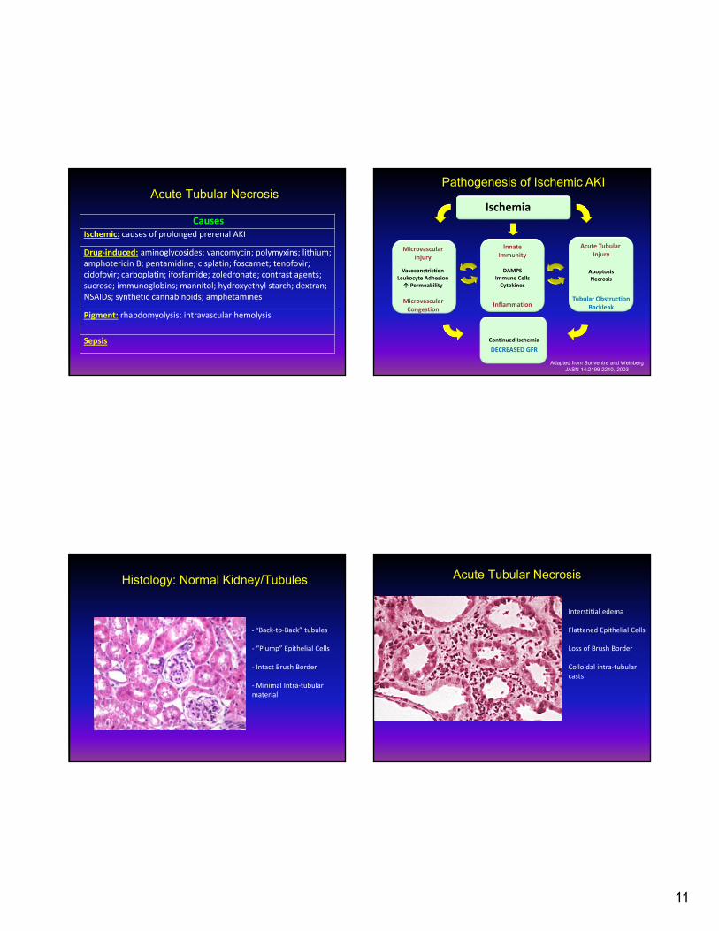
Histology: Normal Kidney/Tubules
‐ “Back‐to‐Back” tubules
‐ “Plump” Epithelial Cells
‐ Intact Brush Border
‐ Minimal Intra‐tubular material
Acute Tubular Necrosis
Interstitial edema
Flattened Epithelial Cells
Loss of Brush Border
Colloidal intra‐tubular casts

12
Acute Tubular Necrosis Urine Sediment
Muddy Brown Granular Casts and Renal Tubular Epithelial Cells
Contrast Media-Nephrotoxicity
Contrast Media
Blood Flow OxygenDelivery
OxygenConsumption
Renal Medullary Hypoxia
SystemicHypoxemiaBlood viscosity
PGE2 EndothelinANP VasopressinAdo PGI2
Osmotic Load
Direct Cellular Toxicity
Rudnick et. al. Seminars in Nephrology 17:15-26, 1997
Increase in serum creatinine occurs within 24 to 48 hours following contrast exposure
Risk Factors for Contrast-Associated AKI
Patient Related
Preexisting renal insufficiency
Diabetes mellitus
Intravascular volume depletion
Reduced cardiac output
Concomitant nephrotoxins
Procedure related
Increased dose of radiocontrast
Multiple procedures within 72 hours
Intra‐arterial administration
Type of radiocontrast
Strategies for Prevention of Contrast-Associated AKI
Effective Low- or Iso-osmolal contrast agents Intravenous isotonic fluids Avoidance of concomitant nephrotoxins
Ineffective or harmful Furosemide Mannitol Dopamine Fenoldopam Prophylactic RRT
Uncertain Intravenous sodium bicarbonate N-acetylcysteine Theophyliine ANP Statins Iron chelators RIPC

13
Acute Interstitial Nephritis
Clinical Suspicion
Fever, Rash
Culprit Drug or Disease Process
Blood Tests
Increased serum creatinine (AKI)
Leukocytosis, eosinophilia, anemia, elevated ESR, transaminitis
Urine Studies
Dipstick/low grade proteinuria
Pyuria, hematuria, WBC casts
Eosinophiluria
Imaging Tests
Renal US/CT Scan
Gallium Scan
FDG‐PET Scan
Kidney Biopsy
Gold Standard
The triad of Fever, Rash and Eosinophilia: <5‐10%
Acute Interstitial Nephritis
CausesDrug‐induced: cephalosporins; penicillin; methicillin; fluoroquinolones; sulfonamides; rifampin; NSAIDs; COX‐2 inhibitors; proton pump inhibitors; 5‐aminosalicylates; indinavir; abacavir; allopurinol; phenytoin; triamterene; furosemide; thiazide diuretics; phenytoin; carbamazepine; Chinese herb nephropathy
Infection: pyelonephritis; viral nephritides; leptospirosis; Legionella; Mycobacterium tuberculosis
Autoimmune: Sjögren syndrome; sarcoidosis; SLE; TINU syndrome; IgG4‐related disease
Malignancy: lymphoma; leukemia; multiple myeloma
Acute Interstitial NephritisAcute Interstitial Nephritis Urine Sediment
White Blood Cell Cast

14
Acute Interstitial Nephritis:Eosinophiluria
Muriithi AK, et al. CJASN 2013; 8: 1857-1862
Drug Induced-AIN All Etiologies of AIN
All cases (n=548) Pyuria (n=452) All cases (566) Pyuria ( 467)
>1% >5% >1% >5% >1% >5% >1% >5%
Sensitivity 35.6 23.3 44.8 29.3 30.8 19.8 38.4 24.7
Specificity 68.2 91.2 61.7 89.3 68.2 91.2 61.7 89.3
PPV 14.7 28.8 14.7 28.8 15.6 30.0 15.6 30.0
NPV 87.3 88.6 88.4 89.6 83.7 85.6 84.4 86.5
Positive LR 1.1 2.6 1.2 2.7 0.97 2.3 1.0 2.3
Negative LR 0.9 0.8 0.9 0.8 1.01 0.9 1.0 0.8
Insensitive test with specificity and positive LR only potentially acceptable using Urine Eos >5% cutoff in setting of high pretest probability
Acute Interstitial Nephritis - Summary
Most commonly drug induced
Complete “classic” triad is rarely present
Common urinary findings include
Pyuria
WBC casts
Eosinophiluria neither sensitive nor specific
Primary treatment is discontinuation of offending agent/treatment of underlying etiology
Role of glucocorticoids remain uncertain
Acute Glomerulonephritis
Nephritic presentation
Proteinuria (may be in nephrotic range (> 3.5 g/day))
Hematuria (dysmorphic RBCs)
RBC casts
Diagnosis usually requires renal biopsy
Infection‐related glomerulonephritis
Cryoglobulinemia
RPGN
Acute Glomerulonephritis: Dysmorphic RBCs and RBC Casts

15
Acute Vascular Syndromes
Causes
Macrovascular: renal artery occlusion; renal vein thrombosis; polyarteritis nodosa
Microvascular: TMA; HUS; TTP; APLS; HELLP; scleroderma renal crisis; hypertensive emergency; drugs (clopidogrel, cyclosporine, tacrolimus, anti‐angiogenesis drugs, interferon, m‐TOR inhibitors); drug‐induced TMA (caused by quinine, cancer therapies [gemcitabine, mitomycin, bevacizumab, bortezomib, sunitinib], calcineurin inhibitors [cyclosporine, tacrolimus], drugs of abuse [cocaine, ecstasy, intravenous extended‐release oxymorphone])
Atheroembolic disease
Atheroembolic Disease
Atheroembolic Disease
Risk factors
Atherosclerosis CAD
AAA
PVD
Hypertension
Hypercholesterolemia
Diabetes Mellitus
Precipitating factors
Arterial catheterization
Arteriography
Vascular surgery
Anticoagulation
Thrombolytic therapy
Atheroembolic Disease:Non-Renal Manifestations
General
Fever
Myalgias
Weight loss
Cutaneous
Livedo reticularis
Digital ischemia
Neurologic
TIA/CVA
Altered mental status
Peripheral neuropathy
Spinal cord infarct
Gastrointestinal
Anorexia
Nausea and vomiting
Nonspecific abdominal pain
GI bleeding
Ileus
Bowel ischemia/infarction
Pancreatitis
Hepatitis
Musculoskeletal
Myositis
Eyes
Amaurosis fugax
Retinal cholesterol emboli

16
Atheroembolic Disease: Renal Manifestations
Renal infarction
Acute kidney injury
Subacute kidney injury
Exacerbation of hypertension
Proteinuria (may be nephrotic)
Hematuria
Atheroembolic Disease:Laboratory Features
Serum chemistries
BUN and creatinine
Amylase
CPK
LFTs
Hematology
Leukocytosis
Eosinophilia
Anemia
Thrombocytopenia
Serologic
ESR
Serum complement
Urine
Eosinophiluria
Proteinuria
Hematuria
Pyuria
Atheroembolic Disease: Treatment
Avoid anticoagulation
Avoid vascular interventions
ACE inhibitors / angiotensin receptor blockers
Statin therapy
Nutrition support
Dialysis for management of volume status and uremia
Role of steroid therapy is uncertain
Intrinsic Renal Disease: Intratubular Obstruction
Common factors: Include high excretion in urine
Low solubility in acidic urine
Exacerbated by hypovolemia
Common crystals Uric acid (tumor lysis syndrome)
Acyclovir
Sulfa
Methotrexate
Ethylene glycol (calcium oxalate deposition)
Intratubular protein deposition Multiple myeloma (Bence‐Jones protein deposition)

17
Tumor Lysis Syndrome Tumor Lysis Syndrome
Rapid lysis of malignant cells leads to hyperuricemia, hyperkalemia, hyperphosphatemia, hypocalcemia, and AKI
Management of patients at risk or presenting with TLS Aggressive volume expansion to achieve a urine output of at least 80 to 100
mL/m2/h
Allopurinol to prevent formation of new uric acid (recommended as prophylaxis for patients at low/intermediate risk for TLS)
Rasburicase for patients at high risk of TLS or with TLS (contraindicated in patients with G6PD deficiency)
Urinary alkalinization is no longer recommended due to an increase in calcium phosphate crystal deposition
Management of hyperkalemia and hyperphosphatemia
RRT in refractory cases
Prevention and Management of AKI
Drug Biological rationale
Animal experiments
Uncontrolled human data
Small RCT
Large RCT
Loop diuretics Present Favorable Favorable Negative N/A
Low-dose dopamine Present Favorable Favorable Variable Negative
Mannitol Present Favorable Favorable N/A N/A
Ca antagonist Present Favorable Favorable Variable N/A
Theophylline Present Favorable Favorable Positive N/A
Prostaglandins Present Favorable Favorable N/A N/A
Natriuretic peptide Present Favorable Favorable Negative N/A
-receptor antagonist Present N/A N/A Positive N/A
Endothelin antagonist Present Favorable N/A N/A N/A
Thromboxane antagonist Present Favorable N/A N/A N/A
Thyroxine Present Favorable N/A Negative N/A
Saline Present Favorable Favorable Positive N/A
NAC Present Favorable N/A Positive N/A
Non-ionic media Present Favorable Favorable Positive positive
Interventions in AKI
The only FDAapproved treatment of
AKI is dialysis

18
Prevention of ATN
Recognition of underlying risk factors
Diabetes
CKD
Age
Cardiac/liver dysfunction
Early recognition is key
Changes in creatinine are a late manifestation of renal injury
A “normal” normal serum creatinine may reflect significant renal insufficiency, particularly in the elderly
Maintenance of renal perfusion
Avoidance of nephrotoxins
AKI Summary
AKI is defined by a standardized creatinine‐based definition
AKI is common
AKI is associated with mortality in a stage‐dependent fashion
Methods for earlier diagnosis of AKI and its progression may result in improved outcomes by facilitating targeted and timely treatment of AKI
There is no treatment of ATN and prevention of precipitating factors is paramount