Embed Size (px)
Citation preview

Diabetic Foot:an Orthopaedic Perspective:
Ulceration & Deformity
Chris Birks
Summary
• Introduction
• Ulceration
• Deformity (Charcot)
Cause
Assessment
Orthopaedic Surgical Treatment
• Amputation

Introduction: (USA Stats 2005)
17 million (6.2%) with Diabetes
5.9 million Undiagnosed
20 % of population over 65 have Diabetes
20% Diabetic admissions with foot ulcers
5-15% Diabetic require LL amputation
25% Adult diabetics with plantar insensitivity
0.5-1% Diabetics may develop charcot
Up to 30% Charcot have vascular insufficiency

Epidemiology
• 60% of non traumatic amputations are a
consequence of diabetes
• Mortality in these patients is between 50 -
80% at 5 years. (especially ischaemic ulcers)
• 50% patients will have an amputation of the
contralateral limb within 5 years.
• Cost US$ 11 Billion/year.
Co-morbidities:
• Cardio-respiratory
• Renal
• Vascular
• Neuropathic
• Eyes
• ? Smoking
• Obesity

Eitiology: Pressure vs Healing
Pressure
• Peripheral Neuropathy
– Sensory
– Autonomic (scaly/cracked skin)
– Motor (Claw toes/MTP Dislocation)
• Contractures
• Deformity/Charcot
• Poor foot care
Healing
• Aging process
• Vascular supply
• Nutritional state
• Poor foot care
The Team Members
• Endocrinologist/Diabetic Nurse
Educator
• Podiatrist/Orthotist/Plaster Technician
• Wound care specialist
• Vascular Lab / Surgeon
• Orthopaedic specialist

Mechanical Failure
Contractures
Boney prominences
Malalignment
Instability

contractures
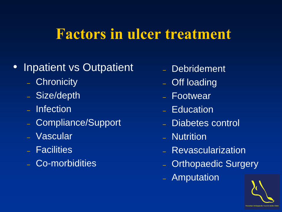
Factors in ulcer treatment
• Inpatient vs Outpatient
– Chronicity
– Size/depth
– Infection
– Compliance/Support
– Vascular
– Facilities
– Co-morbidities
– Debridement
– Off loading
– Footwear
– Education
– Diabetes control
– Nutrition
– Revascularization
– Orthopaedic Surgery
– Amputation

Take Home Messages
• No skin break, infection very unlikely
• Probe goes to bone over 85% deep OM
• More than grade 1 ulcer, not suitable for TCC
• Deep infection and unstable joints need frame
or amputation
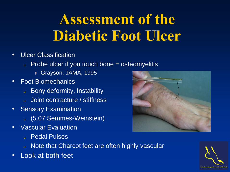
Assessment of the Diabetic Foot Ulcer
• Ulcer Classification
u Probe ulcer if you touch bone = osteomyelitis
F Grayson, JAMA, 1995
• Foot Biomechanics
u Bony deformity, Instability
u Joint contracture / stiffness
• Sensory Examination
u (5.07 Semmes-Weinstein)
• Vascular Evaluation
u Pedal Pulses
u Note that Charcot feet are often highly vascular
• Look at both feet

Investigation of the Diabetic Foot Ulcer
• Weight bearing AP, Lateral,
Oblique foot radiographs
• CRP, Se albumin, HBalc
• +/- Vascular studies
• +/- MRI
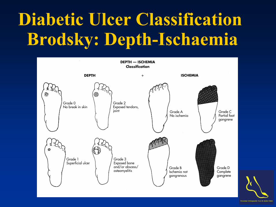
Diabetic Ulcer ClassificationBrodsky: Depth-Ischaemia
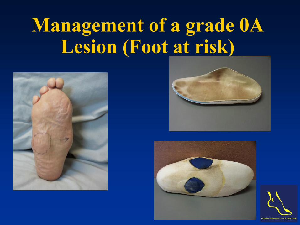
Management of a grade 0A Lesion (Foot at risk)
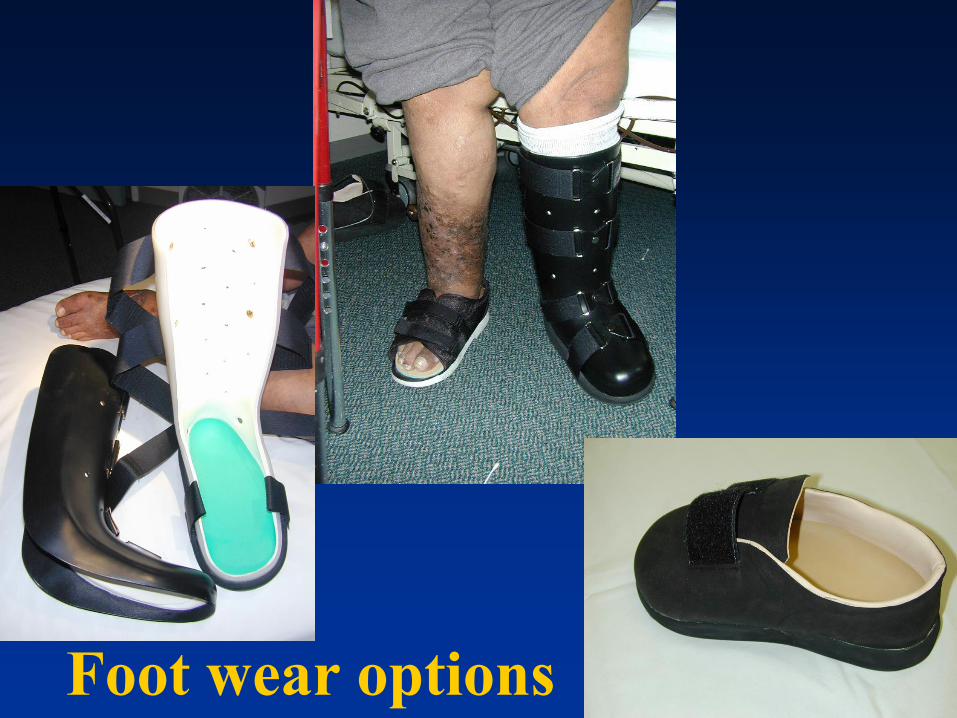
Foot wear options
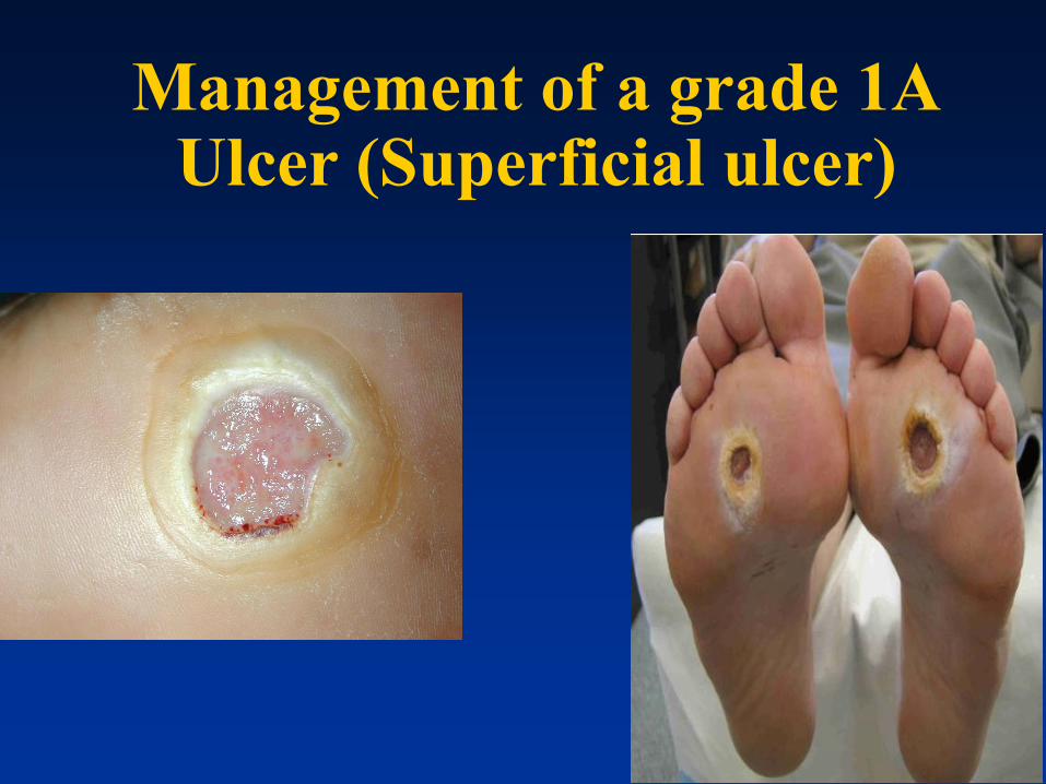
Management of a grade 1A Ulcer (Superficial ulcer)
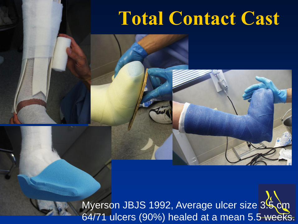
Total Contact Cast
Myerson JBJS 1992, Average ulcer size 3.5 cm
64/71 ulcers (90%) healed at a mean 5.5 weeks.

2A Ulcer 3A Ulcer
Surgical Management Ulcers: Options
• Debridement / VAC/ TCC
• Debridement + Ostectomy
• Amputation
• Debridement + Reconstruction
(acute /delayed)
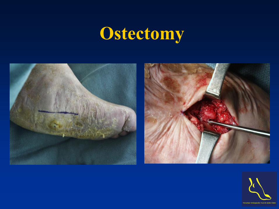
Ostectomy
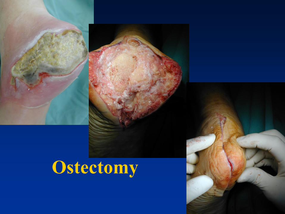
Ostectomy
Charcot Foot; Definition
• Chronic & progressive disease of joints and
bones… … painful or painless bone and
joint destruction in limbs that have lost
sensory innervation
• Joints exhibit synovitis, instability,
subluxation & destruction
Peripheral Neuropathy
• Causes (DINTMINI)
– Diabetes
– Alcoholism
– Congenital
insensitivity to pain
– Renal disease
– Leprosy
– Syphilis
• Diabetic feet
– Up to 5% of all
diabetic patients
– Up to 29% of patients
with PN
– Average duration of
DM 15 years
– Bilateral in 6 - 39%
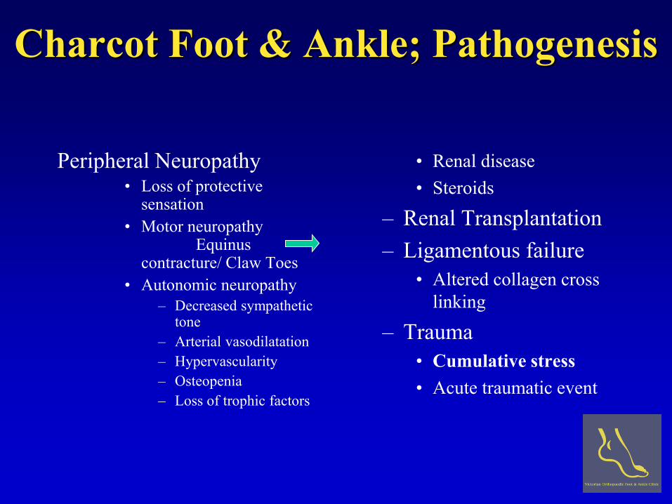
Charcot Foot & Ankle; Pathogenesis
Peripheral Neuropathy• Loss of protective
sensation
• Motor neuropathyEquinus
contracture/ Claw Toes
• Autonomic neuropathy
– Decreased sympathetic tone
– Arterial vasodilatation
– Hypervascularity
– Osteopenia
– Loss of trophic factors
• Renal disease
• Steroids
– Renal Transplantation
– Ligamentous failure
• Altered collagen cross
linking
– Trauma
• Cumulative stress
• Acute traumatic event
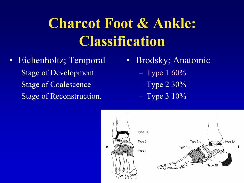
Charcot Foot & Ankle:
Classification
• Eichenholtz; Temporal
Stage of Development
Stage of Coalescence
Stage of Reconstruction.
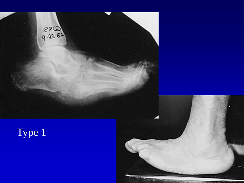
• Brodsky; Anatomic
– Type 1 60%
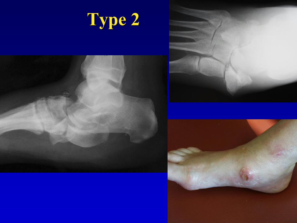
– Type 2 30%
– Type 3 10%

Type 1

Type 2

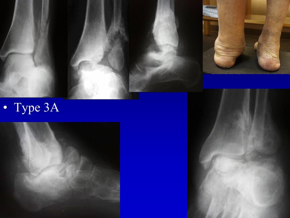
Type 3A
• Type 3A

Type 3A (Ankle) Charcot
Arthropathy
• Require immobilisation for 1-2 years
• Malleolar ulceration
• Deep sepsis
• Amputation
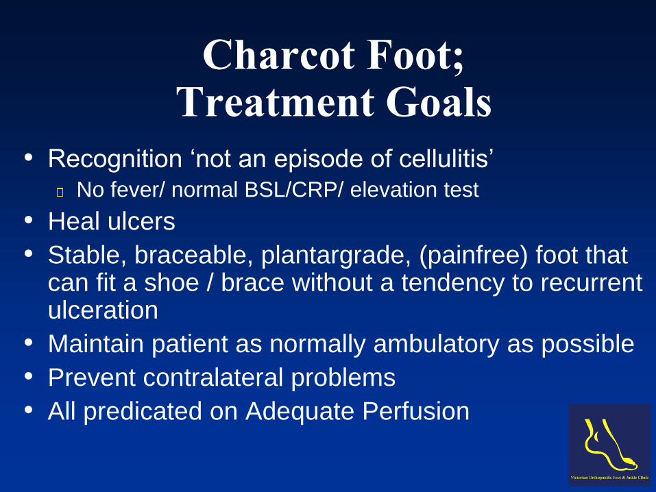
Charcot Foot; Treatment Goals
• Recognition ‘not an episode of cellulitis’ No fever/ normal BSL/CRP/ elevation test
• Heal ulcers
• Stable, braceable, plantargrade, (painfree) foot that can fit a shoe / brace without a tendency to recurrent ulceration
• Maintain patient as normally ambulatory as possible
• Prevent contralateral problems
• All predicated on Adequate Perfusion

NON -Operative treatment
• TCC total contact cast
• CROW Charcot restraint orthotic
walker
• BAFO Bivalved Ankle foot
orthosis
• CFLO custom full length
orthosis
• ? Medical Management
Operative
• Re-vascularisation
• Ulcer treatment
• Ostectomy
• Reconstruction
Fuse short, Instrument long
Two forms of fixation
• Amputation
Type 1
Type 2
Type 3A
• Type 3A
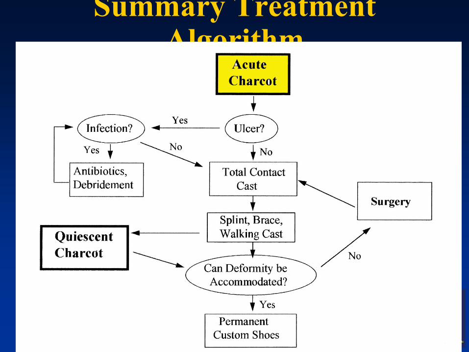
Summary Treatment Algorithm
Issues With Surgery
• Technically challenging
• Multi disciplinary team approach
• Risks of failure of internal fixation
• Wound healing problems
• Deep infections
• Amputation
• Loss of Mobility/Independence

Arthrodesis
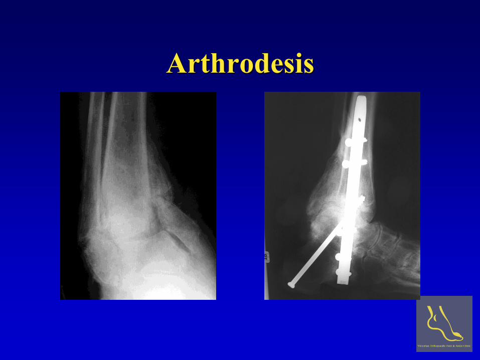
Arthrodesis
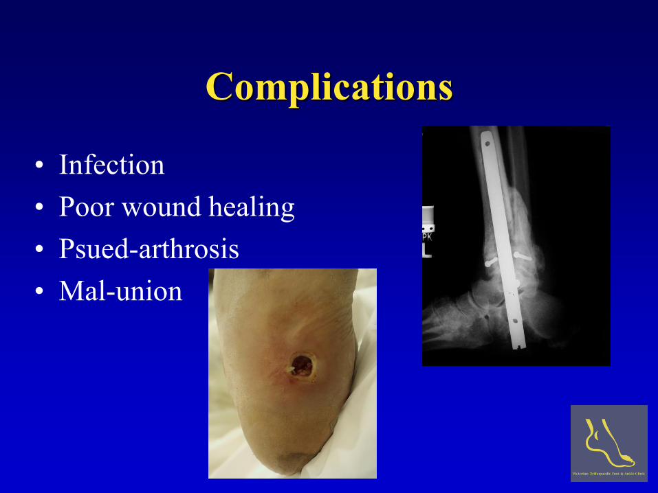
Complications
• Infection
• Poor wound healing
• Psued-arthrosis
• Mal-union
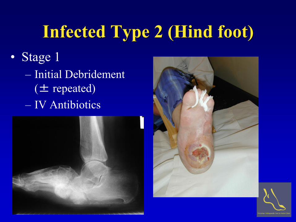
Infected Type 2 (Hind foot)
• Stage 1
– Initial Debridement
(± repeated)
– IV Antibiotics
– TCC
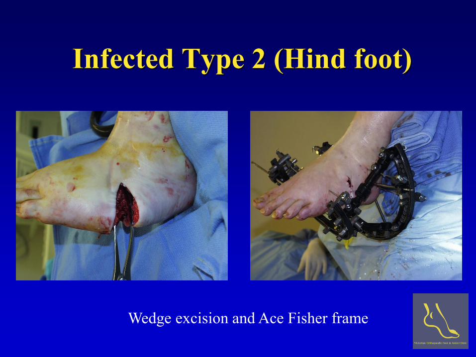
Infected Type 2 (Hind foot)
Wedge excision and Ace Fisher frame

Infected Type 3A

Infected Type 3A
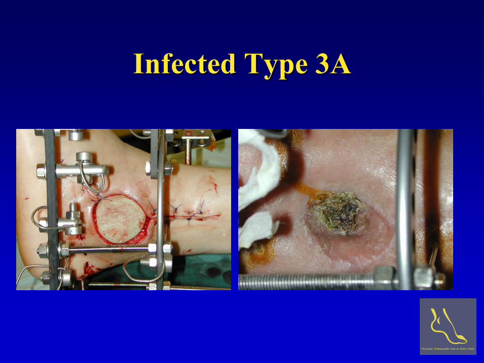
Infected Type 3A

Role of Amputation
• Potentially good option especially with
deep infection
• More rapid rehab (elderly population)
• ? More reliable healing
• ? Mid / Hind foot versus BKA
• ? Contralateral side
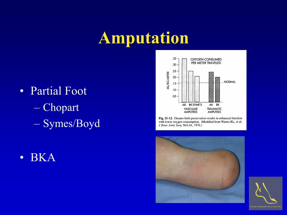
Amputation
• Partial Foot
– Chopart
– Symes/Boyd
• BKA
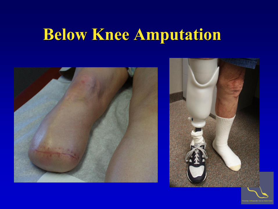
Below Knee Amputation
Conclusion: Ulcers
• Common Problem
• Surgical and Orthotic Management is Becoming More Sophisticated
• Off-loading pressure is the Key
• Multi-disciplinary Approach Required
• Ulcers Need Appropriate Investigation and Staging
Conclusion : Charcot
• Hot swollen foot think Charcot as well as infection
(Do an x-ray)
• Early treatment is usually non-operative (midfoot)
• Early surgery for Ulcers / Deformity (ankle)
• Surgery: Recurrent ulceration (Midfoot)
Uncontrolled deformity (hindfoot)
• Don’t forget Amputation as an option


![Patient education for preventing diabetic foot ulceration … · [Intervention Review] Patient education for preventing diabetic foot ulceration Johannes AN Dorresteijn 1, Didi MW](https://img.dokumen.tips/doc/110x75/5ecb99029d0d2c34515c80a4/patient-education-for-preventing-diabetic-foot-intervention-review-patient.jpg)