Embed Size (px)
Citation preview

Introduction
Chenopodiaceae family is a member ofCentrospermae, which covers about 100 genera and1500 species (Datta, 2003). The members of this familyare distributed chiefly in Australia, the Karro (SouthAfrica), the Red Sea shores, the South-West Caspiancoast, central Asia, and the salt steppes of East Asia. Mostof the Chenopodiaceae are halophytes, adapted to grow insalty or alkaline soil. Since this necessitates the reductionof transpiration, these plants show xerophytic characters(Singh & Jain, 1999).
In Iran, Chenopodiaceae is represented by 41 genera,one important of which in Iran is Chenopodium L.(Assadii, 2001). This genus is characterized by itsdistinctive reduced succulent leaves, spike-like compound
inflorescences, comprised of paired cymules of tinyflowers that are sessile within succulent free bracts. Theseflowers have 5 free perianth lobes, 5 stamens, a single,unilocular, and superior ovary; 1 bitegmic, crassinucellar,and campylotropous ovule with a basal solitary (Zhu Gelinet al., 2003; Shepherd et al., 2005). In the genusChenopodium, like other members of Centrospermae, thewhole seed is occupied by a long curved embryo, exceptfor a central mass of perisperm (Siew Young et al.,1975).
There exist comprehensive studies involving membersof this family on the embryo sac (Fischer, 1880;Dahlgren, 1916; Maheshwari, 1950, 1963; Davis,1966), on endosperm (Hegelmaier, 1885), and on seedcoats (Netolitzky, 1926).
1
Developmental Stages of Ovule and Megagametophyte inChenopodium botrys L. (Chenopodiaceae)
Abdolkarim CHEHREGANI1,*, Behrouz MALAYERI1, Nafiseh YOUSEFI2
1Department of Biology, Bu-Ali Sina University, Hamedan, IRAN2Colleges of Basic Science, Islamic Azad University, Hamedan, IRAN
Received: 27,05.2008Accepted: 31,03.2009
Abstract: The ovule ontogenesis and the megasporogenesis stages in Chenopodium botrys L. were studied with light microscopy.Flowers and young pods were removed from natural plants and fixed in FAA 70, stored in 70% ethanol, embedded in paraffin, andsectioned at 7 μm with a microtome. Staining was carried out with Hematoxylin and Eosin and developmental stages of ovule werestudied. The results of this research showed that ovule development, including megasporogenesis and initial stages ofmegagametogenesis, occurred while flowers were still in bud. In C. botrys the female gametophyte has a monosporic origin and thedevelopmental pattern exhibited by this species is referred to as the polygonum type. Development of ovule starts with the formationof a primordium. In this primordium, an archesporial cell produces a megaspore mother cell, which undergoes meiosis, forming alinear tetrad. The micropylar cell is a functional megaspore that survives and will function in megagametophyte development. Themature gametophyte is composed of 7 cells: 1 secondary nucleus, 2 synergids, 1 egg cell, and 3 antipodal cells.
Key Words: Chenopodium botrys, embryo sac, megagametogenesis, megasporogenesis, ovule development, Chenopodiaceae
* E-mail: [email protected]
Turk J Bot33 (2009) © TÜBİTAK
Research Article
Other studies dealing specifically with Chenopodiaceaealso emphasize these phases of seed study, only brieflydescribing the extensive embryo work has been done(Soueges, 1920; Marion, 1932; Ernest et al., 1961;Prego et al., 1998).
The study of the development of the embryo inChenopodiaceae is suggested, because although manystudies have described the megagametogenesis andmegasporogenesis in Angiosperms, few ofChenopodiaceae have been studied from a viewpoint ofthe embryo development, and also Chenopodiaceae is ataxonomically difficult group, largely due to the lack ofdiagnostic characters available; identification of theembryological characters in this family can be useful fordetermining taxonomic relationship between its members(Marion, 1932 ; Shepherd et al., 2005). Moreover, theflowers appear as the stem elongates, mature seeds maybe found on the same branch unfertilized ovules. For thisreason, selection of embryo in successive stages ofdevelopment is easily made (Marion, 1932).
The objective of this work was to determine thedevelopmental stages of ovule and embryo sac inChenopodium botrys L. Based on our knowledge, this isthe first report about ovule and megagametophytedevelopment in C. botrys.
Materials and Methods
Plant material
Young flowers and pods at different sizes werecollected from natural habitats. Inflorescence material forthe characterization of stages of reproductivedevelopment was collected in the summer of 2007 fromHame Kasi (Hamedan, Iran). A voucher specimen wasplaced in Bu-Ali Sina Herbarium (BASUH, 1037), BotanyDepartment at the same university.
Cytological studies
The flowers and buds were fixed in FAA 70(formaldehyde, glacial acetic acid, and 70% ethanol,5:5:90 v/v), (Johansen, 1940), stored in 70% ethanol.Specimens were embedded in paraffin. The blocks weresectioned at 7 μm with a Leitz 1512 microtome(Germany). Staining was carried out with Hematoxylin-Eosin according to the protocol suggested by Meyer(Yuang, 1984).
Several sections were studied under a lightmicroscope, Zeiss Axiostar Plus (Germany), for each
embryonic sac and ovule developmental stage.Developmental stages that were examined in this researchwork were according to Maheshwari’s criteria (1963)that are including ovular primordial, archeosporial cell,megaspore cell, diad and tetrad cells, mitotic division incoenocytic embryo sac, and mature embryo sac. At least20 samples were studied for each above mentioned stagesof reproductive developmental characters.
Results
Ovule development was investigated in Chenopodiumbotrys. L. Female gametophyte development, includingmegasporogenesis and initial stages ofmegagametogenesis, occurred while flowers were still inbud. The carpel is already closed when the firstprimordium appears (Figure 1). Ovule initiation isbasipetal and starts with mitotic activity in meristematicregions organized in 3 layers: dermal (nucellarepidermis), subdermal, and central (Figure 1). In general,one of hypodermal cells enlarges and functions as anarchesporium. In fact, this cell differentiates immediatelybelow the nucellar epidermis. The initial archesporial cellis distinguished from the other subdermal cells, because itpresents a larger volume, dense cytoplasm, and distinctnucleolus. This cell divides periclinally to give rise to theprimary parietal cell outerly and the archesporial cellproper innerly (Figure 2).
The primary parietal cell undergoes periclinal,anticlinal, and/or oblique divisions, contributing to formnucellar parietal layers. Simultaneously with the divisionof the initial archesporial cells, 2 integuments initiatefrom divisions of dermal cells. The inner integumentdevelops asymmetrically, rising from the distalprimordium flank (Figure 3).
The outer one differentiates simultaneously as a ringaround nucellus (Figure 4) but in some samplesdifferentiation of outer integument occurs sooner thanthe inner one.
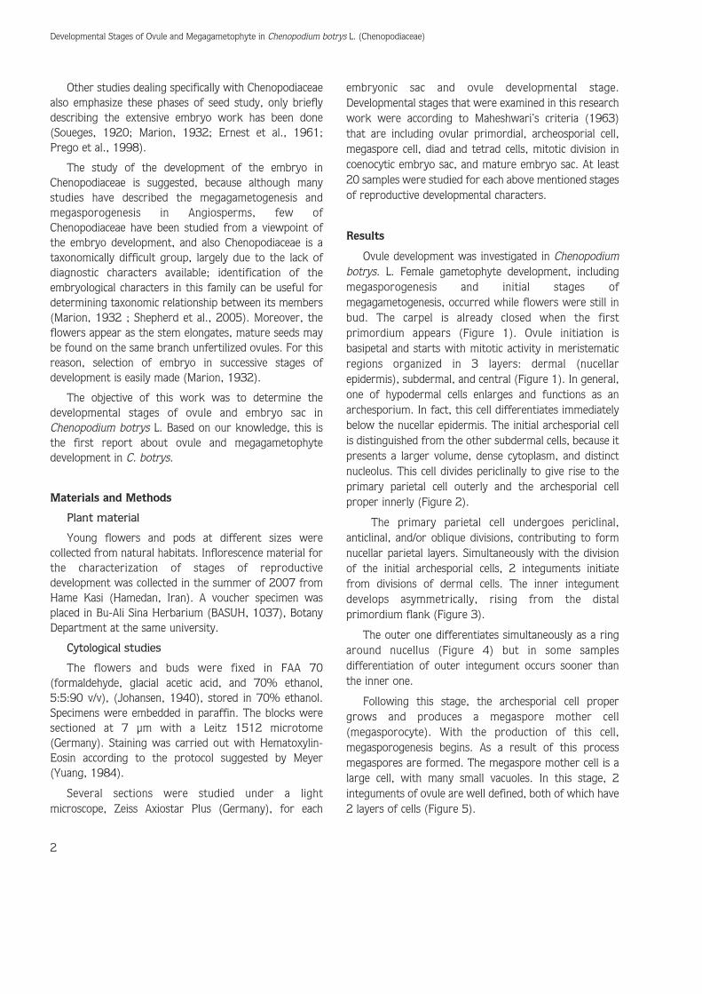
Following this stage, the archesporial cell propergrows and produces a megaspore mother cell(megasporocyte). With the production of this cell,megasporogenesis begins. As a result of this processmegaspores are formed. The megaspore mother cell is alarge cell, with many small vacuoles. In this stage, 2integuments of ovule are well defined, both of which have2 layers of cells (Figure 5).
Developmental Stages of Ovule and Megagametophyte in Chenopodium botrys L. (Chenopodiaceae)
2

Megasporogenesis follows and the diploid megasporemother cell undergoes meiosis and gives rise to 4 haploidmegaspore nuclei. The first meiotic division ofmegasporocyte (megaspore mother cell) producesequally-sized diad cells that are easily identified.Concomitant with this stage, periclinal divisions in parietalcells add to the nonsporogenous layers that are typical ofcrassinucellate ovules (Figure 6). During this floral stage,megasporogenesis within the developing ovule has alsobeen progressing. A second meiotic division produces alinear shaped tetrad of megaspores (Figure 7).
Only 1 of 4 megaspores, usually the one that isfarthest from the micropyle, develops in to embryo sacbut in C. botrys the chalazal megaspores soon degenerateand micropylar megaspore survives and functions as afunctional megaspore. In fact in each ovule examined, themicropylar cell was larger and showed no signs ofdegenerating. In this functional cell vacuolation beginsrapidly (Figure 8). The development of the functionalmegaspore through meiotic divisions of megasporocyteand degeneration of the 3 chalazal megaspores sets thestage for megagametogenesis (Figures 9-12).
A. CHEHREGANI, B. MALAYERI, N. YOUSEFI
3
� �
� �
Figures 1-4. Photomicrography of longitudinal section of the carpel.
1. Ovule primordium initiation with 3-zonate organization: dermal, subdermal, and central, (6500×).
2. Parietal cell that produces nucellar parietal layers and archesporial cell proper in subdermal position that produces megasporocyte, (6500×).
3. Dermal origin of the integuments, outer integument, (4000×).
4. The outer and inner integuments have initiated the formation of the inner integument; the integuments develops asymmetrically and differentiationof outer integument occurs sooner than the inner one, (5100×).
d = dermal layer; s = subdermal layer; c = central layer; p = primary parietal cell; ar = archesporial cell proper; oi = outer integument; ii = innerintegument. Scale Bar = 40 μm in Figures 1 and 2; 50 μm in Figures 3 and 4.
In C. botrys in this stage, several changes take place ingross ovule morphology. The micropyle becomes welldefined by the final elongation of the inner integumentand expansion of integumentary cells in the micropylararea. The outer integument undergoes further elongationand is barely subequal to the inner integument. Duringthis interval, the functional megaspore shows dramaticenlargement and the vacuole expands and occupies mostof the cell volume (Figure 9). At the next stage, a mitoticdivision within the functional megaspore produces 2
nuclei, 1 of which moves towards the chalazal end andother one towards the micropylar pole of the embryo sac(Figure 10). A second mitotic division results in the 4-nucleate stage (Figure 11). The embryo sac nowundergoes a dramatic increase in width and length andthis growth occurs with slow consumption of nucellus,thus in this stage of ovule development the nucellar tissuearound the embryonic sac degenerates slowly (Figure 12).
The third mitotic division produces 8 nuclei, thusafter the third mitotic cycle, the 8-nucleated gametophyte
Developmental Stages of Ovule and Megagametophyte in Chenopodium botrys L. (Chenopodiaceae)
4
� �
Figures 5-8. Photomicrography of longitudinal section of the carpel.
5. Megaspore mother cell that begins megasporogenesis, 2 integuments of ovule are defined, both of them have 2 layers of cells, (4000×).
6. Equally-sized diad cells, produced by the first meiotic division of megasporocyte, divisions in parietal cells add to the nonsporogenous layers,(6500×).
7. Linear shaped tetrad megaspores produced by the second meiotic division of megaspore mother cell, (5100×).
8. Functional micropylar megaspore when vacuolation begins and 3 degenerating chalazal megaspores, (5100×).
mec = megasporocyte; di = dyade; te = tetrad; fm = functional megaspore. Scale Bar = 50 μm in Figures 5, 7, and 8; 40 μm in Figure 6.

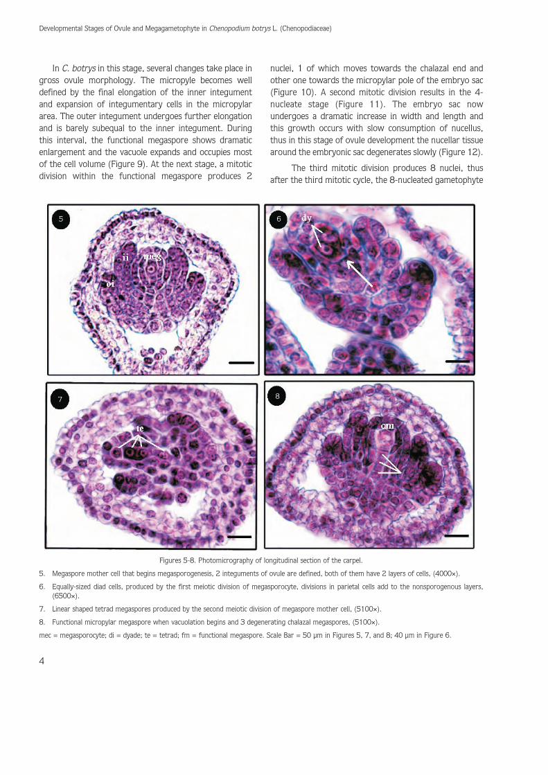
is presented with 4 chalazal and 4 micropylar nuclei(Figure 12). Development of a 8-celled megagametophyteoccurs during the final floral bud stage (Figures 13-16).According to this stage nucellus cells slowly degenerateand their remains are visible around the embryo sac, also3 of 4 chalazal nuclei, after cellularization, establish a rowof wall-less cells known as the antipodals that degeneratelater. Besides, 3 of 4 nuclei at the micropylar end of theembryo sac become organized in to the egg apparatus,consisting of 2 synergid cells and 1 egg cell. The last
chalazal nucleus and its micropylar neighbor migrate tothe mid-region of the central cell, and compose the 2polar nuclei. The polar nuclei fuse and form a diploidnucleus that calls the secondary endosperm nucleus(Figure 14). Thus in this stage embryo sac consist of asecondary endosperm nucleus within the central cell, 2synergids, and an egg cell at the micropylar end and 3antipodals at the chalazal end (Figures 14,16).Furthermore, all of the cells within the femalegametophyte differentiate into polar strictures. In C.
A. CHEHREGANI, B. MALAYERI, N. YOUSEFI
5
� ��
�� ��
Figures 9-12. Photomicrography of longitudinal section of the carpel.
9. Megaspore mother cell, (5100×).
10. Two-nucleated embryonic sac, one of the nuclei stablishes in the chalazal end and other one in the micropylar end of the embryo sac, (8100×).
11. Four-nucleated embryo sac, (6500×).
12. Eight-nucleated embryo sac, expansion of the embryo sac that occurs with the consumption of nucellus, (8500×).
cm = mother cell megaspore; n = nucleus; es = embryo sac, Scale Bar = 80 μm in Figure 9; 30 μm in Figures 10-12.
botrys, the egg cell nucleus is located toward the chalazalend and its vacuole occupies the micropylar end, bycontrast synergid and central cells have the oppositepolarity.
Discussion
This report is the first to provide a detailed ovule andembryo sac development in Chenopodium botrys. Theresults presented indicate that many sexual reproductivefeatures expressed in C. botrys are similar to those of
other members of the Chenopodiaceae. Ovaries of C.botrys exhibited 1 locular carpel and 1 ovule withincarpel. Current observations showed that patterns ofembryo sac development are evidence of standardpolygonum type and pattern of the megasporogenesis inthis species is monosporic, which is the most commontype of development in Angiosperms.
Megasporangium or ovule in C. botrys is basipetal andconsists of nucellus and 2 integuments. In this species, theovule is curved, called as campylotropous, which is inagreement with observation reported by Bocquet (1959).
Developmental Stages of Ovule and Megagametophyte in Chenopodium botrys L. (Chenopodiaceae)
6
��
�� ��
��
Figures 13-16. Photomicrography of longitudinal section of the carpel.
13. Embryo sac in Chenopodium botrys, remains of nucellar cell, (5100×).
14. Two polar nuclei that fuse and form the secondary nucleus, (5600×).
15. Egg apparatus, consisting of 2 synergid cells and 1 egg cell at the micropylar end, and antipodal cells at the chalazal end, (5600×).
16. Mature Campylotropous ovule in Chenopodium botrys L., (5100×).
es = embryo sac; os = oospher; at = antipodal cells; syn = synergid cells; eg = egg cell, Scale Bar = 80 μm in Figures 13-16.

It has 2 integuments, each being 2 thick cells that is inaccordance with the findings of Sherry et al., (1933) andCorner (1976). Differentiation of 2 integuments isconcomitance but in some cases differentiation of theinner one occurs sooner than that of the outer one.
Depending on the extent of development of thenucellus, ovule in C. botrys is crassinucellate type, becausein this species there is a well-developed parietal tissue andan archesporial cell is separated from the nucellarepidermis by 1 layer of cell. This finding is in agreementwith the finding of Connor (1984).
During the megasporogenesis, as a result of meioticdivisions, a linear tetrad of 4 megaspores is formed. The
micropylar megaspore of tetrad serves and gives rise tothe female gametophyte, while the remaining 3megaspores degenerate and disappear; this is a functionalmegaspore. Our report is also the first about micropylarfunctional megaspores.
The mature form of female gametophyte or embryosac of C. botrys like embryonic sac of otherChenopodiaceae has 7 cells, 8 nucleate consisting of 3antipodal cells; a large binucleate central cell and an eggcell adjacent to 2 synergids, that is in accordance with thefindings of previous researchers regarding othermembers of Chenopodiaceae (Stebbins, 1974 ; Marion,1932) (Figures 13-16).
A. CHEHREGANI, B. MALAYERI, N. YOUSEFI
7
Assadii M (2001). Flora of Iran, No. 38: Chenopodiaceae. ResearchInstitute of Forests and Rangelands.
Bocquet G (1959). The camylotropous ovule. Phytomorphology 9: 222-227.
Connor HE (1984). Gynodioecism in Sarcocornia quinqueflora(Chenopodiaceae) in New Zealand. N Z J Bot 22: 433-9.
Corner E (1976). The seeds of dicotyledons. London: Cambridgeuniversity Press.
Dahlgren KVO (1916). Zytologische und embryologische studien uberdie Reihen Primulales und Plumbaginales. Kunglia svenskavetenskapskademiens handlingar 56: 1-80.
Datta SC (2003). Systematic botany, Fourth edition. New ageintrenational (P) Ltd., Publishers.
Davis GL (1966). Systematic embryology of the Angiosperms. JohnWiley & Sons, NewYork.
Ernest M, Gifford JR, & Herbert B (1961). Ontogeny of theinflorescence in Chenopodium album. Am J Bot 48: 657-67.
Fischer A (1880). Kenntniss der embryo-sac entwicklung einigerAngiospermen. Jenaische Zeitschrift Bd 14: 90-132.
Hegelmaier F (1885). Untersuchungen uber olie morphologie desdikotyledonen endosperms. Nova Acta Akad Leop Carol 49: 1-123.
Johansen DA (1940). Plant microtechnique. McGraw-Hill, New York.
Maheshwari P (1950). An introduction to the embryology ofangiosperms. McGraw-Hill book company, Inc. New York.
Maheshwari P (1963). Embryology in relation to taxonomy. In VistasBotany, IV. Turrill, W.B. (Ed). Oxford: Pergamon Press.
Marion K (1932). The development of the embryo of Kochia scoparia(Chenopodiaceae). Bulletin of the torrey Bot. club 56: 391-400.
Netolitzky F (1926). Anatomie der Angiosperms. Samen Handbuch derpflanzenanatomie Bd.X, Borntraeger, Berlin.
Prego I, Maldonado S, & Otegut M (1998). Seed structure andLocalization of Reserves in Chenopodium quinoa. Annals of Bot82: 481-8.
Shepherd KA, Mac Farlance TD & Colmer TD (2005). Morphology,Anatomy and Histology of Salicornioideae (Chenopodiaceae) fruitsand seeds Annals of Bot 95: 917-33.
Sherry R, Eckard K & Lord E (1933). Flower development in DioeciousSpinacia oleracea (Chenopodiaceae). Am J Bot 80: 283-91.
Siew young NG, Philipson WR & Walker JRL (1975). Hectorellaceae amember of Centrospermae. N Z J Bot 13: 567-70.
Singh V & Jain DK (1999). Taxonomy of angiosperms. second edition.Rastugi publications.
Soueges R (1920). Embryogenie des Solanacees. Development de Iembryon chezles Nicotiana. C.R. Acad. Sci. Paris 170: 1125-7.
Stebbins GL (1974). Flowering Plants: Evolution Above the speciesLevel. Cambridge: Harvard University Press.
Yeung EC (1984). Histochemical and Histochemical staining procedures.In: Cell culture and Somatic Cell Genetics of Plants (E.d.) Vasil,I.K., Academic Press, Orlando, Fla, Pp: 686-97.
Zhu G, Mosyakin S & Clemants S (2003). Chenopodium. Flora of China5: 351-414
References