Embed Size (px)
Citation preview

Developmental relations between amygdala volume and anxietytraits: Effects of informant, sex, and age
KATHERINE RICE WARNELL,a MEREDITH PECUKONIS,b AND ELIZABETH REDCAYb
aTexas State University; and bUniversity of Maryland
Abstract
Although substantial human and animal evidence suggests a role for the amygdala in anxiety, literature linking amygdala volume to anxiety symptomatology isinconclusive, with studies finding positive, negative, and null results. Clarifying this brain–behavior relation in middle to late childhood is especiallyimportant, as this is a time both of amygdala structural maturation and the emergence of many anxiety disorders. The goal of the current study was to clarifyinconsistent findings in previous literature by identifying factors moderating the relation between amygdala volume and anxiety traits in a large sample oftypically developing children aged 6–13 years (N ¼ 72). In particular, we investigated the moderating effects of informant (parent vs. child), age, and sex.We found that children’s reports (i.e., self-reports) were related to amygdala volume; children who reported higher anxiety levels had smaller amygdalae.This negative relation between amygdala volume and anxiety weakened with age. There was also an independent effect of sex, such that relations werestronger in males than in females. These results indicate the importance of considering sample and informant characteristics when charting the neurobiologicalmechanisms underlying developmental anxiety.
Across species and methodologies, the amygdala has beenimplicated in fear expression and conditioning (for reviewssee Etkin & Wager, 2007; Grupe & Nitschke, 2013; Kimet al., 2011; and Tovote, Fadok, & Luthi, 2015), suggestingthat this structure may be core to the exaggerated and persis-tent fear that characterizes anxiety disorders (Shin & Liber-zon, 2010). Attesting to the role of the amygdala in anxiety,both clinically anxious populations and typical individualswith high levels of anxiety exhibit amygdala hyperactivityand altered patterns of amygdala functional connectivity(e.g., Bishop, Duncan, & Lawrence, 2004; Etkin et al.,2004; Hahn et al., 2011; Hyde, Gorka, Manuck, & Hariri,2011; Kim, Gee, Loucks, Davis, & Whalen, 2011; Sehlmeyeret al., 2011; Sripada, Wang, Sripada, & Liberzon, 2012). Incontrast to robust findings from functional neuroimaging,the link between amygdala structure and anxiety remains in-conclusive, with adult studies finding both positive (e.g.,Baur, Hanggi, & Jancke, 2012; Lyons-Ruth, Pechtel, Yoon,Anderson, & Teicher, 2016; Schienle, Ebner, & Schafer,2011) and negative (e.g., Fisler et al., 2013; Hayano et al.,2009; Rogers et al., 2009; Spampinato, Wood, De Simone, &Grafman, 2009) relations between anxiety and amygdalavolume. Given the mixed adult literature, studies examining
the link between amygdala volume and anxiety at earlier de-velopmental time points will help clarify the structural brainbases of anxiety.
Understanding the structural brain bases of anxiety isimportant because structural magnetic resonance imaging(MRI) data can inform our understanding of neural mecha-nisms while avoiding some practical confounds of functionalneuroimaging work. Recent human and animal literature hasfound important links between amygdala structure and anxi-ety behaviors. Smaller amygdalae in mice have been linked toincreased fear responsivity and glucocorticoid response(Yang et al., 2008) and, in depression and bipolar disorder, in-dividual differences in amygdala volume are related to func-tional hyperactivity (Kalmar et al., 2009; Siegle, Konecky,Thase, & Carter, 2003). Further, interventions designed toimpact stress and anxiety can alter amygdala volume (Holzelet al., 2009), suggesting that volumetric data could provide aneurobiological measure of intervention efficacy. Practically,structural studies allow for data to be collected from a widerange of ages and functioning levels, including from thosewho cannot complete task-based designs, and structuraldata acquisition is less susceptible to motion artifacts andin-scanner anxiety (Dennis, Gotlib, Thompson, & Thoma-son, 2011). Recent evidence also suggests that structural neu-roimaging can be used to differentiate anxiety subtypes (e.g.,Hilbert, Lueken, Muehlhan, & Beesdo-Baum, 2017; Hilbertet al., 2015), indicating the method’s potential promise as abiomarker. Thus, structural neuroimaging studies of develop-mental anxiety offer important complementary insight tofunctional neuroimaging, helping to illuminate neurobiolog-ical mechanisms of anxious behaviors.
Address correspondence and reprint requests to: Katherine Rice Warnell,Department of Psychology, Texas State University, San Marcos, TX 78666;E-mail: [email protected].
We thank Laura Anderson Kirby, Dan Levitas, Dustin Moraczewski, JazlynNketia, Harrison Pierce, Eleonora Sadikova, and Kayla Velnoskey for assis-tance with data collection and analysis and the Maryland Neuroimaging Cen-ter and staff for project assistance.
Development and Psychopathology, 2017, page 1 of 13# Cambridge University Press 2017doi:10.1017/S0954579417001626
1
https://www.cambridge.org/core/terms. https://doi.org/10.1017/S0954579417001626Downloaded from https://www.cambridge.org/core. Texas State University - San Marcos, on 26 Nov 2017 at 16:10:31, subject to the Cambridge Core terms of use, available at

In particular, elucidating the relation between amygdalavolume and anxiety in middle to late childhood (roughlyages 6–13) is important for two reasons. First, although anx-iety disorders continue to develop into adolescence, middle tolate childhood is a time when many anxiety disorders first be-gin to emerge (Beesdo, Knappe, & Pine, 2009; Kessler et al.,2005, 2007). Thus, examining whether and how the relationbetween amygdala volume and anxiety changes in this agerange, even in typical samples, may help illuminate neuralmechanisms underlying the development of more severe anx-iety later in life (Beesdo et al., 2009). Second, middle to latechildhood represents an important time in amygdala develop-ment. Recent research suggests that amygdala volume peaksaround 11 years, with slightly earlier maturation in femalesversus males and in the left versus right amygdala (Uematsuet al., 2012), although other studies have found peaks in early(Hu, Pruessner, Coupe, & Collins, 2013) and late adolescence(Wierenga, Langen, Oranje, & Durston, 2014), with inconsis-tent evidence for sex differences (either opposite trajectoriesBramen et al., 2011; or no differences, Wierenga et al., 2014).Despite the mixed findings of structural maturation, func-tional data does suggest that approximately 10 years of agemay represent an inflection point in connectivity betweenthe prefrontal cortex and amygdala (Gabard-Durnam et al.,2014; Gee et al., 2013), a circuit that has been linked to fearfulor anxious behaviors.
Despite the importance of understanding brain–behaviorrelations in developmental populations, few studies have ex-amined the link between amygdala volume and anxiety inchildren and adolescents. Further, even this small set of stud-ies has yielded inconsistent findings: anxiety has been linkedto both increased (Albaugh et al., 2017; De Bellis et al., 2000;Qin et al., 2014; Tottenham et al., 2010; van der Plas, Boes,Wemmie, Tranel, & Nopoulos, 2010; Weems, Klabunde,Russell, Reiss, & Carrion, 2015) and decreased amygdalavolume (McQueeny et al., 2011; Milham et al., 2005; Muelleret al., 2013; Strawn et al., 2015). Understanding the reasonsfor these contradictory findings may clarify the mechanismslinking amygdala volume to anxiety. Specifically, there areseveral candidate explanations for why previous develop-mental studies have found divergent results.
First, given that the amygdala continues to developthrough middle to late childhood, one possibility is that therelation between anxiety and amygdala volume changeswith age. Most of the studies finding a positive relation exam-ined participants into middle adolescence (e.g., around ages16–17), with one study finding that a positive relation be-tween amygdala volume and posttraumatic stress disorder di-agnosis was only present in participants older than 15.5 years,with younger participants showing a trend toward negative re-lations (Weems et al., 2015). However, one study examiningchildren aged 7–9 years found a positive relation betweenanxiety traits and amygdala volume (Qin et al., 2014), andnegative relations between amygdala volume and anxietyhave also been found in adults (e.g., Fisler et al., 2013;Hayano et al., 2009; Rogers et al., 2009; Spampinato et al.,
2009). Further complicating the study of age effects, amyg-dala development appears to follow different trajectories formales and females (Bramen et al., 2011; but see Wierengaet al., 2014), and there is some evidence that the develop-mental relations between volume and anxiety are impactedby sex (van der Plas et al., 2010). Anxiety also has differentprevalence between males and females, although sex differ-ences typically do not emerge until late childhood or earlyadolescence (Beesdo et al., 2009; Lewinsohn, Gotlib, Lewin-sohn, Seeley, & Allen, 1998). Thus, it is unknown if age orsex may be responsible for contradictory findings in previousdevelopmental samples.
Second, another potential explanation for the inconsistentliterature is that different methods of anxiety assessment (e.g.,differences in informants, measures, and sample characteris-tics) may yield different results. Although most studies exam-ining amygdala volume and anxiety in typical adults haveused self-reported anxiety (e.g., Baur et al., 2012; Spampi-nato et al., 2009), almost all studies of amygdala volume intypical children have relied on parent report (e.g., Albaughet al., 2017; Qin et al., 2014; Tottenham et al., 2010; vander Plas et al., 2010), with the one exception being in olderadolescents (McQueeny et al., 2011). This reliance on parentreport may be problematic given that the discrepancy betweenparent-reported anxiety and child self-report begins early inchildhood (e.g., Comer & Kendall, 2004; Engel, Rodrigue, &Geffken, 1994). Previous research has indicated that, at leastin typical samples, child report may better reflect real-worldsymptomatology (Cosi, Canals, Hernandez-Martinez, &Vigil-Colet, 2010) and that parental relative underreportingof anxiety symptoms (Cosi et al., 2010; Muris, Merckelbach,Van Brakel, & Mayer, 1999) may lead to statistical floor ef-fects. In addition to issues of informant, some previous stud-ies have used scales that measure internalizing behaviorsmore broadly (e.g., a combined anxiety/depression scale; Al-baugh et al., 2017; Qin et al., 2014). Further, it is possible thata more precise investigation of different types of anxiety (e.g.,social anxiety vs. generalized anxiety) may reveal differentpatterns of correlation with amygdala volume, as functionalneuroimaging literature has indicated dissociations betweenanxiety subtypes in adults (Blair et al., 2008) and children(Kessel, Kujawa, Proudfit, & Klein, 2015). Finally, much de-velopmental research has examined clinical populations.Research in a typical sample may offer insight into thebrain-based precursors of later clinical anxiety (Westenberg,Gullone, Bokhorst, Heyne, & King, 2007) and give insightinto the relation between amygdala volume and anxious traits,traits that may be disruptive even at subclinical levels (Bell-Dolan, Last, & Strauss, 1990). Ultimately, examiningbrain–behavior relations across multiple ages, different infor-mants, and varied types of anxiety within typical samples iscritical, given that the small set of existing studies, with theirwide range of methods and samples, precludes a meta-analy-tic approach.
Thus, the goal of the current study is to disambiguate, in asingle developmental sample, factors that may influence the
K. R. Warnell, M. Pecukonis, and E. Redcay2
https://www.cambridge.org/core/terms. https://doi.org/10.1017/S0954579417001626Downloaded from https://www.cambridge.org/core. Texas State University - San Marcos, on 26 Nov 2017 at 16:10:31, subject to the Cambridge Core terms of use, available at

relation between amygdala volume and anxiety traits, and indoing so, to clarify both past findings and the potentialmechanisms driving these brain–behavior associations. Tothis end, we examined the relation between parent-reportedand child-reported anxiety in a sample of typical childrenaged 6–13 years. We analyzed three specific moderators ofthe relation between amygdala volume and anxiety: (a) infor-mant (i.e., parent vs. child report), (b) sex, and (c) age. We didnot hypothesize about whether the link between amygdalavolume and anxiety traits would be positive or negative acrossthese moderators. Rather, the goal of the study was to attemptto disambiguate factors driving the mixed findings of priordevelopmental research.
Method
Subjects
Eighty-four children were recruited via random samplingfrom a database of local families. All children were full-term, native English speakers, with normal or corrected tonormal hearing and vision. As assessed via parent report,children had no history of psychiatric, psychological, or neu-rological conditions, no first-degree relatives with autism orschizophrenia, and no contraindications for MRI scanning.Racial and ethnic information were collected from a subsetof n ¼ 46 participants. Of this subset, 34 children (74%)were White and non-Hispanic/Latino, 6 were of more thanone race, 2 were Black, 2 were White and Hispanic/Latino,1 was Asian, and 1 was Native Hawaiian or Pacific Islander.Out of the subset of participants (n ¼ 73) with household in-come and parental education data, the majority (81.0%) had ahousehold income of over $75,000/year and the majority(63.0%) also had at least one parent with a graduate degree.Structural MRI and behavioral data were collected at two sep-arate visits within 6 weeks of each other. Minor and parentalconsents were obtained from all participants, as approved bythe Internal Review Board at the University of Maryland.
Behavioral data acquisition
The Screen for Child Anxiety Related Emotional Disorders(SCARED; Birmaher et al., 1999) is a 41-item questionnairethat yields five anxiety factors: somatic/panic, generalizedanxiety, separation anxiety, social phobia, and school phobia.We used two versions of the SCARED, one in which the childreported on his or her own anxiety (child SCARED) and onein which the parent reported on the anxiety levels of the child(parent SCARED). The same items are used on the child andparent SCARED, but pronouns are adjusted based on theinformant, that is, “I feel . . .” versus “My child feels . . .”(Birmaher et al., 1997). Participants respond to prompts ona 3-point Likert scale (0 ¼ not true or hardly ever true, 1¼ sometimes true, and 2 ¼ true or often true), with higherscores indicating greater anxiety. The SCARED has goodinternal validity, test–retest reliability, discriminant validity,
and cross-cultural external validity (Birmaher et al., 1999;Su, Wang, Fan, Su, & Gao, 2008; Weitkamp, Romer,Rosenthal, Wiegand-Grefe, & Daniels, 2010). Parent-reportand child-report have shown low to moderate levels of agree-ment (Cosi et al., 2010; Dirks et al., 2014; Muris et al., 1999;Pereira et al., 2015), with children tending to report higherlevels of anxiety overall and larger discrepancies emergingin somatic/panic and generalized anxiety (Cosi et al., 2010;Wren et al., 2007).
In the current study, each child and each parent indepen-dently completed the SCARED, yielding both a child-reportand a parent-report measure of anxiety. Children completedthe SCARED by hand with the help of a researcher. The re-searcher read each item to the child and asked him or her tomark the box that corresponded with his or her feelings aboutthat item. The researcher told the child that his or her answerswould be kept private to ensure accurate responses on the self-report. One parent of each participant was instructed to com-plete the parent SCARED independently by hand. Consistentwith previous SCARED research, the current study computedraw scores for each factor, which were then summed to obtainan overall parent SCARED score and child SCARED scorefor each participant.
MRI data acquisition
High-resolution T1-weighted images were acquired using asingle Siemens 3.0-T scanner (MAGNETOM Trio Tim Sys-tem, Siemens Medical Solutions) using one three-dimensionalT1 magnetization-prepared rapid gradient-echo sequence (176contiguous sagittal slices, voxel size ¼ 1.0�1.0�1.0 mm;repetition time/echo time/inversion time ¼ 1900 ms/2.52 ms/900 ms; flip angle ¼ 98; pixel matrix ¼ 256�256). For thefinal sample, data from 22 children were collected using a12-channel head coil, and data for the other 50 childrenwere collected on a 32-channel head coil. Age, amygdala vol-ume, and SCARED scores did not significantly differ betweenthe two groups of children ( ps . .05), and inclusion of chan-nel type as a covariate in analyses did not alter any patterns ofresults. Thus, we collapsed together both groups into a singlesample for subsequent analyses.
The imaging data were analyzed using FreeSurfer software(Version 5.1.0). Freesurfer’s automated segmentation ofamygdala volume is highly correlated with expert hand trac-ing methods (Morey et al., 2009). FreeSurfer first automati-cally compared each participant’s T1-weighted image to aprobabilistic atlas, the use of which has been validated in pe-diatric populations (Burgund et al., 2002; Ghosh et al., 2010),and then established boundaries for gray matter, white matter,and pial. Two trained, blind coders visually inspected the vol-umetric parcellation map of each participant, and edited thepial parcellation boundaries where necessary. After edits weremade and agreement was reached between coders, automaticsegmentation was rerun, yielding estimates of total gray mat-ter volume and left and right amygdala volume. Total graymatter volume was calculated using the automatic algorithm
Amygdala volume and anxiety in development 3
https://www.cambridge.org/core/terms. https://doi.org/10.1017/S0954579417001626Downloaded from https://www.cambridge.org/core. Texas State University - San Marcos, on 26 Nov 2017 at 16:10:31, subject to the Cambridge Core terms of use, available at

in FreeSurfer. This algorithm sums together values from leftcortex, right cortex, subcortical structures and cerebellum.For cortex, gray matter is calculated based on the volumeinside the pial boundaries, subtracting out white matter andsubcortical structures. Given concerns about unsupervisedamygdala identification in pediatric populations (Schoe-maker et al., 2016), a trained coder compared the amygdalamask identified by FreeSurfer to the original T1 image toensure accuracy (Figure 1).
From the original sample of 84 children, 5 participants (5female, mean age ¼ 9.82 years) did not have the parentSCARED measure, 1 participant (1 male, age ¼ 8.49 years)did not complete the structural scan due to discomfort inthe scanner, 4 participants (2 male, mean age ¼ 9.27 years)did not have usable FreeSurfer data due to excessivemotion in the scanner, as identified by overall poor imagecontrast, and 2 participants (2 male, mean age ¼ 9.81 years)were excluded due to inaccurate amygdala/hippocampal
segmentation revealed during manual inspection. The finalsample (N ¼ 72) did not differ from the excluded sample inage, sex, or anxiety (Table 1).
Statistical methods
Previous investigations of amygdala volume and anxiety haveused a variety of methods to normalize across head size, in-cluding statistical corrections for total intracranial volume(e.g., Fisler et al., 2013; van der Plas, 2010) and total graymatter (Lyons-Ruth et al., 2016), as well as dividing amyg-dala volume by total intracranial volume (e.g., Rogers et al.,2009). A meta-analysis has indicated that subcortical struc-tures most closely scale with gray matter volume and that con-trolling via regression may best prevent bias (Van Petten,2004). We thus controlled for total gray matter volume inall of our brain-based analyses.
We conducted both planned and exploratory brain–behav-ior analyses. In our planned analysis, we had two core goals:(a) to establish the relation between amygdala volume andanxiety, and, if this relation was significant, (b) to determineif this brain–behavior relation was moderated by informant,sex, or age. To accomplish the first goal, we calculated the re-lation between amygdala volume, which was corrected forage, sex, and total gray matter volume, and total childSCARED score. We then repeated this analysis for total par-ent SCARED score. For these tests, we analyzed the right andleft amygdala separately, as previous developmental literaturehas found relations between anxiety and amygdala volumespecific to both the right (e.g., De Bellis et al., 2000; Muelleret al., 2013; Weems et al., 2015) and the left (e.g., Milhamet al., 2005; Qin et al., 2014) amygdala. Although there issubstantial literature on amygdala lateralization (for reviews,see Fusar-Poli, Placentino, Carletti, Landi, & Abbamonte,2009; McMenamin & Marsolek, 2013; and Sergerie, Cho-chol, & Armony, 2008), the lack of a clear laterality patternin previous brain–behavior results meant that we did not
Figure 1. (Color online) Amygdala volume in childhood. The figure depicts asample coronal slice from a participant (aged 7.58 years) after anatomicaldata were processed. After automatic parcellation was completed, coders in-spected each map, made any needed edits to pial surface segmentation, andthen reran the automatic parcellation. As a final step, the anatomical bound-aries of the amygdala on the T1 image were manually compared to the com-puter-generated amygdala segmentation (depicted in teal online). Left andright amygdala volumes were computed separately.
Table 1. Descriptive statistics
Included Sample Excluded Sample Group ComparisonMeasure (n ¼ 72) (n ¼ 12) p
Sex 35 M/37 F 5 M/7 F .66Age (years) 10.21 (1.90) 9.52 (1.24) .23Verbal IQ 118.82 (11.15) 120.30 (10.84) .69Nonverbal IQ 115.86 (16.23) 112.30 (16.07) .52Full-scale IQ 120.17 (12.90) 118.90 (13.08) .77Total child SCARED 23.06 (10.87) 24.60 (7.96) .67Total parent SCARED 12.00 (9.80) 10.57 (9.61) .71Left amygdala vol. (mm3) 1629.3 (204.3)Right amygdala vol. (mm3) 1680.2 (179.0)Total gray matter vol. (mm3) 800,721 (67,205)
Note: All values are means (standard deviations). For sex, the groups were compared using a chi-squared test; for all other variables,a t test was used. Two children in the excluded sample were missing IQ data, two were missing child Screen for Child AnxietyRelated Emotional Disorders (SCARED) scores, and five were missing parent SCARED scores. IQ was assessed using the Kauf-man Brief Intelligence Test (Kaufman & Kaufman, 2004). No brain-based averages are given for the excluded sample, becausemany in this sample were excluded due to poor data quality.
K. R. Warnell, M. Pecukonis, and E. Redcay4
https://www.cambridge.org/core/terms. https://doi.org/10.1017/S0954579417001626Downloaded from https://www.cambridge.org/core. Texas State University - San Marcos, on 26 Nov 2017 at 16:10:31, subject to the Cambridge Core terms of use, available at

have specific laterality hypotheses. Given the analysis of boththe left and the right amygdala, and both parent and childSCARED scores, we examined our data using Bonferroni-corrected p values (a ¼ 0.0125).
Our planned moderation analysis had two phases. In thefirst phase, we tested whether informant significantly affectedbrain–behavior relations. Specifically, for both the left andthe right amygdala, we compared brain–behavior relationsfor the child SCARED to brain–behavior relations for theparent SCARED. The second phase of moderation analysesexamined whether age or sex moderated the brain–behaviorrelations found to be significant after correcting for multiplecomparisons. As we had no a priori reason to expect an inter-action between age and sex, we planned to test two models:(a) a moderation model testing whether males showed signif-icantly different relations between amygdala volume andanxiety as compared to females and (b) a moderation modeltesting whether children’s age affected the strength of thisbrain–behavior relation. We did not have specific hypothesesabout the direction of age or sex effects.
After conducting our planned analyses, we continued withexploratory analyses in order to determine whether brain–be-havior relations differed across various SCARED subscales.Given the number of subscales (k ¼ 5) and their interdepen-dence, we did not correct for multiple comparisons in thisanalysis. Rather, given that much past literature has sepa-rately examined SCARED subscales (e.g., Becker, Jensen-Doss, Kendall, Birmaher, & Ginsburg, 2016; Kessel et al.,2015; Scaini et al., 2017), the goal of these analyses wasto give descriptive statistics and suggest future directionsfor analysis.
Data were analyzed in SPSS 24.0. For moderation analy-ses, we used the SPSS add-on package PROCESS (Hayes,2013) in order to determine the conditional effects of anxietyon amygdala volume. For dichotomous moderators (e.g.,sex), PROCESS produces effect estimates at each valueof the moderator. For continuous moderators (i.e., age),PROCESS produces effect estimates at the mean value ofthe moderator, as well as at +1 SD. We further employedthe Johnson–Neyman technique (Bauer & Curran, 2005;Preacher, Curran, & Bauer, 2006) in PROCESS, in order todetermine the moderator value(s) at which the relation be-tween amygdala volume and anxiety transitioned insignificance.
Results
Behavioral results
Consistent with prior literature, there was low agreementbetween the child SCARED total and parent SCARED total(Table 2), and scores were consistently higher on the childSCARED. For both child and parent report, child age wasnegatively correlated with total anxiety, although thiscorrelation was not significant for parent report (child:r¼ –.24, p¼ .047; parent: r¼ –.15, p¼ .22). Total anxiety
scores did not differ by sex for either child or parent report( ps . .1). Neither age nor sex moderated informantdiscrepancy (Fs , 1). That is, informant discrepancieswere not significantly different for males versus femalesor for children of different ages.
Neuroanatomical results
Right amygdala volume was significantly larger than leftamygdala volume, t (71) ¼ 2.62, p ¼ .01, although the vol-umes of the structures were significantly correlated (r ¼.636, p , .001). Left and right amygdala volumes were sig-nificantly correlated with total gray matter volume (left: r ¼.250, p , .05; right: r¼ .368, p , .01), which was controlledfor in subsequent brain–behavior analyses. Controlling for to-tal gray matter volume, left and right amygdala volume weresignificantly correlated with age (left: r ¼ .344, p , .01;right: r ¼ .393, p , .001). Males showed significantly largerleft and right amygdala volumes than females: left, 1694.80versus 1567.24 mm3, t (70) ¼ 2.77, p ¼ .007; right, 1751.91versus 1612.41 mm3, t (70) ¼ 3.57, p , .001, when not con-trolling for total gray matter volume. However, after controllingfor total gray matter volume, the effects of sex on amygdalavolume were no longer significant: right, F (1, 69) ¼ 3.72,p ¼ .058; left, F (1, 69) ¼ 3.20, p ¼ .078.
Given recent evidence that the rate of amygdala maturationmay differ between males and females (e.g., Hu et al., 2013;Uematsu et al., 2012), we examined the moderating effect ofsex on the relation between amygdala volume and age. Malesand females had very similar age distributions in the currentsample (male: 10.23 years, SD ¼ 1.92, range ¼ 6.89–
Table 2. Distributions of child- and parent-reportedanxiety and correlations between parent and child report
MeasureChild
SCAREDParent
SCARED
Child–ParentAgreement
r
Somatic/panic 2.003Mean (SD) 4.71 (3.59) 1.04 (1.51)Range 1–18 0–8
General .242*Mean (SD) 5.14 (3.31) 3.47 (3.41)Range 0–13 0–15
Social .519***Mean (SD) 6.76 (3.28) 4.65 (3.80)Range 1–14 0–14
School .142Mean (SD) 1.67 (1.65) 0.51 (0.92)Range 0–8 0–4
Separation .177Mean (SD) 5.00 (3.40) 2.32 (3.08)Range 0–14 0–14
Total .267*Mean (SD) 23.06 (10.87) 12.00 (9.80)Range 7–66 0–43
Note: SCARED, Screen for Child Anxiety Related Emotional Disorders.*p , .05. ***p , .001.
Amygdala volume and anxiety in development 5
https://www.cambridge.org/core/terms. https://doi.org/10.1017/S0954579417001626Downloaded from https://www.cambridge.org/core. Texas State University - San Marcos, on 26 Nov 2017 at 16:10:31, subject to the Cambridge Core terms of use, available at
13.99; female: 10.18 years, SD¼ 1.90, range¼ 6.64–13.99).For the left amygdala, sex had a marginal moderation effect,F (1, 67)¼ 3.04, p¼ .086, such that, controlling for total graymatter volume, males had a stronger relation betweenamygdala volume and age (male: r¼ .451, p¼ .007; female:r ¼ .140, p ¼ .41). The effect was in the same direction butnot significant for the right amygdala, F (1, 67) ¼ 2.73,p¼ .11; male r¼ .512, p¼ .002; female r¼ .248, p¼ .145.
Relation between amygdala volume and anxiety
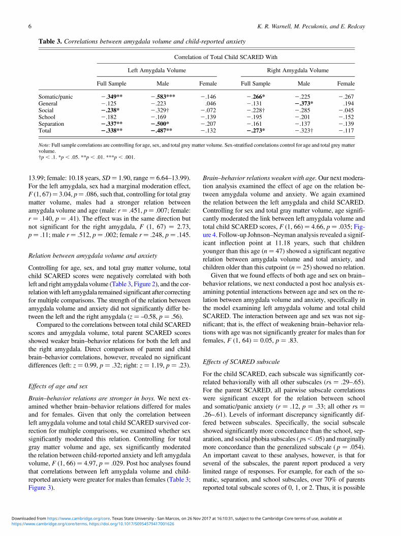
Controlling for age, sex, and total gray matter volume, totalchild SCARED scores were negatively correlated with bothleft and right amygdala volume (Table 3, Figure 2), and the cor-relation with left amygdala remained significant after correctingfor multiple comparisons. The strength of the relation betweenamygdala volume and anxiety did not significantly differ be-tween the left and the right amygdala (z ¼ –0.58, p ¼ .56).
Compared to the correlations between total child SCAREDscores and amygdala volume, total parent SCARED scoresshowed weaker brain–behavior relations for both the left andthe right amygdala. Direct comparison of parent and childbrain–behavior correlations, however, revealed no significantdifferences (left: z ¼ 0.99, p ¼ .32; right: z ¼ 1.19, p ¼ .23).
Effects of age and sex
Brain–behavior relations are stronger in boys. We next ex-amined whether brain–behavior relations differed for malesand for females. Given that only the correlation betweenleft amygdala volume and total child SCARED survived cor-rection for multiple comparisons, we examined whether sexsignificantly moderated this relation. Controlling for totalgray matter volume and age, sex significantly moderatedthe relation between child-reported anxiety and left amygdalavolume, F (1, 66) ¼ 4.97, p ¼ .029. Post hoc analyses foundthat correlations between left amygdala volume and child-reported anxiety were greater for males than females (Table 3;Figure 3).
Brain–behavior relations weaken with age. Our next modera-tion analysis examined the effect of age on the relation be-tween amygdala volume and anxiety. We again examinedthe relation between the left amygdala and child SCARED.Controlling for sex and total gray matter volume, age signifi-cantly moderated the link between left amygdala volume andtotal child SCARED scores, F (1, 66) ¼ 4.66, p ¼ .035; Fig-ure 4. Follow-up Johnson–Neyman analysis revealed a signif-icant inflection point at 11.18 years, such that childrenyounger than this age (n ¼ 47) showed a significant negativerelation between amygdala volume and total anxiety, andchildren older than this cutpoint (n¼ 25) showed no relation.
Given that we found effects of both age and sex on brain–behavior relations, we next conducted a post hoc analysis ex-amining potential interactions between age and sex on the re-lation between amygdala volume and anxiety, specifically inthe model examining left amygdala volume and total childSCARED. The interaction between age and sex was not sig-nificant; that is, the effect of weakening brain–behavior rela-tions with age was not significantly greater for males than forfemales, F (1, 64) ¼ 0.05, p ¼ .83.
Effects of SCARED subscale
For the child SCARED, each subscale was significantly cor-related behaviorally with all other subscales (rs ¼ .29–.65).For the parent SCARED, all pairwise subscale correlationswere significant except for the relation between schooland somatic/panic anxiety (r ¼ .12, p ¼ .33; all other rs ¼.26–.61). Levels of informant discrepancy significantly dif-fered between subscales. Specifically, the social subscaleshowed significantly more concordance than the school, sep-aration, and social phobia subscales ( ps , .05) and marginallymore concordance than the generalized subscale ( p ¼ .054).An important caveat to these analyses, however, is that forseveral of the subscales, the parent report produced a verylimited range of responses. For example, for each of the so-matic, separation, and school subscales, over 70% of parentsreported total subscale scores of 0, 1, or 2. Thus, it is possible
Table 3. Correlations between amygdala volume and child-reported anxiety
Correlation of Total Child SCARED With
Left Amygdala Volume Right Amygdala Volume
Full Sample Male Female Full Sample Male Female
Somatic/panic 2.349** 2.583*** 2.146 2.266* 2.225 2.267General 2.125 2.223 .046 2.131 2.373* .194Social 2.238* 2.329† 2.072 2.228† 2.285 2.045School 2.182 2.169 2.139 2.195 2.201 2.152Separation 2.337** 2.500* 2.207 2.161 2.137 2.139Total 2.338** 2.487** 2.132 2.273* 2.323† 2.117
Note: Full sample correlations are controlling for age, sex, and total grey matter volume. Sex-stratified correlations control for age and total grey mattervolume.†p , .1. *p , .05. **p , .01. ***p , .001.
K. R. Warnell, M. Pecukonis, and E. Redcay6
https://www.cambridge.org/core/terms. https://doi.org/10.1017/S0954579417001626Downloaded from https://www.cambridge.org/core. Texas State University - San Marcos, on 26 Nov 2017 at 16:10:31, subject to the Cambridge Core terms of use, available at

that more nuanced measures would reveal different patterns ofinformant discrepancy.
In additional, exploratory analyses, we repeated our brain–behavior analyses for the five subscales of the child SCAREDby correlating each subscale with left and right amygdala vol-ume. The child reports for the somatic/panic subscale showedthe strongest brain–behavior relation and generalized anxietythe weakest, but the differences between subscales were notsignificant (Table 3).
Discussion
The current study examined, in a single typical, develop-mental sample, the moderating effects of informant, sex,and age on the relation between amygdala volume and indi-vidual differences in anxiety traits. Overall, we found a signif-
icant negative relation between children’s self-reported anxi-ety and amygdala volume. Although we did not findsignificant effects of informant, the strongest correlationsemerged between children’s self-reported anxiety and amyg-dala volume. We further found significant moderating effectsfor sex and age; boys showed stronger brain–behavior rela-tions than girls, and brain–behavior relations significantlyweakened with age. These findings help to clarify previouslycontradictory literature and help suggest neurobiologicalmechanisms underlying anxiety in middle to late childhood.
Using brain–behavior relations to understand informantdiscrepancy
Previous research on informant discrepancy has beenhampered by difficulty in determining true symptomatology
Figure 2. Partial relations between amygdala volume and child- and parent-reported anxiety. Children’s (a) left and (b) right amygdala volumewas correlated with their self-reported anxiety, although only the correlation with (a) the left amygdala volume survived correction for multiplecomparisons. Children’s (c) left and (d) right amygdala volume was not significantly correlated with parent-reported anxiety. Anxiety was mea-sured by the total score on the Screen for Child Anxiety Related Emotional Disorders. All values are corrected for total gray matter volume, age,and sex. Gray lines around the line of best fit represent 95% confidence intervals. The correlation between child-reported anxiety and amygdalavolume is not significantly different than the correlation between parent-reported anxiety and amygdala volume.
Amygdala volume and anxiety in development 7
https://www.cambridge.org/core/terms. https://doi.org/10.1017/S0954579417001626Downloaded from https://www.cambridge.org/core. Texas State University - San Marcos, on 26 Nov 2017 at 16:10:31, subject to the Cambridge Core terms of use, available at

or impairment (see De Los Reyes & Kazdin, 2005; Dirks,De Los Reyes, Briggs-Gowan, Cella, & Wakschlag, 2012;Martel, Markon, & Smith, 2017, for theoretical and methodo-logical reviews of informant discrepancy). Examining correla-tions with physiological (e.g., Bitsika, Sharpley, Andronicos,& Agnew, 2015) or brain-based measures provides a novelmethod to assess the validity of various informants.
In this sample, consistent with previous studies of non-clinical samples (Cosi et al., 2010; Pereira et al., 2015; Wren,Bridge, & Birmaher 2004; although see Kircanski et al.,2017), correlations between child and parent SCAREDwere fairly weak, with self-reported anxiety significantlyhigher than parent-reported anxiety. That is, children reportedthemselves to be more anxious than their parents reportedthem to be. Two possibilities thus arise: that children overre-port their anxiety or that parents underreport their child’sanxiety. The brain–behavior correlations provide a way to
address these two possibilities; assuming a biological modelfor anxiety disorders, the fact that children’s reports of theirown anxiety show a significant correlation with amygdalavolume supports the validity of child report. The brain-basedevidence from the current sample converges with existingfindings that child SCARED scores better correlate with clin-ical interviews than parent SCARED scores (Cosi et al.,2010), that self-reported anxiety is more strongly correlatedwith cortisol levels (Bitsika et al., 2015), and that parentsoverestimate children’s well-being more generally (Lagattuta,Sayfan, & Bamford, 2012).
Although the findings from the current study support thevalidity of child self-report, they do not explain why infor-mant discrepancy arises. One explanation for parental under-report, at least in typical populations, is that some anxietysymptoms are not behaviorally observable and that agreementtends to be higher for observable symptoms (Comer &
Figure 3. Moderating effect of sex on brain–behavior relations. (a) Pyramid histogram of the distribution of age by sex. (b) Relations between leftamygdala volume and total child-reported anxiety, stratified by sex. All values are corrected for total gray matter volume and age. Gray linesaround the line of best fit represent 95% confidence intervals. Sex significantly moderated the relation between anxiety and amygdala volume,such that males showed stronger correlations than females ( p , .05).
K. R. Warnell, M. Pecukonis, and E. Redcay8
https://www.cambridge.org/core/terms. https://doi.org/10.1017/S0954579417001626Downloaded from https://www.cambridge.org/core. Texas State University - San Marcos, on 26 Nov 2017 at 16:10:31, subject to the Cambridge Core terms of use, available at

Kendall, 2004). In the current study, and consistent with priorresearch (e.g., Wren et al., 2007), informant discrepancy waslowest for the social phobia subscale and largest for the so-matic/panic subscale, on which children self-reported overfour times the level of symptomatology than their parentsreported. The somatic/panic subscale contains items aboutinternal physical states (e.g., heart racing), whereas socialphobia items may be more observable, especially at youngerages when parents are more involved in their children’s socialschedules. Complicating the study of informant discrepancy,however, is that parent-reported SCARED values also suf-fered from restriction of range due to their low estimates ofchild anxiety (e.g., the mean parental score for somatic/panicwas 1.04). The restriction of range on the parent SCARED isan important consideration in future studies of anxiety, as it ispossible that more nuanced measures of parental report mayreveal brain–behavior relations.
Future studies of amygdala volume and anxiety shouldalso measure parents’ own anxiety and amygdala volume,in both clinical and typical samples. There is some evidencethat parental overreporting may be linked to parental anxietyand psychopathology (Becker et al., 2016; Frick, Silverhorn, &Evans, 1994), and, although nonclinical samples generallyshow parental underreporting versus child report, some clin-ical samples show the opposite pattern (Blakeley-Smith, Re-aven, Ridge, & Hepburn, 2012; Phares & Danforth, 1994;Salbach-Andrae, Klinkowski, Lenz, & Lehmkuhl, 2009, butsee Weitkamp et al., 2010). Further, there may be genetic fac-tors underlying both susceptibility for internalizing disordersand amygdala volume (Montag, Weber, Fliessbach, Elger, &Reuter, 2009). Ultimately, although examining links with
neurobiology may provide new perspectives on informantdiscrepancy, the critical question of interest to children, par-ents, and clinicians is how these convergent anxiety measuresrelate to real-world functioning. The current study does notlink amygdala volume to long-term scholastic or socialoutcomes or to response to an anxiety intervention. Goingforward, combining neurobiological studies with these be-havioral and clinical approaches will offer the best insightinto informant discrepancy.
Using moderators of brain–behavior relationsto understand neural mechanisms of anxiety
The finding of a negative relation between children’s self-report of anxiety and amygdala volume is consistent withsome existing adult (e.g., Fisler et al., 2013; Hayano et al.,2009; Rogers et al., 2009; Spampinato et al., 2009) and devel-opmental (e.g., McQueeny et al., 2011; Milham et al., 2005;Mueller et al., 2013; Strawn et al., 2015) literature and is con-trary to previous findings of a nonexistent or positive relationbetween anxiety and amygdala volume (e.g., De Bellis et al.,2000; Qin et al., 2014; Tottenham et al., 2010; van der Plaset al., 2010). Whereas positive associations may suggestthat increased dendritic branching or spine density underliesanxiety (e.g., Qin et al., 2014; Tottenham et al., 2010),negative relations suggest alternative mechanisms. Althoughthese precise mechanisms are yet unknown, there are a fewcandidate explanations. Persistent amygdala hyperactivation,which may characterize anxiety, could lead to glutamatergicexcitotoxicity and subsequent reduction of amygdala volume(Siegle et al., 2003). Although this exact causal mechanismhas not been proven in humans, decreased amygdala volumehas been linked to individual differences in amygdala hyper-activity in depression and bipolar disorder (Kalmar et al.,2009; Siegle et al., 2003). An alternative explanation is thatrather than a history of hyperactivity causing a reduction inamygdala size, a smaller amygdala may predispose one toanxiety. Supporting this possibility, mice with smaller amyg-dalae, specifically basolateral amygdala, show stronger fearresponses and increased glucocorticoid response (Yanget al., 2008). A third possibility is that some other factor isdriving both the reduction in amygdala volume and the in-crease in anxiety. This factor remains unknown, but certaingenotypes have been associated with both traits (Montaget al., 2009; Mueller et al., 2013). Further complicating anydiscussion of mechanisms is that the direction of the relationbetween amygdala volume and anxiety may change through-out development.
In the current sample, the relation between amygdala vol-ume and anxiety was strongest at younger ages, with an in-flection point emerging around age 11. After this age, thelink between amygdala volume and anxiety was no longersignificant. The mechanisms driving these age effects are un-known. One simple explanation is that decreased accuracy inself-report around age 11 could lead to the null finding ob-served in the older age group. Although future research
Figure 4. (Color online) The relation between left amygdala volume andchild-reported anxiety, as measured by the Screen for Child Anxiety RelatedEmotional Disorders, is significantly moderated by age, such that older chil-dren show weaker brain–behavior relations than younger children. Modera-tion analyses controlled for sex and total gray matter volume. The lines repre-sent fit at the mean age and +1 SD. Johnson–Neyman analysis revealed aninflection point at 11.18 years of age, where the relation between anxiety andamygdala volume changed from significant to nonsignificant.
Amygdala volume and anxiety in development 9
https://www.cambridge.org/core/terms. https://doi.org/10.1017/S0954579417001626Downloaded from https://www.cambridge.org/core. Texas State University - San Marcos, on 26 Nov 2017 at 16:10:31, subject to the Cambridge Core terms of use, available at

should include more clinical and behavioral anxiety measuresto address this possibility, there is no evidence to suggest thatolder children are worse at self-report than younger children.
Several other explanations of the age effects are possible,and future longitudinal studies would be needed to disambig-uate between them. One explanation of the age-related find-ings is a delayed growth model. In this model, smaller amyg-dala volume may predispose children to develop anxiety, butas children age, their amygdala volume “catches up,” al-though the anxiety remains, and the statistical relation be-tween amygdala size and anxiety disappears. In an alterna-tive, but not orthogonal, model, early versus later onsetanxiety may show different brain bases. Speculatively, earlyonset anxiety may be caused by neurobiological vulnerabil-ities rather than environmental experiences, whereas anxietysymptoms that emerge after puberty may be more varied intheir etiology, producing, at the group level, a null correlationwith amygdala volume. Yet another possibility is that themechanisms linking amygdala volume and anxiety are con-sistent throughout development, but the onset of pubertycomplicates the discovery of that relation at a group level,given that puberty has been linked to differential rates ofamygdala growth (Bramen et al., 2011). Finally, if the mainmechanism linking anxiety and amygdala volume is that anx-iety causes amygdala reduction, one might expect an increasein brain–behavior relations with age. The fact that we did notfind this pattern indicates that these effects may have a sensi-tive period, such that late onset anxiety does not have thesame effect as early onset anxiety. The current cross-sectionalstudy, however, can only speculate about the causal relationbetween variables.
In addition to the effects of age, the current study foundstronger brain–behavior relations in boys than in girls. Onepotential explanation is that boys’ amygdalae mature moreslowly, potentially due to differences in pubertal timing (Bra-men et al., 2011), and thus, the age range of the current studycaptures a key period of amygdala development for boys, butnot for girls. This possibility is supported by the fact thatboys, but not girls, show a significant relation between amyg-dala volume and age in the current sample. If this explanationis true, then studying girls in a younger age range (such as 4–10 years) might reveal similar negative correlations withamygdala volume. Alternatively, the brain bases of anxietymay be different across sexes (Bangasser & Valentino,2014; Hamann, 2005; Maeng & Milad, 2015). For example,in typical adults, sex moderates the relation between amyg-dala hyperactivity and trait anxiety (Dickie & Armony,2008), and in adolescents, differences in amygdala bloodflow mediate the relation between sex and trait anxiety (Kacz-kurkin et al., 2016). Further, testosterone has been linked toincreased amygdala activity (Derntl et al., 2009; van Wingenet al., 2009), suggesting that pubertal onset may have differ-ent effects for males versus females. Future studies shouldalso include pubertal measures, both hormonal and self-re-port, to disentangle age, sex, and puberty effects (cf. Blake-more, Burnett, & Dahl, 2010; Sisk & Zehr, 2005).
Although we did not find significant differences for theright versus left amygdala, effects were numerically strongerin the left amygdala, a finding that should be explored furtherin future work. How these findings fit with existing literatureis unclear, particularly because the existing volumetric litera-ture on laterality of anxiety–amygdala relations is inconclu-sive. Functional literature suggests that the left and the rightamygdala may play different roles in processing threat-relevant stimuli, such that the left amygdala is involved inmore sustained responses (Sergerie et al., 2008), which maylead to a greater involvement in the ongoing fear that under-lies anxiety. Future studies involving both structural andfunctional neuroimaging, using tasks designed to tap intoboth left and right amygdala processes, may provide moreinsight into the role of laterality. Such research should alsocontinue to explore whether there is an effect of specificanxiety type (e.g., social anxiety and generalized anxiety)on brain–behavior relations.
Ultimately, although the current study offers insight intopotential moderators of brain–behavior relations in anxiety,more research needs to be done before making firm conclu-sions about the relationship between amygdala volume andanxiety traits, and before applying findings from structuralneuroimaging to clinical assays or pharmacological interven-tions. Although the current study had a large sample com-pared to many other developmental studies of amygdala vol-ume and anxiety, future research should include even largerclinical and nonclinical samples that will allow for morenuanced investigation of the effects of various moderators.For example, although we did not find an interaction betweenage and sex, that may have been due to limited power. One ofthe advantages of structural neuroimaging is the ability tocombine data across a wide variety of sites (e.g., BRAINSproject; Job et al., 2017), and utilizing such approachesmay resolve ongoing debates in this literature. Furthermore,multimodal approaches, such as examining functional andstructural data in single samples, and including genotyping,will also help clarify the role of the amygdala in anxiety(Mueller et al., 2013). Finally, the amygdala is a complexstructure made up of multiple nuclei, and these nuclei havedifferent functions and show different patterns of functionalconnectivity with the rest of the brain (Saygin, Osher, Augus-tinack, Fischl, & Gabrieli, 2011). For example, there is somehuman (Qin et al., 2014) and animal evidence (Yang et al.,2008) that basolateral amygdala volume may be especiallypredictive of individual differences in anxiety. Future re-search should continue to address this possibility. In theend, inconsistencies in the current literature will likely needmultimethod, large-scale studies in order to be resolved.
Conclusions
The current study adds to the limited developmental literatureon amygdala volume and anxiety by examining brain–behavior relations in a large, typical sample aged 6–13 years.We find that self-reported anxiety is negatively related to
K. R. Warnell, M. Pecukonis, and E. Redcay10
https://www.cambridge.org/core/terms. https://doi.org/10.1017/S0954579417001626Downloaded from https://www.cambridge.org/core. Texas State University - San Marcos, on 26 Nov 2017 at 16:10:31, subject to the Cambridge Core terms of use, available at

amygdala volume. Further, our results indicate that the rela-tion between amygdala volume and anxiety weakens withage, which suggests that anxiety may have different neurobi-ological antecedents or consequences at different points indevelopment. Such developmental trajectories may also inter-
act with sex, as we find stronger brain–behavior relations inmales. Although these findings do not fully explain previousinconsistencies in the literature, they provide importantvariables to consider for future large-scale, longitudinal,and multimodal studies that chart the brain bases of anxiety.
References
Albaugh, M. D., Nguyen, T., Ducharme, S., Collins, D. L., Botteron, K. N.,D’Alberto, N., . . . Hudziak, J. J. (2017). Age-related volumetric changeof limbic structures and subclinical anxious/depressed symptomatologyin typically developing children and adolescents. Biological Psychology,124, 133–140. doi:10.1016/j.biopsycho.2017.02.002
Bangasser, D. A., & Valentino, R. J. (2014). Sex differences in stress-relatedpsychiatric disorders: Neurobiological perspectives. Frontiers in Neu-roendocrinology, 35, 303–319. doi:10.1016/j.yfrne.2014.03.008
Bauer, D. J., & Curran, P. J. (2005). Probing interactions in fixed and multi-level regression: Inferential and graphical techniques. MultivariateBehavioral Research, 40, 373–400. doi:10.1207/s15327906mbr4003_5
Baur, V., Hanggi, J., & Jancke, L. (2012). Volumetric associations betweenuncinate fasciculus, amygdala, and trait anxiety. BMC Neuroscience, 13,4. doi:10.1186/1471-2202-13-4
Becker, E. M., Jensen-Doss, A., Kendall, P. C., Birmaher, B., & Ginsburg, G.S. (2016). All anxiety is not created equal: Correlates of parent/youthagreement vary across subtypes of anxiety. Journal of Psychopathologyand Behavioral Assessment, 38, 528–537. doi:10.1007/s10862-016-9544-z
Beesdo, K., Knappe, S., & Pine, D. S. (2009). Anxiety and anxiety disordersin children and adolescents: Developmental issues and implications forDSM-V. Psychiatric Clinics of North America, 32, 483–524. doi:10.1016/j.psc.2009.06.002
Bell-Dolan, D. J., Last, C. G., & Strauss, C. C. (1990). Symptoms of anxi-ety disorders in normal children. Journal of the American Academy ofChild & Adolescent Psychiatry, 29, 759–765. doi:10.1097/00004583-199009000-00014
Birmaher, B., Brent, D. A., Chiappetta, L., Bridge, J., Monga, S., & Baugher,M. (1999). Psychometric properties of the Screen for Child Anxiety Re-lated Emotional Disorders (SCARED): A replication study. Journal ofthe American Academy of Child & Adolescent Psychiatry, 38, 1230–1236. doi:10.1097/00004583-199910000-00011
Birmaher, B., Khetarpal, S., Brent, D., Cully, M., Balach, L., Kaufman, J., &Neer, S. M. (1997). The Screen for Child Anxiety Related Emotional Dis-orders (SCARED): Scale construction and psychometric characteristics.Journal of the American Academy of Child & Adolescent Psychiatry,36, 545–553. doi:10.1097/00004583-199704000-00018
Bishop, S. J., Duncan, J., & Lawrence, A. D. (2004). State anxietymodulation of the amygdala response to unattended threat-related stimuli.Journal of Neuroscience, 24, 10364–10368. doi:10.1523/jneurosci.2550-04.2004
Bitsika, V., Sharpley, C. F., Andronicos, N. M., & Agnew, L. L. (2015).Agreement between self-vs parent-ratings of general anxiety disordersymptoms and salivary cortisol in boys with an ASD. Journal of Devel-opmental and Physical Disabilities, 27, 467–477. doi:10.1007/s10882-015-9431-7
Blair, K., Shaywitz, J., Smith, B. W., Rhodes, R., Geraci, M., Jones, M., . . .Jacobs, M. (2008). Response to emotional expressions in generalized so-cial phobia and generalized anxiety disorder: Evidence for separate dis-orders. American Journal of Psychiatry, 165, 1193–1202. doi:10.1176/appi.ajp.2008.07071060
Blakeley-Smith, A., Reaven, J., Ridge, K., & Hepburn, S. (2012). Parent–child agreement of anxiety symptoms in youth with autism spectrum dis-orders. Research in Autism Spectrum Disorders, 6, 707–716. doi:10.1016/j.rasd.2011.07.020
Blakemore, S. J., Burnett, S., & Dahl, R. E. (2010). The role of puberty in thedeveloping adolescent brain. Human Brain Mapping, 31, 926–933.doi:10.1002/hbm.21052
Bramen, J. E., Hranilovich, J. A., Dahl, R. E., Forbes, E. E., Chen, J., Toga,A. W., . . . Sowell, E. R. (2011). Puberty influences medial temporal lobeand cortical gray matter maturation differently in boys than girls matchedfor sexual maturity. Cerebral Cortex, 21, 636–646. doi:10.1093/cercor/bhq137
Burgund, E. D., Kang, H. C., Kelly, J. E., Buckner, R. L., Snyder, A. Z.,Petersen, S. E., & Schlaggar, B. L. (2002). The feasibility of a commonstereotactic space for children and adults in fMRI studies of development.NeuroImage, 17, 184–200. doi:10.1006/nimg.2002.1174
Comer, J. S., & Kendall, P. C. (2004). A symptom-level examination ofparent–child agreement in the diagnosis of anxious youths. Journal ofthe American Academy of Child & Adolescent Psychiatry, 43, 878–886. doi:10.1097/01.chi.0000125092.35109.c5
Cosi, S., Canals, J., Hernandez-Martinez, C., & Vigil-Colet, A. (2010).Parent–child agreement in SCARED and its relationship to anxietysymptoms. Journal of Anxiety Disorders, 24, 129–133. doi:10.1016/j.janxdis.2009.09.008
De Bellis, M. D., Casey, B. J., Dahl, R. E., Birmaher, B., Williamson, D. E.,Thomas, K. M., . . . Ryan, N. D. (2000). A pilot study of amygdalavolumes in pediatric generalized anxiety disorder. Biological Psychiatry,48, 51–57. doi:10.1016/S0006-3223(00)00835-0
De Los Reyes, A., & Kazdin, A. E. (2005). Informant discrepancies in theassessment of childhood psychopathology: A critical review, theoreticalframework, and recommendations for further study. Psychological Bulle-tin, 131, 483. doi:10.1037/0033-2909.131.4.483
Dennis, E. L., Gotlib, I. H., Thompson, P. M., & Thomason, M. E. (2011).Anxiety modulates insula recruitment in resting-state functional magneticresonance imaging in youth and adults. Brain Connectivity, 1, 245–254.doi:10.1089/brain.2011.0030
Derntl, B., Windischberger, C., Robinson, S., Kryspin-Exner, I., Gur, R. C.,Moser, E., & Habel, U. (2009). Amygdala activity to fear and anger inhealthy young males is associated with testosterone. Psychoneuroendo-crinology, 34, 687–693. doi:10.1016/j.psyneuen.2008.11.007
Dickie, E. W., & Armony, J. L. (2008). Amygdala responses to unattended fear-ful faces: Interaction between sex and trait anxiety. Psychiatry Research:Neuroimaging, 162, 51–57. doi:10.1016/j.pscychresns.2007.08.002
Dirks, M. A., De Los Reyes, A., Briggs-Gowan, M., Cella, D., & Wakschlag,L. S. (2012). Annual Research Review: Embracing not erasing contextualvariability in children’s behavior—Theory and utility in the selection anduse of methods and informants in developmental psychopathology. Jour-nal of Child Psychology and Psychiatry, 53, 558–574. doi:10.1111/j.1469-7610.2012.02537.x
Dirks, M. A., Weersing, V. R., Warnick, E., Gonzalez, A., Alton, M., Dauser,C., . . . Woolston, J. (2014). Parent and youth report of youth anxiety:Evidence for measurement invariance. Journal of Child Psychologyand Psychiatry, 55, 284–291. doi:10.1111/jcpp.12159
Engel, N. A., Rodrigue, J. R., & Geffken, G. R. (1994). Parent-childagreement on ratings of anxiety in children. Psychological Reports, 75,1251–1260. doi:10.2466/pr0.1994.75.3.1251
Etkin, A., Klemenhagen, K. C., Dudman, J. T., Rogan, M. T., Hen, R.,Kandel, E. R., & Hirsch, J. (2004). Individual differences in trait anxietypredict the response of the basolateral amygdala to unconsciouslyprocessed fearful faces. Neuron, 44, 1043–1055. doi:10.1016/j.neuron.2004.12.006
Etkin, A., & Wager, T. D. (2007). Functional neuroimaging of anxiety: Ameta-analysis of emotional processing in PTSD, social anxiety disorder,and specific phobia. American Journal of Psychiatry, 164, 1476–1488.doi:10.1176/appi.ajp.2007.07030504
Fisler, M. S., Federspiel, A., Horn, H., Dierks, T., Schmitt, W., Wiest, R., . . .Soravia, L. M. (2013). Spider phobia is associated with decreased leftamygdala volume: A cross-sectional study. BMC Psychiatry, 13, 70.doi:10.1186/1471-244X-13-70
Frick, P. J., Silverthorn, P., & Evans, C. (1994). Assessment of childhoodanxiety using structured interviews: Patterns of agreement among infor-mants and association with maternal anxiety. Psychological Assessment,6, 372. doi:10.1037/1040-3590.6.4.372
Fusar-Poli, P., Placentino, A., Carletti, F., Landi, P., & Abbamonte, M.(2009). Functional atlas of emotional faces processing: A voxel-based
Amygdala volume and anxiety in development 11
https://www.cambridge.org/core/terms. https://doi.org/10.1017/S0954579417001626Downloaded from https://www.cambridge.org/core. Texas State University - San Marcos, on 26 Nov 2017 at 16:10:31, subject to the Cambridge Core terms of use, available at

meta-analysis of 105 functional magnetic resonance imaging studies.Journal of Psychiatry and Neuroscience, 34, 418–432.
Gabard-Durnam, L. J., Flannery, J., Goff, B., Gee, D. G., Humphreys, K. L.,Telzer, E., . . . Tottenham, N. (2014). The development of human amyg-dala functional connectivity at rest from 4 to 23 years: A cross-sectionalstudy. NeuroImage, 95, 193–207. doi:10.1016/j.neuroimage.2014.03.038
Gee, D. G., Humphreys, K. L., Flannery, J., Goff, B., Telzer, E. H., Shapiro,M., . . . Tottenham, N. (2013). A developmental shift from positive tonegative connectivity in human amygdala–prefrontal circuitry. Journalof Neuroscience, 33, 4584–4593. doi:10.1523/jneurosci.3446-12.2013
Ghosh, S. S., Kakunoori, S., Augustinack, J., Nieto-Castanon, A., Kovelman,I., Gaab, N., . . . Fischl, B. (2010). Evaluating the validity of volume-based and surface-based brain image registration for developmental cog-nitive neuroscience studies in children 4 to 11years of age. NeuroImage,53, 85–93. doi:10.1016/j.neuroimage.2010.05.075
Grupe, D. W., & Nitschke, J. B. (2013). Uncertainty and anticipation in anx-iety: An integrated neurobiological and psychological perspective. Na-ture Reviews Neuroscience, 14, 488–501. doi:10.1038/nrn3524
Hahn, A., Stein, P., Windischberger, C., Weissenbacher, A., Spindelegger,C., Moser, E., . . . Lanzenberger, R. (2011). Reduced resting-state func-tional connectivity between amygdala and orbitofrontal cortex in socialanxiety disorder. NeuroImage, 56, 881–889. doi:10.1016/j.neuro-image.2011.02.064
Hamann, S. (2005). Sex differences in the responses of the human amygdala.Neuroscientist, 11, 288–293. doi:10.1177/1073858404271981
Hayano, F., Nakamura, M., Asami, T., Uehara, K., Yoshida, T., Roppongi, T.,. . . Hirayasu, Y. (2009). Smaller amygdala is associated with anxiety inpatients with panic disorder. Psychiatry and Clinical Neurosciences, 63,266–276. doi:10.1111/j.1440-1819.2009.01960.x
Hayes, A. F. (2013). Introduction to mediation, moderation, and conditionalprocess analysis: A regression-based approach. New York: GuilfordPress.
Hilbert, K., Lueken, U., Muehlhan, M., & Beesdo-Baum, K. (2017). Separ-ating generalized anxiety disorder from major depression using clinical,hormonal, and structural MRI data: A multimodal machine learningstudy. Brain and Behavior. Advance online publication. doi:10.1002/brb3.633
Hilbert, K., Pine, D. S., Muehlhan, M., Lueken, U., Steudte-Schmiedgen, S.,& Beesdo-Baum, K. (2015). Gray and white matter volume abnormalitiesin generalized anxiety disorder by categorical and dimensional character-ization. Psychiatry Research: Neuroimaging, 234, 314–320. doi:10.1016/j.pscychresns.2015.10.009
Holzel, B. K., Carmody, J., Evans, K. C., Hoge, E. A., Dusek, J. A., Morgan,L., . . . Lazar, S. W. (2009). Stress reduction correlates with structuralchanges in the amygdala. Social Cognitive and Affective Neuroscience,5, 11–17. doi:10.1093/scan/nsp034
Hu, S., Pruessner, J. C., Coupe, P., & Collins, D. L. (2013). Volumetricanalysis of medial temporal lobe structures in brain development fromchildhood to adolescence. NeuroImage, 74, 276–287. doi:10.1016/j.neuroimage.2013.02.032
Hyde, L. W., Gorka, A., Manuck, S. B., & Hariri, A. R. (2011). Perceivedsocial support moderates the link between threat-related amygdala reac-tivity and trait anxiety. Neuropsychologia, 49, 651–656. doi:10.1016/j.neuropsychologia.2010.08.025
Job, D. E., Dickie, D. A., Rodriguez, D., Robson, A., Danso, S., Pernet, C.,. . . Waiter, G. D. (2017). A brain imaging repository of normal structuralMRI across the life course: Brain Images of Normal Subjects (BRAINS).NeuroImage, 144, 299–304. doi:10.1016/j.neuroimage.2016.01.027
Kaczkurkin, A. N., Moore, T. M., Ruparel, K., Ciric, R., Calkins, M. E., Shi-nohara, R. T., . . . Gennatas, E. D. (2016). Elevated amygdala perfusionmediates developmental sex differences in trait anxiety. Biological Psy-chiatry, 80, 775–785. doi:10.1016/j.biopsych.2016.04.021
Kalmar, J. H., Wang, F., Chepenik, L. G., Womer, F. Y., Jones, M. M., Pitt-man, B., . . . Blumberg, H. P. (2009). Relation between amygdala struc-ture and function in adolescents with bipolar disorder. Journal of theAmerican Academy of Child & Adolescent Psychiatry, 48, 636–642.doi:10.1097/CHI.0b013e31819f6fbc
Kaufman, A. S., & Kaufman, N. L. (2004). Kaufman Brief Intelligence Test(2nd ed.). Bloomington, MN: Pearson.
Kessel, E. M., Kujawa, A., Hajcak Proudfit, G., & Klein, D. N. (2015).Neural reactivity to monetary rewards and losses differentiates socialfrom generalized anxiety in children. Journal of Child Psychology andPsychiatry, 56, 792–800. doi:10.1111/jcpp.12355
Kessler, R. C., Angermeyer, M., Anthony, J. C., De Graaf, R. O. N., Demyt-tenaere, K., Gasquet, I., . . . Kawakami, N. (2007). Lifetime prevalenceand age-of-onset distributions of mental disorders in the World HealthOrganization’s World Mental Health Survey Initiative. World Psychiatry,6, 168–176.
Kessler, R. C., Berglund, P., Demler, O., Jin, R., Merikangas, K. R., & Walters,E. E. (2005). Lifetime prevalence and age-of-onset distributions ofDSM-IV disorders in the National Comorbidity Survey Replication. Ar-chives of General Psychiatry, 62, 593–602. doi:10.1001/archpsyc.62.6.593
Kim, M. J., Gee, D. G., Loucks, R. A., Davis, F. C., & Whalen, P. J. (2011).Anxiety dissociates dorsal and ventral medial prefrontal cortex functionalconnectivity with the amygdala at rest. Cerebral Cortex, 21, 1667–1673.doi:10.1093/cercor/bhq237
Kim, M. J., Loucks, R. A., Palmer, A. L., Brown, A. C., Solomon, K. M.,Marchante, A. N., & Whalen, P. J. (2011). The structural and functionalconnectivity of the amygdala: From normal emotion to pathological anx-iety. Behavioural Brain Research, 223, 403–410. doi:10.1016/j.bbr.2011.04.025
Kircanski, K., Zhang, S., Stringaris, A., Wiggins, J. L., Towbin, K. E., Pine,D. S., . . . Brotman, M. A. (2017). Empirically derived patterns of psychi-atric symptoms in youth: A latent profile analysis. Journal of AffectiveDisorders, 216, 109–116. doi:10.1016/j.jad.2016.09.016
Lagattuta, K. H., Sayfan, L., & Bamford, C. (2012). Do you know how I feel?Parents underestimate worry and overestimate optimism compared tochild self-report. Journal of Experimental Child Psychology, 113, 211–232. doi:10.1016/j.jecp.2012.04.001
Lewinsohn, P. M., Gotlib, I. H., Lewinsohn, M., Seeley, J. R., & Allen, N. B.(1998). Gender differences in anxiety disorders and anxiety symptoms inadolescents. Journal of Abnormal Psychology, 107, 109. doi:10.1037/0021-843X.107.1.109
Lyons-Ruth, K., Pechtel, P., Yoon, S. A., Anderson, C. M., & Teicher, M. H.(2016). Disorganized attachment in infancy predicts greater amygdalavolume in adulthood. Behavioural Brain Research, 308, 83–93. doi:10.1016/j.bbr.2016.03.050
Maeng, L. Y., & Milad, M. R. (2015). Sex differences in anxiety disorders:Interactions between fear, stress, and gonadal hormones. Hormones andBehavior, 76, 106–117. doi:10.1016/j.yhbeh.2015.04.002
Martel, M. M., Markon, K., & Smith, G. T. (2017). Research Review: Multi-informant integration in child and adolescent psychopathology diagnosis.Journal of Child Psychology and Psychiatry, 58, 116–128. doi:10.1111/jcpp.12611
McMenamin, B. W., & Marsolek, C. J. (2013). Can theories of visual repre-sentation help to explain asymmetries in amygdala function? Cognitive,Affective, & Behavioral Neuroscience, 13, 211–224. doi:10.3758/s13415-012-0139-1
McQueeny, T., Padula, C. B., Price, J., Medina, K. L., Logan, P., & Tapert, S.F. (2011). Gender effects on amygdala morphometry in adolescent mar-ijuana users. Behavioural Brain Research, 224, 128–134. doi:10.1016/j.bbr.2011.05.031
Milham, M. P., Nugent, A. C., Drevets, W. C., Dickstein, D. S., Leibenluft,E., Ernst, M., . . . Pine, D. S. (2005). Selective reduction in amygdalavolume in pediatric anxiety disorders: A voxel-based morphometry in-vestigation. Biological Psychiatry, 57, 961–966. doi:10.1016/j.biopsych.2005.01.038
Montag, C., Weber, B., Fliessbach, K., Elger, C., & Reuter, M. (2009). TheBDNF Val66Met polymorphism impacts parahippocampal and amyg-dala volume in healthy humans: Incremental support for a genetic riskfactor for depression. Psychological Medicine, 39, 1831–1839. doi:10.1017/S0033291709005509
Morey, R. A., Petty, C. M., Xu, Y., Hayes, J. P., Wagner, H. R., Lewis, D. V.,. . . McCarthy, G. (2009). A comparison of automated segmentation andmanual tracing for quantifying hippocampal and amygdala volumes.NeuroImage, 45, 855–866. doi:10.1016/j.neuroimage.2008.12.033
Mueller, S. C., Aouidad, A., Gorodetsky, E., Goldman, D., Pine, D. S., &Ernst, M. (2013). Gray matter volume in adolescent anxiety: An impactof the brain-derived neurotrophic factor val 66 met polymorphism? Jour-nal of the American Academy of Child & Adolescent Psychiatry, 52, 184–195. doi:10.1016/j.jaac.2012.11.016
Muris, P., Merckelbach, H., Van Brakel, A., & Mayer, A. B. (1999). The re-vised version of the Screen for Child Anxiety Related Emotional Disor-ders (SCARED-R): Further evidence for its reliability and validity. Anx-iety, Stress & Coping, 12, 411–425. doi:10.1080/10615809908249319
Pereira, A. I., Muris, P., Barros, L., Goes, R., Marques, T., & Russo, V.(2015). Agreement and discrepancy between mother and child in the
K. R. Warnell, M. Pecukonis, and E. Redcay12
https://www.cambridge.org/core/terms. https://doi.org/10.1017/S0954579417001626Downloaded from https://www.cambridge.org/core. Texas State University - San Marcos, on 26 Nov 2017 at 16:10:31, subject to the Cambridge Core terms of use, available at

evaluation of children’s anxiety symptoms and anxiety life interference.European Child & Adolescent Psychiatry, 24, 327–337. doi:10.1007/s00787-014-0583-2
Phares, V., & Danforth, J. S. (1994). Adolescents’, parents’, and teachers’distress over adolescents’ behavior. Journal of Abnormal Child Psychol-ogy, 22, 721–732. doi:10.1007/BF02171998
Preacher, K. J., Curran, P. J., & Bauer, D. J. (2006). Computational tools forprobing interactions in multiple linear regression, multilevel modeling,and latent curve analysis. Journal of Educational and Behavioral Statis-tics, 31, 437–448. doi:10.3102/10769986031004437
Qin, S., Young, C. B., Duan, X., Chen, T., Supekar, K., & Menon, V. (2014).Amygdala subregional structure and intrinsic functional connectivity pre-dicts individual differences in anxiety during early childhood. BiologicalPsychiatry, 75, 892–900. doi:10.1016/j.biopsych.2013.10.006
Rogers, M. A., Yamasue, H., Abe, O., Yamada, H., Ohtani, T., Iwanami, A.,. . . Kasai, K. (2009). Smaller amygdala volume and reduced anterior cin-gulate gray matter density associated with history of post-traumatic stressdisorder. Psychiatry Research: Neuroimaging, 174, 210–216. doi:10.1016/j.pscychresns.2009.06.001
Salbach-Andrae, H., Klinkowski, N., Lenz, K., & Lehmkuhl, U. (2009).Agreement between youth-reported and parent-reported psychopathol-ogy in a referred sample. European Child & Adolescent Psychiatry, 18,136–143. doi:10.1007/s00787-008-0710-z
Saygin, Z. M., Osher, D. E., Augustinack, J., Fischl, B., & Gabrieli, J. D.(2011). Connectivity-based segmentation of human amygdala nuclei us-ing probabilistic tractography. NeuroImage, 56, 1353–1361. doi:10.1016/j.neuroimage.2011.03.006
Scaini, S., Ogliari, A., De Carolis, L., Bellodi, L., Di Serio, C., & Brombin,C. (2017). Evaluation of mother-child agreement and factorial structuresof the SCARED questionnaire in an Italian clinical sample. Frontiers inPsychology, 8, 242. doi:10.3389/fpsyg.2017.00242
Schienle, A., Ebner, F., & Schafer, A. (2011). Localized gray matter volumeabnormalities in generalized anxiety disorder. European Archives of Psy-chiatry and Clinical Neuroscience, 261, 303–307. doi:10.1007/s00406-010-0147-5
Schoemaker, D., Buss, C., Head, K., Sandman, C. A., Davis, E. P., Chakra-varty, M. M., . . . Pruessner, J. C. (2016). Hippocampus and amygdalavolumes from magnetic resonance images in children: Assessing accu-racy of FreeSurfer and FSL against manual segmentation. NeuroImage,129, 1–14. doi:10.1016/j.neuroimage.2016.01.038
Sehlmeyer, C., Dannlowski, U., Schoning, S., Kugel, H., Pyka, M., Pflei-derer, B., . . . Konrad, C. (2011). Neural correlates of trait anxiety infear extinction. Psychological Medicine, 41, 789–798. doi:10.1017/S0033291710001248
Sergerie, K., Chochol, C., & Armony, J. L. (2008). The role of the amygdalain emotional processing: A quantitative meta-analysis of functional neu-roimaging studies. Neuroscience & Biobehavioral Reviews, 32, 811–830.doi:10.1016/j.neubiorev.2007.12.002
Shin, L. M., & Liberzon, I. (2010). The neurocircuitry of fear, stress, and anx-iety disorders. Neuropsychopharmacology, 35, 169–191. doi:10.1038/npp.2009.83
Siegle, G. J., Konecky, R. O., Thase, M. E., & Carter, C. S. (2003). Relation-ships between amygdala volume and activity during emotional informa-tion processing tasks in depressed and never-depressed individuals. An-nals of the New York Academy of Sciences, 985, 481–484. doi:10.1111/j.1749-6632.2003.tb07105.x
Sisk, C. L., & Zehr, J. L. (2005). Pubertal hormones organize the adolescentbrain and behavior. Frontiers in Neuroendocrinology, 26, 163–174.doi:10.1016/j.yfrne.2005.10.003
Spampinato, M. V., Wood, J. N., De Simone, V., & Grafman, J. (2009).Neural correlates of anxiety in healthy volunteers: A voxel-based mor-phometry study. Journal of Neuropsychiatry and Clinical Neurosciences,21, 199–205. doi:10.1176/jnp.2009.21.2.199
Sripada, R. K., Wang, X., Sripada, C. S., & Liberzon, I. (2012). Altered rest-ing-state amygdala functional connectivity in men with posttraumatic
stress disorder. Journal of Psychiatry and Neuroscience, 37, 241.doi:10.1503/jpn.110069
Strawn, J. R., Hamm, L., Fitzgerald, D. A., Fitzgerald, K. D., Monk, C. S., &Phan, K. L. (2015). Neurostructural abnormalities in pediatric anxiety dis-orders. Journal of Anxiety Disorders, 32, 81–88. doi:10.1016/j.janxdis.2015.03.004
Su, L., Wang, K., Fan, F., Su, Y., & Gao, X. (2008). Reliability and validity ofthe Screen for Child Anxiety Related Emotional Disorders (SCARED) inChinese children. Journal of Anxiety Disorders, 22, 612–621. doi:10.1016/j.janxdis.2007.05.011
Tottenham, N., Hare, T. A., Quinn, B. T., McCarry, T. W., Nurse, M., Gil-hooly, T., . . . Thomas, K. M. (2010). Prolonged institutional rearing isassociated with atypically large amygdala volume and difficulties in emo-tion regulation. Developmental Science, 13, 46–61. doi:10.1111/j.1467-7687.2009.00852.x
Tovote, P., Fadok, J. P., & Luthi, A. (2015). Neuronal circuits for fear andanxiety. Nature Reviews Neuroscience, 6, 317–331. doi:10.1038/nrn3945
Uematsu, A., Matsui, M., Tanaka, C., Takahashi, T., Noguchi, K., Suzuki,M., & Nishijo, H. (2012). Developmental trajectories of amygdala andhippocampus from infancy to early adulthood in healthy individuals.PLOS ONE, 7, e46970. doi:10.1371/journal.pone.0046970
van der Plas, E. A., Boes, A. D., Wemmie, J. A., Tranel, D., & Nopoulos, P.(2010). Amygdala volume correlates positively with fearfulness in nor-mal healthy girls. Social Cognitive and Affective Neuroscience, 5, 424–431. doi:10.1093/scan/nsq009
Van Petten, C. (2004). Relationship between hippocampal volume andmemory ability in healthy individuals across the lifespan: Review andmeta-analysis. Neuropsychologia, 42, 1394–1413. doi:j.neuropsycholo-gia.2004.04.006
van Wingen, G. A., Zylicz, S. A., Pieters, S., Mattern, C., Verkes, R. J., Bui-telaar, J. K., & Fernandez, G. (2009). Testosterone increases amygdalareactivity in middle-aged women to a young adulthood level. Neuropsy-chopharmacology, 34, 539–547. doi:10.1038/npp.2008.2
Weems, C. F., Klabunde, M., Russell, J. D., Reiss, A. L., & Carrion, V. G.(2015). Post-traumatic stress and age variation in amygdala volumesamong youth exposed to trauma. Social Cognitive and Affective Neu-roscience, 10, 1661–1667. doi:10.1093/scan/nsv053
Weitkamp, K., Romer, G., Rosenthal, S., Wiegand-Grefe, S., & Daniels, J.(2010). German Screen for Child Anxiety Related Emotional Disorders(SCARED): Reliability, validity, and cross-informant agreement in aclinical sample. Child and Adolescent Psychiatry and Mental Health,4, 19. doi:10.1186/1753-2000-4-19
Westenberg, P. M., Gullone, E., Bokhorst, C. L., Heyne, D. A., & King, N. J.(2007). Social evaluation fear in childhood and adolescence: Normativedevelopmental course and continuity of individual differences. BritishJournal of Developmental Psychology, 25, 471–483. doi:10.1348/026151006X173099
Wierenga, L. M., Langen, M., Oranje, B., & Durston, S. (2014). Unique de-velopmental trajectories of cortical thickness and surface area. Neuro-Image, 87, 120–126. doi:10.1016/j.neuroimage.2013.11.010
Wren, F. J., Berg, E. A., Heiden, L. A., Kinnamon, C. J., Ohlson, L. A.,Bridge, J. A., . . . Bernal, M. P. (2007). Childhood anxiety in a diverseprimary care population: Parent-child reports, ethnicity and SCAREDfactor structure. Journal of the American Academy of Child & AdolescentPsychiatry, 46, 332–340. doi:10.1097/chi.0b013e31802f1267
Wren, F. J., Bridge, J. A., & Birmaher, B. (2004). Screening for childhoodanxiety symptoms in primary care: Integrating child and parent reports.Journal of the American Academy of Child & Adolescent Psychiatry,43, 1364–1371. doi:10.1097/01.chi.0000138350.60487.d3
Yang, R. J., Mozhui, K., Karlsson, R. M., Cameron, H. A., Williams, R.W., & Holmes, A. (2008). Variation in mouse basolateral amygdalavolume is associated with differences in stress reactivity and fearlearning. Neuropsychopharmacology, 33, 2595–2604. doi:10.1038/sj.npp.1301665
Amygdala volume and anxiety in development 13
https://www.cambridge.org/core/terms. https://doi.org/10.1017/S0954579417001626Downloaded from https://www.cambridge.org/core. Texas State University - San Marcos, on 26 Nov 2017 at 16:10:31, subject to the Cambridge Core terms of use, available at

https://www.cambridge.org/core/terms. https://doi.org/10.1017/S0954579417001626Downloaded from https://www.cambridge.org/core. Texas State University - San Marcos, on 26 Nov 2017 at 16:10:31, subject to the Cambridge Core terms of use, available at


![LIST OF PUBLICATIONS BY ALERVY I. LEVITAS...BY ALERVY I. LEVITAS Monographs [1] Large Deformation of Materials with Complex Rheological Properties at Normal and High Pressure. Levitas](https://img.dokumen.tips/doc/110x75/608166051db7f93d2c7f1cd7/list-of-publications-by-alervy-i-levitas-by-alervy-i-levitas-monographs-1.jpg)


