Embed Size (px)
Citation preview
Çanakkale Onsekiz Mart Üniversitesi Fen Bilimleri Enstitüsü Dergisi, 2019:5,2, 243-250
Çanakkale Onsekiz Mart University Journal of Graduate School of Natural and Applied
Sciences, 2019:5,2, 243-250
Araştırma / Research
*Sorumlu Yazar (Corresponding Author): Aslıhan Çetinbaş Genç (e-posta: [email protected])
243
Developmental Features of Reproductive Organs in Viburnum tinus L.
Aslıhan Çetinbaş Genç
*
Department of Biology, Marmara University, Kadıköy, 34722 Istanbul, Turkey
27.05.2019 Geliş/Received, 29.09.2019 Kabul/Accepted
Abstract
Viburnum tinus, which belongs to Adoxaceae family, is a plant that commonly used in
pharmacy and landscape architecture. Flowers located in corymb-type of the inflorescence are
white, fragrant, hermaphrodite. The development of the flower buds begins with the
differentiation of the apical meristem as a small bulge. Afterwards, the apical apex is
expanded, flattened and transforms into a floral meristem. Floral meristem cells have large
volume and abundant cytoplasm. Concomitant with the development, firstly five stamen
primordia and then three carpel primordia differentiate from the floral meristem. Anthers are
tetrasporangiate. Anther wall is formed by epidermis, endothecium with fibrous thickening,
ephemeral middle layer and, plasmodial tapetum. Tapetal cells degenerate at the young pollen
stage. Pollen grains are discharged by the opening of stomium. Carpel primordial cells
lengthen upwards, merge and form a style above the ovary. An ovule differentiates into each
ovarian loculi. But the development continues only in one them and transforms into the
mature embryo sac. Ovules are unitegmic and tenuinucellate. The style has a transmitting
channel. Stigma is three-lobed and wet typed. The transmitting tissue and stigmatic papillae
start to degenerate after pollination.
Keywords: developmental biology, flower ontogeny, reproductive biology, sexual
reproduction, Viburnum tinus
Viburnum tinus L.’ nin Üreme Organlarının Gelişimsel Özellikleri
Öz
Adoxaceae familyasına ait olan Viburnum tinus eczacılık ve peyzaj mimarisinde sıklıkla
kullanılan bir bitkidir. Korimbus tip çiçek durumunda bulunan beyaz ve hoş kokulu çiçekler
hermafrodittir. Çiçek tomurcuklarında gelişim apikal meristemin küçük bir çıkıntı olarak
farklılaşması ile başlar. İlerleyen evrelerde apikal tepe genişleyip düzleşerek floral meristeme
dönüşür. Floral meristem hücreleri büyük hacimli ve bol sitoplazmalıdır. Gelişimin ilerlemesi
ile beraber floral meristemden ilk önce beş adet stamen taslağı, ardından 3 adet karpel taslağı
farklılaşır. Anterler tetrasporangiyattır. Anter çeperi epidermis, fibroz kalınlaşma gösteren
endotesyum, kısa ömürlü ara tabaka ve plazmodyal tapetumdan oluşur. Tapetum hücreleri
genç polen evresinde körelir. Polen taneleri stomiumların açılması ile doğaya salınır. Karpel
taslakları yukarı doğru uzayıp, birleşerek ovaryumun üzerinde stilusu oluşturur. Her ovaryum
Çetinbaş Genç., 2019 Araştırma / Research
244
lokusunda bir tohum taslağı farklılaşır. Ancak tohum taslaklarından sadece bir tanesi olgun
embriyo kesesine dönüşür. Tohum taslakları tek integümentli ve tenuinusellat tiptedir. Stilus
nakil doku içerir. Stigma üç loblu ve ıslak tiptedir. Stilusun nakil dokusu ve stigma papilleri
tozlaşmadan sonra körelmeye başlar.
Anahtar Kelimeler: çiçek ontogenisi, eşeyli üreme, gelişim biyolojisi, üreme biyolojisi,
Viburnum tinus
1.Introduction
Viburnum tinus L. belongs to the Adoxaceae family comprises more than 200 species of
shrubs or small trees. It is mainly used as a medicinal plant and, is preferred in landscape
architecture (Konarska, 2017). Viburnum tinus is characterized by a corymb-like
inflorescence containing 15-50 flowers. Flowers are white, lightly scented and hermaphrodite
with five stamens and one pistil (Jin et al, 2010). Fruits are dark, blue-black and have
ornamental importance (Darras et al., 2010).
Sexual development of a flower includes two main stages; floral initiation and floral organ
development (Sandoval-Oliveros et al., 2017). Floral initiation starts by the transformation of
the apical meristem into the floral meristem which forms flower organ primordia in
forthcoming stages (Çetinbaş-Genç and Ünal, 2017). Flower organ development continues
with the maturation of these organ primordia and proceeds with the embryological process
(Bernier et al., 1993). Reproductive features seen in these durations are very considerable
since the reproductive biology of plants supply beneficial information for the working area of
programmed cell death of cell biology, systematic and seed production (Kinney et al., 2008).
Despite extensive morphological and pharmacological studies in Viburnum tinus L., minor
information is known about reproductive biology. The main goal of the paper is to analyze the
developmental features of male and female reproductive organs during flower development.
Information on the developmental features of reproductive organs will help advance our
comprehension of reproductive behaviour.
2.Material-Methods
Materials were collected from Marmara University Campus (Istanbul/Turkey). Flower buds
were fixed by ethyl alcohol-glacial acetic acid solution, grounded in paraffin and sliced by
rotation microtome. Slides were stained by Hematoxylin for general structure, by Periodic
acid-Schiff (PAS) for insoluble polysaccharide (Feder and O’Brian, 1968), by Coomassie
Brilliant Blue (CBB) for protein (Fisher, 1968), by Auramine O for cuticle (Heslop-Harrison
and Shivanna, 1977). Sections were stained by 4,6-Diamidino-2-phenylindole (DAPI)
solution to detect the nuclear disorders (Schweizer, 1976). Preparations were investigated by
an Olympus BX-51 light and fluorescence microscope (Auramine O and DAPI).
3.Results and Discussion
V. tinus have corymb-like inflorescences contains only fertile hermaphrodite flowers as in
V. lantana and V. dilatatum. However, some inflorescence of Viburnum species such as
V. macrocephalum, V. sympodiale, and V. sargentii have both fertile and sterile flowers. Also
some species such as V. macrocephalum, inflorescence includes only sterile flowers
(Donoghue et al., 2003; Jin et al., 2007).
Çanakkale Onsekiz Mart University Journal of Graduate School of Natural and Applied Sciences
245
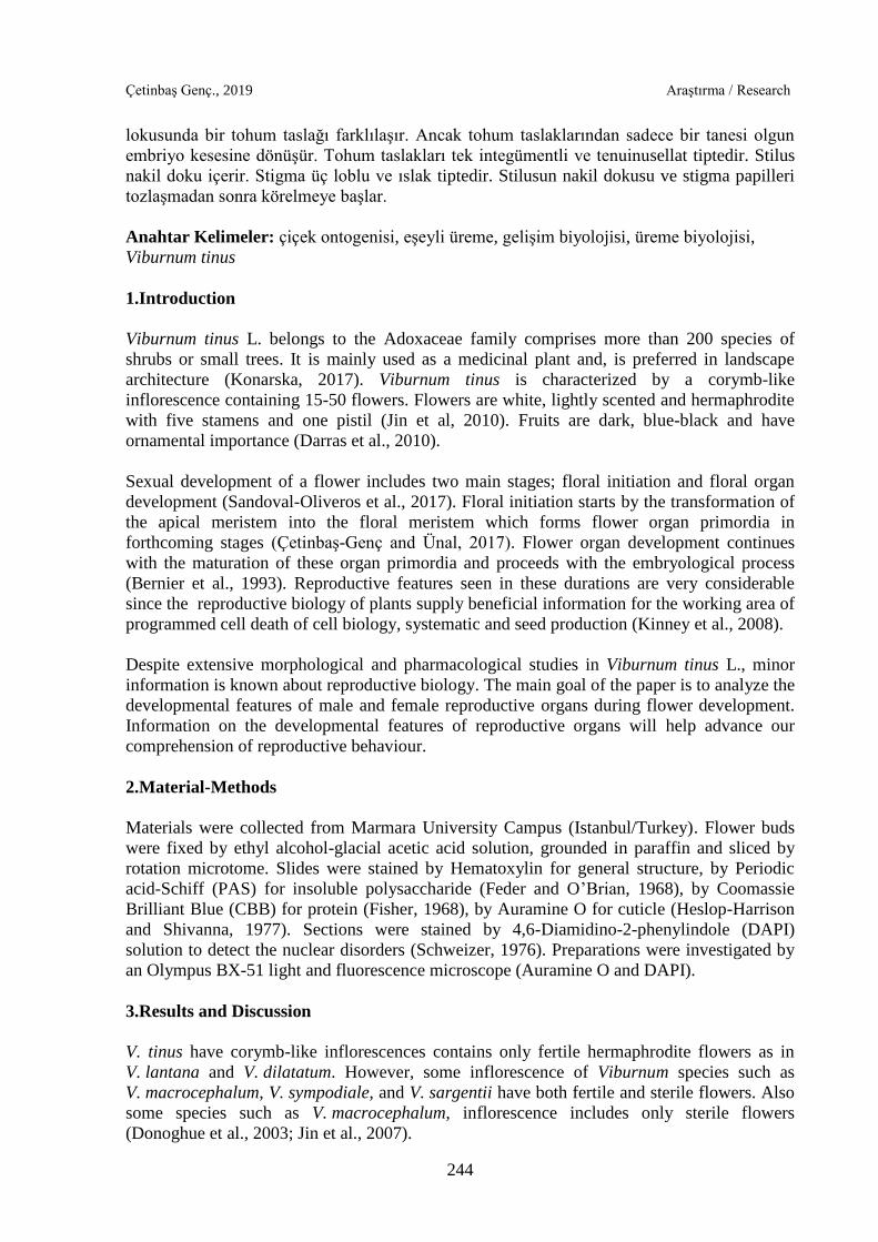
Flower development starts with the differentiation of apical meristem consisting of
consecutive layers of the cell (Figure 3.1a). Afterward, the apical meristem becomes flattened
and transforms into the floral meristem. Teeri et al. (2006) indicated that enlargement of floral
meristem appears as a result of the enhancement in division ratio of the floral meristem cells.
Floral meristem cells have a bigger volume and dense cytoplasm (Figure 3.1b). In the
following stages, floral meristem forms floral organ primordia. Firstly, five stamen primordia
differentiate as a roundish bulge from the floral meristem (Figure 3.1c). Shortly after the
stamen primordia induction, floral meristem cells differentiate into the three carpel primordia
(Figure 3.1d). The similar organ primordia differentiation model is seen in Adoxa
moschatellina belongs to the Adoxaceae family (Roels and Smets, 1994).
Figure 3.1. Flower ontogeny in V. tinus. a. Apical meristem, b. Floral meristem, c. Stamen
primordia initiation, d. Carpel primordia initiation. AM: Apical meristem, FM: Floral
meristem, S: Stamen primordia, C: Carpel primordia. Bar: 50 µm.
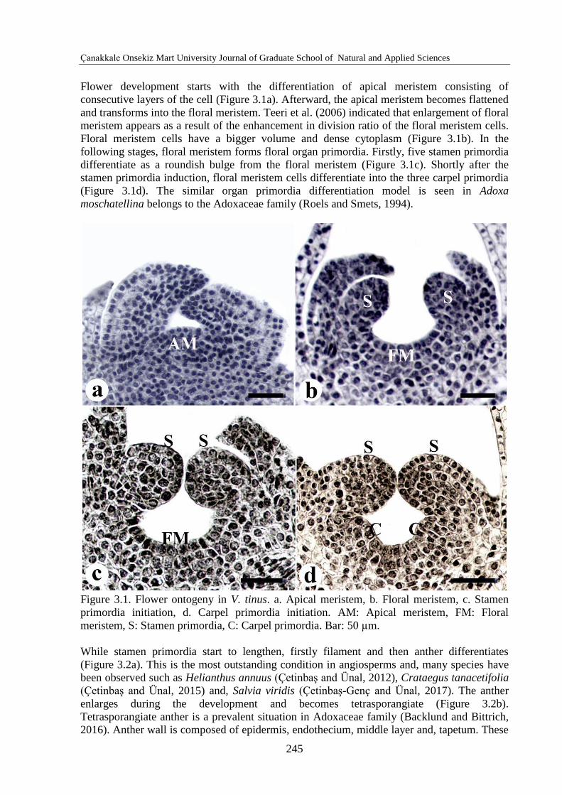
While stamen primordia start to lengthen, firstly filament and then anther differentiates
(Figure 3.2a). This is the most outstanding condition in angiosperms and, many species have
been observed such as Helianthus annuus (Çetinbaş and Ünal, 2012), Crataegus tanacetifolia
(Çetinbaş and Ünal, 2015) and, Salvia viridis (Çetinbaş-Genç and Ünal, 2017). The anther
enlarges during the development and becomes tetrasporangiate (Figure 3.2b).
Tetrasporangiate anther is a prevalent situation in Adoxaceae family (Backlund and Bittrich,
2016). Anther wall is composed of epidermis, endothecium, middle layer and, tapetum. These
Çetinbaş Genç., 2019 Araştırma / Research
246
anther layers locate as proper cell lines at the beginning of the development (Figure 3.2b).
Epidermis and endothecium with fibrous thickening stay unspoiled until anther dehiscence;
however, middle layer and tapetum dissolve during development (Figure 3.2c). Fibrous
endothecium is very common in the Adoxaceae family (Ghimire at al., 2018). Mature pollen
grains are discharged by the opening of stomium (Figure 3.2d). Ghimire et al., (2018) also
have reported anther dehiscence by the longitudinal slit in some species of Adoxaceae family.
Tapetum is plasmodial typed which tapetal cells break down during microspore development.
Benko-Iseppon and Morawetz (2000) indicated that Viburnum species can also have glandular
tapetum. Disorders in tapetal nuclei start to be visible from the microspore tetrad stage
(Figure 3.2e, h). Disorganization and nuclear fragmentations are more prominent at the young
pollen stage (Figure 3.2f, i) and finally, tapetum cannot be observed at the mature pollen stage
(Figure 3.2g, j). Similar tapetal nuclei disorders have been shown in many species such as
Lathyrus undulatus L. (Vardar and Ünal, 2012) and Crataegus tanacetifolia (Çetinbaş and
Ünal, 2015).
Figure 3.2. Anther development in V. tinus. a. Differantiation of anther and filament, b.
Young tetrasporangiate anther, c. Mature tetrasporangiate anther, d. Anther dehiscence by
stomium (arrow), e. Ordered wall layer at young anther, f. Deteriorated tapetum layer at
young pollen stage, g. Anther wall layers at mature pollen stage, h. Tapetal nuclei at tetrad
stage (asteriks), i. Tapetal cells with degenerated nuclei at young pollen stage (asteriks), j.
Mature anther. A: Anther, F: Filament, E: Epidermis, END: Endothecium, ML: Middle layer,
TA: Tapetum, PMC: Pollen mother cells, YP: Young pollen, MP: Mature pollen. Bar: 50 µm.
Floral meristem cells differentiate into the three carpel primordia shortly after the stamen
initiation (Figure 3.3a) as in A. moschatellina (Roels and Smets, 1994). Gasser and Beers
(1993) stated that carpel primordium initiates in the core of the floral meristem. Three carpel
primordia start developing and forming three ovarian loculi between them (Figure 3.3b). In
Çanakkale Onsekiz Mart University Journal of Graduate School of Natural and Applied Sciences
247
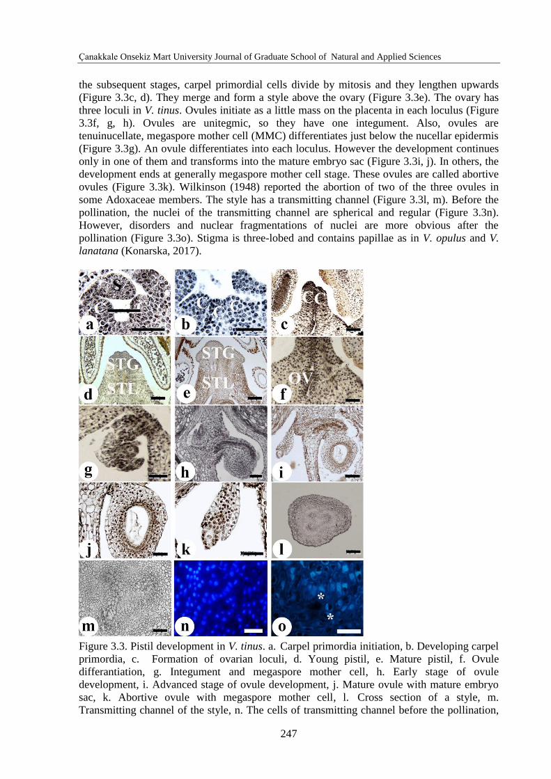
the subsequent stages, carpel primordial cells divide by mitosis and they lengthen upwards
(Figure 3.3c, d). They merge and form a style above the ovary (Figure 3.3e). The ovary has
three loculi in V. tinus. Ovules initiate as a little mass on the placenta in each loculus (Figure
3.3f, g, h). Ovules are unitegmic, so they have one integument. Also, ovules are
tenuinucellate, megaspore mother cell (MMC) differentiates just below the nucellar epidermis
(Figure 3.3g). An ovule differentiates into each loculus. However the development continues
only in one of them and transforms into the mature embryo sac (Figure 3.3i, j). In others, the
development ends at generally megaspore mother cell stage. These ovules are called abortive
ovules (Figure 3.3k). Wilkinson (1948) reported the abortion of two of the three ovules in
some Adoxaceae members. The style has a transmitting channel (Figure 3.3l, m). Before the
pollination, the nuclei of the transmitting channel are spherical and regular (Figure 3.3n).
However, disorders and nuclear fragmentations of nuclei are more obvious after the
pollination (Figure 3.3o). Stigma is three-lobed and contains papillae as in V. opulus and V.
lanatana (Konarska, 2017).
Figure 3.3. Pistil development in V. tinus. a. Carpel primordia initiation, b. Developing carpel
primordia, c. Formation of ovarian loculi, d. Young pistil, e. Mature pistil, f. Ovule
differantiation, g. Integument and megaspore mother cell, h. Early stage of ovule
development, i. Advanced stage of ovule development, j. Mature ovule with mature embryo
sac, k. Abortive ovule with megaspore mother cell, l. Cross section of a style, m.
Transmitting channel of the style, n. The cells of transmitting channel before the pollination,
Çetinbaş Genç., 2019 Araştırma / Research
248
o. The cells of transmitting channel after the pollination. C: Carpel primordia, S: Stamen
primordia, STG: Stigma, STL: Style, OV: Ovule. Bar: 50 µm (b, f, g, j, k, l, m, n, o) and 100
µm (a, c, d, e, h, i).
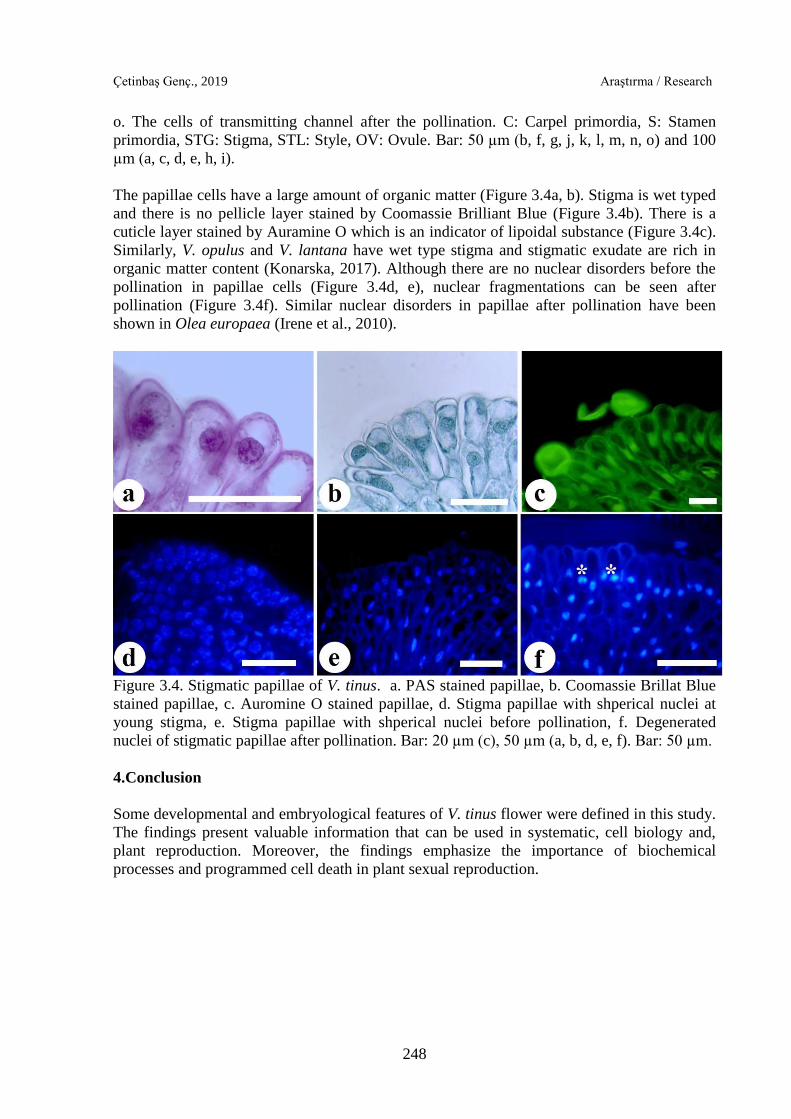
The papillae cells have a large amount of organic matter (Figure 3.4a, b). Stigma is wet typed
and there is no pellicle layer stained by Coomassie Brilliant Blue (Figure 3.4b). There is a
cuticle layer stained by Auramine O which is an indicator of lipoidal substance (Figure 3.4c).
Similarly, V. opulus and V. lantana have wet type stigma and stigmatic exudate are rich in
organic matter content (Konarska, 2017). Although there are no nuclear disorders before the
pollination in papillae cells (Figure 3.4d, e), nuclear fragmentations can be seen after
pollination (Figure 3.4f). Similar nuclear disorders in papillae after pollination have been
shown in Olea europaea (Irene et al., 2010).
Figure 3.4. Stigmatic papillae of V. tinus. a. PAS stained papillae, b. Coomassie Brillat Blue
stained papillae, c. Auromine O stained papillae, d. Stigma papillae with shperical nuclei at
young stigma, e. Stigma papillae with shperical nuclei before pollination, f. Degenerated
nuclei of stigmatic papillae after pollination. Bar: 20 µm (c), 50 µm (a, b, d, e, f). Bar: 50 µm.
4.Conclusion
Some developmental and embryological features of V. tinus flower were defined in this study.
The findings present valuable information that can be used in systematic, cell biology and,
plant reproduction. Moreover, the findings emphasize the importance of biochemical
processes and programmed cell death in plant sexual reproduction.
Çanakkale Onsekiz Mart University Journal of Graduate School of Natural and Applied Sciences
249
References
Backlund A., Bittrich V., 2016. Adoxaceae. In Flowering Plants. Eudicots (pp. 19-29).
Springer, Cham.
Benko-Iseppon A.M., Morawetz, W., 2000. Viburnales: Cytological Features and a New
Circumscription. Taxon 5-16.
Bernier G., Havelange A., Houssa C., Petitjean A., Lejeune P., 1993. Physiological Signals
That İnduce Flowering. Plant Cell 5:1147-1155.
Çetinbaş A., Ünal M., 2012. Comparative Ontogeny of Hermaphrodite and Pistillate Florets
in Helianthus annuus L. (Asteraceae). Notulae Scientia Biologicae 4(2):30-40.
Çetinbaş-Genç A., Ünal M., 2017. Flower Ontogeny and Reproductive Biology of Salvia
viridis L. Pakistan Journal of Botany 49: 891-896.
Darras A.I., Akoumianaki-Ioannidou A., Pompodakis N.E., 2010. Evaluation and
Improvement of Post-Harvest Performance of Cut Viburnum tinus Inflorescence. Scientia
horticulturae 124(3):376-380.
Donoghue M.J., Bell Charles D., Winkworth Richard C., 2003. The Evolution of
Reproductive Characters in Dipsacales. International Journal of Plant Sciences 164(5):
453–464.
Feder N., O’Brien T.P., 1968. Plant Microtechnique: Some Principles and New Methods.
Amerikan Journal of Botany 55(1): 123-142.
Fisher D.B., Jensen W.A., Ashton M.E., 1968. Histochemical Studies of Pollen: Storage
Pockets in the Endoplasmic Reticulum. Histochemie 13: 169-182.
Gasser S., Beers K., 1993. Pistil Development. Plant Cell 5:1231-1239.
Ghimire B., Suh G.U., Lee C.H., Heo K., Jeong M.J., 2018. Embryological Studies on Abelia
tyaihyoni Nakai (Caprifoliaceae). Flora 242:79-88.
Heslop-Harrison Y., 1977. The Pollen Stigma İnteraction: Pollen Tube Penetration in Crocus.
Annals of Botany 41:913–922.
Irene S., Salvatore P., Adela O., 2010. Programmed-cell-death Hallmarks in Incompatible
Pollen and Papillar Stigma Cells of Olea europaea L. Under Free Pollination. Plant Cell
Reports 29(6):561-572.
Jin B., Li N., Jia N., Zhou W., Wang L., Xiang Q., 2007. Observations on the Anatomy of
Reproductive Organs and the Pollinators of Viburnum macrocephalum f . keteleeri
(Caprifoliaceae). Acta Phytotaxonomica Sinica 45:753–768.
Çetinbaş Genç., 2019 Araştırma / Research
250
Jin B., Wang L., Wang J., Teng N.J., He X.D., Mu X.J., Wang Y.L., 2010. The Structure and
Roles of Sterile Flowers in Viburnum macrocephalum f. keteleeri (Adoxaceae). Plant
Biology 12: 853–862.
Kinney M.S., Columbus J.T., Friar, E.A., 2008. Unisexual Flower, Spikelet, and
Inflorescence Development in Monoecious/Dioecious Bouteloua dimorpha (Poaceae,
Chloridoideae). American Journal of Botany 95(2): 123-132.
Konarska A., 2017. Comparative Micromorphology and Anatomy of Flowers and Floral
Secretory Structures in Two Viburnum Species. Protoplasma 254(1): 523-537.
Roels P., Smets E., 1994. A Comparative Floral Ontogenetical Study Between Adoxa
moschatellina and Sambucus ebulus. Belgian Journal of Botany 127: 157-170.
Sandoval-Oliveros R., Guevara-Olvera L., Beltrán J.P., Gómez-Mena C., Acosta-García G.,
2017. Developmental Landmarks During Floral Ontogeny of jalapeño chili pepper
(Capsicum annuum L.) and the Effect of Gibberellin on Ovary Growth. Plant
reproduction, 30(3): 119-129.
Schweizer D., 1976. Reverse Fluorescent Chromosome Banding with Chromomycin and
DAPI. Chromosoma 58: 307-324.
Teeri T.H., Uimari A., Kotilainen M., Laitinen R., Help H., Elomaa P., Albert V.A., 2006.
Reproductive Meristem Fates in Gerbera. Journal of Experimental Botany 57:3445-3455.
Vardar F., Ünal M., 2012. Ultrastructural Aspects and Programmed Cell Death in the Tapetal
Cells of Lathyrus undulatus Boiss. Acta Biologica Hungarica 63(1):52-66.
Wilkinson A.M., 1948. Floral Anatomy and Morphology of Some Species of the Tribe
Lonicereae of the Caprifoliaceae. American Journal of Botany 35: 261–271.


