Embed Size (px)
Citation preview

Vol. 1: 169-182, 1986 DISEASES OF AQUATIC ORGANISMS Dis. aquat. Org.
Published October 15
Development of the PKX myxosporean in rainbow trout Salmo gairdneri
Michael L. Kent* & Ronald P. Hedricka*
Department Medicine, School of Veterinary Medicine. University of California, Davis. California 95616, USA
ABSTRACT Sequenhal development of PKX, the causative agent of prohferahve h d n e y dlsease, is descnbed in rainbow trout Salmo ga~rdnen hchardson by light and electron mlcroscopy Fish were exposed to water contaming the infechous stage of PKX and the parasite was detected 3 wk later Parasites were prominent the following week and they mulhplied by endogeny, binary fission and possibly plasmotomy in the blood and hdney intershhum Intershbal nephnbs that is typically assoclated \nth PKD was fust observed 5 wk after exposure and was most prominent between 8 to 11 wk At 7 wk parasites were found in the lumens of the tubules, many were daughter cells released from PKX Myxosporean sporoblasts formed within these daughter cells (enveloping cells) The sporoblasts consisted of 6 cells and some organized into spores w ~ t h 2 spherical polar capsules Although the spores persisted for several months after the interstitial PKX and assoclated inflammation had subsided they dld not complete their development They remained within the enveloping cell and apparently dld not form complete valves Because only incomplete spores were observed salmonids may be abnormal hosts and the preclse taxonomic status of PKX was not determined Epizoohological and sequential development data support the hypothesis that the interstitial form of PKX and the intraluminal myxosporeans belong to the same organism
INTRODUCTION
Proliferative kidney dlsease (PKD), caused by an unclassified protozoan (PKX), is considered a serious obstacle to the successful culture of certain salmonid fishes. The disease has been reported most often in rainbow trout Salmo yairdnen kchardson, and mor- talities may reach 90 %, but usually remain below 30 to 50 '10 (Clifton-Hadley et al. 1984). Other salmonid species have been affected and PKD is considered one of the most important diseases of cultured salmonids in Europe (Clifton-Hadley et al. 1984). It is also wide- spread in western North America (Hedrick et al. 1984a, b , 1985; Smith et al. 1984, Hosluns 1985). Even in the absence of high mortality, morbidity may be 100 % and fish with PKD often exhibit decreased food conversion, poor tolerance to stress, and greater susceptibihty to secondary infection (Clifton-Hadley et al. 1984). The &sease inficts additional economic burdens on pro- duction fachties because husbandry practices must be
Present address: Center for Marine Disease Control, Bat- telle Marine Research Laboratory, 439 West Sequvn Bay Road, Sequim, Washington 98382, USA " Addressee for reprint requests
O Inter-Research/Printed in F. R. Germany
modified and the planting of exposed fishes is often restricted by regulatory agencies.
The disease is characterized by chronic inflamma- tion of the ludney. Tissue sections reveal the PKX protozoan primarily in the ludney interstitium, associ- ated with a granulomatous interstitial nephritis and tubular atrophy (Ferguson & Needham 1978, Clifton- Hadley et al. 1984, Smith et al. 1984). In heavily infected fish, PKX is often found in the spleen, gut, gills, liver and muscle, where it also invokes a granulomatous response (Ferguson & Needham 1978, Smith et al. 1984). The PKX parasite most likely reaches these organs via the circulatory system and may adhere to vessel walls, provoking a necrotizing vasculitis (Smith et al. 1984). If fish survive PKD, the interstitial hyperplasia subsides in 12 to 20 wk, and the ludneys of recovered fish show little if any signs of previous infection (Clifton-Hadley et al. 1985).
A parasite that was most Likely PKX was described as an amoeba by Plehn (1924). Ghlttino et al. (1977) and Ferguson & Needham (1978) also suggested that PKX may be an amoeba because it formed pseudopodia. Although no spores were observed, Seagrave et al. (1980a) proposed that the organism might be a haplo- spondan (phylum Ascetospora). Division by internal
170 Dis. aquat. Org. 1: 169-182, 1986
cleavage, the presence of multivesicular bodies, and the existence of electron-dense cytoplasmic inclusions reminiscent of the haplosporosomes of Marteilia spp. were cited as common features with PKX.
However, internal cleavage and organelles similar to haplosporosomes also occur among members of the Myxozoa (Current & Janovy 1977, Desser and Paterson 1978, Current et al. 1979, Lom et al. 1982. Desser et al. 1983), and Ferguson & Needham (1978) and Seagrave e t al. (1980a, b) did not exclude the possibility that PKX might belong to this phylum. Hedrick et al. (1984b) and Kent & Hedrick (1985a) observed myxo- sporean trophozoites and developing spores in the renal tubules of fish with PKD, and they proposed that the observed organisms might be later stages of PKX and that PKX belongs most appropeately to the phy- lum Myxozoa.
We present here a detailed description of the sequential development of PKX from the renal intersti-
tial form (typical PKX) to an immature myxosporean spore in the lumens of the tubules. We also present evidence that substantiates that both forms belong to the same organism. These findings indicate that PKX is a member of the phylum Myxozoa.
MATERIALS AND METHODS
A group of PKX-free rainbow trout under 1 yr of age (Eagle Lake, California strain) was introduced into water containing the infective stage of PKX at the American River hatchery, Rancho Cordova, California, USA on 16 August 1984. The parasite is only transmit- ted by water-borne exposure or by injecting infected tissues (D'Silva et al. i984), and the infective stage under natural conditions is yet to be identified. Approximately 500 of these fish were transferred to the Fish Patho!ogy Laboratory at the University of Califor-
90 A Intra lurn~nal forms X P K X 0 I n te rs f i t~a l nephrit~s
a I
Wks ofter I V 6'- Introduction 0 5 10 15 20 2 5 30 35
Ten
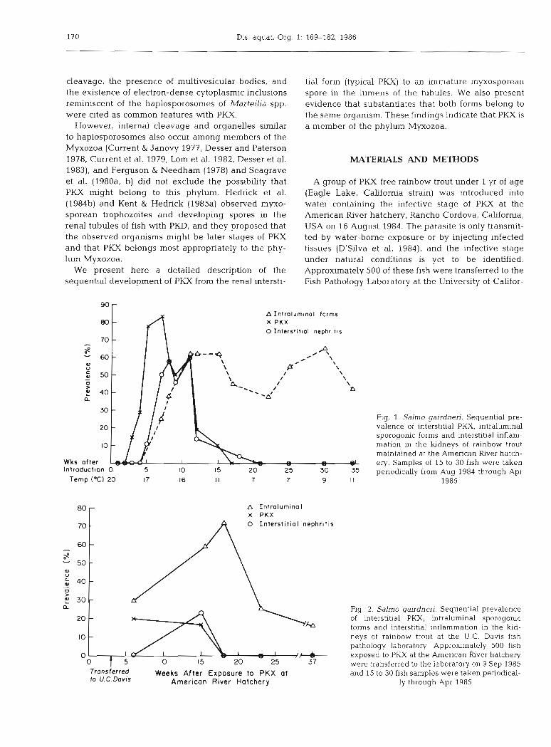
Fig. 1. Salmo gairdnen. Sequenhal pre- valence of interstitial PKX, intraluminal sporogonic forms and interstitial inflam- mation in the kidneys of rainbow trout maintained at the American River hatch- ery. Samples of 15 to 30 flsh were taken periodically from Aug 1984 through Apr
1985
80
70
60
S 50-
Q) 0
40- - 0 >
30- a
20
l0
0
- A Introlurninol X PKX
- 0 I n t e r s t ~ t i a l n e p h r i t ~ s
-
Fig. 2. Salmo gairdnen. Sequential prevalence - of interstitial PKX, intraluminal sporogonic
forms and interstitial inflammation in the kid- - neys of rainbow trout at the U.C. Davis fish pathology laboratory. Approximately 500 fish
I - V exposed to PKX at the American h v e r hatchery 0 5 10 15 20 25 37 were transferred to the laboratory on 9 Sep 1985 Trons ferred Weeks After Exposure to P K X ot and 15 to 30 fish samples were taken penodical- to U. C. Dovis American River Hatchery ly through Apr 1985

Kent & Hedrick: PKX in rainbow trout
nia (U.C.), Davis, California, on 9 September 1984 and maintained in PKX-free well water at 15 to 18 'C. Samples of 15 to 30 fish were taken at selected times from the population at the hatchery and from the fish at U.C. Davis until May 1985.
To determine the morphology of PKX, preparations of fresh kidney were squashed between a cover slip and glass slide (wet mounts), and then examined by bright field and phase contrast microscopy. For his- tological examinations, tissues were fixed in David- son's solution, embedded in paraffin, sectioned at 5 Km, and stained with either hematoxylin and eosin (H & E) or Prices's Giemsa as described by Luna (1968) and Humason (1979). Fish from the sequential samples taken at the American River hatchery and U.C. Davis were examined for the prevalence of interstitial PKX, associated pathology, and intraluminal myxosporean stages using paraffin sections of the kidneys. Kidney tissue samples for electron microscopy were fixed in 3 % (v/v) glutaraldehyde in phosphate buffer as described by Bullock (1978). These specimens were post-fixed in 1 O/O OsO,, embedded in Epon, sectioned, stained with lead citrate and uranyl acetate, and examined with a Phillips EM 400 transmission electron nlicroscope. Similar developmental stages of PKX were observed in all epizootics of PKD in California, and fish from several locations in the state were used for the light and electron microscopical descriptions.
RESULTS
Temporal prevalence
The PKX parasite was first detected in one of 15 fish examined 3 wk after transfer to the American River hatchery (Fig. l ) . Parasites were easily detected the following week and reached a peak prevalence of 83 % at 7 wk. Associated interstitial inflammation was evi- dent at 5 wk, and was most prominent between 8 and 11 wk. The PKX parasite in the epithelium of the tubules and intraluminal sporogonic stages were first detected at 7 wk. These forms persisted at approxi- mately the same prevalence for at least 12 wk after interstitial PKX and associated lesions were no longer detectable (Fig. l ) . However, the intensity of the intraluminal sporogonic forms diminished with time. The prevalence of intraluminal forms increased and interstitial PKX and associated lesions decreased as the water temperature at the hatchery declined (Fig. 1).
The development of PKX in the fish transferred to U.C. Davis was similar to that observed in the fish held at the hatchery (Fig. 2). However, even though the tem- perature at U.C. Davis was above 15 "C throughout the study, the intraluminal forms became prevalent in these fish as the disease progressed (Fig. 2).
Light microscopy
The organism seen at 3 wk was small, condensed, eosinophilic, and contained a daughter cell (Fig. 3). Tissue sections revealed larger, typical parasites in the blood and kidney interstitium the following week (Fig. 4). These forms were also detected in blood smears at this time. Primary cells with as many as 7 daughter cells were evident in these tissues (Fig. 5), and were weakly eosinophilic compared to the sur- rounding host cells. The nuclei of the parasites con- tained large endosomes that were particularly promi- nent in the primary cells. Degenerating primary cells and small condensed cells were observed in the inter- stitium of some fish. The small cells may represent released daughter cells (Fig. 6).
Parasites that were in the blood occasionally formed thrombi in the renal vessels (Fig. 7). Hypercellularity of the kidney interstitium due to mononuclear cell proli- feration and infiltration was associated with the para- sites (Fig. 8), and infected fish exhibited a reduced number of tubules mm-* when compared with un- infected fish (Fig. 9). Many parasites were surrounded by attached macrophages and lymphocytes (Fig.6). Although most fish in our experiment exhibited gross ludney hypertrophy, they did not exhibit other clinical signs of PKD and no significant mortality could be directly attributed to PKX.
Organisms with typical PKX morphology were ob- served in the epithelium of the renal tubules (Fig. 101, and the daughter cell of one was seen in binary fission (Fig. 11). Some primary cells in the epithelium de- generated, and their daughter cells, surrounded by primary cell remnants, migrated to the lumen (Fig. 12). Small, condensed eosinophilic cells, presumably re- leased daughter cells, were observed in the lumen of tubules at this time. The simplest intraluminal forms were uninucleate cells that resembled the secondary cells of interstitial PKX (Fig. 13). More complex intraluminal forms contained secondary and tertiary daughter cells. Continued development of these stages by endogeny and binary fission resulted in sporoblasts with up to 6 internal sporogonic cells (Fig. 14 & 15).
Developing myxosporean spores as evidenced by polar capsule formation were observed in the more advanced intraluminal parasites (Fig. 16 to 18). These spores were oblong, 12 X 7 km, with 2 spherical polar capsules 2 pm in diameter (measured from wet mounts, n = 20). Capsulogenic nuclei and a large sporoplasm nucleus were prominent in paraffin sections stained with hematoxylin and eosin (Fig. 16). The intraluminal organisms were considered to be myxosporean tro- phozoites because they formed multicellular spores with polar capsules. Only one spore developed withln each enveloping cell and no large multinucleated


Dis. aquat. Org. 1: 169-182, 1986
plasmodia, typical of most myxosporeans, were ob- served. However, up to 3 nuclei were observed in some enveloping cells. This monosporous development within an enveloping cell, described as a 'pseudoplas- modium' by Lom et al. (1982) in Sphaerospora spp., was confirmed in wet mounts (Fig. 18). Enveloping cells observed in wet mounts were characterized by numerous refractile granules.
The spores apparently did not complete their development in the kidney. No clear spore valves or well defined valvogenic cells were observed in either paraffin sections or wet mounts. The spores were fragile and lacked rigidity as evidenced by the readi- ness with which they changed shape under pressure of the cover slip in wet mounts; they degenerated within 4 h in minced kidneys rlldirll&lled in water dt 10 "C. Further, spores could no longer be detected after 24 h under these same conditions.
The intraluminal trophozoites and spores were most frequently observed in the second proximal and distal segments of the tubules. Although they were not found in the collecting ducts, ureters or urinary bladder, 3 sporoblasts with polar capsules were observed in one of 10 urine samples obtained from fish at the American River hatchery.
Electron microscopy
The primary cell of PKX often contained internal secondary and terbary daughter cells (Fig. 19) and always contained 'haplosporosomes' identical to those described by Seagrave et al. (1980a, b) (Fig. 20). The primary cells contained a prominent endoplasmic reticulum. They also contained large, electron-dense, membrane-bound inclusions which were presumed to be secondary lysosomes. The daughter cells, either secondary or tertiary, were encircled by a membrane of the outer cell. They never contained haplosporosomes and rarely lysosomes. Degenerating primary cells, with condensed organelles and disrupted membranes, contained intact daughter cells (Fig.21). The latter may be released to continue the vegetative develop- ment of the parasite in the interstitium.
The PKX parasites in the epithelium of the tubules (Fig. 22) had an ultrastructural morphology identical to those in the interstitium. They migrated to the lumen by penetrating between the epithelia1 cells.
Daughter cells wi thn PKX and early intraluminal forms had a similar ultrastructural morphology. The simplest and possibly earliest intraluminal forms were uninucleate (Fig. 23), and presumably they were daughter cells released from typical PKX. These forms and daughter cells within typical PKX shared a similar size, nucleus to cytoplasm ratio, chromatin pattern, and
plate-like cristae in the mitochondria. Additionally, as with secondary cells, they contained little endoplasmic reticula compared to the primary cell of PKX, and had similar ribosome content and cytoplasmic matnces.
More complex intraluminal forms had enveloping cells with electron-dense multilaminate bodies and up to 5 sporogonic cells (Fig. 24 & 25). The most fully developed forms observed were spores containing polar capsules with coiled internal filaments (Fig. 26). Although valves were not observed, early valvogenic cells pahally surrounded the capsulogenic cells.
Macrophages usually surrounded the parasites in the blood and ludney interstitium, but they were not observed in association with the intralurninal stages. The only intracellular stage of PKX observed was a small form that had been completely engulfed by a macrophage.
A diagramatic representation of the sequential de- velopment of PKX in the kidney of salmonid fishes is shown in Fig. 27. Stages represented were observed either by light or electron microscopy.
DISCUSSION
Evidence from this study verifies that PKX belongs to the phylum Myxozoa. Myxosporeans are ubiquitous, usually host-specific parasites of fishes (Mitchell 1977) and the prespore stages typically do not induce a significant inflammatory response (Dykova & Lom 1978). Salmonids exhibit a severe inflammatory response to interstitial PKX, which is the pre- sporogonic form, and only immature spores were observed. This suggests that salmonids are abnormal hosts for PKX.
Proliferative ludney disease parasites (PKX) with identical morphology to those reported by Ferguson & Needham (1978), Seagrave et al. (1980a, b) and Smith et al. (1984) were frequently observed in the blood and kidney interstitium. The parasite was detected in fish introduced to water containing the infectious stage 1 wk earlier than previously reported (Ferguson & Ball 1979, D'Silva et al. 1984), and at this time was smaller and more condensed than typical PKX (Fig. 3). Numer- ous typical forms were observed in the blood, particu- larly early in the infection, which may indicate that it reaches the kidney via the circulatory system.
The PKX parasite most likely undergoes vegetative reproduction within the fish host. This is supported by the observation of numerous daughter cells in the ludney interstitium of heavily infected fish (Fig. 5 & 6). Additionally, fish injected with very few parasites became heavily infected (Kent & Hedrick 1985b). Fish transferred from the hatchery to PKX-free water at U.C. Davis before parasites could be detected later

$2 g 2 E Opt
,g '
g6
,~
.. a
.
2 a
zE
~
&;so
c 0
23
9
%,
iU
~
alg
.a3
a" D
E.2
G o
E.c
h
.5 3+
a2
a
m c
.S $
;i
f?
c
0 3 .y
a
OQ
E m
2
2 g
g,
-%
2&
O
@'
Z $2
2u
al0
,
BP
ss
z
z; 0 a
c
.p
al6
2 2
6 s;.z
hz
Nd
~
~1
1
"3
5(
z 2 F
U
gb
rn
a~
a
pi.2
;
Z
m 8 a
,c
,2=m2
a E .c
D
2" g
, c
L(
0 $S.;;?
22
>a
CS al c
Da
U
eU
--~9
2 2
5 2d
E4
E m
m
cc
aa
ei,
o
on
EE
3
U-
0
$3
a i
Gr
nZ
%
4 E"
'S, u
,o
o
S
Q,
4 a11
33
22
m
e
Q .a
2 3
.- m C
&a a-
E $l s's

f N g
-a
I


178 Dis. aquat. Org. 1: 169-182, 1986
Fig. 26. Salmo gairdnen. Intralurninal sporoblast with polar capsules. Valvogenic cells (v) surrounding capsulogenic cells (c); pc: polar capsule; E: enveloping cell
developed heavy infections (Fig. 21, indicating that continual exposure is not needed to induce heavy infections. A mechanism for the development of the daughter cells by internal cleavage was reported by Seagrave et al. (1980b). However, binary fission of the internal daughter cells, as reported in the present study, is a second method for vegetative development. No fixed developmental sequence within the kidney interstltium could be determined, and plasmotomy (external budding), endogeny (internal budding), and binary fission may all occur in the same parasite (Fig. 27). This lack of rigidity in the vegetative development is consistent with the phylum Myxozoa (Lom et al. 1983a, Johnstone 1984).
Ferguson & Needham (1978) reported PKX in the renal tubules, but migration of the parasite through the epithelium and release of daughter cells into the lumen were not described. The PKX parasites in the epithelium of the tubules and its intraluminal stages were more prevalent in convalescing fish than in clinically affected fish (Fig. l ) , and Ferguson & Needham (1978) have suggested that the tubules may provide a mode of exit for the parasite.
The simplest intraluminal stage detected in this
study was a small unicellular organism. Similar forms contained single endogenous daughter cells (Fig. 23). These stages appeared identical to the internal daugh- ter cells of the interstitial and epithelia1 PKX (Fig. 19 & 22) and are most Likely the same cells. Haplosporo- somes are only observed in the primary cell of PKX (Ferguson & Needham 1978, Seagrave et al. 1980a, Smith et al. 1984). This accounts for their absence in the intraluminal forms because this stage seems to arise from released secondary cells of typical PKX. The intraluminal forms represent pseudoplasmodia as described by Lom et al. (1982) for Sphaerospora spp. and the internal cells are the sporoblast. The envelop- ing cell of the pseudoplasmo~um in Sphaerospora spp. is uninucleate (Lom et al. 1982), but this cell of PKX often contained 2 or 3 nuclei. This cell also con- tained prominent refractile granules as seen in wet moints (Fig. 18), and these are most Likely analogous to the electron-dense multilaminated bodies (Fig. 24 to 26) and the periodic acid Shiff's (PAS) positive bodies reported by Kent & Hedrick (1985a). Polar capsule formation in PKX indicated spore formation. However, trophozoites were more numerous than spores, even late in the infection, as Bond (1938) reported for

Kent & Hedrick: PKX in rainbow trout 179
Sphaerospora r e n d s in the killifish Fundulus hetero- clitus L.
Polar capsules may form before valvogenesis occurs, as in other myxosporeans (Desser & Paterson 1978, Current et al. 1979), and valves were not apparent in the PKX spore. The spores remained within their pseudoplasmodium throughout the infections, and this, combined with the apparent lack of valves, indi- cates that the spores were not fully developed. Five to 6 sporogonic cells were observed in maturing sporo- blasts, which is consistent with myxosporean sporo- genesis. Two of the cells correspond to the capsul- ogenic cells, 1 or 2 to the sporoplasm cells, and 2 presumably to the valve-forming cells (Mitchell 1977). Although valves were not observed, PKX spores may form vestigial valvogenic cells in salmonids. These spores were fragile, and it is unlikely that they would survive long enough for valvogenesis to occur if they were released from this host.
Although many spores were observed by light mi- croscopy, few were found by electron microscopy. In one spore, early valvogenic cells partially surrounded capsulogenic cells (Fig. 26). If more advanced stages of valvogenesis occur, they were not detected by light microscopy. More spores must also be examined by electron microscopy to identify the most advanced stage of spore development that occurs in salmonids.
Such studies are made difficult because of the paucity of PKX spores in most fish.
A clear sequential development from the interstitial PKX to spores with polar capsules demonstrated the myxosporean nature of the parasite. This is further supported by epizootiological evidence and experi- mental data. The myxosporean of this study was only observed in PKX-exposed fishes, and although it has not been reported previously, it was detected in kidney sections of infected fish from all cases of PKD that we have examined. These involved coho salmon Oncorhynchus kisutch (Walbaum) and steelhead trout from 2 enzootic areas on Vancouver Island, British Columbia, Canada; convalescing rainbow trout from the Hagerman State hatchery, Idaho, USA; coho salmon from the State of Washington, USA; and from all cases investigated in California, USA (Hedrick et al. 1984a, b , 1985). A similar myxosporean has been found in PKX- exposed Atlantic salmon Salrno salar (L.) and brown trout Salrno trutta (L.) in Europe (R. S. Clifton-Hadley, Ministry of Aquaculture, Fisheries and Food, Wey- mouth, England, pers. comm.). The coincidence of typi- cal PKX with the intraluminal myxosporean forms was further demonstrated in studies by Hedrick et al. (1985). Fish introduced to the American River hatchery during the spring and summer showed both typical PKX and the lntraluminal forms. In contrast, fish exposed during
Fig. 27. Sequential development of PKX in kidney of salmonid fishes as interpreted from wet mounts, paraffin sections and elec- tron micrographs. (a) PKX parasite in a blood vessel. (b) PKX enters hdney inter- stitium. (c) Uninucleate PKX. (d) Daughter cell produced by endogenous budding. (e) Secondary daughter cells divide by blnary fission and produce tertiary cells by en- dogenous budding. Nuclear division in primary cell may also occur. (f) Daughter cells are released from degenerated primary cell and continue vegetative development. ( f ' ) Plasmotomy or binary fission of primary cell may also occur. Vegetative develop- ment also may occur in the blood vessels. (g) PKX parasite penetrates between epithelia1 cells of tubule. (h) PKX enters the lumen; primary cell degenerates and releases sec- ondary cell (= pseudoplasmodium). (h') Primary cells may release daughter cells into lumen from epithelium. Multiplication of released secondary cells may occur in the lumen. (i) Released secondary cell (envelop- ing cell) with tertiary (sporogonic) cell. U) Tertiary cell multiplies resulting in a nlul- ticellular sporoblast. (k) Sporulation con- tinues; capsulogenic cells differentiate and form polar capsules and sporoplasm de- velops a prominent nucleus. No valvo- genesis observed, and spore remains within
enveloping cell throughout infection

180 Dis. aquat. Org.
the winter when the infectious stage was not present did not become infected with either stage of PKX.
A blood and spleen transmission experiment by Kent & Hedrick (1985b) also supports the conclusion that both forms belong to the same parasite. Fish injected with the blood and spleen of PKX-infected fish became infected with both interstitial PKX and the intralumi- nal myxosporeans. Since only the typical PKX is found in blood and spleen, the myxosporeans must have arisen from this stage.
Seagrave et al. (1980a) suggested that PKX is a haplosporidan (phylum Ascetospora) because of its similarities with the invertebrate pathogens Marteha spp. However, they did not exclude the possibility that it may belong to the phylum Myxozoa. The higher taxonomy of these phyla is in a state of confusion. Desportes & Ginsburger-Vogel (1977) demonstrated affinities of the family Martediidae with the class Para- myxea (Ascetospora), Actinosporea, and Myxosporea (Myxozoa), and suggested they be grouped in a com- mon taxon. Of the myxosporeans examined by trans- mission electron microscopy, most form electron-dense cytoplasmic inclusions, similar to haplosporosomes in the Ascetospora (Current & Janovy 1977, Desser & Paterson 1978, Current et al. 1979, Lorn et al. 1982, Desser et al. 1983), and organisms of both phyla repro- duce by internal cleavage (endogenous budding).
Although the P M myxosporean could not be iden- tified because only immature spores were found, it shows similarities to Sphaerospora spp. (family Sphaerospondae). Kent & Hedrick (1985a) compared the prespore form of PKX in the kidney interstitium to a protozoan of carp blood (UBO), which may be an early stage of Sphaerospora renicola (Lom et al. 1983a). A protozoan similar to UBO found in the swimbladder of carp was shown to be an early stage of S. renicola (angulata) (Csaba et al. 1984, Molnar 1984). The PKX parasite is similar to UBO and the swimbladder proto- zoan in that all 3 form endogenous secondary and tertiary daughter cells which are released and may subsequently sporulate in the lumen of the tubules. As with PKX, the secondary cells of UBO reproduce by binary fission in addition to endogenous budding (Lom et al. 1983a). Furthermore, sporogenesis of PKX is similar to that of some Sphaerospora spp. in that spores of both are formed with a pseudoplasmodium in the lumen of the kidney tubules. The enveloping cell of pseudoplasmodium in both parasites contains promi- nent refractile granules (Lom et al. 1985), and PKX is monosporous like S. molnari (Lom et al. 1983b).
A companson of immature spores of Sphaerospora spp. with those of PKX shows similar features such as an elongated shape, small polar capsules, and indis- tinct valves. In contrast to PKX, the mature spores of Sphaerospora spp. have a spherical shape and valves
are evident. Ferguson (1984) reported Sphaerospora- like myxosporeans in the kidneys of feral brown trout from PKD enzootic waters, and Lorn et al. (1985) observed pseudoplasmodia without spores in brown trout and grayling in Czechoslovakia. These fishes are hosts for PKX, and the pseudoplasmodia were possibly the intraluminal stages of PKX.
Although the PKX myxosporean shows many similarities to members of the family Sphaerosporidae, its spore also resembles those described for Parvicap- sula (Parvicapsulidae). Members of this genus are also elongated, and have 2 polar capsules similar in size to those of PKX. Furthermore, a Pam'capsula sp. is known to sporulate in the epithelium of the kidney tubules in coho salmon (Hoffman 1984, Johnstone 1984). Althouy h Pdrvicdpsula develops in the usual myxospo- rean manner with large plasmodia and pansporoblasts, it may also form pseudoplasmodia (J. Lom, Czecho- slovak Academy of Sciences, C. Budejovice, Erani- Sovska, Czechoslovalua, pers. comm., Johnstone 1984).
Spore morphology has been the most important criterion for species descriptions in the Myxozoa (Mitchell 1977), but the origin of sporoblasts (either within true plasmodia or pseudoplasmodia) should also be included in these descriptions (Lom et al. 1982, Lorn & Noble 1984). Identification of the primary host and completely formed spores are needed before the precise taxonomic status of the PKX myxosporean can be determined.
Salmonids may be abnormal hosts for PKX (Seagrave et al. 1980a). The severe inflammation associated with the prespore (typical) PKX stage and the lack of com- plete development of the spore support this hypothesis. The severe inflammation with PKD is in contrast to the lack of tissue reaction to the prespore forms in hosts in which myxosporeans complete sporogenesis (Dykova & Lorn 1978). No significant pathological changes were associated with the intraluminal stages of PKX, and the interstitial nephritis disappeared shortly after the typi- cal PKX form was no longer detectable (Fig. 1). Fergu- son (1981) reported that fish with PKD recover faster in cold water. However, low temperatures are not required for disappearance of the interstitial PKX, recovery from PKD, migration of PKX to the tubules, or sporulation, because all of these events occurred in the fish transferred to U.C. Davis where temperatures remained above 15 "C.
This suggests that the stage of development of the parasite is an important factor in the inflammatory response associated with PKD. Furthermore, the in- flammatory response in an abnormal host may inhibit the migration of PKX to the tubules and subsequent sporulation. Kent (1985) immunosuppressed PKX- infected fish with cortisol implants and found that suppression of the inflammatory response to PKX
Kent & Hedrick: PKX in rainbow trout
enhanced the development of the parasite. In PKX- infected fish with cortisol implants, more interstitial forms, but less interstitial hypercellularity were observed. These fish also exhibited more sporogonic stages, but the cortisol treatments did not induce com- plete development of the spores (Kent 1985, Kent & Hedrick 1986). Possibly the kidney tubules of rainbow trout do not provide the proper physiological environ- ment for complete sporulation. This is substantiated by the observations from fish in natural epizootics, in which spores persisted in an immature state in the tubules several months after the inflammation had subsided (Fig. 1).
This study has demonstrated that the causative agent of PKD is the presporogonic form of a myxosporean, which undergoes vegetative development and induces a severe tissue response in the kidney interstitium and blood of rainbow trout. As the disease progresses, the parasites migrate to the lumen of the kidney tubules and sporulate within a pseudoplasmodium. Although inflammation was not associated with these sporogonic forms, only immature spores were observed in the tubules. These observations all indicate that salmonids are abnormal hosts for PKX and that the parasite may complete sporulation in a non-salmonid fish. Identifi- cation of the primary host is needed to determine the precise identity of PKX and the reservoir of infection.
We have recently observed immature spores similar to those of PKX and more fully developed Sphaero- spora spores in the lumens of kidney tubules from tui chub Gila bicolor (Girad) (Cypnnidae), and stickle- backs Gasterosteus aculeatus L. (Gasterosteidae), from watersheds where PKX is prevalent (Hedrick et al. 1986). Studies are underway to determine the associa- tion of these myxosporeans with PKX and the role of non-salmonids as reservoir hosts.
Acknowledgements. This work is a result of research spon- sored in part by NOAA, National Sea Grant College Program, Department of Commerce, under Grant Number NA80AA-D- 00120, project number R/F-100, through the California Sea Grant College Program, and in part by the California State Resources Agency. The U.S. Government is authorized to reproduce and distribute for governmental purposes. The authors also thank R. Munn and P. Lee for assistance with the electron microscopy and Drs. J. Lom and C. Smith for their helpful advice. The assistance of the staff at California Fish and Game, Fish Disease Laboratory, and the American &ver hatchery was invaluable.
LITERATURE CITED
Bond, F. F. (1938). Cnidosporidia from Fundulus heteroclihls Lin. Trans. Am. microsc. Soc. 57 (2): 107-122
Bullock, A. M. (1978). Laboratory methods. In: Roberts, R. J. (ed.) Fish pathology. Bailliere-Tindall, London, p. 245-267
Clifton-Hadley, R. S., Bucke, D., hchards, R. H. (1984).
Proliferative kidney disease of salmonid fish: a review. J. Fish Dis. 7: 36S377
Clifton-Hadley, R. S., &chards, R. H., Bucke, D. (1985). The sequential pathological changes in proliferative kidney disease. In: Ellis, A. E. (ed.) Fish and shellfish pathology. Academic Press, London, p. 359-367
Csaba. G.. Kovacs-Gayer, E., Bekesi, L.. Buscek, M., Szakolc- zai, J.. Molnar, K. (1984). Studies into the possible proto- zoan aeitiology of swimbladder inflammation in carp fry. J. Fish Dis. 7: 39-56
Current, W. L., Janovy, J. (1977). Sporogenesis in Henneguya exihs infecting the channel catfish: an ultrastructure study. Protistoloqca 13: 157-167
Current, W. L., Janovy, J . , Knight, S, A. (1979). Myxosoma funduh Kudo (Myxosporida) in Fundulus kansae: ultra- structure of the plasmodium wall and of sporogenesls. J. Protozool. 26: 574-583
Desportes, I., Ginsburger-Vogel, T. (1977). Affinites du genre Marteilia, parasite de'Huitres (maladie des Abers) et du Crustace Orchestia gammarella (Pallas), avec les Myxo- spondies. Actinomyxides et Paramyxides. C. r. hebd. Seanc. Acad. Sci., Pans D 285: 111 1-1 114
Desser, S. S., Molnar, K., Horvath, I. (1983). An ultrastructural study of the myxosporeans, Sphaerospora angulata and Sphaerospora carassii, in the common carp, Cyprinus car- pio L. J. Protozool. 30: 415422
Desser, S. S.. Paterson, W. B. (1978). Ultrastructural and cytochemical observations on sporogenesis of Myxobolus sp. (Myxosporida: Myxobolidae) from the common shiner Notropis cornutus. J. Protozool. 25: 314-326
D'Sllva, J., Mulcahy, M. F., de knkelin, P. (1984). Experimen- tal transmission of proliferative ludney disease in rainbow trout Salmo gavdnerj hchardson. J. Fish Dis. 7: 235-240
Dykova, I . , Lom, J. (1978). Histopathological changes m fish 9111s infected w t h myxosporidian parasites of the genus Henneguya. J. Fish Biol. 12: 197-202
Ferguson, H. W (1981). The effects of water temperature on the development of proliferative kidney bsease in rain- bow trout, Salrno galrdneri hchardson. J. Fish Dis. 4: 17.5177
Ferguson. H. W. (1984). Proliferative kidney disease in North- ern Ireland: epidemiology and control. S. F. Sniesko Com- memorative Workshop: Fish Health Sec./Am. Fish Soc. Ann. conf., L~ttle Rock, Arkansas, 10-12 July, p. 6
Ferguson. H. W., Ball. H. J. (1979). Epidemiological aspects of proliferative kidney disease amongst rainbow trout Salmo gairdneri Richardson in Northern Ireland. J. Fish Dis. 2: 219-225
Ferguson, H. W., Needham, E. A. (1978). Proliferative ludney disease In rainbow trout Salmo gairdnen hchardson. J. Fish Dis. 1: 91-108
Ghittino, P., Andruetto, S. , Vigliani, E. (1977). L'amebiasl della trota iridea d'Allevamento. h v , ital. Piscic. Itio- pathol. 12: 74-89
Hedrick, R. P., Kent, M. L., Rosemark, R., Manzer, D. (1984a). Proliferative kidney disease (PKD) in Pacific salmon and steelhead trout. J. World Maricul. Soc. 15: 3 1 a 3 2 5
Hedrick, R. P,, Kent, M. L., Rosemark, R., Manzer, D. (1984b). Occurrence of proliferative kidney disease (PKD) among Pacific salmon and steelhead trout. Bull. Eur. Ass. Fish Pathol. 4 (3): 34-37
Hedrick, R. P., Kent. M. L., Foott, M. S., Rosemark, R., Manzer, D. (1985). Proliferative kidney disease among salmonid fish in California, USA; a second look. Bull. Eur. Ass. Fish Pathol. 5 (1): 3 6 3 8
Hedrick, R. P., Kent, M. L., Toth, R. J. (1986). Myxosporeans found in the kidneys of non-salmonid fish from waters

Dis. aquat. Org. 1: 169-182, 1986
enzootic for proliferahve kidney disease (PKD). Fish Health Sec./Am. Fish. Soc. Newsletter 14 (2): 4
Hoffman, G. L. (1984). Two fish pathogens, Parvicapsula sp. and Mtraspora c y p h i Myxosporea, new to North America. Symp. Biol. Hung. 23: 127-135
Hoskins, G. (1985). In brief notes. Fish Health Sec./Am. Fish. Soc. Newsletter 13 (1): 8
Humason. G. L. (1979). Animal tissue techniques. W. H. Freeman Co., San Francisco
Johnstone, A. K. (1984). Pathogenesis and life cycle of the myxozoan Parvicapsula sp. infecting marine cultured coho salmon. Doctoral dissertation, Univ. of Washington
Kent, M. L. (1985). The development of the PKX myxospo- rean, the causative agent of proliferative kidney disease, in rainbow trout Salmo gairdneri Richardson. Doctoral dissertation, Univ. of California, Davis
Kent, M. L., Hedrick. R. P. (1985a). PKX, the causative agent of proliferative ludney disease (PKD) in Pacific salmonid flshes and its affinities with the Myxozoa. J. Protozool. 32: 254-260
Kent, M. L., Hedrick, R. P. (1985b). Transmission of the causa- tive agent of proliferative kidney disease with the blood and spleen of infected fish; further evidence that the PKX parasite belongs to the phylum Myxozoa. Bull. Eur. Ass. Fish Pathol. 5 (2): 39-42
Lom, J., Dykova. I.. Lhotbkova, S. (1982). Fine structure of Sphaerospora renicola Dykova and Lom, 1982, a myxo- sporean from carp kidney and comments on the origin of pansporoblasts. Protistologica 18: 48%502
Lom, J., Dykova, I., PavlBskova, M. (1983a). 'Unidentified' mobile protozoans from the blood of carp and some unsol- ved problems of the myxosporean life cycles. J. Protozool. 30: 497-508
Lom, J., Dykova, I., Pavl~skova, M., Grupcheva, G. (1983b).
Sphaerospora rnolnari sp, nov. (Myxozoa : Myxosporea), an agent of gill, skin and blood sphaerosporosis of com- mon carp in Europe. Parasitology 86: 52S.535
Lom, J., Noble, E. R. (1984). Revised classification of the class Myxosporea Butschli, 1881. Folia Parasitol. (Praha) 31: 193-205
Lom, J . , PavlaskovA, M,, Dykova, 1. (1985). Notes on kidney- infecting species of the genus Sphaerospora Thelohan (Myxosporea), including a new species S. gobionis sp. nov., and myxosporean life cycle stages in the blood of some freshwater fish. J. Fish Dis. 8: 221-232
Luna, L. G. (1968). Manual of histologic staining. McGraw- H d , New York
Mitchell, L. G. (1977). Myxosporidla. In: Krier, J. P. (ed.) Parasitic protozoa. Vol. IV. Academic Press, New York, p. 115-154
Molnar, K. (1984). Experimental evidence that protozoans causing swim-bladder inflammation in common carp (Cyynrinrls carpio L ) are stages of ,Sphaerospora rem'cola. Bull. Eur. Ass. Fish Pathol. 4: 14-15
Plehn, M. (1924). Praktikum der Fischkrankheiten. E. Schweizerbart'sche Verlagsbuchhandlung, Stuttgart
Seagrave, C . P., Bucke, D.. Mderroan. D. J. (1980a). Ultra- structure of a haplosporean-like organism: the possible causative agent of proliferative kidney disease in rainbow trout. J. Fish Biol. 16: 453459
Seagrave, C . , Bucke, D., Alderman, D. (1980b). The causative agent of proliferative kidney disease may be a member of the Haplosporidia. In: Ahne, W. (ed.) Fish chseases. T h ~ r d COPRAQ Session. Springer-Verlag, Heidelberg. Berlin, p. 174-181
Smith, C. E., Morrison, J. K., Rarnsey, H. W.. Ferguson, H. W. (1984). Proliferative ludney disease: first reported out- break in North America. J. Fish Dls. 7: 207-216
Responsible Subject Editor: Dr. T. Evelyn; accepted for printing on June 30, 1986