Embed Size (px)
Citation preview

Development of Resistance towards Artesunate in MDA-MB-231 Human Breast Cancer CellsBeatrice Bachmeier1,2, Iduna Fichtner3, Peter H. Killian1, Emanuel Kronski1, Ulrich Pfeffer2, Thomas
Efferth4*
1 Department of Clinical Chemistry and Clinical Biochemistry, Ludwig-Maximilians-University, Munich, Germany, 2 Functional Genomics, Advanced Biotechnology Center,
Genoa, Italy, 3 Department of Experimental Pharmacology, Max Delbruck-Center for Molecular Medicine, Berlin, Germany, 4 Department of Pharmaceutical Biology,
Institute of Pharmacy and Biochemistry, Johannes Gutenberg University, Mainz, Germany
Abstract
Breast cancer is the most common cancer and the second leading cause of cancer death in industrialized countries.Systemic treatment of breast cancer is effective at the beginning of therapy. However, after a variable period of time,progression occurs due to therapy resistance. Artesunate, clinically used as anti-malarial agent, has recently revealedremarkable anti-tumor activity offering a role as novel candidate for cancer chemotherapy. We analyzed the anti-tumoreffects of artesunate in metastasizing breast carcinoma in vitro and in vivo. Unlike as expected, artesunate inducedresistance in highly metastatic human breast cancer cells MDA-MB-231. Likewise acquired resistance led to abolishment ofapoptosis and cytotoxicity in pre-treated MDA-MB-231 cells. In contrast, artesunate was more cytotoxic towards the lesstumorigenic MDA-MB-468 cells without showing resistance. Unraveling the underlying molecular mechanisms, we foundthat resistance was induced due to activation of the tumor progression related transcription factors NFkB and AP-1. Therebytranscription, expression and activity of the matrix-degrading enzyme MMP-1, whose function is correlated with increasedinvasion and metastasis, was up-regulated upon acquisition of resistance. Additionally, activation of the apoptosis-relatedfactor NFkB lead to increased expression of ant-apoptotic bcl2 and reduced expression of pro-apoptotic bax. Application ofartesunate in vivo in a model of xenografted breast cancer showed, that tumors growth was not efficiently abolished ascompared to the control drug doxorubicin. Taken together our in vitro and in vivo results correlate well showing for the firsttime that artesunate induces resistance in highly metastatic breast tumors.
Citation: Bachmeier B, Fichtner I, Killian PH, Kronski E, Pfeffer U, et al. (2011) Development of Resistance towards Artesunate in MDA-MB-231 Human BreastCancer Cells. PLoS ONE 6(5): e20550. doi:10.1371/journal.pone.0020550
Editor: Andrei L. Gartel, University of Illinois at Chicago, United States of America
Received September 9, 2010; Accepted May 4, 2011; Published May 26, 2011
Copyright: � 2011 Bachmeier et al. This is an open-access article distributed under the terms of the Creative Commons Attribution License, which permitsunrestricted use, distribution, and reproduction in any medium, provided the original author and source are credited.
Funding: The authors have no support or funding to report.
Competing Interests: The authors have declared that no competing interests exist.
* E-mail: [email protected]
Introduction
Breast cancer belongs to the most fatal cancer types in
industrialized countries [1]. While treatment options have consid-
erably improved over the past decades, cure from the disease is still
not a reality for all women suffering from breast cancer. Among the
reasons for this situation are the development of drug resistance and
severe side effects of chemotherapy, which still are unresolved
problems in clinical oncology. Therefore, the search for novel anti-
cancer compounds with improved features is mandatory.
A couple of years ago, we focused our efforts on artesunate
[2,3]. This is a semi-synthetic derivative of artemisinin, the active
principle of Artemisia annua L. Artemisinin and its derivatives are
valuable drugs treating multidrug-resistant Plasmodium falciparum
and P. vivax infections. In addition to their efficacy in malaria
treatment, they are cytotoxic towards cancer cells and multidrug-
resistant tumor cells. More than 70 cell lines from different tumor
types have been reported to be inhibited by artesunate and
its related compound artemisinin [2,4]. Over-expressing ATP-
binding cassette-type drug transporters (MDR1/P-gp, MRP1,
BCRP) do not reveal cross-resistance to artesunate [4]. We have
also shown that normal cells are minimally or not affected by
artesunate [5]. In addition, there are several reports by us and
others that artesunate and artemisinin inhibit tumor growth in
xenograft tumors in vivo [6,7,8,9]. Case reports on the activity of
this drug class in tumor patients [10] and a clinical study on 120
non-small cell lung cancer prove the anticancer activity of
artesunate [11].
Despite the far-reaching lack of resistance in malaria and
cancer, the first reports appeared concerning development of
resistance in Plasmodia [12,13,14] implying that resistance to
artesunate may also occur in cancer cells. To address the question
of development of artesunate resistance in cancer cells, we have
chosen breast cancer as suitable tumor type. The response rates of
breast cancer towards standard chemotherapy show that this entity
belongs to the tumor types, where women can benefit from
cytotoxic treatment. Therefore, further improving treatment
strategies in breast cancer might be more promising than in other
tumor types poorly responding the chemotherapy. For this reason,
we used MDA-MB-231 breast cancer cells. This cell line reveals
several features of an aggressive phenotype such as invasiveness
and formation of metastasis in vivo and insensitivity to anticancer
drugs.
In the present investigation, we demonstrated that a resistance
phenotype could be induced in MDA-MB-231 cells. Up-regulation
of the transcription factors NFkB and Ap-1 associated with
PLoS ONE | www.plosone.org 1 May 2011 | Volume 6 | Issue 5 | e20550

increased expression of ant-apoptotic bcl-2 and reduced expression
of pro-apoptotic bax can be discussed as underlying mechanism of
action. These results obtained in vitro correspond with the weak
activity of artesunate in MDA-MB-231 xenograft tumors in vivo.
Materials and Methods
Cell Culture ConditionsWe obtained the estrogen receptor negative cell lines MDA-
MB-231 and MDA-MB-468 [15,16] (referred to as 231 and 468
cells) from ATCC_LGC (Wesel, Germany). MDA-MB-231 cells
injected into the mammary fat pad of nude mice result in the
formation of tumors and distant metastases in lungs, brain, and
lymph nodes of most mice [17], whereas MDA-MB-468 are less
tumorigenic and do not form metastasis in vivo.
Cells were grown at 37uC in a humidified atmosphere of 5% CO2
in MEM (Eagle’s) with Earle’s salts supplemented with 5% heat
inactivated fetal calf serum, 1% L-glutamine solution (200 mM),
1% sodium pyruvate solution (100 mM), non-essential amino acids
and vitamins. All cell culture material was obtained from PAA
(Colbe, Germany). Medium was changed every two days.
Treatment of CellsArtesunate was obtained from Saokim Ltd (Hanoi, Vietnam).
The drug was dissolved in sterile DMSO (SIGMA-Aldrich;
Taufkirchen, Germany) as a 1 mM stock solution and stored at
220uC. For the use in cell culture sterile dilutions in culture media
were prepared. The maximal dilution of DMSO in the cell
cultures did not exceed 1:100. Therefore, toxic effects by DMSO
can be excluded.
Preparation of Conditioned MediaCell culture supernatants of artesunate and carrier-treated-
treated MDA-MB-231 cells were collected and centrifuged 15 min
at 40006g. The supernatants were used for Western Blots and
zymography analyses.
Determination of Protein ConcentrationProtein concentrations were determined by the BCA protein
assay (Pierce, Oud-Bejierland, Netherlands) with bovine serum
albumin as standard.
Preparation of RNA and cDNA synthesisTotal RNAs were isolated from cells treated with artesunate for
several time periods using the RNeasy Mini Kit (Qiagen, Hilden,
Germany) according to the manufacturer’s instructions. Thereaf-
ter, oligo dT primed cDNAs were synthesized using the Super-
ScriptH III First-Strand Synthesis SuperMix (Invitrogen, Irvine,
CA) following the manufacturer’s instructions.
Quantitative RT-PCRExpression analysis of a variety of genes was performed by
quantitative real-time RT-PCR. All primers for the genes tested
were designed using primer3 software [18] with a Tm optimum of
approximately 60uC and a product length of 100–150 nt (see
primer list, Table S1). Real time PCR was performed on an I-
Cycler (Biorad Hercules, CA) using iQ Supermix (Biorad)
supplemented with 10 nM fluorescein (Biorad), 0.16 SYBR-
Green I (Sigma-Aldrich), 2.5 mL of cDNA (56 diluted), 3 pmol
sense and antisense primers in a final reaction volume of 25 mL.
After an initial denaturation step of 3 min during which the well
factor was measured, 40 cycles of 15 sec at 95uC followed by
30 sec at 60uC were performed. Fluorescence was measured
during the annealing step in each cycle. After amplification
melting curves with 80 steps of 15 sec and 0.5uC increase were
performed to monitor amplicon identity. Amplification efficiency
was assessed for all primer sets utilized in separate reactions, and
primers with efficiencies .94% were used. Expression data were
normalized on HPRT, GAPDH and on RNA polymerase II
(RPII) gene expression data obtained in parallel using the software
BestKeeper [19]. Relative expression values with standard errors
and statistical comparisons (unpaired two-tailed t-test) were
obtained using Qgene software [20].
Western BlotsConditioned media from artesunate treated (3, 5, 7, 15, or 24 h)
and non-treated control cells were analyzed using antibodies
against MMP-1 (kind gift from Ralf Lichtinghagen, Medical
School, Hannover) as previously described [21]. Enhanced
chemiluminescence was used for visualization of the protein bands
as recommended by the manufacturer (GE Healthcare, Little
Chalfont, U.K.). Semiquantitative evaluation of the bands was
performed by densitometric analysis with the ImageJ software
provided by the NIH (http://rsb.info.nih.gov/ij/).
MTT assayThe anti-proliferative effects of artesunate on MDA-MB-231 and
MDA-MB-468 breast carcinoma cell lines were determined by the
MTT (3-[4,5-dimethylthiazol-2-yl]-2,5-diphenyltetrazolium bro-
mide) dye uptake method as previously described [22]. Pretreat-
ment with artesunate was performed for 24 h by adding 20 mM of
the drug to the culture media. Afterwards cells were washed 3 times
with PBS and incubation with different concentrations of artesunate
(2, 5, 10, 20, or 50 mM) was continued for another 24 h.
Apoptosis AssayApoptotic cell death was determined by an enzyme-linked
immunoassay (Cell Death Detection ELISAPLUS, Roche) to detect
fragmented DNA and histones (mononucleosomes and oligonu-
cleosomes). Human breast cancer cells MDA-MB-231 were seeded
on 24-well plates and pretreated with 20 mM artesunate or carrier
for 24 h. Afterwards cells were washed with PBS and treated with
the carrier alone or different concentrations of artesunate in for
another 24 h. Lysates prepared from the cells were processed
following the instructions of the manufacturer.
The breast cancer cell lines MDA-MB-231 and MDA-MB-468
were grown in 24-well plates and incubated with Artesunate as
described in the result section. The cells were harvested and
treated with (FITC)-conjugated annexin V and propidium iodide
(Annexin-V-FLUOS Staining kit from Roche Diagnostics (Mann-
heim, Germany) according to the recommendations of the
manufacturer. Ten thousand events were counted for each
sample. Data were analyzed using a Flow-Cytometer (Beckmann
Coulter XL-MCL, Software: System II).
Electrophoretic Mobility Shift and Supershift AssaysCells were seeded onto 150 cm2 culture dishes with 25 mL
culture medium and treated with artesunate or the carrier alone
for several time periods (2, 4 or 6 h). Nuclear extracts were
prepared as previously described [23]. Oligonucleotides corre-
sponding to the consensus sequences (AP-1 site: 59-GAT CTG
TGA CTC AGC GCG AG-39; NFkB site: 59-GTT AGT TGA
GGG GAC TTT CCC A-GGC-39) were labeled with [32P]dATP
(3000 Ci/mM) and Klenow enzyme and were incubated with
10 mg of nuclear protein in 20 mL of 7 mM Hepes-KOH
(pH 7.9), 100 mM KCl, 3.6 mM MgCl2, and 10% glycerol on
Artesunate Resistance in Cancer Cells
PLoS ONE | www.plosone.org 2 May 2011 | Volume 6 | Issue 5 | e20550

ice for 20 min. Poly[d(I-C)] (0.05 mg/mL) was added as an
unspecific competitor. The samples were run on a 5% non-
denaturing polyacrylamide gel in a buffer containing 25 mM
Tris-HCl (pH 8.0), 190 mM glycine, and 1 mM EDTA. Gels
were dried and analyzed by autoradiography. In order to prove
the specificity of the probes, a 50-fold excess of unlabeled probe
was incubated with the binding reaction mixture for 45 min on
ice before adding the radiolabeled DNA fragment. Specificity of
binding ascertained by Superhift was performed using a speci-
fic antibodies against the p65 subunit of NFkB and c-jun
(Santa Cruz, USA).
Casein ZymographyGelatin zymography was essentially performed as previously
described in detail [24]. A molecular mass standard (Biorad,
Germany) was used in all experiments. These experiments were
repeated three times.
Promoter analysisPromoter sequences were analyzed for the presence of
transcription factor binding sites using oPossum (www.cisreg.ca/
oPOSSUM; [25]. Sequence 5000 nts upstream and 2000 nts
downstream of the transcription start site were considered, the
matrix match threshold was set to 85%.
Data analysisStatistical significance was assessed by comparing mean (6SD)
values, which were normalized to the control group with Student’s
t-test for independent groups. One-way analysis of variance was
used to test for statistical significance (P,0.05), and when
Figure 1. Cell viability. A) Different concentrations of artesunate reduce cell viability measured by MTT assay up to approximately 40% (50 mM)after 24 h in MDA-MB-231 cells (right panel), which was statistically significant (* P,0.05; ** P,0.01; *** P,0.001; one way Anova with Bonferroni’spost test). In contrast, MDA-MB-231 cells pretreated with 20 mM artesunate for 24 h acquired resistance and did not longer respond to application ofthe drug at various concentrations for further 24 h (left panel). B) Human non-metastastc breast cancer cells MDA-MB-468 responded to artesunatetreatment at various concentrations with a statistically significant decline in cell viability up to 50% (** P,0.01; *** P,0.001; one way Anova withBonferroni’s post test) (right panel). After 24 h pretreatment with 20 mM artesunate, MDA-MB-468 cells still responded to the drug with decreased cellviability down to about 55%, which was statistically significant (*** P,0.001; one way Anova with Bonferroni’s post test).doi:10.1371/journal.pone.0020550.g001
Artesunate Resistance in Cancer Cells
PLoS ONE | www.plosone.org 3 May 2011 | Volume 6 | Issue 5 | e20550
significance was determined, Bonferroni’s multiple comparison test
was performed post hoc, as indicated in the figure legends.
Statistical analysis was performed using the Prism software
(GraphPad, San Diego, CA).
Animal ExperimentsAnimals: For the animal experiments, female nude mice
(Ncr:nu/nu), ages 4 to 6 weeks and weighing 20–24 g, were used
as described earlier [26]. All animal experiments were performed
according to the German Animal Protection Law and by approval
of the local responsible authorities (Approval Number: GV247/98;
Ethics Committee of the Landesamt fur Gesundheit und Soziales, Berlin,
Germany).
Tumor Inoculation: Each mouse received 107 tumor cells
subcutaneously without anesthesia. The injected cells were diluted
in isotonic salt solution and subcutaneously injected into the mice.
The diameter of the tumors was measured twice weekly using a
caliper-like mechanical instrument and the tumor volume (V) was
calculated according to the empirical equation V = (length6width2)/2. The median volumes of each group were normalized
to the initial tumor volume resulting in the relative tumor volume.
Treatment modalities: Each six MDA-MD-231-transplanted
animals received artesunate (200 and 400 mg/kg, respectively) or
vehicle (10% Tween80 in saline) intraperitoneally (i.p.) at five
consecutive days. Treatment started when tumors were palpable
(about 5–6 mm diameter). An additional group of xenografted
animals were treated with doxorubicin (i.v., 8 mg/kg) once a week
for two weeks.
Results
Acquired resistance in terms of cell viabilityAs a first step, the effect of artesunate on the viability of the
human breast cancer cell lines MDA-MB-231 and MDA-MB-468
was analyzed by MTT assays. Artesunate induced resistance in the
highly metastatic MDA-MB-231 cells (Fig. 1A) and to a lesser
extent also in non-metastatic MDA-MB-468 cells (Fig. 1B).
In comparison to carrier-treated cells, cell viability of MDA-
MB-231 cells treated for 24 h with different concentrations of
artesunate (2, 5, 10, 20 and 50 mM) declined in response to the
dose applied to about 55% (50 mM) with high statistical
significance: P,0.05 for 2 mM; P,0.01 for 5, 10 and 20 mM;
P,0.001 for 50 mM (Fig. 1A, right panel).
When MDA-MB-231 cells were pretreated for 24 h with 20 mM
artesunate, the cells did not respond to any further treatments
showing that the cells acquired drug resistance (Fig. 1A, left panel).
MDA-MB-468 cells responded to application of artesunate and
cell viability dropped in a dose-dependent manner to about 45%
with statistical significance: P,0.01 for 2, 5, 10 and 20 mM;
P,0.001 for 50 mM (Fig. 1B, right panel). In comparison to MDA-
MB-231 cells, pretreatment of MDA-MB-468 cells with 20 mM
artesunate for 24 h induced resistance to a lesser extent with a
constant decline in cell viability in a dose-dependent manner to
further application of the drug with statistical significance: P,0.05
for 10 mM, P,0.01 for 5 mM and P,0.001 for 50 mM (Fig. 1B,
left panel). In contrast to the carrier-treated controls, cell viability
dropped to further 55%.
Acquired resistance in terms of apoptosisApoptosis measured by means of increased nucleosomes in cell
lysates was induced in MDA-MB-231 and MDA-MB-468 cells in a
dose-dependent manner after 24 h treatment (Fig. 2 (&)).
Thereby, the apoptosis rate was increased about 2-fold by
20 mM and about 3-fold by 50 mM artesunate. Pretreatment of
MDA-MB-231 and MDA-MB-468 cells with 20 mM ART for
24 h induced resistance to apoptosis in the cells and likewise the
number of nucleosomes in lysates of the pretreated cells did not
increase upon further treatment with various doses of artesunate
(Fig. 2 (m)).
In order to confirm our results obtained by measuring the
increase of nucleosomes and to clearly distinguish between early
and late apotosis, we performed flow cytometry analysis of
apoptosis and necrosis in non-resistant MDA-MB-468 and
resistant MDA-MB-231 cells (Fig. 3). MDA-MB-468 cells treated
with ART for 24 h, showed clear evidence of early and late
apotosis, no matter whether they were pretreated for 24 h with
ART. In contrast, ART treatment induced early and late apotosis
only in non-pretreated MDA-MB-231 cells, whereas pretreated
MDA-MB-231 cells did not further undergo early and late
apotosis.
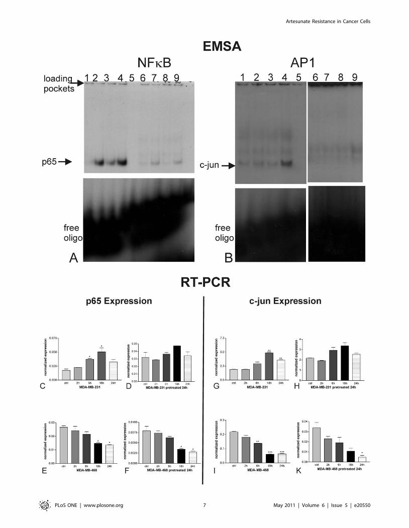
Activation of NFkB and AP-1 (EMSA) by artesunateIn order to unravel the molecular mechanism of acquired
resistance in MDA-MB-231 cells, we analyzed the effect of
artesunate on the transcription factors NFkB and AP-1. In case of
nuclear activation of these factors, apoptosis is down-regulated due
to over-expression of cell survival related genes and repression of
pro-apoptotic genes.
Nuclear extracts from artesunate-treated and carrier-treated
cells were subjected to electrophoretic mobility shift assays
(EMSA), where the binding of transcription factors is revealed
by the retarded electrophoretic migration of radioactively labeled
oligonucleotides of the specific binding sequence. Binding of the
transcription factors NFkB and AP-1, which are both associated
with resistance to standard anti-cancer drugs, was monitored
(Fig. 4A and B). Artesunate treatment for 2, 4, and 6 h of MDA-
MB-231 cells (lanes 2, 3, and 4, respectively) showed a clear
induction in the specific bands for the NFkB transcription factor
subunit p65 (Fig. 4A) and the AP-1 subunit c-jun (Fig. 4B) in
comparison to carrier-treated cells (lanes 1). The specificity of the
binding was assessed by addition of a 50-fold excess of cold
oligonucleotides that abolished the band shifts observed (lanes 5).
Furthermore, the bands disappeared upon addition of appropriate
Figure 2. Detection of nucleosomes in cytoplasmic fractions(Cell Death ELISA, Roche Applied Biosystems) of MDA-MB-231and MDA-MB-468 cells treated for 24 h with various concen-trations of artesunate in comparison to the cells pre-treatedfor 24 h with 25 mM artesunate. While MDA-MB-468 cells undergoapoptosis upon artesunate treatment no matter if cells are pretreatedfor 24 h with artesunate (indicated with %) or not (indicated with #),MDA-MB-231 cells acquired resistance to artesunate treatment and donot undergo apoptosis (indicated with & or m respectively) whenpretreated for 24 h with artesunate.doi:10.1371/journal.pone.0020550.g002
Artesunate Resistance in Cancer Cells
PLoS ONE | www.plosone.org 4 May 2011 | Volume 6 | Issue 5 | e20550
Artesunate Resistance in Cancer Cells
PLoS ONE | www.plosone.org 5 May 2011 | Volume 6 | Issue 5 | e20550

specific antibodies (supershift) against p65 or c-jun respectively.
The interference of the antibodies with the binding of the proteins
(transcription factors) to the labelled probes resulted in the
formation of very faint or rather diffuse double bands (lanes
6–9). Addition of an unrelated mutant oligonucleotide had no
effect on NFkB binding (data not shown). Experiments were
repeated at least three times.
Expression of the NFkB subunit p65 (Fig. 4C–F) and the AP-1
subunit c-jun (Fig. 4G–K) were analyzed by quantitative RT-PCR.
mRNA expression of p65 was induced in MDA-MB-231 cells
upon treatment throughout 24 h, whereby the peak of induction
was reached after 18 h (* P,0.05; one way Anova with
Bonferroni’s post test). After pre-treatment of MDA-MB-231 cells
with ART for 24 h, no statistically significant changes in p65
expression could be observed. In non-resistant MDA-MB-468
cells p65 expression is down-regulated statistically significantly
(* P,0.05; one way Anova with Bonferroni’s post test) after 18 h
and 24 h upon ART treatment no matter whether the cells were
pretreated with ART or not. The effect of acquired resistance as
seen for p65 expression could also be found concerning the
expression of c-jun. While c-jun was statistically significantly
(** P,0.01; one way Anova with Bonferroni’s post test) up-
regulated in MDA-MB-231 cells after 18 h and 24 h, no
significant change could be observed for cells pretreated 24 h
with ART. Again MDA-MB-468 cells responded always to ART
treatment with down-regulation of c-jun expression, which was
statistically significant at least after 24 h no matter if the cells were
pretreated with ART or not.
Our results show that human metastatic breast cancer cells
MDA-MB-231 acquired resistance to ART treatment and thereby
expression of p65 and c-jun do not alter upon further treatment
with the substance.
Acquired resistance becomes also clear by comparing the
percentage of cells undergoing early and late apotosis upon further
ART treatment in pretreated or non pretreated breast cancer cells
(Table 1). While MDA-MB-468 cells have a significant increase in
early (6.18%) and late apoptosis (14.63%), MDA-MB-231 cells
showed only a minimal increase in early apoptosis (2.29%) and no
increase in late apoptosis 24 h after additional ART treatment.
Bcl-2 and bax are involved in the development ofresistance
As recent evidence accumulates that the ratio between bcl-2 and
bax is of special interest for the induction of apoptosis in cancer
cells, we examined the expression of these two members of the
bcl-2-family, in order to evaluate their role in the development of
resistance against artesunate.
Treatment of the human metastatic breast cancer cell line
MDA-MB-231 with 20 mM artesunate, led to induction of bcl-2
expression already after 2 h (** P,0.01). Up-regulation of this
anti-apoptotic factor reached a level of about two fold after 18 h
with statistical significance of * P,0.05 in comparison to carrier-
treated control cells (Fig. 5A). Pretreatment with ART rendered
MDA-MB-231 cells resistant to this compound and hence bcl-2
expression could not be induced in these cells (Fig. 5B). In contrast,
pretreatment with ART did not lead to resistance in MDA-MB-
468 cells. Consequently, bcl-2 expression was diminished in these
cells already after 2 h with further decline up to 24 h, no matter
whether cells were pretreated (Fig. 5C) or not (Fig. 5D).
Expression of the pro-apoptotic factor bax was repressed in
MDA-MB-231 cells upon artesunate treatment. This anti-
apoptotic effect became statistically significant after 6 h with
ongoing decline up to 24 h with a significance of *** P,0.001 in
comparison to carrier-treated cells (Fig. 5E). As already seen for
bcl-2 expression, pretreatment with ART rendered MDA-MB-231
cells resistant and, therefore, bax expression could not be inhibited
by ART in these cells (Fig. 5F). On the other hand, expression of
bax in MDA-MB-468, which did not acquire resistance against
ART, could be induced by the compound, already after 2 h with
further significant increase up to 24 h, no matter whether cells
were pretreated (Fig. 5H) or not (Fig. 5G).
Apoptosis results from the balance of pro- and anti-apoptotic
members of the bcl-2 family. Therefore, we calculated the ratio of
the pro-apoptotic bax and the anti-apoptotic bcl-2 as indicator for
induction or repression of apoptosis by artesunate. High ratios
indicate cellular proficiency to induce apoptosis, while low ratios
may occur in more apoptosis-resistant cells. As shown in Figure 5
(I), the bax/bcl-2 ratio was high in non-pretreated MDA-MB-231
cells and decreased over time, indicating that the cells acquired
resistance to artesunate-induced apoptosis. In contrast, the bax/
bcl-2 ratio was low in pretreated cells and did not increase up to
18 h and only a little bit after 24 h. this may indicate that the cells
remained apoptosis-resistant towards artesunate. The strongly
increasing bax/bcl-2 ratios in both pretreated and non-pretreated
MDA-MB-468 cells indicate that artesunate pretreatment did not
result in apoptosis-resistance towards artesunate.
The correlation between the induction of the transcription
factors NFkB and the regulation of the apoptotis-related genes
bcl-2 and bax was confirmed by promoter analysis. Both genes
share all of the transcription binding factor sites identified by high
stringency analysis of the promoter region from 25000 to +2000
(ELK4,ELF5, SPIB, NFkB, ZNF354C, SP1, ELK1, MZF1_1–4,
MZF 5–13, Bapx1) indicating a common regulation. bax had a
bona fide NFkB in position 2363 (sequence: GGGCCTGCCC)
and bcl-2 had two of binding sites in position 2641 and +139
(sequences: GGCAATTTAC), relative to the transcription start
site (Table 2).
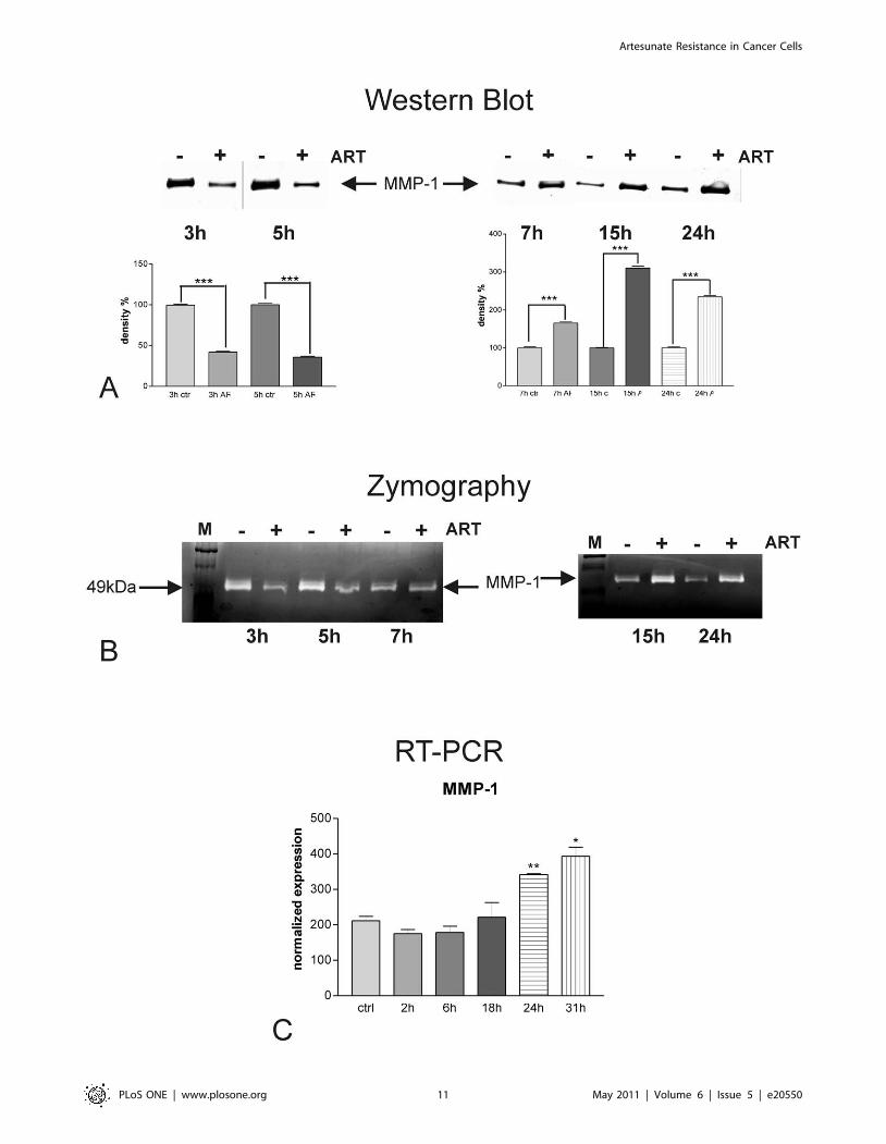
Effect of artesunate on expression and activity of MMP-1in resistant breast cancer cells
We analyzed the amount of the metastasis-related protease
MMP-1 secreted into the cell supernatants of artesunate treated
human breast cancer cells MDA-MB-231 by Western blots with
specific antibodies against MMP-1 (Fig. 6A). Thereby, accumu-
lation of newly released MMP-1 secreted into fresh serum-free
medium added at the beginning of the time course was monitored
in the presence (+) or in the absence (2) of 20 mM artesunate. We
found that MMP-1 protein was released into the supernatants of
MDA-MB-231 cell cultures. For shorter time periods (3 and 5 h,
left upper panel), the amount of MMP-1 protein secreted into the
Figure 3. Flow cytometry analysis of early and late apoptosis in non-resistant MDA-MB-468 and resistant MDA-MB-231 cells.Evidence of early apoptosis in terms of translocation of phospholipid phosphatidylserine from the inner to the outer leaflet of the plasma membrane,where it becomes accessible to annexin V staining, can be seen in quadrants 4 and 2. Cells reaching a late apoptotic propidium iodide positive statecan be seen in quadrant 1. MDA-MB-468 cells treated with ART for 24 h, showed clear evidence of early and late apoptosis, no matter if they werepretreated for 24 h with ART. The two arrows indicate the shift from the lower left quarter to the upper right and upper left quarters. In contrast ARTtreatment induced early and late apoptosis only in non pretreated MDA-MB-231 cells, whereas pretreated MDA-MB-231 cells did not further undergoearly or late apoptosis as illustrated by the two circles and the arrow in the lower panel of the figure.doi:10.1371/journal.pone.0020550.g003
Artesunate Resistance in Cancer Cells
PLoS ONE | www.plosone.org 6 May 2011 | Volume 6 | Issue 5 | e20550
Artesunate Resistance in Cancer Cells
PLoS ONE | www.plosone.org 7 May 2011 | Volume 6 | Issue 5 | e20550
media of treated cells was lower compared to those of non-treated
cells. After 7 h of artesunate treatment, the effect switched and
supernatants of treated cells had elevated MMP-1 concentrations
in comparison to non-treated breast cancer cells. Up-regulation
of MMP-1 secretion was observed for up to 24 h artesunate
treatment (7, 15 or 24 h, upper right panel). All experiments have
been performed in triplicates. According to determination of
protein concentrations by Pierce assay equal amounts of secreted
protein were subjected to SDS-PAGE, and as loading control, the
amount of protein blotted onto the membranes was visualized with
Ponceau red before blocking (data not shown). Densitometric
analysis of the bands (Fig. 6A, lower panels) revealed that the
down-regulation of MMP-1 protein secretion in response to
artesunate treatment, seen for early time points (3 and 5 h; Fig. 6A,
lower left panel), was about 60–65%. Up-regulation of MMP-1
secretion was about 1.6-fold after 7 h, 3 fold after 15 h and 2.3-
fold after 24 h of artesunate treatment (Fig. 6A, lower right panel).
The statistical power for all observed regulation events was
P,0.001 throughout the whole experiments (one way Anova with
Bonferroni’s post test).
In order to visualize the enzymatic activities present in cell
culture supernatants, we performed zymography analyses on
casein-containing gels (Fig. 6B). As MMP-1 is able to degrade
casein, electrophoretically separated proteases, present in cell
culture supernatants, were detected as translucent bands on the
Coomassie brillant blue stained substrate background.
MDA-MB-231 breast cancer cells responded to the artesunate
treatment (lanes labeled with ‘‘+’’) with a transient (3 and 5 h, left
panel) reduction of MMP-1 activity released into fresh serum-free
medium in comparison to carrier-treated cells (lanes labeled with
‘‘2’’). In contrast, cells treated for longer periods (15 h and 24 h)
with artesunate (+) showed a clear induction of MMP-1 as
depicted here in Fig. 6A, right panel. Interestingly, treatment with
artesunate for 7 h did not result in any differences in MMP-1
secretion into the culture medium of MDA-MB-231 cells when
compared to carrier-treated cells (2). MMP-1 was identified
according to its migration behavior as compared to a molecular
mass standard (lanes labeled with M).
In order to monitor the effect of artesunate on the level of
MMP-1 transcription, we performed quantitative Real Time RT-
PCR (Fig. 6C) and normalized expression values on those
obtained for the housekeeping genes RPII and HPRT. In
accordance with the results from Western blots and zymography,
shown in the sections before, artesunate treatment of MDA-MB-
231 cells resulted in slightly diminished levels of MMP-1 mRNA
after 2 and 6 h. However, after 18 h treatment the reduction of
MMP-1 mRNA expression by artesunate faded out and treatment
with 20 mM of the drug for longer time periods led to statistically
significant inductions of MMP-1 of 1.6- and 1.9-fold after 24 and
31 h respectively. The levels of statistical significance were P,0.01
at 24 h and P,0.05 at 31 h (student’s t-test).
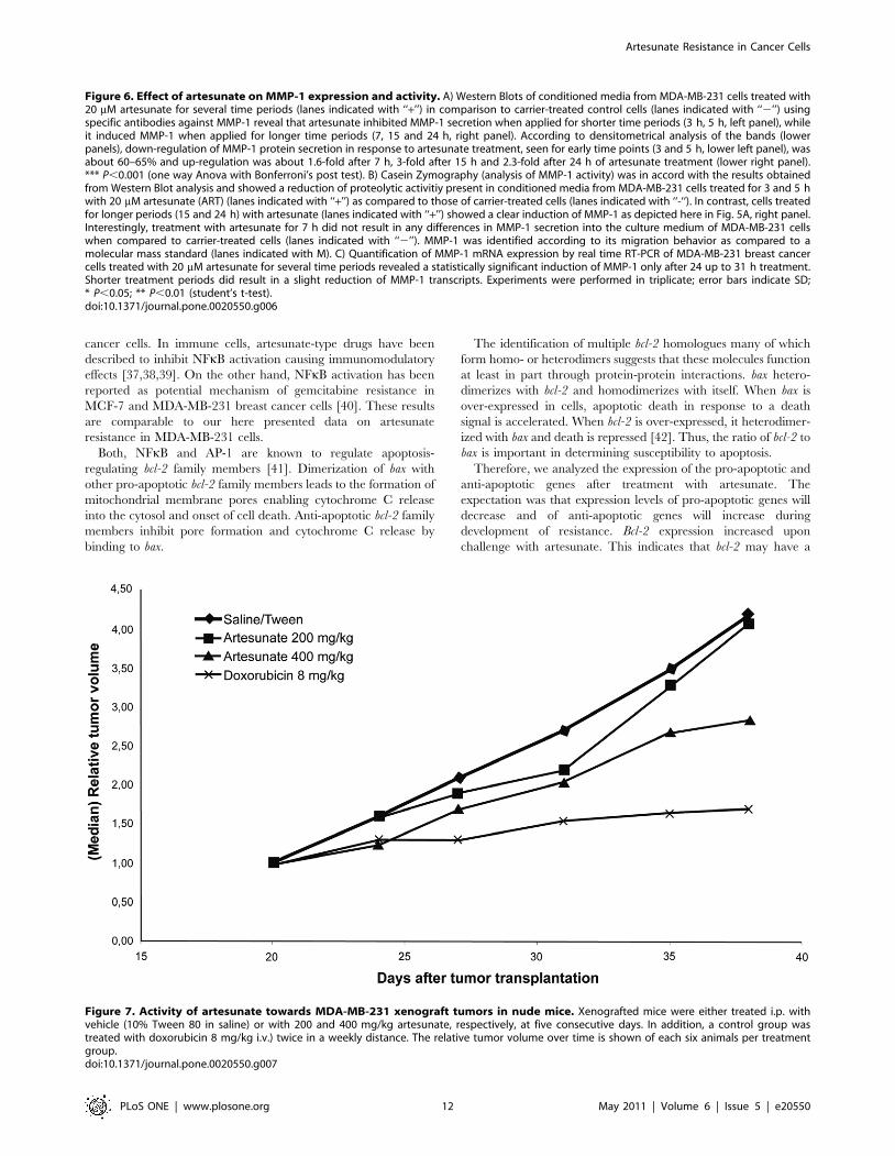
Activity of artesunate towards MDA-MB-231 xenografttumors in nude mice
The anti-tumor activity of artesunate was examined in vivo in
MDA-MB-231 xenografted tumors in nude mice. The results are
shown in Fig. 7. While treatment with 200 mg/kg artesunate did
not result in inhibition of tumor growth as compared to vehicle-
treated tumors, a marginal inhibition was observed with 400 mg/
kg artesunate. By contrast, a considerable inhibition of tumor
growth was reached using a control drug, doxorubicin. We did not
observe any signs of toxicity of artesunate (loss of weight etc.).
Discussion
In the present investigation, we showed that sub-lethal doses of
artesunate resulted in the development of resistance towards this
drug. While inherent differences of cancer cell lines in response
towards artesunate and modulation of inherent resistance by
ferrous iron were previously described [4,27], the present study is
to the best of our knowledge the first to show an acquired
resistance phenomenon towards artesunate in cancer cells. The
Figure 4. Effects of artesunate on NFkB (A) and AP-1 (B). Binding of 32P-labeled NFkB and AP-1 specific oligonucleotides to the cognatetranscription factor present in nuclear extracts of MDA-MB-231 cells was monitored by EMSA. Artesunate treatment of MDA-MB-231 cells for differenttime intervals (lane 2, 2 h; lane 3, 4 h; lane 4, 6 h) led to a induced binding of NFkB (A) and AP-1 (B) to its response element as compared to carrier-treated cells (lane 1). The specificity of the binding was assessed for all three oligos by addition of a 506molar excess of cold oligonucleotides (lanes5). Furthermore, addition of appropriate specific antibodies(supershift) against p65 or c-jun respectively resulted in the formation of very faint orrather diffuse double bands (lanes 6–9). Experiments were repeated at least three times. Fig. 4 C-K illustrates the expression of p65 and c-jun in MDA-MB-231 and -468 cells. mRNA expression of p65 was induced in MDA-MB-231 cells upon artesunate treatment only in non-pretreated cells (C), whilep65 expression did not alter upon artesunate treatment in pre-treated cells (D). MDA-MB-468 cells p65 expression was down-regulated statisticallysignificantly after 18 h and 24 h upon artesunate treatment, no matter whether the cells were pretreated with artesunate (F) or not (E). The effect ofacquired resistance as seen for p65 expression could also be found concerning the expression of c-jun. While c-jun was statistically significantlyup-regulated in MDA-MB-231 cells after 18 h and 24 h, no significant change could be observed for cells pretreated 24 h with artesunate. (* P,0.05;** P,0.01; *** P,0.001; one way Anova with Bonferroni’s post test). Experiments were performed in triplicates.doi:10.1371/journal.pone.0020550.g004
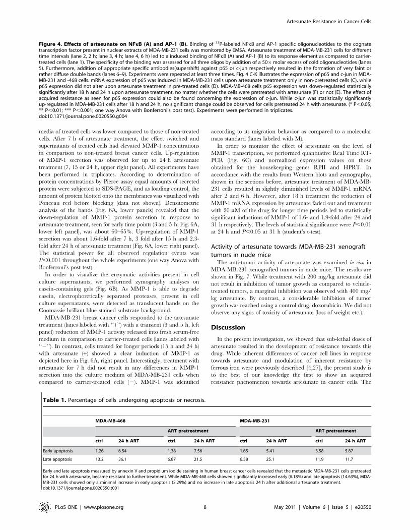
Table 1. Percentage of cells undergoing apoptosis or necrosis.
MDA-MB-468 MDA-MB-231
ART pretreatment ART pretreatment
ctrl 24 h ART ctrl 24 h ART ctrl 24 h ART ctrl 24 h ART
Early apoptosis 1.26 6.54 1.38 7.56 1.65 5.41 3.58 5.87
Late apoptosis 13.2 36.1 6.87 21.5 6.58 25.1 11.9 11.7
Early and late apoptosis measured by annexin V and propidium iodide staining in human breast cancer cells revealed that the metastatic MDA-MB-231 cells pretreatedfor 24 h with artesunate, became resistant to further treatment. While MDA-MB-468 cells showed significantly increased early (6.18%) and late apoptosis (14.63%), MDA-MB-231 cells showed only a minimal increase in early apoptosis (2.29%) and no increase in late apoptosis 24 h after additional artesunate treatment.doi:10.1371/journal.pone.0020550.t001
Artesunate Resistance in Cancer Cells
PLoS ONE | www.plosone.org 8 May 2011 | Volume 6 | Issue 5 | e20550

Artesunate Resistance in Cancer Cells
PLoS ONE | www.plosone.org 9 May 2011 | Volume 6 | Issue 5 | e20550

induced resistance observed in vitro corresponds to the unrespon-
siveness at lower artesunate doses (200 mg/kg) and only minor
responsiveness after treatment with high artesunate doses
(400 mg/kg) after repeated injection of the compound to nude
mice as shown here.
Recently, it was reported that artemisinin treatment can confer
resistance to other anti-cancer drugs such as doxorubicin [28].
Resistance towards artesunate-type drugs occurs only infrequently
in malaria parasites, but has been reported both in patient samples
and under in vitro conditions [12,13,14]. The role of several
plasmodial proteins, including multidrug resistance transporters
(pfmdr1), sarcoendoplasmatic Ca2+-dependent ATPase (SERCA),
translationally controlled tumor protein (TCTP) and others have
been discussed.
Here, we present evidence that MDA-MB-231 human breast
cancer cells reveal reduced sensitivity after repeated artesunate
treatment. Likewise diminished cytotoxicity was seen in pre-
treated highly metastatic MDA-MB-231 cells in contrast to less
tumorigenic non-metastatic MDA-MB-468 breast cancer cells.
This observation implicates that the drug could be beneficially
applied as therapy in less advanced breast cancer. Additionally
apoptosis was down-regulated in metastatic breast cancer cells pre-
treated with the drug for 24 h.
Analysis of molecular mechanisms underlying the acquired
resistance against ART in the breast cancer cells showed that this
was due to activation of the transcription factors, NFkB and AP-1.
NFkB represents a central player for many cellular processes such
as proliferation, adhesion, angiogenesis, inflammation and others.
It mediated apoptosis resistance towards various stimuli, including
anti-cancer agents and is activated by DNA damage [29].
NF-kB is a resistance factor for established anticancer drugs by
inhibiting apoptosis [30,31]. Recently, we have shown that
artesunate induces double stand breaks [32]. In non-activated
cells, NFkB is complexed with IkB in the cytosol. Upon
stimulation by appropriate stimuli, the complex is dissociated
and NFkB translocates into the nucleus, where it binds to specific
promoters of target genes, e.g. survival-related genes, and
stimulates their transcriptional activation.
The AP-1 complex consists of c-Fos, FosB, Fra-1, or Fra-2, each
of which can dimerize with c-Jun, JunB, or JunD. This complex
binds to specific binding motifs in the promoter of target genes
regulating, apoptosis, proliferation, or differentiation. A role for
AP-1 for resistance to anti-cancer drugs has also been proposed
[33]. In a previous investigation, we found that AP-1 (together
with Sp1) was activated in host cells upon infection with human
cytomegalovirus and that a single artesunate treatment suppressed
AP-1 activation and production of virus-specific proteins in host
cells [34]. AP-1 acts as transcription factor for anticancer drug
resistance genes such as P-glycoprotein/MDR1 or glutathione S-
transferase-pi. Glutathione S-transferases detoxify harmful xeno-
biotic molecules by binding them to glutathione. Then, glutathi-
one-drug conjugates are transported out of cells. The expression of
glutathione S-transferases was also associated with cellular
response of tumor cells to artesunate [35,36].
Here, we observed that repeated treatment of highly metastatic
breast cancer cells induced AP-1 activation leading to artesunate
resistance. Whether a similar phenomenon can also be found in
anti-viral therapy with artesunate is unknown yet.
Similar to AP-1, we found an activation of NFkB associated
with development of artesunate resistance in MDA-MB-231
Figure 5. Expression of apoptosis-related genes as determined by RT-PCR. (A–D) Treatment with 20 mM leads to induction of bcl-2expression in MDA-MB-231 cells after 2 h (** P,0.01) and reaches a level of about two fold after 18 h with statistical significance of * P,0.05 incomparison to carrier-treated control cells (A). Pretreatment with artesunate rendered MDA-MB-231 cells resistant to this compound and, hence, bcl-2expression could not be induced in these cells (B). In contrast, pretreatment with artesunate did not lead to resistance in MDA-MB-468 cells.Consequently, bcl-2 expression was statistically significantly diminished in these cells already after 6 h with further decline up to 24 h, no matter ifcells were pretreated (C) or not (D). (E–H) Expression of the pro-apoptotic factor bax was repressed in MDA-MB-231 cells upon treatment between 6and 24 h(E). As already seen for bcl-2 expression, pretreatment with artesunate rendered MDA-MB-231 cells resistant and, therefore, bax expressioncould not be inhibited by artesunate in these cells (F). On the other hand, expression of bax in MDA-MB-468, which did not acquire resistance againstartesunate, could be induced by the compound, already after 2 h with further significant increase up to 24 h, no matter whether cells werepretreated or not. Experiments were performed in triplicates (* P,0.05; ** P,0.01; *** P,0.001; one way Anova with Bonferroni’s post test). (I) Sinceapoptosis results from the balance of pro- and anti-apoptotic members of the bcl-2 family, we calculated the expression ratios of the pro-apoptoticbax and the anti-apoptotic bcl-2 as indicator for induction or repression of apoptosis by artesunate. High ratios indicate cellular proficiency to induceapoptosis, while low ratios may occur in more apoptosis-resistant cells.doi:10.1371/journal.pone.0020550.g005
Table 2. Analysis of NFkB binding sites in the apoptosis genes.
Promoter Analysis
Gene ENSEMBL ID Chr.Transcription factorbinding site
Position relative totranscription start Orientation Z-Score
BAX ENSG00000087088 19 GGGCCTGCCC 354 21 7.01e+00
BCL2 ENSG00000171791 18 GGGACTTCCA 3745 21 9.80e+00
GGGACTTCCA 2966 21 9.80e+00
GGCAATTTAC 650 1 7.04e+00
GGCAATTTAC 2130 1 7.04e+00
The promoter regions of BAX and BCL2 from 25000 to +2000 relative to the transcription start site were analyzed for the occurrence of NFkB binding sites using theOpossum web-service. The identity of the sequence used for analysis (ENSEMBL ID), the chromosome number (Chr.), the actual transcription factor binding site, theposition relative to the transcription start site and the orientation are indicated. The Z-score uses the normal approximation to the binomial distribution to compare therate of occurrence of a TFBS in the target set of genes to the expected rate estimated from the pre-computed background set. The likelihood that the number of TFBSnucleotides detected for the included target genes was significant as compared with the number of TFBS nucleotides detected for the background set. Z-score wasexpressed in units of magnitude of the standard deviation.doi:10.1371/journal.pone.0020550.t002
Artesunate Resistance in Cancer Cells
PLoS ONE | www.plosone.org 10 May 2011 | Volume 6 | Issue 5 | e20550
Artesunate Resistance in Cancer Cells
PLoS ONE | www.plosone.org 11 May 2011 | Volume 6 | Issue 5 | e20550
cancer cells. In immune cells, artesunate-type drugs have been
described to inhibit NFkB activation causing immunomodulatory
effects [37,38,39]. On the other hand, NFkB activation has been
reported as potential mechanism of gemcitabine resistance in
MCF-7 and MDA-MB-231 breast cancer cells [40]. These results
are comparable to our here presented data on artesunate
resistance in MDA-MB-231 cells.
Both, NFkB and AP-1 are known to regulate apoptosis-
regulating bcl-2 family members [41]. Dimerization of bax with
other pro-apoptotic bcl-2 family members leads to the formation of
mitochondrial membrane pores enabling cytochrome C release
into the cytosol and onset of cell death. Anti-apoptotic bcl-2 family
members inhibit pore formation and cytochrome C release by
binding to bax.
The identification of multiple bcl-2 homologues many of which
form homo- or heterodimers suggests that these molecules function
at least in part through protein-protein interactions. bax hetero-
dimerizes with bcl-2 and homodimerizes with itself. When bax is
over-expressed in cells, apoptotic death in response to a death
signal is accelerated. When bcl-2 is over-expressed, it heterodimer-
ized with bax and death is repressed [42]. Thus, the ratio of bcl-2 to
bax is important in determining susceptibility to apoptosis.
Therefore, we analyzed the expression of the pro-apoptotic and
anti-apoptotic genes after treatment with artesunate. The
expectation was that expression levels of pro-apoptotic genes will
decrease and of anti-apoptotic genes will increase during
development of resistance. Bcl-2 expression increased upon
challenge with artesunate. This indicates that bcl-2 may have a
Figure 7. Activity of artesunate towards MDA-MB-231 xenograft tumors in nude mice. Xenografted mice were either treated i.p. withvehicle (10% Tween 80 in saline) or with 200 and 400 mg/kg artesunate, respectively, at five consecutive days. In addition, a control group wastreated with doxorubicin 8 mg/kg i.v.) twice in a weekly distance. The relative tumor volume over time is shown of each six animals per treatmentgroup.doi:10.1371/journal.pone.0020550.g007
Figure 6. Effect of artesunate on MMP-1 expression and activity. A) Western Blots of conditioned media from MDA-MB-231 cells treated with20 mM artesunate for several time periods (lanes indicated with ‘‘+’’) in comparison to carrier-treated control cells (lanes indicated with ‘‘2’’) usingspecific antibodies against MMP-1 reveal that artesunate inhibited MMP-1 secretion when applied for shorter time periods (3 h, 5 h, left panel), whileit induced MMP-1 when applied for longer time periods (7, 15 and 24 h, right panel). According to densitometrical analysis of the bands (lowerpanels), down-regulation of MMP-1 protein secretion in response to artesunate treatment, seen for early time points (3 and 5 h, lower left panel), wasabout 60–65% and up-regulation was about 1.6-fold after 7 h, 3-fold after 15 h and 2.3-fold after 24 h of artesunate treatment (lower right panel).*** P,0.001 (one way Anova with Bonferroni’s post test). B) Casein Zymography (analysis of MMP-1 activity) was in accord with the results obtainedfrom Western Blot analysis and showed a reduction of proteolytic activitiy present in conditioned media from MDA-MB-231 cells treated for 3 and 5 hwith 20 mM artesunate (ART) (lanes indicated with ‘‘+’’) as compared to those of carrier-treated cells (lanes indicated with ‘‘-‘‘). In contrast, cells treatedfor longer periods (15 and 24 h) with artesunate (lanes indicated with ‘‘+’’) showed a clear induction of MMP-1 as depicted here in Fig. 5A, right panel.Interestingly, treatment with artesunate for 7 h did not result in any differences in MMP-1 secretion into the culture medium of MDA-MB-231 cellswhen compared to carrier-treated cells (lanes indicated with ‘‘2’’). MMP-1 was identified according to its migration behavior as compared to amolecular mass standard (lanes indicated with M). C) Quantification of MMP-1 mRNA expression by real time RT-PCR of MDA-MB-231 breast cancercells treated with 20 mM artesunate for several time periods revealed a statistically significant induction of MMP-1 only after 24 up to 31 h treatment.Shorter treatment periods did result in a slight reduction of MMP-1 transcripts. Experiments were performed in triplicate; error bars indicate SD;* P,0.05; ** P,0.01 (student’s t-test).doi:10.1371/journal.pone.0020550.g006
Artesunate Resistance in Cancer Cells
PLoS ONE | www.plosone.org 12 May 2011 | Volume 6 | Issue 5 | e20550

specific function for induction of artesunate resistance. Among the
pro-apoptotic genes, bax expression decreased after artesunate
treatment suggesting that down-regulation of bax participated in
development of artesunate resistance. The stable expression of bax
at 18 and 24 h may be explained by a rheostat of anti-and
pro-apoptotic bcl-2 family members, of which bax is only one
pro-apoptotic factor. It cannot be excluded that other pro-
apoptotic members are also down-regulated supporting the
function of bax. Functional analysis of apoptosis by detection of
nucleosomes in cytoplasmic fractions of MDA-MB-231 cells
treated with various concentrations of artesunate in comparison
to MDA-MB-231 cells pre-treated with artesunate revealed that
artesunate induced apoptosis only in cells that have not been pre-
treated. These results strongly suggest that MDA-MB-231 cells
acquire resistance against cell death. Bcl-2 is up-regulated and bax
is down-regulated upon treatment with artesunate, though
transiently. However, both share similar TF-binding sites,
especially sites for NFkB. The differential regulation can therefore
not simply be explained by enhanced NFkB activity following
ART treatment. bax is also under control of p53 which might be
induced by artesunate induced double strand breaks. MDA-MB
231 cells have a mutation in exon 5 of the p53 gene (www-p53.
iarc.fr) leading to an almost non functional transcriptional activity
conserving, however, some activity on the bax promoter [43]. It is
conceivable that NFkB has an immediate survival effect mediated
by bcl-2 with a potential feedback via bax, with the latter being
dependent on other factors such as p53. Moreover, bcl-2 has two
bona fide binding sites and bax only one; kinetics or the strength of
induction might therefore vary between the two genes. It should
however be taken into account that NFkB affects other signaling
routes (e.g. JNK phosphorylation) as well to regulate drug-induced
apoptosis [44].
The human metastatic breast cancer cells MDA-MB-231 are a
well-known model for studying tumor aggressiveness, invasion and
metastasis. Matrix metalloproteinases (MMP), especially MMP-1
that is able to degrade fibrillar native collagen type I, are involved
in the metastatic process, and also in the inhibition of apoptosis
and drug resistance [45,46]. Likewise it has been shown that
multidrug resistant cell lines produced more MMP-1, -2 and -9
compared to their analogous non resistant cells [47].
Therefore, we also analyzed the effect of artesunate on MMP
expression and observed that for short treatment periods
artesunate transiently down-regulates MMP-1 on the levels of
expression, secretion and enzymatic activity in metastatic MDA-
MB-231. At later time points, when the cells have already acquired
resistance, MMP-1 expression, secretion and activity are induced
upon artesunate treatment. The induction of MMP-1 is well in line
with the activation of the tumor progression-associated transcrip-
tion factors NFkB and AP-1, which are induced upon artesunate
treatment as evidenced here by EMSA analyses. It has been
reported that both NFkB and AP-1 binding sites are present in the
promoter region of MMP-1 [48,49,50] and enhanced production
of MMP-1 is associated with a more aggressive tumor growth, a
higher metastatic potential, and poor clinical outcome of
malignant tumors [51,52,53].
In conclusion, treatment of MDA-MB-231 cells with artesunate
results in development of resistance, which was associated by
NFkB and AP-1-mediated apoptosis resistance with up-regulation
of the anti-apoptotic bcl-2 and down-regulations of the pro-
apoptotic bax genes. The development of resistance towards
artesunate may have important implications for the application of
this drug in cancer chemotherapy.
Supporting Information
Table S1 Primer Sequences.
(DOC)
Author Contributions
Conceived and designed the experiments: BB UP IF TE. Performed the
experiments: BB IF PHK EK UP. Analyzed the data: BB IF UP TE.
Contributed reagents/materials/analysis tools: BB IF UP TE. Wrote the
paper: BB TE.
References
1. Jemal A, Siegel R, Ward E, Murray T, Xu J, et al. (2007) Cancer statistics, 2007.CA Cancer J Clin 57: 43–66.
2. Efferth T, Dunstan H, Sauerbrey A, Miyachi H, Chitambar CR (2001) The
anti-malarial artesunate is also active against cancer. Int J Oncol 18: 767–773.
3. Efferth T, Rucker G, Falkenberg M, Manns D, Olbrich A, et al. (1996)
Detection of apoptosis in KG-1a leukemic cells treated with investigationaldrugs. Arzneimittelforschung 46: 196–200.
4. Efferth T, Sauerbrey A, Olbrich A, Gebhart E, Rauch P, et al. (2003) Molecular
modes of action of artesunate in tumor cell lines. Mol Pharmacol 64:382–394.
5. Sertel S, Eichhorn T, Sieber S, Sauer A, Weiss J, et al. Factors determining
sensitivity or resistance of tumor cell lines towards artesunate. Chem BiolInteract 185: 42–52.
6. Dell’Eva R, Pfeffer U, Vene R, Anfosso L, Forlani A, et al. (2004) Inhibition of
angiogenesis in vivo and growth of Kaposi’s sarcoma xenograft tumors by theanti-malarial artesunate. Biochem Pharmacol 68: 2359–2366.
7. Hou J, Wang D, Zhang R, Wang H (2008) Experimental therapy of hepatoma
with artemisinin and its derivatives: in vitro and in vivo activity, chemosensitiza-tion, and mechanisms of action. Clin Cancer Res 14: 5519–5530.
8. Chen H, Sun B, Pan S, Jiang H, Sun X (2009) Dihydroartemisinin inhibits
growth of pancreatic cancer cells in vitro and in vivo. Anticancer Drugs 20:131–140.
9. Du JH, Zhang HD, Ma ZJ, Ji KM Artesunate induces oncosis-like cell death invitro and has antitumor activity against pancreatic cancer xenografts in vivo.
Cancer Chemother Pharmacol 65: 895–902.
10. Berger TG, Dieckmann D, Efferth T, Schultz ES, Funk JO, et al. (2005)Artesunate in the treatment of metastatic uveal melanoma–first experiences.
Oncol Rep 14: 1599–1603.
11. Zhang ZY, Yu SQ, Miao LY, Huang XY, Zhang XP, et al. (2008) [Artesunatecombined with vinorelbine plus cisplatin in treatment of advanced non-small cell
lung cancer: a randomized controlled trial]. Zhong Xi Yi Jie He Xue Bao 6:
134–138.
12. Meshnick SR (2002) Artemisinin: mechanisms of action, resistance and toxicity.Int J Parasitol 32: 1655–1660.
13. Duraisingh MT, Cowman AF (2005) Contribution of the pfmdr1 gene to
antimalarial drug-resistance. Acta Trop 94: 181–190.
14. Afonso A, Hunt P, Cheesman S, Alves AC, Cunha CV, et al. (2006) Malaria
parasites can develop stable resistance to artemisinin but lack mutations incandidate genes atp6 (encoding the sarcoplasmic and endoplasmic reticulum
Ca2+ ATPase), tctp, mdr1, and cg10. Antimicrob Agents Chemother 50:
480–489.
15. Cailleau R, Olive M, Cruciger QV (1978) Long-term human breast carcinoma
cell lines of metastatic origin: preliminary characterization. In Vitro 14:
911–915.
16. Cailleau R, Young R, Olive M, Reeves WJ, Jr. (1974) Breast tumor cell lines
from pleural effusions. J Natl Cancer Inst 53: 661–674.
17. Zhang RD, Fidler IJ, Price JE (1991) Relative malignant potential of humanbreast carcinoma cell lines established from pleural effusions and a brain
metastasis. Invasion Metastasis 11: 204–215.
18. Rozen S, Skaletsky H (2000) Primer3 on the WWW for general users and forbiologist programmers. Methods Mol Biol 132: 365–386.
19. Pfaffl MW, Tichopad A, Prgomet C, Neuvians TP (2004) Determination of
stable housekeeping genes, differentially regulated target genes and sampleintegrity: BestKeeper–Excel-based tool using pair-wise correlations. Biotechnol
Lett 26: 509–515.
20. Muller PY, Janovjak H, Miserez AR, Dobbie Z (2002) Processing of gene
expression data generated by quantitative real-time RT-PCR. Biotechniques 32:
1372–1379.
21. Bachmeier BE, Albini A, Vene R, Benelli R, Noonan D, et al. (2005) Cell
density-dependent regulation of matrix metalloproteinase and TIMP expression
in differently tumorigenic breast cancer cell lines. Exp Cell Res 305: 83–98.
22. Bachmeier B, Nerlich AG, Iancu CM, Cilli M, Schleicher E, et al. (2007) The
chemopreventive polyphenol Curcumin prevents hematogenous breast cancer
metastases in immunodeficient mice. Cell Physiol Biochem 19: 137–152.
Artesunate Resistance in Cancer Cells
PLoS ONE | www.plosone.org 13 May 2011 | Volume 6 | Issue 5 | e20550
23. Bachmeier BE, Vene R, Iancu CM, Pfeffer U, Mayer B, et al. (2005)
Transcriptional control of cell density dependent regulation of matrixmetalloproteinase and TIMP expression in breast cancer cell lines. Thromb
Haemost 93: 761–769.
24. Bachmeier BE, Nerlich AG, Boukamp P, Lichtinghagen R, Tschesche H, et al.(2000) Human keratinocyte cell lines differ in the expression of the collagenolytic
matrix metalloproteinases-1,-8, and -13 and of TIMP-1. Biol Chem 381:509–516.
25. Ho Sui SJ, Mortimer JR, Arenillas DJ, Brumm J, Walsh CJ, et al. (2005)
oPOSSUM: identification of over-represented transcription factor binding sitesin co-expressed genes. Nucleic Acids Res 33: 3154–3164.
26. Becker M, Sommer A, Kratzschmar JR, Seidel H, Pohlenz HD, et al. (2005)Distinct gene expression patterns in a tamoxifen-sensitive human mammary
carcinoma xenograft and its tamoxifen-resistant subline MaCa 3366/TAM. MolCancer Ther 4: 151–168.
27. Kelter G, Steinbach D, Konkimalla VB, Tahara T, Taketani S, et al. (2007)
Role of transferrin receptor and the ABC transporters ABCB6 and ABCB7 forresistance and differentiation of tumor cells towards artesunate. PLoS ONE 2:
e798.28. Riganti C, Doublier S, Viarisio D, Miraglia E, Pescarmona G, et al. (2009)
Artemisinin induces doxorubicin resistance in human colon cancer cells via
calcium-dependent activation of HIF-1a and P-glycoprotein overexpression.Br J Pharmacol 156: 1054–1066.
29. Russell JS, Raju U, Gumin GJ, Lang FF, Wilson DR, et al. (2002) Inhibition ofradiation-induced nuclear factor-kappaB activation by an anti-Ras single-chain
antibody fragment: lack of involvement in radiosensitization. Cancer Res 62:2318–2326.
30. Arlt A, Schafer H (2002) NFkB-dependent chemoresistance in solid tumors.
Int J Clin Pharmacol Ther 40: 336–347.31. Katsman A, Umezawa K, Bonavida B (2009) Chemosensitization and
immunosensitization of resistant cancer cells to apoptosis and inhibition ofmetastasis by the specific NFkB inhibitor DHMEQ. Curr Pharm Des 15:
792–808.
32. Li PC, Lam E, Roos WP, Zdzienicka MZ, Kaina B, et al. (2008) Artesunatederived from traditional Chinese medicine induces DNA damage and repair.
Cancer Res 68: 4347–4351.33. Efferth T, Marschall M, Wang X, Huong SM, Hauber I, et al. (2002) Antiviral
activity of artesunate towards wild-type, recombinant, and ganciclovir-resistanthuman cytomegaloviruses. J Mol Med 80: 233–242.
34. el-Deiry WS (1997) Role of oncogenes in resistance and killing by cancer
therapeutic agents. Curr Opin Oncol 9: 79–87.35. Efferth T, Oesch F (2004) Oxidative stress response of tumor cells: microarray-
based comparison between artemisinins and anthracyclines. Biochem Pharmacol68: 3–10.
36. Efferth T, Volm M (2005) Glutathione-related enzymes contribute to resistance
of tumor cells and low toxicity in normal organs to artesunate. In Vivo 19:225–232.
37. Aldieri E, Atragene D, Bergandi L, Riganti C, Costamagna C, et al. (2003)Artemisinin inhibits inducible nitric oxide synthase and nuclear factor NFkB
activation. FEBS Lett 552: 141–144.
38. Li WD, Dong YJ, Tu YY, Lin ZB (2006) Dihydroarteannuin ameliorates lupus
symptom of BXSB mice by inhibiting production of TNF-a and blocking the
signaling pathway NFkB translocation. Int Immunopharmacol 6: 1243–1250.
39. Xu H, He Y, Yang X, Liang L, Zhan Z, et al. (2007) Anti-malarial agent
artesunate inhibits TNF-a-induced production of proinflammatory cytokines via
inhibition of NFkB and PI3 kinase/Akt signal pathway in human rheumatoid
arthritis fibroblast-like synoviocytes. Rheumatology (Oxford) 46: 920–926.
40. Hernandez-Vargas H, Rodriguez-Pinilla SM, Julian-Tendero M, Sanchez-
Rovira P, Cuevas C, et al. (2007) Gene expression profiling of breast cancer cells
in response to gemcitabine: NFkB pathway activation as a potential mechanism
of resistance. Breast Cancer Res Treat 102: 157–172.
41. Sevilla L, Zaldumbide A, Pognonec P, Boulukos KE (2001) Transcriptional
regulation of the bcl-x gene encoding the anti-apoptotic Bcl-xL protein by Ets,
Rel/NFkB, STAT and AP1 transcription factor families. Histol Histopathol 16:
595–601.
42. Oltvai ZN, Milliman CL, Korsmeyer SJ (1993) Bcl-2 heterodimerizes in vivo
with a conserved homolog, Bax, that accelerates programmed cell death. Cell
74: 609–619.
43. Kato S, Han SY, Liu W, Otsuka K, Shibata H, et al. (2003) Understanding the
function-structure and function-mutation relationships of p53 tumor suppressor
protein by high-resolution missense mutation analysis. Proc Natl Acad Sci U S A
100: 8424–8429.
44. Perona R, Sanchez-Perez I (2007) Signalling pathways involved in clinical
responses to chemotherapy. Clin Transl Oncol 9: 625–633.
45. Stetler-Stevenson WG, Yu AE (2001) Proteases in invasion: matrix metallopro-
teinases. Semin Cancer Biol 11: 143–152.
46. Minn AJ, Gupta GP, Siegel PM, Bos PD, Shu W, et al. (2005) Genes that
mediate breast cancer metastasis to lung. Nature 436: 518–524.
47. Yang JM, Xu Z, Wu H, Zhu H, Wu X, et al. (2003) Overexpression of
extracellular matrix metalloproteinase inducer in multidrug resistant cancer
cells. Mol Cancer Res 1: 420–427.
48. Westermarck J, Kahari VM (1999) Regulation of matrix metalloproteinase
expression in tumor invasion. FASEB J 13: 781–792.
49. Bond M, Chase AJ, Baker AH, Newby AC (2001) Inhibition of transcription
factor NFkB reduces matrix metalloproteinase-1, -3 and -9 production by
vascular smooth muscle cells. Cardiovasc Res 50: 556–565.
50. Benbow U, Brinckerhoff CE (1997) The AP-1 site and MMP gene regulation:
what is all the fuss about? Matrix Biol 15: 519–526.
51. Airola K, Karonen T, Vaalamo M, Lehti K, Lohi J, et al. (1999) Expression of
collagenases-1 and -3 and their inhibitors TIMP-1 and -3 correlates with the
level of invasion in malignant melanomas. Br J Cancer 80: 733–743.
52. Murray GI, Duncan ME, O’Neil P, McKay JA, Melvin WT, et al. (1998) Matrix
metalloproteinase-1 is associated with poor prognosis in oesophageal cancer.
J Pathol 185: 256–261.
53. Murray GI, Duncan ME, O’Neil P, Melvin WT, Fothergill JE (1996) Matrix
metalloproteinase-1 is associated with poor prognosis in colorectal cancer. Nat
Med 2: 461–462.
Artesunate Resistance in Cancer Cells
PLoS ONE | www.plosone.org 14 May 2011 | Volume 6 | Issue 5 | e20550