Embed Size (px)
Citation preview
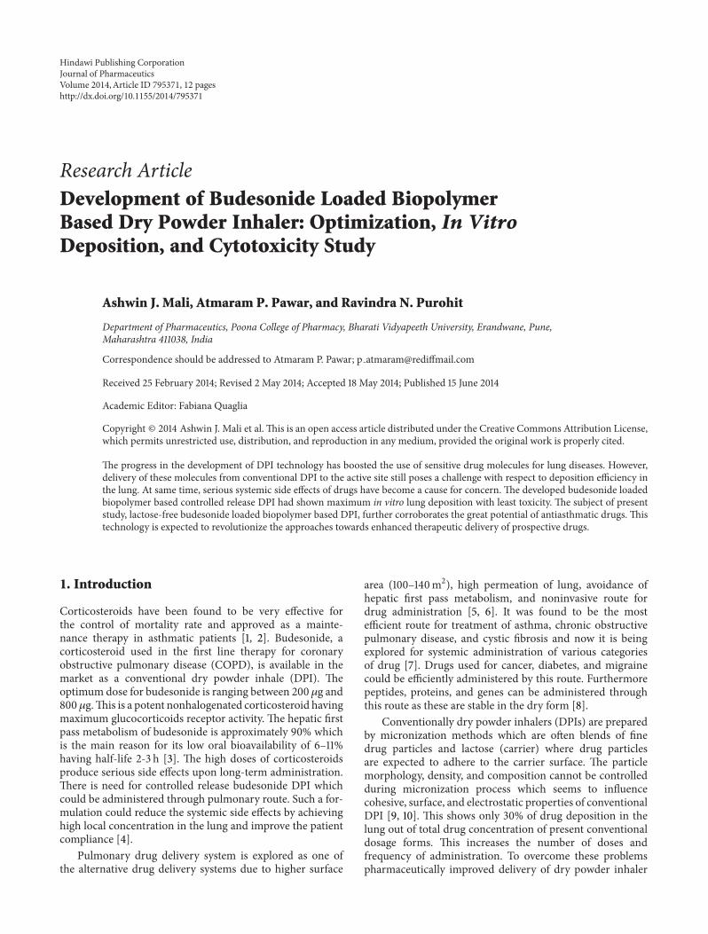
Research ArticleDevelopment of Budesonide Loaded BiopolymerBased Dry Powder Inhaler: Optimization, In VitroDeposition, and Cytotoxicity Study
Ashwin J. Mali, Atmaram P. Pawar, and Ravindra N. Purohit
Department of Pharmaceutics, Poona College of Pharmacy, Bharati Vidyapeeth University, Erandwane, Pune,Maharashtra 411038, India
Correspondence should be addressed to Atmaram P. Pawar; p [email protected]
Received 25 February 2014; Revised 2 May 2014; Accepted 18 May 2014; Published 15 June 2014
Academic Editor: Fabiana Quaglia
Copyright © 2014 Ashwin J. Mali et al. This is an open access article distributed under the Creative Commons Attribution License,which permits unrestricted use, distribution, and reproduction in any medium, provided the original work is properly cited.
The progress in the development of DPI technology has boosted the use of sensitive drug molecules for lung diseases. However,delivery of these molecules from conventional DPI to the active site still poses a challenge with respect to deposition efficiency inthe lung. At same time, serious systemic side effects of drugs have become a cause for concern. The developed budesonide loadedbiopolymer based controlled release DPI had shown maximum in vitro lung deposition with least toxicity. The subject of presentstudy, lactose-free budesonide loaded biopolymer based DPI, further corroborates the great potential of antiasthmatic drugs. Thistechnology is expected to revolutionize the approaches towards enhanced therapeutic delivery of prospective drugs.
1. Introduction
Corticosteroids have been found to be very effective forthe control of mortality rate and approved as a mainte-nance therapy in asthmatic patients [1, 2]. Budesonide, acorticosteroid used in the first line therapy for coronaryobstructive pulmonary disease (COPD), is available in themarket as a conventional dry powder inhale (DPI). Theoptimum dose for budesonide is ranging between 200 𝜇g and800 𝜇g.This is a potent nonhalogenated corticosteroid havingmaximum glucocorticoids receptor activity. The hepatic firstpass metabolism of budesonide is approximately 90% whichis the main reason for its low oral bioavailability of 6–11%having half-life 2-3 h [3]. The high doses of corticosteroidsproduce serious side effects upon long-term administration.There is need for controlled release budesonide DPI whichcould be administered through pulmonary route. Such a for-mulation could reduce the systemic side effects by achievinghigh local concentration in the lung and improve the patientcompliance [4].
Pulmonary drug delivery system is explored as one ofthe alternative drug delivery systems due to higher surface
area (100–140m2), high permeation of lung, avoidance ofhepatic first pass metabolism, and noninvasive route fordrug administration [5, 6]. It was found to be the mostefficient route for treatment of asthma, chronic obstructivepulmonary disease, and cystic fibrosis and now it is beingexplored for systemic administration of various categoriesof drug [7]. Drugs used for cancer, diabetes, and migrainecould be efficiently administered by this route. Furthermorepeptides, proteins, and genes can be administered throughthis route as these are stable in the dry form [8].
Conventionally dry powder inhalers (DPIs) are preparedby micronization methods which are often blends of finedrug particles and lactose (carrier) where drug particlesare expected to adhere to the carrier surface. The particlemorphology, density, and composition cannot be controlledduring micronization process which seems to influencecohesive, surface, and electrostatic properties of conventionalDPI [9, 10]. This shows only 30% of drug deposition in thelung out of total drug concentration of present conventionaldosage forms. This increases the number of doses andfrequency of administration. To overcome these problemspharmaceutically improved delivery of dry powder inhaler
Hindawi Publishing CorporationJournal of PharmaceuticsVolume 2014, Article ID 795371, 12 pageshttp://dx.doi.org/10.1155/2014/795371

2 Journal of Pharmaceutics
formulation should be achieved for efficacious drug deliveryto achieve local effects in the asthma and COPD whichprominently comprises the larger airways region of the lung.Moreover, most of the DPI formulations rely on lactosemonohydrate as a carrier where lactose has major drawbackssuch as presence of transmissible spongiform encephalopathyand endotoxins obtained from bovine source. Also it cannotbe used in the compounds with the reducing sugar such asproteins, peptides, budesonide, and formoterol [11].
Thedesired performance of dry powder inhaler (DPI)wasindicated by its fine particle fraction (FPF) and emitted dose(ED) which in turn mainly depends upon the particle massmedian aerodynamic diameter (MMAD). To achieve max-imum deposition in the lung, particles exhibiting MMADranging from 1 to 5 𝜇m were required [12–14].
Most of the research work and patents came out with var-ious novel systems to achieve required range of MMAD thatincludes nanoparticles, microspheres, solid-lipid nanoparti-cles, liposomes, and porous particles. Particle penetration anddeposition in the lungs depend on the aerodynamic behaviorof particle which changes the particle velocity and direction.Thus particle trajectories depend upon particle dynamicswhich were governed by the particle density, size, shape,surface nature, and charge of particles [15, 16].
Hickey et al. observed that the static, bulk, and solid stateproperty of lactose in DPI was responsible for better aero-dynamic behavior and respiratory deposition [17]. Recently,Divey et al. achieved good lung deposition forDPI containingelectrostatically driven hybrid nanoparticles [18]. Telko et al.also studied the effect of triboelectrification on the cohesiveand noncohesive types of DPI [19].
Sodium alginate and chitosan, two naturally occur-ring polymers, were used widely in the formulation devel-opment due to their unique properties such as bio-compatibility, biodegradability, and forms complexationwith polyelectrolyte ions (CaCl
2) which were attractive to
many researchers to formulate carriers like nanoparticlesand microparticles with controlled drug release [20–25].Pluronic F-68 is an amphiphilic synthetic polymer con-taining hydrophilic poly(ethylene oxide) (PEO) blocks andhydrophobic poly(propylene oxide) (PPO) blocks arrangedin triblock structure which has unique property in theencapsulation of drug moiety in the delivery system [7].
Notably, there were no pulmonary formulations presentin the market prepared by biopolymer based controlled drugrelease which would be beneficial to produce local effect withreduced systemic side effect and overcome lung depositionobstacles with improved local inhalation therapy. To furtheradvance the therapeutic utility of budesonide, the presentinvestigation deals with development of hydrophobic budes-onide loaded biopolymer (sodium alginate, chitosan, andpluronic F-68) based controlled release microparticulate drypowder inhaler (DPI) with desired physical characteristicsand aerosolization in order to improve aerodynamic behaviorand lung deposition. The microparticles were prepared bycontrolled pregelation of sodium alginate solution containingpluronic F-68 followed by polycationic (chitosan) cross-linking technique and 32 factorial design adapted to optimize
the amount of chitosan and calcium chloride [26]. Theformulations were lyophilized using mannitol as a cryopro-tectant to get stable formulations and evaluated in termsof respirable fraction using twin stage impinger (TSI) andpowder properties.The optimized formulation was subjectedto mass median aerodynamic diameter and fine particlefraction along with static properties on particle dynamicsand fluidization evaluation using Andersen cascade impactor(ACI) in comparison with commercial DPI. Further, in vitrocell viability against alveolar epithelial cancer cell line A549was studied to prove the safety of formulation.
2. Experiment
2.1. Materials and Method. Budesonide was obtained fromLupin Ltd., Pune, India. Sodium alginate (medium viscosity,3500 cps) and dialysis bag with a 12,000 molecular weightcutoff was purchased from Sigma Aldrich Chemicals PrivateLtd. (Bangalore, India). Budesonide dry powder inhala-tion commercial product was purchased from local market.Deacetylated chitosan (deacetylation degree 37.08, molecu-lar weight 50 kDa) was obtained from Marine Chemicals,Cochin, India. Pluronic F-68 was provided by Cipla Pharma-ceuticals (Mumbai, India). Acetone, potassium dihydrogenphosphate, sodium dihydrogen phosphate, calcium chloride,and all the solvents used in the study were obtained fromMerck Ltd. (Mumbai, India).
2.2. Fabrication of Budesonide DPI. Budesonide DPI wasprepared by the principle involving cation induced controlledgelification of alginate, method reported by Rajaonarivony etal., with slight modifications [27]. Acetone solution (10mL)of drug (25mg) and pluronic F-68 (100mg) was added tothe sodium alginate (0.063% w/v) solution under magneticstirring at 250 rpm, to which optimized 4mL of calciumchloride (10mM) solutionwas added dropwise for 15min fol-lowed by 1mL of optimized chitosan (2mg) solution added;stirring was continued for 24 hours until evaporation oforganic solvent was completed. The obtained microparticlessuspensionwas subjected to lyophilized usingmannitol (2.5%w/v) as a cryoprotectant to get budesonide DPI.
2.3. Experimental Design. Process parameters were opti-mized based on the preliminary data by applying the 32factorial designs for formulated DPI. The response surfacesof the obtained results were plotted. The coded values arelisted in Table 1.The obtained data was analyzed by the resultsobserved from the multiple regression analysis using DesignExpert 8.0.6.1 software (Stat-Ease Inc., USA).
The following equation was obtained:
𝑌 = 𝛽0+ 𝛽1𝑋1+ 𝛽2𝑋2+ 𝛽11𝑋1𝑋1+ 𝛽22𝑋2𝑋2+ 𝛽12𝑋1𝑋2,
(1)
where 𝑌 is the measured response, 𝑋 is the levels of factors,𝛽 is the regression coefficient, and 𝑋
1and 𝑋
2indicate the
amount of calcium chloride and chitosan.

Journal of Pharmaceutics 3
Table 1: Factorial design of all formulations.
Formulation Factorial design Calciumchloride (mL)
Chitosan(mg)
F1 (−1, −1) 1 2F2 (−1, 0) 1 3F3 (−1, +1) 1 4F4 (0, −1) 2 2F5 (0, 0) 2 3F6 (0, +1) 2 4F7 (+1, −1) 3 2F8 (+1, −1) 3 3F9 (+1, +1) 3 4
2.4. Characterization of Budesonide DPI
2.4.1. Particle Size Analysis of Suspension and LyophilizedFormulation. The mean particle size was determined bylaser diffraction technique using Malvern 2000 SM (MalvernInstruments, Malvern, UK) which allows sample measure-ment in the range of 0.05–20,000𝜇m. Analysis was carriedout at room temperature keeping angle of detection 90∘. Themean particle size was expressed in terms of D (0.9), thatis, size of the 90% of the particle. The data presented aremean values of three independent samples produced underidentical production conditions.
The particle size of the prepared lyophilized formulationwas checked by laser diffraction technique using Malvern2000 SM (Malvern Instruments, Malvern, UK) with the helpof dry assembly.
2.4.2. Entrapment Efficiency Analysis. The amount of drugentrapped in the formulations was calculated by estimat-ing the amount of unentrapped drug by centrifugation at25,000 rpm for 30min.The obtained supernatant was assayedspectrophotometrically at 246 nm for free drug content. Inthis process the percent entrapment efficiency (EE)was calcu-lated as the percentage of drug entrapped in the final dosageform to its initial concentration. The %EE was calculatedusing
%EE = ( (Total drug concentration
−Drug concentration in supernatant)
× (Initial drug concentration)−1) × 100.
(2)
2.4.3. Flow Properties of Formulated DPI. The fixed heightcone method was used to check the flow property of theformulations and commercial DPI. A glass funnel with 5mminternal diameter was fixed at height of 2.5 cm over the flatsurface. The gentle flowing of the powder through the funnelwas carried out.The diameter of the powder cone formedwas
measured.The angle of reposewas calculated by the followingequation:
Tan 𝜃 =heightradius. (3)
The tapped and untapped densities were evaluated using asmall graduated tube with a defined volume size into whichthe known weight of the powders was added. Bulk densityis determined by dividing the mass of the powder by thevolume. Tapped volume is calculated by using a tap densitytester (Electrolab, tap density tester, USP) following 100 taps.Tapped density is determined by dividing themass of powderby volume. Carr’s index (Ci) is calculated using the values ofbulk and tapped density:
Ci =(tapped density − bulk density)
tapped density× 100. (4)
Hausner ratio defines the flowability of powder mixture. Thevalue indicates the ratio of bulk and tapped density:
Hausner ratio =bulk densitytapped density
. (5)
Carr’s index, Hausner’s ratio, and percentage porosity arethe tools used to quantify flow properties of powders. TheHausner ratio is <1.25 and Carr’s index is in the range of 5–15%.
Percent porosity (𝜀) is used to determine compressibilityof powder which is the degree of volume reduction due toan applied pressure which is the measurement of porositychanges during compaction and is calculated using thefollowing formula:
𝐸 = 1 − (PbPt) × 100, (6)
where Pb and Pt are bulk and taped density of DPI.
2.4.4. In Vitro Deposition Study Using Twin Stage Impinger.Rotahaler was used as the delivery device for determi-nations using twin stage impinger (TSI), Andersen cas-cade impactor (ACI), and dosage unit sampling apparatus(DUSA). The obtained 25mg of powder equivalent to 200𝜇gbudesonide was encapsulated in hydroxyl propryl methylcellulose (HPMC) stick-free capsule = 3. Initially respirablefraction of optimized budesonide and commercial DPI wasdetermined by TSI (Model number WP-SSGI-0289, WestechInstruments, UK) after aerosolization at 60±5 L/min for 5 secwith 7mL and 30mL of phosphate buffer saline (PBS pH 7.4)in stages 1 and 2 of the impinger, respectively. Each stage wasrinsed with PBS and drug content was determined by theUV spectrophotometry method after appropriate dilution.Rotahaler with filled capsule to be tested was placed intoa rubber mouthpiece attached to the throat of the TSI andthe pump was switched on. The pump was operated soas to get the flow rate of 60 ± 5 L/min. The capsule wasreleased by operating the inhalation device and the pumpwas allowed to run for another 5 seconds which allowed the

4 Journal of Pharmaceutics
aspiration of 5 L of air in the apparatus, as recommended bythe European Pharmacopoeia (2000). Each section (inhaler,capsule shell, stages 1 and 2) was rinsed with PBS pH 7.4.The rinsed buffer was collected and diluted to an appropriatevolume. The budesonide content was determined by UVspectrophotometer at 246 nm (Jasco-v-530).The formulationhaving the highest respirable fraction was chosen for furtherdeposition studies using an ACI [28–30].
2.4.5. Zeta Potential Analysis. The finalised DPI formulationwas checked for the charge assessment. The zeta potentialof the formulated DPI was measured by the laser Dopplerelectrophoretic mobility measurement using Zetasizer 300HSA (Malvern Instruments Ltd., UK) at temperature of 25∘C.
2.4.6. Transmission Electron Microscopy. Transmission elec-tron micrograph (TEM) was obtained for budesonide DPIusing a JEOL 1200 EXII TEM. Initially, carbon-coated gridswere floated on a droplet of the formulation on a flexible plas-tic film (Parafilm) to permit the adsorption of the particlesonto the grid. After that, the grid was blotted with a filterpaper and air-dried for 1 h. Obtained data was used to analyzethe size and morphological data of formulated DPI.
2.4.7. Scanning Electron Microscopy. Crystal characteristicof the final formulations was studied by scanning electronmicroscopy (SEM). Samples were mounted on the aluminumstub and coatedwith a thin gold-palladium layer byAuto FineCoater (JEOL, JEC-1600, Tokyo, Japan) and analyzed witha scanning electron microscope (JEOL, JSM-6360A, Tokyo,Japan) operated at an 10 kV acceleration voltage.
2.4.8. Fourier Transform-Infrared Spectroscopy. IR spectrawere recorded from 4,000 to 400 cm−1 with a Fouriertransform-infrared spectrometer (FTIR-8400; ShimadzuCorporation, Kyoto, Japan) equipped with a diffusereflectance accessory (DRS-8000; Shimadzu Corporation,Japan) and a data station to confirm drug entrapment in thepolymer. About 2-3mg samples were prepared by processingcompressed KBR discs.
2.4.9. Differential Scanning Calorimetry. The differentialscanning calorimetry (DSC) thermograms of formulatedDPIwere obtained using DSC 821e (Mettler-Toledo, Greifensee,Switzerland). Indium standards were used to calibrate thetemperature and enthalpy scale. Samples were (5–10mg)heated in hermetically sealed aluminium pan with a heatingrate of 10∘C/min over a range of 0–300∘C under a nitrogenatmosphere (flow rate 50mL/min).
2.4.10. Powder X-Ray Diffraction. Powder X-ray diffraction(PXRD) patterns of particles were recorded by X-ray diffrac-tometer (PW 1729; Philips, Almelo, The Netherlands) usingCu K𝛼 radiation (1.542A) with a voltage of 30 kV and acurrent of 30mA. Samples were scanned from 10∘ to 30∘ at2𝜃.
2.4.11. Release Profiles. The in vitro release for budesonideloaded biopolymer based DPI was carried out in phosphatebuffer saline (pH 7.4) using dialysis bag diffusion tech-nique. Formulation equivalent to 200𝜇g of budesonide wasadded into the dialysis bag (cellulose membrane, mw cutoff12,000Da), whichwas hermetically sealed and immersed into100mL of release medium. The entire system was kept at37±0.5
∘Cwith continuousmagnetic stirring at 100 rpm/min.At selected time interval, sample was removed and replacedwith fresh medium in order to maintain sink conditions.Thesample was analyzed by UV spectrophotometry at 246 nm.
2.4.12. In Vitro Deposition Study Using ACI. An aerodynamiccharacteristic of optimized budesonideDPI havingminimumparticle size, maximum entrapment efficiency, and excellentflow properties was assessed and compared with the com-mercial DPI (Budecort Rotacpas) by using an eight-stage,nonviable cascade impactor (Westech private instruments,Model Number WP-ACISS-0289). The obtained 25mg ofpowder equivalent to 200𝜇g budesonide was encapsulated inhydroxyl propryl methyl cellulose (HPMC) stick-free capsule= 3. Rotahaler was used as delivery device. The capsule to be
tested was placed in the Rotahaler, which had been fitted intomoulded rubber mouthpiece attached to the throat piece ofthe impactor. Once assembly had been checked and foundto be vertical and stable, run was conducted at a flow rateof 60 L/min for 5 sec. The capsule shell was removed fromthe inhaler device and four more capsules were actuated inthe samemanner.The test was conducted in triplicate. Cutoffparticle aerodynamic diameters at 60 L/min for each stage ofthe impactor were preseparator (8.6 𝜇m), stage 0 (6.5 𝜇m),stage 1 (4.4𝜇m), stage 2 (3.3 𝜇m), stage 3 (2.0 𝜇m), stage 4(1.1 𝜇m), stage 5 (0.54 𝜇m), and stage 6 (0.25 𝜇m). After thecompletion of dosing, different plates were collected; theywere washed with 10mL of acetonitrile: phosphate buffersaline (pH 3.2). The dispersion was sonicated in a bath-typesonicator for 15min. Then the solution was centrifuged at25,000 rpm for 30min and the amount of budesonide inthe supernatant was determined using a high performanceliquid chromatography (HPLC) assay method. The deposi-tion of formulated and commercial DPI on each stage of theimpactor was determined. MMAD and GSD were calculatedfrom the deposition data using the MMAD calculator forAnderson apparatus [13, 31, 32].
The HPLC system specifications were as follows: pump,PU-1580 (JASCO, Japan); injector, Autosampler (AS-1555; JASCO); column, Phenomenex C18, 250 × 4.6mm,5 𝜇m (Thermo Electron Corporation, USA); and detector,UV/visible (UV-1575; JASCO). Data acquisition and analysiswere carried out using Borwin/HSS 2000 software (LG 1580-04; JASCO). The mobile phase was a mixture of acetonitrile:phosphate buffer saline pH 3.2 (34 : 66 v/v). The columntemperature and flow rate were 40∘C and 1.5mL/min and thewavelength was 240 nm.
2.5. Cell Viability Assay. In vitro cell viability was evaluatedfor formulated budesonide DPI against alveolar epithelial

Journal of Pharmaceutics 5
cancer cell line A549 (obtained from NCCS, Pune, Maha-rashtra, India) using MTT assay. The results were comparedwith free budesonide and formulation excipients. The cellswere cultured in DMEM/F12 medium and supplementedwith 10% v/v fetal bovine serum and 2mM L-glutamine.The medium maintained humidity atmosphere less than 5%carbon dioxide at 37∘C. Trypsin-EDTA solution was used forsubculturing and cell isolation.
The cells were harvested on the fourth day of subculture.The cells were seeded at the density of 5 × 10³ cells per welland grown in 96-well tissue culture plates in a final volume of150 𝜇L in humidified atmosphere for 48 hours. Each formula-tion was dispersed in water and tested in varying budesonideconcentration over the range of 15𝜇M to 1000 𝜇M. After24 hr of incubation, 10 𝜇L ofMTT labeling agent (5mg/mL inPBS) was added and incubated for further 4 h in humidifiedcondition. After incubation, 100𝜇L of solubilizing solution(10% SDS in 0.01M HCl) was added to each well. The platewas incubated overnight. The optical density was measuredat 570 nm with a reference wavelength at 630 nm using anELISA reader.The cell viabilitywas calculated using followingequation:
Viability (%) =𝐴 test𝐴control× 100, (7)
where𝐴 test is the absorbance of the test solutions and𝐴controlis the absorbance of control (PBS).
3. Result and Discussion
Budesonide is a potent corticosteroid used in the firstline therapy for coronary obstructive pulmonary diseases(COPD). The low oral bioavailability of budesonide dueto hepatic metabolism and short half-life continues to behighlighted as a major challenge in developing formulationsfor clinical efficacy. However, budesonide is available in themarket as a conventional dry powder inhaler (DPI) whichshows only 30% of drug deposition in the lung out of totaldrug concentration. Besides, high/frequent dose is neededto achieve optimum therapeutic efficacy, which often causessevere side effects.
In present study we fabricated budesonide loadedbiopolymer carriers based DPI via controlled gelation ofsodium alginate where calcium ions react with guluronicacid units of the sodium alginate to form the negativecharged calcium alginate polyelectrolyte complex in whichdrug molecules were entrapped followed by enveloping withchitosan in order to overcome commercial DPI problems.In preliminary study, the amount of calcium chloride andchitosan showed pronounced effect on biopolymer DPI [21,32]. To investigate the effect of independent variables such ascalcium chloride (𝑋
1) and chitosan (𝑋
2) on the dependent
variables such as particle size (𝑌1), entrapment efficiency (𝑌
2),
bulk density (𝑌3), and Carr’s index (𝑌
4) which are major
contributing factors for the lung deposition were optimizedby using 32 factorial design [26, 28].
3.1. Characterization of Budesonide Loaded Biopolymer BasedDPI
3.1.1. Particle Size. Significant particle size variations wereobserved with different concentration of calcium chlorideand chitosan. The particle size distribution for formulationsF1 to F9 showed values in the range of 1.192 ± 0.03 𝜇m to3.424 ± 0.04 𝜇m as listed in Table 2. For the commercial DPIparticle size was 1.521 ± 0.04 𝜇m. The multiple regressionanalysis for themean particle size of factorial batches revealedthe fair fit (𝑅2 = 0.460). The positive coefficient for bothindependent variables influencing the size of the particle wasgiven by the following equation:
𝑌1= 7.368 + 3.513𝑋
1+ 1.136𝑋
2+ 0.346𝑋
1𝑋1
− 0.257𝑋2𝑋2+ 0.152𝑋
1𝑋2.
(8)
As per the 32 factorial design surface response graph(Figure 1(a)) and polynomial equation (2), the concentrationof calcium chloride (𝑋
1) was found to influence change in
the particle size. The calcium ions react with glucuronic acidmolecules present in sodium alginate, leading to formation ofcompact polyelectrolyte crosslinked structures.The increasedconcentration of calcium chloride results in gelation andcrosslinking of the biopolymer which was responsible forincrease in particle size. Similarly, the chitosan showed thesame response as that of calcium chloride in the particle size.The particle size was increased with increasing chitosan (𝑋
2)
concentration which may be due to interaction of cationicchitosan polymer with sodium alginate and formation ofthick layer coating of excessive chitosan around the particles[21, 28, 33, 34].
3.1.2. Entrapment Efficiency. The effect of independent vari-ables𝑋
1and𝑋
2on the percent entrapment efficiency of drug
for all the formulations was observed. EE was in the rangeof 80.68 ± 2.68% to 92.64 ± 2.12% as listed in Table 2. Themultiple regression analysis for the EE as per the factorialdesigns revealed the good fit (𝑅2 = 0.943) with the followingequation:
𝑌2= 60.018 + 5.323𝑋
1+ 5.303𝑋
2− 0.0362𝑋
1𝑋1
+ 0.116𝑋2𝑋2− 0.884𝑋
1𝑋2.
(9)
As per the 32 factorial design response surface graph(Figure 1(b)) and polynomial equation (3), EE was mainlygoverned by concentration of calcium chloride (𝑋
1) which
results in lower entrapment in the initial formulations dueto weak gel strength and increased entrapment as theconcentration of calcium chloride was increased [21]. Theformulated DPI showed less entrapment due to “calciumsaturation phenomenon” as compared to F8 and F9 [35]. Thehigher concentration of chitosan (𝑋
2) was also responsible
for increasing the EE of drug as it has a film forming propertyencapsulating the inner core of the particle [33, 36]. Theuse of triblock polymer showed positive effect in case of EEwhichmight be due to its self-assembling property in aqueousenvironment with hydrophobic core and intercalation ofhydrophilic chain with alginate chitosan complex [37].

6 Journal of Pharmaceutics
Table 2: Characterization of in vitro deposition of formulations by TSI.
Formulation number 𝐷[0.9] [𝜇m]a Entrapmentefficiency [%]a
Recovereddose [𝜇g]a
Fine particledose [𝜇g]a
Respirable fraction[𝜇g]a
F1 1.761 ± 0.05 80.68 ± 2.68 123.80 ± 0.04 52.15 ± 0.02 42.12 ± 0.02F2 1.192 ± 0.03 86.43 ± 1.15 116.28 ± 0.02 48.84 ± 0.01 42.00 ± 0.03F3 2.147 ± 0.03 90.92 ± 2.21 112.15 ± 0.03 44.87 ± 0.04 40.00 ± 0.01F4 3.204 ± 0.01 85.94 ± 2.12 110.6 ± 0.06 44.24 ± 0.06 40.00 ± 0.05F5 1.926 ± 0.03 86.66 ± 1.25 130.28 ± 0.04 51.81 ± 0.03 39.76 ± 0.02F6 3.424 ± 0.04 92.64 ± 2.12 129.41 ± 0.03 47.51 ± 0.02 36.71 ± 0.04F7 1.937 ± 0.06 87.16 ± 1.11 139.41 ± 0.03 60.09 ± 0.01 43.10 ± 0.02F8 1.537 ± 0.08 91.39 ± 1.98 97.86 ± 0.01 29.75 ± 0.02 30.40 ± 0.03F9 3.218 ± 0.09 92.20 ± 2.25 66.06 ± 0.04 30.03 ± 0.03 37.50 ± 0.01Commercial DPI — — 48.31 ± 0.03 10.82 ± 0.03 22.39 ± 0.05(+1) = higher values and (−1) = lower values.aAll the determinations performed in triplicate and values are expressed as mean (values = average ± SD).
3.5
3
2.5
2
1.51
4.00
3.50
3.00
2.50
2.00 2.913.68
4.415.14
5.88
PS
Chitosan
Calcium chloride
(a)
4.003.50
3.002.50
2.00 2.913.68
4.415.14
5.88
9492908886848280
Chitosan Calcium chloride
EE
(b)
0.140.120.10.080.060.010.02
Chitosan
Calcium chloride
Bulk
den
sity
2.91
3.68
4.41
5.14
5.88 2.002.50
3.003.50
4.00
(c)
40
30
20
10
0
ChitosanCalcium chloride
Carr
’s in
dex
4.00
3.50
3.00
2.50
2.00 2.913.68
4.415.14
5.88
(d)
Figure 1: Response surface plots of (a) particle size, (b) entrapment efficiency, (c) bulk density, and (d) Carr’s index.
3.1.3. Bulk Density and Carr’s Index of the Budesonide DPI.Bulk density of all formulations was in the range of 0.037 ±0.06 g/cm3 to 0.123 ± 0.03 g/cm3 as listed in Table 3. Themultiple regression analysis for the bulk density as per thefactorial designs revealed the good fit (𝑅2 = 0.823) with thefollowing equation:
𝑌3= 0.147 + 0.033𝑋
1+ 0.125𝑋
2+ 1.696𝑋
1𝑋1
− 9.333𝑋2𝑋2− 0.018𝑋
1𝑋2.
(10)
As per the 32 factorial design response surface graph(Figure 1(c)) and polynomial equation (4), the formulationinteraction term 𝑋
1𝑋1has positive influence on the bulk

Journal of Pharmaceutics 7
8000070000600005000040000300002000010000
0−200 −100 0 100 200
Zeta potential (mV)
Tota
l cou
nts
Figure 2: Zeta potential of formulated DPI.
density than the interaction term 𝑋1𝑋2as indicated in (10).
The calcium chloride and chitosan demonstrated positiveimpact on the density of formulations. Insignificant changesin the densities were observed with change in concentrationsof calcium chloride and chitosan. The incorporation ofmaterials like chitosan, calciumchloride, and sodiumalginatereduced the density with least variations which was helpful toimprove flow properties of the formulated DPI.
The Carr index of all the formulations was in the range of4.65 ± 0.01% to 47.88 ± 0.07% as listed in Table 3, resulting infair fit (𝑅2 = 0.629). The following equation was observed:
𝑌4= − 21.017 + 2.596𝑋
1+ 24.523𝑋
2− 2.347𝑋
1𝑋1
− 8.353𝑋2𝑋2+ 6.639𝑋
1𝑋2.
(11)
As per the 32 factorial response surface graph (Figure 1(d))and polynomial equation (5), positive influence of 𝑋
2was
seen on the flow property of formulated DPI. Chitosan maybe helpful in getting spherical particles by forming thincoat around the formulated DPI which in turn may help toincrease the flow property of formulated DPI [21]. From thepolynomial equation, response parameters such as EE anddensity showed good fit which were more significant due tocontrolled gelation of sodium alginate.
3.1.4. Flow Properties. The aerosolization efficiency of theformulated DPI was governed by the flow properties. Theangle of repose, Carr’s index, and Hausner’s ratio for F1 to F9formulations were in the range of 24 ± 0.09∘ to 28 ± 0.02∘,4.05 ± 0.01% to 47.88 ± 0.07%, and 0.52 ± 0.08 to 0.95 ± 0.08as compared to 24 ± 0.07∘, 19.48 ± 0.03%, and 0.80 ± 0.04for commercial DPI, respectively, as listed in Table 3. Thebetter angle of repose and Carr’s index were observed foroptimized budesonide DPI as compared to the commercialDPI and remaining formulations. The percentage porosity ofall the formulations ranges from 10 ± 0.05% to 48 ± 0.04% ascompared to the 20 ± 0.04% of the commercial product.
3.1.5. In Vitro Deposition Study Using Twin Stage Impinger.Theamount of drug deposited in the second stage of impinger(effective cutoff diameter <6.4 𝜇m) was considered as fineparticle dose (FPD). The recovered dose (RD) is the amountof drug present in stage 1 and stage 2 of the impinger, inhalerdevice, and capsule shell. Respirable fraction (RF) was theratio of FPD toRDandwas expressed in percentage. RF for all
the formulations ranges from 30.40±0.03% to 43.10±0.02%.As per the obtained results depicted in Table 2, FPD for all theformulations ranges from 29.75 ± 0.02 𝜇g to 60.09 ± 0.01 𝜇gand RDwas in the range of 66.06±0.03 𝜇g to 139.41±0.03 𝜇g.The respirable fraction for F7 was 43.10± 0.02% as comparedto 22.39 ± 0.05% for commercial DPI. The high FPD of F7can be attributed to the collective effect of uniform sphericalnature, lack of surface van der Waals forces, less bulk density,and good flow property of formulated DPI.
Considering the results of 32 factorial design, the F7batch showed optimum entrapment efficiency, fine particledose, respirable fraction, angle of repose, bulk density, tappeddensity, Carr’s index, Hausner’s ratio, and percentage porositywhich were subjected to further evaluation. The optimizedbatch F7 showed increased particle size (3.059 ± 0.03 𝜇m)after lyophilization which may be due to aggregation duringlyophilization process. The final composition of formulatedDPI with respect drug to powder ratio was 1 : 30mg.
3.1.6. Zeta Potential. The final formulation has shown−17.5mV of surface charge (Figure 2). This has resulted fromhigher concentration of calcium chloride than the chitosan inthe final formulation where calcium ions cooperatively bindthe alginates molecules preventing chitosan from formingthe coat around the alginate molecules. Also it may happendue to inadequate deacylation of chitosan used in the finalformulation where stretching of deacetylated chains was notfully carried out due to electrostatic repulsion between theNH3groups thatmay yield irregular and nonuniform coating
of the chitosan resulting in negative charge of the particles[21, 33, 36, 37]. The charge on the human respiratory tractis negative due to presence of mucin [38]. As per the chargetheory, negatively charged particles are more responsible forrepulsion in between the particles.Therefore, negative chargeon the respiratory tract and formulated DPI was responsiblefor more prominent repulsive forces and was responsible forincreasing the time of flight of the budesonide DPI whichleads to increasing the deposition of drug in the larger airwayregion of the lung.
3.1.7. Transmission Electron Microscopy. As observed fromthe TEM depicted in Figure 3(a) the image clearly indicatesthe presence of drug particles encapsulated in the micropar-ticles of formulated DPI. Observed particles have uniformspherical nature.
3.1.8. Scanning Electron Microscopy. The surface nature andmorphology of the formulated DPI were verified by SEMtechnique. Optimized budesonide DPI as evident from thephotograph depicted more uniform spherical particles withsmooth surface as shown in Figure 3(b). The SEM imagealso significantly specifies the uniformity of size and leastamount of fines in the formulated DPI at specific range ofmagnification.
3.1.9. Fourier Transform-Infrared Spectroscopy. Potentialintermolecular interactions between the polymers and drugswere analyzed by the FTIR spectra (Figure 4). Budesonide

8 Journal of Pharmaceutics
(a) (b)
Figure 3: (a) TEM image and (b) SEM image of formulated DPI.
Table 3: Flowability characteristics of budesonide DPI.
Formulations Angle ofreposea [𝜃]
Bulkdensitya [g/cm3]
Tappeddensitya [g/cm3]
Carr’sindexa [Ci%] Hausner ratioa Percentage
porositya
F1 26 ± 0.01 0.084 ± 0.04 0.105 ± 0.05 20.00 ± 0.04 0.80 ± 0.09 20 ± 0.04F2 26 ± 0.07 0.071 ± 0.02 0.123 ± 0.02 42.27 ± 0.02 0.57 ± 0.08 43 ± 0.07F3 24 ± 0.04 0.123 ± 0.03 0.129 ± 0.04 04.65 ± 0.01 0.95 ± 0.08 10 ± 0.05F4 27 ± 0.01 0.097 ± 0.02 0.131 ± 0.07 25.95 ± 0.03 0.74 ± 0.01 26 ± 0.02F5 25 ± 0.02 0.101 ± 0.07 0.124 ± 0.02 18.54 ± 0.05 0.81 ± 0.03 19 ± 0.01F6 24 ± 0.09 0.073 ± 0.02 0.089 ± 0.01 17.97 ± 0.03 0.80 ± 0.04 18 ± 0.08F7 25 ± 0.06 0.076 ± 0.08 0.095 ± 0.02 20.00 ± 0.02 0.80 ± 0.10 20 ± 0.03F8 28 ± 0.02 0.079 ± 0.02 0.124 ± 0.03 36.29 ± 0.03 0.63 ± 0.03 37 ± 0.06F9 26 ± 0.05 0.037 ± 0.06 0.071 ± 0.02 47.88 ± 0.07 0.52 ± 0.08 48 ± 0.04Commercial DPI 24 ± 0.07 0.124 ± 0.05 0.154 ± 0.09 19.48 ± 0.03 0.80 ± 0.04 20 ± 0.04aAll the determinations performed in triplicate and values are expressed as mean (values = average ± SD).
4000
3600
3300
3000
3000
2700
2400
2100
1800
1500
1200
900
600
400
0
(a)
(b)
(c)
(d)
(e)
Figure 4: The FTIR of (a) budesonide, (b) formulated DPI, (c)chitosan, (d) sodium alginate, and (e) pluronic F-68.
showed peaks at 3499 cm−1, 2956 cm−1, 1722 cm−1, and1690 cm−1 due to O–H stretching, C–H stretching, and C=Ostretching. The characteristic peaks of sodium alginate wereobserved at 3357 cm−1, 1601 to 1407 cm−1, and 1029 cm−1due to hydroxyl group, COO− group, symmetric andasymmetric stretching vibrations, and C–O–C groupstretching vibrations, respectively. Chitosan spectra showed
peaks at 3414 cm−1, 1538 cm−1, 1402 cm−1, and 1101 cm−1due to presence of N–H stretching of amine group andpresence of secondary hydroxyl group. Pluronic F-68showed functional group peak at 1154.19 cm−1. However, inthe final spectrum of formulation, budesonide showedminorshifting of peaks to 3487 cm−1, 2971 cm−1, 1705 cm−1, and1638 cm−1 for O–H stretching, C–H stretching, and C=Ostretching. Minor shifting in the peaks of sodium alginatewas observed at 3987 cm−1, 1638 cm−1 to 1562 cm−1, and963 cm−1 for OH, COO−, and C–O–C groups, respectively.Furthermore, in chitosan, shifting of NH
2group, amide
group, and N–H stretching and hydroxyl group was carriedout to 3487 cm−1, 1467 cm−1, 1459 cm−1, and 1136 cm−1,respectively.This shifting of functional groups was attributedto the formation of hydrogen bonding and conversion toamorphous form [4, 39].
3.1.10. Differential Scanning Calorimetry. In Figure 5(a), DSCscan of budesonide showed sharp endothermic peak at 260∘Cdue to melting transition point of drug. Chitosan exhib-ited endothermic peak at 104.93∘C and exothermic peak at265.30∘C due to the melting and consequently degradation ofpolymer at higher temperature. DSC scan of sodium alginateshowed broad endothermic peak at 105.69∘C due to evapo-ration of water content. Pluronic F-68 showed endothermic

Journal of Pharmaceutics 9
50 100 150 200 250 300 350 400Temperature
(A)
(B)
(C)
(D)
(E)
(a)
(2𝜃)0 10 20 30 40 50
Inte
nsity
(A)
(B)
(C)
(D)
(E)
(b)
Figure 5: (a) DSC plots and (b) PXRD plots of (A) budesonide, (B) formulated DPI, (C) chitosan, (D) sodium alginate, and (E) pluronicF-68.
peak at 35.20∘C due to the melting of polymer. In thephysicalmixture endothermic peaks at 49.98∘C, 118.70∘C, and309.34∘C were observed. These peaks may be attributed toloss of water, interaction between the polymers, and meltingof polymers at respective temperatures.The final formulationshowed the endothermic peak at 81.05∘C and exothermicpeak at 260.29∘C. These peaks mainly represent melting ofpolymer and degradation of system at higher temperature.The absence of endothermic peak of budesonide in the entirespectrum of formulation pointed out complete entrapmentand reduction of drug crystallinity in polymer matrix [36].
3.1.11. Powder X-Ray Diffraction. Peaks with reduced inten-sity were observed at the formulated DPI as compared to thepure drug. The PXRD diffraction data of pure drug revealedcharacteristic peaks at 2𝜃 of 6.2∘, 12.2∘, 15.6∘, 16.1∘, and 23∘representing high crystalline nature (Figure 5(b)). Completedisappearance of high intensity peaks in the lyophilizedpowder was due to formation of complex in the polymermatrix. The intermolecular interaction between polymermatrix and drug molecules results in the molecular complexwhich was responsible for less intensity peaks.
3.1.12. Release Profile. In vitro drug release profiles of budes-onide from DPI were carried out by dialysis technique usingdiffusion bag. The release studies were carried out in PBS(pH 7.4) at 37∘C. As shown in Figure 6, the rapid releaseof budesonide from commercial DPI was observed, nearly100% in 8 h due to rapid diffusion of budesonide in PBS.The obtained DPI showed a biphasic release pattern withinitial burst release (25%) within the first 2 h followed bycontrolled release up to 24 h. The initial burst release maybe due to the presence of free drug or adsorption on thesurface of the microparticles, while a controlled release couldbe caused by diffusion of the drug from rigid polymericchains of gelled biodegradable sodiumalginate [40].Thedrug
100
80
60
40
20
00 5 10 15 20 25
Time (h)
Budesonide DPICommercial product
Dru
g re
leas
e (%
)
Figure 6: In vitro drug release profile of formulated budesonide andcommercial DPI. Data are presented as mean ± SD, 𝑛 = 3.
entrapped into the inner core compartment stayed firmlyinside the microparticles showing a very slow release even atsink conditions with 16% of the initially incorporated drugstill being associated with the microparticles even after 24 h.The controlled release reflects the longer retention of drug inthe lung which reduces the exhalation and systemic toxicityof budesonide.
3.1.13. In Vitro Deposition Study Using Andersen CascadeImpactor. The aerodynamic diameter is the key factor fordrug deposition in the lung. The key parameters such as FPF,MMAD, and GSD prominently decided the aerosolizationefficiency and deposition of drug in the lungs. According toEuropean Pharmacopeia, the HPLC analytical method and

10 Journal of Pharmaceutics
Table 4: Characterization of in vitro deposition of final formulated and commercial DPI by ACI.
Formulation
Particle sizeof drypowder[0.9]a
Angle ofrepose [𝜃]
Bulk densitya[g/cm3]
Tappeddensity[g/cm3]
Carr’s indexa Hausnerratioa
MMADa
[𝜇m]GSDa
[𝜇m] FPFa [%]
OptimizedDPI (F7) 3.059 ± 0.03 25 ± 0.01 0.076 ± 0.01 0.095 ± 0.02 20.00 ± 0.01 0.80 ± 0.10 1.16 ± 0.01 3.78 ± 0.07 56.18 ± 0.05
CommercialDPI 1.521 ± 0.04 24 ± 0.01 0.124 ± 0.01 0.154 ± 0.01 19.48 ± 0.03 0.80 ± 0.01 5.04 ± 0.03 1.44 ± 0.02 22.83 ± 0.06aAll the determinations performed in triplicate and values are expressed as mean (values = average ± SD).
100
90
80
70
60
50
40
30
20
10
00 15.62 31.25 62.5 125 250 500 1000
Concentration (𝜇M)
FormulationBlankDrugSodium alginate
Calcium chloridePluronic F-68Chitosan
Cel
l via
bilit
y (%
)
Figure 7: Percentage cell viability against alveolar epithelial cancercell line A549 of formulated budesonide and blank DPI and itsexcipients. Data are presented as mean ± SD, 𝑛 = 3.
process of extractionwerewell validated inwhich budesonideactive metabolites peaks were eluted at 17.6min and 19.2minin phosphate buffer pH 3.2. The peaks areas of metaboliteswere used for quantification. The metabolites calibrationcurve was linear (𝑦 = 22352𝑥 + 41669) at a concentrationrange of 0.001–50𝜇g/mL. In order to determine the drugdeposition in various stages, Rotahaler was connected tothe cascade impactor at 60 L/min and drug content wascalculated on each stage.
The optimized budesonide DPI showed the MMAD1.16±0.01 𝜇m as compared to 5.04±0.03 𝜇m for commercialDPI as stated in Table 4. This was observed due to thelower density of formulated budesonide DPI [4, 8]. Particleswith MMAD of 1–3𝜇m are responsible for efficient alveolardeposition. Therefore, the formulated DPI having MMAD1.16±0.01 𝜇m is expected to deposit prominently in the lowerregion of lung as compared to the commercial DPI.
The %RF, referred to also as the fine particle fractionof the total dose (FPF), was calculated as the percentage ofaerosolized particles that reached the lower seven stages of theimpactor (corresponding to aerodynamic diameters below
5.8 𝜇m) or the lower five stages (corresponding to aerody-namic diameters below 3.3 𝜇m) according to the followingequation [13, 41]. The FPF is calculated as
%FPF = ( (Powder mass recovered from the terminal
stages of impactor)
× (Total particle mass recovered)−1) × 100.(12)
The FPF for formulated budesonide DPI was 56.18 ± 0.05%.The commercial DPI has FPF of 22.83±0.06%.The optimizedbudesonide loaded biopolymer based DPI exhibited one-and-half-fold increase in deposition at the terminal stagesof impactor with efficient aerosolization as compared to thecommercial DPI. Most of the commercial DPI formulationsare blend of micronized drug with larger carrier particles in aspecific ratio where particle separation is the most importantperformance characteristic for effective aerosol generation,but due to the micronization and blending process there isthe chance of induction of surface and electrostatic chargeson the drug particles [10]. The particle morphology, density,and composition cannot be effectively controlled. Therefore,powder turns to be more cohesive and poorly flowable whichmainly affects the particle trajectories and lung depositionat adequate shear force of the inhaled air [42, 43]. In theoptimized budesonide DPI, there were least chances ofcohesiveness due to bypass of micronization and blendingmethod. The least differences in the bulk and tapped densityof the formulated DPI as compared to the commercial DPIwere due to the presence of uniformity in the particles whichimparts higher fluidization and trajectories in the powderbed and helps in efficient deposition of formulated DPI [44].Moreover, formulatedDPI has shown negative surface chargeof −17.5mV (Figure 2). As per the charge theory, negativesurface charge on the respiratory tract and formulated DPIwas more prominently responsible for repulsive forces whichmay increase the time of flight and consequently lungdeposition of the budesonide DPI.
3.2. Cell Viability Assay. As themicroparticles are intended toprovide control release, it is necessary to test for local toxicityof the formulation and its excipients [45]. Therefore, in vitrocell viability for optimized budesonide loaded biopolymer

Journal of Pharmaceutics 11
based DPI was evaluated against alveolar epithelial cancercell line A549 using MTT assay and compared with blankformulation, free budesonide, and formulation excipients(Figure 7). At 500 𝜇M concentration, all the tested formu-lations showed more than 80% cell viability, whereas theblank formulation and chitosan showed 64% and 4.9% cellviability, respectively. However, the concentrations of all theexcipients used were less than 500 𝜇M. Even, at 1000𝜇Mconcentration, the formulated budesonide DPI showed 71.7%cell viability. The improved cell viability in the formulatedDPI due to negative charge of engineered particles andcontrolled release of the drug from rigid polymeric chainsof gelled biodegradable sodium alginate microparticles leadsto lower cellular internalization [46]. The results indicatedthat the formulated biopolymer based DPI was safe up to1000 𝜇M.
4. Conclusion
Formulation of statistically optimized budesonide loadedbiopolymer based DPI was carried out by using biocompat-ible sodium alginate polymer which was useful to enhancethe fluidization with increased regional lung deposition.The characteristic of the formulated DPI was predominantlyinfluenced by calcium chloride and chitosan. The optimizedbiopolymer based DPI results in better in vitro lung deposi-tion as compared to the commercial DPI by using TSI andACI. The study revealed predominant correlations betweenthe flowability, surface charges, and physical properties ascompared to particle size for particle dynamics in the res-piratory tract. From the results it can be concluded that,for effective particle fluidization and trajectories, along withmorphological properties, therewas higher probing influenceof surface charge of formulated DPI and acts as merit forevaluation of lung deposition. In vitro cell viability againstalveolar epithelial cancer cell line A549 proved safety offormulation. Further in vivo regionallung deposition studyisin the pipeline.
Conflict of Interests
The authors declare that there is no conflict of interestsregarding the publication of this paper.
Acknowledgments
The authors are thankful to the All India Council for Techni-cal Education, NewDelhi, India, andUniversity Grants Com-mission, New Delhi, India, for providing financial support inthe form of major research project.
References
[1] A. H. Morice, S. Peterson, O. Beckman, and D. Osmanliev,“Therapeutic comparison of a new budesonide/formoterolpMDI with budesonide pMDI and budesonide/formoterol DPIin asthma,” International Journal of Clinical Practice, vol. 61, no.11, pp. 1874–1883, 2007.
[2] S. Lahelma, M. Kirjavainen, M. Kela et al., “Equivalent lungdeposition of budesonide in vivo: a comparison of dry powderinhalers using a pharmacokinetic method,” British Journal ofClinical Pharmacology, vol. 59, no. 2, pp. 167–173, 2005.
[3] N. A. Hanania, “The impact of inhaled corticosteroid and long-acting 𝛽-agonist combination therapy on outcomes in COPD,”Pulmonary Pharmacology and Therapeutics, vol. 21, no. 3, pp.540–550, 2008.
[4] S. R. Naikwade, A. N. Bajaj, P. Gurav, M. M. Gatne, and P.Singh Soni, “Development of budesonide microparticles usingspray-drying technology for pulmonary administration: design,characterization, in vitro evaluation, and in vivo efficacy study,”AAPS PharmSciTech, vol. 10, no. 3, pp. 993–1012, 2009.
[5] W. Yang, J. I. Peters, and R. O. Williams III, “Inhalednanoparticles—a current review,” International Journal of Phar-maceutics, vol. 356, no. 1-2, pp. 239–247, 2008.
[6] J. Fu, J. Fiegel, E. Krauland, and J. Hanes, “New polymericcarriers for controlled drug delivery following inhalation orinjection,” Biomaterials, vol. 23, no. 22, pp. 4425–4433, 2002.
[7] T. Sebti, G. Pilcer, B. Van Gansbeke et al., “Pharmacoscinti-graphic evaluation of lipid dry powder budesonide formula-tions for inhalation,” European Journal of Pharmaceutics andBiopharmaceutics, vol. 64, no. 1, pp. 26–32, 2006.
[8] G. Pilcer, F. Vanderbist, and K. Amighi, “Preparation andcharacterization of spray-dried tobramycin powders containingnanoparticles for pulmonary delivery,” International Journal ofPharmaceutics, vol. 365, no. 1-2, pp. 162–169, 2009.
[9] C. Kumaresan, N. Subramanian, M. Gover Antoniraj, andK. Ruckmani, “Dry powder inhaler—formulation aspects,”Pharma Times, vol. 44, no. 10, pp. 14–18, 2012.
[10] G. Saint-Lorant, P. Leterme, A.Gayot, andM. P. Flament, “Influ-ence of carrier on the performance of dry powder inhalers,”International Journal of Pharmaceutics, vol. 334, no. 1-2, pp. 85–91, 2007.
[11] H. Steckel and N. Bolzen, “Alternative sugars as potentialcarriers for dry powder inhalations,” International Journal ofPharmaceutics, vol. 270, no. 1-2, pp. 297–306, 2004.
[12] W. S. Cheow, S. Li, and K. Hadinoto, “Spray drying formulationof hollow spherical aggregates of silica nanoparticles by experi-mental design,” Chemical Engineering Research and Design, vol.88, no. 5-6, pp. 673–685, 2010.
[13] N. El-Gendy, E. M. Gorman, E. J. Munson, and C. Berkland,“Budesonide nanoparticle agglomerates as dry powder aerosolswith rapid dissolution,” Journal of Pharmaceutical Sciences, vol.98, no. 8, pp. 2731–2746, 2009.
[14] F. J. Ahmad, G. Mittal, G. K. Jain, G. Malhotra, R. K. Khar,and A. Bhatnagar, “Nano-salbutamol dry powder inhalation:a new approach for treating broncho-constrictive conditions,”European Journal of Pharmaceutics and Biopharmaceutics, vol.71, no. 2, pp. 282–291, 2009.
[15] J. C. Sung, B. L. Pulliam, and D. A. Edwards, “Nanoparticles fordrug delivery to the lungs,” Trends in Biotechnology, vol. 25, no.12, pp. 563–570, 2007.
[16] H. W. Frijlink and A. H. de Boer, “Dry powder inhalers forpulmonary drug delivery,”ExpertOpinion onDrugDelivery, vol.1, no. 1, pp. 67–86, 2004.
[17] A. J. Hickey, H. M. Mansour, M. J. Telko et al., “Physicalcharacterization of component particles included in dry powderinhalers—I. Strategy review and static characteristics,” Journalof Pharmaceutical Sciences, vol. 96, no. 5, pp. 1282–1301, 2007.

12 Journal of Pharmaceutics
[18] S. Divey, C. U. Yurteri, R. A. Grable, and M. K. Mazumder,“Effect of Charge on the deposition of electrostatically chargedinhalable aerosol in lung model,” Journal of the ArkansasAcademy of Science, vol. 56, pp. 146–152, 2002.
[19] M. J. Telko, J. Kujanpaa, and A. J. Hickey, “Investigation oftriboelectric charging in dry powder inhalers using electricallow pressure impactor (ELPIŮ),” International Journal of Phar-maceutics, vol. 336, no. 2, pp. 352–360, 2007.
[20] S. Alipour, H. Montaseri, and M. Tafaghodi, “Preparation andcharacterization of biodegradable paclitaxel loaded alginatemicroparticles for pulmonary delivery,”Colloids and Surfaces B:Biointerfaces, vol. 81, no. 2, pp. 521–529, 2010.
[21] S. De and D. Robinson, “Polymer relationships duringpreparation of chitosan-alginate and poly-l-lysine-alginatenanospheres,” Journal of Controlled Release, vol. 89, no. 1, pp.101–112, 2003.
[22] B. C. Lehtovaara, M. S. Verma, and F. X. Gu, “Synthe-sis of curdlan-graft-poly(ethylene glycol) and formulation ofdoxorubicin-loaded core-shell nanoparticles,” Journal of Bioac-tive and Compatible Polymers, vol. 27, no. 1, pp. 3–17, 2012.
[23] Y. J. Son and H. S. Yoo, “PH-responsive microspheres encap-sulated with iron oxide nanoaggregates for gastrointestinaldelivery,” Journal of Bioactive and Compatible Polymers, vol. 27,no. 1, pp. 54–66, 2012.
[24] U. H. Park, E. J. Lee, J. N. Knowles, andH.W. Kim, “Preparationof in situ hardening compositemicrocarrier: calciumphosphatecement combined with alginate for bone regeneration,” Journalof Biomaterials Applications, vol. 28, pp. 1079–1084, 2014.
[25] B. Witold, M. Kucharska, T. Ciach, L. Koperski, Z. Jastrzębski,and M. Szałwinski, “Bone regeneration potential of the newchitosan-based alloplastic biomaterial,” Journal of BiomaterialsApplications, vol. 28, pp. 1060–1068, 2014.
[26] J. Malakar and A. K. Nayak, “Formulation and statisticaloptimization ofmultiple-unit ibuprofen-loaded buoyant systemusing 23-factorial design,” Chemical Engineering Research andDesign, vol. 90, no. 11, pp. 1834–1846, 2012.
[27] M. Rajaonarivony, C. Vauthier, G. Couarraze, F. Puisieux, andP. Couvreur, “Development of a new drug carrier made fromalginate,” Journal of Pharmaceutical Sciences, vol. 82, no. 9, pp.912–917, 1993.
[28] K. Kristo, J. Bajdik, and K. Pintye-Hodi, “Optimization of theformulation of solid multiparticulate dosage forms containingpancreatin,” Chemical Engineering Research and Design, vol. 88,no. 8, pp. 1033–1036, 2010.
[29] K. Gilani, A. R. Najafabadi, M. Darabi, M. Barghi, and M.Rafiee-Tehrani, “Influence of formulation variables and inhala-tion device on the deposition profiles of cromolyn sodium drypowder aerosols,” Daru, vol. 12, no. 3, pp. 123–130, 2004.
[30] K. Iida, Y. Hayakawa, H. Okamoto, K. Danjo, and H. Leuen-berger, “Evaluation of flow properties of dry powder inhalationof salbutamol sulfate with lactose carrier,” Chemical and Phar-maceutical Bulletin, vol. 49, no. 10, pp. 1326–1330, 2001.
[31] R. S. Dhumal, S. V. Biradar, A. R. Paradkar, and P. York, “Particleengineering using sonocrystallization: salbutamol sulphate forpulmonary delivery,” International Journal of Pharmaceutics,vol. 368, no. 1-2, pp. 129–137, 2009.
[32] A. Abbas, M. Srour, P. Tang, H. Chiou, H.-K. Chan, and J. A.Romagnoli, “Sonocrystallisation of sodium chloride particlesfor inhalation,” Chemical Engineering Science, vol. 62, no. 9, pp.2445–2453, 2007.
[33] R. C. Nagarwal, R. Kumar, and J. K. Pandit, “Chitosan coatedsodium alginate-chitosan nanoparticles loaded with 5-FU for
ocular delivery: in vitro characterization and in vivo study inrabbit eye,” European Journal of Pharmaceutical Sciences, vol. 47,no. 4, pp. 678–685, 2012.
[34] K. Mobus, J. Siepmann, and R. Bodmeier, “Zinc-alginatemicroparticles for controlled pulmonary delivery of proteinsprepared by spray-drying,” European Journal of Pharmaceuticsand Biopharmaceutics, vol. 81, no. 1, pp. 121–130, 2012.
[35] S. Patil, A. Pawar, and S. K. Sahoo, “Effect of additives on thephysicochemical and drug release properties of pioglitazonehydrochloride spherical agglomerates,”Tropical Journal of Phar-maceutical Research, vol. 11, no. 1, pp. 18–27, 2012.
[36] M. Simonoska Crcarevska, M. Glavas Dodov, and K. Gora-cinova, “Chitosan coated Ca-alginate microparticles loadedwith budesonide for delivery to the inflamed colonic mucosa,”European Journal of Pharmaceutics and Biopharmaceutics, vol.68, no. 3, pp. 565–578, 2008.
[37] R. K. Das, N. Kasoju, and U. Bora, “Encapsulation of cur-cumin in alginate-chitosan-pluronic composite nanoparticlesfor delivery to cancer cells,” Nanomedicine: Nanotechnology,Biology, and Medicine, vol. 6, no. 1, pp. e153–e160, 2010.
[38] F. Andrade, F. Goycoolea, D. A. Chiappetta, J. das Neves, A.Sosnik, and B. Sarmento, “Chitosan-grafted copolymers andchitosan-ligand conjugates as matrices for pulmonary drugdelivery,” International Journal of Carbohydrate Chemistry, vol.2011, Article ID 865704, 14 pages, 2011.
[39] B. Chellampillai and A. P. Pawar, “Andrographolide, a novelbioactive phytoconstituent encapsulated in sustained releasebiodegradable nanoparticles,” International Journal of Nan-otechnology, vol. 8, no. 8-9, pp. 764–778, 2011.
[40] L. F. Zhang, D. J. Yang, H. C. Chen et al., “An ionicallycrosslinked hydrogel containing vancomycin coating on aporous scaffold for drug delivery and cell culture,” InternationalJournal of Pharmaceutics, vol. 353, no. 1-2, pp. 74–87, 2008.
[41] M. S. Hassan and R. Lau, “Inhalation performance of pollen-shape carrier in dry powder formulation: effect of size andsurface morphology,” International Journal of Pharmaceutics,vol. 413, no. 1-2, pp. 93–102, 2011.
[42] M. J. Telko and A. J. Hickey, “Dry powder inhaler formulation,”Respiratory Care, vol. 50, no. 9, pp. 1209–1227, 2005.
[43] S. P. Newman, D. Pavia, F. Moren, N. F. Sheahan, and S.W. Clarke, “Deposition of pressurised aerosols in the humanrespiratory tract,”Thorax, vol. 36, no. 1, pp. 52–55, 1981.
[44] X. Kou, L. W. Chan, H. Steckel, and P. W. S. Heng, “Physico-chemical aspects of lactose for inhalation,” Advanced DrugDelivery Reviews, vol. 64, no. 3, pp. 220–232, 2012.
[45] A. Saigal, W. K. Ng, R. B. H. Tan, and S. Y. Chan, “Developmentof controlled release inhalable polymeric microspheres fortreatment of pulmonary hypertension,” International Journal ofPharmaceutics, vol. 450, no. 1-2, pp. 114–122, 2013.
[46] O. Harush-Frenkel, M. Bivas-Benita, T. Nassar et al., “A safetyand tolerability study of differently-charged nanoparticles forlocal pulmonary drug delivery,” Toxicology and Applied Phar-macology, vol. 246, no. 1-2, pp. 83–90, 2010.