Embed Size (px)
Citation preview

Full Terms & Conditions of access and use can be found athttp://www.tandfonline.com/action/journalInformation?journalCode=ynns20
Download by: [University of Illinois at Urbana-Champaign] Date: 11 May 2017, At: 04:26
Nutritional NeuroscienceAn International Journal on Nutrition, Diet and Nervous System
ISSN: 1028-415X (Print) 1476-8305 (Online) Journal homepage: http://www.tandfonline.com/loi/ynns20
Determinants of fluid intelligence in healthy aging:Omega-3 polyunsaturated fatty acid status andfrontoparietal cortex structure
Marta K. Zamroziewicz, Erick J. Paul, Chris E. Zwilling & Aron K. Barbey
To cite this article: Marta K. Zamroziewicz, Erick J. Paul, Chris E. Zwilling & Aron K. Barbey(2017): Determinants of fluid intelligence in healthy aging: Omega-3 polyunsaturated fatty acidstatus and frontoparietal cortex structure, Nutritional Neuroscience
To link to this article: http://dx.doi.org/10.1080/1028415X.2017.1324357
Published online: 11 May 2017.
Submit your article to this journal
View related articles
View Crossmark data

Determinants of fluid intelligence in healthyaging: Omega-3 polyunsaturated fatty acidstatus and frontoparietal cortex structureMarta K. Zamroziewicz 1,2,3, Erick J. Paul1,2, Chris E. Zwilling 1,2,Aron K. Barbey 1,2,3,4,5,6,7
1Decision Neuroscience Laboratory, University of Illinois Urbana-Champaign, Urbana, IL 61801, USA, 2BeckmanInstitute for Advanced Science and Technology, University of Illinois Urbana-Champaign, Urbana, IL 61801,USA, 3Neuroscience Program, University of Illinois Urbana-Champaign, Urbana, IL 61801, USA, 4Department ofPsychology, University of Illinois Urbana-Champaign, Urbana, IL 61801, USA, 5Carle Neuroscience Institute,Carle Foundation Hospital, Urbana, IL 61801, USA, 6Department of Internal Medicine, University of IllinoisUrbana-Champaign, Urbana, IL 61801, USA, 7Institute for Genomic Biology, University of Illinois Urbana-Champaign, Champaign, IL 61801, USA
Introduction: Accumulating evidence indicates that cognitive decline depends not only upon changes inbrain health, but critically, also upon nutritional status. Decline in fluid intelligence, one of the mostdebilitating aspects of cognitive aging, has been linked to omega-3 polyunsaturated fatty acid (PUFA)status; however, it is not known whether this phenomenon results from specific omega-3 PUFAs acting onparticular aspects of brain health. Therefore, this study aims to explore whether particular patterns ofomega-3 PUFAs influence fluid intelligence by supporting specific neural structures.Methods:Wemeasured six plasma phospholipid omega-3 PUFAs, fluid intelligence, and regional gray mattervolume in the frontal and parietal cortices in 100 cognitively intact older adults (65–75 years old). A four-stepmediation analysis was implemented using principal component analysis and multivariate linear regressions,adjusted for age, gender, education, and body mass index.Results: The mediation analysis revealed that one pattern of omega-3 PUFAs, consisting of alpha-linolenicacid, stearidonic acid, and eicosatrienoic acid, was linked to fluid intelligence, and that total gray mattervolume of the left frontoparietal cortex (FPC) fully mediated the relationship between this omega-3 PUFApattern and fluid intelligence.Discussion: These data demonstrate that fluid intelligence may be optimally supported by specific omega-3PUFAs through preservation of FPC gray matter structure in cognitively intact older adults. This reportprovides novel evidence for the benefits of particular omega-3 PUFA patterns on fluid intelligence andunderlying gray matter structure.
Keywords: Nutrient biomarkers, Nutrient biomarker patterns, Cognitive performance, Cognitive aging, Cortical integrity, Brain aging, Nutritional cognitiveneuroscience
IntroductionNutrition has increasingly been recognized for itsability to help prevent and protect against disease,and at the frontiers of this effort is research withinthe emerging interdisciplinary field of NutritionalCognitive Neuroscience. This line of work demon-strates that cognitive decline depends not only uponchanges in brain structure and brain function, but cri-tically, also upon dietary intake and nutritionalstatus.1 As the United States experiences rapid
growth in the proportion of older adults, the searchfor effective strategies to promote healthy brainaging provides a catalyst for research to investigatethe beneficial effects of nutrition on the aging brain.In the absence of neurodegenerative disease, decline
in fluid intelligence presents as one of the most debil-itating aspects of cognitive aging.2 Fluid intelligencerefers to the intellectual abilities required for adaptiveproblem solving in novel situations, and reflects thecapacity to creatively and flexibly grapple with theworld in ways that do not rely on prior knowledge.3
A fundamental issue in the study of cognitive aginghas historically been whether fluid intelligence canbe maintained in late adulthood.4 Indeed, recent
Correspondence to: Aron K. Barbey, Decision Neuroscience Laboratory,Beckman Institute for Advanced Science and Technology, University ofIllinois, Urbana, IL 61801, USA.Email: [email protected]; http://DecisionNeuroscienceLab.org/
© 2017 Informa UK Limited, trading as Taylor & Francis GroupDOI 10.1080/1028415X.2017.1324357 Nutritional Neuroscience 2017 1
evidence indicates that age-related decline in fluidintelligence is mediated by nervous system health,highlighting the potential for intervention by neuro-protective nutrients.5
Increasing evidence suggests that omega-3 (n-3)polyunsaturated fatty acids (PUFAs) benefit theaging brain.6 PUFAs are known to contribute tostructural integrity of neuronal membranes, controlinflammation and oxidation, and promote energymetabolism.7 Omega-3 PUFAs have been linked tothe preservation of cognitive functions vulnerable toage-related decline, including fluid intelligence.8
However, it is not known which brain structures n-3PUFAs may act upon to support fluid intelligence,and whether particular patterns of n-3 PUFAs prefer-entially provide support.Fluid intelligence engages a distributed brain circuit
within the frontal and parietal cortex.9,10 Specifically,fluid intelligence is linked to structural integrity andneural activity within the lateral prefrontal andposterior parietal cortices, regions cumulativelyreferred to as the frontoparietal cortex (FPC).11,12
Importantly, n-3 PUFAs slow age-related structuraldecline in the FPC,13,14 and in this way, couldprevent age-related decline in fluid intelligence. Thus,the FPC plays a critical role in fluid intelligence andis amenable to n-3 PUFAs, making it a target regionof interest for investigating the impact of n-3 PUFAson the cognitive and neural mechanisms of fluidintelligence.In summary, one of the most debilitating aspects of
cognitive aging, decline in fluid intelligence anddegeneration of the underlying FPC, may be amelio-rated by n-3 PUFA intake. However, it is not knownwhether particular patterns of n-3 PUFAs influencecore brain regions to support fluid intelligence.Therefore, this study aims to (i) identify nutritionalbiomarkers of fluid intelligence by empirically deriv-ing patterns of n-3 PUFAs and (ii) distinguish theneural structures that mediate the beneficial effect ofn-3 PUFAs on fluid intelligence.
Materials and methodsStudy participantsThis cross-sectional study enrolled 122 healthy elderlyadult patients from Carle Foundation Hospital, a localand readily available cohort of well-characterizedelderly adults. No participants were cognitivelyimpaired, as defined by a score of lower than 26 onthe Mini-Mental State Examination.15 Participantswith a diagnosis of mild cognitive impairment, demen-tia, psychiatric illness within the last 3 years, strokewithin the past 12 months, and cancer within the last3 years were excluded. Participants were also excludedfor current chemotherapy or radiation, an inability tocomplete study activities, prior involvement in
cognitive training or dietary intervention studies, andcontraindications for magnetic resonance imaging(MRI). All participants were right handed withnormal, or corrected to normal vision and no contra-indication for MRI. Of these 122 participants, 22 par-ticipants did not have a complete dataset, whichincluded neuropsychological testing, MRI, andblood biomarker analysis. Therefore, 100 participantswere considered in the current analysis.
Standard protocol approval and participantconsentThis study was approved by the University of IllinoisInstitutional Review Board and the Carle HospitalInstitutional Review Board and, in accordance withthe stated guidelines, all participants read and signedinformed consent documents.
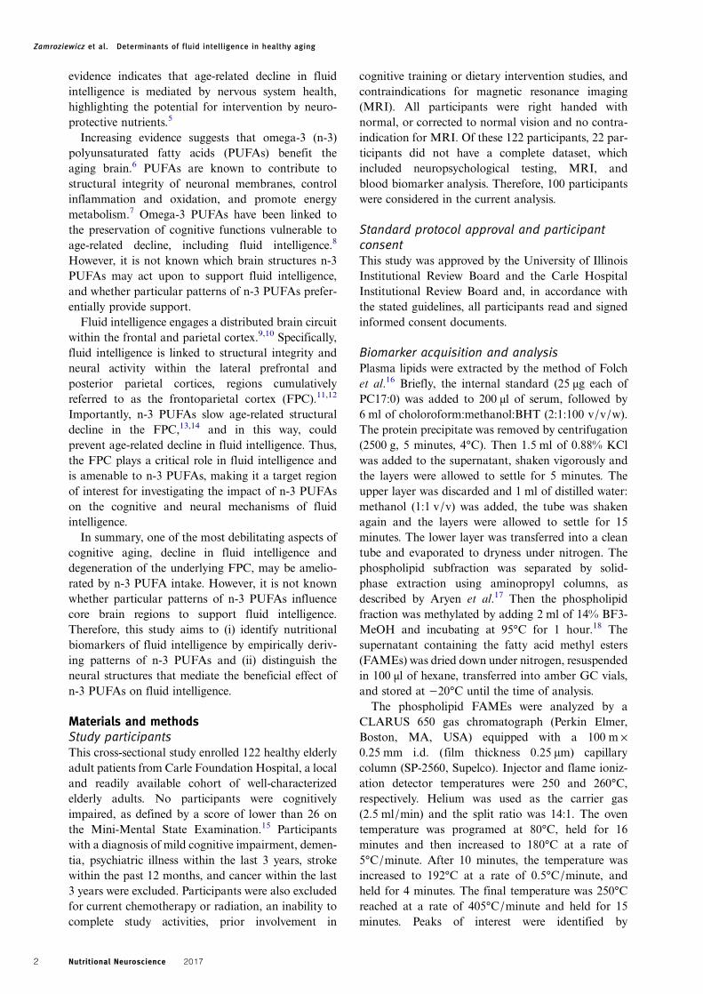
Biomarker acquisition and analysisPlasma lipids were extracted by the method of Folchet al.16 Briefly, the internal standard (25 μg each ofPC17:0) was added to 200 μl of serum, followed by6 ml of choloroform:methanol:BHT (2:1:100 v/v/w).The protein precipitate was removed by centrifugation(2500 g, 5 minutes, 4°C). Then 1.5 ml of 0.88% KClwas added to the supernatant, shaken vigorously andthe layers were allowed to settle for 5 minutes. Theupper layer was discarded and 1 ml of distilled water:methanol (1:1 v/v) was added, the tube was shakenagain and the layers were allowed to settle for 15minutes. The lower layer was transferred into a cleantube and evaporated to dryness under nitrogen. Thephospholipid subfraction was separated by solid-phase extraction using aminopropyl columns, asdescribed by Aryen et al.17 Then the phospholipidfraction was methylated by adding 2 ml of 14% BF3-MeOH and incubating at 95°C for 1 hour.18 Thesupernatant containing the fatty acid methyl esters(FAMEs) was dried down under nitrogen, resuspendedin 100 μl of hexane, transferred into amber GC vials,and stored at −20°C until the time of analysis.
The phospholipid FAMEs were analyzed by aCLARUS 650 gas chromatograph (Perkin Elmer,Boston, MA, USA) equipped with a 100 m ×0.25 mm i.d. (film thickness 0.25 µm) capillarycolumn (SP-2560, Supelco). Injector and flame ioniz-ation detector temperatures were 250 and 260°C,respectively. Helium was used as the carrier gas(2.5 ml/min) and the split ratio was 14:1. The oventemperature was programed at 80°C, held for 16minutes and then increased to 180°C at a rate of5°C/minute. After 10 minutes, the temperature wasincreased to 192°C at a rate of 0.5°C/minute, andheld for 4 minutes. The final temperature was 250°Creached at a rate of 405°C/minute and held for 15minutes. Peaks of interest were identified by
Zamroziewicz et al. Determinants of fluid intelligence in healthy aging
Nutritional Neuroscience 20172

comparison with authentic fatty acid standards (Nu-Chek Prep, Inc., Waterville, MN, USA) and expressedas absolute concentration (μmol/l). The plasma phos-pholipid lipids of interest were n-3 PUFAs, includingα-linolenic acid (ALA, 18:3n-3), stearidonic acid(SDA, 18:4n-3), eicosatrienoic acid (20:3n-3, ETE),eicosapentaenoic acid (EPA, 20:5n-3), docosapentae-noic acid (DPA, 22:5n-3), and docosahexaenoic acid(DHA, 22:6n-3).
Nutrient biomarker pattern analysis of PUFAsNutrient biomarker pattern (NBP) analysis was con-ducted in the IBM SPSS statistical software, version24 for Macintosh. Principal component analysis wasused to identify NBPs from the six n-3 PUFAs of inter-est. Of these, five n-3 PUFAs (ALA, ETE, EPA, DPA,and DHA) were non-normally distributed as indicatedby Shapiro–Wilk test (all P-values < 0.05), and there-fore log-transformed to correct for skewness of vari-ables and subsequently considered in the analysis.The appropriate rotation method was determined byexamining the factor correlation matrix: varimaxrotation was chosen for a correlation matrix withvalues less than 0.32 and direct oblimin rotation waschosen for a correlation matrix with values greaterthan 0.32.19 Statistical validity of the factor analysiswas confirmed via the Kaiser–Meyer–Olkin measureof sampling adequacy (≥0.50)20 and Bartlett’s test ofsphericity (P< 0.05).21 The number of NBPs to beretained was determined by a combination of eigen-values greater than 1.0, variance accounted for byeach component, and scree plot inflection point.Interpretation of each factor was based on identifyingbiomarkers with an absolute loading value of greaterthan 0.50 on an NBP (i.e. identifying the dominantbiomarkers contributing to each particular NBP).Each participant received a standardized NBP scorefor each pattern that corresponded to a linear combi-nation of the nutrient biomarkers.
Neuropsychological testsFluid intelligence was measured by the WechslerAbbreviated Scale of Intelligence – second edition(WASI-II).22 This assessment measured fluid intelli-gence by way of a perceptual reasoning index, whichwas the product of two subtests: a block designsubtest and a matrix reasoning subtest. In the blockdesign subtest, participants were asked to reproducepictured designs using specifically designed blocks asquickly and accurately as possible. In the matrixreasoning subtest, participants were asked to completea matrix or serial reasoning problem by selecting themissing section from five response items. Subjects’raw scores were converted to normalized scaledscores and subsequently combined into a perceptual
reasoning index, which provided a measure of nonver-bal reasoning and fluid intelligence.
Volumetric brain MRIVolumetric analysis was performed on data from a 3Dhigh-resolution T1-weighted scan using MPRAGEacquisition (0.9 mm isotropic voxel; TR: 1900 ms,TI: 900 ms, TE: 2.32 ms, with GRAPPA and an accel-eration factor of 2). Cortical reconstruction was per-formed with the Freesurfer image analysis suite,which is documented and freely available for down-load online (http://surfer.nmr.mgh.harvard.edu/).The technical details of these procedures are describedin prior publications.23–35 All cortical reconstructionswere manually checked for accuracy, as recommendedby the software developers. The volumetric analysesfocused on gray matter volume in the FPC, given therole of this cortical region in fluid intelligence11,12
and its sensitivity to n-3 PUFAs.13,14 As provided byFreesurfer parcellation, the FPC consisted of the fol-lowing regions of interest: superior frontal cortex,rostral middle frontal cortex, caudal middle frontalcortex, pars opercularis, pars triangularis, pars orbita-lis, superior parietal cortex, supramarginal cortex, andprecuneus.36,37 The volumetric analyses took into con-sideration total gray matter volume of the FPC as wellas gray matter volume of individual regions within theFPC.
CovariatesCovariates were included according to the previousassociation with cognitive decline.38–43 The covariatesincluded age (continuous), gender (nominal, man/woman), education (nominal, five fixed levels), andbody mass index (continuous). Volumetric analysesof the total FPC additionally accounted for intracra-nial volume (continuous), and volumetric analyses ofindividual regions within the FPC additionallyaccounted for total FPC volume (continuous) in aneffort to isolate the contribution of each individualregion.
Statistical analysisA formal mediation framework was applied to: (i)identify predictive nutritional biomarkers of fluidintelligence, as derived by NBP analysis, and (ii) dis-tinguish the neural structures that mediate the ben-eficial effect of n-3 PUFA patterns on fluidintelligence. First, regression models characterizedthe three relationships within the mediation frame-work: (i) the relationship between NBPs and fluidintelligence, (ii) the relationship between gray mattervolume within the FPC and fluid intelligence, and(iii) the relationship between NBPs and gray mattervolume within the FPC. Second, taking into accountresults of the regression analyses, a mediation modelassessed whether gray matter volume within the FPC
Zamroziewicz et al. Determinants of fluid intelligence in healthy aging
Nutritional Neuroscience 2017 3

mediated the relationship between NBPs and fluidintelligence (Fig. 1). Statistics were performed asfollows:(1) In the first step, one linear regression model was used
to characterize the relationship between NBPs andfluid intelligence (Fig. 3 path a). This analysisaccounted for covariates listed in Covariates. Theresults of this regression model indicated independentvariables for consideration in the mediation model.
(2) In the second step, linear regression models wereapplied to characterize the relationship betweeneach gray matter volume within the FPC, includingtotal FPC volume and volume of individual regionswithin the FPC, and fluid intelligence (Fig. 3 pathc). This analysis accounted for covariates listed inCovariates and applied a false discovery rate(FDR) correction for multiple comparisons (q<0.05, one-tailed).44 The results of these regressionmodels indicated mediatory variables for consider-ation in the mediation model.
(3) In the third step, linear regression models were usedto characterize the relationship between NBPs andeach gray matter volume within the FPC, includingtotal FPC volume and volume of individual regionswithin the FPC (Fig. 3 path b). This analysisaccounted for covariates listed in Covariates andapplied an FDR correction for multiple compari-sons (q< 0.05, one-tailed).44 The results of theseregression models further specified mediatory vari-ables for consideration in the mediation model.
(4) In the fourth step, the PROCESS macro designedfor SPSS was applied to implement the bootstrap-ping method to estimate mediation effects.45 Thisanalysis drew 1000 bootstrapped samples with repla-cement from the dataset to estimate a sampling dis-tribution for indirect and direct mediation effects,controlling for covariates listed in Covariates. Theindirect mediation effect refers to the pathwayfrom NBPs to gray matter volume within the FPCto fluid intelligence (Fig. 3 paths b–c). The directmediation effect refers to the direct pathway fromNBPs to fluid intelligence, accounting for theeffect of gray matter volume within the FPC(Fig. 3 path a′). As shown in Fig. 1, the primaryrequirement for mediation is a significant indirect
mediation effect, or the effect of the independentvariable (NBPs) through the mediator (gray mattervolume within the FPC) on the dependent variable(fluid intelligence).46 To further validate the pro-posed mediation model, an alternative mediationmodel, incorporating FPC as the independent vari-able, NBPs as the mediating variable, and fluidintelligence as the dependent variable, was alsotested.
Results are reported using (i) R2 and P for eachmodel, (ii) unstandardized regression coefficients (β),unstandardized regression coefficient standard error(SE β), and P of each individual regression relation-ship, and (iii) a 95% bias-corrected confidence interval(95% CI) for the direct and indirect effects of themediation. Significance was accepted at P≤ 0.05. Astatistically significant mediation that matches thehypothesized framework is indicated by: (i) an indirectmediation effect that does not include zero within 95%CI, and (ii) a direct mediation effect that does includezero within 95% CI.46
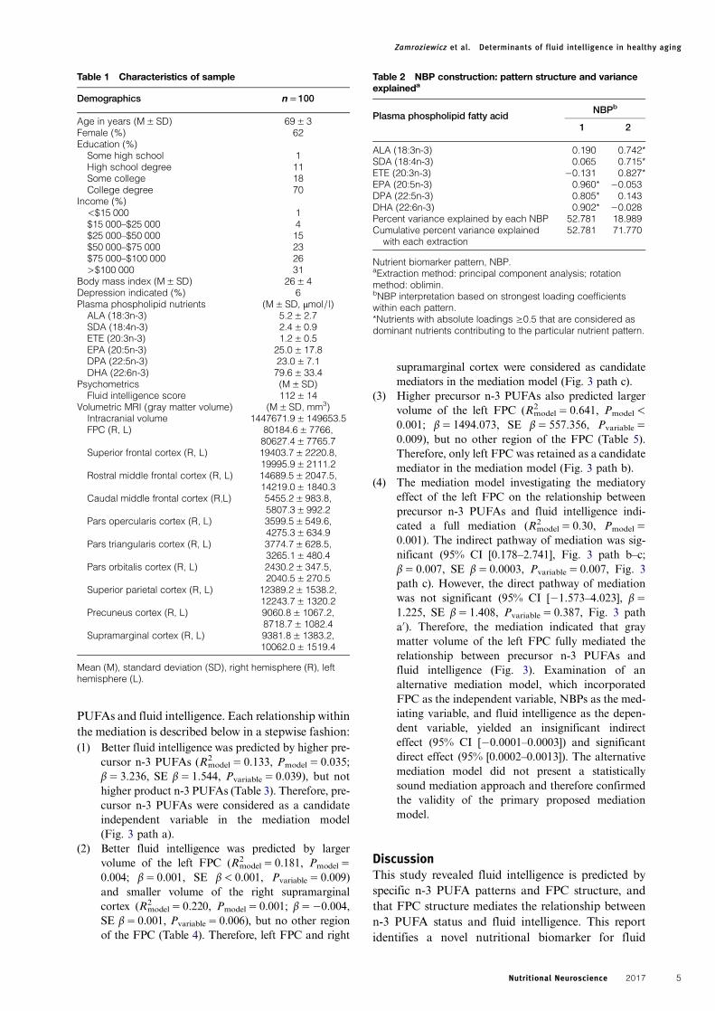
ResultsParticipant characteristicsParticipants (n= 100) had a mean age of 69 years and62% of participants were females (n= 62). All otherparticipant characteristics are reported in Table 1.
Nutrient biomarker patternsPrincipal component analysis generated two NBPs(Table 2). The factor correlation matrix containedvalues greater than 0.32; therefore, direct obliminrotation was implemented. Statistical validity of thefactor analyses was confirmed via the Kaiser–Meyer–Olkin measure of sampling adequacy (0.728) andBartlett’s test of sphericity (P< 0.001). Two NBPswere selected for retention because (i) after thesecond NBP extraction with principal componentanalysis, 71.8% of the total variance was accountedfor in the original set of nutrient biomarkers, and (ii)inspection of the scree plot indicated that the inflectionpoint occurred after the second NBP (Fig. 2).Hereafter, the first NBP is described as product n-3PUFAs (i.e. it is composed of downstream n-3PUFAs, including EPA, DPA n-3, and DHA), andthe second NBP is described as precursor n-3PUFAs (i.e. it is composed of three n-3 PUFAs thatserve as precursors to EPA and DHA).
Nutrient biomarker patterns, fluid intelligence,and gray matter volume with the FPCThe mediation analyses indicated that fluid intelli-gence was linked to precursor n-3 PUFAs as well astotal gray matter volume within the FPC, and further-more, that total gray matter volume of the left FPCfully mediated the relationship between precursor n-3
Figure 1 Proposed mediation model. the primaryrequirement for mediation is a significant indirect mediationeffect, defined as the effect of the independent variable(NBPs) through the mediator (gray matter volume within theFPC) on the dependent variable (fluid intelligence).
Zamroziewicz et al. Determinants of fluid intelligence in healthy aging
Nutritional Neuroscience 20174
PUFAs and fluid intelligence. Each relationship withinthe mediation is described below in a stepwise fashion:(1) Better fluid intelligence was predicted by higher pre-
cursor n-3 PUFAs (R2model= 0.133, Pmodel= 0.035;
β= 3.236, SE β= 1.544, Pvariable= 0.039), but nothigher product n-3 PUFAs (Table 3). Therefore, pre-cursor n-3 PUFAs were considered as a candidateindependent variable in the mediation model(Fig. 3 path a).
(2) Better fluid intelligence was predicted by largervolume of the left FPC (R2
model= 0.181, Pmodel=0.004; β= 0.001, SE β< 0.001, Pvariable= 0.009)and smaller volume of the right supramarginalcortex (R2
model= 0.220, Pmodel= 0.001; β=−0.004,SE β= 0.001, Pvariable= 0.006), but no other regionof the FPC (Table 4). Therefore, left FPC and right
supramarginal cortex were considered as candidatemediators in the mediation model (Fig. 3 path c).
(3) Higher precursor n-3 PUFAs also predicted largervolume of the left FPC (R2
model= 0.641, Pmodel<0.001; β= 1494.073, SE β= 557.356, Pvariable=0.009), but no other region of the FPC (Table 5).Therefore, only left FPC was retained as a candidatemediator in the mediation model (Fig. 3 path b).
(4) The mediation model investigating the mediatoryeffect of the left FPC on the relationship betweenprecursor n-3 PUFAs and fluid intelligence indi-cated a full mediation (R2
model= 0.30, Pmodel=0.001). The indirect pathway of mediation was sig-nificant (95% CI [0.178–2.741], Fig. 3 path b–c;β= 0.007, SE β= 0.0003, Pvariable= 0.007, Fig. 3path c). However, the direct pathway of mediationwas not significant (95% CI [−1.573–4.023], β=1.225, SE β= 1.408, Pvariable= 0.387, Fig. 3 patha′). Therefore, the mediation indicated that graymatter volume of the left FPC fully mediated therelationship between precursor n-3 PUFAs andfluid intelligence (Fig. 3). Examination of analternative mediation model, which incorporatedFPC as the independent variable, NBPs as the med-iating variable, and fluid intelligence as the depen-dent variable, yielded an insignificant indirecteffect (95% CI [−0.0001–0.0003]) and significantdirect effect (95% [0.0002–0.0013]). The alternativemediation model did not present a statisticallysound mediation approach and therefore confirmedthe validity of the primary proposed mediationmodel.
DiscussionThis study revealed fluid intelligence is predicted byspecific n-3 PUFA patterns and FPC structure, andthat FPC structure mediates the relationship betweenn-3 PUFA status and fluid intelligence. This reportidentifies a novel nutritional biomarker for fluid
Table 1 Characteristics of sample
Demographics n = 100
Age in years (M± SD) 69± 3Female (%) 62Education (%)
Some high school 1High school degree 11Some college 18College degree 70
Income (%)<$15 000 1$15 000–$25 000 4$25 000–$50 000 15$50 000–$75 000 23$75 000–$100 000 26>$100 000 31
Body mass index (M± SD) 26± 4Depression indicated (%) 6Plasma phospholipid nutrients (M± SD, μmol/l)
ALA (18:3n-3) 5.2± 2.7SDA (18:4n-3) 2.4± 0.9ETE (20:3n-3) 1.2± 0.5EPA (20:5n-3) 25.0± 17.8DPA (22:5n-3) 23.0± 7.1DHA (22:6n-3) 79.6± 33.4
Psychometrics (M± SD)Fluid intelligence score 112± 14
Volumetric MRI (gray matter volume) (M± SD, mm3)Intracranial volume 1447671.9± 149653.5FPC (R, L) 80184.6± 7766,
80627.4± 7765.7Superior frontal cortex (R, L) 19403.7± 2220.8,
19995.9± 2111.2Rostral middle frontal cortex (R, L) 14689.5± 2047.5,
14219.0± 1840.3Caudal middle frontal cortex (R,L) 5455.2± 983.8,
5807.3± 992.2Pars opercularis cortex (R, L) 3599.5± 549.6,
4275.3± 634.9Pars triangularis cortex (R, L) 3774.7± 628.5,
3265.1± 480.4Pars orbitalis cortex (R, L) 2430.2± 347.5,
2040.5± 270.5Superior parietal cortex (R, L) 12389.2± 1538.2,
12243.7± 1320.2Precuneus cortex (R, L) 9060.8± 1067.2,
8718.7± 1082.4Supramarginal cortex (R, L) 9381.8± 1383.2,
10062.0± 1519.4
Mean (M), standard deviation (SD), right hemisphere (R), lefthemisphere (L).
Table 2 NBP construction: pattern structure and varianceexplaineda
Plasma phospholipid fatty acidNBPb
1 2
ALA (18:3n-3) 0.190 0.742*SDA (18:4n-3) 0.065 0.715*ETE (20:3n-3) −0.131 0.827*EPA (20:5n-3) 0.960* −0.053DPA (22:5n-3) 0.805* 0.143DHA (22:6n-3) 0.902* −0.028Percent variance explained by each NBP 52.781 18.989Cumulative percent variance explained
with each extraction52.781 71.770
Nutrient biomarker pattern, NBP.aExtraction method: principal component analysis; rotationmethod: oblimin.bNBP interpretation based on strongest loading coefficientswithin each pattern.*Nutrients with absolute loadings ≥0.5 that are considered asdominant nutrients contributing to the particular nutrient pattern.
Zamroziewicz et al. Determinants of fluid intelligence in healthy aging
Nutritional Neuroscience 2017 5

intelligence as well as a novel mediatory relationshipbetween n-3 PUFAs, FPC structure, and fluid intelli-gence. The individual relationships reported withinthe mediation, including those between n-3 PUFAsand fluid intelligence (Fig. 3 path a), between FPCand fluid intelligence (Fig. 3 path c), and between n-3 PUFAs and FPC (Fig. 3 path b), are each supportedby previous work reviewed in turn below.First, precursor n-3 PUFAs positively associated
with fluid intelligence. Red blood cell phospholipidtotal n-3 PUFAs have been previously linked to intel-ligence in older adults.47 More specifically, serum con-centration of EPA, DPA n-3, and DHA has beenlinked to better performance on tests of frontal func-tion in older adults8,48; however, to our knowledge,
no studyhas examined the effects ofALAor its immedi-ate downstream products, including SDA and ETE, onintelligence or tests of frontal function in older adults.Importantly, ALA in serum,49 red blood cell phospho-lipids,50 andplasma51 has been linked to risk for demen-tia. Decline in fluid intelligence is a key feature of thecognitive changes that precede dementia,2 thus ALAand its immediate downstream products, includingSDA and ETE, could serve as predictive biomarkersfor fluid intelligence.
Second, structural integrity of the FPC was linkedto fluid intelligence. More specifically, gray mattervolume of the left FPC positively predicted fluid intel-ligence. Evidence indicates that fluid intelligence relieson the structure and function of regions within theFPC.9,10,12 The unilateral effect is supported byprior work, which suggests that regions within theleft hemisphere may be selectively susceptible todegeneration.52 Conversely, gray matter volume ofthe right supramarginal cortex negatively predictedfluid intelligence. Although the supramarginalcortex is considered part of the FPC,36,37 neuralactivity in this region decreases during tests of intelli-gence.53 In line with prior evidence, our resultssuggest that while the supramarginal cortex may con-tribute to the FPC as a whole, its individual contri-butions to intelligence are not congruent to that ofthe entire FPC.
Figure 2 Scree plot. inspection of the scree plot indicated that the inflection point occurred after the second component, orNBP, was extracted using a direct oblimin rotation.
Table 3 Linear regression models: n-3 PUFA patternsassociated with fluid intelligence
NBP Fluid intelligenceModel 1a
NBP1 β −0.645SE 1.604
NBP2 β 3.236*SE 1.544
Model R2 0.133*
Nutrient biomarker pattern, NBP.aModel: fluid intelligence=NBP1+NBP2+ age+ gender+education+ body mass index.*P< 0.05, **P< 0.01, ***P< 0.001.
Zamroziewicz et al. Determinants of fluid intelligence in healthy aging
Nutritional Neuroscience 20176

Third, precursor n-3 PUFAs positively predictedstructural integrity of the left FPC. Higher red bloodcell levels of DHA,8 combined EPA and DHA,54
and ALA55 have been linked to greater total brain
volume and markers of reduced brain atrophy. Inaddition, supplementation of EPA and DHA increasesgray matter volume in the frontal and parietal corticesof the left hemisphere in healthy, older adults.14
However, to our knowledge, no study has examinedthe effects of ALA or its immediate downstream pro-ducts, including SDA and ETE, on FPC gray matterstructure.Lastly, gray matter volume of the left FPC fully
mediated the relationship between precursor n-3PUFAs and fluid intelligence. Thus, precursor n-3PUFAs may influence fluid intelligence by promotingstructural integrity of the left FPC. Each of the threerelationships within the mediation is supported byprior findings, described above, but the mediationanalysis provides a novel link between particular n-3PUFAs, a cognitive function that is particularly vul-nerable to age-related decline, and an underlying neu-roanatomical network. These findings contribute toaccumulating evidence, suggesting that certain nutri-ents may slow or prevent aspects of age-related cogni-tive decline by influencing particular aspects of brainstructure.1,56–61
The predictive power of one NBP, the precursor n-3PUFA pattern, has noteworthy implications for theneuroprotective potential of n-3 PUFAs on fluid intel-ligence. The precursor n-3 PUFA pattern is reflectiveof either metabolic processing of n-3 PUFAs ordietary intake of n-3 PUFA-rich oils, nuts, andseeds.62,63 Metabolic processing of n-3 PUFAs withinthe precursor n-3 PUFA pattern may be neuroprotec-tive because ALA, SDA, and ETE are converted toEPA, and to a smaller extent, DHA. Although DHAis the most abundant long-chain n-3 PUFA in the
Figure 3 Mediation model statistics. a mediation model was used to characterize the relationship between NBP2, left FPC graymatter volume, and fluid intelligence. NBP2 positively associated with fluid intelligence (path a). NBP2 positively associated withtotal gray matter volume of the left FPC (path b). The indirect pathway of mediation (i.e. the effect of NBP2 through total graymatter volume of the left FPCon fluid intelligence; paths b–c) was statistically significant. The direct pathway of mediation (i.e. theeffect of NBP2 on fluid intelligence, accounting for total gray matter volume of the left FPC; path a′) was not significant.Therefore, total gray matter volume of left FPC fully mediated the relationship between NBP2 and fluid intelligence.
Table 4 Linear regression models: gray matter regionsassociated with fluid intelligence
Region Hemisphere
Fluid intelligence
β β SEModelR2
FPC Lefta 0.001**# <0.001**# 0.181**Righta 0.001 <0.001 0.153*
Superior frontal Leftb −0.001 0.001 0.179**Rightc 0.001 0.001 0.165**
Rostral middlefrontal
Leftb <0.001 0.001 0.178**Rightc <0.001 0.001 0.153*
Caudal middlefrontal
Leftb 0.001 0.002 0.179**Rightc <0.001 0.002 0.153*
Pars opercularis Leftb 0.002 0.002 0.184**Rightc 0.003 0.003 0.159*
Pars triangularis Leftb 0.003 0.003 0.187**Rightc 0.001 0.003 0.154*
Pars orbitalis Leftb −0.007 0.006 0.190**Rightc 0.004 0.005 0.160*
Superior parietal Leftb <0.001 0.002 0.178**Rightc 0.001 0.001 0.154*
Precuneus Leftb −0.001 0.002 0.180**Rightc 0.001 0.002 0.156*
Supramarginal Leftb <0.001 0.002 0.178**Rightc −0.004**# 0.001**# 0.220**
Frontoparietal cortex, FPC.aModel: fluid intelligence= regional gray matter volume+ age+gender+ education+ body mass index+ intracranial volume.bModel: gray matter volume= regional gray matter volume+age+ gender+ education+ body mass index+ left FPCvolume.cModel: gray matter volume= regional gray matter volume+age+ gender+ education+ body mass index+ right FPCvolume.*P< 0.05, **P< 0.01, ***P< 0.001, #P< 0.05, FDR-corrected.
Zamroziewicz et al. Determinants of fluid intelligence in healthy aging
Nutritional Neuroscience 2017 7

brain,64 but both EPA and DHA have physiologicaleffects that can improve brain health. These includereducing inflammation, reducing oxidative stress,reducing platelet aggregation, improving bloodpressure, and improving arterial compliance.65
Alternatively, dietary consumption of precursor n-3PUFAs may support neuronal health through theunique neuroprotective benefits of ALA and itsimmediate downstream products. Previous work hasshown that phospholipid ALA may prevent brainatrophy55 by providing glucose to the brain throughefficient ketogenesis,66 increasing serotonin and dopa-minergic neurotransmission in the frontal cortex,67
and increasing plasma levels of brain-derived neuro-trophic factor, thereby indirectly promoting neurogen-esis and neuronal survival.68 Importantly, few studieshave investigated the neuroprotective potential of n-3PUFAs within the precursor n-3 PUFA pattern, andeven fewer have derived empirical patterns of plasmaphospholipid n-3 PUFAs. The methodology employedin the current study allowed for an unprecedentedcomprehensive assessment of nutritional status of n-3PUFAs, and provided support for novel nutritionalbiomarkers of fluid intelligence and underlying corti-cal structure. Future mechanistic studies are neededto investigate whether precursor n-3 PUFAs are neuro-protective by way of conversion to EPA and DHAor whether these precursors possess unique neuro-protective benefits, as well as the endogenous andexogenous factors that contribute to the neuropro-tective effects. Future longitudinal studies are also
warranted to investigate the time scale on whichprecursor n-3 PUFAs influence fluid intelligenceand underlying cortical structure. While measure-ment of n-3 PUFAs in plasma phospholipidsreveals that short-term intake of these nutrientsinfluences cognition and brain health, measurementof n-3 PUFAs in adipose tissue will indicate theneuroprotective effects of long-term n-3 PUFAintake.69
The strengths of this study include: (i) the use ofblood biomarkers to measure physiological status ofn-3 PUFAs, (ii) the use of NBP analysis to empiricallyderive patterns of n-3 PUFAs, (iii) the use of structuralMRI to measure cortical integrity with high spatialresolution, and (iv) the assessment of a particular cog-nitive function that is known to be sensitive to age-related decline, rather than a global measure of cogni-tive function that presents with little variability inhealthy aging adults. The limitations of this studyinclude: (i) relatively small sample size (n= 100), (ii)cross-sectional design, (iii) limited neuropsychologicaltesting (i.e. only fluid intelligence), (iv) limited neuroi-maging domains (i.e. only structural neuroimaging),(v) inability to explore mechanisms that support therelationship between precursor n-3 PUFAs and FPCstructure, (vi) inability to explore contributions ofdiet and metabolic processes to n-3 PUFA patterns,and (vii) isolation of a specific dietary component.Thus, directions for future research include: (i) replica-tion of results in a larger sample, (ii) implementationof a longitudinal study to examine how changes in
Table 5 Linear regression models: n-3 PUFA patterns associated with gray matter structure of the frontoparietal cortex
Region Hemisphere
NBP1 NBP2
β β SE β β SE Model R2
FPC Lefta −389.183 580.267 1494.073**# 557.356**# 0.641***Righta −508.141 560.816 1424.340* 538.816* 0.673***
Superior frontal Leftb −44.492 129.989 −213.372 128.783 0.754***Rightc −238.769 131.753 34.029 130.181 0.772***
Rostral middle frontal Leftb 107.278 125.255 12.413 124.093 0.700***Rightc 82.595 153.719 −15.483 151.885 0.634***
Caudal middle frontal Leftb −165.806 95.989 53.767 95.098 0.393***Rightc 9.079 96.967 1.954 95.810 0.370***
Pars opercularis Leftb 23.717 67.597 59.900 66.970 0.265***Rightc 83.108 53.686 15.063 53.046 0.381***
Pars triangularis Leftb 90.942 47.264 9.672 46.826 0.372***Rightc 79.649 64.295 −37.704 63.528 0.321***
Pars orbitalis Leftb 15.187 28.148 −21.612 27.887 0.298***Rightc 46.995 35.625 −27.554 35.200 0.318***
Superior parietal Leftb 102.797 92.112 56.757 91.257 0.684***Rightc 42.707 122.006 −10.367 120.550 0.592***
Precuneus Leftb −172.987 73.955 −91.045 73.269 0.697***Rightc −118.534 80.577 −6.624 79.616 0.630***
Supramarginal Leftb 43.363 103.833 133.520 102.870 0.697***Rightc 13.211 121.662 46.687 120.211 0.498***
Nutrient biomarker pattern, NBP; frontoparietal cortex, FPC.aModel: gray matter volume=NBP1+NBP2+ age+ gender+ education+ body mass index+ intracranial volume.bModel: gray matter volume=NBP1+NBP2+ age+ gender+ education+ body mass index+ left FPC volume.cModel: gray matter volume=NBP1+NBP2+ age+ gender+ education+ body mass index+ right FPC volume.*P< 0.05, **P< 0.01, ***P< 0.001, #P< 0.05, FDR-corrected.
Zamroziewicz et al. Determinants of fluid intelligence in healthy aging
Nutritional Neuroscience 20178
n-3 PUFAs relate to changes in fluid intelligence andintegrity of the FPC, (iii) examination of other facetsof cognitive function, (iv) investigation of other neu-roimaging domains, such as white matter microstruc-ture and functional activity, (v) examination of themechanisms that support the relationship between pre-cursor n-3 PUFAs and FPC structure, (vi) investi-gation of the relative contributions of diet andmetabolic processes to n-3 PUFA patterns, (vii) exam-ination of potential synergistic interactions between n-3 PUFAs and other known neuroprotective dietarycomponents, such as antioxidant vitamins (i.e. caro-tenoids, vitamin E), that may reduce oxidation ofingested fatty acids and therefore optimize neuropro-tective effects.Research at the frontline of Nutritional Cognitive
Neuroscience suggests that certain nutrients mayslow or prevent aspects of age-related cognitivedecline by influencing particular age-related changesin brain structure.1,56–61 The present finding contrib-utes to this research program, and provides a novellink between nutritional and neuroanatomical bio-markers for fluid intelligence in healthy, olderadults. Ultimately, this line of work can inform clini-cal studies of personalized and comprehensiveapproaches to nutritional intervention for healthybrain aging.
AcknowledgementsAuthors are grateful to Tapas Das and Suzette Pereirafor their feedback in the drafting of this manuscript.We are also grateful to Joachim Operskalski, KelseyCampbell, Michael Kruepke, Jack Kuhns, andNikolai Sherepa for their invaluable help with thetesting of participants and organization of this study.
Disclaimer statementsContributors MKZ, EJP, and AKB contributed toexperiment concept and design; MKZ conductedresearch; MKZ and EJP analyzed data; all authorswere involved in interpretation and writing of manu-script; AKB had primary responsibility for the finalcontent. All authors read and approved themanuscript.
Funding This work was supported by a grant fromAbbott Nutrition through the Center for Nutrition,Learning, and Memory at the University of Illinois(ANGC1205; PI: Barbey).
Conflicts of interest The authors declare grants fromthe Center for Nutrition, Learning, and Memory(funded by Abbott Nutrition) during the conduct ofthe study.
Ethics approval None.
ORCIDMarta K. Zamroziewicz http://orcid.org/0000-0002-8227-0711Chris E. Zwilling http://orcid.org/0000-0002-2873-0115Aron K. Barbey http://orcid.org/0000-0002-6092-0912
References1 Zamroziewicz MK, Barbey AK. Nutritional cognitive neuro-science: innovations for healthy brain aging. Front Neurosci2016;10(June):1–10.
2 Schaie KW. The course of adult intellectual development. AmPsychol 1994;49:304–13.
3 Horn JL, Cattell RB. Age differences in fluid and crystallizedintelligence. Acta Psychol (Amst) 1967;26:107–29.
4 Tranter LJ, Koutstaal W. Age and flexible thinking: an exper-imental demonstration of the beneficial effects of increased cog-nitively stimulating activity on fluid intelligence in healthy olderadults. Aging Neuropsychol Cogn 2008;15(2):184–207.
5 Bergman I, Almkvist O. The effect of age on fluid intelligence isfully mediated by physical health. Arch Gerontol Geriatr2013;57(1):100–9.
6 Parletta N, Milte CM,Meyer BJ. Nutritional modulation of cog-nitive function and mental health. J Nutr Biochem 2013;24(5):725–43.
7 Cunnane SC, PlourdeM, Pifferi F, BéginM, Féart C, Barberger-Gateau P. Fish, docosahexaenoic acid and Alzheimer’s disease.Prog Lipid Res 2009;48:239–56.
8 Tan ZS, Harris WS, Beiser AS, Au R, Himali JJ, Debette S, et al.Red blood cell ω-3 fatty acid levels and markers of acceleratedbrain aging. Neurology 2012;78(9):658–64.
9 Woolgar A, Parr A, Cusack R, Thompson R, Nimmo-smith I,Torralva T, et al. Fluid intelligence loss linked to restrictedregions of damage within frontal and parietal cortex. Proc NatlAcad Sci 2015;112(35):E4969--.
10 Barbey AK, Colom R, Paul EJ, Grafman J. Architecture of fluidintelligence and working memory revealed by lesion mapping.Brain Struct Funct 2014;219(2):485–94.
11 Raz N, Lindenberger U, Ghisletta P, Rodrigue KM, KennedyKM, Acker JD. Neuroanatomical correlates of fluid intelligencein healthy adults and persons with vascular risk factors. CerebCortex 2008;18(3):718–26.
12 Cole MW, Yarkoni T, RepovšG, Anticevic A, Braver TS. Globalconnectivity of prefrontal cortex predicts cognitive control andintelligence. J Neurosci 2012;32(26):8988–99.
13 Titova OE, Ax E, Brooks SJ, Sjögren P, Cederholm T, KilanderL, et al. Mediterranean diet habits in older individuals: associ-ations with cognitive functioning and brain volumes. ExpGerontol 2013;48(12):1443–8.
14 Witte AV, Kerti L, Hermannstadter HM, Fiebach JB, SchreiberSJ, Schuchardt JP, et al. Long-chain omega-3 fatty acids improvebrain function and structure in older adults. Cereb Cortex2014;24(11):3059–68.
15 Folstein MF, Folstein SE, McHugh PR. “Mini-mental state” apractical method for grading the cognitive state of patients forthe clinician. J Psychiatr Res 1975;12(3):189–98.
16 Folch J, Lees M, Sloane Stanley GH. A simple method for theisolation and purification of total lipides from animal tissues. JBiol Chem 1957;226:497–509.
17 Aryen JJ, Julkunen A, Penttila I. Rapid separation of serumlipids for fatty acid analysis by a single aminopropyl column. JLipid Res 1992;33:1871–6.
18 Morrison WR, Smith LM. Preparation of fatty acid methylesters and dimethylacetals from lipids with boron fluoride-methanol. J Lipid Res 1964;55:600–8.
19 Tabachnick BG, Fidell LS. Using multivariate statistics. 5th ed.Upper Saddle River, NJ: Pearson Allyn & Bacon; 2007. 646p.
20 Kaiser HF. A second generation little jiffy. Psychometrika1970;35(4):401–15.
21 Bartlett MS. Tests of significance in factor analysis. Br J MathStat Psychol 1950;3:77–85.
22 Wechsler D. Wechsler abbreviated scale of intelligence. NewYork, NY: Psychol Corp; 1999.
Zamroziewicz et al. Determinants of fluid intelligence in healthy aging
Nutritional Neuroscience 2017 9

23 Dale A, Sereno M. Improved localization of cortical activity bycombining EEG and MEG with MRI cortical surface recon-struction. J Cogn Neurosci 1992;5(2):162–76.
24 Dale A, Fischl B, Sereno M. Cortical surface-based analysis.Neuroimage 1999;9:179–94.
25 Fischl B, Dale AM. Measuring the thickness of the human cer-ebral cortex from magnetic resonance images. PNAS 2000;97(20):11050–5.
26 Fischl B. Automatically parcellating the human cerebral cortex.Cereb Cortex 2004;14(1):11–22.
27 Fischl B, Sereno MI, Tootell RB, Dale AM. High-resolutionintersubject averaging and a coordinate system for the corticalsurface. Hum Brain Mapp 1999;8(4):272–84.
28 Fischl B, Sereno M, Dale A. Cortical surface-based analysis.Neuroimage 1999;9:195–207.
29 Fischl B, Liu A, Dale a M. Automated manifold surgery: con-structing geometrically accurate and topologically correctmodels of the human cerebral cortex. IEEE Trans MedImaging 2001;20(1):70–80.
30 Fischl B, Salat DH, Busa E, Albert M, Dieterich M, HaselgroveC, et al. Whole brain segmentation: neurotechnique automatedlabeling of neuroanatomical structures in the human brain.Neuron 2002;33(1):341–55.
31 Han X, Jovicich J, Salat D, van der Kouwe A, Quinn B,Czanner S, et al. Reliability of MRI-derived measurements ofhuman cerebral cortical thickness: the effects of field strength,scanner upgrade and manufacturer. Neuroimage 2006;32(1):180–94.
32 Jovicich J, Czanner S, Greve D, Haley E, van der Kouwe A,Gollub R, et al. Reliability in multi-site structural MRIstudies: effects of gradient non-linearity correction on phantomand human data. Neuroimage 2006;30(2):436–43.
33 Reuter M, Rosas HD, Fischl B. Highly accurate inverse consist-ent registration: a robust approach. Neuroimage 2010;53(4):1181–96.
34 Reuter M, Schmansky NJ, Rosas HD, Fischl B. Within-subjecttemplate estimation for unbiased longitudinal image analysis.Neuroimage 2012;61(4):1402–18.
35 Ségonne F, Dale AM, Busa E, Glessner M, Salat D, Hahn HK,et al. A hybrid approach to the skull stripping problem in MRI.Neuroimage 2004;22(3):1060–75.
36 Vijayakumar N, Whittle S, Yücel M, Dennison M, Simmons J,Allen NB. Thinning of the lateral prefrontal cortex during ado-lescence predicts emotion regulation in females. Soc CognAffect Neurosci 2014;9(11):1845–54.
37 Colom R, Karama S, Jung RE, Haier RJ. Human intelligenceand brain networks. Dialogues Clin Neurosci 2010;12:489–501.
38 Raz N, Ghisletta P, Rodrigue KM, Kennedy KM, LindenbergerU. Trajectories of brain aging in middle-aged and older adults:Regional and individual differences. Neuroimage 2010;51(2):501–11.
39 Coffey CE, Lucke JF, Saxton JA, Ratcliff G, Unitas LJ, Billig B,et al. Sex differences in brain aging. Arch Neurol 1998;55:169–79.
40 Coffey CE, Saxton JA, Ratcliff G, Bryan RN, Lucke JF.Relation of education to brain size in normal aging: implicationsfor the reserve hypothesis. Neurology 1999;53(1):189–96.
41 Fotenos AF, MintumMA, Snyder AZ, Morris JC, Buckner RL.Brain volume decline in aging. Arch Neurol 2008;65(1):113–20.
42 Gunstad J, Paul RH, Cohen RA, Tate DF, Spitznagel MB,Grieve S, et al. Relationship between body mass index andbrain volume in healthy adults. Int J Neurosci 2008;118(11):1582–93.
43 van Tol M-J, van der Wee NJA, van den Heuvel OA, NielenMMA, Demenescu LR, Aleman A, et al. Regional brainvolume in depression and anxiety disorders. Arch GenPsychiatry 2010;67(10):1002–11.
44 Benjamini Y, Hochberg Y. Controlling the false discovery rate: apractical and powerful approach to multiple testing. J R Stat Soc1995;57(1):289–300.
45 Preacher KJ, Hayes AF. Asymptotic and resampling strategiesfor assessing and comparing indirect effects in multiple mediatormodels. Behav Res Methods 2008;40(3):879–91.
46 Zhao X, Lynch Jr. JG, Chen Q. Reconsidering baron andKenny: myths and truths about mediation analysis. J ConsumRes 2010;37:197–206.
47 Whalley LJ, Deary IJ, Starr JM, Wahle KW, Rance KA, BourneVJ, et al. n-3 fatty acid erythrocyte membrane content, APOE 4,and cognitive variation: an observational follow-up study in late.Am J Clin Nutr 2008;87:449–54.
48 D’Ascoli TA, Mursu J, Voutilainen S, Kauhanen J, TuomainenTP, Virtanen JK. Association between serum long-chainomega-3 polyunsaturated fatty acids and cognitive performancein elderly men and women: the Kuopio Ischaemic heart diseaserisk factor study. Eur J Clin Nutr 2016;70:970–5.
49 Yamagishi K, Ikeda A, Chei CL, Noda H, Umesawa M, Cui R,et al. Serum a-linolenic and other n-3 fatty acids, and risk of dis-abling dementia: Community-based nested case-control study.Clin Nutr 2017;36(3)793–7.
50 Kim M, Nam JH, Oh DH, Park Y. Erythrocyte α-linolenic acidis associated with the risk for mild dementia in Korean elderly.Nutr Res 2010;30(11):756–61.
51 Cherubini A, Andres-Lacueva C, Martin A, Lauretani F, IorioAD, Bartali B, et al. Low plasma N-3 fatty acids and dementiain older persons: the InCHIANTI study. J Gerontol 2007;62A(10):1120–6.
52 Thompson PM, Hayashi KM, de Zubicaray G, Janke AL, RoseSE, Semple J, et al. Dynamics of gray matter loss in Alzheimer’sdisease. J Neurosci 2003;23(3):994–1005.
53 Haier RJ, White NS, Alkire MT. Individual differences ingeneral intelligence correlate with brain function during nonrea-soning tasks. Intelligence 2003;31(5):429–41.
54 Pottala J V., Yaffe K, Robinson JG, Espeland MA, Wallace R,Harris WS. Higher RBC EPA+DHA corresponds with largertotal brain and hippocampal volumes: WHIMS-MRI study.Neurology 2014;82(5):435–42.
55 Virtanen JK, Siscovick DS, Lemaitre RN, Longstreth WT,Spiegelman D, Rimm EB, et al. Circulating omega-3 polyunsa-turated fatty acids and subclinical brain abnormalities on MRIin older adults: the cardiovascular health study. J Am HeartAssoc 2013;2(5):1–11.
56 Bowman GL, Silbert LC, Howieson D, Dodge HH, Traber MG,Frei B, et al. Nutrient biomarker patterns, cognitive function,and MRI measures of brain aging. Neurology 2012;78(4):241–9.
57 Zamroziewicz MK, Paul EJ, Rubin RD, Barbey AK. Anteriorcingulate cortex mediates the relationship between O3PUFAsand executive functions in APOE e4 carriers. Front AgingNeurosci 2015;7(87):1–7.
58 Gu Y, Vorburger RS, Gazes Y, Habeck CG, Stern Y, LuchsingerJA, et al.White matter integrity as a mediator in the relationshipbetween dietary nutrients and cognition in the elderly. AnnNeurol 2016;79:1014–25.
59 Zamroziewicz MK, Paul EJ, Zwilling CE, Johnson EJ, KuchanMJ, Cohen NJ, et al. Parahippocampal cortex mediates therelationship between lutein and crystallized intelligence inhealthy, older adults. Front Aging Neurosci 2016;8(297):1–9.
60 Zamroziewicz MK, Zwilling CE, Barbey AK. Inferior prefrontalcortex mediates the relationship between phosphatidylcholineand executive functions in healthy, older adults. Front AgingNeurosci 2016;8(226):1–8.
61 Zamroziewicz MK, Paul EJ, Zwilling CE, Barbey AK.Predictors of memory in healthy aging: polyunsaturated fattyacid balance and fornix white matter integrity. Aging Dis2018; 9(1):1–12.
62 James MJ, Ursin VM, Cleland LG. Metabolism of stearidonicacid in human subjects: comparison with the metabolism ofother n-3 fatty acids 1–3. Am J Clin Nutr 2003;77:1140–5.
63 Walker CG, Jebb SA, Calder PC. Stearidonic acid as asupplemental source of w-3 polyunsaturated fatty acids toenhance status for improved human health. Nutrition 2013;29(2):363–9.
64 Kuratko CN, Salem N. Biomarkers of DHA status.Prostaglandins Leukot Essent Fat Acids 2009;81(2–3):111–8.
65 Mozaffarian D, Wu JHY. (n-3) fatty acids and cardiovascularhealth: are effects of EPA and DHA shared or complementary?J Nutr 2012;142:614S–625S.
66 Freemantle E, Vandal M, Tremblay-Mercier J, Tremblay S,Blachère JC, Bégin ME, et al. Omega-3 fatty acids, energy sub-strates, and brain function during aging. Prostaglandins LeukotEssent Fat Acids 2006;75(3):213–20.
67 Delion S, Chalon S, Herault J, Guilloteau D, Besnard J-C,Durand G. Chronic dietary a-linolenic acid deficiency altersdopaminergic and serotoninergic neurotransmission in rats. JNutr 1994;124(12):2466–76.
68 Hadjighassem M, Kamalidehghan B, Shekarriz N, Baseerat A,Molavi N, Mehrpour M, et al. Oral consumption of α-linolenicacid increases serum BDNF levels in healthy adult humans. NutrJ 2015;14(1):1–5.
69 Arab L. Biomarkers of fat and fatty acid intake. J Nutr 2003;133:925–32.
Zamroziewicz et al. Determinants of fluid intelligence in healthy aging
Nutritional Neuroscience 201710