Embed Size (px)
Citation preview

Detection value of free cancer cells in peritoneal washingin gastric cancer: a systematic review and meta-analysisFrancisco Tustumi,* WanderleyMarques Bernardo, Andre Roncon Dias, Marcus Fernando Kodama Pertille Ramos,
Ivan Cecconello, Bruno Zilberstein, Ulysses Ribeiro-Junior
Hospital das Clınicas da Faculdade de Medicina da Universidade de Sao Paulo, Sao Paulo/SP, Brazil.
Intraperitoneal free cancer cells in gastric adenocarcinoma are associated with a poor outcome. However, thetrue prognostic value of intraperitoneal free cancer cells is still unclear, leading to a lack of consensus in themanagement of gastric cancer. The aim of the present study is to perform a systematic review and meta-analysisto analyze intraperitoneal free cancer cells-positive patients with regard to tumor oncologic stage, recurrence,grade of cellular differentiation, and survival rates and to analyze the clinical significance of intraperitonealfree cancer cells with regard to prognosis. Databases were searched up to January 2016 for prognostic factorsassociated with intraperitoneal free cancer cells, including oncologic stage, depth of neoplasm invasion, lymphnodal spread, differentiation grade of the tumor, and recurrence and survival rates. A total of 100 studies wereidentified. Meta-analysis revealed a clear association between intraperitoneal free cancer cells and a poorprognosis. intraperitoneal free cancer cells -positive patients had higher rates of nodal spread (risk difference:0.29; po0.01), serosal invasion (risk difference: 0.43; po0.01), recurrence (after 60 months of follow-up, riskdifference: 0.44; po0.01), and mortality (after 60 months of follow-up, risk difference: 0.34; po0.01). Intra-peritoneal free cancer cells are associated with a poor outcome in gastric cancer. This surrogate biomarkershould be used to guide therapy both prior to and after surgery.
KEYWORDS: Gastric Carcinoma; Peritoneal Washing; Peritoneal Lavage; Cytology; Carcinoembryonic Antigen; RT-PCR.
Tustumi F, Bernardo WM, Dias AR, Ramos MF, Cecconello I, Zilberstein B, et al. Detection value of free cancer cells in peritoneal washing ingastric cancer: a systematic review and meta-analysis. Clinics. 2016;71(12):733-745
Received for publication on July 23, 2016; First review completed on August 29, 2016; Accepted for publication on September 9, 2016
*Corresponding author. E-mail: [email protected]
’ INTRODUCTION
Peritoneal dissemination is the most common pattern ofrecurrence in gastric cancer, even after a potentially cura-tive resection. This characteristic may be attributable to pos-sible intraperitoneal dissemination of malignant cells alreadypresent at the time of surgery or to surgical manipulations.Current knowledge on intraperitoneal free cancer cell (IFCC)positivity in gastric cancer demonstrates that these cells areassociated with a poor prognosis and advanced oncologicstages. Additionally, high recurrence rates, mainly due toperitoneal dissemination, and poor median survival areassociated with cytology detection (1-3).Based on these data, the Japanese Classification of Gastric
Carcinoma: 3rd English Edition (4) and the 7th Edition of theAJCC Cancer Staging Manual: Stomach (5) consider conven-tional cytology positivity in peritoneal fluid to be an indi-cator of stage IV disease.Several institutional protocols are used to manage IFCC-
positive patients, including chemotherapy, prompt gastrect-omy, neoadjuvant treatment, peritoneal infusion, hyperthermic
peritoneal chemotherapy, or palliation alone. However, noneof these techniques are accepted worldwide as a gold stan-dard therapy.The investigation of peritoneal washing for IFCCs in gastric
cancer patients remains controversial. Little is known about theactual burden of IFCC positivity and its accuracy for predictingan outcome. Moreover, a lack of consensus exists in its routinepractice, methods of detection (6), and association with clinicalpathological variables.Thus, the aim of this study was to perform a systematic
review and meta-analysis, investigating patients positive forIFCCs detected via different methods, regarding the neo-plasm oncologic stage, recurrence rates, grade of cellulardifferentiation, and survival rates and to analyze the clinicalsignificance of IFCCs with regard to prognosis.
’ METHODS
The construction and modeling of the present studywere guided by the Preferred Reporting Items for SystematicReviews and Meta-Analyses (PRISMA) statement (7).
Database searchA literature search was performed in MEDLINE using the
following search terms: (((‘‘Stomach Neoplasms/cytology’’[Mesh]) AND ((Peritoneum OR Peritoneal OR abdominalcavity OR ascitic fluid OR washing OR lavage)))) OR(((cytology AND gastric cancer)) AND ((Peritoneum ORDOI: 10.6061/clinics/2016(12)10
Copyright & 2016 CLINICS – This is an Open Access article distributed under theterms of the Creative Commons License (http://creativecommons.org/licenses/by/4.0/) which permits unrestricted use, distribution, and reproduction in anymedium or format, provided the original work is properly cited.
733
REVIEW

Peritoneal OR abdominal cavity OR ascitic fluid))). Otherdatabases searched included LILACS, CENTRAL, Cochrane,CINAHL, and Scopus as well as grey literature.No attempts were made to locate unpublished material.
Inclusion criteria
� Patients with confirmed gastric adenocarcinoma submittedto preoperative peritoneal washing/lavage evaluation (open,laparoscopic, or by paracentesis) for IFCCs (conventionalcytology with Papanicolaou, Giemsa, or Hematoxylin-eosinstaining); molecular methods, such as RT-PCR for carcino-embryonic antigen (CEA), cytokeratin (CK20), and melanoma-associated gene (MAGE); and immunohistochemistry.
� Studies that evaluated the prognosis (i.e., oncologic stage,survival, recurrence rate, or grade of cellular differentiation).
� Prospective or retrospective studies.� Studies selected by both of two reviewers.
Exclusion criteria
� Data could not be extracted from pooled results.� Patients submitted to a neoadjuvant approach prior to theperitoneal washing/lavage procedure.
� Presence of other primary malignancy.� Case series, case reports, animal models, conference pro-ceedings, editorials, and letters.
� Review articles and meta-analyses were excluded frommeta-analysis.
� Studies with no full-text.
IdiomNo restriction.
Search periodNo restriction. The search was performed up to January
2016.
Outcomes
� Recurrence rate� Recurrence site: lymph node, peritoneal, or other organs(local recurrence or hematogenous spread)
� Mortality� Oncologic stage� Serosal invasion� Lymph node spread� Grade of cellular differentiation
Statistical analysisAbsolute numbers for the outcome parameters were
extracted and analyzed with Review Manager Version 5.3software (Copenhagen: The Nordic Cochrane Centre; TheCochrane Collaboration, 2014).
We performed subgroup analysis and sensitivity teststo explore the causes of statistical heterogeneity in whichthe effect of single studies on the heterogeneity value wastested. Forest plots were used for graphical exploration ofheterogeneity. A funnel plot was used to identify publica-tion bias.
’ RESULTS
734
IFCC in gastric cancerTustumi F et al.
CLINICS 2016;71(12):733-745

Studies characteristicsOf the selected articles, 20 were excluded because they
lacked the information necessary for meta-analysis, such asserosal invasion, oncologic stage, neoplasm dissemination,and grade of cellular differentiation. In total, 100 (1-3, 8-104)eligible trials were identified and reviewed, and 91 wereincluded in the meta-analysis. Cumulatively, 16,913 gastriccancer patients were evaluated. In 41 studies analyzed, allpatients were submitted to curative intention surgery.We assessed the quality of the studies using the Newcastle-
Ottawa Scale (NOS). In terms of study quality, the cohortstudies were considered to be of fair (scores of 4–6) to good(scores of 7–9) quality, but two articles were considered lowquality (scores of 1-3).Of the 100 eligible trials, data describing conventional
cytology were available for 68 papers; data regarding PCR-CEA were available for 27; and data regarding PCR-CK20were available for 5. Other studies also evaluated Ber-Ep4,MAGE, RT-LAMP, or a combination of techniques used todetect IFCCs.Most studies performed peritoneal washing/lavage simi-
larly to the method described by Nakajima et al. (69). Theperitoneal cavity was washed with 50 to 200 ml of normalsaline. After stirring, the fluid was collected. Thirty-threestudies collected fluid from the Douglas space, 16 collected
fluids from the Douglas and left subphrenic spaces, and5 collected fluid from the perigastric surroundings. Theremaining studies collected fluid from different combinations ofrecesses. Peritoneal washing/lavage was performed by lapa-rotomy in 81%, by laparoscopy in 15.2%, and by drainage tubein 3.8% of the studies.Data were collected from 16 countries. The median follow-
up across all studies was 36 months (range 12-108 months).The prevalence of IFCCs ranged from 2 to 72%, with a
median of 27%. Considering only conventional cytologystudies, the median prevalence was 19.3% (range 2-61%).Considering only PCR-CEA, the median prevalence was27.8% (range 15-63%). Considering only PCR-CK20, themedian prevalence was 27.9% (range 15-39%).
IFCC and oncologic stageThe present study analyzed the oncologic stage according
to the UICC/AJCC system 6th edition (105). For this purpose,IFCC detection alone was not considered stage IV.The pooled data of the network meta-analysis showed
that IFCC detection was associated with a significantlyhigher risk of stage III or IV compared with stage I or II (riskdifference: 0.41; 95% CI: 0.33–0.49; n=4,258 patients; I2=88%,po0.00001) (see Figure 1). The sensitivity analysis failed to
Figure 1 - Oncologic stage according to the AJCC 6th edition. A strong association was observed between IFCC detection and stages III and IV.
735
CLINICS 2016;71(12):733-745 IFCC in gastric cancerTustumi F et al.

identify outliers. A random-effects analysis method was usedto adjust for inter-study heterogeneity.For the subgroup analysis, conventional cytology studies
(1-3,12,16-19,24,49,54,79,94,96) (risk difference: 0.34; 95% CI:0.2–0.48; n=2,373 patients; I2=93%; po0.00001) and PCR-CEA(31,36,38,49,77,93,94,97,104) (risk difference: 0.5; 95% CI: 0.36–0.63; n=1,073 patients; I2=83%; po0.00001) were reviewed bycomparing stage III or IV patients with stage I or II patients.Comparable results were identified (risk difference: 0.32;
95% CI: 0.19–0.44; n=600 patients; I2=55%; po0.00001) whenanalyzing studies that evaluated oncologic stages accordingto the UICC/AJCC system 7th edition (26,31,38,59,97).
IFCC and serosal invasionThe pooled data of the network meta-analysis showed that
IFCC detection was associated with a significantly higherrisk of serosal invasion than tumors that did not invadethe serosa (risk difference: 0.43; 95% CI: 0.38–0.48; n=11,511patients; I2=89%, po0.00001) (see Figure 2). The sensitivityanalysis failed to identify outliers. A random-effects analysismethod was used to adjust for inter-study heterogeneity.For the subgroup analysis, conventional cytology studies
(2,3,10,11,15,16,18,19,22,25,29,34,40,41,43,46,48-57,60,63,69,70,78,81,83,87,92,94,96,99,101) (risk difference: 0.39; 95% CI: 0.35–0.43; n=2,374 patients; I2=93%; po0.00001) and PCR-CEA(28,36-38,48-51,57,58,64,70,76,77,83,89,93,94,97,101,104) (risk dif-ference: 0.51; 95% CI: 0.45–0.57; n=2,612 patients; I2=66%;po0.00001) were reviewed.
IFCC and lymph node spreadThe pooled data of the network meta-analysis showed that
IFCC detection was associated with a significantly increasedrisk of lymph node spread compared to cancer with no lymphnode involvement (risk difference: 0.29; 95% CI: 0.23–0.34;n=7,718 patients; I2=89%, po0.00001) (see Figure 3). The sensi-tivity analysis failed to identify outliers. A random-effects anal-ysis method was used to adjust for inter-study heterogeneity.For the subgroup analysis, conventional cytology studies
(1-3,12,14,16,18,19,25,29,41,43,46,51,52,54-57,61,63,78,81,92,94,96,101) (risk difference: 0.25; 95% CI: 0.18–0.31; n=5,008 patients;I2=87%; po0.00001) and PCR-CEA (31,36,38,48,57,58,64,76,77,93,94,97,104) (risk difference: 0.3; 95% CI: 0.15–0.45; n=1,464 patients; I2=93%; po0.00001) were reviewed.
IFCC and grade of cellular differentiationThe pooled data of the network meta-analysis showed that
IFCC detection was associated with a significantly increasedprobability of having poorly differentiated tumors comparedto well or moderately differentiated tumors (risk difference:0.15; 95% CI: 0.12–0.17; n=7,232; I2=65%, po0.00001).A sensitivity analysis was performed by repeating the net-
work analysis after omitting 3 studies with a high risk ofbias (31,102,103). The final result revealed a risk differenceof 0.15 (95% CI: 0.13–0.18; n=6,784; I2=43%, po0.00001) (seeFigure 4).For the subgroup analysis, conventional cytology studies
(2,10,11,18,19,34,40,41,43,55-57,69,78,81,87,89,92,94-96,102,103) (risk difference after excluding 2 outliers (102,103):0.17; 95% CI: 0.14–0.2; n=5,437 patients; I2=39%; po0.00001)and PCR-CEA (31,57,64,77,93,94,97) (risk difference: 0.08;95% CI: 0.01–0.15; n=805 patients; I2=55%; po0.04) werereviewed.
IFCC and recurrenceThe recurrence rate was assessed for gastric cancers treated
with curative intention surgery.The pooled data of the network meta-analysis showed that
IFCC detection was associated with a significantly increasedrisk of recurrence. For recurrence after 24 months of follow-up (15,16,28,72), the risk difference was 0.38 (95% CI: 0.25–0.51; n=360 patients; I2=57%, po0.00001). For recurrenceafter 60 months, the risk difference was 0.44 (95% CI: 0.32–0.56; n=2,176 patients; I2=88%, po0.00001) (see Figure 5).
For IFCC-positive patients, the mean recurrence rate was55.35% after 24 months and 68.73% after 60 months. ForIFCC-negative patients, the mean recurrence rate was 16.77%after 24 months and 31.36% after 60 months.
IFCC and sites of recurrenceFor gastric cancers treated with curative intent surgery,
studies were assessed regarding peritoneal recurrence, lymphnodal recurrence, or recurrence in other organs.
For peritoneal recurrence, the presence of IFCCs predicteda risk difference of 0.48 (95% CI: 0.38–0.59; n=2,683 patients;I2=86%, po0.00001) (see Figure 6). The sensitivity analysisfailed to identify outliers. A random-effects analysis methodwas used to adjust for inter-study heterogeneity.
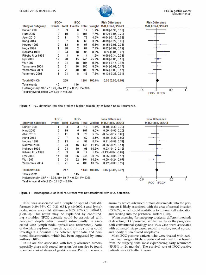
For lymph nodal recurrence, the presence of IFCCs pre-dicted a risk difference of 0.05 (95% CI: 0.00–0.1; n=1,553patients; I2=29%, p=0.05) (see Figure 7).
For local or hematogenous recurrence, the presence ofIFCCs did not predict a poor prognosis (risk difference: 0.02;95% CI: -0.03, 0.07; n=1,355 patients; I2=23%, p=0.22) (seeFigure 8).
IFCC and mortalityThe pooled data of the network meta-analysis showed that
IFCC detection was associated with a significantly increasedrisk of mortality.
For mortality after 12 months of follow-up (9,13,15,20,56,57,63,85,92), the risk difference was 0.26 (95% CI: 0.19–0.33;n=1,765 patients; I2=48%, po0.00001). One study was omit-ted after sensitivity analysis (92).
For mortality after 24 months (9,13,20,29,61,63,84,92), therisk difference was 0.4 (95% CI: 0.33–0.48; n=934 patients;I2=35%, po0.00001). One study was omitted after sensitivityanalysis (9).
For mortality after 60 months, the risk difference was 0.34(95% CI: 0.29–0.38; n=1,811 patients; I2=50%, po0.00001).Two studies were omitted after sensitivity analysis (69,72)(see Figure 9).
For IFCC-positive patients, the mean mortality rate was43.5% after 12 months, 75% after 24 months, and 72.3% forstudies that analyzed mortality after 60 months. For IFCC-negative patients, the mean mortality rate was 16.6% after12 months, 43.2% after 24 months, and 41.2% after 60 months.
For the subgroup analysis, studies that exclusively eval-uated patients who submitted to curative intention surgerywere assessed.
For mortality after 12 months, the risk difference was 0.35(95% CI: 0.24–0.45; n=799 patients; I2=13%, po0.00001). Onestudy was omitted after sensitivity analysis (92).
For mortality after 24 months, the risk difference was 0.34(95% CI: 0.24–0.44; n=717 patients; I2=35%, po0.00001). Onestudy was omitted after sensitivity analysis (9).
736
IFCC in gastric cancerTustumi F et al.
CLINICS 2016;71(12):733-745

Figure 2 - Evaluation of serosal invasion. An association between IFCC detection and serosal invasion was demonstrated.
737
CLINICS 2016;71(12):733-745 IFCC in gastric cancerTustumi F et al.

Figure 3 - Evaluation of lymph node dissemination. A clear association between IFCC detection and lymph node metastasis was noted.
738
IFCC in gastric cancerTustumi F et al.
CLINICS 2016;71(12):733-745
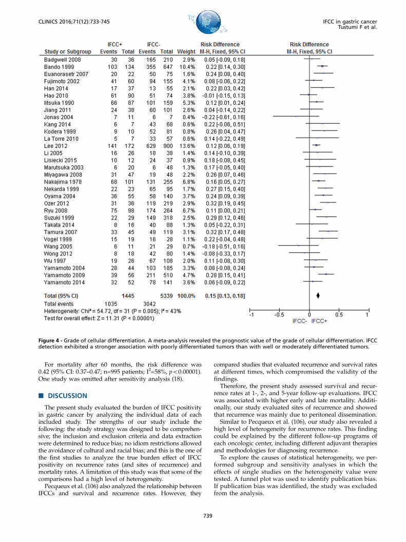
For mortality after 60 months, the risk difference was0.42 (95% CI: 0.37–0.47; n=995 patients; I2=58%, po0.00001).One study was omitted after sensitivity analysis (18).
’ DISCUSSION
The present study evaluated the burden of IFCC positivityin gastric cancer by analyzing the individual data of eachincluded study. The strengths of our study include thefollowing: the study strategy was designed to be comprehen-sive; the inclusion and exclusion criteria and data extractionwere determined to reduce bias; no idiom restrictions allowedthe avoidance of cultural and racial bias; and this is the one ofthe first studies to analyze the true burden effect of IFCCpositivity on recurrence rates (and sites of recurrence) andmortality rates. A limitation of this study was that some of thecomparisons had a high level of heterogeneity.Pecqueux et al. (106) also analyzed the relationship between
IFCCs and survival and recurrence rates. However, they
compared studies that evaluated recurrence and survival ratesat different times, which compromised the validity of thefindings.Therefore, the present study assessed survival and recur-
rence rates at 1-, 2-, and 5-year follow-up evaluations. IFCCwas associated with higher early and late mortality. Additi-onally, our study evaluated sites of recurrence and showedthat recurrence was mainly due to peritoneal dissemination.Similar to Pecqueux et al. (106), our study also revealed a
high level of heterogeneity for recurrence rates. This findingcould be explained by the different follow-up programs ofeach oncologic center, including different adjuvant therapiesand methodologies for diagnosing recurrence.To explore the causes of statistical heterogeneity, we per-
formed subgroup and sensitivity analyses in which theeffects of single studies on the heterogeneity value weretested. A funnel plot was used to identify publication bias.If publication bias was identified, the study was excludedfrom the analysis.
Figure 4 - Grade of cellular differentiation. A meta-analysis revealed the prognostic value of the grade of cellular differentiation. IFCCdetection exhibited a stronger association with poorly differentiated tumors than with well or moderately differentiated tumors.
739
CLINICS 2016;71(12):733-745 IFCC in gastric cancerTustumi F et al.

Figure 5 - Recurrence rate. IFCCs were associated with a higher probability of recurrence after 60 months of follow-up.
Figure 6 - A strong association between IFCC detection and peritoneal recurrence was noted.
740
IFCC in gastric cancerTustumi F et al.
CLINICS 2016;71(12):733-745
IFCC was associated with lymphatic spread (risk dif-ference: 0.29; 95% CI: 0.23–0.34, po0.00001) and lymphnodal recurrence (risk difference 0.05; 95% CI: 0.00–0.1,po0.05). This result may be explained by confound-ing variables (IFCC actually could be associated withneoplasm depth, which would subsequently be asso-ciated with lymph nodal spread and recurrence). Noneof the trials explored these data, and future studies couldinvestigate a possible link between lymphatic and peri-toneal dissemination, which has been suggested by someauthors (107).IFCCs are also associated with locally advanced tumors,
especially those with serosal invasion, but can also be foundin earlier clinical stages of gastric cancer. Part of the mech-
anism by which advanced tumors disseminate into the peri-toneum is likely associated with the area of serosal invasion(53,54,79), which could contribute to tumoral cell exfoliationand seeding into the peritoneal surface (108).When assessing for subgroup analysis, different methods
of detecting IFCC presented similar results for the prognosis.Both conventional cytology and PCR-CEA were associatedwith advanced stage cases, serosal invasion, nodal spread,and poorly differentiated neoplasms.Most IFCC-positive patients who were treated with cura-
tive intent surgery likely experienced minimal to no benefitfrom the surgery, with most experiencing early recurrence(55.35% in 24 months). The survival rate of IFCC-positivepatients was 25% after 2 years.
Figure 7 - IFCC detection can also predict a higher probability of lymph nodal recurrence.
Figure 8 - Hematogenous or local recurrence was not associated with IFCC detection.
741
CLINICS 2016;71(12):733-745 IFCC in gastric cancerTustumi F et al.

Accordingly, preoperative peritoneal washing/lavage ingastric cancer should be strongly advised for high sur-gical risk patients (e.g., the elderly, low status performancepatients, and patients with incapacitating comorbidities).If IFCC positivity is determined, palliative therapy may beconsidered.In low surgical risk and oncologic low risk (no serosa inva-
sion, no lymph nodal spread, moderate or well differentiatedneoplasm) patients, immediate surgery should be performed,and intraoperative peritoneal washing/lavage should beadded. If IFCC positivity is determined, postoperative chemo-therapy could be indicated. Clinical trials of hyperthermic intra-peritoneal chemotherapy may be proposed.The pooled data demonstrate that IFCC findings are an
independent prognostic factor in gastric cancer. From thiswork, it can be concluded that the prognosis in surgicallytreated patients with gastric carcinoma is significantly affectedby the presence of IFCCs at the time of gastrectomy and shouldguide gastric cancer management.
’ AUTHOR CONTRIBUTIONS
Tustumi F was responsible for the elaboration of the project and manu-script writing. Bernardo WM was responsible for the statistical analysis.Dias AR and Ramos MF helped with manuscript revision. Cecconello I andZilberstein B selected the articles. Ribeiro-Junior U was responsible for theelaboration of the project.
’ REFERENCES
1. Bentrem D, Wilton A, Mazumdar M, Brennan M, Coit D. The value ofperitoneal cytology as a preoperative predictor in patients with gastriccarcinoma undergoing a curative resection. Ann Surg Oncol. 2005;12(5):347-53, http://dx.doi.org/10.1245/ASO.2005.03.065.
2. Kodera Y, Yamamura Y, Shimizu Y, Torii A, Hirai T, Yasui K, et al.Peritoneal washing cytology: prognostic value of positive findingsin patients with gastric carcinoma undergoing a potentially curativeresection. J Surg Oncol. 1999;72(2):60-4, http://dx.doi.org/10.1002/(SICI)1096-9098(199910)72:2o60::AID-JSO343.0.CO;2-1.
3. Ribeiro U Jr, Safatle-Ribeiro AV, Zilberstein B, Mucerino D, Yagi OK,Bresciani CC, et al. Does the intraoperative peritoneal lavage cyto-logy add prognostic information in patients with potentially curativegastric resection? J Gastrointest Surg. 2006;10(2):170-6, http://dx.doi.org/10.1016/j.gassur.2005.11.001.
4. Ajani JA, Bentrem DJ, Besh S, D’Amico TA, Das P, Denlinger C, et al.2013 NCCN clinical practice guidelines in oncology: gastric cancer. 2013;Version 2. www.nccn.org.
5. Washington K. 7th edition of the AJCC cancer staging manual: stomach.Ann Surg Oncol. 2010;17(12):3077-9, http://dx.doi.org/10.1245/s10434-010-1362-z.
6. Brar SS, Mahar AL, Helyer LK, Swallow C, Law C, Paszat L, et al.Processes of care in the multidisciplinary treatment of gastric cancer:results of a RAND/UCLA expert panel. JAMA Surg. 2014;149(1):18-25,http://dx.doi.org/10.1001/jamasurg.2013.3959.
7. Moher D, Shamseer L, Clarke M, Ghersi D, Liberati A, Petticrew M, et al.Preferred reporting items for systematic review and meta-analysis pro-tocols (PRISMA-P) 2015 statement. Syst Rev. 2015;4:1, http://dx.doi.org/10.1186/2046-4053-4-1.
8. Andrade RJ, Iturriza JF, Loyo EL, Martinez LE. Adenocarcinoma delSistema digestivo: utilidad diagnóstica de la citología peritoneal. RevVenez Oncol. 2005;17(2):79-88.
9. Asao T, Fukuda T, Yazawa S, Nagamachi Y. Carcinoembryonic antigenlevels in peritoneal washings can predict peritoneal recurrence aftercurative resection of gastric cancer. Cancer. 1991;68(1):44-7, http://dx.doi.org/10.1002/1097-0142(19910701)68:1o44::AID-CNCR282068010943.0.CO;2-J.
10. Badgwell B, Cormier JN, Krishnan S, Yao J, Staerkel GA, Lupo PJ, et al.Does neoadjuvant treatment for gastric cancer patients with positiveperitoneal cytology at staging laparoscopy improve survival? Ann SurgOncol. 2008;15(10):2684-91, http://dx.doi.org/10.1245/s10434-008-0055-3.
11. Bando E, Yonemura Y, Takeshita Y, Taniguchi K, Yasui T, Yoshimitsu Y,et al. Intraoperative lavage for cytological examination in 1,297 patientswith gastric carcinoma. Am J Surg. 1999;178(3):256-62, http://dx.doi.org/10.1016/S0002-9610(99)00162-2.
12. Benevolo M, Mottolese M, Cosimelli M, Tedesco M, Giannarelli D,Vasselli S, et al. Diagnostic and prognostic value of peritoneal immuno-cytology in gastric cancer. J Clin Oncol. 1998;16(10):3406-11.
13. Bonenkamp JJ, Songun I, Hermans J, van de Velde CJ. Prognostic valueof positive cytology findings from abdominal washings in patients withgastric cancer. Br J Surg. 1996;83(5):672-4, http://dx.doi.org/10.1002/bjs.1800830526.
14. Brito AM, Sarmento BJ, Mota ED, Fraga AC Jr, Campoli PM, MilhomemLM, et al. Prognostic role of positive peritoneal cytology in patients withresectable gastric cancer. Rev Col Bras Cir. 2013;40(2):121-6, http://dx.doi.org/10.1590/S0100-69912013000200007.
15. Burke EC, Karpeh MS Jr, Conlon KC, Brennan MF. Peritoneal lavagecytology in gastric cancer: an independent predictor of outcome. AnnSurg Oncol. 1998;5(5):411-5, http://dx.doi.org/10.1007/BF02303859.
16. Chuwa EW, Khin LW, Chan WH, Ong HS, Wong WK. Prognostic signi-ficance of peritoneal lavage cytology in gastric cancer in Singapore.Gastric Cancer. 2005;8(4):228-37, http://dx.doi.org/10.1007/s10120-005-0343-6.
17. Dalal KM, Woo Y, Kelly K, Galanis C, Gonen M, Fong Y, et al. Detectionof micrometastases in peritoneal washings of gastric cancer patients bythe reverse transcriptase polymerase chain reaction. Gastric Cancer.2008;11(4):206-13, http://dx.doi.org/10.1007/s10120-008-0483-6.
Figure 9 - Mortality rate. IFCC detection was associated with a higher mortality rate after 60 months of follow-up.
742
IFCC in gastric cancerTustumi F et al.
CLINICS 2016;71(12):733-745
18. Euanorasetr C, Lertsithichai P. Prognostic significance of peritonealwashing cytology in Thai patients with gastric adenocarcinoma under-going curative D2 gastrectomy. Gastric Cancer. 2007;10(1):18-23, http://dx.doi.org/10.1007/s10120-006-0402-7.
19. Fujimoto T, Zhang B, Minami S, Wang X, Takahashi Y, Mai M.Evaluation of intraoperative intraperitoneal cytology for advancedgastric carcinoma. Oncology. 2002;62(3):201-8, http://dx.doi.org/10.1159/000059566.
20. Fujimura T, Kinami S, Ninomiya I, Kitagawa H, Fushida S, Nishimura G,et al. Diagnostic laparoscopy, serum CA125, and peritoneal metastasis ingastric cancer. Endoscopy. 2002;34(7):569-74, http://dx.doi.org/10.1055/s-2002-33228.
21. Fujiwara Y, Okada K, Hanada H, Tamura S, Kimura Y, Fujita J, et al. Theclinical importance of a transcription reverse-transcription concerted(TRC) diagnosis using peritoneal lavage fluids in gastric cancer withclinical serosal invasion: A prospective, multicenter study. Surgery.2014;155(3):417-23, http://dx.doi.org/10.1016/j.surg.2013.10.004.
22. Fukagawa T, Katai H, Saka M, Morita S, Sasajima Y, Taniguchi H, et al.Significance of lavage cytology in advanced gastric cancer patients.World J Surg. 2010;34(3):563-8, http://dx.doi.org/10.1007/s00268-009-0355-1.
23. Fukuda N, Sugiyama Y, Wada J. Prognostic factors of T4 gastric cancerpatients undergoing potentially curative resection. World J Gastro-enterol. 2011;17(9):1180-4, http://dx.doi.org/10.3748/wjg.v17.i9.1180.
24. Fukumoto Y, Ikeguchi M, Matsumoto S, Inoue M, Osaki T, Fukuda K,et al. Detection of cancer cells and gene expression of cytokines in theperitoneal cavity in patients with gastric cancer. Gastric Cancer. 2006;9(4):271-6, http://dx.doi.org/10.1007/s10120-006-0390-7.
25. Funami Y, Tokumoto N, Miyauchi H, Ochiai T, Kuga K. Prognostic valueof peritoneal lavage cytology and chemotherapy during surgery foradvanced gastric cancer. Int Surg. 1999;84(3):220-4.
26. Han J, Lv P, Yu JL, Wu YC, Zhu X, Hong LL, et al. Circulating methy-lated MINT2 promoter DNA is a potential poor prognostic factor ingastric cancer. Dig Dis Sci. 2014;59(6):1160-8, http://dx.doi.org/10.1007/s10620-013-3007-0.
27. Hao YX, Zhong H, Yu PW, Qian F, Zhao YL, Shi Y, et al. Influence oflaparoscopic gastrectomy on the detection rate of free gastric cancer cellsin the peritoneal cavity. Ann Surg Oncol. 2010;17(1):65-72, http://dx.doi.org/10.1245/s10434-009-0703-2.
28. Hara M, Nakanishi H, Jun Q, Kanemitsu Y, Ito S, Mochizuki Y, et al.Comparative analysis of intraperitoneal minimal free cancer cellsbetween colorectal and gastric cancer patients using quantitative RT-PCR: possible reason for rare peritoneal recurrence in colorectal cancer.Clin Exp Metastasis. 2007;24(3):179-89, http://dx.doi.org/10.1007/s10585-007-9067-9.
29. Hayes N, Wayman J, Wadehra V, Scott DJ, Raimes SA, Griffin SM.Peritoneal cytology in the surgical evaluation of gastric carcinoma. Br JCancer. 1999;79(3-4):520-4, http://dx.doi.org/10.1038/sj.bjc.6690081.
30. Homma Y, Ushida S, Yamada M, Kobayashi H, Suzuki K. Positiveperitoneal washing cytology in multiple cavities can predict poor pro-gnosis of advanced gastric cancer patients. Ann Surg Oncol. 2010;17(2):455-60, http://dx.doi.org/10.1245/s10434-009-0764-2.
31. Horikawa M, Iinuma H, Inoue T, Ogawa E, Fukushima R. Clinicalsignificance of intraperitoneal CD44 mRNA levels of magneticallyseparated CD45-negative EpCAM-positive cells for peritoneal recurrenceand prognosis in stage II and III gastric cancer patients. Oncol Rep.2011;25(5):1413-20.
32. Iida T, Iwahashi M, Katsuda M, Ishida K, Nakamori M, Nakamura M,et al. Prognostic significance of IL-17 mRNA expression in peritoneallavage in gastric cancer patients who underwent curative resection.Oncol Rep. 2014;31(2):605-12.
33. Iitsuka Y, Kaneshima S, Tanida O, Takeuchi T, Koga S. Intraperitonealfree cancer cells and their viability in gastric cancer. Cancer. 1979;44(4):1476-80, http://dx.doi.org/10.1002/1097-0142(197910)44:4o1476::AID-CNCR282044044243.0.CO;2-R.
34. Iitsuka Y, Shiota S, Matsui T, Murata Y, Kimura A, Koga S. Relationshipbetween the cytologic characteristics of intraperitoneal free cancer cellsand the prognosis in patients with gastric cancer. Acta Cytol. 1990;34(3):437-42.
35. Ishigami S, Uenosono Y, Arigami T, Yanagita S, Okumura H, UchikadoY, et al. Clinical utility of perioperative staging laparoscopy for advancedgastric cancer. World J Surg Oncol. 2014;12:350, http://dx.doi.org/10.1186/1477-7819-12-350.
36. Ishii T, Fujiwara Y, Ohnaka S, Hayashi T, Taniguchi H, Takiguchi S, et al.Rapid genetic diagnosis with the transcription-reverse transcriptionconcerted reaction system for cancer micrometastasis. Ann Surg Oncol.2004;11(8):778-85, http://dx.doi.org/10.1245/ASO.2004.12.043.
37. Ito S, Nakanishi H, Kodera Y, Mochizuki Y, Tatematsu M, Yamamura Y.Prospective validation of quantitative CEA mRNA detection in perito-neal washes in gastric carcinoma patients. Br J Cancer. 2005;93(9):986-92,http://dx.doi.org/10.1038/sj.bjc.6602802.
38. Jeon CH, Kim IH, Chae HD. Prognostic value of genetic detection usingCEA and MAGE in peritoneal washes with gastric carcinoma after
curative resection: result of a 3-year follow-up. Medicine (Baltimore).2014;93(11):e83, http://dx.doi.org/10.1097/MD.0000000000000083.
39. Jeon CH, Shin IH, Park JB, Chae HD. Prognostic significance ofMAGE in peritoneal washes in gastric carcinoma patients withoutperitoneal metastasis: results of a 5-year follow-up study. J Clin Gas-troenterol. 2010;44(10):682-6, http://dx.doi.org/10.1097/MCG.0b013e3181d6bb0b.
40. Jiang CG, Xu Y, Wang ZN, Sun Z, Liu FN, Yu M, et al. Clinico-pathological analysis and prognostic significance of peritoneal cytologyin Chinese patients with advanced gastric cancer. ANZ J Surg. 2011;81(9):608-13, http://dx.doi.org/10.1111/j.1445-2197.2010.05536.x.
41. Jonas S, Weinrich M, Tullius SG, Al-Abadi H, Steinbrich R, Radke C,et al. Microscopic tumor cell dissemination in gastric cancer. Surg Today.2004;34(2):101-6, http://dx.doi.org/10.1007/s00595-003-2666-4.
42. Kanetaka K, Ito S, Susumu S, Yoneda A, Fujita F, Takatsuki M, et al.Clinical significance of carcinoembryonic antigen in peritoneal lavagefrom patients with gastric cancer. Surgery. 2013;154(3):563-72, http://dx.doi.org/10.1016/j.surg.2013.03.005.
43. Kang KK, Hur H, Byun CS, Kim YB, Han SU, Cho YK. Conventionalcytology is not beneficial for predicting peritoneal recurrence after cura-tive surgery for gastric cancer: results of a prospective clinical study.J Gastric Cancer. 2014;14(1):23-31, http://dx.doi.org/10.5230/jgc.2014.14.1.23.
44. Kano Y, Kosugi S, Ishikawa T, Otani T, Muneoka Y, Sato Y, et al. Prog-nostic significance of peritoneal lavage cytology at three cavities inpatients with gastric cancer. Surgery. 2015;158(6):1581-9, http://dx.doi.org/10.1016/j.surg.2015.04.004.
45. Katsuragi K, Yashiro M, Sawada T, Osaka H, Ohira M, Hirakawa K.Prognostic impact of PCR-based identification of isolated tumour cells inthe peritoneal lavage fluid of gastric cancer patients who underwent acurative R0 resection. Br J Cancer. 2007;97(4):550-6, http://dx.doi.org/10.1038/sj.bjc.6603909.
46. Ki YJ, Ji SH, Min JS, Jin SH, Park S, Yu HJ, et al. Test execution variationin peritoneal lavage cytology could be related to poor diagnostic accu-racy and stage migration in patients with gastric cancer. J Gastric Cancer.2013;13(4):214-25, http://dx.doi.org/10.5230/jgc.2013.13.4.214.
47. Kodera Y, Nakanishi H, Ito S, Mochizuki Y, Ohashi N, Yamamura Y, et al.Prognostic significance of intraperitoneal cancer cells in gastric carci-noma: analysis of real time reverse transcriptase-polymerase chainreaction after 5 years of followup. J Am Coll Surg. 2006;202(2):231-6,http://dx.doi.org/10.1016/j.jamcollsurg.2005.09.008.
48. Kodera Y, Nakanishi H, Ito S, Yamamura Y, Kanemitsu Y, Shimizu Y,et al. Quantitative detection of disseminated free cancer cells in perito-neal washes with real-time reverse transcriptase-polymerase chain reaction:a sensitive predictor of outcome for patients with gastric carcinoma. AnnSurg. 2002;235(4):499-506, http://dx.doi.org/10.1097/00000658-200204000-00007.
49. Kodera Y, Nakanishi H, Yamamura Y, Shimizu Y, Torii A, Hirai T, et al.Prognostic value and clinical implications of disseminated cancer cellsin the peritoneal cavity detected by reverse transcriptase-polymerasechain reaction and cytology. Int J Cancer. 1998;79(4):429-33, http://dx.doi.org/10.1002/(SICI)1097-0215(19980821)79:4o429::AID-IJC2043.0.CO;2-Z.
50. Kodera Y, Nakanishi H, Ito S, Yamamura Y, Fujiwara M, Koike M,et al. Prognostic significance of intraperitoneal cancer cells in gastriccarcinoma: detection of cytokeratin 20 mRNA in peritoneal washes, inaddition to detection of carcinoembryonic antigen. Gastric Cancer. 2005;8(3):142-8, http://dx.doi.org/10.1007/s10120-005-0318-7.
51. Kodera Y, Nakanishi H, Ito S, Yamamura Y, Kanemitsu Y, Shimizu Y,et al. Quantitative detection of disseminated cancer cells in the greateromentum of gastric carcinoma patients with real-time RT-PCR: a com-parison with peritoneal lavage cytology. Gastric Cancer. 2002;5(2):69-76,http://dx.doi.org/10.1007/s101200200012.
52. Kodera Y, Yamamura Y, Ito S, Kanemitsu Y, Shimizu Y, Hirai T, et al. IsBorrmann type IV gastric carcinoma a surgical disease? An old problemrevisited with reference to the result of peritoneal washing cytology.J Surg Oncol. 2001;78(3):175-81, http://dx.doi.org/10.1002/jso.1144.
53. Koga S, Kaibara N, Iitsuka Y, Kudo H, Kimura A, Hiraoka H. Prognosticsignificance of intraperitoneal free cancer cells in gastric cancer patients.J Cancer Res Clin Oncol. 1984;108(2):236-8, http://dx.doi.org/10.1007/BF00402474.
54. Kostić Z, Cuk V, Bokun R, Ignjatović D, Usaj-Knezević S, Ignjatović M.Detection of free cancer cells in peritoneal cavity in patients surgicallytreated for gastric adenocarcinoma. Vojnosanit Pregl. 2006;63(4):349-56,http://dx.doi.org/10.2298/VSP0604349K.
55. La Torre M, Ferri M, Giovagnoli MR, Sforza N, Cosenza G, Giarnieri E,et al. Peritoneal wash cytology in gastric carcinoma. Prognostic sig-nificance and therapeutic consequences. Eur J Surg Oncol. 2010;36(10):982-6, http://dx.doi.org/10.1016/j.ejso.2010.06.007.
56. Lee SD, Ryu KW, Eom BW, Lee JH, Kook MC, Kim YW. Prog-nostic significance of peritoneal washing cytology in patients withgastric cancer. Br J Surg. 2012;99(3):397-403, http://dx.doi.org/10.1002/bjs.7812.
743
CLINICS 2016;71(12):733-745 IFCC in gastric cancerTustumi F et al.

57. Li JK, Zheng M, Miao CW, Zhang JH, Ding GH, Wu WS. Peritoneallavage cytology and carcinoembryonic antigen determination in pre-dicting peritoneal metastasis and prognosis of gastric cancer. WorldJ Gastroenterol. 2005;11(46):7374-7, http://dx.doi.org/10.3748/wjg.v11.i46.7374.
58. Li Z, Zhang D, Zhang H, Miao Z, Tang Y, Sun G, et al. Prediction ofperitoneal recurrence by the mRNA level of CEA and MMP-7 in peri-toneal lavage of gastric cancer patients. Tumour Biol. 2014;35(4):3463-70,http://dx.doi.org/10.1007/s13277-013-1458-8.
59. Lisiecki R, Spycha"a A, Pater K, Murawa D. Analysis of risk factorsof positive peritoneal cytology in patients treated for gastric cancer -preliminary report. Pol Przegl Chir. 2015;87(10):506-12.
60. Majima T, Ichikura T, Mochizuki H. Prognostic significance of thecytologic features of free cancer cells in the peritoneal cavity of patientswith gastric cancer. Surg Today. 2002;32(1):35-9, http://dx.doi.org/10.1007/s595-002-8110-6.
61. Makino T, Fujiwara Y, Takiguchi S, Miyata H, Yamasaki M, Nakajima K,et al. The utility of pre-operative peritoneal lavage examination inserosa-invading gastric cancer patients. Surgery. 2010;148(1):96-102,http://dx.doi.org/10.1016/j.surg.2009.11.025.
62. Mandorwski S, Lourenco LG, Forones NM. CA72-4 e CEA no soroe no lavado peritonial de doentes com câncer gástrico. Arq. Gastro-enterol. 2002;39(1):17-21, http://dx.doi.org/10.1590/S0004-28032002000100004.
63. de Manzoni G, Verlato G, Di Leo A, Tomezzoli A, Pedrazzani C, Pasini F,et al. Peritoneal cytology does not increase the prognostic informationprovided by TNM in gastric cancer. World J Surg. 2006;30(4):579-84,http://dx.doi.org/10.1007/s00268-005-7901-2.
64. Marutsuka T, Shimada S, Shiomori K, Hayashi N, Yagi Y, Yamane T,et al. Mechanisms of peritoneal metastasis after operation for non-Serosa-invasive gastric carcinoma: an ultrarapid detection system forintraperitoneal Free cancer cells and a prophylactic strategy for perito-neal metastasis. Clin Cancer Res. 2003;9(2):678-85.
65. Miyagawa K, Sakakura C, Nakashima S, Yoshikawa T, Fukuda K, Kin S,et al. Overexpression of RegIV in peritoneal dissemination of gastriccancer and its potential as A novel marker for the detection of peritonealmicrometastasis. Anticancer Res. 2008;28(2B):1169-79.
66. Miyashiro I, Takachi K, Doki Y, Ishikawa O, Ohigashi H, Murata K, et al.When is curative gastrectomy justified for gastric cancer with positiveperitoneal lavage cytology but negative macroscopic peritoneal implant?World J Surg. 2005;29(9):1131-4, http://dx.doi.org/10.1007/s00268-005-7703-6.
67. Nakagawa S, Nashimoto A, Yabusaki H. Role of staging laparoscopywith peritoneal lavage cytology in the treatment of locally advancedgastric cancer. Gastric Cancer. 2007;10(1):29-34, http://dx.doi.org/10.1007/s10120-006-0406-3.
68. Nakagohri T, Yoneyama Y, Kinoshita T, Konishi M, Inoue K, Takahashi S.Prognostic significance of peritoneal washing cytology in patientswith potentially resectable gastric cancer. Hepato Gastroenterol. 2008;55(86-87):1913-5.
69. Nakajima T, Harashima S, Hirata M, Kajitani T. Prognostic and ther-apeutic values of peritoneal cytology in gastric cancer. Acta Cytol.1978;22(4):225-9.
70. Nakanishi H, Kodera Y, Yamamura Y, Ito S, Kato T, Ezaki T, et al. Rapidquantitative detection of carcinoembryonic antigen-expressing free tumorcells in the peritoneal cavity of gastric-cancer patients with real-timeRT-PCR on the LightCycler. Int J Cancer. 2000;89(5):411-7, http://dx.doi.org/10.1002/1097-0215(20000920)89:5o411::AID-IJC343.0.CO;2-5.
71. Nakanishi H, Kodera Y, Yamamura Y, Kuzuya K, Nakanishi T, Ezaki T,et al. Molecular diagnostic detection of Free cancer cells in the peritonealcavity of patients with gastrointestinal and gynecologic malignancies.Cancer Chemother Pharmacol. 1999;43 Suppl:S32-6, http://dx.doi.org/10.1007/s002800050869.
72. Nekarda H, Gess C, Stark M, Mueller JD, Fink U, Schenck U, et al.Immunocytochemically detected free peritoneal tumour cells (FPTC)are a strong prognostic factor in gastric carcinoma. Br J Cancer. 1999;79(3-4):611-9, http://dx.doi.org/10.1038/sj.bjc.6690096.
73. Nishiyama M, Takashima I, Tanaka T, Yoshida K, Toge T, Nagata N, et al.Carcinoembryonic antigen levels in the peritoneal cavity: useful guide toperitoneal recurrence and prognosis for gastric cancer. World J Surg.1995;19(1):133-7, http://dx.doi.org/10.1007/BF00316997.
74. Nishizawa M, Seshimo A, Miyake K, Amano K, Kameoka S. Usefulnessof the TRC method in the peritoneal washing cytology for gastric cancer.Hepatogastroenterology. 2014;61(129):240-4.
75. Oh CA, Bae JM, Oh SJ, Choi MG, Noh JH, Sohn TS, et al. Long-termresults and prognostic factors of gastric cancer patients with only posi-tive peritoneal lavage cytology. J Surg Oncol. 2012;105(4):393-9, http://dx.doi.org/10.1002/jso.22091.
76. Ohashi N, Nakanishi H, Kodera Y, Ito S, Mochizuki Y, Koike M, et al.Intraoperative quantitative detection of CEA mRNA in the peritoneallavage of gastric cancer patients with transcription reverse-transcriptionconcerted (TRC) method. A comparative study with real-time quantita-tive RT-PCR. Anticancer Res. 2007;27(4C):2769-77.
77. Oyama K, Terashima M, Takagane A, Maesawa C. Prognostic sig-nificance of peritoneal minimal residual disease in gastric cancer detec-ted by reverse transcription-polymerase chain reaction. Br J Surg. 2004;91(4):435-43, http://dx.doi.org/10.1002/bjs.4455.
78. Ozer I, Bostanci EB, Dalgic T, Karaman K, Ulas M, Ozogul YB,et al. Presence of free cancer cells in the peritoneal cavity of patients whounderwent curative gastrectomy with lymph node dissection. Hepa-togastroenterology. 2012;59(117):1657-60, http://dx.doi.org/10.5754/hge11562.
79. Ribeiro U Jr, Gama-Rodrigues JJ, Safatle-Ribeiro AV, Bitelman B, IbrahimRE, Ferreira MB, et al. Prognostic significance of intraperitoneal freecancer cells obtained by laparoscopic peritoneal lavage in patients withgastric cancer. J Gastrointest Surg. 1998;2(3):244-9, http://dx.doi.org/10.1016/S1091-255X(98)80019-X.
80. Rosenberg R, Nekarda H, Bauer P, Schenck U, Hoefler H, Siewert JR.Free peritoneal tumour cells are an independent prognostic factor incuratively resected stage IB gastric carcinoma. Br J Surg. 2006;93(3):325-31, http://dx.doi.org/10.1002/bjs.5196.
81. Ryu CK, Park JI, Min JS, Jin SH, Park SH, Bang HY, et al. The ClinicalSignificance and Detection of Intraperitoneal Micrometastases byThinPrep(R) Cytology with Peritoneal Lavage Fluid in Patients withAdvanced Gastric Cancer. J Korean Gastric Cancer Assoc. 2008;8(4):189-97.
82. Saito H, Kihara K, Kuroda H, Matsunaga T, Tatebe S, Ikeguchi M.Surgical outcomes for gastric cancer patients with intraperitoneal freecancer cell, but no macroscopic peritoneal metastasis. J Surg Oncol.2011;104(5):534-7, http://dx.doi.org/10.1002/jso.21983.
83. Sakakura C, Hagiwara A, Shirasu M, Yasuoka R, Fujita Y, Nakanishi M,et al. Polymerase chain reaction for detection of carcinoembryonic anti-gen-expressing tumor cells on milky spots of the greater omentum ingastric cancer patients: a pilot study. Int J Cancer. 2001;95(5):286-9,http://dx.doi.org/10.1002/1097-0215(20010920)95:5o286::AID-IJC104943.0.CO;2-Q.
84. Schott A, Vogel I, Krueger U, Kalthoff H, Schreiber HW, Schmiegel W,et al. Isolated tumor cells are frequently detectable in the peritonealcavity of gastric and colorectal cancer patients and serve as a new pro-gnostic marker. Ann Surg. 1998;227(3):372-9, http://dx.doi.org/10.1097/00000658-199803000-00009.
85. Song KY, Kim JJ, Kim SN, Park CH. Staging laparoscopy for advancedgastric cancer: is it also useful for the group which has an aggressivesurgical strategy? World J Surg. 2007;31(6):1228-3, http://dx.doi.org/10.1007/s00268-007-9017-3.
86. Suzuki O, Fukuchi M, Mochiki E, Ishiguro T, Sobajima J, Onozawa H,et al. Prognostic role of gastrectomy in patients with gastric cancer withpositive peritoneal cytology. Int Surg. 2014;99(6):830-4, http://dx.doi.org/10.9738/INTSURG-D-14-00119.1.
87. Suzuki T, Ochiai T, Hayashi H, Hori S, Shimada H, Isono K. Peritoneallavage cytology findings as prognostic factor for gastric cancer. SeminSurg Oncol. 1999;17(2):103-7, http://dx.doi.org/10.1002/(SICI)1098-2388(199909)17:2o103::AID-SSU443.0.CO;2-Q.
88. Takata A, Kurokawa Y, Fujiwara Y, Nakamura Y, Takahashi T, YamasakiM, et al. Prognostic value of CEA and CK20 mRNA in the peritoneallavage fluid of patients undergoing curative surgery for gastric cancer.World J Surg. 2014;38(5):1107-11, http://dx.doi.org/10.1007/s00268-013-2385-y.
89. Tamura N, Iinuma H, Takada T. Prospective study of the quantitativecarcinoembryonic antigen and cytokeratin 20 mRNA detection in perito-neal washes to predict peritoneal recurrence in gastric carcinoma patients.Oncol Rep. 2007;17(3):667-72.
90. Tamura S, Fujiwara Y, Kimura Y, Fujita J, Imamura H, Kinuta M, et al.Prognostic information derived from RT-PCR analysis of peritonealfluid in gastric cancer patients: results from a prospective multicenterclinical trial. J Surg Oncol. 2014;109(2):75-80, http://dx.doi.org/10.1002/jso.23472.
91. Tourani SS, Cabalag C, Link E, Chan ST, Duong CP. Laparoscopyand peritoneal cytology: important prognostic tools to guide treatmentselection in gastric adenocarcinoma. ANZ J Surg. 2015;85(1-2):69-73,http://dx.doi.org/10.1111/ans.12197.
92. Vogel P, Rüschoff J, Kümmel S, Zirngibl H, Hofstädter F, Hohenberger W,et al. Immunocytology improves prognostic impact of peritoneal tumourcell detection compared to conventional cytology in gastric cancer. Eur JSurg Oncol. 1999;25(5):515-9, http://dx.doi.org/10.1053/ejso.1999.0688.
93. Wang JY, Lin SR, Lu CY, Chen CC, Wu DC, Chai CY, et al. Gastric cancercell detection in peritoneal lavage: RT-PCR for carcinoembryonic antigentranscripts versus the combined cytology with peritoneal carcinoem-bryonic antigen levels. Cancer Lett. 2005;223(1):129-35, http://dx.doi.org/10.1016/j.canlet.2004.09.031.
94. Wong J, Kelly KJ, Mittra A, Gonen M, Allen P, Fong Y, et al. RT-PCRincreases detection of submicroscopic peritoneal metastases in gastriccancer and has prognostic significance. J Gastrointest Surg. 2012;16(5):889-96, http://dx.doi.org/10.1007/s11605-012-1845-2.
95. Wu CC, Chen JT, Chang MC, Ho WL, Chen CY, Yeh DC, et al. Optimalsurgical strategy for potentially curable serosa-involved gastric
744
IFCC in gastric cancerTustumi F et al.
CLINICS 2016;71(12):733-745

carcinoma with intraperitoneal free cancer cells. J Am Coll Surg. 1997;184(6):611-7.
96. Yamamoto M, Matsuyama A, Kameyama T, Okamoto M, Okazaki J,Utsunomiya T, et al. Prognostic re-evaluation of peritoneal lavage cyto-logy in Japanese patients with gastric carcinoma. Hepatogastroenterology.2009;56(89):261-5.
97. Yamamoto M, Yoshinaga K, Matsuyama A, Tsutsui S, Ishida T. CEA/CA72-4 levels in peritoneal lavage fluid are predictive factors in patientswith gastric carcinoma. J Cancer Res Clin Oncol. 2014;140(4):607-12,http://dx.doi.org/10.1007/s00432-014-1601-y.
98. Yamamoto M, Baba H, Kakeji Y, Endo K, Ikeda Y, Toh Y, et al. Prog-nostic significance of tumor markers in peritoneal lavage in advancedgastric cancer. Oncology. 2004;67(1):19-26, http://dx.doi.org/10.1159/000080281.
99. Yamashita K, Sakuramoto S, Kikuchi S, Katada N, Kobayashi N,Watanabe M. Strong association of lymph node metastasis with intra-peritoneal free cancer cell (IFCC) in advanced gastric cancer. Hepato-gastroenterology. 2008;55(86-87):1873-7.
100. Yoneda A, Taniguchi K, Torashima Y, Susumu S, Kanetaka K, Kuroki T,et al. The detection of gastric cancer cells in intraoperative peritoneallavage using the reverse transcription–loop-mediated isothermal ampli-fication method. J Surg Res. 2014;187(1):e1-6, http://dx.doi.org/10.1016/j.jss.2013.01.001.
101. Yonemura Y, Endou Y, Fujimura T, Fushida S, Bandou E, Kinoshita K,et al. Diagnostic value of preoperative RT-PCR-based screening methodto detect carcinoembryonic antigen-expressing free cancer cells in the
peritoneal cavity from patients with gastric cancer. ANZ J Surg. 2001;71(9):521-8, http://dx.doi.org/10.1046/j.1440-1622.2001.02187.x.
102. Yonemura Y, Fujimura T, Ninomiya I, Kim BS, Bandou E, Sawa T, et al.Prediction of peritoneal micrometastasis by peritoneal lavaged cytologyand reverse transcriptase-polymerase chain reaction for matrix metal-loproteinase-7 mRNA. Clin Cancer Res. 2001;7(6):1647-53.
103. Yoshikawa T, Tsuburaya A, Kobayashi O, Sairenji M, Motohashi H,Noguchi Y. Peritoneal cytology in patients with gastric cancer exposed tothe serosa--a proposed new classification based on the local and distantcytology. Hepatogastroenterology. 2003;50(52):1183-6.
104. Zhang YS, Xu J, Luo GH, Wang RC, Zhu J, Zhang XY, et al. Detectionof carcinoembryonic antigen mRNA in peritoneal washes from gastriccancer patients and its clinical significance. World J Gastroenterol.2006;12(9):1408-11, http://dx.doi.org/10.3748/wjg.v12.i9.1408.
105. Greene FL, Page DL, Fleming ID, Fritz A, Balch CM, Morrow M (eds).AJCC Cancer Staging Manual. 6th ed.. New York: Springer-Verlag; 2002.
106. Pecqueux M, Fritzmann J, Adamu M, Thorlund K, Kahlert C, Rei�felderC, et al. Free intraperitoneal tumor cells and outcome in gastric cancerpatients: a systematic review and meta-analysis. Oncotarget. 2015;6(34):35564-78, http://dx.doi.org/10.18632/oncotarget.5595.
107. Maehara Y, Tomisaki S, Oda S, Sakaguchi Y, Ichiyoshi Y, Sugimachi K.Lymphatic advancement to peritoneal dissemination and liver metas-tasis in gastric cancer patients. Anticancer Res. 1994;14(6B):2755-7.
108. Kusamura S, Baratti D, Zaffaroni N, Villa R, Laterza B, Balestra MR, et al.Pathophysiology and biology of peritoneal carcinomatosis. World J Gastro-intest Oncol. 2010;2(1):12-8, http://dx.doi.org/10.4251/wjgo.v2.i1.12.
745
CLINICS 2016;71(12):733-745 IFCC in gastric cancerTustumi F et al.