Embed Size (px)
Citation preview

DETECTION OF PDGFRA MUTATIONS
AT EXONS 12 AND 18
AMONG CHRONIC MYELOID LEUKEMIA
PATIENTS TREATED WITH IMATINIB MESYLATE
By
DR RUZI HAMIMI RAZALI
Dissertation Submitted In
Partial Fulfilment Of The Requirements For The Degree Of
Master Of Pathology
(Medical Genetics)
UNIVERSITI SAINS MALAYSIA
2015

ii
ACKNOWLEDGEMENT
First of all I would like to thank my supervisor which is also the Director of Human
Genome Centre, Dr Sarina Sulong and my co supervisor Professor Rosline Hassan. I owe
them a huge debt of gratitude for providing me with a wealth of knowledge in medical
genetics and for their guidance and support throughout the study.
My gratitude also goes to Professor Dr Zilfalil Alwi for his assistance and guidance in
getting my post graduate career started on the right path and providing me with the
foundation for becoming a medical geneticist.
To our most respected ‘guru’ Professor Ravindran Ankathil who offered ample
encouragement and understanding, a thousand thank you.
I have been privileged to have shared working area with Dr Hashima Hashim and Dr
Rose during the course of my thesis. I have learnt so many things from them and highly
indebted to them. Tremendous supports, cooperation and selfless help extended by them
during various stages of my work have been extremely valuable to me.
I would like to extend my sincere gratitude to the senior member of Human Genome
Centre; Ms Amira, Ms Syakila, Ms Najla, Mrs Shafawati, Mrs Hayati, Mr Aizat, Ms Hasnah,
Ms Sathiya, Ms Fatin, Ms Fazreen, Dr Nazihah, Puan Atifah. Thank you for spending your
valuable time in sharing a vast input regarding laboratory techniques and tasks.

iii
To a panel of the most admired and expert technologists; Mrs Hidayah, Mrs Hafizah,
Mrs Alia, Mrs Sima, Mrs Shimah, Mr Nik, Mr Qais and Ms Ika your valuable friendship and
thoughtful help would be fondly remembered and cherish.
Most importantly, I would like to thank my husband and daughter for their tolerance,
encouragement, quiet patience and unwavering love. I thank my parents, for their faith in me
and it was under their watchful eyes that I have gained so much drive and an ability to tackle
challenges head on.
Finally I would like to acknowledge the Ministry of Education and Universiti
Technology Mara for providing me the scholarship to fulfil my career goals and to Universiti
Sains Malaysia for supporting this study under one of the grants.

iv
LIST OF CONTENTS
CONTENTS PAGE
TITLE i
ACKNOWLEDGEMENT ii
LIST OF CONTENTS iii
LIST OF TABLES vii
LIST OF FIGURES viii
LIST OF ABBREVIATION x
ABSTRAK xiv
ABSTRACT xvi
CHAPTER 1 LITERATURE REVIEW 1
1.1 Introduction 1
1.1.1 Blood malignancies 2
1.1.2 Chronic myeloid leukemia (CML) and its genetic basis 4
1.1.2.1 Classification and clinical phases 5
1.1.2.2 Treatment and management 6
1.1.2.2(a) Imatinib mesylate (IM) 7
1.1.3 Role of tyrosine kinases (TKs) in CML 10
1.1.4 Platelet-derived growth factor alpha (PDGFRA) 11
1.1.4.1 IM resistance in PDGFRA mutation 15
1.2 Rationale of the study 17
1.3 Objectives of the study 18
1.3.1 General objective 18
1.3.2 Specific objectives 18

v
CHAPTER 2 MATERIALS AND METHODS 19
2.1 Study design and study subject 20
2.2 Sample size 20
2.3 Flow of the study 21
2.4 Inclusion criteria 23
2.5 Exclusion criteria 23
2.6 Methodology 24
2.6.1 Genomic DNA extraction 24
2.6.2 DNA quantification and purity 25
2.6.3 PCR amplifications of exons 12 and 18 of PDGFRA 26
2.6.3.1 Selection of PCR primers 26
2.6.3.2 Polymerase Chain Reaction (PCR) amplications 26
2.6.4 Preparation of reagents used in gel electrophoresis 27
2.6.4.1 2% agarose gel preparation 27
2.6.4.2 Ladder/DNA marker 31
2.6.4.3 Loading dye buffer 31
2.6.4.4 Staining material 31
2.6.4.5 1X TBE buffer solution 32
2.6.5 2% agarose gel electrophoresis protocol 32
2.6.6 PCR purification 33
2.6.7 DNA sequencing 33
2.6.8 Data analysis 34

vi
CHAPTER 3 RESULTS 35
3.1 Analysis of demographic data 36
3.1.1 Summary of subjects’ data 36
3.2 Determination of genomic DNA from blood samples 43
3.3 Analysis of PCR amplifications of exons 12 and 18 of PDGFRA 46
3.4 Interpretation of DNA sequencing results 46
3.4.1 Electropherogram analysis of exon 18 and exon 12 of PDGFRA 46
3.4.2 Alignment of sequence by using Basic Local Alignment 50
Tools (BLAST)
CHAPTER 4 DISCUSSION 51
4.1 PDGFRA and its mutation status in CML 54
4.2 Mechanisms of IM resistance in cancers 57
4.3 Strengths and limitations of the study 59
CHAPTER 5 CONCLUSION 61
REFERENCES 63
APPENDICES 71
vii
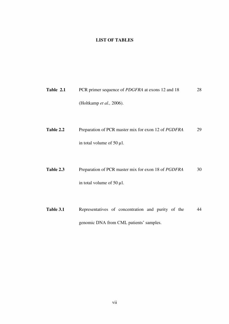
LIST OF TABLES
Table 2.1 PCR primer sequence of PDGFRA at exons 12 and 18
(Holtkamp et al., 2006).
28
Table 2.2 Preparation of PCR master mix for exon 12 of PGDFRA
in total volume of 50 µl.
29
Table 2.3 Preparation of PCR master mix for exon 18 of PGDFRA
in total volume of 50 µl.
30
Table 3.1 Representatives of concentration and purity of the
genomic DNA from CML patients’ samples.
44

viii
LIST OF FIGURES
Figure 1.1 Schematic representation of the most frequent activating
mutations of the homologous PDGFRA kinase in patients
with GIST.
14
Figure 1.2 Diagrammatic representation of the structure of the KIT and
PDGFRA showing the corresponding identified mutations of
both genes in GIST.
16
Figure 2.1 Flowchart of the study.
22
Figure 3.1 Distribution of CML samples from various hospitals recruited
in the study.
37
Figure 3.2 Study subjects distribution by gender and their response
towards IM.
38
Figure 3.3 Ethnic breakdown of CML patients in this study and their
response towards IM therapy.
40
Figure 3.4 Age group distribution and respond of CML patients towards
IM treatment.
41

ix
Figure 3.5
Distribution of IM-resistant patients in different phases of
CML.
42
Figure 3.6 Representatives of genomic DNAs bands of CML patients on
2% agarose gel for integrity checking.
45
Figure 3.7 Representatives of PCR products for exon 12 (lane 1 to 3) and
exon 18 (lane 4 to 7) on 2% of agarose gel.
47
Figure 3.8 Representatives of electropherogram results of selected region
on exon 12.
48
Figure 3.9 Representatives of electropherogram results of selected region
on exon 18.
49
Figure 3.10 Representatives of sequence samples for exon 12 after
aligning by BLAST.
51
Figure 3.11 Representatives of sequence samples for exon 12 after
aligning by BLAST.
52

x
LIST OF ABBREVIATIONS
% percent
µl microlitre
µM micromolar
A (Ala) alanine
A260/280 optical spectrometer measurement of absorbance at the
wavelengths of 260 nm over 280 nm
ALL acute lymphoid leukemia
AML acute myeloid leukemia
AP accelerated phase
ATP adenosine triphosphate
BCR-ABL BCR-ABL gene
BCR-ABL BCR-ABL protein
BLAST Basic Local Alignment Search Tool
BP blastic phase
bp
CCyR
base pair
complete cytogenetic response
CML chronic myeloid leukemia
CP
CyR
chronic phase
cytogenetic response
D (Asp) aspartic acid
ddH2O deionized distilled water
DMSA dimethyl sulphoxide

xi
DNA deoxyribonucleic acid
dNTP dinucleotide triphosphotase
EDTA ethylenediamine tetraacetic acid
FGFR1 fibroblast growth factor receptor 1 gene
g gram
gDNA genomic deoxyribonucleic acid
GIST gastrointestinal stromal tumour
HCL hydrochloride
HES hypereosinophilic syndrome
HPP Hospital Pulau Pinang
HRM high-resolution melting
HRPB Hospital Raja Permaisuri Bainun
HSA Hospital Sultanah Aminah
HUKM Hospital Universiti Kebangsaan Malaysia
HUSM Hospital Universiti Sains Malaysia
I (Ile) isoleucine
IM imatinib mesylate
IRIS International Randomized Study of Interferon versus
STI571
K (Lys) Lysine
KIT SCFR stem cell factor receptor gene
L (Leu) leucine
LNs
MCyR
lymphoid neoplasms
major cytogenetic response
MDS myelodysplastic syndrome

xii
MgCl2 magnesium chloride
ml mililiter
mM milimolar
MNs myeloid neoplasms
MPN myeloproliferative neoplasms
N (Asn) Asparagine
NCBI National Center for Biotechnology Information
NGS next generation sequencing
NK natural killer
nm nanometer
oC degree celsius
P (Pro) proline
PCR polymerase chain reaction
PDGF platelet-derived growth factor
PDGFA platelet-derived growth factor alpha protein
PDGFR platelet-derived growth factor receptor
PDGFRA platelet-derived growth factor receptor alpha protein
PDGFRA platlet-derived growth factor receptor alpha gene
PDGFRB platelet-derived growth factor receptor beta protein
PDGFRB platelet-derived growth factor receptor beta gene
Ph Philadelphia
S (Ser) serine
T (Thr) threonine
taq Thermophilus aquaticus
TBE Tris-Boric Acid-EDTA

xiii
TERT Telomerase reverse transcriptase gene
TKI tyrosine kinase inhibitor
UK United Kingdom
USA United States of America
UV ultra violet
V volt
V (Val) valine
WHO World Health Organisation

xiv
ABSTRAK
PENGESANAN MUTASI DI PDGFRA PADA EKSON 12 DAN 18 DI KALANGAN
PESAKIT-PESAKIT LEUKEMIA MEILOID KRONIK YANG TELAH DIRAWAT
DENGAN IMATINIB MESYLATE
Rintangan terhadap rawatan imatinib mesylate (IM) merupakan cabaran baru dan utama
dalam merawat pesakit leukemia mieloid kronik (CML). Rintangan terhadap IM boleh
dibahagikan kepada dua laluan iaitu laluan yang bergantung kepada BCR-ABL dan laluan
yang tidak bergantung kepada BCR-ABL. Mekanisma yang berada di laluan yang tidak
bergantung kepada BCR-ABL diuji dalam projek ini; yang melibatkan mutasi di PDGFRA
sebagai mekanisma rintangan pada pesakit -pesakit CML positif Philadelphia yang dirawat
dengan IM. PDGFRA adalah tergolong dalam kumpulan tyrosine kinase Kelas III; bukan
sahaja menyumbang kepada pembentukan haematopoiesis tetapi juga telah dikaitkan dengan
kanser. Mutasi PDGFRA membawa kepada pengaktifan kendirian menyebabkan penambahan
spontan sel. Analisis penjujukan PCR-DNA telah dijalankan untuk mengesan mutasi
PDGFRA pada ekson 12 dan 18. Lapan puluh enam pesakit CML positif Philadelphia dari
lima hospital rujukan di seluruh Semenanjung Malaysia yang berada dalam fasa penyakit
yang berbeza dirawat dengan IM dari tahun 2010 sehingga 2013 telah dinilai (pesakit yang

xv
responsif, n= 43 ; pesakit yang rintang, n=43). Sampel pesakit-pesakit ini diperoleh daripada
kajian projek berkaitan CML terdahulu. Daripada kesemua pesakit-pesakit dalam kumpulan
yang rintang terhadap IM, 32 pesakit berada dalam fasa CML kronik; 7 pesakit dalam fasa
percepatan CML; dan 4 pesakit dalam fasa blastik CML. Pesakit yang berumur dari 20
hingga 73 tahun telah dikategorikan ke dalam pelbagai kumpulan umur dari pesakit yang
lebih muda (umur yang lebih muda daripada 60 tahun) sehingga pesakit yang lebih tua (umur
60 tahun atau lebih tua). Amplifikasi tindak balas rantai polimerase di lokasi yang terpilih
telah dijalankan iaitu pada ekson 12 dan 18 diikuti oleh saringan mutasi oleh penjujukan
langsung untuk kesemua sampel. Keputusan penjujukan telah sejajar dengan menggunakan
Asas Tempatan Penjajaran Search Tool (BLAST) untuk membandingkan urutan pertanyaan
dengan rujukan urutan. Kebanyakan pesakit yang tidak memberi respon terhadap rawatan IM
adalah wanita. Median umur ialah 43 tahun. Sebanyak 74 % daripada pesakit CML yang
tidak memberi respon terhadap IM berada dalam fasa kronik. Namun begitu, tiada pesakit
CML dalam kajian ini menunjukkan mempunyai mutasi pada ekson potensi; ekson 12 dan 18
dalam PDGFRA. Kajian ini menunjukkan wanita mempunyai kekerapan rintang yang lebih
tinggi terhadap rawatan IM dan berlaku terutamanya di kalangan pesakit yang lebih muda.
Ketiadaan mutasi PDGFRA di ekson 12 dan 18 kemungkinan kerana mutasi berada di lokasi-
lokasi lain pada gen ini. Majoriti sampel kajian adalah dari pesakit fasa kronik mungkin
menyumbang kepada mutasi negatif. Kajian pada masa depan haruslah berpandukan ke arah
mencari ekson-ekson lain yang berpotensi dengan memilih lebih pesakit dalam peringkat
berbahaya untuk meperolehi kekerapan mutasi yang lebih tinggi. Kajian klinikal dan
tambahan ‘pathogenetic’ diperlukan untuk memahami kaitan antara PDGFRA dan IM rintang
CML.

xvi
ABSTRACT
DETECTION OF PDGFRA MUTATIONS AT EXONS 12 AND 18 AMONG CHRONIC
MYELOID LEUKEMIA PATIENTS TREATED WITH IMATINIB MESYLATE
Imatinib mesylate (IM) resistance is an emerging and major challenge in the treatment of
patients with chronic myeloid leukaemia (CML). Resistance toward IM can be divided into
BCR-ABL dependent pathways and BCR-ABL independent pathway. In this study, the BCR-
ABL independent pathway was investigated; the involvement of PDGFRA mutation as a
mechanism of resistance in Philadelphia positive CML patients treated with IM. The
PDGFRA belongs to the tyrosine kinase Class III; not only contribute to haematopoiesis
development but also has been implicated in cancers. Mutation of PDGFRA leads to
constitutive activation causing spontaneous proliferation. PCR-DNA sequencing analyses
were carried out to detect exons 12 and 18 of PDGFRA mutations. Eighty-six patients from
five tertiary hospitals around peninsular Malaysia in different phases of Philadelphia-positive
CML who were treated with IM from 2010 until 2013 were evaluated (IM-responsive

xvii
patients, n= 43; IM-resistant patients, n=43). These samples were archived from the earlier
CML project. Of all the patients in resistant group, 32 patients in chronic phase CML; 7
patients in accelerated phase CML; and 4 patients in blastic phase CML. Patients aged from
20 until 73 years were categorized into a range of age group from younger patients (age
younger than 60 years) till older patients (age 60 years or older). Polymerase chain reaction
amplifications were performed on the selected hotspots; exons 12 and exon 18 followed by
screening for mutations by direct sequencing in 43 resistant and 43 responsive CML samples.
Sequencing results were aligned by using Basic Local Alignment Search Tool (BLAST) to
compare a query sequence with a reference sequences. Resistant patients predominate by
female and the median age was 43. There were 74% of the resistant CML in chronic phase.
None of the CML patients in this study exhibit any mutation on the hotspot exons 12 and 18
in PDGFRA. This study shows higher frequency of IM resistance notable in female and
occurring mainly in younger age population. The absence of PDGFRA mutation at exons 12
and 18 may suggest that other regions of this gene could be involved. Majority of the study
samples were from chronic phase patients might contribute to the negative mutation finding.
Future study should be directed towards finding more potential exons by selecting more
patients in advance stage in order to yield higher mutation frequency. Additional clinical and
pathogenetic studies are needed to understand the association between PDGFRA and IM-
resistant CML.

1
CHAPTER 1
LITERATURE REVIEW

2
1.1 Introduction
1.1.1 Blood malignancies
Blood malignancies or haematological neoplasms are comparatively common,
accounting for around 9% of all cancers and being the fourth most frequently diagnosed
cancer in both men (after prostate, lung, and colorectum) and women (after breast, lung, and
colorectum) in economically developed regions of the world (Smith et al., 2011).
These neoplasms are forms of cancer that begin in the cells of blood-forming tissue, such
as the bone marrow, or in the cells of the immune system. It may derive from either of the
two major blood cell lineages: myeloid and lymphoid cell lines. The myeloid cell line
normally produces granulocytes, erythrocytes, thrombocytes, macrophages and mast cells;
the lymphoid cell line produces B, T, natural killer (NK) and plasma cells. Malignancies such
as lymphomas, lymphocytic leukemias, and myeloma are from the lymphoid line, while acute
and chronic myeloid leukemia (CML), myelodysplastic syndromes (MDS) and
myeloproliferative neoplasms (MPNs) are myeloid in origin.
Unlike many other cancers, haematological neoplasms are diagnosed using multiple
parameters; such as morphology, cytochemistry, immunophenotype, genetics and clinical
features in order to define clinically significant disease entities. The current classification
system was established by the World Health Organisation (WHO) in 2001 and more recently
modified in 2008 to refine the diagnosis (Swerdllow et al., 2008). This classification stratifies
neoplasms according to their lineage (myeloid, lymphoid, histiocytic/dendritic) and
distinguishes neoplasms of precursor cells from those comprised of functionally mature cells.

3
Brief overviews on lymphoid neoplasms (LN), LNs are derived from the clonal
expansion and proliferation of B- and T-lymphocytes. They encompass a heterogeneous
group of lymphomas and leukemias including B-cell, T-cell, and NK-cell disorders. Whereas
for myeloid neoplasm (MNs), five major subgroups of MNs are recognized based mainly on
their degree of maturation and biologic properties: MPNs which are comprised primarily of
mature cells with effective proliferation; myeloid (and lymphoid) neoplasms with
eosinophilia and abnormalities of platelet-derived growth factor alpha gene (PDGFRA),
platelet-derived growth factor beta gene (PDGFRB) and fibroblast growth factor receptor 1
gene (FGFR1), defined largely by the finding of significant eosinophilia and specific genetic
abnormalities; MDS/MPN comprised mainly of mature cells with both effective and
ineffective proliferation of various lineages; MDS, in which immature and mature cells are
found with abnormal, dysplastic and ineffective maturation, and acute myeloid leukemia
(AML), comprised of precursor cells with impaired maturation (Vardiman, 2010).
According to the revised WHO Classification Scheme in year 2008, CML was assigned
under MPN category together with polycythemia vera, essential thrombocytopenia, primary
myelofibrosis, chronic neutrophilic leukemia, chronic eosinophilic leukemia (not otherwise
specified), mast cell disease and MPN unclassifiable (Tefferi et al., 2009).

4
1.1.2 Chronic Myeloid Leukemia and its genetic basis
CML is characterized by uncontrolled expansion of myeloid cells particularly the
granulocytic cell line without the loss of their capacity to differentiate. The incidence of CML
were 1 to 2 cases per 100,000 adults, and accounts for 15% of newly diagnosed cases
of leukemia in adults (Jemal et al., 2010) .
This is the first human cancer in which a consistent genetic abnormality was
demonstrated to cause the disease (Sherbenou and Druker, 2007). CML is associated with
the presence of the Philadelphia (Ph) chromosome leading to a BCR-ABL fusion. The Ph
chromosome is the result of a balanced t(9;22)(q34;q11) translocation, and is observed in
more than 90% of CML cases, with variant Ph translocations being observed in the remainder
(O'Brien et al., 1997).
This oncogenic fusion BCR-ABL, produce a constitutively active tyrosine kinase and
is important in the pathogenesis and expression of CML. CML ensues when an abnormal
pluripotent hematopoietic progenitor cell initiates excessive production of granulocytes,
primarily in the bone marrow but also manifested in extramedullary sites (eg. spleen, liver).
Although granulocyte production predominates, the neoplastic clones include red blood cells,
megakaryocytes, monocytes, and even some T and B cells.
With a rapid expansion of granulocytes and advance disease ensues, metastasis may
come to the picture which later progress to organ failure and death. Epidemiologic data
indicates that almost 5000 new cases are reported every year and 10% of these patients
eventually succumb to the disease (An et al., 2010).

5
1.1.2.1 Classification and clinical phases
CML is classified into three phases based on clinical features and laboratory findings;
chronic, accelerated, and blast phase. Approximately 90% of patients are diagnosed in the
chronic phase (CP) and, historically have a median survival close to 5 years (Cortes et al.,
2006). In the CP of disease, mature cells proliferate exceeding the normal rate. Patients at this
phase are asymptomatic but CML progression is insidious, with a nonspecific “benign” stage
(malaise, anorexia, weight loss). Unless the disease is treated, CML evolves from a CP
characterized by the Ph chromosome as the sole genetic abnormality into blast crisis or blast
phase (BP), which is often associated with additional chromosomal and molecular secondary
changes (Calabretta and Perrotti, 2004). This is considered as a natural progression of
disease.
During this phase, patient manifests ominous signs such as splenomegaly, pallor, easy
bruising and bleeding, fever, lymphadenopathy and skin changes. Two-thirds of patients who
develop BP go through an intermediate accelerated phase (AP) with median survival of
patient in AP is 1 to 2 years (Cortes et al., 2006). The BP is the last stage or blast crisis where
immature cells rapidly proliferate and more or less resembling the acute leukemia and at this
point of progression the patient deteriorates considerably fast leading to mortality.
The term “advanced phase” is sometimes used to describe both the AP and BP. There
are two distinct types of blast phases defined by the types of blast cells present. In most
patients the blast cells (immature white cells) resemble those seen in AML, whereas in about
a quarter of patients, the blast cells look more like those seen in acute lymphoblastic

6
leukaemia (ALL). This latter form of the disease is known as lymphoid blast crisis and tends
to have a poorer response to treatment.
CML cases were account for 20% of all adult leukemias worldwide (Singer et al.,
2011). It typically affects middle-aged individuals. Uncommonly, the disease occurs in
younger individuals. Younger patients may present with a more aggressive form of CML,
such as in accelerated phase or blast crisis. Uncommonly, CML may appear as a disease of
new onset in elderly individuals.
1.1.2.2 Treatment and management
The treatment of CML and the survival rate largely depends on the stage where the
diagnosis is made. The time of diagnosis does contribute to the prognosis of the patient with
this disease and directly related to the outcome of the patient as in chronic stage the
progression can be stopped. Most of the patients seen in AP transition to blast crisis, it is
imminent and the outcome is poor.
The elucidation of the molecular pathogenesis of CML led to the development of its
targeted therapy. This was unprecedented until CML was regarded as a life-threatening
disease with a median life expectancy of around six to seven years with the only exception
being the minority of patients who could receive a compatible stem cell transplant. Previous
years, cytotoxic drugs are the main modality as therapeutic option for CML before the
discovery of BCR-ABL. Nevertheless, effective chemotherapy does not change the natural
history of CML thus most patients would ultimately progress to blast phase.

7
The drugs that have been used by clinician to treat CML were hydroxyurea, busulfan
and interferon. Hydroxyurea is the easiest therapy to be managed and has the fewest adverse
effects. Unfortunately cytogenetic responses are rare and the onset to blast crisis is not
delayed, with transformation occurring within a median of 4 to 6 years (Salesse and
Verfaillie, 2002).
Busulfan has been known to cause unexpected general myelosuppression, and
interferon causes a flu-like syndrome that frequently is unacceptable to patients. In contrary,
these therapies alleviate the distressing splenomegaly and adenopathy and help in control of
the tumor burden to reduce the incidence of tumor lysis and gout. Too unfortunate, none of
these therapies prolongs median survival more than 1 year compared with untreated patients;
thus, reduction in symptoms is the major goal, and therapy is not continued when patients
have significant toxic symptoms (Porter et al., 2006).
1.1.2.2 (a) Imatinib mesylate (IM)
A very essential lesson learned from IM is that the discovery of the primary genetic
abnormality in a malignancy together with the development of an agent that targets that
abnormality, can lead to therapeutic success. IM is a 2-phenylaminopyrimidine derivative
developed originally as a general tyrosine kinase inhibitor (TKI) that was modified
chemically so as to compete with adenosine triphosphate (ATP) for the ATP-binding site or
P-loop in the ABL protein and thereby block the deregulated enzymatic function of the BCR-
ABL oncoprotein (Goldman, 2009).

8
This BCR-ABL TKI (Glivec®, formerly STI571, Novartis Pharma AG, Basel,
Switzerland) prevent tyrosine kinase turning into its active form thus inhibits cellular
proliferation without the induction of apoptosis. An in vitro study reported that IM produced
a 92% to 98% decrease in CML colony growth without significantly inhibiting normal colony
growth (An et al., 2010). It has been proven to produces complete hematologic and
cytogenetic responses in a substantial percentage of CML patients and it is effective in CP,
AP and blast crisis, with lower response rates in patients with more advanced disease
(Buchdunger et al., 2002).
The clinical use of IM has resulted in a significantly improved prognosis, response
rate, overall survival, and patient outcome in CML patients compared to previous therapeutic
regimens by busulfan, hydroxyurea and interferon. Results from the International
Randomized Study of Interferon and STI571 (IRIS) trial in newly diagnosed CML-
CP showed that, cumulatively, 98% of patients who received IM as initial therapy achieved a
complete haematological remission and 87% achieved a complete cytogenetic remission
(Assouline and Lipton, 2011). The above phases of disease progression have changed
dramatically in the IM era. For most patients, the CP lasts at least ten years, possibly much
longer, and only a minority of patients who start treatment in the CP fail to respond well to
IM.
Relapses have been common in blast crisis associated with reactivation of BCR-ABL
kinase activity. Approximately 33% of patients with CML treated with IM do not achieve a
complete cytogenetic response (CCyR), while others have drug resistance or cannot tolerate
drug-related toxicities (Bixby and Talpaz, 2009; Hochhaus et al., 2009). Two categories of
IM resistance have been characterized: primary resistance is the failure to achieve any of the

9
landmark responses established by the European LeukemiaNet (ELN) (Baccarani et al.,
2006). Primary resistance also known as intrinsic resistance can be further divided into
primary hematologic resistance and primary cytogenetic resistance. Primary hematologic
resistance accounts for 2 to 4% of cases who fail to normalize peripheral counts within 3 to 6
months of initiation of treatment whereas primary cytogenetic resistance, which is more
common, observed in approximately 15 to 25% of patients who fail to achieve any level of
cytogenetic response (CyR) at 6 months, a major CyR (MCyR) at 12 months or a complete
CyR (CCyR) at 18 months (Shah, 2007). Secondary resistance occurs in those who have
previously achieved and subsequently lost their response in accordance with the guideline
(Bhamidipati et al., 2013). The mechanisms of resistance to IM can be either BCR-ABL
dependent (gene amplification or point mutations) or BCR-ABL independent (La Rosée and
Deininger, 2010). The present study was designed to investigate the mechanisms of resistance
involving BCR-ABL independent pathway.
The use of pre-imatinib-era treatment strategies such as hydroxyurea, busulfan or
interferon by some physicians as salvage treatment after IM failure and unsuitability of stem
cell transplantation still occurs despite the growing availability of newer TKIs (Rohrbacher
and Hasford, 2009). This has reflected that not all CML cases are caused by solely Ph
chromosome translocation, some other molecular mechanism must have contributed to its
pathogenesis and disease progression making treatment with the most effective drug turned
into a complicated non effective effort.

10
1.1.3 Role of tyrosine kinases (TKs) in cancer
TKs are a subclass of protein kinase. Protein TKs are enzymes that transfer phosphate
groups to tyrosine residues on protein substrates. Phosphorylation of proteins cause changes
in their function and/or enzymatic activity resulting in specific biological responses (Gocek et
al., 2014). It functions as a switch in many cellular functions and is the key role in diverse
biological processes like growth, differentiation, metabolism and apoptosis in response to
external and internal stimuli.
As the sequencing of the human genome is completed by the Human Genome Project,
90 unique kinase genes can be identified. Of the 90 TKs, 58 are receptor type, receptor
tyrosine kinases (RTKs), distributed into 20 subfamilies and 32 non-receptor tyrosine kinases
(NRTKs) can be placed in 10 subfamilies (Robinson et al., 2000) . RTKs are part of the
larger family of protein tyrosine kinases which contain a transmembrane domain, whereas the
NRTKs do not possess transmembrane domains. Haematopoiesis is controlled by a number
of growth factors and cytokines, a number of which act through binding to high-affinity
RTKs (Reilly, 2003). RTKs are activated by ligand binding to the extracellular domain
followed by dimerization of receptors facilitating trans-phosphorylation in the cytoplasmic
domain (Paul and Mukhopadhyay, 2004). The most important downstream signalling
cascades activated by RTKs include the Ras–extracellular regulated kinase (ERK)–mitogen
activated protein kinase (MAPK) pathways, the phosphoinositide 3-kinase (PI 3-kinase)–Akt
and the Janus kinase and Signal Transducer and Activator of Transcription (JAK/STAT)
pathway leading either to a complex activation or repression of various subsets of genes for
running biological functions.

11
TKs have been implicated in the pathophysiology of cancer. Though their activity is
tightly regulated in normal cells, deregulation may result in deregulated TK activity with
constitutive or strongly enhanced signalling capacity, leading to malignancy. The most
important mechanisms leading to constitutive RTK signalling include: overexpression and/or
gene amplification of RTKs, genetic alterations such as deletions and mutations within the
extracellular domain as well as alterations of the catalytic site, or autocrine–paracrine
stimulation through aberrant growth factor loops (Zwick et al., 2002).
In CML, the important and related TK gene involves directly is ABL which is
classified under NRTK whereas the gene in this study is PDGFRA which is under class III
RTK. Class III RTK is arguably one of the most intriguing RTK classes in terms of
evolutionary, functional and clinical considerations (Grassot et al., 2006; Verstraete and
Savvides, 2012). Characterized by present of five immunoglobulin-like domains in the
extracellular ligand-binding region, this class of RTK has grown to harbour another four
members include PDGFRB (platelet-derived growth factor receptor beta), c-KIT (cellular
component-stem cell factor receptor, CD117), FLT3 (FMS-like tyrosine kinase, CD135) and
c-FMS (colony-stimulating factor-1 receptor).
1.1.4 Platelet derived growth factor receptor alpha (PDGFRA)
Platelet-derived growth factor receptors (PDGFRs) and their ligands, platelet-derived
growth factor (PDGFs) play critical roles in mesenchymal cell migration and proliferation
(Jones and Cross, 2004). These growth factors are mitogens for cells of mesenchymal origin.
Two types of PDGFRs have been identified: alpha-type and beta-type.

12
The PDGFRA belongs to the sub-family of proteins within the family of receptor
tyrosine kinases which is class III. The structure of the PDGFA receptor comprises an
extracellular domain (the portion extending outside of the cell membrane), a juxtamembrane
domain (the portion just inside of the cell membrane), and two sections inside the cell known
as the two catalytic parts of the kinase domain. (Refer Figure 1.1) One part of the kinase
domain is essential for the binding of ATP and the other part is needed for the transfer of a
phosphate group that leads to kinase activation. PDGFRA can forms homo- or heterodimers
by binding to the endogenous PDGFR ligands; PDGF-A, -B, -C and –D. Upon binding of the
receptor and its ligand, this will induce receptor dimerization and transphosphorylation at
specific tyrosine residues thus activate the intracellular kinase activity, initiating intracellular
signalling.
PDGFRA is the approved gene symbol according to the HUGO Gene Nomenclature
Committee (HGNC). The other names that also encodes for this gene were CD140A and
PDGFR2. The full length PDGFRA is encoded by a 6.4 kb transcript but there is also
evidence for a 3.0 kb transcript as a result of alternative splicing, potentially encoding a
truncated, dominant-negative receptor isoform (Mosselman et al., 1996). Both these
transcripts are generated from a promoter upstream of exon 1 (P1 promoter). This gene can
be found at chromosome 4 located at 4q12. This gene has 23 exons and 1089 amino acid
residues.
PDGFRA and other members in the same class were originally grouped under the
PDGFR family. They are crucially important in the development and homeostasis of the
cellular repertoire of the haematopoietic and immune systems but both PDGFR subtypes have
primary roles in mesenchymal processes such as angiogenesis, fibroblast proliferation, bone

13
formation and tissue repair (Verstraete and Savvides, 2012). Recently in Frenette`s group,
PDGFRA was shown to be one of the markers of hematopoiesis-supporting nestin-
expressing multipotent stromal cells in the human system (Pinho et al., 2013).

14
Figure 1.1 Structure of PDGFA receptor is comprised of five extracellular
immunoglobulin- like domains, a juxtamembrane domain and two catalytic part of
kinase domains. (Adapted from Pierotti et.al., 2011). (Pierotti et al., 2011)

15
1.1.4.1 IM resistance in PDGFRA mutation
The expanding understanding of the basis of IM-mediated tyrosine kinase inhibition
has revealed a spectrum of potential new antitumor applications apart from its excellence
capability in the treatment of CML. IM is a potent inhibitor of both receptors, PDGFRA and
PDGFRB and has shown activity in vivo against platelet-derived growth factor (PDGF)-
driven tumor models including gastrointestinal stromal tumours (GISTs), glioblastoma,
dermatofibrosarcoma protuberans and chronic myelomonocytic leukemia (Buchdunger et al.,
2002) .
GIST commonly harbours oncogenic mutations of the KIT or PDGFRA kinases,
which are targets for IM. (Heinrich et al., 2003b; Hirota et al., 2003). The PDGFRA is very
similar to the KIT (both are the member of same RTK family) and the genes coding
for KIT and PDGFA receptors are found in close proximity on chromosome 4q12 (Spritz et
al., 1994). Mutations of PDGFRA lead to independent activation of the receptor in the
absence of ligand; this is called constitutive activation (Heinrich et al., 2003b). This means
the cell is carrying out spontaneous proliferation without the normal signalling ligand telling
the cell to grow. The result is uncontrolled growth of a tumour. The “downstream” pathways
that come into play in the cell following activation of mutant PDGFRA are thought to be
fairly similar with KIT in GIST (Duensing et al., 2004). However, different sets of genes are
expressed depending on which gene is mutant (Antonescu et al., 2004; Subramanian et al.,
2004; Kang et al., 2006).
Approximately 80% of GISTs express the mutated receptor tyrosine kinase KIT.
However about 8% of GISTs have a normal KIT (wildtype) but 5% show mutations in the
gene for PDGFRA (Joensuu, 2006). PDGFRA mutations in GIST have been found in exons
12, 14 and 18 (corresponding to exons 11, 13 and 17 of KIT) (Refer Figure 1.2).

16
Figure 1.2 Diagrammatic representation of the structure of the KIT and PDGFRA showing the
corresponding identified mutations of both genes in GIST. The most frequent activating mutations
of the homologous PDGFRA kinase in patients with GIST are in exon 18, such as the D842V
substituition that shows resistance to IM. Mutation in the juxtamembrane domain (exon 12;
V561D most common) and exon 14 of tyrosine kinase 1 (TK1) domain (e.g., N659K) are less
common. (Adapted from Heinrich,2006). (Heinrich, 2006) Molecular basis for
Exon 9
Exon 11
Exon 13
Exon 12 (V561D)
Exon 14
Exon 18 (D842V)

17
According to a study done by Corless et.al., the leading mutation of all GIST caused by
PDGFRA activating mutations, is found on exon 18 which accounted for 89.6% followed by
9.3% in exon 12 (Corless et al., 2005). Of all the mutations in exon 18 of PDGFRA, the
D842V mutation is the most common and has been proven to be IM resistant (Heinrich et al.,
2003a). The similar mutational status might also happen in CML that leads to the resistant to
the treatment given.
1.2 Rationale of the study
This study may generate new data on potential involvement of PDGFRA mutations
among CML patients treated with IM. Since many data from previous studies have implicated
a subset of PDGFRA mutation has caused the IM resistance in GIST cases, this has instigate
the curiosity of knowing whether the same mechanism play a role in the resistance cases of
IM in CML patients. Thus in this research, since the most common sites of mutations in GIST
cases which resistant to IM occur at exons 12 and 18, we adapted the facts as hypotheses to
be tested in CML cases treated with IM that manifest similar behaviour.
Identifying key components involved in the CML pathogenesis may lead to the
exploration of new approaches that might eventually overcome the resistance of this
particular TKI. This may shed some light on how to treat resistance cases and might also
reflect a better prognosis of CML patients.
18
1.3 Objectives of the study
1.3.1 General objective
To determine whether there are PDGFRA mutations in exons 12 and exon 18 in CML
patients and any association with the resistance the patients experience on the treatment of
IM.
1.3.2 Specific objectives
To perform the PCR amplification of selected hot spots (exons 12 and 18) of
PDGFRA.
To sequence the PCR product to determine the presence of any PDGFRA mutations.
To relate frequency of any PDGFRA mutations with CML status in association with
IM treatment.

19
CHAPTER 2
MATERIALS AND METHODS

20
2.1 Study design and study subject
This is a comparative cross-sectional study conducted in the Human Genome Centre,
School of Medical Sciences, Universiti Sains Malaysia, Health Campus. This study was
approved by the Research Committee, School of Medical Sciences, Universiti Sains Malaysia
(Reference no:USMKK/PPP/JEPem(221.3[9]). The blood samples used in this study were
archival from the previous study entitled “Influence of TERT gene copy number status and its
mRNA expression level in association with telomerase activity in Chronic Myeloid Leukemia
patients resistance to Imatinib Mesylate treatment”.
Total of 86 samples from two different groups of patients enrolled by random
sampling included in this study which comprised of 43 samples from each group; resistant
group and responsive group. The samples were obtained from patients who were clinically
confirmed diagnosis of CML.
2.2 Sample size
Power and sample size (PS) software was employed to calculate sample size. A study
of independent cases and controls with 1 control(s) per case was designed. Prior data from a
study done in a group of patients with rearrangement of PDGFRA treated with IM indicate
that the failure rate among controls is 0.5 (Cools et al., 2003). If the true failure rate for
experimental subjects is 0.214, we will need to study 43 experimental subjects and 43 control
subject (which in this situation refers to response group) to be able to reject the null

21
hypothesis that the failure rates for experimental and control subjects are equal with
probability (power) 0.8. The Type 1 error probability associated with this test of this null
hypothesis is 0.05. An uncorrected chi-squared statistic was constructed to evaluate this null
hypothesis. Therefore, the total sample size is 86; 43 for the resistant and response group.
2.3 Flow of the study
The CML patients were categorized into two groups, the CML-IM resistant labeled as
“Resistant” (R) group and the CML-IM responsive labeled as “Good response” (G) group
based on their response towards three monitoring parameters; cytogenetic, molecular and
haematological. Commercial deoxyribonucleic acid (DNA) extraction kit, GeneAll Exgene
Blood SV mini was used to extract genomic DNA from blood. Polymerase Chain Reaction
(PCR) for PDGFRA on exon 12 and exon 18 were carried out to amplify the amplicons. To
detect the presence of DNA fragments from PCR products, the products were loaded to
agarose gel for electrophoresis and the stained gel was visualized under ultra violet (UV)
transilluminator. This followed by purification of the amplicons using Illustra TM
Exoprostar
TM Enzymatic PCR and Sequencing Clean-Up. The purified PCR products then were sent to
First Base Labarotory for DNA sequencing service. The sequencing results were screened by
using Basic Local Alignment Search Tool (BLAST) software from National Center for
Biotechnology Information (NCBI) by comparing with the sequence from the reference
journal; Holtkamp et al., 2006. To the final touch, data from the results were converted by
statistical analysis into the form of graphs and charts as shown in the Result chapter. The
flow chart of the study is shown in Figure 2.1.

22
Collection of DNA samples
(archival samples)
43 samples for resistant group
43 samples for responsive group
Selection of PCR primers of exons 12 & 18
(based on Holtkamp et al., 2006)
PCR amplifications of exons 12 & 18
PCR purification & quantification
DNA sequencing analysis
&
interpretation
2% agarose gel electrophoresis
&
Gel visualization using UV transilluminator
Figure 2.1 Flowchart of the study

23
2.4 Inclusion criteria
IM-resistant CML patient included in this study was those;
i. Confirmed clinically and hematologically to have CML with presence of Ph
chromosome.
ii. Undergone IM treatment (400 mg, 600 mg or 800mg dose) for at least 12 months
iii. Experienced signs of primary or secondary resistance to the treatment.
iv. Showed suboptimal response and failure to IM treatment at cytogenetic,
hematological and molecular level (based on European LeukemiaNet 2013 (ELN)
guidelines) (Baccarani et al., 2009).
IM-responsive CML patient included in this study was those;
i. Confirmed clinically and hematologically to have CML with presence of Ph
chromosome.
ii. Undergone IM treatment (400 mg, 600 mg or 800mg dose) for at least 12 months
iii. Showed optimal response to IM treatment at cytogenetic, hematological and
molecular level (based on ELN 2013 guidelines) (Baccarani et al., 2009).
2.5 Exclusion criteria
i. CML patients with absence of Ph chromosome confirmed by molecular test.
ii. CML patients who were treated with IM but less than 12 months.
iii. CML patients who are not treated with IM.

24
2.6 Methodology
2.6.1 Genomic DNA extraction
Genomic DNA (gDNA) was extracted from the patient’s peripheral blood which was
stored in EDTA tube using a commercial GeneAll Exgene Blood SV mini kit (GeneAll
Biotechnology, Korea) according to manufacturer’s instruction. Included in this kit are
GeneAll Proteinase K, Buffer CL, Buffer BL, Buffer BW, Buffer TW and Buffer AE. The
protocol begins with 20 µl of Proteinase K solution was pipetted into the bottom of a 1.5 ml
tube. This followed by 200 µl of blood from each sample was transferred to the tube and then
200 µl of Buffer BL was pipetted into the same tube. The tube was vortexed for 15 seconds to
mix it. The mixture was incubated at 56⁰C for 10 minutes using Thermomixer (Eppendorf,
Germany) before being centrifuged briefly to remove any drops from internal lid wall.
Next, 200 µl of absolute ethanol was added to the sample to precipitate the DNA. The
tube was placed on pulse-vortex for 15 seconds to mix the samples thoroughly, and then
centrifuged briefly for about 10 seconds. The mixture was transferred to the SV column
carefully, followed by centrifugation at 8000 rpm for 1 minute to resuspend the cell pellet.
The collection tube was then discarded and replaced with the new one. Next step was the
addition of 600 µl of Buffer BW into each SV column and then was centrifuged for 1 minute
at 8000 rpm. Once again, the collection tube was discarded and replaced with new collection
tube. A volume of 700 µl of Buffer TW was added and subjected to 1 minute centrifugation
at 6000 rpm. The pass-through was discarded and the SV column was reinserted back into the
collection tube. Centrifugation was then done at full speed for 1 minute to remove residual