Embed Size (px)
Citation preview

Detection of Early Recurrence with 18F-FDG PET in Patients with Cervical Cancer Sang-Young Ryu, MD1; Moon-Hong Kim, MD1; Suck-Chul Choi, MD1; Chang-Woon Choi, MD2; and Kyung-Hee Lee, MD1
1Department of Obstetrics & Gynecology, Korea Cancer Center Hospital, Seoul, Korea; and 2Department of Nuclear Medicine, Korea Cancer Center Hospital, Seoul, Korea
This study investigated the feasibility of PET with 18F-FDG to evaluate retrospectively early recurrence in patients with cervical cancer. Methods: From September 1997 to March 2000, 249 patients with no evidence of cervical cancer after treatment were investigated with 18F-FDG PET. 18F-FDG PET scanning, beginning 50 min after injection of 370 –555 MBq 18F-FDG, was performed. 18F-FDG uptake other than physiologic uptake was evaluated with the standardized uptake value and was analyzed by 2 observers who were unaware of CT or MRI data. CT or MRI and needle biopsies were performed to evaluate the positive lesions on 18F-FDG PET, and all patients were monitored closely for 6 mo for recurrence. Results: Of the 249 patients, 80 patients (32.1%) showed positive lesions with 18F-FDG PET, and 28 patients (11.2%) were clinically or histologically confirmed as having recurrences. Eighty-two percent of recurrence was detected within 6 –18 mo after diagnosis, and 89% of recurrence occurred in Fede ration Internationale de Gyne cologie et d’Obste trique (FIGO) stage IIb and stage III patients. The sensitivity and specificity of 18F-FDG PET for detection of early recurrence were 90.3% and 76.1%, respectively. The sensitivity of 18F-FDG PET was high in mediastinal, hilar, and scalene lymph nodes, spine, and liver; however, the sensitivity was relatively low in lung, retrovesical lymph nodes, and paraaortic lymph nodes. Three false-negative cases were detected in lung, retrovesical lymph nodes, and paraaortic lymph nodes. Conclusion: 18F-FDG PET was effective in detecting early recurrences in cervical cancer patients with no evidence of disease. 18F-FDG PET may be a useful follow-up method for cervical cancer, thereby providing the patients with early opportunities for sophisticated treatments.
Key Words: 18F-FDG PET; cervical cancer; recurrence
J Nucl Med 2003; 44:347–352
Cervical cancer is one of the most common gynecologic malignancies throughout the world. Although the overall mortality from cervical cancer has decreased because of early detection and treatment of preinvasive disease, it still
Received May 20, 2002; revision accepted Sep. 27, 2002. For correspondence or reprints contact: Sang-Young Ryu, MD, Department
of Obstetrics & Gynecology, Korea Cancer Center Hospital, 215-4, Kongneung-dong, Nowon-ku, Seoul, Korea.
E-mail: [email protected]
remains one of the leading causes of cancer death (1). Despite of carefully planned and executed treatments, approximately 30% of cervical cancer is known to eventually recur after treatment (2). Conventional follow-up methods, such as physical examination, Papanicolaou smear (Pap smear), and tumor markers, and radiologic imaging methods, such as CT or MRI, have been used to detect early recurrence; however, it is very difficult to achieve an early diagnosis of pelvic recurrence of cervical cancer (3).
PET with 18F-FDG, which is preferentially trapped in tumor cells, reveals a functional image of high glucose metabolism (4,5). Recently, 18F-FDG PET has been widely used for detection of early recurrence that cannot be diagnosed with conventional radiologic imaging studies and is known to be more accurate than CT or MRI in detecting recurrent lymph node metastases in several human cancers (6). In lung cancer, 18F-FDG PET showed 81% accuracy on the involvement of mediastinal lymph nodes, whereas CT showed only 52% accuracy (7). Similar observations have also been reported in breast cancer, melanoma, and other cancers (8,9).
In cervical cancer, the role of 18F-FDG PET has not been well established. Recently, Sugawara et al. (10) reported that 18F-FDG PET could detect 100% of cancers and 86% of lymph node metastasis, whereas CT was positive in 57% of lymph node metastasis in 21 cervical cancers, suggesting a promising role of 18F-FDG PET in cervical cancer detection. In other studies, 18F-FDG PET is known to be very effective not only in detecting early recurrences but also in preoperative staging and evaluating the response of treatment (11– 17). However, the feasibility of 18F-FDG PET in the early detection in cervical cancer recurrence is not well established.
The purpose of this study was to assess the feasibility of 18F-FDG PET in detecting early cancer recurrences in patients with no evidence of the ailment unmasked by conventional imaging methods.
MATERIALS AND METHODS
Patients From September 1997 to March 2000, 249 patients with cervi
cal cancer, showing no evidence of disease after treatment, under-
18F-FDG PET IN CERVICAL CANCER • Ryu et al. 347

went 18F-FDG PET as part of their investigations. The patients had histologically proven cervical cancers and were treated with surgery or radiation combined with or without chemotherapy according to the Federation Internationale de Gynecologie et d’Obstetrique (FIGO) clinical stage. The detection rate of 18F-FDG PET for recurrences was analyzed retrospectively.
All patients were treated and monitored according to standard protocol. In brief, most of the patients with stage Ib and stage IIa were treated with radical hysterectomy and bilateral pelvic lymphadenectomy. Postoperative adjuvant radiation therapy was performed on patients with high-risk factors such as full-thickness involvement of the cervix, parametrial invasion, lymphatic invasion, and positive resection margin. Definitive radiation therapy without (before 1999) or with (after 1999) chemotherapy was performed on patients with stage IIb or higher, and chemotherapy was performed on patients with distant metastasis. The chemotherapy regimen was based mainly on cisplatin, such as 5-fluorouracil (500 mg/m2) + cisplatin (50 mg/m2) or cyclophosphamide (500 mg/m2) + cisplatin (50 mg/m2). After treatment, patients were monitored every 3 mo in the first 2 y and every 6 mo thereafter for 5 y with tumor markers, Pap smears, chest radiography, and annual pelvic CT or MRI.
In patients who had undergone surgery, no evidence of disease was defined as normal follow-up tests, including physical examination, chest radiography, tumor marker (squamous cell carcinoma antigen), Pap smear, and annual radiologic imaging studies. In patients who were treated with radiation, those who showed complete disappearance of the lesion on radiologic imaging studies performed at least after 6 mo of treatment and who showed normal follow-up tests (described above) were defined as having no evidence of disease.
18F-FDG PET was recommended as a part of the work-up on all patients with high risk factors for recurrence such as full-thickness involvement of the cervix, parametrial invasion, lymphatic invasion, and positive resection margin. Among them, 249 patients, who showed no evidence of disease on previous annual pelvic CT or MRI, physical examination, chest radiography, tumor marker, and Pap smear, were selected retrospectively for analysis.
PET Patients were prepared with overnight fasting before 18F-FDG
injection. 18F-FDG PET was performed on an Advance HR+ scanner (General Electric, Waukesha, WI), starting 50 min after injection of 370 –555 MBq (10 –15 mCi) 18F-FDG with the bladder emptied by Foley catheter insertion and injection of diuretics to reduce tracer activity in the bladder.
18F-FDG PET images were interpreted by using a dedicated system (ECAT EXACT 921; Siemens/CTI, Knoxville, TN) with a 10.8-cm transverse field of view and a 2-dimensional acquisition mode. Three- to 5-min transmission scans and 8-min emission scans were obtained. Five or 6 bed positions were used to cover the area from the orbitomeatal line to the midfemoral line. Images were reconstructed on transaxial, sagittal, and coronal planes by means of the ordered-subset expectation maximization algorithm and segmented photon absorption correction and were interpreted by 2 observers on both film and computer displays, who were unaware of the clinical information of previous treatment and of annual CT or MRI data. Any focal uptake of 18F-FDG, which is considered not be physiologic on PET images, was measured on the basis of the standardized uptake value, being the radioactive
concentration in a hot spot divided by the injected dose and the patient’s body weight.
Diagnosis of Recurrence Any positive lesion on 18F-FDG PET was evaluated with CT or
MRI and was confirmed for recurrence histologically by fine-needle aspiration (FNA) as quickly as possible. For the lymph nodes in the mediastinum, hilum, paraaorta, and pelvis, lymph nodes of >1 cm in the short axis on CT were interpreted as positive for metastasis. Lymph nodes that had prominent uptake on 18F-FDG PET but were <1 cm in the short axis were reevaluated with CT 3 mo later. If there was no change in the size on the follow-up CT scan, the patients were recommended to follow-up every 3 mo for 1 y. All scalene lymph nodes with obvious 18F-FDG uptake were evaluated for recurrence with FNA or node dissection.
Any prominent lesion in the lung parenchyma on 18F-FDG PET was evaluated for recurrence with chest CT and histologically confirmed by FNA or lung biopsy. Small lung lesions of <0.5 cm, which were difficult to access with FNA, were evaluated with CT 3 mo later. If there was no change in the size on the follow-up CT scan, the patients were recommended to follow-up every 3 mo for 6 mo.
In other body regions such as the retrovesical area, liver, and chest wall, any lesion of >1 cm in size on CT was confirmed by FNA. A prominent uptake on 18F-FDG PET of <1 cm in the short axis was reevaluated with CT 3 mo later. If there was no change in size on the follow-up CT scan, the patients were recommended to follow-up every 3 mo for 6 mo.
CT or MRI images to confirm recurrences were analyzed by 2 separate observers, who were unaware of 18F-FDG PET data and clinical information. If there was no histologic evidence of recurrence, we decided there was no evidence of disease after close follow-up for 1 y.
RESULTS
The median age of the patients was 51 y (range, 31–78 y), and 59.7% of the patients were classified as FIGO stage Ib and stage IIa. Histologically, 90.7% of the cervical cancer was squamous cell carcinoma. The duration of no evidence of disease at the point of 18F-FDG PET was not statistically different in stages Ib, IIa, and IIb; however, 18F-FDG PET scans were obtained earlier in patients with stage III than in patients with other stages. The median interval from the last CT or MRI to 18F-FDG PET was 6 mo (Table 1).
Of the 249 patients with cervical cancer who showed no evidence of disease after treatment, 80 patients (32.1%) showed positive lesions on 18F-FDG PET (Table 2). Among the 80 patients with positive 18F-FDG PET scans, 28 patients (11.2% [28/249 patients]) were clinically or histologically confirmed to have recurrent lesions (Fig. 1). The sensitivity and specificity of 18F-FDG PET in detecting recurrences of cervical cancer were 90.3% and 76.1%, respectively. Positive and negative predictive values of 18FFDG PET in detecting recurrence of cervical cancers were 35% and 98.2%, respectively.
Most recurrences were detected within 18 mo after diagnosis of the disease, and the peak period of detection was 9 –12 mo after diagnosis (Fig. 2). The detection rate of
348 THE JOURNAL OF NUCLEAR MEDICINE • Vol. 44 • No. 3 • March 2003

TABLE 1 Patient Characteristics
Characteristic Value
No. of patients 249 Age (y) (range) 51 (31–78) Stage*
Ib 100 (40.1) IIa 49 (19.6) IIb 83 (33.3) III or IV 17 (6.8)
Histology* Squamous 226 (90.7) Adenocarcinoma 10 (4) Adenosquamous 5 (2) Other† 8 (3.2)
Treatment* Surgery 86 (34.5) Radiation 90 (36.1) Surgery + radiation 68 (27.3) Chemotherapy 5 (2)
Median duration of 18F-FDG PET from last CT or MRI (mo) (range) 6 (3–12)
Median duration of NED (mo) (range) 30 (6–282) Ib 30 (7–129) IIa 35 (7–108) IIb 31 (6–282) III or IV 16 (6–165)
*Values in parentheses are percentages. †Six undifferentiated, 1 clear, and 1 glassy cell. NED no evidence of disease.
18F-FDG PET according to the stage was higher in FIGO stages IIb and III than in stages Ib and IIa (20.4% and 29.4% vs. 6% and 6.1%, respectively) (Fig. 3).
The sensitivity of 18F-FDG PET was relatively high in lesions such as the mediastinum, hilum, chest wall, scarlene lymph node, iliac, spine, and liver; however, it was relatively low in lesions including the lung, retrovesical area, and paraaortic lymph node. The specificity of 18F-FDG PET was relatively low in lesions such as the lung, retrovesical lymph node, and paraaortic lymph node (Table 3). Incidentally, as shown in Figure 4, 18F-FDG PET detected 2 cases each of thyroid cancer and tuberculosis.
DISCUSSION
This study showed that 18F-FDG PET is a useful method to detect early recurrences in patients with cervical cancer who showed no evidence of disease after treatment. Because all patients in the study showed no evidence of disease on physical examination, tumor markers, chest radiography, and annual pelvic CT or MRI, the 28 patients who were confirmed to have recurrence were detected only by 18FFDG PET.
It is estimated that approximately 35% of patients with invasive cervical cancer will have recurrent or persistent disease after therapy (2). Conventional imaging modalities
such as CT or MRI were performed to detect early recurrent lesions; however, the detection rate is low (18). Sugawara et al. (10) reported that 18F-FDG PET could detect lymph node metastasis more accurately than CT or MRI in patients with cervical cancer. 18F-FDG PET could detect recurrences in small lesions of <1 cm and in the retrovesical area, which are frequently obscured by postradiation fibrosis. In a retrospective study performed on 13 patients with cervical cancer, 18F-FDG PET could detect recurrences in 10 patients who had recurrences in the iliac lymph node, liver, lung, and paraaortic lymph node, suggesting a promising role of 18F-FDG PET in cervical cancer (16).
The higher feasibility of 18F-FDG PET over CT or MRI in detecting recurrences of cervical cancer may be explained by several factors. First, because 18F-FDG PET scans can provide functional information on the lesions rather than anatomic images, it can detect recurrent lesions independent of the size (7–9). Furthermore, as already well recognized in head and neck cancers (6,19), 18F-FDG PET provides more important images when anatomy has been distorted after surgery or radiation treatment. In our study, 18F-FDG PET could detect occult recurrent metastasis in lesions such as the vaginal cuff, retrovesical area, and pelvic sidewall, where it is difficult to differentiate between fibrosis and recurrence.
The other advantage of 18F-FDG PET is that it can show a whole-body image at one time. Approximately 70% of recurrences of cervical cancer are estimated to be distant or a combination of local and distant metastases (2). Most of the distant metastasis is detected in an already far-advanced state with clinical symptoms such as cough, hemoptysis, and pain. Because 18F-FDG PET can provide a whole-body image at one time, distant metastases, which are usually not evident on routine pelvic CT or MRI, can easily be detected by 18F-FDG PET. In this study, 18F-FDG PET was useful in detecting metastasis in lesions such as the scarlene lymph node, lung, and mediastinum, where it was difficult to detect recurrence with conventional imaging modalities. Two cases each of pulmonary tuberculosis and thyroid cancer, incidentally detected in this study, also benefited from the whole-body image of 18F-FDG PET.
Most recurrence in cervical cancer is known to occur within 2 y after therapy (2); however, the peak period of
TABLE 2 Detection of Early Recurrence with 18F-FDG PET
in Cervical Cancer
Parameter No. of patients %
No. of patients 249 Negative PET scan 169 67.8
True-negative 166 66.6 False-negative 3 1.2
Positive PET scan 80 32.1 True-positive 28 11.2 False-positive 52 20.8
18F-FDG PET IN CERVICAL CANCER • Ryu et al. 349
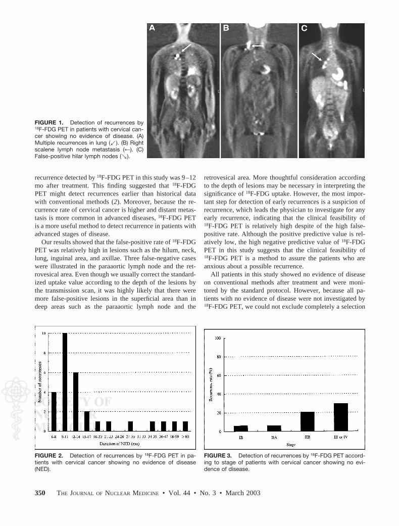
FIGURE 2. Detection of recurrences by 18F-FDG PET in patients with cervical cancer
FIGURE 3. Detection of recurrences by 18F-FDG PET according to stage of patients with cervical cancer showing no evidence of disease. (NED).
FIGURE 1. Detection of recurrences by 18F-FDG PET in patients with cervical cancer showing no evidence of disease. (A) Multiple recurrences in lung (,). (B) Right scalene lymph node metastasis (4). (C) False-positive hilar lymph nodes (n).
recurrence detected by 18F-FDG PET in this study was 9 –12 mo after treatment. This finding suggested that 18F-FDG PET might detect recurrences earlier than historical data with conventional methods (2). Moreover, because the recurrence rate of cervical cancer is higher and distant metastasis is more common in advanced diseases, 18F-FDG PET is a more useful method to detect recurrence in patients with advanced stages of disease.
Our results showed that the false-positive rate of 18F-FDG PET was relatively high in lesions such as the hilum, neck, lung, inguinal area, and axillae. Three false-negative cases were illustrated in the paraaortic lymph node and the retrovesical area. Even though we usually correct the standardized uptake value according to the depth of the lesions by the transmission scan, it was highly likely that there were more false-positive lesions in the superficial area than in deep areas such as the paraaortic lymph node and the
retrovesical area. More thoughtful consideration according to the depth of lesions may be necessary in interpreting the significance of 18F-FDG uptake. However, the most important step for detection of early recurrences is a suspicion of recurrence, which leads the physician to investigate for any early recurrence, indicating that the clinical feasibility of 18F-FDG PET is relatively high despite of the high false-positive rate. Although the positive predictive value is relatively low, the high negative predictive value of 18F-FDG PET in this study suggests that the clinical feasibility of 18F-FDG PET is a method to assure the patients who are anxious about a possible recurrence.
All patients in this study showed no evidence of disease on conventional methods after treatment and were monitored by the standard protocol. However, because all patients with no evidence of disease were not investigated by 18F-FDG PET, we could not exclude completely a selection
showing no evidence of disease
350 THE JOURNAL OF NUCLEAR MEDICINE • Vol. 44 • No. 3 • March 2003
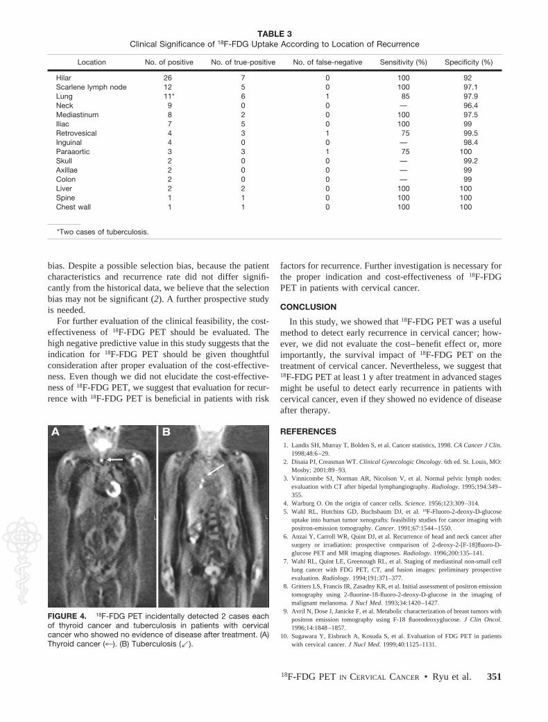
FIGURE 4. 18F-FDG PET incidentally detected 2 cases each of thyroid cancer and tuberculosis in patients with cervical
TABLE 3 Clinical Significance of 18F-FDG Uptake According to Location of Recurrence
Location No. of positive No. of true-positive No. of false-negative Sensitivity (%) Specificity (%)
Hilar 26 7 0 100 92 Scarlene lymph node 12 5 0 100 97.1 Lung 11* 6 1 85 97.9 Neck 9 0 0 — 96.4 Mediastinum 8 2 0 100 97.5 Iliac 7 5 0 100 99 Retrovesical 4 3 1 75 99.5 Inguinal 4 0 0 — 98.4 Paraaortic 3 3 1 75 100 Skull 2 0 0 — 99.2 Axillae 2 0 0 — 99 Colon 2 0 0 — 99 Liver 2 2 0 100 100 Spine 1 1 0 100 100 Chest wall 1 1 0 100 100
*Two cases of tuberculosis.
bias. Despite a possible selection bias, because the patient characteristics and recurrence rate did not differ significantly from the historical data, we believe that the selection bias may not be significant (2). A further prospective study is needed.
For further evaluation of the clinical feasibility, the cost-effectiveness of 18F-FDG PET should be evaluated. The high negative predictive value in this study suggests that the indication for 18F-FDG PET should be given thoughtful consideration after proper evaluation of the cost-effectiveness. Even though we did not elucidate the cost-effectiveness of 18F-FDG PET, we suggest that evaluation for recurrence with 18F-FDG PET is beneficial in patients with risk
cancer who showed no evidence of disease after treatment. (A) Thyroid cancer (4). (B) Tuberculosis (,).
factors for recurrence. Further investigation is necessary for the proper indication and cost-effectiveness of 18F-FDG PET in patients with cervical cancer.
CONCLUSION
In this study, we showed that 18F-FDG PET was a useful method to detect early recurrence in cervical cancer; however, we did not evaluate the cost– benefit effect or, more importantly, the survival impact of 18F-FDG PET on the treatment of cervical cancer. Nevertheless, we suggest that 18F-FDG PET at least 1 y after treatment in advanced stages might be useful to detect early recurrence in patients with cervical cancer, even if they showed no evidence of disease after therapy.
REFERENCES
1. Landis SH, Murray T, Bolden S, et al. Cancer statistics, 1998. CA Cancer J Clin. 1998;48:6 –29.
2. Disaia PJ, Creasman WT. Clinical Gynecologic Oncology. 6th ed. St. Louis, MO: Mosby; 2001:89 –93.
3. Vinnicombe SJ, Norman AR, Nicolson V, et al. Normal pelvic lymph nodes: evaluation with CT after bipedal lymphangiography. Radiology. 1995;194:349 – 355.
4. Warburg O. On the origin of cancer cells. Science. 1956;123:309 –314. 5. Wahl RL, Hutchins GD, Buchsbaum DJ, et al. 18F-Fluoro-2-deoxy-D-glucose
uptake into human tumor xenografts: feasibility studies for cancer imaging with positron-emission tomography. Cancer. 1991;67:1544 –1550.
6. Anzai Y, Carroll WR, Quint DJ, et al. Recurrence of head and neck cancer after surgery or irradiation: prospective comparison of 2-deoxy-2-[F-18]fluoro-Dglucose PET and MR imaging diagnoses. Radiology. 1996;200:135–141.
7. Wahl RL, Quint LE, Greenough RL, et al. Staging of mediastinal non-small cell lung cancer with FDG PET, CT, and fusion images: preliminary prospective evaluation. Radiology. 1994;191:371–377.
8. Gritters LS, Francis IR, Zasadny KR, et al. Initial assessment of positron emission tomography using 2-fluorine-18-fluoro-2-deoxy-D-glucose in the imaging of malignant melanoma. J Nucl Med. 1993;34:1420 –1427.
9. Avril N, Dose J, Janicke F, et al. Metabolic characterization of breast tumors with positron emission tomography using F-18 fluorodeoxyglucose. J Clin Oncol. 1996;14:1848 –1857.
10. Sugawara Y, Eisbruch A, Kosuda S, et al. Evaluation of FDG PET in patients with cervical cancer. J Nucl Med. 1999;40:1125–1131.
18F-FDG PET IN CERVICAL CANCER • Ryu et al. 351

11. Shimotsu Y, Ishida Y, Fukuchi K, et al. Fluorine-18-fluorodeoxyglucose PET identification of cardiac metastasis arising from uterine cervical carcinoma. J Nucl Med. 1998;39:2084 –2087.
12. Rose PG, Adler LP, Rodriguez M, et al. Positron emission tomography for evaluating para-aortic nodal metastasis in locally advanced cervical cancer before surgical staging: a surgicopathologic study. J Clin Oncol. 1999;17:41– 45.
13. Umesaki N, Tanaka T, Miyama M, et al. Early diagnosis and evaluation of therapy in postoperative recurrent cervical cancers by positron emission tomography. Oncol Rep. 2000;7:53–56.
14. Grigsby PW, Siegel BA, Dehdashti F. Lymph node staging by positron emission tomography in patients with carcinoma of the cervix. J Clin Oncol. 2001;19:3745–3749.
15. Reinhardt MJ, Ehritt-Braun C, Vogelgesang D, et al. Metastatic lymph nodes in
patients with cervical cancer: detection with MR imaging and FDG PET. Radiology. 2001;218:776 –782.
16. Kerr IG, Manji MF, Powe J, et al. Positron emission tomography for the evaluation of metastases in patients with carcinoma of the cervix: a retrospective review. Gynecol Oncol. 2001;81:477– 480.
17. Narayan K, Hicks RJ, Jobling T, et al. A comparison of MRI and PET scanning in surgically staged loco-regionally advanced cervical cancer: potential impact on treatment. Int J Gynecol Cancer. 2001;11:263–271.
18. Hricak H, Yu KK. Radiology in invasive cervical cancer. AJR. 1996;167:1101– 1108.
19. Strasuss LG, Clorius JH, Schlag P, et al. Recurrence of colorectal tumors: PET evaluation. Radiology. 1989;170:329 –332.
352 THE JOURNAL OF NUCLEAR MEDICINE • Vol. 44 • No. 3 • March 2003
Integrating PET and PET/CT into the Risk-Adapted Therapy of Lymphoma
Yvette L. Kasamon1,2, Richard J. Jones1, and Richard L. Wahl1,3
1Department of Oncology, Johns Hopkins Medical Institutions, Baltimore, Maryland; 2Department of Medicine, Johns Hopkins Medical Institutions, Baltimore, Maryland; and 3Division of Nuclear Medicine, The Russell H. Morgan Department of Radiology and Radiological Science, Johns Hopkins Medical Institutions, Baltimore, Maryland
Imaging with 18F-FDG PET is increasingly accepted as a valuable tool for lymphoma management. A recent shift in the use of PET and PET/CT in medical practice has become evident. We selected aggressive lymphomas as a platform for the discussion of these imaging modalities in oncology patients and the resulting management questions. Methods: On the basis of our clinical experience and a review of the literature, we evaluated the emerging role of 18F-FDG PET in staging, response assessment, risk stratification, and tailored therapy. We explored the biologic meaning of true-positive or true-negative PET results in assessing tumor killing and the implications for risk-adapted therapy of lymphoma. Results: PET/CT improves the accuracy of staging and response assessment over that of conventional anatomic imaging. The strong prognostic value of PET for aggressive lymphomas is established, whether the imaging is performed at the end of therapy or after only a few cycles of chemotherapy. How to modify therapy on the basis of PET results is not yet established, although it is clear that high-risk patient subsets can be reliably identified. Conclusion: PET/CT improves the accuracy of staging and response assessment over that of CT alone. A negative midtreatment PET result does not indicate the absence of a viable tumor or that therapy can be abbreviated or reduced in intensity. Similarly, a positive PET result does not necessarily indicate a viable tumor or that extending or intensifying treatment will benefit the patient. In assessing response, it is possible that prognosis rests not only on whether the PET result is positive or negative but also on the intensity of the signal. Although the prognostic value of PET for lymphoma is now clear, how to tailor therapy accordingly is a separate matter that requires further investigation.
Key Words: PET; PET/CT; lymphoma; prognosis; response
J Nucl Med 2007; 48:19S–27S
Metabolic imaging with 18F-FDG PET has recently come to the forefront of cancer management. This change has been quite pronounced for both Hodgkin’s lymphoma and non-Hodgkin’s lymphoma (NHL).
Received Jan. 9, 2006; revision accepted Jun. 8, 2006. For correspondence or reprints contact: Richard L. Wahl, MD, 601
N. Caroline St., Room 3223A, Baltimore, MD 21287. E-mail: [email protected] COPYRIGHT ª 2006 by the Society of Nuclear Medicine, Inc.
In patients with lymphoma, the size of a mass is only somewhat indicative of the number of viable tumor cells, especially after therapy. Metabolic imaging with 18F-FDG PET provides a more reliable measure of cancer burden, as the intensity of uptake reflects the number of viable cancer cells (1,2). PET addresses this and other limitations of anatomic methods of staging and response assessment. Accordingly, in the past few years, the clinical applications of PET and PET/CT for lymphoma have evolved from staging to response assessment and now to response-adapted therapy.
STAGING
18F-FDG PET improves the detection of occult splenic disease (3), bony lesions, and small tumor foci over that of CT and is superior to 67Ga scintigraphy for the detection of infradiaphragmatic disease (4). However, because of partial-volume effects, PET may fail to detect tumors that are smaller than the spatial resolution of the scanner and may incorrectly estimate their sizes (5,6). As a functional imaging tool, PET also may not permit the precise localization of lesions. Consequently, nontumoral 18F-FDG uptake (e.g., that attributable to physiologic uptake, infection, or inflammation) may be less readily distinguishable from and may be misinterpreted as tumor.
PET combined with CT, however, provides complementary information. PET/CT allows more precise anatomic localization as well as more reliable tumor measurements. Such images have usually been acquired separately, but dedicated fusion scanners are becoming more widely available. CT generates anatomic maps or full-quality diagnostic scans and attenuation correction data for PET (7), thereby improving diagnostic accuracy (8,9). For example, in an analysis of 48 discordant sites on dedicated combination scans, PET was determined to be correct in 83% of cases, of which 78% involved a site with positive PET but negative CT results often attributable to small lesion size (7).
The contribution of PET to the primary staging of lymphoma has been established (10). PET complements but cannot replace bone marrow biopsy for lymphoma (11,12). Compared with anatomic imaging, metabolic imaging often correctly leads to either upstaging or downstaging in approximately 10%–40% of patients with Hodgkin’s lymphoma or
PET/CT FOR LYMPHOMA • Kasamon et al. 19S

NHL, variably influencing management (Fig. 1) (7,10). For lymphoma, metabolic imaging is particularly important in distinguishing disseminated disease from localized disease that might be amenable to irradiation. It cannot be overemphasized, however, that one should not defer urgent treatment initiation (such as that for symptomatic or highly aggressive lymphomas) to obtain a PET or PET/CT scan.
RESPONSE ASSESSMENT
Residual, even bulky masses after therapy completion are frequent in both Hodgkin’s lymphoma and NHL but correlate poorly with survival (13). Masses often do not regress completely after adequate (curative) treatment because of fibrosis and necrotic debris. The anatomic response categories of ‘‘complete remission unconfirmed’’ or ‘‘clinical complete remission’’ were created in recognition of the problem that, particularly in patients with lymphoma, anatomic response criteria often underestimate the chemotherapeutic effect. However even patients described as having stable disease by conventional anatomic criteria may be cured. It has been demonstrated that adding PET to post-therapy CT is especially useful in identifying which of these patients have achieved satisfactory functional remission (5,14).
It therefore makes sense to adopt a response classification for lymphoma that integrates tumor size and metabolic response. The reasons are many and include the improved accuracy of PET/CT over that of CT alone (8,9), the ability of metabolic imaging to help differentiate viable tumor
from fibrosis or necrosis in residual masses (15), and the prognostic and potential therapeutic implications. Additionally, changes in tumor size can be slow and may not reflect the real-time treatment effect.
Such a classification was recently proposed for aggressive NHL (5). This classification combines traditional (largely anatomic) response definitions with the PET result, which is scored as ‘‘completely negative’’ or ‘‘positive.’’ On retrospective analysis, these new criteria predicted progression-free survival more accurately than traditional anatomic response criteria (5). These criteria are an important step forward and require validation in prospective studies. Integrated response criteria are similarly needed for Hodgkin’s lymphoma.
However, a central and as-yet-unresolved question is how and when to best define a metabolic response. Conventional response criteria can be easily standardized because they are based on relatively straightforward tumor measurements (16). However, 18F-FDG uptake is not binary but lies on a continuum, as does tumor size (Fig. 2). The prognostic implications were illustrated in an analysis of midtreatment PET for NHL (17), in which patients with minimal residual uptake had survival outcomes intermediate between those of patients with positive scan results and those of patients with negative scan results (Table 1).
An arbitrary designation of positive or negative results is attractive for formulating standardized metabolic response criteria as well as for planning clinical trials in which treatment is modified on the basis of the PET result.
FIGURE 1. PET/CT for staging of Hodgkin’s lymphoma. CT showed involvement only in right neck. PET/CT (A: coronal views; B: transverse views; MIP 5 maximum-intensity projection) showed that normal-size (9-mm) upper mediastinal lymph node was clearly metabolically active, changing stage from I to II. This finding is relevant if consolidative radiation after chemotherapy is planned. Incidental normal scalene muscle uptake was noted on coronal PET.
FIGURE 2. Defining positive PET results after treatment. After 3 cycles of chemotherapy for NHL, midtreatment PET/CT showed persistent, metabolically active disease in mediastinum (enhancing rim with central necrosis [arrow] in A; nodular pattern in B). After BMT in clinical trial, PET/CT showed decreased but persistent metabolic activity (C) compatible with either inflammation or residual malignancy, raising questions about management and prognosis. Uptake was in location of prior residual mass and was cephalad and distinct from thymus.
20S THE JOURNAL OF NUCLEAR MEDICINE • Vol. 48 • No. 1 (Suppl) • January 2007

PET/CT FOR LYMPHOMA • Kasamon et al. 21S
TA
BL
E 1
M
idtr
eatm
ent
18
F-F
DG
PE
T f
or
NH
L
% E
FS
(no
. o
f y)
for
patients
with
the
follo
win
gN
o.
of
patients
PE
T r
esults:
Cycle
s b
efo
reN
o.
of
with
po
sitiv
eP
PV
NP
VM
ed
ian
Stu
dy
T
yp
e
PE
T
Tre
atm
ent
patients
P
ET
results
(%
) (%
) P
ositiv
e
Neg
ative
fo
llow
-up
(m
o)
Med
ian
TT
F (
mo
)
Mik
haeel
Retr
osp
ective
2–4
F
irst
line
23
8
88
100
—
—
30
—
et
al.
(18)
Sp
aep
en
Retr
osp
ective
2–4
F
irst
line
70
33
100
84
4
(2)*
85
(2)*
36
1.5
if P
ET
po
sitiv
e,
35
ifet
al.
(19)
PE
T n
eg
ative
Jeru
sale
mP
rosp
ective
2–5
F
irst
line
or
28
5
100
67
20
(1),
0 (
2)
81
(1),
62
(2)
17.5
—
et
al.
(20)
salv
ag
e
Ko
sta
ko
glu
Pro
sp
ective
1
F
irst
line
or
30
(17
with
NH
L,
15
87
87
20
(1)*
85
(1)*
19
—
et
al.
(21)
salv
ag
e
13
with
HL)
Mik
haeel
Retr
osp
ective
2
or
3
First
line
102
y
52
71
90
16
(5)
89
(5)
24
z 10
if P
ET
po
sitiv
e,
7 if
et
al.
(17)
MR
U,
24
if P
ET
neg
ative
H
aio
un
Pro
sp
ective
2
F
irst
line,
with
or
90
36
—
—
43
(2)
82
(2)
24
—
et
al.
(22)
witho
ut
BM
T§
*Estim
ate
d f
rom
Kap
lan–M
eie
r curv
es.
y N
inete
en
ad
ditio
nal p
atients
had
MR
U a
nd
were
analy
zed
sep
ara
tely
, w
ith
5-y
EF
S o
f 59%
.z
Valu
e o
f 24
mo
was f
or
all
patients
; valu
e f
or
surv
ivin
g p
atients
was 2
8.5
mo
.§
Fo
rty
perc
ent
of
co
ho
rt r
eceiv
ed
auto
log
ous
BM
T a
s p
art
of
pla
nned
thera
py,
irre
sp
ective
of
PE
T r
esults.
Results
were
rep
ort
ed
fo
r w
ho
le g
roup
.P
PV
5 p
ositiv
e p
red
ictive
valu
e;
NP
V 5
neg
ative
pre
dic
tive
valu
e;
EF
S 5
event-
free
surv
ival;
TT
F 5
tim
e t
o t
reatm
ent
failu
re;
— 5
no
data
; N
HL
5 n
on-H
od
gkin
’s l
ym
pho
ma;
HL
5
Ho
dg
kin
’s ly
mp
ho
ma; M
RU
5 m
inim
al r
esid
ual u
pta
ke; B
MT
5 b
loo
d o
r m
arr
ow
transp
lanta
tio
n. D
efinitio
n o
f EF
S v
ariab
ly r
ep
resents
freed
om
fro
m d
isease
pro
gre
ssio
n, r
ela
pse, i
nco
mp
lete
rem
issio
n,
dis
ease
-rela
ted
death
, o
r d
eath
fro
m a
ny c
ause
.

However, the reproducibility of the response designation may be compromised if it is based on qualitative (visual) criteria. Quantitative or semiquantitative measures, such as standardized uptake values, although more complex and time-consuming, are potentially highly reproducible (23). A clear cutoff for an adequate (clinically meaningful) reduction in the standardized uptake value remains to be defined in large trials (24) and may vary on the basis of tumor histology and type of treatment. It should be noted, however, that conventional anatomic response definitions are also quite arbitrary and are not based on strong outcome data (6).
RISK STRATIFICATION AND RESPONSE ASSESSMENT
Midtreatment (interim) 18F-FDG PET has emerged as a powerful prognostic tool that complements and is more informative than established prognostic indices for lymphoma (19,25).
PET and PET/CT have clearly enhanced the ability to risk stratify patients. Independent groups have established that 18F-FDG PET, whether performed after treatment (at the completion of all therapy) (18,26) or midtreatment (after only a few cycles of chemotherapy) (17,19) for aggressive NHL, is highly predictive of progression-free and overall survival. In patients with newly diagnosed NHL, representative studies have demonstrated disease progression rates of 71%–100% if the midtreatment PET scan result is regarded as positive but only 8%–16% if the midtreatment PET scan result is regarded as negative (Table 1). Time to treatment failure also tends to be significantly shorter in patients with a persistently abnormal midtreatment PET result (Table 1). For example, in patients with NHL, the median times to treatment failure have been found to be 1.5–10 mo in patients determined to have a positive midtreatment PET result and 24–35 mo if the midtreatment PET result is determined to be negative (17,19).
More recently, dedicated studies of midtreatment PET for Hodgkin’s lymphoma were also published (Table 2). The negative predictive value of midtreatment PET (i.e., the probability of patients with negative PET results achieving durable remission) has been consistently high (at least 94%). Notably, however, the positive predictive value (i.e., the probability of patients with positive PET results having disease progression) has been quite variable (approximately 62%–90%).
Survival outcomes depend not simply on whether the PET result becomes negative but also on the rapidity with which it happens. Of particular clinical significance is that most patients who have lymphoma and who achieve durable remission will have negative PET results after the first few (2–4) chemotherapy cycles. In fact, the kinetics of the metabolic response during even the first week of chemotherapy have been found to be prognostic (29). PET thus permits the earlier identification of high-risk patients (Fig. 3) and could shape individualized, response-adapted therapy.
RESPONSE-ADAPTED THERAPY
It has become increasingly clear that PET, whether performed midtreatment or after therapy completion, brings new meaning to the definition of an adequate therapeutic response. The management implications are many. However, to better understand the role of PET as a measure of lymphoma treatment effectiveness, a brief discussion of the biology underpinning the clinical observations is in order.
Meaning of Midtreatment or Posttreatment PET Results Cancers are usually not diagnosed until they reach a size
of 10–100 g, or 1010–1011 cells (Fig. 4). In the idealized setting, external-beam radiation and cytotoxic chemotherapy kill cancer cells by first-order kinetics; that is, a given treatment dose will kill the same fraction, not the same number, of cancer cells regardless of the size of the tumor (30). Thus, a dose of therapy that produces a 90% (1-log unit) reduction in tumor mass will have to be repeated at least 10 times to eliminate a newly diagnosed cancer (obviously ignoring immunologic effects that could potentially improve treatment efficacy or resistant subpopulations of cancer cells that would worsen it). Moreover, cure of lymphoma with 6 cycles of therapy, assuming no interval regrowth, requires at least 1.5 log units of tumor cell killing per cycle, or a 99.9% reduction in the number of viable cancer cells after 2 cycles. The limit of resolution of 18F-FDG PET for detecting lymphoma generally ranges between 0.5 and 1.0 cm (7,31), which translates to a tumor size of approximately 0.1–1.0 g, or 108–109 cells. It therefore follows that PET likely can only measure the first 2–3 log units of tumor cell killing, depending on the initial size of the tumor (Fig. 4).
Accordingly, a true-positive PET scan result at the end of 6 cycles of therapy likely signifies that the cancer is resistant because probably fewer than 2 or 3 log units of tumor cells have been eliminated. Conversely, a true-negative PET scan result at the end of therapy might be expected to have less predictive value because the tumor cell killing could be quite heterogeneous, including patients whose tumors were completely eliminated and those whose tumor cell killing was as small as 2 log units. Whereas a negative PET scan result at the end of treatment is probably not able to distinguish between 2 and 10 log units of tumor cell killing, a midtreatment scan may be able to do so. Because a true-positive PET scan result at the end of 2 cycles of therapy suggests that fewer than 2 or 3 log units of tumor cells have been eliminated, it is unlikely that the 10 or 11 log units needed for cure will be eradicated by 6–8 cycles. A true-negative PET scan result after 2 cycles of therapy implies the opposite; that is, the rate of tumor cell killing for this lymphoma is sufficient to produce cure (Fig. 4).
False-Positive Results Relatively common potential causes of false-positive
readings on 18F-FDG PET for lymphoma patients include inflammation, infection, supraclavicular adipose tissue
22S THE JOURNAL OF NUCLEAR MEDICINE • Vol. 48 • No. 1 (Suppl) • January 2007

TA
BL
E 2
18
F-F
DG
PE
T D
uring
Initia
l Thera
py
fo
r H
od
gkin
’s L
ym
pho
ma
% E
FS
(no
. o
f y)
for
patients
No
. o
f p
atients
with
the
fo
llow
ing
PE
T r
esults:
Cycle
sN
o.
of
with
po
sitiv
e P
ET
PP
VN
PV
Med
ian
Stu
dy
T
yp
e
befo
re P
ET
p
atients
re
sults
(%
) (%
) P
ositiv
e
Neg
ative
fo
llow
-up
(m
o)
Med
ian
TT
F (
mo
)
Fried
berg
et
al.
(3)
Pro
sp
ective
3
22*
5
80
94
—
—
24
—
Galla
min
i et
al.
(27) y
P
rosp
ective
2
61
10
90
98
10
(3)
98
(3)
19
—
Hutc
hin
gs
et
al.
(28)
Retr
osp
ective
2
or
3
85
z 13
61.5
94
46
(2),
38.5
(5)
97
(2),
91.5
(5)
40
24
if P
ET
po
sitiv
e,
9 if
PE
T
neg
ative
(in
clu
din
g M
RU
)
Hutc
hin
gs
et
al.
(25)
Pro
sp
ective
2
77
16
69
95
0
(2)
96
(2)
23
—
Hutc
hin
gs
et
al.
(25)
Pro
sp
ective
4
64
§ 13
85
96
19
(2)
96
(2)
23
—
*Thirty
-six
ad
ditio
nal p
atients
had
PE
T s
cans
aft
er
thera
py
co
mp
letio
n;
results
fo
r 8
were
po
sitiv
e,
with
PP
V o
f 50%
and
NP
V o
f 96%
.y
Inte
rim
data
.z
Nin
e p
atients
had
MR
U;
for
analy
sis,
their
PE
T r
esults w
ere
co
nsid
ere
d n
eg
ative.
§ P
ET
was a
lso
perf
orm
ed
aft
er
thera
py c
om
ple
tio
n f
or
65
patients
; re
sults f
or
9 w
ere
po
sitiv
e,
with
PP
V o
f 78%
and
NP
V o
f 96%
.P
PV
5 p
ositiv
e p
red
ictive
valu
e; N
PV
5 n
eg
ative
pre
dic
tive
valu
e; E
FS
5 e
vent-
free
surv
ival;
TT
F 5
tim
e t
o t
reatm
ent
failu
re; —
5 n
o d
ata
; MR
U 5
min
imal r
esid
ual u
pta
ke. D
efinitio
n o
f
EF
S v
ariab
ly r
ep
resents
fre
ed
om
fro
m r
ela
pse,
pro
gre
ssio
n o
n t
hera
py,
inco
mp
lete
rem
issio
n,
dis
ease-r
ela
ted
death
, o
r d
eath
fro
m a
ny
cause
.
FIGURE 3. PET/CT for early risk stratification. Midtreatment PET/CT after 3 cycles of chemotherapy for diffuse large B-cell lymphoma showed dramatic anatomic response (baseline imaging not shown) but persistent metabolic activity in multiple mediastinal and para-aortic lymph nodes. Despite modification of chemotherapy in clinical trial, 2 mo later patient developed abdominal pain and was found to have fulminant disease progression (not shown). MIP 5 maximum-intensity projection.
(brown fat) (32), thymic hyperplasia (thymic rebound), and bone marrow uptake attributable to granulocyte colony-stimulating factors. Experienced interpreters and the use of PET/CT likely can reduce but not totally eliminate false-positive readings on initial imaging or imaging after therapy.
Timing of Metabolic Imaging The optimal number of cycles before midtreatment PET
and the optimal interval between last treatment and PET are matters of debate. After chemotherapy, a minimum 10-d window has been advised to permit the chemotherapeutic effect and to bypass transient fluctuations in 18F-FDG
FIGURE 4. Kinetics of tumor cell killing and relationship to PET. Line B represents minimum rate of tumor cell killing that would lead to cure. Line A represents even more brisk tumor response that would produce cure after only 4 cycles of chemotherapy. Both of these lines would be associated with negative PET scan results after 2 cycles of chemotherapy. In contrast, line C represents rate of tumor cell killing that would be associated with negative PET scan results after 4–6 cycles of chemotherapy but would not produce cure. Importantly, PET scan results for line C would be positive after 2 or 3 cycles.
PET/CT FOR LYMPHOMA • Kasamon et al. 23S

uptake that may occur early after treatment, that is, ‘‘stunning’’ of tumor uptake (2).
Most of the outcome data for PET after treatment are from studies involving chemotherapy; relatively few data are thus far available for patients treated with radiation, radioimmunotherapy, or other biologic therapies. Longer and more variable intervals (spanning weeks to months) have been advised after radiation therapy (33), because tumor response is more gradual and because inflammation can confound the PET result. The optimal timing is not yet known and may depend on the radiation dose (33). The time course of the metabolic response to radioimmunotherapy has begun to be defined for lymphoma (34).
Histologic Evaluation The clinical utility of 18F-FDG PET depends on the path
ologic subtype but not necessarily on the grade of tumor (12). For example, in 1 series, 18F-FDG PET detected 98% of follicular (low-grade) lymphomas but only 67% of marginal-zone lymphomas (which are also low grade) (12). Most of the PET data are for B-cell lymphomas, as T-cell lymphomas are comparatively rare.
Classical Hodgkin’s lymphoma deserves special consideration in this regard. In NHL, as in most solid-tumor malignancies, the bulk of the tumor is composed of malignant cells. Curiously, in Hodgkin’s lymphoma, typically less than 1% of the tumor mass comprises malignant cells; the remainder is a benign inflammatory infiltrate. Thus, the PET signal almost certainly originates not only from the malignant cells but also from the infiltrating lymphocytes that comprise the bulk of the tumor. This PET signal that originates from infiltrating lymphocytes is expected to affect overall 18F-FDG uptake before as well as after treatment. The variable positive predictive value of PET for Hodgkin’s lymphoma (Table 2), as opposed to NHL, may simply be attributable to the relatively small number of high-risk patients but may also reflect this difference in tumor histology.
MANAGING POSITIVE POSTTHERAPY PET RESULTS
Whereas there are defined approaches to managing relapsing or refractory lymphoma, how to manage positive PET results in an otherwise ‘‘responding’’ patient is not established and is the basis for ongoing and emerging trials. Certainly, positive PET results after the completion of therapy raise concern, and it may be tempting to extend or escalate therapy in patients with such results. However, it is not yet known which management strategies are most likely to translate into a clinical benefit. For the purposes of illustration, we consider several scenarios involving positive posttreatment PET results outside a clinical trial.
Extending Course of Chemotherapy Viable lymphoma that persists despite 6 cycles of CHOP
(cyclophosphamide-doxorubicin-vincristine-prednisone) or ABVD (doxorubicin-bleomycin-vinblastine-dacarbazine)
treatment is very likely to be inherently resistant to that regimen. This conclusion is based on the kinetics of tumor killing (30). Therefore, it is doubtful that additional cycles of the same chemotherapy will benefit a patient, even if there has been a seemingly brisk response on the basis of CT criteria.
Adding Radiation Because of its cumulative late toxicities and questionable
impact on overall survival, the role of consolidative radiation for Hodgkin’s lymphoma and NHL is controversial. This is particularly the case for bulky or limited-stage disease. There is promise for PET/CT in helping to guide not only radiation planning but also the decision to use radiation.
Let us assume that, after a full course of chemotherapy, residual 18F-FDG uptake in a mediastinal mass is known to represent viable tumor rather than inflammation. It is possible that radiation therapy may eradicate disease that has persisted despite a full course of chemotherapy. On the other hand, such disease may very well be radioresistant as well as chemoresistant; thus, consolidative radiation would increase the risk of therapeutic toxicities without significantly reducing the tumor burden. These toxicities, in turn, could complicate future and potentially curative treatments, such as blood or marrow transplantation (BMT). For example, pulmonary function in a patient with Hodgkin’s lymphoma may deteriorate because of the combined insult of bleomycin and radiation.
Chemoresistance and radioresistance coexist commonly in patients with relapsing lymphoma. For example, salvage radiation is less likely to be beneficial for Hodgkin’s lymphoma that relapses early (less than 1 y) after chemotherapy (35), and it is not uncommon for disease to recur in a previously irradiated site. It follows that there may be even less benefit to the use of radiation for disease that remains 18F-FDG avid after a full course of chemotherapy. Efforts are needed to better guide patient selection in this regard. Outside a clinical trial, one should not assume that radiation is the natural next step for eradicating residual lymphoma.
Intensifying Treatment with BMT High-dose therapy with autologous BMT is superior to
nonmyeloablative therapy for patients with relapsing aggressive NHL, but only provided that the disease is chemosensitive (i.e., first responds to a trial of salvage chemotherapy) (36). The benefit of early transplantation (in first remission) is a matter of debate but is most apparent in high-risk patients (37). Because of the morbidity, the 5%–8% mortality rate, and the expense of autologous BMT, better ways of selecting patients for this intensive approach are needed. Traditionally, such patients have been stratified on the basis of validated prognostic indices (38); however, these are population-based, rather than patient-specific, parameters. Given the prognostic power of PET, it is possible that PET/CT may help to optimize patient selection for BMT. For example, early BMT could be avoided in patients who were identified as high-risk patients by standard prognostic
24S THE JOURNAL OF NUCLEAR MEDICINE • Vol. 48 • No. 1 (Suppl) • January 2007

indices but whose PET results became negative after 2 or 3 cycles of chemotherapy.
In the nonprotocol setting, we would not advocate BMT solely on the basis of a residually positive PET scan result after first-line therapy. This is because the positive predictive value of PET is not 100%. Because of the clinical consequences, we would first advocate either biopsy confirmation of disease persistence or follow-up radiographic assessment to confirm disease progression.
It has been appreciated that PET has significant prognostic value when performed before transplantation (39,40). Metabolic imaging before transplantation has thus expanded the concept of chemosensitive or chemoresistant relapse (39). Because of relatively poor outcomes, skepticism has been generated about the appropriateness of BMT for patients who have persistently positive PET results after salvage nonmyeloablative chemotherapy. However, although it is tempting to regard a PET result as positive or negative for the purposes of treatment decisions, there clearly is a continuum. It is possible that lymphoma with ‘‘mild’’ 18FFDG uptake may be less resistant (and hence more amenable to cure) than lymphoma with intense uptake. The effectiveness of BMT, then, may rest not only on whether the PET result is positive but by how much. Because such a scenario is unlikely to be an all-or-nothing situation, we would not deny patients BMT solely on this basis. Indeed, some of these patients may stand to benefit most from treatment intensification.
MANAGING NEGATIVE PET RESULTS
What about de-escalation of therapy on the basis of negative PET results? It should be emphasized that, in studies to date, patients with negative midtreatment PET results and a favorable outcome still completed a full course of therapy. Some may find it tempting to shorten the chemotherapy course or omit consolidative radiation therapy if an interim PET result is regarded as negative. Data are not yet available to support this approach, although trials are ongoing or planned.
It is also critical to keep in mind that a negative PET result does not necessarily indicate total eradication of disease (Fig. 5). Rather, as discussed previously, it simply implies a certain amount of cell killing. Thus, patients with true-negative midtreatment or posttreatment PET results represent a heterogeneous group in terms of relapse risk.
INDIVIDUALIZED THERAPY BASED ON PET OR PET/CT
We propose a conservative algorithm for integrating PET/ CT into the management of aggressive lymphomas on the basis of available published data. The addition of PET is certainly helpful in staging and improves diagnostic accuracy but should not unduly delay prompt initiation of treatment if such is indicated. In our experience, it is generally very helpful to obtain a baseline PET study for future comparison. At present, for early therapy monitoring and risk stratifica-
FIGURE 5. PET/CT for monitoring response and remission status. After 4 cycles of chemotherapy for peripheral T-cell lymphoma (baseline imaging not shown), PET/CT (A) was negative for active disease, and patient completed 2 more cycles. Two months after therapy completion, worrisome symptoms developed, and PET/CT (B) showed multiple 18F-FDG–avid lymph nodes above and below diaphragm. CT at that time was not definitively abnormal but at 2 mo later showed definitive tumor progression. This case indicates that negative PET after treatment does not mean absence of active tumor and also indicates how PET/CT can be more sensitive than CT for detecting early recurrence. MIP 5 maximum-intensity projection.
tion, midtreatment PET/CT is best obtained in the context of a clinical trial, because of the great uncertainties about how to manage the results. It is, however, clear that a true-positive midtreatment PET result is associated with a significantly increased risk of treatment failure.
PET/CT can be more routinely considered after therapy completion to document the depth of remission. Beforehand, however, one should consider whether and how the information will influence patient management. Outside a clinical trial, if a PET result after therapy is positive but there is otherwise no evidence of persistent or progressive disease, other confirmation of disease persistence should be sought before treatment is modified. One option is to obtain a biopsy of the suspected lesion. However, this option may be risky, impractical, or impossible, depending on the site. An attractive, noninvasive alternative is to wait and reassess soon afterward with repeat imaging (e.g., repeating PET or PET/CT in 1 or 2 mo).
PET/CT FOR LYMPHOMA • Kasamon et al. 25S

Uptake on 18F-FDG PET commonly precedes the development of morphologically or clinically evident disease progression (Fig. 5). At present, however, the role of PET/ CT rather than CT for routine surveillance is still in evolution. One must weigh the added expense and radiation exposure of sequential PET/CT scans and also consider the particular clinical situation. The clinical impact of detecting relapse early depends on the types of treatment available (palliative vs. curative) and the biology of the lymphoma (indolent vs. aggressive). For example, early detection is less important for patients with indolent NHL treated with palliative rather than curative intent. On the other hand, relapse of a highly aggressive lymphoma is best detected early, so as to permit the institution of therapy before clinical deterioration occurs. Potentially curative therapies, such as BMT, may also be available, as in patients with diffuse large B-cell lymphoma or Hodgkin’s lymphoma. Because radiographic surveillance is advised for aggressive lymphomas, PET/CT may have an expanding role for patients with such lymphomas.
Because the management implications are potentially great, the importance of the oncologist clarifying a positive PET finding with the radiologist cannot be overemphasized.
CONCLUSION
The integration of PET and PET/CT adds a new dimension to response and risk assessment in lymphoma. There is potential not only to improve the outcomes of suboptimally responding patients through earlier intervention but also to spare low-risk patients from overly aggressive treatments. Thus, more precise tailoring of the treatment plan to the individual patient on the basis of the PET/CT result should be feasible.
Many of the diagnostic and management questions considered here are relevant to other tumor types. For instance, how positive is positive after treatment? What constitutes an adequate metabolic response? What is the appropriate threshold for changing management on the basis of a mid-treatment or posttreatment PET result? Given the many potential causes of a false-positive or false-negative PET result and until more clinical data emerge, a conservative strategy seems best in the nonprotocol setting. The prognostic value of PET for lymphoma has been established, and the next step is to define how to use this information to optimize patient outcomes. Ideally, through the use of PET/CT, the choice of therapy, its intensity, and its duration will become better suited to the biology of the individual patient.
ACKNOWLEDGMENTS
This study was supported by an ASCO Foundation Young Investigator Award, an AACR-Bristol-Myers Squibb Oncology Fellowship in Clinical Cancer Research, and research grants or honoraria from GE Healthcare and GlaxoSmithKline. R.L.W. has received consulting fees
from Nihon Mediphysics and holds licensed patents on GlaxoSmithKline Biogen Idec.
REFERENCES
1. Spaepen K, Stroobants S, Dupont P, et al. [(18)F]FDG PET monitoring of
tumour response to chemotherapy: does [(18)F]FDG uptake correlate with the
viable tumour cell fraction? Eur J Nucl Med Mol Imaging. 2003;30:682–688.
2. Engles JM, Quarless SA, Mambo E, Ishimori T, Cho SY, Wahl RL. Stunning and
its effect on 3H-FDG uptake and key gene expression in breast cancer cells
undergoing chemotherapy. J Nucl Med. 2006;47:603–608.
3. Friedberg JW, Fischman A, Neuberg D, et al. FDG-PET is superior to gallium
scintigraphy in staging and more sensitive in the follow-up of patients with de novo
Hodgkin lymphoma: a blinded comparison. Leuk Lymphoma. 2004;45:85–92.
4. Kostakoglu L, Leonard JP, Kuji I, Coleman M, Vallabhajosula S, Goldsmith
SJ. Comparison of fluorine-18 fluorodeoxyglucose positron emission tomogra
phy and Ga-67 scintigraphy in evaluation of lymphoma. Cancer. 2002;94:
879–888.
5. Juweid ME, Wiseman GA, Vose JM, et al. Response assessment of aggressive
non-Hodgkin’s lymphoma by integrated International Workshop Criteria and
fluorine-18-fluorodeoxyglucose positron emission tomography. J Clin Oncol. 2005;23:4652–4661.
6. Avril NE, Weber WA. Monitoring response to treatment in patients utilizing
PET. Radiol Clin North Am. 2005;43:189–204.
7. Tatsumi M, Cohade C, Nakamoto Y, Fishman EK, Wahl RL. Direct comparison
of FDG PET and CT findings in patients with lymphoma: initial experience.
Radiology. 2005;237:1038–1045.
8. Allen-Auerbach M, Quon A, Weber WA, et al. Comparison between 2-deoxy-2
[18F]fluoro-D-glucose positron emission tomography and positron emission
tomography/computed tomography hardware fusion for staging of patients with
lymphoma. Mol Imaging Biol. 2004;6:411–416.
9. Freudenberg LS, Antoch G, Schutt P, et al. FDG-PET/CT in re-staging of
patients with lymphoma. Eur J Nucl Med Mol Imaging. 2004;31:325–329.
10. Schiepers C, Filmont JE, Czernin J. PET for staging of Hodgkin’s disease and non
Hodgkin’s lymphoma. Eur J Nucl Med Mol Imaging. 2003;30(suppl 1):S82–S88.
11. Pakos EE, Fotopoulos AD, Ioannidis JP. 18F-FDG PET for evaluation of bone
marrow infiltration in staging of lymphoma: a meta-analysis. J Nucl Med. 2005;
46:958–963.
12. Elstrom R, Guan L, Baker G, et al. Utility of FDG-PET scanning in lymphoma
by WHO classification. Blood. 2003;101:3875–3876.
13. Jochelson M, Mauch P, Balikian J, Rosenthal D, Canellos G. The significance of
the residual mediastinal mass in treated Hodgkin’s disease. J Clin Oncol. 1985;
3:637–640.
14. Reinhardt MJ, Herkel C, Altehoefer C, Finke J, Moser E. Computed tomography
and 18F-FDG positron emission tomography for therapy control of Hodgkin’s
and non-Hodgkin’s lymphoma patients: when do we really need FDG-PET? Ann Oncol. 2005;16:1524–1529.
15. Jerusalem G, Beguin Y, Fassotte MF, et al. Whole-body positron emission
tomography using 18F-fluorodeoxyglucose for posttreatment evaluation in
Hodgkin’s disease and non-Hodgkin’s lymphoma has higher diagnostic and
prognostic value than classical computed tomography scan imaging. Blood. 1999;94:429–433.
16. Cheson BD, Horning SJ, Coiffier B, et al. Report of an international workshop to
standardize response criteria for non-Hodgkin’s lymphomas. NCI Sponsored
International Working Group. J Clin Oncol. 1999;17:1244.
17. Mikhaeel NG, Hutchings M, Fields PA, O’Doherty MJ, Timothy AR. FDG-PET
after two to three cycles of chemotherapy predicts progression-free and overall
survival in high-grade non-Hodgkin lymphoma. Ann Oncol. 2005;16:1514–1523.
18. Mikhaeel NG, Timothy AR, O’Doherty MJ, Hain S, Maisey MN. 18-FDG-PET
as a prognostic indicator in the treatment of aggressive non-Hodgkin’s
lymphoma: comparison with CT. Leuk Lymphoma. 2000;39:543–553.
19. Spaepen K, Stroobants S, Dupont P, et al. Early restaging positron emission
tomography with (18)F-fluorodeoxyglucose predicts outcome in patients with
aggressive non-Hodgkin’s lymphoma. Ann Oncol. 2002;13:1356–1363.
20. Jerusalem G, Beguin Y, Fassotte MF, et al. Persistent tumor 18F-FDG uptake
after a few cycles of polychemotherapy is predictive of treatment failure in non
Hodgkin’s lymphoma. Haematologica. 2000;85:613–618.
21. Kostakoglu L, Coleman M, Leonard JP, Kuji I, Zoe H, Goldsmith SJ. PET
predicts prognosis after 1 cycle of chemotherapy in aggressive lymphoma and
Hodgkin’s disease. J Nucl Med. 2002;43:1018–1027.
22. Haioun C, Itti E, Rahmouni A, et al. [18F]Fluoro-2-deoxy-D-glucose positron
emission tomography (FDG-PET) in aggressive lymphoma: an early prognostic
tool for predicting patient outcome. Blood. 2005;106:1376–1381.
26S THE JOURNAL OF NUCLEAR MEDICINE • Vol. 48 • No. 1 (Suppl) • January 2007

23. Minn H, Zasadny KR, Quint LE, Wahl RL. Lung cancer: reproducibility of
quantitative measurements for evaluating 2-[F-18]-fluoro-2-deoxy-D-glucose
uptake at PET. Radiology. 1995;196:167–173.
24. Young H, Baum R, Cremerius U, et al. Measurement of clinical and subclinical
tumour response using [18F]-fluorodeoxyglucose and positron emission tomog
raphy: review and 1999 EORTC recommendations. European Organization for
Research and Treatment of Cancer (EORTC) PET Study Group. Eur J Cancer. 1999;35:1773–1782.
25. Hutchings M, Loft A, Hansen M, et al. FDG-PET after two cycles of chemo
therapy predicts treatment failure and progression-free survival in Hodgkin
lymphoma. Blood. 2006;107:52–59.
26. Spaepen K, Stroobants S, Dupont P, et al. Prognostic value of positron emission
tomography (PET) with fluorine-18 fluorodeoxyglucose ([18F]FDG) after first-
line chemotherapy in non-Hodgkin’s lymphoma: is [18F]FDG-PET a valid
alternative to conventional diagnostic methods? J Clin Oncol. 2001;19:
414–419.
27. Gallamini A, Tavera S, Biggi A, et al. Predictive value of 18FDG-PET scan
performed after two courses of standard therapy on treatment outcome in
advanced-stage Hodgkin’s disease [abstract]. Paper presented at: European
Society for Medical Oncology (ESMO) Scientific and Educational Conference;
June 2–5, 2005; Budapest, Hungary.
28. Hutchings M, Mikhaeel NG, Fields PA, Nunan T, Timothy AR. Prognostic value
of interim FDG-PET after two or three cycles of chemotherapy in Hodgkin
lymphoma. Ann Oncol. 2005;16:1160–1168.
29. Romer W, Hanauske AR, Ziegler S, et al. Positron emission tomography in non
Hodgkin’s lymphoma: assessment of chemotherapy with fluorodeoxyglucose.
Blood. 1998;91:4464–4471.
30. Skipper HE, Schabel FM Jr, Wilcox WS. Experimental evaluation of potential
anticancer agents, XIII: on the criteria and kinetics associated with ‘‘curability’’
of experimental leukemia. Cancer Chemother Rep. 1964;35:1–111.
31. Humm JL, Rosenfeld A, Del Guerra A. From PET detectors to PET scanners.
Eur J Nucl Med Mol Imaging. 2003;30:1574–1597.
32. Cohade C, Osman M, Pannu HK, Wahl RL. Uptake in supraclavicular area fat
(‘‘USA-Fat’’): description on 18F-FDG PET/CT. J Nucl Med. 2003;44:170–176.
33. Castellucci P, Zinzani P, Nanni C, et al. 18F-FDG PET early after radiotherapy in
lymphoma patients. Cancer Biother Radiopharm. 2004;19:606–612.
34. Torizuka T, Zasadny KR, Kison PV, Rommelfanger SG, Kaminski MS, Wahl RL.
Metabolic response of non-Hodgkin’s lymphoma to 131I-anti-B1 radioimmuno
therapy: evaluation with FDG PET. J Nucl Med. 2000;41:999–1005.
35. Fox KA, Lippman SM, Cassady JR, Heusinkveld RS, Miller TP. Radiation
therapy salvage of Hodgkin’s disease following chemotherapy failure. J Clin Oncol. 1987;5:38–45.
36. Philip T, Guglielmi C, Hagenbeek A, et al. Autologous bone marrow trans
plantation as compared with salvage chemotherapy in relapses of chemotherapy-
sensitive non-Hodgkin’s lymphoma. N Engl J Med. 1995;333:1540–1545.
37. Haioun C, Lepage E, Gisselbrecht C, et al. Survival benefit of high-dose therapy
in poor-risk aggressive non-Hodgkin’s lymphoma: final analysis of the prospec
tive LNH87-2 protocol—a groupe d’Etude des lymphomes de l’Adulte study.
J Clin Oncol. 2000;18:3025–3030.
38. A predictive model for aggressive non-Hodgkin’s lymphoma. The International
Non-Hodgkin’s Lymphoma Prognostic Factors Project. N Engl J Med. 1993;329:
987–994.
39. Spaepen K, Stroobants S, Dupont P, et al. Prognostic value of pretransplantation
positron emission tomography using fluorine 18-fluorodeoxyglucose in patients
with aggressive lymphoma treated with high-dose chemotherapy and stem cell
transplantation. Blood. 2003;102:53–59.
40. Cremerius U, Fabry U, Wildberger JE, et al. Pre-transplant positron emission
tomography (PET) using fluorine-18-fluoro-deoxyglucose (FDG) predicts outcome
in patients treated with high-dose chemotherapy and autologous stem cell transplan
tation for non-Hodgkin’s lymphoma. Bone Marrow Transplant. 2002;30:103–111.
PET/CT FOR LYMPHOMA • Kasamon et al. 27S

Is 18F-FDG PET/CT Useful for Imaging and Management of Patients with Suspected Occult Recurrence of Cancer? Ora Israel, MD1,2; Maya Mor, MD1; Luda Guralnik, MD3; Nirit Hermoni, MD1; Diana Gaitini, MD2,3; Rachel Bar-Shalom, MD1; Zohar Keidar, MD, PhD1,2; and Ron Epelbaum, MD2,4
1Department of Nuclear Medicine, Rambam Medical Center, Haifa, Israel; 2The B. Rappaport School of Medicine, Technion, Israel Institute of Technology, Haifa, Israel; 3Department of Diagnostic Imaging, Rambam Medical Center, Haifa, Israel; and 4Department of Oncology, Rambam Medical Center, Haifa, Israel
Rising serum tumor markers may be associated with negative imaging in the presence of cancer. CT and 18F-FDG PET may yield incongruent results in the assessment of tumor recurrence. The present study evaluates the incremental role of 18F-FDG PET/CT for the diagnosis and management of cancer patients with increasing levels of tumor markers as the sole indicator of potential recurrence after initial successful treatment. Methods: Thirty-six cancer patients with increasing levels of tumor markers during follow-up and negative CT underwent 18F-FDG PET/ CT, which showed 111 sites of increased tracer uptake. PET/CT was compared with PET results on a site-based analysis for characterization of 18F-FDG foci and on a patient-based analysis for diagnosis of recurrence. The clinical impact of PET/CT on further patient management was evaluated. Results: Thirty patients (83%) had recurrence in 85 malignant sites (77%). For the site-based analysis, PET had a sensitivity, specificity, accuracy, positive predictive value, and negative predictive value of 96%, 50%, 85%, 85%, and 82%, respectively, as compared with the performance indices of PET/CT of 100%, 89%, 97%, 97%, and 100%, respectively. There was a statistically significant difference between the specificity (P < 0.05) and accuracy (P < 0.001) of PET and PET/CT for precise characterization of suspected lesions. For the patient-based analysis, PET had a sensitivity, specificity, and accuracy of 93%, 50%, and 86%, respectively, as compared with PET/CT with values of 93%, 67%, and 89%, respectively (P = not significant). PET/CT was the single modality that directed further management and treatment planning in 12 patients (33%). Conclusion: The results of this study indicate that PET/CT may improve the accuracy of occult cancer detection and further lead to management changes in patients with increasing levels of tumor markers as the sole suspicion of recurrent malignancy.
Key Words: PET; PET/CT; cancer recurrence; tumor markers
J Nucl Med 2004; 45:2045–2051
Received Jun. 2, 2004; revision accepted Aug. 12, 2004. For correspondence or reprints contact: Ora Israel, MD, Department of
Nuclear Medicine, Rambam Medical Center, Haifa 35254, Israel. E-mail: [email protected]
Early detection of tumor recurrence is currently the main clinical application of serum cancer markers (1) in an attempt to diagnose a small tumor load with the potential improved outcome of second-line treatment (2,3). Increasing concentrations of tumor markers may be the earliest indication of recurrent disease after treatment, the possibility of false-positive (FP) results notwithstanding. Further evaluation of cancer patients showing increasing tumor marker serum values during follow-up, however, may be difficult, involving sophisticated technology and invasive procedures, while, at the same time, raising the level of the patients’ anxiety.
Diagnosis of recurrent cancer by CT is based on the detection of a new abnormal mass or changes in the size of a known lesion caused by renewed cancer growth (4,5). Diagnosis of recurrent malignancy by PET using 18F-FDG is based on increased utilization of glucose by malignant cells. These 2 imaging modalities do not always yield congruent findings. As previously demonstrated, cancer relapse can be diagnosed by PET months and even years before it becomes evident on conventional, anatomic imaging modalities (5–7). However, diagnosis of early recurrent cancer-induced metabolic changes by PET is impaired by the lack of precise anatomic landmarks and by the presence of increased radiotracer uptake of physiologic or nonmalignant etiology associated with benign and treatment-related conditions and distorted anatomy after surgery (8,9).
PET/CT hybrid imaging, performed using a single device in a single diagnostic session, combines noninvasive structural and metabolic tumor assessment and, therefore, provides precise anatomic localization of areas of increased 18F-FDG uptake (10 –12).
The objectives of the present study were to assess whether the fused metabolic and anatomic information provided by PET/CT has an incremental value in the diagnosis and localization of recurrence and in the subsequent clinical management of cancer patients with increasing concentra
PET/CT IN EVALUATION OF OCCULT RECURRENCE • Israel et al. 2045

tions of tumor serum markers and negative conventional imaging performed earlier.
MATERIALS AND METHODS
Patient Population Forty-one cancer patients referred for 18F-FDG PET/CT be
tween October 2000 and December 2002 in search of occult recurrent cancer were evaluated. The entry criteria for this prospective study included (a) cancer patients during follow-up after treatment for their known primary tumors; (b) normal-range baseline serum tumor marker values after completion of treatment, with subsequent increasing concentrations on serial examinations during routine follow-up; and (c) negative high-resolution, contrast-enhanced CT performed before the present 18F-FDG hybrid imaging. The Institutional Review Board approved the study, and each patient signed a written informed consent form.
Consecutive patients with matching inclusion criteria were included and no power analysis was performed. Five patients were excluded from further analysis because of lack of follow-up data after their PET/CT study. The final study population, therefore, included 36 patients: 19 women and 17 men with a mean age of 61 y (range, 32– 84 y). The clinical characteristics of the study population are presented in Table 1. The last CT study was performed within a mean time of 52 d (range, 11–99 d) before the
TABLE 1 Clinical Characteristics of 36 Cancer Patients with
Increasing Concentrations of Tumor Markers and Negative CT
Characteristic Value
Sex (no. of patients) Male 17 Female 19
Age (y) Mean 61 Range 32–84
Histology of primary tumor (no. of patients) Colorectal 21 Breast 7 Lung 4 Pancreas 1 Prostate 1 Ovary 1 Thyroid 1
Time from last treatment (mo) Mean 27.6 Range 2–184
Rising levels of tumor markers (no. of patients) CEA 25 CA 15-3 5 CA 19-9 4 CA-125 3 MCA 2 PSA 1 Thyroglobulin 1
CEA = carcinoembryonic antigen; CA = carbohydrate antigen; MCA = mucin-like carcinoma-associated antigen; PSA = prostate-specific antigen.
PET/CT study, without intervening therapeutic interventions. Additional investigations, such as physical examination, endoscopy, ultrasound, and bone scintigraphy, were also negative. The final diagnosis of the presence or absence of recurrent cancer was based on histologic findings obtained during surgery or biopsy, subsequent imaging, and clinical follow-up. Patients were considered to have no evidence of recurrent cancer if they showed a subsequent decrease in tumor marker levels or had a negative clinical and radiologic follow-up of at least 12 mo after their PET/CT examination.
Imaging Technique The patients were studied by a dedicated PET/CT system (Dis
covery LS; General Electric Medical Systems). Patients were instructed to fast for 4 – 6 h before injection of 18F-FDG, except for glucose-free oral hydration. Blood glucose was measured before injection of the tracer to ensure levels of <11 mmol/L. The injected dose of 18F-FDG was 370 – 444 MBq (10 –12 mCi). After injection, patients were kept lying comfortably. No urinary bladder catheterization was performed and oral muscle relaxants were not administered. PET/CT was started 60 min after 18F-FDG injection. No oral and intravenous contrast material was administered for the purpose of the CT.
The PET/CT system is composed of a dedicated PET scanner with a full-ring bismuth germanate detector and a multislice CT (11,12). The protocol of the present study included an initial CT acquisition followed by the PET study. CT parameters used for acquisition included 140 kV, 80 mA, 4-slices helical, 0.5 s per rotation, and pitch of 6:1, with a slice thickness of 4.25 mm, equal to that of the PET. CT images were reconstructed onto a 512 X 512 matrix. PET was acquired by sequential fields of view, each covering 15 cm during an acquisition time of 5 min. PET acquisition was performed in 2-dimensional mode using a matrix of 128 X 128. PET data were reconstructed using ordered-subsets expectation maximization. Data obtained from the CT acquisition were used for low-noise attenuation correction of PET emission data and for fusion of attenuation-corrected PET images with the corresponding CT images.
After completion of PET acquisition, the reconstructed x-ray attenuation-corrected PET images, CT images, and fused images were available for review in axial, coronal, and sagittal planes, and in maximum-intensity-projection 3-dimensional cine mode, using the manufacturer’s review station (eNTEGRA; General Electric Medical Systems).
Study Interpretation Two experienced nuclear medicine physicians who were aware
of each patient’s clinical history initially interpreted together the stand-alone 18F-FDG PET images with the previously performed high-resolution, contrast-enhanced CT studies available for side-by-side visual comparison and with the knowledge that these studies had been initially reported as negative for the presence of cancer. The presence and localization of any area of increased 18F-FDG uptake was recorded, and each lesion was characterized as benign, malignant, or equivocal. A focus of increased 18F-FDG uptake was defined as benign when related to physiologic biodistribution of 18F-FDG or to a known nonmalignant process. A focal abnormal 18F-FDG activity, of higher intensity than that of surrounding tissues—not related to the physiologic or benign 18FFDG uptake—was defined as malignant. Any area of increased 18F-FDG uptake that could not be clearly characterized was defined as equivocal.
2046 THE JOURNAL OF NUCLEAR MEDICINE • Vol. 45 • No. 12 • December 2004
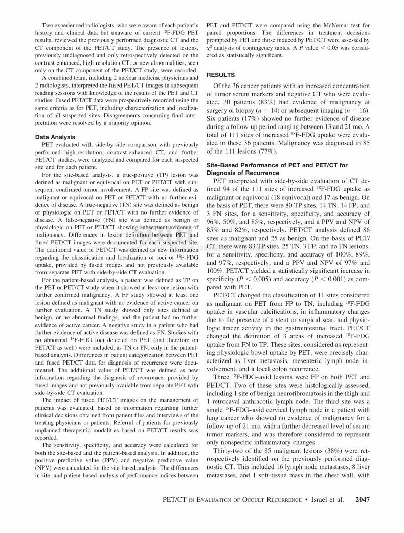
Two experienced radiologists, who were aware of each patient’s history and clinical data but unaware of current 18F-FDG PET results, reviewed the previously performed diagnostic CT and the CT component of the PET/CT study. The presence of lesions, previously undiagnosed and only retrospectively detected on the contrast-enhanced, high-resolution CT, or new abnormalities, seen only on the CT component of the PET/CT study, were recorded.
A combined team, including 2 nuclear medicine physicians and 2 radiologists, interpreted the fused PET/CT images in subsequent reading sessions with knowledge of the results of the PET and CT studies. Fused PET/CT data were prospectively recorded using the same criteria as for PET, including characterization and localization of all suspected sites. Disagreements concerning final interpretation were resolved by a majority opinion.
Data Analysis PET evaluated with side-by-side comparison with previously
performed high-resolution, contrast-enhanced CT, and further PET/CT studies, were analyzed and compared for each suspected site and for each patient.
For the site-based analysis, a true-positive (TP) lesion was defined as malignant or equivocal on PET or PET/CT with subsequent confirmed tumor involvement. A FP site was defined as malignant or equivocal on PET or PET/CT with no further evidence of disease. A true-negative (TN) site was defined as benign or physiologic on PET or PET/CT with no further evidence of disease. A false-negative (FN) site was defined as benign or physiologic on PET or PET/CT showing subsequent evidence of malignancy. Differences in lesion definition between PET and fused PET/CT images were documented for each suspected site. The additional value of PET/CT was defined as new information regarding the classification and localization of foci of 18F-FDG uptake, provided by fused images and not previously available from separate PET with side-by-side CT evaluation.
For the patient-based analysis, a patient was defined as TP on the PET or PET/CT study when it showed at least one lesion with further confirmed malignancy. A FP study showed at least one lesion defined as malignant with no evidence of active cancer on further evaluation. A TN study showed only sites defined as benign, or no abnormal findings, and the patient had no further evidence of active cancer. A negative study in a patient who had further evidence of active disease was defined as FN. Studies with no abnormal 18F-FDG foci detected on PET (and therefore on PET/CT as well) were included, as TN or FN, only in the patient-based analysis. Differences in patient categorization between PET and fused PET/CT data for diagnosis of recurrence were documented. The additional value of PET/CT was defined as new information regarding the diagnosis of recurrence, provided by fused images and not previously available from separate PET with side-by-side CT evaluation.
The impact of fused PET/CT images on the management of patients was evaluated, based on information regarding further clinical decisions obtained from patient files and interviews of the treating physicians or patients. Referral of patients for previously unplanned therapeutic modalities based on PET/CT results was recorded.
The sensitivity, specificity, and accuracy were calculated for both the site-based and the patient-based analysis. In addition, the positive predictive value (PPV) and negative predictive value (NPV) were calculated for the site-based analysis. The differences in site- and patient-based analysis of performance indices between
PET and PET/CT were compared using the McNemar test for paired proportions. The differences in treatment decisions prompted by PET and those induced by PET/CT were assessed by x2 analysis of contingency tables. A P value < 0.05 was considered as statistically significant.
RESULTS
Of the 36 cancer patients with an increased concentration of tumor serum markers and negative CT who were evaluated, 30 patients (83%) had evidence of malignancy at surgery or biopsy (n = 14) or subsequent imaging (n = 16). Six patients (17%) showed no further evidence of disease during a follow-up period ranging between 13 and 21 mo. A total of 111 sites of increased 18F-FDG uptake were evaluated in these 36 patients. Malignancy was diagnosed in 85 of the 111 lesions (77%).
Site-Based Performance of PET and PET/CT for Diagnosis of Recurrence
PET interpreted with side-by-side evaluation of CT defined 94 of the 111 sites of increased 18F-FDG uptake as malignant or equivocal (18 equivocal) and 17 as benign. On the basis of PET, there were 80 TP sites, 14 TN, 14 FP, and 3 FN sites, for a sensitivity, specificity, and accuracy of 96%, 50%, and 85%, respectively, and a PPV and NPV of 85% and 82%, respectively. PET/CT analysis defined 86 sites as malignant and 25 as benign. On the basis of PET/ CT, there were 83 TP sites, 25 TN, 3 FP, and no FN lesions, for a sensitivity, specificity, and accuracy of 100%, 89%, and 97%, respectively, and a PPV and NPV of 97% and 100%. PET/CT yielded a statistically significant increase in specificity (P < 0.005) and accuracy (P < 0.001) as compared with PET.
PET/CT changed the classification of 11 sites considered as malignant on PET from FP to TN, including 18F-FDG uptake in vascular calcifications, in inflammatory changes due to the presence of a stent or surgical scar, and physiologic tracer activity in the gastrointestinal tract. PET/CT changed the definition of 3 areas of increased 18F-FDG uptake from FN to TP. These sites, considered as representing physiologic bowel uptake by PET, were precisely characterized as liver metastasis, mesenteric lymph node involvement, and a local colon recurrence.
Three 18F-FDG–avid lesions were FP on both PET and PET/CT. Two of these sites were histologically assessed, including 1 site of benign neurofibromatosis in the thigh and 1 retrocaval anthracotic lymph node. The third site was a single 18F-FDG–avid cervical lymph node in a patient with lung cancer who showed no evidence of malignancy for a follow-up of 21 mo, with a further decreased level of serum tumor markers, and was therefore considered to represent only nonspecific inflammatory changes.
Thirty-two of the 85 malignant lesions (38%) were retrospectively identified on the previously performed diagnostic CT. This included 16 lymph node metastases, 8 liver metastases, and 1 soft-tissue mass in the chest wall, with
PET/CT IN EVALUATION OF OCCULT RECURRENCE • Israel et al. 2047

TABLE 2 suspicious by PET and precisely defined by PET/CT as Comparative Analysis of PET and PET/CT Site-Based Performance in 111 Suggestive Lesions in 36 Patients
with Suspected Occult Cancer Recurrence Due to Rising Levels of Tumor Markers
Performance PET PET/CT
TP (n) 80 83 TN (n) 14 25 FP (n) 14 3 FN (n) 3 — Sensitivity (%) 96 100 Specificity* (%) 50 89 Accuracy* (%) 85 97 PPV (%) 85 97 NPV (%) 82 100
*P statistically significant for specificity (P < 0.05) and accuracy (P < 0.001).
diameters ranging between 8 and 20 mm, and 7 bone metastases. New structural abnormalities were observed on the CT component of the hybrid imaging study in 30 sites (35%).
The site-based comparative performance of PET and PET/CT is summarized in Table 2.
Patient-Based Performance of PET and PET/CT for Diagnosis of Recurrence
PET interpreted with side-by-side evaluation of CT defined 28 of the 36 studies as TP, 3 TN, 3 FP, and 2 FN, for a sensitivity of 93%, a specificity of 50%, and an accuracy of 86%. On the basis of PET/CT analysis, there were 28 TP, 4 TN, 2 FP, and 2 FN studies, for a sensitivity of 93%, a specificity of 67%, and an accuracy of 89%. There was no statistically significant difference between the performance indices of PET and PET/CT for the patient-based analysis.
Tumor marker serum levels decreased during follow-up in 4 of the 6 patients with no further evidence of disease. None of the 6 patients showed any suggestive lesions on imaging studies performed during the follow-up period.
PET/CT changed the classification of 1 site considered as malignant on PET from FP to TN. This patient had a single abdominal focus of increased 18F-FDG uptake considered as
physiologic bowel activity. The patient showed no evidence of disease for a follow-up of 15 mo (Fig. 1).
The 2 patients defined as FP on both PET and PET/CT included 1 patient with anthracosis in an enlarged retrocaval lymph node and a second patient with nonspecific inflammatory changes in a cervical lymph node. The 2 patients defined as FN by both PET and PET/CT included a local recurrence of prostate cancer diagnosed 6 wk later and 1 patient with breast cancer showing a 7-mm malignant lesion in the second breast 2 mo later.
The patient-based comparative performance of PET and PET/CT is summarized in Table 3.
Clinical Impact of PET/CT on Patient Management Of the 28 patients with TP PET/CT studies, 9 patients
underwent surgery with curative intent, 15 patients were referred for chemotherapy, 3 patients received radiotherapy, and 1 patient was referred for radioiodine treatment. In 12 of the 28 patients (43%). treatment could be planned based only on the incremental diagnostic localization data provided by PET/CT.
Surgery with curative intent was performed in 9 patients with solitary malignant lesions. Eight of these 9 patients were referred for surgery based on the PET/CT diagnosis and precise localization of single tumor foci, including 3 lymph node metastases, 3 local recurrences of colon cancer (Fig. 2), 1 liver metastasis, and 1 second primary gastric cancer. Additional investigations guided by PET/CT findings (colonoscopy or gastroscopy) were performed in 2 of these 8 patients before the tumor resection.
Previously unplanned chemotherapy was administered to 15 patients with disseminated metastatic disease. Two of the 15 patients received chemotherapy after the PET/CT diagnosis of unresectable, extensive disease.
Radiation treatment was administered to 4 patients. One patient with a single bone metastasis received local-field radiotherapy, and 131I treatment was administered to a second patient with metastatic thyroid cancer. PET/CT findings induced changes in the localization and size of radiation fields in 2 patients, including 1 patient with a single bone metastasis and a second patient with a single soft-tissue metastasis, precisely localized by PET/CT. There was a
FIGURE 1. An 81-y-old man with cancer of sigmoid colon, after surgery, increasing carcinoembryonic antigen (CEA) serum levels, and a repeated negative CT study. (A) PET shows focal area of increased 18FFDG uptake in right upper abdomen. (B) Hybrid PET/CT images precisely localized this uptake to colon, consistent with physiologic bowel excretion. Malignancy was excluded and study was reported as normal. Patient had no evidence of recurrence in colon during 15 mo of clinical follow-up.
2048 THE JOURNAL OF NUCLEAR MEDICINE • Vol. 45 • No. 12 • December 2004
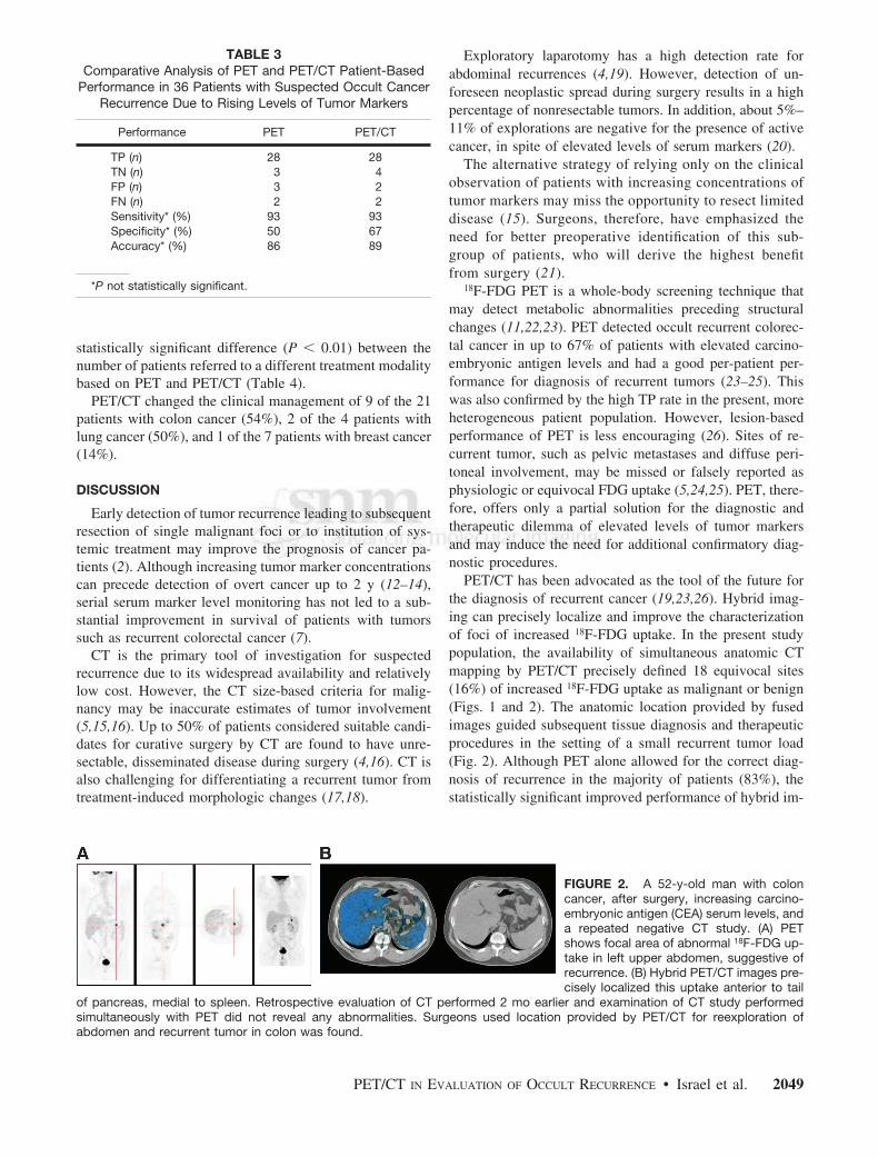
TABLE 3 Exploratory laparotomy has a high detection rate for Comparative Analysis of PET and PET/CT Patient-Based
Performance in 36 Patients with Suspected Occult Cancer Recurrence Due to Rising Levels of Tumor Markers
Performance PET PET/CT
TP (n) 28 28 TN (n) 3 4 FP (n) 3 2 FN (n) 2 2 Sensitivity* (%) 93 93 Specificity* (%) 50 67 Accuracy* (%) 86 89
*P not statistically significant.
statistically significant difference (P < 0.01) between the number of patients referred to a different treatment modality based on PET and PET/CT (Table 4).
PET/CT changed the clinical management of 9 of the 21 patients with colon cancer (54%), 2 of the 4 patients with lung cancer (50%), and 1 of the 7 patients with breast cancer (14%).
DISCUSSION
Early detection of tumor recurrence leading to subsequent resection of single malignant foci or to institution of systemic treatment may improve the prognosis of cancer patients (2). Although increasing tumor marker concentrations can precede detection of overt cancer up to 2 y (12–14), serial serum marker level monitoring has not led to a substantial improvement in survival of patients with tumors such as recurrent colorectal cancer (7).
CT is the primary tool of investigation for suspected recurrence due to its widespread availability and relatively low cost. However, the CT size-based criteria for malignancy may be inaccurate estimates of tumor involvement (5,15,16). Up to 50% of patients considered suitable candidates for curative surgery by CT are found to have unresectable, disseminated disease during surgery (4,16). CT is also challenging for differentiating a recurrent tumor from treatment-induced morphologic changes (17,18).
abdominal recurrences (4,19). However, detection of unforeseen neoplastic spread during surgery results in a high percentage of nonresectable tumors. In addition, about 5%– 11% of explorations are negative for the presence of active cancer, in spite of elevated levels of serum markers (20).
The alternative strategy of relying only on the clinical observation of patients with increasing concentrations of tumor markers may miss the opportunity to resect limited disease (15). Surgeons, therefore, have emphasized the need for better preoperative identification of this subgroup of patients, who will derive the highest benefit from surgery (21).
18F-FDG PET is a whole-body screening technique that may detect metabolic abnormalities preceding structural changes (11,22,23). PET detected occult recurrent colorectal cancer in up to 67% of patients with elevated carcinoembryonic antigen levels and had a good per-patient performance for diagnosis of recurrent tumors (23–25). This was also confirmed by the high TP rate in the present, more heterogeneous patient population. However, lesion-based performance of PET is less encouraging (26). Sites of recurrent tumor, such as pelvic metastases and diffuse peritoneal involvement, may be missed or falsely reported as physiologic or equivocal FDG uptake (5,24,25). PET, therefore, offers only a partial solution for the diagnostic and therapeutic dilemma of elevated levels of tumor markers and may induce the need for additional confirmatory diagnostic procedures.
PET/CT has been advocated as the tool of the future for the diagnosis of recurrent cancer (19,23,26). Hybrid imaging can precisely localize and improve the characterization of foci of increased 18F-FDG uptake. In the present study population, the availability of simultaneous anatomic CT mapping by PET/CT precisely defined 18 equivocal sites (16%) of increased 18F-FDG uptake as malignant or benign (Figs. 1 and 2). The anatomic location provided by fused images guided subsequent tissue diagnosis and therapeutic procedures in the setting of a small recurrent tumor load (Fig. 2). Although PET alone allowed for the correct diagnosis of recurrence in the majority of patients (83%), the statistically significant improved performance of hybrid im-
FIGURE 2. A 52-y-old man with colon cancer, after surgery, increasing carcinoembryonic antigen (CEA) serum levels, and a repeated negative CT study. (A) PET shows focal area of abnormal 18F-FDG uptake in left upper abdomen, suggestive of recurrence. (B) Hybrid PET/CT images precisely localized this uptake anterior to tail
of pancreas, medial to spleen. Retrospective evaluation of CT performed 2 mo earlier and examination of CT study performed simultaneously with PET did not reveal any abnormalities. Surgeons used location provided by PET/CT for reexploration of abdomen and recurrent tumor in colon was found.
PET/CT IN EVALUATION OF OCCULT RECURRENCE • Israel et al. 2049

TABLE 4 Change in Patient Management Based on PET
and PET/CT Results
Based only on Based incremental Total based
on contribution of on PET/CT Referral PET PET/CT results
For surgery 1 8 9 For chemotherapy 13 2 15 For radiotherapy 2 2 4 Total 16 12 28
Differences between therapeutic strategy based on PET and PET/CT results were statistically significant (P < 0.01).
aging for evaluating individual lesions indicates that PET/CT has an impact on the subsequent management of cancer patients, beyond the diagnosis of recurrence. The precise PET/CT localization and definition of suspicious 18F-FDG foci led to a better assessment of the extent of recurrent disease, with subsequent treatment planned on the basis of the unique information provided by hybrid imaging in one third of the total study population.
The clinical contribution of PET/CT is indicated mainly by the number of patients referred for resection with curative intent. Surgery was performed in 8 patients following PET/CT results, in addition to 1 patient who may have been referred to surgery based only on PET results. Six of these 8 patients were referred for surgery based solely on the results of hybrid imaging, sparing further unnecessary diagnostic procedures.
When PET, a highly sensitive test, indicates the presence of widespread disease, the precise localization of each malignant site is, as a rule, less clinically relevant. Of the present study population, 13 patients were referred for chemotherapy based on PET alone. Administration of chemotherapy to 2 additional patients was enabled by PET/CTbased precise definition of equivocal foci of 18F-FDG uptake as additional malignant lesions and diagnosis of multifocal recurrence.
In the present study radiation therapy planning was modified in 2 patients after the use of PET/CT. The potentially important role of hybrid imaging in this clinical setting cannot be fully appreciated from this small patient sample.
In some of the evaluated patients, the relatively long time interval between the previous CT reported as negative and the current PET/CT study as well as the patient sample with different primary malignancies and a preponderance toward colorectal tumors are limitations of the present study. Including tumors with low metabolic rates, such as prostate cancer, may also induce biases in the study results. The benefit of improved selection criteria and the potential value of a positive PET study at baseline need to be assessed. Further studies with long-term follow-up in large homogeneous patient cohorts with single histologic tumor types
need to follow. These studies will have to clarify whether PET/CT should be used, in the future, as the first step in the evaluation of patients with suspected occult recurrent cancer.
CONCLUSION
The results of present study indicate that hybrid PET/CT plays a potential important role in the early diagnosis and assessment of the extent of relapsed disease in cancer patients with increasing concentrations of serum tumor markers as the single suspicion of recurrence, with a significant clinical impact on further treatment planning in one third of the patient population.
ACKNOWLEDGMENTS
The authors are grateful to Drs. Yodphat Krausz and David Groshar for their many useful suggestions and to Judy Buchanan for preparation of this manuscript. This research was supported in part by a Research Grant from the Israeli Cancer Association.
REFERENCES
1. Duffy MJ. Clinical uses of tumor markers: a critical review. Crit Rev Clin Lab Sci. 2001;38:225–262.
2. Sagar PM, Pemberton JH. Surgical management of locally recurrent rectal cancer. Br J Surg. 1996;83:293–304.
3. Bakalakos EA, Burak WE, Young DC, et al. Is carcino-embryonic antigen useful in the follow-up management of patients with colorectal liver metastases? Am J Surg. 1999;177:2– 6.
4. Hida JI, Yasutomi M, Shindoh K, et al. Second-look operation for recurrent colorectal cancer based on carcinoembryonic antigen and imaging techniques. Dis Colon Rectum. 1996;39:74 –79.
5. Torizuka T, Nobezawa S, Kanno T, et al. Ovarian cancer recurrence: role for whole-body positron emission tomography using 2-[fluorine-18]-fluoro-2-deoxyD-glucose. Eur J Nucl Med. 2002;29:797– 803.
6. Spaepen K, Stroobants S, Dupont P, et al. Prognostic value of positron emission tomography (PET) with fluorine-18 fluorodeoxyglucose ([18F]FDG) after first-line chemotherapy in non-Hodgkin’s lymphoma: is [18F]FDG-PET a valid alternative to conventional diagnostic methods? J Clin Oncol. 2001;19:414 – 419.
7. Hung GU, Shiau YC, Tsai SC, et al. Value of 18F-fluoro-2-deoxyglucose positron emission tomography in the evaluation of recurrent colorectal cancer. Anticancer Res. 2001;21:1375–1378.
8. Cook GI, Maisey MN, Fogelman I. Normal variants, artifacts and interpretative pitfalls in PET imaging with 18-fluoro-deoxyglucose and carbon-11 methionine. Eur J Nucl Med. 1999;26:1363–1378.
9. Kostakoglu L, Agress H, Goldsmith J. Clinical role of FDG PET in evaluation of cancer patients. Radiographics. 2003;23:315–340.
10. Beyer T, Townsend DW, Brun T, et al. A combined PET/CT scanner for clinical oncology. J Nucl Med. 2000;41:1369 –1379.
11. Bar-Shalom R, Yefremov N, Altman H, et al. The clinical performance of PET/CT in evaluation of cancer: additional value for diagnostic imaging and patient management. J Nucl Med. 2003;44:1200 –1209.
12. Lardinois D, Weder W, Hany TF, et al. Staging of non-small-cell lung cancer with integrated positron-emission tomography and computed tomography. N Engl J Med. 2003;348:2500 –2507.
13. Patt YZ, Podoloff DA, Curley S, et al. Technetium 99m-labeled IMMU-4, a monoclonal antibody against carcinoembryonic antigen for imaging of occult recurrent colorectal cancer in patients with rising serum carcinoembryonic antigen levels. J Clin Oncol. 1994;12:489 – 495.
14. Mangkharak J, Patanachak C, Podhisuwan K, et al. The evaluation of combined scintimammography and tumor markers in breast cancer patients. Anticancer Res. 1997;17:1611–1614.
15. Arulampalam THA, Costa DC, Loisidou M, et al. Positron emission tomography and colorectal cancer. Br J Surg. 2001;88:176 –189.
16. Buccheri G, Ferrigno D. Serum biomarkers facilitate the recognition of early-stage cancer and may guide the selection of surgical candidates: a study of
2050 THE JOURNAL OF NUCLEAR MEDICINE • Vol. 45 • No. 12 • December 2004

carcinoembryonic antigen and tissue polypeptide antigen in patients with operable non-small cell lung cancer. J Thorac Cardiovasc Surg. 2001;122:891– 899.
17. Zervos EE, Badgwell BD, Burak WE, et al. Fluorodeoxyglucose positron emission tomography as an adjunct to carcinoembryonic antigen in the management of patients with presumed recurrent colorectal cancer and nondiagnostic radio-logic workup. Surgery. 2001;130:636 – 644.
18. Jadvar H, Fischman AJ. Evaluation of pancreatic carcinoma with FDG PET. Abdom Imaging. 2001;26:254 –259.
19. Makhija S, Howden N, Edwards R, et al. Positron emission tomography/computed tomography imaging for the detection of recurrent ovarian and fallopian tube carcinoma: a retrospective review. Gynecol Oncol. 2002;85:53–58.
20. Sijmons EA, Heintz AP. Second look and second surgery: second chance or second best? Semin Oncol. 2000;19:54 – 61.
21. Wexner SD. Invited critique. Arch Surg. 1999;134:311–313.
22. Flamen P, Hoekstra OS, Homans F, et al. Unexplained rising carcinoembryonic antigen (CEA) in the postoperative surveillance of colorectal cancer: the utility of positron emission tomography (PET). Eur J Cancer. 2001;37:862– 869.
23. Kalff V, Hicks RJ, Ware RE, et al. The clinical impact of 18F-FDG PET in patients with suspected or confirmed recurrence of colorectal cancer: a prospective study. J Nucl Med. 2002;43:492– 499.
24. Flanagan FL, Dehdashti F, Ogunbiyi OA, et al. Utility of FDG-PET for investigating unexplained plasma CEA elevation in patients with colorectal cancer. Ann Surg. 1998;227:319 –323.
25. Valk PE, Abella-Columna E, Haseman MK, et al. Whole-body PET imaging with 18F fluorodeoxyglucose in management of recurrent colorectal cancer. Arch Surg. 1999;134:503–511.
26. Czernin J, Phelps ME. Positron emission tomography scanning: current and future applications. Annu Rev Med. 2002;53:89 –112.
PET/CT IN EVALUATION OF OCCULT RECURRENCE • Israel et al. 2051

CONTINUING EDUCATION
18F-FDG PET Evaluation of the Response to Therapy for Lymphoma and for Breast, Lung, and Colorectal Carcinoma* Lale Kostakoglu, MD; and Stanley J. Goldsmith, MD
Division of Nuclear Medicine, Department of Radiology, New York Presbyterian Hospital, Weill Medical College of Cornell University, New York, New York
PET is a unique form of diagnostic imaging that observes in vivo biologic changes using radiopharmaceuticals that closely mimic endogenous molecules. 18F-FDG, which allows the evaluation of glucose metabolism, is the most commonly used tracer in oncology because of the practical half-life of 18F (110 min), compared with other short-lived positron emitters. 18F-FDG uptake in tumors is proportional to the glycolytic metabolic rate of viable tumor cells indicating the increased metabolic demand of tumors for glucose. 18F-FDG PET significantly improves the accuracy of imaging tumors in initial staging, management of recurrent cancer, and monitoring of therapy response. The information provided by this technique is more sensitive and specific than that provided by anatomic imaging modalities. 18F-FDG PET is particularly superior to CT or MRI in the ability to evaluate the effectiveness of various treatment regimens early during therapy or after therapy. In this review, we discuss the role of 18F-FDG PET in evaluating the response to therapy and the impact of this information on patient management.
Key Words: 18F-FDG PET; therapy response; lymphoma; lung cancer; colorectal cancer; breast cancer
J Nucl Med 2003; 44:224 –239
The primary goal of nonsurgical therapy of cancer is the complete eradication of tumor cells through the cytocidal effect of chemotherapy, radiotherapy, hormonal therapy, or biologic therapy. The tumor response to therapy varies widely, however. Differences in tumor response can be caused by disparities in tumor biology, such as growth phase, tumor volume, oxygenation, heterogeneity, presence of drug resistance mechanisms, and radio- or chemosensitivity. It would be most advantageous to identify resistant or nonresponding tumors early during or immediately after
Received May 3, 2002; revision accepted Sep. 25, 2002. For correspondence or reprints contact: Lale Kostakoglu, MD, New York
Presbyterian Hospital, Weill Medical College of Cornell University, 525 E. 68th St., Starr no. 221, New York, NY 10021.
E-mail: [email protected] *NOTE: FOR CE CREDIT, YOU CAN ACCESS THIS ACTIVITY THROUGH
THE SNM WEB SITE (http://www.snm.org/education/ce_online.html) THROUGH FEBRUARY 2004.
therapy to institute a timely alternative treatment that may be more effective. In advanced cancers in particular, systemic anticancer therapy has an evolving role in efforts to increase survival. High-dose chemotherapy with growth factors to stimulate bone marrow recovery after chemotherapy can be an option, as has been shown in breast cancer. Another alternative therapy, so-called tailor-made drug selection, can be based on tumor characteristics specific to the individual patient by determining tumor cell viability after incubation with various cytotoxic agents. Furthermore, emerging novel therapies, including immunotherapy, cancer vaccines, and biologic therapy with cytokines, are entering the oncologic arena as alternative approaches.
The efficacy of a treatment is a direct function of tumor burden (1). Hence, detection of residual tumors at a subclinical level or at a small volume is beneficial to optimize the efficacy of subsequent therapy. Unnecessary morbidity associated with treatment toxicity could also be avoided in patients with a short expected survival and at high risk of experiencing serious side-effects if it is determined that the therapy will fail. Response to therapy is currently evaluated using conventional imaging (CI) modalities. However, the definition of tumor response or progression, using anatomic imaging modalities, is based on size criteria. The disease activity may have completely resolved after therapy, but residual masses may persist on morphologic imaging as resolution of therapy-induced anatomic changes lags behind tumor cell mortality. PET is currently the most sensitive and specific imaging method to obtain information about tumor physiology and metabolism. Tumor cells have increased glucose metabolism due to increased expression of glucose transporters and hexokinase (glucose phosphorylating enzyme) as a result of oncogenic transformation. After phosphorylation, 18F-FDG is essentially trapped within the cell because of 2 phenomena: 18F-FDG is not a substrate for subsequent pathways that take place in glycolysis, and dephosphorylating enzyme— glucose-6-phosphatase—is either scarce or absent in cancer tissue. Therefore, the cellular concentration of 18F-FDG in tumor represents the glycolytic activity of viable tumor cells. Additionally, the results of a
224 THE JOURNAL OF NUCLEAR MEDICINE • Vol. 44 • No. 2 • February 2003

recent study revealed that the activity of tumor hexokinase is a marker of tumor growth rate that can be determined by 18F-FDG PET. Consequently, 18F-FDG PET may provide important prognostic information about the proliferative rates and, therefore, the antiproliferative effect of cancer therapy (2). There is now convincing evidence that reduction or resolution of 18F-FDG uptake in the tumor is an early indicator of response at a clinical or subclinical level (3–7).
Anticancer therapy is frequently individualized, as it is directed toward specific targets depending on the characteristics of the tumors. 18F-FDG PET may provide better and more timely assessment of the efficacy of various specific therapies that may result in significant clinical management alterations.
ASSESSMENT OF RESPONSE TO THERAPY
Monitoring response to therapy requires acquisition of a baseline 18F-FDG PET study before therapy and repeating 18F-FDG PET during or after the completion of therapy. The exact timing of 18F-FDG PET reimaging can be challenging because of the variability in tumor sensitivity to different treatment modalities. In fact, this matter is still in evolution. The method of quantifying tumor 18F-FDG uptake also varies greatly. Some guidelines have been developed in an attempt to standardize acquisition and response criteria for post-therapy changes in 18F-FDG tumor uptake to predict response to therapy (8). Biodistribution of 18F-FDG can be affected by various physiologic factors. Blood glucose levels have an impact on 18F-FDG uptake through the competitive displacement of 18F-FDG by plasma glucose. Patients should fast 4 – 6 h to reduce competition with plasma glucose and thus optimize and standardize tumor 18F-FDG uptake. There is no consensus on the optimal procedure for adjusting glucose levels in diabetic patients. In patients with type I diabetes, insulin is not recommended; in patients with type II diabetes, insulin may be administered at the discretion of the physician, although physiologic muscle uptake is exaggerated by insulin administration. Because the primary route of 18F-FDG excretion is renal, good hydration is required before imaging to encourage urinary excretion. It is recommended that patients drink 500 mL of water after injection of 18F-FDG. Muscle relaxants may be used to reduce muscle uptake, especially in patients with head and neck tumors, in whom uptake by tense cervical muscles may occur. Patients should remain silent during and after injection to reduce laryngeal muscle uptake, which may be confused with a residual or recurrent head and neck tumor.
Evaluation of response to therapy involves careful comparison of pretreatment and post-treatment 18F-FDG PET scans. Imaging 1–2 wk after completion of therapy is recommended to avoid transient fluctuations in 18F-FDG metabolism. When 18F-FDG PET is performed during the course of chemotherapy, current data indicate that images can be obtained as early as after 1 or 2 cycles of therapy
(5,6,9 –12). The relationship between radiotherapy and changes in tumor 18F-FDG uptake has yet to be established.
Interpreting 18F-FDG tumor uptake after therapy may be confounding at times because of therapy-related changes. In the post-therapy setting, approximately 25% of 18F-FDG uptake can occur in nontumor tissues such as macrophages, neutrophils, fibroblasts, and granulation tissue (13). In vitro assays demonstrate that irradiated tumor cells might have a tenfold increased 18F-FDG uptake (14). In colon cancer, it is recommended that 18F-FDG PET studies be delayed for 60 d after the completion of radiotherapy to accurately assess its outcome (14 –16). Generally 18F-FDG uptake 6 mo after radiotherapy is associated with tumor recurrence. Transient and reversible cell damage as well as minimal residual disease may obscure assessment of the true cell kill in the tumor mass (13,14). The negative predictive value of post-therapy 18F-FDG PET is therefore usually lower than the positive predictive value. More important, tumor perfusion and delivery of 18F-FDG into the tumor may be affected to the same extent by both necrosis and therapy-induced changes.
There are essentially 2 types of methods to analyze 18FFDG uptake by tumor: visual, or qualitative, assessment and quantitative analysis. The most straightforward method is dichotomous visual evaluation to differentiate malignant processes from benign lesions. This approach, however, is subjective, requires substantial experience, and is not sufficient for subtle findings. Alternatively, graded visual assessment results in less interobserver variability, provided that well-defined criteria are available for positive and negative findings.
Although visual evaluation is valuable and practical, 18FFDG PET benefits significantly from quantitative assessment of uptake, particularly in predicting outcome by determining tumor aggressiveness and monitoring therapy response. Nevertheless, the optimal method for quantitating prognosis and assessing response to therapy has not yet been defined. Quantitation can be approached in several ways, from simple tumor-to-background calculations to intricate kinetic analyses with dynamic PET acquisitions and blood sampling (17). Currently, it is not clear whether more advanced quantitation techniques are superior to more basic methods in the prediction of prognosis and therapy response, mainly because of the insufficient 18F-FDG PET data obtained thus far on the assessment of various tumors.
All quantitation methods entail attenuation correction to avoid the variability in 18F-FDG uptake due to the differences in tumor depth in the body. Determination of ratios of tumor to normal tissue is the simplest means of quantitation. This method can be applied to images even after reconstruction without the requirement of additional procedures or information. Although this approach is more objective than visual assessment, it significantly limits the placement of regions of interest versus background and the use of count statistics and reconstruction algorithms. A more accurate way of measuring tumor 18F-FDG uptake involves the de-
PET EVALUATION OF RESPONSE TO THERAPY • Kostakoglu and Goldsmith 225
termination of standardized uptake value (SUV), which has been widely used for the measurement of 18F-FDG uptake by tumors. This value normalizes 18F-FDG tumor uptake with injected activity (Qinj) and body weight (W), providing a semiquantitative index of 18F-FDG uptake (SUV = Q X W/Qinj) (18). SUV depends, however, on body weight. Correction with lean body mass is therefore required to avoid erroneous comparisons that can stem from changes in pre- and post-therapy body weight in the same patient. In calculating SUVs, the administered dose, corrected for residual activity in syringe and tubing, must also be accurately determined and the dose must be decay corrected to the time of imaging.
More objective and reliable quantitative methods are available, including simplified kinetic analysis (19), Patlak graphical analysis (20), and kinetic analysis with parameter optimization. Nonetheless, considering the practical constraints, these methods are unlikely to find clinical applications. Kinetic modeling has been used to determine the rate of glucose metabolism over time, expressed in Jmol/min/ mL. This technique is more demanding because it requires arterial catheterization and rapid blood sampling. Using dynamic PET, the net metabolic clearance of 18F-FDG is calculated by the time course of the radioactivity concentrations in tissue and arterial blood. A more promising development has been the demonstration that blood sampling can be replaced by time–activity curves based on dynamic scanning of the blood pool. This type of analysis requires that the aorta be in the field of view during the acquisition of dynamic PET for calculation of the net metabolic clearance of 18F-FDG (21,22). In quantitative analysis, heterogeneous tumor constitution with varying 18F-FDG kinetics may affect the measurement of glucose metabolic rate.
In the post-therapy setting, high sensitivity is preferable to specificity because the consequences of a false-negative interpretation are less desirable than those of a false-positive interpretation. In this regard, the threshold set for positivity depends on multiple variables such as tumor type, interval after therapy, and type of therapy. The required interval for post-therapy evaluation with 18F-FDG PET may be longer for radiotherapy than for chemotherapy, during which treatment response can be assessed even early.
PET is the most favorable noninvasive diagnostic means to assess tumor metabolic status after therapy and determine the presence of residual tumor. Several studies have illustrated that 18F-FDG uptake is an independent predictor of outcome and survival. Findlay et al. showed that pretreatment SUVs do not correlate with tumor response, although response is associated with lower 4- to 5-wk SUVs in patients with liver metastases from colorectal cancer (5). In studying lung cancer, Dhital et al. reported that SUV is of prognostic value before surgical resection (23). An SUV of 20 or more was associated with a 4.7 times increase in poor prognosis, compared with lower levels of SUV; however, Dhital et al. found no significant correlation between tumor
histology and SUVs. In patients with lymphoma, Cremerius et al. found that a tumor SUV of greater than 11 is associated with poorer progression-free survival than are lower tumor SUVs (24). Although these studies indicate that quantitative PET has considerable potential in predicting prognosis and therapy outcome, they also suggest that different types of tumors require different methods of analysis.
Alterations in 18F-FDG uptake as measured by various methods, including visual and quantitative analyses, provide useful information on response to anticancer therapy. The area in which quantitative PET will have its greatest impact is, in fact, the assessment of response to therapy. However, determining which method is more specific and superior for monitoring response is difficult because of the inconsistencies in these analytic methods between investigators. As more tumor-specific markers are developed, more accurate information will be obtained from quantitative analysis of PET images.
CLINICAL STUDIES MONITORING RESPONSE TO THERAPY
Lymphoma Hodgkin’s disease (HD) and non-Hodgkin’s lymphoma
(NHL) compose only 8% of all malignancies, and they are frequently curable. Histology and the extent of the disease (staging) are important factors influencing prognosis and therapy success (25,26).
Patients Who Will Benefit from Monitoring Response to Therapy. Surgical treatment is not an option in lymphoma. Therapeutic implications for patients with HD and NHL emphasize the importance of initially staging the disease accurately. Patients diagnosed with early-stage HD and NHL are treated with combination chemotherapy and radiotherapy or radiotherapy alone, whereas those with stage III or IV disease are typically treated with aggressive chemotherapy. In early-stage lymphoma, 75%–90% of patients respond to therapy regardless of the histologic subtype; however, in advanced-stage lymphoma, less than 50% of newly diagnosed patients are curable with standard treatments (27,28). Hence, evaluation of response to therapy is vital in patients with advanced-stage disease. There is an advantage to assessing the response to therapy early during chemotherapy, since early evidence of persistent disease may suggest that innovative intervention such as bone marrow transplantation be used in advanced-stage lymphoma. Early recognition of resistance to chemotherapy can also result in lower cumulative treatment toxicity and tumor burden at the start of salvage therapy, potentially improving clinical outcome.
Prediction of Response to Therapy After Its Completion. Residual abnormalities frequently occur after therapy in up to 64% of patients with lymphoma (29). Although residual masses are usually considered to be persistent disease, a maximum of 18% of residual masses are found to harbor viable lymphoma after therapy (29). Currently, 18F-FDG PET is considered to be more accurate than anatomic im
226 THE JOURNAL OF NUCLEAR MEDICINE • Vol. 44 • No. 2 • February 2003

aging modalities in assessing treatment effects to correctly identify patients with residual disease and predict therapy outcome (Table 1; Figs. 1 and 2) (9,10,24,30 –38). As a CI modality, 67Ga imaging is an independent predictor of outcome in lymphoma early during chemotherapy (39,40). The value of 67Ga in evaluating response to therapy in intraabdominal tumors and low-grade lymphoma, however, is unclear. 18F-FDG PET can identify nonresponders more accurately than can CT. Nevertheless, after completion of chemotherapy, 18F-FDG PET may not exclude the presence of minimal residual disease, which may lead to a later relapse. In a previous study, comparing 18F-FDG PET and CT in a post-therapy setting, relapse occurred in all patients with positive post-therapy 18F-FDG PET findings and in only 26% of patients with residual masses observed on CT. Consequently, the positive predictive values for 18F-FDG PET and CT were 100% and 42%, respectively, after the completion of therapy (36). In this study, positive 18F-FDG PET findings after therapy were consistently associated with poorer survival than were negative findings, with 1-y pro-gression-free survivals of 0% and 86%, respectively (36). The prognostic significance of post-therapy 18F-FDG PET has been assessed, so far, in patients with various lymphoma subtypes and stages. In a study by Cremerius et al., the population consisted of patients referred for reevaluation within 3 mo of completion of therapy, as well as those with suspected relapse or residual masses who presented within 12 mo of completion of therapy (24). In this mixed group, progression was observed in 84% of patients after a median interval of 2 mo. Among patients with negative 18F-FDG PET findings, only 13% had disease progression after a median follow-up of 21 mo. Positive post-therapy 18F-FDG
PET findings were associated with poorer progression-free survival. Although the results of this study indicate that 18F-FDG PET has a high prognostic value in lymphoma, the study design suffers from a heterogeneous patient population. 18F-FDG PET may prove more useful in patients who are at a higher risk for disease recurrence. 18F-FDG PET evidence of persistent disease may prompt alternative therapy regimens in the poor-prognosis group. Supporting this view was the finding that 18F-FDG PET predicted complete remission better in moderate-risk patients (stage I–III, no relapse, no more than 2 different prior therapy regimens) than in high-risk patients, with negative predictive values of 90% versus 50%– 67% (30).
In a more recent study, Spaepen et al. evaluated the value of 18F-FDG PET in detecting residual disease and, thus, predicting relapse after completion of first-line chemotherapy in 93 patients (32). After first-line chemotherapy, 83.5% of patients with negative 18F-FDG PET findings remained in complete remission after a median follow-up of 653 d. Only 16% of patients had relapse of disease after negative 18FFDG PET findings, with a median progression-free survival of 404 d. 18F-FDG PET showed persistent uptake in 26 patients, all of whom had relapse, with a median progression-free survival of 73 d. The authors concluded that persistent 18F-FDG uptake after first-line chemotherapy in NHL was highly predictive of residual or recurrent disease. Among patients with disease relapse, disease-free survival was clearly shorter in patients with positive 18F-FDG PET findings than in those with negative findings.
The prognostic value of pretherapy 18F-FDG PET was also evaluated in patients with HD and aggressive NHL undergoing high-dose therapy with stem cell transplanta-
TABLE 1 18F-FDG PET in Detection of Residual Lymphoma After Therapy
No. of Author Reference patients Time of therapy Type of disease PPV (%) NPV (%) PFS (-PET) PFS (+PET)
Cremerius 24 56 At completion of Rx NHL + HD 84 86 NR 2 mo Cremerius 30 72 At completion of Rx NHL + HD NA 90 vs 67* NA NA Spaepen 32 93 At completion of Rx NHL 100 83.5 13.5 mo 2.4 mo Becherer 33 16 At completion of Rx NHL + HD 80 100 100%† 18%†
Weihrauch 34 28 At completion of Rx HD 60 100 NR 2 mo Jerusalem 36 54 At completion of Rx NHL + HD 100 90 86%† 0%†
Torizuka 37 14 At completion of Rx‡ NHL NA NA NA NA Mikhaeel 35 49 Early during Rx NHL 100 82 NA NA
At completion of Rx 100 83 Jerusalem 36 28 Early during Rx NHL 100 67 81%† 20%†
Romer 9 11 Early during Rx NHL NA 54.5 NA NA Kostakoglu 10 23¶ Early during Rx NHL + HD 90 85 NR 5 mo
30 At completion of Rx 83 65 NR 0 mo
*Moderate-risk vs. high-risk patients. †PFS at 1 y, percentage of patients in remission. ‡Radioimmunotherapy. ¶23 of 30 patients had 18F-FDG PET after first cycle and completion of therapy. PPV = positive predictive value; NPV = negative predictive value; PFS = progression-free survival; -PET = negative 18F-FDG PET
findings; +PET = positive 18F-FDG PET findings; Rx = therapy; NA = not available; NR = still in remission and PFS not reached at 2 y.
PET EVALUATION OF RESPONSE TO THERAPY • Kostakoglu and Goldsmith 227
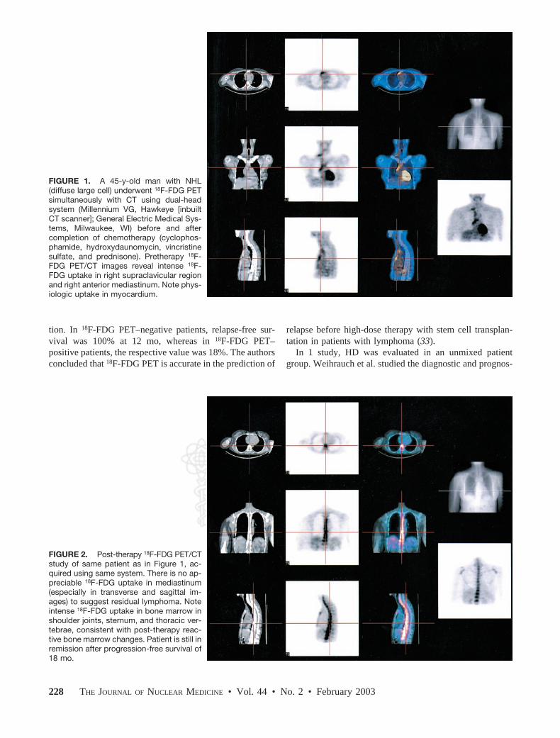
FIGURE 1. A 45-y-old man with NHL (diffuse large cell) underwent 18F-FDG PET simultaneously with CT using dual-head system (Millennium VG, Hawkeye [inbuilt CT scanner]; General Electric Medical Systems, Milwaukee, WI) before and after completion of chemotherapy (cyclophosphamide, hydroxydaunomycin, vincristine sulfate, and prednisone). Pretherapy 18FFDG PET/CT images reveal intense 18FFDG uptake in right supraclavicular region and right anterior mediastinum. Note physiologic uptake in myocardium.
tion. In 18F-FDG PET–negative patients, relapse-free survival was 100% at 12 mo, whereas in 18F-FDG PET– positive patients, the respective value was 18%. The authors concluded that 18F-FDG PET is accurate in the prediction of
FIGURE 2. Post-therapy 18F-FDG PET/CT study of same patient as in Figure 1, acquired using same system. There is no appreciable 18F-FDG uptake in mediastinum (especially in transverse and sagittal images) to suggest residual lymphoma. Note intense 18F-FDG uptake in bone marrow in shoulder joints, sternum, and thoracic vertebrae, consistent with post-therapy reactive bone marrow changes. Patient is still in remission after progression-free survival of 18 mo.
relapse before high-dose therapy with stem cell transplantation in patients with lymphoma (33).
In 1 study, HD was evaluated in an unmixed patient group. Weihrauch et al. studied the diagnostic and prognos
228 THE JOURNAL OF NUCLEAR MEDICINE • Vol. 44 • No. 2 • February 2003

tic value of 18F-FDG PET performed at least 3 mo after therapy on 28 HD patients with residual mediastinal masses determined by CT (34). The results indicated that patients with negative 18F-FDG PET findings after therapy were unlikely to have disease relapse within a year. The negative and positive predictive values at 1 y for 18F-FDG PET were 95% and 60%, respectively. This particular study emphasized the high false-positive rates obtained in HD with post-therapy 18F-FDG PET (34). These results were, however, derived from a very small population that had positive post-therapy 18F-FDG PET findings. Additionally, in this younger age group false-positive findings may be due to thymus hyperplasia, which is a well-known phenomenon (41).
Early Prediction of Response to Therapy. Preliminary studies suggest that 18F-FDG PET can distinguish responders from nonresponders early into the course of chemotherapy or immunotherapy in patients with lymphoma. The extent and time course of changes in 18F-FDG uptake in response to chemotherapy were studied by Romer et al. (9). Dynamic 18F-FDG PET was performed on 11 patients at baseline and 1 and 6 wk after the initiation of chemotherapy. One week after the initiation of chemotherapy, tumor 18FFDG uptake decreased by 60%. A further decrease of 42% was observed at 6 wk, resulting in a total decrease of 79% from baseline. During a follow-up of 16.0 4.2 mo, approximately 54% of patients continued to show complete remission. Seven days after initiation of chemotherapy, this group of patients displayed a significantly lower mean 18FFDG metabolic rate than did the group of patients with relapse. At 6 wk, all parameters of 18F-FDG uptake showed a significant difference for both patient groups. The relative change of 18F-FDG metabolic rate from baseline to week 6,
as well as from week 1 to week 6, was significantly larger, compared with SUV parameters. Standard chemotherapy of patients with NHL causes a rapid decrease of tumor 18FFDG uptake as early as 1 wk after treatment, and uptake continues to decline during therapy, indicating the sensitivity of metabolic signals to chemotherapeutic interventions.
A recent study demonstrated that 18F-FDG PET has a high prognostic value for evaluation of therapy as early as after 1 cycle in aggressive NHL and HD (10). Ninety percent of patients with positive 18F-FDG PET findings after 1 cycle had disease relapse with a median progression-free survival of 5 mo, whereas 85% of patients who had negative 18F-FDG PET findings remained in complete remission after a minimum follow-up of 18 mo. All patients with persistent 18F-FDG uptake both after the first cycle and at completion of therapy had relapse, except 1 patient who had a thymic rebound. The progression-free survival was significantly different between patients with negative and patients with positive 18F-FDG PET findings after 1 cycle of treatment. After completion of chemotherapy, although there was a statistically significant difference in progression-free survival between patients with negative and patients with positive findings, the findings after completion of chemotherapy yielded a significantly lower sensitivity and negative predictive value than did the findings after the first cycle (Fig. 3). In this study, the relapse rate for patients with negative 18F-FDG PET findings at the completion of therapy was higher than the relapse rate for patients with negative 18F-FDG PET findings after the first cycle (35% vs. 15%). The potential of 18F-FDG PET to predict outcome in patients with aggressive lymphoma and HD early during therapy, compared with after completion of therapy, is most likely due to the sensitivity of these lymphomas to chemo-
FIGURE 3. A 55-y-old man with NHL (diffuse large cell) underwent 18F-FDG PET using dual-head gamma camera with attenuation correction (MCD-AC; ADAC Laboratories, Milpitas, CA) before, after first cycle of, and at completion of chemotherapy. Pretherapy 18F-FDG PET image reveals extensive radiotracer uptake in anterior mediastinum. Note physiologic uptake in supraclavicular cervical muscles. Patient underwent chemotherapy with cyclophosphamide, hydroxydaunomycin, vincristine sulfate, and prednisone. After first cycle, residual disease was seen in anterior mediastinum (not shown). 18F-FDG PET image after last cycle (middle image) demonstrates no residual mass in mediastinum, consistent with complete resolution of disease. 18F-FDG PET image 8 mo after completion of therapy, however, reveals recurrence of disease in anterior mediastinum. Note physiologic uptake in heart in all images.
PET EVALUATION OF RESPONSE TO THERAPY • Kostakoglu and Goldsmith 229

therapy. A positive 18F-FDG PET result after 1 cycle reflects the metabolic activity of potentially resistant clones, which, although responding to chemotherapy, do so more slowly than do those homogeneously sensitive tumor cells. This study provides a strong argument for consideration of further trials to evaluate a subsequent change in treatment based on the 18F-FDG PET results (10).
18F-FDG PET has been compared with CT in the assessment of remission after treatment of aggressive NHL (35). In a subset of patients, the prognostic value of interim 18F-FDG PET performed after 2–3 cycles of chemotherapy was also evaluated. Not surprisingly, post-treatment 18FFDG PET was more accurate than CT in assessing remission status after treatment. The respective relapse rates were 100% and 18% for positive and negative 18F-FDG PET results, compared with 41% and 25% for patients with positive and negative CT results, with a median follow-up of 30 mo. Interim 18F-FDG PET revealed no relapses in patients with no or minimal residual uptake, compared with an 87.5% relapse rate in patients with persistent PET activity. The authors concluded that 18F-FDG PET is a more accurate method than CT in assessing remission and estimating prognosis after treatment of aggressive NHL. An interim PET scan after 2–3 cycles of chemotherapy may assist in separating good-prognosis patients, who are likely to be cured with standard chemotherapy, from patients with a poorer prognosis, who require alternative treatment (35). In another study with a similar design, 28 NHL patients were studied for early evaluation of response by 18F-FDG PET performed after a median of 3 cycles of chemotherapy. Persistent tumor 18F-FDG uptake after several cycles of chemotherapy was predictive of clinical remission, progression-free survival, and overall survival. All patients with residual 18F-FDG uptake and 33% of patients without residual 18F-FDG uptake had disease relapse or progression, with a positive predictive value of 100% and a negative predictive value of 67%. Mean progression-free survivals at 1 and 2 y were, respectively, 20% and 0% for 18F-FDG PET–positive patients and 81% and 62% for 18F-FDG PET– negative patients. However, the sensitivity of qualitative 18F-FDG PET in identifying patients with a poor outcome was insufficient (36).
Radioimmunotherapy, with 131I-labeled anti-B1 antibody developed against the surface antigen CD20, has been recognized as a promising approach for treatment of low-grade NHL (42). The tumor response to radioimmunotherapy may be more gradual than the tumor response to chemotherapy. The prognostic value of 18F-FDG PET was evaluated in 14 patients with NHL treated with 131I-anti-B1 therapy. All patients underwent 18F-FDG PET at baseline, 5–7 d, and 1–2 mo after radioimmunotherapy to estimate the response to radioimmunotherapy. 18F-FDG PET metabolic data obtained 1–2 mo after radioimmunotherapy correlated well with the ultimate response of NHL to radioimmunotherapy. The correlation was more significant than that of the early
18F-FDG PET data obtained 5–7 d after radioimmunotherapy (37).
False-Positives. Reactive lymph nodes and inflammatory or infectious processes may cause false-positive findings. Anti-inflammatory cells such as activated macrophages or granulation tissue that are present in areas of inflammation have been shown to avidly take up 18F-FDG. Although 18F-FDG PET is sensitive for identifying disease sites in the chest, false-positive 18F-FDG uptake after therapy can be seen at the site of thymic hyperplasia (41). Also, infectious or inflammatory processes such as toxoplasmosis, Mycobacterium tuberculosis, fungi, and sarcoidosis in the extra-nodal organs, particularly the spleen, can cause false-positive findings (43). Post-therapy reactive bone marrow changes may at times be a potential source of confusion about disease involvement (Fig. 2).
False-Negatives. 18F-FDG PET has been reported to have false-negative results in mucosa-associated lymphoid tissue (44) and in lesions smaller than 1 cm, particularly low-grade lymphoma, although in our series we detected lesions as small as 0.6 cm (45). Although 18F-FDG PET is sensitive in low-grade lymphomas, the degree of uptake can be lower than that observed in intermediate- or high-grade lymphomas. The sensitivity of 18F-FDG PET to detect bone marrow infiltration has been reported to be low (46,47). Because 18F-FDG PET may yield false-positive or -negative results, evaluation of bone marrow should be performed using bone marrow biopsy and MRI, or should be complemented by these, when indicated. Nevertheless, in the evaluation of bone marrow, no single technique is completely reliable. Bone marrow is associated with a high rate of false-negative findings, which can also be obtained with MRI in cases of bone marrow hyperplasia, diffuse lymphoma infiltration, and infectious processes.
Summary. 18F-FDG uptake is predictive of the response to therapy after its completion or early during its course. Patients with a negative PET result after 1 cycle, indicating a good prognosis, would continue with a full course of their first-line treatment, because negative 18F-FDG PET findings may not be sufficiently sensitive in patients with minimal residual disease. Selected patients with a positive 18F-FDG PET result after 1 cycle, and thus a less favorable prognosis, could be randomized to receive second-line chemotherapy with stem cell transplantation without completing a full course of initial chemotherapy.
Breast Carcinoma Breast carcinoma is the most frequently diagnosed ma
lignancy in women in North America and the second most frequent cause of cancer death (48). The relatively constant mortality, despite increases in incidence, may be the result of improved outcome secondary to earlier diagnosis and advances in treatment and follow-up. If diagnosed early, it is a curable disease.
230 THE JOURNAL OF NUCLEAR MEDICINE • Vol. 44 • No. 2 • February 2003

Patients Who Will Benefit from Monitoring Response to Therapy. 18F-FDG PET should be used as a monitoring tool in patients undergoing induction therapy for advanced disease as well as in those receiving preoperative chemo- or radiotherapy for inoperable localized tumors. Large or locally advanced breast cancer (LABC) may account for 15%–25% of cases of breast cancer (49). The definition of LABC is variable. The investigators include, as LABC patients, all patients with inoperable stage IIIB cancer, whereas patients with operable stage IIIB or stage IV cancer can also be included by virtue of positive supraclavicular lymph nodes. Treatment of LABC should include neoadjuvant or preoperative chemotherapy, since there is evidence that such therapy will significantly increase the number of patients who will benefit from breast-conserving surgery (50,51). Early administration of systemic chemotherapy before local treatment in patients with large or LABC is intended to downstage the primary tumor to render subsequent local treatment (surgery or radiotherapy) more effective and less morbid, as well as to eliminate occult distant metastases. Despite the experimental data indicating the survival benefits of neoadjuvant chemotherapy over postoperative adjuvant chemotherapy, the survival rate is still poor because of failure of chemotherapy (51). Studies have also demonstrated that patients with unresponsive tumors may achieve an improved survival with alternative or more prolonged courses of chemotherapy and the timely initiation of radiotherapy (52). It is essential, therefore, to accurately identify patients who would benefit from alternative treatments during the course of chemotherapy.
Prediction of Response to Therapy After Its Completion. In a retrospective study of 61 patients, Vranjesevic et al. compared the value of 18F-FDG PET with CI to predict outcome in breast cancer patients who had previously undergone primary therapy. 18F-FDG PET was more accurate than combined CI modalities for predicting outcome, with positive and negative predictive values of 93% and 84%, respectively, for 18F-FDG PET versus 85% and 59%, respectively, for CI modalities (53). The prognostic accuracy of 18F-FDG PET was superior to that of multiple procedures with CI (90% vs. 75%). Disease-free survival differed significantly between patients with negative and patients with positive 18F-FDG PET findings. The estimates of disease-free survival stratified by CI results, however, showed a marginally significant difference between CI-positive and CI-negative patients. The results of this study should be interpreted in a different context because of the significant differences in its design, compared with the designs of other studies. Briefly, this study is retrospective and the population includes patients with different histologic types and stages of breast cancer. The patients underwent a variety of treatments, including chemotherapy and radiotherapy, and follow-up was performed long after the completion of therapy. 18F-FDG PET performed long after therapy can be used to predict the clinical outcome of previously treated patients relative to what is achievable by CI alone.
Early Prediction of Response to Therapy. The complete pathologic resolution of the tumor after chemotherapy is of considerable prognostic importance and frequently does not correlate with observed clinical response (54,55). In contrast to morphologic imaging modalities, 18F-FDG PET has been reported to detect metabolic changes in breast cancer as early as 8 d after initiation of therapy, preceding appreciable anatomic changes (56). Several studies also indicate that responders may be differentiated from nonresponders using 18F-FDG PET after the first course of chemotherapy (6,11,12). Schelling et al. investigated the predictive value of 18F-FDG PET for evaluating histopathologic response during chemotherapy in patients with LABC (11). Significant differences in tracer uptake were obtained as early as after the first course of chemotherapy between nonresponding and responding tumors. After the first course of chemotherapy, all responders were correctly identified, with a sensitivity of 100% and a specificity of 85%.
Bassa et al. found that 18F-FDG PET was useful in evaluating response to presurgical chemotherapy for the primary LABC tumor in 16 patients (12). The sensitivity for detection of pathologically proven primary lesions was better with 18F-FDG PET (100%) than with mammography (62.5%) or ultrasonography (87.5%). After therapy, however, the sensitivity for 18F-FDG PET was 75%, compared with 71% and 87.5% for mammography and ultrasonography, respectively, for detection of residual primary tumor. A substantial decrease in tumor glucose metabolism of the primary tumor, measured by SUVs, was visible on the 18F-FDG PET images of 69% of patients as early as after the first cycle of chemotherapy. Because of microscopic residual disease, however, the decrease in 18F-FDG uptake did not correlate with favorable clinical outcome for all patients. Thus, 18F-FDG PET was false-negative in 25% of patients with progression of disease at a later course. The main emphasis of this study was that an elevated 18F-FDG uptake at the completion of chemotherapy before surgery correlated with a poor clinical outcome. Patients with persistent 18F-FDG uptake after chemotherapy should be selected for more aggressive therapy and a closer follow-up (12). Similarly, in another study, 18F-FDG PET was able to predict complete pathologic response after a single course of chemotherapy in 30 patients with large tumors (>3 cm), or LABC, with a sensitivity of 90% and a specificity of 74% (57). The mean reduction in relative 18F-FDG uptake after the first course of chemotherapy was significantly greater in lesions that achieved a partial or complete pathologic response than in those with no response or progression. Mean pretreatment glycolytic rates were significantly higher in ultimately responsive cancers. The mean change in relative 18F-FDG uptake and glycolytic rates after the first course of chemotherapy was significantly greater in responding lesions. These conclusions were derived, however, from a small number of patients; thus, the ultimate clinical significance of these results is unknown.
Tiling et al. compared 18F-FDG PET with 99mTc-sesta-
PET EVALUATION OF RESPONSE TO THERAPY • Kostakoglu and Goldsmith 231

mibi scintimammography (MIBI) in the assessment of tumor response to chemotherapy after the first and second cycles of chemotherapy in 7 patients with LABC (58). These preliminary data demonstrated that MIBI is as useful as 18F-FDG PET for monitoring the response to chemotherapy. Patients with complete remission showed decreased 18F-FDG and MIBI uptake as early as 8 d after therapy, followed by complete disappearance of uptake at the end of therapy. In patients with partial or no response, both techniques showed persistent tumor uptake during chemotherapy. An early decline in glucose or MIBI uptake 8 d after initiation of therapy did not necessarily predict complete remission in all patients. After the second chemotherapy cycle, both techniques were able to distinguish between complete and partial or no response. The results of this study, however, should be evaluated in light of 2 shortcomings: Only 7 patients were studied, and MIBI may have limitations as an imaging agent, particularly in the post-therapy setting. MIBI is a transport substrate for the Pglycoprotein pump system, which confers multidrug resistance (59). Thus, the accumulation and retention of MIBI are reduced in multidrug-resistant tumors, especially after chemotherapy. Prior studies have shown an inverse relationship between the levels of P-glycoprotein expression and the magnitude of MIBI uptake and washout in breast cancer cells (60,61). After chemotherapy, the percentage of breast cancers overexpressing P-glycoprotein increases dramatically (62). In the absence of immunohistologic information on P-glycoprotein expression in the tumor, it is impossible to tell whether a decrease in MIBI uptake is due to the effective chemotherapy or to chemotherapy-induced P-glycoprotein overexpression causing extrusion of MIBI out of tumor cells.
In a recent study, the factors influencing the response of LABC to presurgical chemotherapy were investigated using 18F-FDG PET and blood flow measured by 15O-water PET (63). The authors hypothesized that low tumor perfusion would predict poor response to systemic therapy; however, blood flow alone was not predictive of response. There was a strong association between pretherapy metabolism, reflected by the association of the metabolic rate of 18F-FDG, and the degree of response. Tumors with higher rates of glucose metabolism before therapy manifested a poor therapy response. Furthermore, a low ratio of 18F-FDG metabolic rate to blood flow was the best predictor of a better prognosis, as confirmed by survival analysis.
Metabolic Flare. Increased tumor 18F-FDG uptake (i.e., metabolic flare) and the degree of estrogen receptor (ER) blockade early after institution of tamoxifen may predict response to antiestrogen therapy in patients with ER-positive metastatic breast cancer (64). Mortimer et al. reported that the functional status of tumor ERs can be characterized in vivo by PET with 18F-FDG and 16c-18F-fluoro-17[estradiol (FES) (64). 18F-FDG PET was predictive of clinical response to tamoxifen therapy in patients with advanced ER-positive breast cancer. Forty women with biopsy-
proved advanced ER-positive breast cancer underwent PET with 18F-FDG and FES before and 7–10 d after initiation of tamoxifen therapy. In the responders, the tumor 18F-FDG uptake increased after tamoxifen by 28.4%; only 5 of these patients had evidence of a clinical flare reaction. In nonresponders, tumor 18F-FDG uptake did not significantly change from baseline. Lesions of responders had higher baseline FES uptake than did those of nonresponders. All patients had evidence of blockade of the tumor ERs 7–10 d after initiation of tamoxifen therapy; however, the degree of ER blockade was greater in responders than in nonresponders.
False-Positives. False-positive results occur in patients with inflammatory processes in the breast or early after biopsy or surgery. Benign breast tumors usually have low 18F-FDG uptake; only about 10% of fibroadenomas accumulate 18F-FDG (65).
False-Negatives. False-negative results can occur when lesions are less than 1 cm or when the tumor is well differentiated, as is the case with tubular carcinoma and carcinoma in situ. A high rate of false-negative findings has also been reported for lobular carcinomas (66).
Summary. 18F-FDG PET appears to have a high prognostic value for determination of the effectiveness of therapy in LABC, although more data are necessary to confirm the existing findings in extended patient groups. After therapy, 18F-FDG PET reflects the overall biologic response to therapy but is limited in evaluating microscopic residual disease; however, early evidence of persistent disease may allow for more aggressive and novel therapy options. Thus, 18F-FDG PET should be the imaging modality of choice to monitor response to therapy in patients with LABC.
Non–Small Cell Lung Carcinoma Lung cancer is the leading cause of cancer death in both
men and women. Most developed countries have shown declines in death rates from cancer other than lung cancer. Approximately 13% of patients with lung cancer survive 5 y. This rate has been stable in the past 2 decades (48). The histologic classification of lung cancer by the World Health Organization defines 4 subtypes: squamous cell carcinoma, adenocarcinoma, large cell carcinoma, and small cell carcinoma. Small cell carcinoma accounts for 20%–25% of all lung cancers, and spread is often present at the time of diagnosis. The remainder of the lung cancer subtypes constitute the non–small cell lung cancer (NSCLC) group. In North America, adenocarcinoma of the lung is the most common histologic type of lung cancer, accounting for more than 40% of all cases of lung cancer. It appears that other than T1 N0 tumors, adenocarcinoma of the lung has a worse prognosis, stage for stage, than does squamous cell carcinoma (67).
Patients Who Will Benefit from Monitoring Response to Therapy. Tumor shrinkage assessed by anatomic modalities is not a good indicator of response to treatment for detecting resistant clones present even in significantly shrunken
232 THE JOURNAL OF NUCLEAR MEDICINE • Vol. 44 • No. 2 • February 2003

FIGURE 4. A 46-y-old man with NSCLC of right upper lobe. Patient underwent 18F-FDG PET using dual-head gamma camera with attenuation correction (MCD-AC; ADAC Laboratories, Milpitas, CA) before and after completion of chemotherapy. Pretherapy 18F-FDG PET image (left) demonstrates distinct focus of increased 18F-FDG uptake in right upper lung. Post-therapy 18F-FDG PET image (right) reveals almost complete resolution of 18F-FDG uptake in corresponding region. Patient underwent resection of right upper lobe and was free of disease at 10-mo follow-up.
masses. An accurate assessment of the impact of chemotherapy and radiotherapy would help guide treatment for patients with locally advanced lung cancer (stages IIIA and IIIB). Patients with locally advanced NSCLC who have bulky, inoperable disease will benefit most from an evaluation of the efficacy of chemotherapy. This group of patients composed 25%– 40% of newly diagnosed lung cancer patients in the United States (48). In patients with locally advanced NSCLC, induction therapy followed by surgery may improve survival rates over those for surgery alone (68). In advanced-stage disease, at progression after platinum-based chemotherapy, second-line chemotherapy with docetaxel may have a survival benefit in selected patients (69). In stage IIIAN2 disease with no residual nodal disease after induction therapy and surgery, 5-y survival rates were 54%, compared with 17% in all patients treated with induction therapy regardless of the response status. Hence, it would be beneficial to offer surgery only to those patients with objective evidence of therapy response (68).
18F-FDG PET can also be useful in the assessment of the volumetric response to radiotherapy for locally advanced lung cancer. Chemotherapy-assisted novel hyperfractionated accelerated radiotherapy may be used in selected patients. Identification of nonresponders may allow physicians to limit more aggressive and toxic approaches to the subgroups of patients who would benefit from them.
Prediction of Response to Therapy After Its Completion. The reported data on changes of 18F-FDG uptake between pretherapy and post-therapy 18F-FDG PET studies indicated
a significant role for 18F-FDG PET in predicting response to therapy (Figs. 4 and 5). Patz et al. assessed the prognostic value of 18F-FDG PET in 113 patients with NSCLC who were treated with either chemotherapy, surgery, or radiotherapy (70). Patz et al. found a statistically significant difference in survival between patients with positive and patients with negative 18F-FDG PET findings. The median survival for patients with positive post-therapy 18F-FDG PET results was 12.1 mo, whereas 85% of those with negative results were alive at a median of 34.2 mo. Most early-stage patients had negative 18F-FDG PET results after therapy. Currently, there are no data for advocating adjuvant therapy in patients with stage I–II disease; however, a high-risk determination may justify further therapy in this setting. In this study, the authors did not designate a specific time after therapy for 18F-FDG PET to be performed, nor did they recruit a specific patient population in an effort to standardize the stage of disease and therapy modality. Nevertheless, this study may indicate that 18F-FDG PET is a predictor of survival independent of the time of study and the treatment modality.
Bury et al. evaluated 18F-FDG PET in the detection of residual or recurrent disease in 126 patients with stage I–IIIB NSCLC treated with radiotherapy (71). 18F-FDG PET had a sensitivity and a negative predictive value of 100% and a specificity of 92%. In comparison, CT had a
FIGURE 5. A 50-y-old woman with NSCLC of right upper lobe underwent 18F-FDG PET using dual-head gamma camera with attenuation correction (MCD-AC; ADAC Laboratories, Milpitas, CA) before and after completion of chemotherapy. Pretherapy 18F-FDG PET image (left) demonstrates distinct focus of increased 18F-FDG uptake in right upper lung. Post-therapy 18FFDG PET image (right) reveals no interval change in extent of 18F-FDG uptake. Patient underwent resection of right upper lobe but, 8 mo after completion of therapy, presented with bone metastases detected on bone scan (not shown) obtained at another institution.
PET EVALUATION OF RESPONSE TO THERAPY • Kostakoglu and Goldsmith 233
sensitivity of 72%, a negative predictive value of 79%, and a specificity of 95%. The authors concluded that 18F-FDG PET, with its high negative predictive value and sensitivity, is a useful adjunct to CT in monitoring the effects of radiotherapy. In a similar study, a negative postradiotherapy 18F-FDG PET finding associated with equivocal radiographic changes was a reliable indicator of a good prognosis (72). One notion has to be further clarified, however: A reduction in 18F-FDG uptake in the tumor should not be confused with complete resolution of 18F-FDG uptake after therapy. A decrease in tumor 18F-FDG uptake may reflect only partial response, whereas normalization of 18F-FDG uptake usually indicates a good prognosis (73). In accordance with this concept, Hebert et al. noted that the patients with completely negative 18F-FDG findings after therapy survived at least 2 y whereas 50% of patients with residual tumor hypermetabolism, regardless of the percentage of reduction, died within the same 2-y period (72). In a study by Frank et al., 5 patients with post-therapy residual tumor metabolism were further treated, although they were not clinically symptomatic. The results of that study were encouraging, with patients surviving beyond 3 y (74).
In a recent 18F-FDG PET study on 56 patients, Akhurst et al. retrospectively evaluated the value of 18F-FDG PET in detecting residual disease after therapy and the accuracy of restaging NSCLC after induction therapy (75). The data revealed that 18F-FDG PET had a positive predictive value of 98% for detecting residual disease after chemotherapy, radiotherapy, or chemoradiotherapy. 18F-FDG PET over-staged nodal status in 33% of patients with metastatic disease, understaged nodal status in 15%, and accurately staged nodal status in 52%. The authors concluded that although 18F-FDG PET accurately detected residual viable tumor after therapy, its potential to determine pretherapy nodal status is flawed. This study, however, had shortcomings stemming from the heterogeneous patient population that received different therapy modalities and from the various periods that had elapsed between completion of therapy and the 18F-FDG PET study. Hence, the authors could not correlate the changes in 18F-FDG tumor uptake between pre- and post-therapy 18F-FDG PET studies with pathologic response or survival. The results of this study serve as a foundation for prospective studies.
Early Prediction of Response to Therapy. There is a paucity of 18F-FDG PET data on monitoring response to radiotherapy during the course of ongoing therapy. Prospective studies are necessary to address this vital issue, as identification of nonresponders as early as possible would significantly benefit the patients by allowing timely initiation of alternative therapies. Abe et al. investigated the value of pre- and post-therapy 18F-FDG PET changes in 5 patients in the prediction of radiotherapy response (76). All patients with negative 18F-FDG PET findings after completion of radiotherapy had a complete response, and those with residual 18F-FDG uptake showed a partial response accompanying tumor regrowth 2–3 mo after completion of
therapy. In another preliminary study, the prognostic value of serial 18F-FDG PET studies was evaluated during the course of radiotherapy by coregistering pre- and post-therapy 18F-FDG PET images for 2 NSCLC patients (77). The authors investigated the changes in 18F-FDG uptake for 8 wk during radiotherapy and then after therapy. The data showed a progressive decrease in all response parameters for the patient who responded to treatment and an initial decrease followed by a sharp increase starting at the 45-Gy level for the nonresponder. Although further confirmatory studies are required, the authors concluded that ideal radiotherapy monitoring should include 3 18F-FDG PET studies during therapy. In addition to pre- and post-therapy 18FFDG PET studies, they suggested that the third study be acquired at the 50-Gy dose level or 2 wk before the last dose fraction.
False-Positives. Some benign pulmonary lesions have high metabolic rates resulting in false-positive results. These lesions include granulomas such as sarcoidosis, tuberculosis, histoplasmosis, aspergillosis, and coccidiomycosis as well as Mycobacterium avium intracellulare and other infectious processes such as pneumonia (78).
False-Negatives. Tumors such as bronchioloalveolar carcinoma and carcinoid tumors, with low metabolic activity, can give rise to false-negative studies. Occasionally, well-differentiated adenocarcinomas have relatively less intense 18F-FDG accumulation, particularly in lesions smaller than 1.0 cm.
Other PET Tracers. 11C-Thymidine is a PET radiotracer used to evaluate tumor DNA synthesis and thereby proliferative activity, which is closely related to the effectiveness of cytotoxic therapy (79,80). In a comparative study, PET with 11C-thymidine and 18F-FDG was performed to measure tumor response to chemotherapy early after the initiation of treatment in a small group of patients with small cell lung cancer or soft-tissue sarcoma. In the patients with a clinical response to treatment, both 11C-thymidine and 18F-FDG uptake markedly declined 1 wk after therapy. In the 2 patients with progressive disease, 11C-thymidine uptake was essentially unchanged 1 wk after therapy, whereas 18F-FDG SUV increased significantly in 1 of these patients. In this preliminary study, the authors concluded that the assessment of tumor proliferation may reflect response to therapy better than 18F-FDG measurement. Because post-therapy benign tissue changes may cause modest levels of 18F-FDG uptake, this observation is relevant; however, comparative studies with a sufficient number of patients have to be performed to confirm this observation (81).
Summary. 18F-FDG PET after induction therapy accurately detects residual viable tumor. Persistent 18F-FDG uptake after therapy in a primary tumor or metastatic site is strongly predictive of residual viable disease. Early identification of nonresponders would significantly benefit patients with locally advanced NSCLC by allowing timely initiation of alternative therapies. Patients with therapy
234 THE JOURNAL OF NUCLEAR MEDICINE • Vol. 44 • No. 2 • February 2003

resistant disease may greatly benefit from a change in therapeutic regimen.
Colorectal Carcinoma Adenocarcinoma of the colon affects 5% of the popula
tion in the United States and in most western countries. If the disease is diagnosed in early stages, surgical treatment is curative and morbidity and mortality are minimal (82). Potentially curative resection at disease presentation can be performed on only 70%– 80% of the patients, and overall survival at 5 y is less than 60%. Improvements in surgical and adjuvant therapies, more extensive screening programs, and recent advances in detection techniques, including imaging modalities, have reduced colon cancer mortality in the United States (83).
Patients Who Will Benefit from Monitoring Response to Therapy. 18F-FDG PET is most useful in monitoring advanced-stage colorectal cancer. Advanced disease is associated with a poor prognosis. Chemotherapy has demonstrated effective palliation, improvement of quality of life, and improvement of symptoms in such patients. Systemic chemotherapy doubles the survival of these patients, compared with untreated controls. For nearly 4 decades, 5-fluorouracil has been the mainstay of treatment (84). The use of 5-fluorouracil in combination with radiotherapy in primary unresectable colorectal cancer is also associated with improved survival. Chemotherapeutic options in the treatment of advanced colorectal cancer have markedly improved during the last few years, partly because of the high-dose 5-fluorouracil regimen but also because of the development of new cytotoxic agents and drug combinations. Today, most patients are treated by a sequential therapeutic concept that uses the newer drugs mainly for second- or third-line therapy. Combination of oxiliplatin with 5-fluorouracil can downstage previously unresectable liver metastases for potentially curative surgery in some patients (85). Oral fluoropyrimidines mark another progression in the treatment of advanced colorectal cancer.
18F-FDG PET offers great promise in the optimization of therapy, particularly as more targeted therapies become available. 18F-FDG PET enables a very early and more specific indication of response to preoperative therapies or of the presence of residual disease after surgical tumor resection or interventional tumor ablation of metastases or recurrences (86). 18F-FDG PET may play an important role in avoiding major surgery in patients for whom curative surgery is intended after chemotherapy or radiotherapy.
Prediction of Response to Therapy After Its Completion. Guillem et al. assessed response to preoperative radiation and 5-fluorouracil– based chemotherapy in 15 patients (87). 18F-FDG PET was obtained before therapy and at 4 –5 wk after completion of both radio- and chemotherapy. 18F-FDG PET parameters included SUVs, PET-derived tumor volume and visual response score, and change in total lesion glycolysis. All patients demonstrated a pathologic response to preoperative radiation and 5-fluorouracil– based chemo
therapy. This response was confirmed in 100% of the cases by PET, compared with 78% by CT. In addition, the visual response score accurately estimated the extent of pathologic response in 60% of cases, compared with 22% of cases with CT. This pilot study demonstrated that 18F-FDG PET adds incremental information to the preoperative assessment of patients with rectal cancer. However, further studies on a larger series of patients are needed to verify these findings and to determine the value of 18F-FDG PET in a preoperative strategy aimed at identifying patients suitable for sphincter-preserving rectal cancer surgery.
The effect of radiotherapy was also evaluated by 18FFDG PET approximately 6 wk after completion of radiotherapy in a group of patients with recurrent colorectal cancer (16). Post-treatment 18F-FDG PET studies revealed a statistically significant reduction in tumor uptake in only 50% of patients despite satisfactory palliative results. These results may be explained by inflammatory reactions caused by radiation injury immediately after radiotherapy. It is recommended that response to radiotherapy be evaluated at least 6 mo after completion of therapy in colorectal cancer to avoid false-positive results (16). In contrast, normal serum carcinoembryonic antigen levels were associated with increased 18F-FDG uptake in 14 of the 41 examinations, suggesting that PET is more sensitive than carcinoembryonic antigen measurement in patients with tumor recurrence.
Early Prediction of Response to Therapy. There are 2 reports suggesting that 18F-FDG PET can predict response to chemotherapy in patients with hepatic metastases (5). Findlay et al. studied the metabolism of colorectal cancer liver metastases using 18F-FDG PET before and during the first month of 5-fluorouracil chemotherapy in 18 patients. The investigators obtained 18F-FDG PET studies before treatment, after 1–2 wk of treatment, and after 4 –5 wk of treatment. Tumor response was associated with lower tumor-to-liver ratios at the 1- to 2-wk and 4- to 5-wk assessment as well as lower SUVs after 4 –5 wk of therapy. Responding lesions had a greater reduction in metabolism (67% vs. 99%). The 4- to 5-wk tumor-to-liver ratio was able to discriminate response from nonresponse in both a lesion-by-lesion assessment and an overall patient response assessment with a sensitivity of 100% and specificities of 90% and 75%, respectively (5). A clear correlation was observed between reduction of tumor metabolism 5 wk after the initiation of systemic 5-fluorouracil treatment and therapy outcome. The 4- to 5-wk tumor-to-liver ratios and SUVs were able to discriminate responders from nonresponders in both a lesion-by-lesion assessment and an overall patient response assessment with sensitivities of 100% and 75%, respectively. There was no correlation, however, between the changes in tumor metabolism at 1–2 wk and therapy outcome. This study confirmed some limitations of 18F-FDG PET follow-up studies, such as the so-called flare phenomenon seen in breast cancer patients, observed as a marked increase in 18F-FDG metabolism in lesions responding after
PET EVALUATION OF RESPONSE TO THERAPY • Kostakoglu and Goldsmith 235

initiation of chemotherapy, and the importance of correct timing of 18F-FDG PET after therapy.
In patients with metastatic colorectal cancer treated with novel therapies such as radiofrequency ablation or a combination of cryotherapy and hepatic artery chemotherapy, 18F-FDG PET may be more accurate than CT when CT is equivocal in distinguishing post-therapy changes from recurrent or residual tumor. In a preliminary study, the predictive value of 18F-FDG PET was evaluated in a group of patients with liver metastases treated with a combination of cryotherapy and hepatic artery chemotherapy. 18F-FDG PET was superior to CT in differentiating post-therapy changes from active tumor (88).
False-Positives. False-positive results can be obtained immediately after radiotherapy, as 18F-FDG uptake can occur in macrophages, neutrophils, fibroblasts, and granulation tissue (13). In vitro assays demonstrated that irradiated tumor cells may have a tenfold increased 18F-FDG uptake (14). Delaying 18F-FDG PET studies for 60 d after radiotherapy is recommended to accurately assess therapy response in colorectal cancer (16). The so-called flare phenomenon observed shortly after the initiation of chemotherapy may also lead to an increase in 18F-FDG metabolism in responding lesions (5).
False-Negatives. False-negative 18F-FDG PET results may occur in lesions smaller than 1 cm, particularly in the liver (89). False-negative results in metastatic lymph nodes appear to stem from the lesser extent of the involvement (micrometastases) and the proximity of the involved lymph node to the primary site.
Other PET Tracers. It is most helpful to assess individual drug concentrations at the target area before therapy begins. 5-Fluorouracil is the most important cytostatic agent for the therapy of metastatic colorectal cancer. 18F-5-Fluorouracil is biochemically identical to unlabeled 5-fluorouracil, and PET has been reported to be a useful tool to optimize and individualize chemotherapy for metastatic colorectal cancer. The trapping of 18F-5-fluorouracil can be highly variable even for multiple metastases in the same patient. Several studies reported that patients with high tumor 18F5-fluorouracil SUVs (>2.5) are more likely to achieve at least stabilization of disease and survive longer than those with lower SUVs (90,91). Metastases with high 18F-5-fluorouracil uptake values (SUV of >3.0) correlated with negative values for tumor growth rate, whereas metastases with low uptake values (SUV of 2.0) demonstrated positive values for growth rates. Only metastases with a 18F-5fluorouracil SUV exceeding 3.0 at 120 min after injection demonstrated a response to therapy (91). Hence, the outcome of 5-fluorouracil chemotherapy can be predicted using a single 18F-5-fluorouracil PET study before the initiation of therapy.
In another study, a pharmacokinetic model was developed to quantify the intracellular 5-fluorouracil concentration in liver metastases of colorectal adenocarcinoma, because this concentration is expected to correlate closely with
therapy response. In addition, the influence of the biomodulator folinic acid on the action of 5-fluorouracil in the metastases was investigated (92). The authors found that with the quantitative modeling approach, trapping of 5-fluorouracil could be assessed noninvasively on an individual basis, but folinic acid showed no effect on the overall clinical response. This approach may make it possible to adjust the dose for each patient to optimize the treatment schedule. The most sensitive parameters for therapy monitoring were those that characterize the transport of 5-fluorouracil in (k[in]) and out (k[out]) of the intracellular volume of the metastases. Tumor response can be expected only if k[in] is higher than k[out], resulting in trapping of 5-fluorouracil within the tumor. Trapping was observed in 22% of metastases. The same parameters were also used to investigate the influence of the biomodulating agent folinic acid on drug effect. Five of the 6 metastases that showed trapping of 5-fluorouracil were observed in patients who received folinic acid. All patients but 1 who received folinic acid, however, had multiple metastases, of which only 1 was noted to trap 5-fluorouracil. Ideally, patient response can be expected only when all metastases trap 5-fluorouracil; thus, the authors concluded that folinic acid showed no effect on the overall clinical response (92).
Summary. 18F-FDG PET may induce a change in the therapeutic concept in patients with recurrent or advanced colorectal cancer for whom locoregional therapies are considered. If the published data are expanded on the prognostic value of 18F-FDG PET in predicting therapy response in colorectal cancer, 18F-FDG PET may be an integral part of treatment planning and outcome evaluation.
IMAGING OF GENE EXPRESSION WITH PET
PET technology lends itself to being optimal for studying molecular biology through the ability to analyze cellular biochemical processes quantitatively and repetitively. Imaging gene expression is of prime importance in evaluating the delivery of genes and vector products, quantifying gene expression, and monitoring the levels of transgene expression in vivo. Several imaging technologies have been investigated as tools to assess gene expression in vivo. Compared with optical and MRI-based approaches, PET, with its superior quantitative capability and sensitivity, performs favorably for imaging gene expression. A translated enzyme or a receptor can be probed with positron-emitting ligands specific for the expression product. Briefly, reporter gene– specific receptors bind positron-emitting ligand probes or enzymes that modify the positron-emitting substrate probes to produce sequestered products. Cells expressing the PET reporter gene will sequester the radiolabel of the PET reporter probe as a ligand bound to the PET reporter receptor or as a “trapped” product of the enzymatic reaction of the PET reporter enzyme. Ideally, those cells with no expression of the PET reporter genes do not accumulate the PET reporter probe. Imaging has been investigated for marker
236 THE JOURNAL OF NUCLEAR MEDICINE • Vol. 44 • No. 2 • February 2003

genes encoding intracellular enzymes and for marker genes encoding extracellular or cell-surface proteins and peptides. Expression of intracellular genes does not incite an immune response or, thus, allow for repeated studies. Extracellular genes have the disadvantage of inducing an immune response, which might pose limitations for repeated applications. Exogenous PET reporter genes, with the appropriate probe, have the advantage of producing signal in only the tissues in which they are expressed. An ideal reporter gene for longitudinal studies should therefore produce no immune response and not be normally expressed in the organism, or at least not in the organs under consideration (93– 95). Strategies to image gene expression by PET include herpes simplex virus 1 thymidine kinase (HSV1-Tk), which was originally used as a marker to detect viral encephalitis; cytosine deaminase, which is expressed by some tumor cells; and other reporter systems, such as naturally occurring receptors (e.g., somatostatin receptors) or channels (e.g., iodine transporter channel) (94,95). The potential advantage of using HSV1-Tk as a marker gene is that the same gene can also be used to selectively kill targeted tumor cells using therapeutic prodrugs (96). Other prodrug activation strategies are also currently under development. Cytosine deaminase is capable of converting 5-fluorocytosine into lethal 5-fluorouracyl and has been used to monitor gene therapy by PET (97,98). 5-Flourouracil, however, is subject to significant efflux from the cell, rendering detectability and therapeutic efficiency unsatisfactory. Furthermore, uptake studies revealed only a moderate and nonsaturable tumor accumulation of radioactivity, suggesting that 5-flourouracil enters the cells only through diffusion and not by mediated cell internalization. Therefore, cytosine deaminase– based imaging lacks the advantage of HSV-Tk: intracellular retention of the converted radiolabel.
Currently, PET reporter gene or probe imaging allows for quantitative evaluation of gene expression in animals, in particular with micro-PET (93). In the future, the combination of more developed PET systems and molecular probe design will eventually allow investigators to evaluate gene expression in gene-therapy patients and follow the effectiveness of gene therapy in oncologic disorders.
CONCLUSION 18F-FDG PET is a highly sensitive and specific imaging
modality in the evaluation of biochemical changes that occur in tumors during or after therapy. The convincing evidence that 18F-FDG PET can predict response early during the course of therapy opens up new possibilities for optimizing therapy planning and prognostic evaluation. The information derived from 18F-FDG PET images during treatment can now be used to change the management of individual patients and modify their therapy options. 18FFDG PET evaluation after the first cycle may need to be incorporated into standard follow-up procedures.
REFERENCES
1. DeVita VT. Principles of cancer management: chemotherapy. In: Devita VT, Hellman S, Rosenberg SA, eds. Cancer: Principles and Practice of Oncology. 5th ed. Philadelphia, PA: Lippincott; 1997:333–347.
2. Burt BM, Humm JL, Kooby DA, et al. Using positron emission tomography with [(18)F]FDG to predict tumor behavior in experimental colorectal cancer. Neoplasia. 2001;3:189 –195.
3. Price P, Jones T. Can positron emission tomography (PET) be used to detect subclinical response to cancer therapy? The EC PET Oncology Concerted Action and the EORTC PET Study Group. Eur J Cancer. 1995;31A:1924 –1927.
4. Chaiken L, Rege S, Hoh C, et al. Positron emission tomography with fluorodeoxyglucose to evaluate tumor response and control after radiation therapy. Int J Radiat Oncol Biol Phys. 1993;27:455– 464.
5. Findlay M, Young H, Cunningham D, et al. Noninvasive monitoring of tumor metabolism using fluorodeoxyglucose and positron emission tomography in colorectal cancer liver metastases: correlation with tumor response to fluorouracil. J Clin Oncol. 1996;14:700 –708.
6. Jansson T, Westlin JE, Ahlstrom H, et al. Positron emission tomography studies in patients with locally advanced and/or metastatic breast cancer: a method for early therapy evaluation? J Clin Oncol. 1995;13:1470 –1477.
7. Eary JF, Krohn KA. Positron emission tomography: imaging tumor response. Eur J Nucl Med. 2000;27:1737–1739.
8. Young H, Baum R, Cremerius U, et al. Measurement of clinical and subclinical tumour response using [18F]-fluorodeoxyglucose and positron emission tomography: review and 1999 EORTC recommendations. European Organization for Research and Treatment of Cancer (EORTC) PET Study Group. Eur J Cancer. 1999;35:1773–1782.
9. Romer W, Hanauske AR, Ziegler S, et al. Positron emission tomography in non-Hodgkin’s lymphoma: assessment of chemotherapy with fluorodeoxyglucose. Blood. 1998;91:4464 – 4471.
10. Kostakoglu L, Coleman M, Leonard JP, Kuji I, Zoe H, Goldsmith SJ. Positron emission tomography predicts prognosis after one cycle of chemotherapy in aggressive lymphoma and Hodgkin’s disease. J Nucl Med. 2002;43:1018 –1027.
11. Schelling M, Avril N, Nahrig J, et al. Positron emission tomography using [(18)F]fluorodeoxyglucose for monitoring primary chemotherapy in breast cancer. J Clin Oncol. 2000;18:1689 –1695.
12. Bassa P, Kim E, Inoue T, et al. Evaluation of preoperative chemotherapy using PET with fluorine-18-fluorodeoxyglucose in breast cancer. J Nucl Med. 1996;37: 931–938.
13. Kubota R, Yamada S, Kubota K, et al. Intratumoral distribution of F18-fluorodeoxyglucose in vivo: high accumulation in macrophages and granulation tissues studied by micro-autoradiography. J Nucl Med. 1992;33:1972–1980.
14. Higashi K, Clavo AC, Wahl RL, et al. In vitro assessment of 2-fluoro-2-deoxyD-glucose, L-methionine and thymidine as agents to monitor the early response of a human adenocarcinoma cell line to radiotherapy. J Nucl Med. 1993;34:773– 779.
15. Kubota K, Yamada S, Ishiwata K, Ito M, Ido T. Positron emission tomography for treatment evaluation and recurrence detection compared with CT in long-term follow-up cases of lung cancer. Clin Nucl Med. 1992;17:877– 881.
16. Haberkorn U, Strauss LG, Dimitrakopoulou A, et al. PET studies for fluorodeoxyglucose metabolism in patients with recurrent colorectal tumors receiving radiotherapy. J Nucl Med. 1991;32:1485–1490.
17. Graham MM, Peterson LM, Hayward RM. Comparison of simplified quantitative analyses of FDG uptake. Nucl Med Biol. 2000;27:647– 655.
18. Woodward HQ, Bigler RE, Freed B, Russ G. Expression of tissue isotope distribution. J Nucl Med. 1975;16:958 –959.
19. Hunter GJ, Hamberg LM, Alpert NM, Choi NC, Fischman AJ. Simplified measurement of deoxyglucose utilization rate. J Nucl Med. 1996;37:950 –955.
20. Patlak CS, Blasberg RG, Fenstermacher JD. Graphical evaluation of blood-tobrain transfer constants from multiple-time uptake data. J Cereb Blood Flow Metab. 1983;3:1–7.
21. Keiding S, Munk OL, Schiott KM, Hansen SB. Dynamic 2-[18F]fluoro-2-deoxyD-glucose positron emission tomography of liver tumours without blood sampling. Eur J Nucl Med. 2000;27:407– 412.
22. van der Weerdt AP, Klein LJ, Boellaard R, Visser CA, Visser FC, Lammertsma AA. Image-derived input functions for determination of MRGlu in cardiac (18)F-FDG PET scans. J Nucl Med. 2001;42:1622–1629.
23. Dhital K, Saunders CA, Seed PT, O’Doherty MJ, Dussek J. [(18)F]Fluorodeoxyglucose positron emission tomography and its prognostic value in lung cancer. Eur J Cardiothorac Surg. 2000;18:425– 428.
24. Cremerius U, Fabry U, Neuerburg J, et al. Prognostic significance of positron emission tomography using fluorine-18-fluorodeoxyglucose in patients treated for malignant lymphoma. Nuklearmedizin. 2001;40:23–30.
PET EVALUATION OF RESPONSE TO THERAPY • Kostakoglu and Goldsmith 237

25. Armitage JO. Drug therapy: treatment of non Hodgkin’s lymphoma. N Engl J Med. 1993;328:1023–1030.
26. Jotti G, Bonandonna G. Prognostic factors in Hodgkin’s disease: implications for modern treatment. Anticancer Res. 1998;8:749 –760.
27. Brandt L, Kimby E, Nygren P, Glimelius B. A systematic overview of chemotherapy effects in Hodgkin’s disease. Acta Oncol. 2001;40:185–197.
28. Coiffier B, Gisselbrecht C, Vose JM, et al. Prognostic factors in aggressive malignant lymphomas: description and validation of prognostic index that could identify patients requiring a more intensive therapy. J Clin Oncol. 1991;9:211– 219.
29. Surbone A, Longo DL, DeVita VT, et al. Residual abdominal masses in aggressive non-Hodgkin’s lymphoma after combination chemotherapy: significance and management. J Clin Oncol. 1988;6:1832–1837.
30. Cremerius U, Fabry U, Kroll U, et al. Clinical value of FDG PET for therapy monitoring of malignant lymphoma: results of a retrospective study in 72 patients. Nuklearmedizin. 1999;38:24 –30.
31. Mainolfi C, Maurea S, Varrella P, et al. Positron emission tomography with fluorine-18-deoxyglucose in the staging and control of patients with lymphoma: comparison with clinico-radiologic assessment. Radiol Med. 1998;95:98 –104.
32. Spaepen K, Stroobants S, Dupont P, et al. Prognostic value of positron emission tomography (PET) with fluorine-18 fluorodeoxyglucose ([18F]FDG after first line chemotherapy in non-Hodgkins lymphoma: is ([18F]FDG PET a valid alternative to conventional diagnostic methods? J Clin Oncol. 2001;19:414 – 419.
33. Becherer A, Mitterbauer M, Jaeger U, et al. Positron emission tomography with [18F]2-fluoro-D-2-deoxyglucose (FDG PET) predicts relapse of malignant lymphoma after high-dose therapy with stem cell transplantation. Leukemia. 2002; 16:260 –267.
34. Weihrauch MR, Re D, Scheidhauer K, et al. Thoracic positron emission tomography using 18F-fluorodeoxyglucose for the evaluation of residual mediastinal Hodgkin disease. Blood. 2001;98:2930 –2934.
35. Mikhaeel NG, Timothy AR, O’Doherty MJ, Hain S, Maisey T. 18-FDG PET as a prognostic indicator in the treatment of aggressive non-Hodgkin’s lymphoma: comparison with CT. Leuk Lymphoma. 2000;39:543–553.
36. Jerusalem G, Beguin Y, Fassotte MF, et al. Whole-body positron emission tomography using 18F-fluorodeoxyglucose for post-treatment evaluation in Hodgkin’s disease and non-Hodgkin’s lymphoma has higher diagnostic and prognostic value than classical computed tomography scan imaging. Blood. 1999;94:429 – 433.
37. Torizuka T, Zasadny KR, Kison PV, Rommelfanger SG, Kaminski MS, Wahl RL. Metabolic response of non-Hodgkin’s lymphoma to 131I-anti-B1 radioimmunotherapy: evaluation with FDG PET. J Nucl Med. 2000;41:999 –1005.
38. Jerusalem G, Beguin Y, Fassotte MF, et al. Persistent tumor 18F-FDG uptake after a few cycles of polychemotherapy is predictive of treatment failure in nonHodgkin’s lymphoma. Haematologica. 2000;85:613– 618.
39. Front D, Bar-Shalom R, Mor M, et al. Aggressive non-Hodgkin lymphoma: early prediction of outcome with 67Ga scintigraphy. Radiology. 2000;214:253–257.
40. Front D, Bar-Shalom R, Mor M, et al. Hodgkin’s disease: prediction of outcome with 67Ga scintigraphy after one cycle of chemotherapy. Radiology. 1999;210: 487– 491.
41. Weinblatt ME, Zanzi I, Belakhlef A, Babchyck B, Kochen J. False-positive FDG PET imaging of the thymus of a child with Hodgkin’s disease. J Nucl Med. 1997;38:888 – 890.
42. Kaminski MS, Zelenetz AD, Press OW, et al. Pivotal study of iodine I 131 tositumomab for chemotherapy-refractory low-grade or transformed low-grade B-cell non-Hodgkin’s lymphomas. J Clin Oncol. 2001;19:3918 –3928.
43. Sandherr M, von Schilling C, Link T, et al. Pitfalls in imaging Hodgkin’s disease with computed tomography and positron emission. Ann Oncol. 2001;12:719 – 722.
44. Hoffmann M, Kletter K, Diemling M, et al. Positron emission tomography with fluorine-18-2-fluoro-2-deoxy-D-glucose (F18-FDG) does not visualize extra-nodal B-cell lymphoma of the mucosa-associated lymphoid tissue (MALT)-type. Ann Oncol. 1999;10:1185–1189.
45. Kostakoglu L, Leonard JP, Kuji I, Coleman M, Vallabhajosula S, Goldsmith SJ. Comparison of fluorine-18 fluorodeoxyglucose positron emission tomography and Ga-67 scintigraphy in lymphoma. Cancer. 2002;94:879 – 888.
46. Wiedmann E, Baican B, Hertel A, et al. Positron emission tomography (PET) for staging and evaluation of response to treatment in patients with Hodgkin’s disease. Leuk Lymphoma. 1999;34:545–551.
47. Jerusalem G, Warland V, Najjar F, et al. Whole-body 18F-FDG PET for the evaluation of patients with Hodgkin’s disease and non-Hodgkin’s lymphoma. Nucl Med Commun. 1999;20:13–20.
48. Parker SL, Tong T, Bolden S, Wingo PA. Cancer statistics, 1996. CA Cancer J Clin. 1996;46:5–27.
49. Harris J, Morrow M, Norton L. Malignant tumors of the breast. In: Devita VT,
Hellman S, Rosenberg SA, eds. Cancer: Principles and Practice of Oncology. 5th ed. Philadelphia, PA: Lippincott; 1997:1557–1616.
50. Bergh J, Jonsson P-E, Glimelius B, Nygren P. A systematic overview of chemotherapy effects in breast cancer. Acta Oncol. 2001;40:253–281.
51. Wang H-C, Lo S-S. Future prospects of neoadjuvant chemotherapy in treatment of primary breast cancer. Semin Surg Oncol. 1996;12:59 – 66.
52. Heys SD, Eremin JM, Sarkar TK, et al. Role of multimodality therapy in the management of locally advanced carcinoma of the breast. J Am Coll Surg. 1994;179:493–504.
53. Vranjesevic D, Filmont JE, Meta J, et al. Whole-body (18)F-FDG PET and conventional imaging for predicting outcome in previously treated breast cancer patients. J Nucl Med. 2002;43:325–329.
54. Eltahir A, Heys SD, Hutcheon AW, et al. Treatment of large and locally advanced breast cancers using neoadjuvant chemotherapy. Am J Surg. 1998;175:127–132.
55. Gazet JC, Coombes RC, Ford HT, et al. Assessment of the effect of pretreatment with neoadjuvant therapy on primary breast cancer. Br J Cancer. 1996;73:758 – 762.
56. Wahl RL, Zasadny K, Helvie M, Hutchins GD, Weber B, Cody R. Metabolic monitoring of breast cancer chemohormonotherapy using positron emission tomography: initial evaluation. J Clin Oncol. 1993;11:2101–2111.
57. Smith IC, Welch AE, Hutcheon AW, et al. Positron emission tomography using [(18)F]-fluorodeoxy-D-glucose to predict the pathologic response of breast cancer to primary chemotherapy. J Clin Oncol. 2000;18:1676 –1688.
58. Tiling R, Linke R, Untch M, et al. 18F-FDG PET and 99mTc-sestamibi scintimammography for monitoring breast cancer response to neoadjuvant chemotherapy: a comparative study. Eur J Nucl Med. 2001;28:711–720.
59. Piwnica-Worms D, Chiu ML, Budding M, Kronauge JF, Kramer RA, Croop JM. Functional imaging of multidrug-resistant Pgp with an organotechnetium complex. Cancer Res. 1993;53:977–984.
60. Del Vecchio S, Ciarmiello A, Potena MI, et al. In vivo detection of multi-drug resistant (MDR1) phenotype by technetium-99m sestamibi scan in untreated breast cancer patients. Eur J Nucl Med. 1997;24:150 –159.
61. Kostakoglu L, Elahi N, Kiratli P, et al. Clinical validation of the influence of Pgp on the uptake of Tc-99m-sestamibi in patients with malignant tumors. J Nucl Med. 1997;38:1003–1008.
62. Noonan KE, Beck C, Holzmayer TA, et al. Quantitative analysis of MDR1 (multidrug resistance) gene expression in human tumors by polymerase chain reaction. Proc Natl Acad Sci USA. 1990;87:7160 –7164.
63. Mankoff DA, Dunnwald LK, Gralow JR, et al. Blood flow and metabolism in locally advanced breast cancer: relationship to response to therapy. J Nucl Med. 2002;43:500 –509.
64. Mortimer JE, Dehdashti F, Siegel BA, Trinkaus K, Katzenellenbogen JA, Welch MJ. Metabolic flare: indicator of hormone responsiveness in advanced breast cancer. J Clin Oncol. 2001;19:2797–2803.
65. Avril N, Schelling M, Dose J, et al. Utility of PET in breast cancer. Clin Positron Imaging. 1999;2:261–271.
66. Avril N, Rose CA, Schelling M, et al. Breast imaging with positron emission tomography and fluorine-18 fluorodeoxyglucose: use and limitations. J Clin Oncol. 2000;18:3495–3502.
67. Ginsberg RJ, Vokes EE, Raben A. Non-small cell cancer. In: Devita VT, Hellman S, Rosenberg SA, eds. Cancer Principles and Practice of Oncology. Philadelphia, PA: Lippincott; 1997:858 –911.
68. Martini N, Kris MG, Flehinger BJ, et al. Preoperative chemotherapy for stage IIIa (N2) lung cancer: the Sloan-Kettering experience with 136 patients. Ann Thorac Surg. 1993;55:1365–1373.
69. Sorenson S, Glimelius B, Nygren P. A systematic overview of chemotherapy effects in non-small cell lung cancer. Acta Oncol. 2001;40:327–339.
70. Patz EF, Connolly J, Herndon J. Prognostic value of thoracic FDG PET imaging after treatment for non-small cell lung cancer. AJR. 2000;174:769 –774.
71. Bury T, Corhay JL, Duysinx B, et al. Value of FDG PET in detecting residual or recurrent non-small cell lung cancer. Eur Respir J. 1999;14:1376 –1380.
72. Hebert ME, Lowe VJ, Hoffman JM, Patz EF, Anscher MS. Positron emission tomography in the pretreatment evaluation and follow-up of non-small cell lung cancer patients treated with radiotherapy: preliminary findings. Am J Clin Oncol. 1996;19:4:416 – 421.
73. Ichiya Y, Kuwabara Y, Otsuka M, et al. Assessment of response to cancer therapy using fluorine-18-fluorodeoxyglucose and positron emission tomography. J Nucl Med. 1991;32:1655–1660.
74. Frank A, Lefkowitz D, Jaeger S, et al. Decision logic for retreatment of asymptomatic lung cancer recurrence based on positron emission tomography findings. Int J Radiat Oncol Biol Phys. 1995;32:1495–1512.
75. Akhurst T, Downey RJ, Ginsberg MS, et al. An initial experience with FDG PET in the imaging of residual disease after induction therapy for lung cancer. Ann Thorac Surg. 2002;73:259 –264.
238 THE JOURNAL OF NUCLEAR MEDICINE • Vol. 44 • No. 2 • February 2003
76. Abe Y, Matsuzawa T, Fujiwara T, et al. Clinical assessment of therapeutic effects on cancer using 18F-2-fluoro-2-deoxy-D-glucose and positron emission tomography: preliminary study of lung cancer. Int J Radiat Oncol Biol Phys. 1990;19: 1005–1010.
77. Erdi YE, Macapinlac H, Rosenzweig KE, et al. Use of PET to monitor the response of lung cancer to radiation treatment. Eur J Nucl Med. 2000;27:861– 866.
78. Goldsmith SJ, Kostakoglu L. Nuclear medicine imaging of lung cancer. Radiol Clin North Am. 2000;38:511–524.
79. Vander Borght T, Labar D, Pauwels S, Lambotte L. Production of 2-[11C]thymidine for quantification of cellular proliferation with PET. Appl Radiat Isotopes. 1991;42:103–104.
80. Shields A, Lim K, Grierson J, Link J, Krohn K. Utilization of labeled thymidine in DNA synthesis: studies for PET. J Nucl Med. 1990;31:337–342.
81. Shields AF, Mankoff DA, Link JM, et al. Carbon-11-thymidine and FDG to measure therapy response. J Nucl Med. 1998;39:1757–1762.
82. Cohen AM, Minsky B, Schilsky RL. Cancer of colon. In: Devita VT, Hellman S, Rosenberg SA, eds. Cancer Principles and Practice of Oncology. Philadelphia, PA: Lippincott; 1997:1144 –1197.
83. Chu KC, Tarone RE, Chow Wh, et al. Temporal patterns in colorectal cancer incidence, survival and mortality from 1950 through 1960. J Natl Cancer Inst. 1994;86:997–1006.
84. Bertino JR. Biomodulation of 5-fluorouracil with antifolates. Semin Oncol. 1997; 24(suppl 18):S18-52–S18-56.
85. Reimer P, Ruckle-Lanz H. New therapeutic options in chemotherapy of advanced colorectal cancer [in German]. Med Klin. 2001;96:593–598.
86. Schlag PM, Amthauer H, Stroszczynski C, Felix R. Influence of positron emission tomography on surgical therapy planning in recurrent colorectal cancer. Chirurg. 2001;72:995–1002.
87. Guillem JG, Puig-La Calle J Jr, Akhurst T, et al. Prospective assessment of primary rectal cancer response to preoperative radiation and chemotherapy using 18-fluorodeoxyglucose positron emission tomography. Dis Colon Rectum. 2000; 43:18 –24.
88. Akhurst T, Larson S, Macapinlac H, et al. Fluorodeoxyglucose (FDG) positron emission tomography (PET) immediately post hepatic cryotherapy predicts recurrence of tumor in the liver [abstract]. Proc Am Soc Clin Oncol. 1999:625a.
89. Ruers TJ, Langenhoff BS, Neeleman N, et al. Value of positron emission tomography with [F-18]fluorodeoxyglucose in patients with colorectal liver metastases: a prospective study. J Clin Oncol. 2002;20:388 –395.
90. Moehler M, Dimitrakopoulou-Strauss A, Gutzler F, Raeth U, Strauss LG, Stremmel W. 18F-labeled fluorouracil positron emission tomography and the prognoses of colorectal carcinoma patients with metastases to the liver treated with 5-fluorouracil. Cancer. 1998;83:245–253.
91. Dimitrakopoulou-Strauss A, Strauss LG, Schlag P, et al. Fluorine-18-fluorouracil to predict therapy response in liver metastases from colorectal carcinoma. J Nucl Med. 1998;39:1197–1202.
92. Kissel J, Brix G, Bellemann ME, et al. Pharmacokinetic analysis of 5-[18F]fluorouracil tissue concentrations measured with positron emission tomography in patients with liver metastases from colorectal adenocarcinoma. Cancer Res. 1997;57:3415–3423.
93. MacLaren DC, Toyokuni T, Cherry SR, et al. PET imaging of transgene expression. Biol Psychiatry. 2000;48:337–348.
94. Wunderbaldinger P, Bogdanov A, Weissleder R. New approaches for imaging in gene therapy. Eur J Radiol. 2000;34:156 –165.
95. Gambhir SS, Herschman HR, Cherry SR, et al. Imaging transgene expression with radionuclide imaging technologies. Neoplasia. 2000;2:118 –138.
96. Glorioso J. Gene therapy 1995: developments on the horizon. Gene Ther. 1995; 2:81– 83.
97. Haberkorn U, Oberdorfer F, Gebert J, et al. Monitoring gene therapy with cytosine deaminase: in vitro studies using tritiated-5-fluorocytosine. J Nucl Med. 1996;37:87–94.
98. Huber BE, Austin EA, Richards CA, Davis ST, Good SS. Metabolism of 5-fluorocytosine to 5-fluorouracil in human colorectal tumor cells transduced with the cytosine deaminase gene: significant antitumor effects when only a small percentage of tumor cells express cytosine deaminase. Proc Natl Acad Sci USA. 1994;91:8302– 8306.
PET EVALUATION OF RESPONSE TO THERAPY • Kostakoglu and Goldsmith 239
![FDG-PET in Large Vessel Vasculitis...FDG-PET in Large Vessel Vasculitis 61 5. [18 F]FDG-PET and [18 F]FDG-PET/CT [18 F]FDG-PET is an operator-independent, non- invasive imaging modality](https://img.dokumen.tips/doc/110x75/5f6c13132f0609183b646bce/fdg-pet-in-large-vessel-vasculitis-fdg-pet-in-large-vessel-vasculitis-61-5.jpg)


![[18F]FDG uptake of bone marrow on PET/CT for predicting ......BLR ≥ 0.91 had a distant recurrence rate of 40.7%. Conclusions: BLR on pretreatment [18F]FDG PET/CT were significant](https://img.dokumen.tips/doc/110x75/60de3dd8893f706a1901a451/18ffdg-uptake-of-bone-marrow-on-petct-for-predicting-blr-a-091-had.jpg)


![Integrazione dell’imaging morfologico e metabolico in ... · PET Positron Emission Tomography ... CT + PET 0.97 Zimny 2002. TAC [18F] FDG PETF] FDG PET PET/TAC images fusion. Riproducibilità](https://img.dokumen.tips/doc/110x75/5f09f5777e708231d42953e2/integrazione-dellaimaging-morfologico-e-metabolico-in-pet-positron-emission.jpg)