Embed Size (px)
Citation preview

Detection and prediction of neuropathic pain:the role of neurophysiological techniques
Luis Garcia-Larrea
NeuroPain lab - Central Integration of Pain in HumansInserm U1028 – Cnrs UMR5092 – Univ. Claude Bernard Lyon 1
University Hospital Pain Centre (CETD)Hôpital Neurologique Lyon, France

Clinical Neurophysiology in :
• Diagnosis
• Prediction
• Detection of pain states

Diagnosis

Chronic pain
nociceptiveneuropathic
Diagnosis of definite neuropathic pain needs:
1. neuroanatomically plausible distribution of pain, 2. history suggestive of a somatosensory lesion / disease, and 3. at least one objective confirmatory test of the existence of “relevant
somatosensory lesion or disease”
Neuropathic pain (IASP SIG 2008) :« Pain resulting as a direct consequence of a lesion or a disease of somatosensory systems »
dysfunctional
Treede, Jensen et al, Neurology 2008

Treede, Jensen et al, Neurology 2008

We need markers

Pain No pain
Images are not enough

Pain? Sensory deficit?
Images are not enough
https://www.theguardian.com/media/2017/jun/15/
abc-journalist-adam-harvey-hit-in-neck-by-stray-bullet-while-reporting-in-philippines
It literally just looks like I’ve been hit with a cricket ball”.
“I’ve got to go, but I’m OK.”

a) Definite diagnosis of Neuropathic pain needsobjective demonstration of lesion or diseaseof somatosensory pathways
b) Neurophysiology provides such objectiveindicators

Peripheral side Central side
• Aβ-mediated• electrical stimulus (SEPs)• tactile stimulus (t-SEPs)• air puff (a-SEPs)
• Aδ-mediated• laser stimulus (LEPs)• contact heat/cold stimulus (CHEPs)• intraepidermal electric currents (iEPs)
• C-fibre mediated• laser stimulus (C-LEPs)
Dorsal columns –medial lemniscus
Spino-thalamicsystem (STS)
… and multiple stimulus modes to activate nociceptive and non-nociceptives pathways

Modified from Treede, Lorenz & Baumgartner, Neurophysiol Clin 2003, 6: 303-14
Neuropathic pain is most often the
result of lesions in thin peripheral
fibres, and/or spino-thalamo-
cortical pathways.

Truini, Garcia-Larrea, Cruccu; Nat Neurol Reviews 2013
Nociceptive (RIII) reflexes
Sympathetic skin
responses
0 10 sec
Voluntary motor reactions
Multiple techniques at our disposal
Laser, contact heat/cold, intraepidermal

How can we stimulate systems for pain & temperature in the clinics?
Electrical Thermal Mechanical ChemicalStandard intraepidermal thermode light bulb laser
Specificity --- ++? ++ +++ +++ --- +++
Noninvasiveness +++ +++ +++ +++ +++ +++ ---
Reproducibility +++ ++ ± ± ++ ± ±
Time locking +++ +++ -- --- ++ ± ---
Adapted from an original idea of Léon Plaghki

N2
P2
N1
N/P1-bip
P1
Cz-nose
T4-Fp1
T4-nose
Fp1-nose
0 200 400 600 800ms
early response (N1:150-180 ms)
«Vertex response» (N2-P2:200-350ms)
3. Sympathetic skinresponse (nociceptive stim)
VAS = 0-10/102. Subjective ratings
4. Motor reaction time (250-300ms)
Four pillars leading to interpretation
1. Evoked responses

Laser-evoked potentials detect abnormal transmisison in thermo-algesic pathways
(thin fibres, spinothalamic tracts)
…which pass unnoticed to conventional CN techniques (EMNG, standard SEPs)…

Woman, 60y
Syringomyelia decompressed 15 years before
10 year after operation burning feet
No lemniscal signs
Tendon reflexes present symmetrical
Strandard EMG / ENG normal
LEPs, stim L5 (foot)
CZ-CZ-
-0.10 0.00 0.10 0.20 0.30 0.40 0.50 0.60 0.70 0.80 0.90
+25 uV
Average Controls
Patient (Right/Left)
+25 uV
CZ-CZ-
-0.10 0.00 0.10 0.20 0.30 0.40 0.50 0.60 0.70 0.80 0.90
+25 uV
Average Controls
Patient (Right/Left)
+25 uV
Small fibre neuropathy
L3P257

Cruccu & Garcia-Larrea, Clin Neurophysiol Suppl., 2004
Peripheral / radicular lesions

Montes et al Pain 2005
Fz
Cz
Pz
C3 C4
240ms 306ms
T4-Fz
T3-FzLateralised negativity
Focal thalamic lesions

Insular – opercular lesions
Normal short-latency somatosensory evoked
potentials (SSEPs)
N20
P45
0 20 40 60 80 100ms
3µV
+
_
F7* F3* FZ* F4* F8*
FT7* FC3* FCZ* FC4* FT8*
T3* C3* CZ* C4* T4*
TP7* CP3* CPZ* CP4* TP8*
T5* P3* PZ* P4* T6*
O1* O2*A1 (TP9)* A2 (TP10)*
FCZ*
CZ*
CPZ*
normal side
affected
side
SSR
Abnormal nocieptive laser evoked potentials (LEPs)
0 400 800ms
10µV
+
_

Lesions of nerve branches
• often iatrogenic
• often non recognised
Thoracotomy
inguinal hernia repair
Knee arthroplasty
Marchettini 2003

L3P0293
LEP
RCS
LEP
RCS
LEP
RCS
Genito-femoral lesion or malingering ?

Stim laser Saph int D
DOB…L3P302
Motor reaction
Sympathetic skin response
Cortical evoked
potentials (LEPs)
Stim laser Saph int G
-1000 uV
0
500 uV
0 4 8 sec 0 4 8 sec
0
Knee surgery 9 years before. Persisting pain and hypo/dysesthesiae.
Still in conflict with surgeon
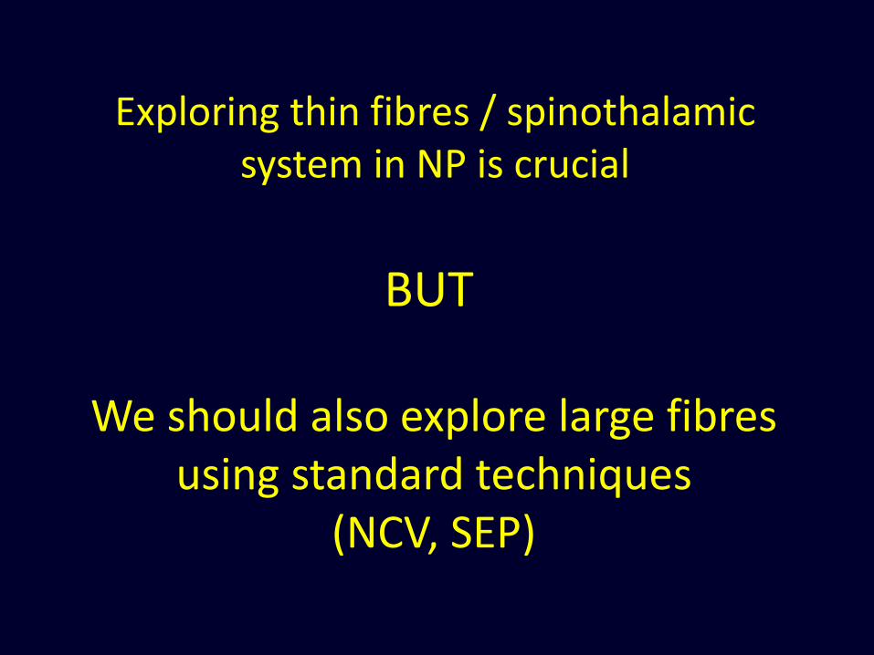
We should also explore large fibres using standard techniques
(NCV, SEP)
Exploring thin fibres / spinothalamic system in NP is crucial
BUT

« (…) electric shock-like sensations probably arise from high-frequency bursts generated in demyelinated non-
nociceptive Aβ fibres.

Paroxysmal pain following implantation of a spinal stimulator… for pain relief !
«Lhermitte’s sign » type of pain
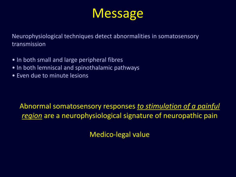
Neurophysiological techniques detect abnormalities in somatosensory transmission
• In both small and large peripheral fibres• In both lemniscal and spinothalamic pathways • Even due to minute lesions
Message
Abnormal somatosensory responses to stimulation of a painful region are a neurophysiological signature of neuropathic pain
Medico-legal value

II. Predicting pain
Clinical neurophysiology allows characterising sensory abnormalities in patients who are already in pain.
Could these techniques predict the occurrence of pain in patients who have not yet developed painful symptoms ?

Conditions able to induce chronic neuropathic pain* Brain or spinal stroke
* Spinal injury
* Multiple sclerosis (MS)
* Syringomyelia / syringobulbia
* Tumours, abscesses
* Systemic disease (Lyme, Lupus, Syphilis…)
* Peripheral neuropathy (diabetes, ischaemia, trauma, MNM)
* Plexus avulsion
* radiculopathy
• …
…and some ideas about prevalence
•Central post-stroke pain:
•8% (Andersen et al 1995, n>200)
•18% if somatosensory signs (Hansson 2004)
• 25-30% if thalamus involved (Bogousslavsky et al)
•MS: 28% (Boivie 1999, n=372)
•Syringomyelia : 67% (Attal et al 1999, n=18)
•Wallenberg : 27-50% (MacGowan et al 97, n=63)
•Anterolateral cordotomy : <10% !! (Tasker 1990)

Thalamic stroke: ~25% of patients will develop pharmaco-resistant thalamic pain
How to detect patients at risk ?

Montes et al Pain 2005
Fz
Cz
Pz
C3 C4
240ms 306ms
T4-Fz
T3-FzLateralised negativity
A clinical certitude: central pain most often concomitant with loss of pain-temperature sensations (Boivie et al 1988)

Anatomical data: posterior thalamic nuclei are predominantly affected in thalamic pain
A 9
R
STh
SN
RN
CL
mc
Pf CeM
CM
LD
VLpd
VLpl
VM
pl
MDpc
VLpv
VPMpc
A 7.2
R
LP
RN
SN
CM
Pf
mc
CL
LD
STh
pl
MDpc
VLpv
VPLa
VPM
VPMpc
A 5.4
R
LGN
SN
RN
LP
Pf
CM
CL
PuA
LD
ZI
MDpc
VPM
VPLp
VPMpc
VPLa
CL
A 3.6
R
LGN
SN
RN
LP
PuA
VPM
VPI
CM
Pf
PG
Pf
VPLp
MDpc
VPMpc
A 2.7
R
LGN
PG
SN
RN
LP
PuA
CM
Pf
Li
CLPuM
Hb
Po
MD
VPLp
VPIVMpo
A 5.4
A 1.8
R
LP
VPI
PoPf
CMPuA
PuM
CL
Hb
RN
SN
LGN
Li
SGVPL
p
VMpo
VMpo unaffected
VPL-VPM-VPI-PuA
VMpo
VMpo
VPL
VPL
VLp
VPM
A 9
R
STh
SN
RN
CL
mc
Pf CeM
CM
LD
VLpd
VLpl
VM
pl
MDpc
VLpv
VPMpc
A 9
R
STh
SN
RN
CL
mc
Pf CeM
CM
LD
VLpd
VLpd
VLpl
VLpl
VM
pl
MDpc
MDpc
VLpv
VLpv
VPMpc
VPMpc
A 7.2
R
LP
RN
SN
CM
Pf
mc
CL
LD
STh
pl
MDpc
VLpv
VPLa
VPM
VPMpc
A 7.2
R
LP
RN
SN
CM
Pf
mc
CL
LD
STh
pl
MDpc
MDpc
VLpv
VLpv
VPLa
VPLa
VPM
VPMpc
VPMpc
A 5.4
R
LGN
SN
RN
LP
Pf
CM
CL
PuA
LD
ZI
MDpc
VPM
VPLp
VPMpc
VPLa
A 5.4
R
LGN
SN
RN
LP
Pf
CM
CL
PuA
LD
ZI
MDpc
MDpc
VPM
VPLp
VPLp
VPMpc
VPMpc
VPLa
VPLa
CL
A 3.6
R
LGN
SN
RN
LP
PuA
VPM
VPI
CM
Pf
PG
Pf
VPLp
MDpc
VPMpc
CL
A 3.6
R
LGN
SN
RN
LP
PuA
VPM
VPI
CM
Pf
PG
Pf
VPLp
VPLp
MDpc
MDpc
VPMpc
VPMpcposterior
anterior
A 2.7
R
LGN
PG
SN
RN
LP
PuA
CM
Pf
Li
CLPuM
Hb
Po
MD
VPLp
VPIVMpo
A 2.7
R
LGN
PG
SN
RN
LP
PuA
CM
Pf
Li
CLPuM
Hb
Po
MD
VPLp
VPLp
VPIVMpo
A 5.4A 5.4
A 1.8
R
LP
VPI
PoPf
CMPuA
PuM
CL
Hb
RN
SN
LGN
Li
SGVPL
p
VMpo
A 1.8
R
LP
VPI
PoPf
CMPuA
PuM
CL
Hb
RN
SN
LGN
Li
SGVPL
pVPL
p
VMpo
VMpo unaffected
VPL-VPM-VPI-PuA
VMpoVMpo
VMpoVMpo
VPLVPL
VPL
VLpVLp
VPMVPM
*receiving STT projections*affected by the infarct

Combining MRI and spinothalamic-evoked potentials
to classify patients with / without thalamic pain
following a thalamic lesion

Thalamic stroke with or without thalamic pain
(n=42 patients)
Vartiainen et al, Brain 2016

Phenotyping patients to predict / prevent neuropathic pain
Pain No Pain
LEP suppression + PuA involvement 80-90% thalamic pain development(see also Sprenger et al Brain 2012, and Kurtz et al JNNP 2012))
“Both abnormal LEP and involvement of the
anterior pulvinar nucleus were independently
associated with the development of thalamic pain.
The combination of both indices increased their
predictive value (PPV 87%, NPV 77.7%)”

Much more difficult:
Discriminating pain patientsamong those having undergone a selective spinothalamic lesion
Wallenberg syndrome: 25-50% of cases will develop central pain

ms-100.0 0.0 100.0 200.0 300.0 400.0 500.0 600.0 700.0 800.0 900.0
µV 0.0
-2.5
-5.0
-7.5
-10.0
2.5
5.0
7.5
10.0
__ normal side
__ affected side, non painful (n=9)
__ affected side, painful (n=11)
Different LEP abnormalities after a brainstem lesion
Szy
Valo
Ama
neuropathic pain

Normal
Affected
Time-frequency transform of LEPs
Controls
Wallenberg – non pain
Wallenberg - pain
Fré
quency
time
controls
Wall, no pain
Wall, pain

s
3.A
3.B
AMAX
AMAX
ΔT/ΔF
1/ [ΔT/ΔF]
Controls
Non-pain patients
Pain patients
Predicting pain in brainstem syndromes ?

III. Objective detection of a pain state
…using bodily reactions

Sympathetic responses to assess the arousal effectof nociceptive stimuli
Heart rate
Chouchou et al, EJP 2011
RR intervals (ms)
Pupillary Diameter (cm)
Systolic Pressure
Diastolic Pressure
Sympathetic skin response (µS, %)
Chouchou et al, submitted

Sympathetic skin responses reflect the arousal effectof nociceptive stimuli
Abnormal sudomotor skin responses to temperature and
pain stimuli in syringomyelia .
Veciana M, Valls-Solé J, Schestatsky P, Montero J, Casado V.
J Neurol. 2007 May;254(5):638-45
Skin autonomic reactivity to thermoalgesic stimuli
Schestatsky P, Valls-Solé J, Costa J, León L, Veciana M, Chaves ML..
Clin Auton Res. 2007;17:349-55.

Enhanced subjective pain is associated wth higher sympathetic skin responses
•Laser stimuli do not produce idential sensation in every single trial (changes in
energy delivery, receptor density, etc)
•Laser stimuli yielding higher SSR were associated with Higher BOLD responses in
the insula and S1/S2, and with higher amplitudes of laser-evoked cortical responses
Mobascher et al, NeuroImage 2009; 44: 1081-1092

Cotation contre RCSga
R2 = 0,5498
0
1000
2000
3000
4000
5000
6000
7000
8000
9000
0 1 2 3 4 5 6
Cotation
Am
plitu
de
de
RC
Sg
a
The P value is < 0.0001, considered extremely
significant.
Cotation contre N2P2 R2 = 0,3953
-20
0
20
40
60
80
100
0 1 2 3 4 5 6
Cotation
Am
plitu
de
de
N2
-P2
The P value is < 0.0001, considered
extremely significant.
sec-1.0 0.0 1.0 2.0 3.0 4.0 5.0 6.0 7.0 8.0 9.0
0 900ms
Subject: Math…
VAS=5
VAS=2
VAS=5
VAS=2
In healthy subjects subjective intensity, cortical response and SSR covary

-1000 0 5000 9000
1.5J hey, it hurts!
1.25J stronger!
1J yes, it pricks
0.75J perhaps?
0.5J no resp
-3000
-2000
-1000
0
1000
2000
3000
Defining objective thresholds using sympathetic responses
Mme Cap… L3P0490

Objectivising hypoesthesia using sympathetic cutaneous responses
-1000 uV
0
500 uV
0 4 8 sec
-1000 uV
0
500 uV
0 4 8 sec
Stim Right T5; energy=1J Ø= 4mm
« very clear, pricking »
Stim Left T5; energy: 1J Ø= 4mm
« was there anything? »
Laser stim on the back,
SSR recorded over palm of the hand
Pain & hypoesthesia over left T5/T6, following back surgery + chiropractic manipulation

Objective assessment of thermal hyperalgesia
Patient L3P0492
FC1
0
FC2
0
C3
0
C4
CP2
0
P4
00
P3
CP1
Cz
0
0 400 800ms
Pz
0
Probable lesion right femorocutaneous n.Normal tactile (FVF) and thermal (laser) thresholdsMechanical allodyniaThermal hyperalgesia
Higly abnormal cortical responses
to laser stimulus of the painful side (red)
RT (EMG)
0
Abnormally delayed motor response times

Thermal hyperalgesia: sequential sympathetic responses
L3P0492
0 5000 9000 ms
-3000
-2000
-1000
0
1000
2000
3000
Average n=10 SSR
Probable surgical lesion right femorocutaneous n.Normal tactile (FVF) and thermal (laser) thresholdsMechanical allodyniaThermal hyperalgesia
Sequentially obtained SSR. Superimpsed traces to stim of right (red) and left FC territory

-1000 0 5000 9000
-3000
-2000
-1000
0
1000
2000
3000
Stim painful side:
thermal hyperalgesia with enhanced SSR
Stim normal side

Ham… Sa L3P047..
Traumatic lesion, right foot. Allodynic sequelae (mechanical & thermal). Clinical exam
impossible due to pain to any cutaneous contact. Doubt about veracity of allodynic symptoms
Laser evoked potentials impossible (intensity needed to record could not be reached)
Detecting allodynia using sympathetic responses
RCS Avg-chevdr-iee-RCS
-1000-800-600-400-200020040060080010001200140016001800200022002400260028003000320034003600380040004200440046004800500052005400560058006000620064006600680070007200740076007800800082008400
-800
-600
-400
-200
0
200
400
600
800
Intraepidermal stimuli, low intensity (0.2 mA)
Stim Right foot:«ouch, painful !!»
Stim Left foot:barely perceived

Anaesthesiol Intensive Ther. 2013 Jul-Sep;45(3):134-7.
Objective assessment of pain-related stress in mechanically ventilated newborns based on skin conductance fluctuations.
J Karpe, A Misiołek, A Daszkiewicz, H Misiołek.
Differentiating between heat pain intensities: the combined effect
of multiple autonomic parameters.
R Treister, M Kliger, G Zuckerman, I Goor Aryeh, E Eisenberg.
Pain 153 (2012) 1807-14.
Differentiating between heat pain intensities: the combined effect
of multiple autonomic parameters.
R Treister, M Kliger, G Zuckerman, I Goor Aryeh, E Eisenberg.
Differentiating between heat pain intensities: the combined effect
of multiple autonomic parameters.
R Treister, M Kliger, G Zuckerman, I Goor Aryeh, E Eisenberg.
Pain 153 (2012) 1807-14.
Sympathetic electrodermal activityto assess pain in the clinics
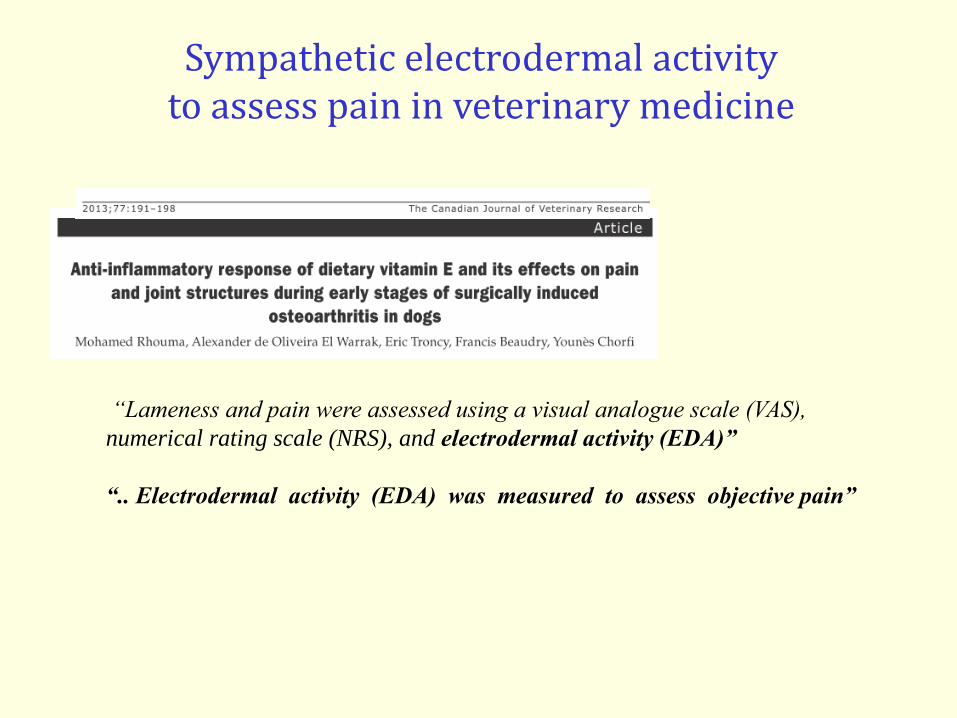
Sympathetic electrodermal activityto assess pain in veterinary medicine
“Lameness and pain were assessed using a visual analogue scale (VAS),
numerical rating scale (NRS), and electrodermal activity (EDA)”
“.. Electrodermal activity (EDA) was measured to assess objective pain”

Robocop …?
arterial pressure
EEG 128 chann
Pupillometrics
EKG
Painful
Stimuli
Subjective pain
assessment
Electrodermal activity

Detection of altered somatosensory transmission-> essential for doagnosis; medico-legal value
Prediction of neuropathic pain after neural lesion -> seems possible (thalamic stroke, brainstem lesion…)
but urgent need of prospective studies
Objectivation of pain combining behavioural, cortical and vegetative responses
-> Useful to detect objectively perceptive thresholds -> Probably useful to detect and follow allodynia (need more data!)
Why neurophysiology?

A conclusion needed ?
What is simple is false; what is complex is unusable
(Paul Valéry)

All this was done with :
Caroline Perchet
Philippe Convers
Michel Magnin
Roland Peyron
Maud Frot
François Mauguière
Patrick Mertens
Bernard Laurent
Christelle Creac’h
…