Embed Size (px)
Citation preview

Detachment of single- and multi-species bacterial biofilms by crude enzymes extracted from wastewater biofilms and
bacteria
by
Alicia van der Merwe
Submitted in partial fulfilment of the requirements of the degree Master of Science
in the Faculty of Natural and Agricultural Sciences Department of Microbiology and Plant Pathology
University of Pretoria Pretoria
December 2008
©© UUnniivveerrssiittyy ooff PPrreettoorriiaa

DECLARATION
I declare that the dissertation, which I hereby submit for the degree M.Sc (Microbiology) at
the University of Pretoria, is my own work and had not previously been submitted by me for a
degree at this or any other tertiary institution.
Signed: …………………………………….. Date: …………………………..
i

“There is hope in dreams, imagination, and in the courage of those who
wish to make those dreams a reality” Jonas Salk
ACKNOWLEDGEMENTS
My heavenly Father, for his constant unwavering love and providing me with strength
and wisdom to complete this study.
Prof. Cloete and Prof. Theron, for their valuable guidance and support throughout this
study.
My parents, for their support, love and encouragement and for always setting a great
example.
Heinrich, for always uplifting my spirit.
Chrizelle, for giving great advice and a lot of laughs.
My lab colleges, Leandra, Charisse, Melanie, Itumileng, Rasheed and Boitumelo for
creating a friendly lab environment.
Karen Surridge, for assisting with molecular techniques.
Freddie, for teaching me the fundamentals of SDS-PAGE.
Chris and Allan, for their assistance with the Scanning Electron Microscopy.
Eskom, for funding this study.
ii

SUMMARY
Detachment of single- and multi-species bacterial biofilms by crude enzymes extracted from wastewater biofilms and bacteria
by
Alicia van der Merwe
Supervisor: Prof. T.E. Cloete Department of Microbiology and Plant Pathology University of Pretoria Co-supervisor: Prof. J. Theron Department of Microbiology and Plant Pathology University of Pretoria
for the degree M.Sc
Biofilms are bacterial communities that adhere to biotic and abiotc surfaces, and are
embedded in a polymeric matrix composed mainly of polysaccharides and proteins. Not only
are biofilms a public health problem, but they are also a hindrance in industrial practices. Due
to their intractability by conventional cleaning agents, a number of alternative agents,
including enzymes, have been investigated as potential biofilm detachment-promoting agents.
Two major types of enzymes, i.e. proteases and polysaccharases, have been used for biofilm
removal and their use is aimed at degrading or promoting the collapse of the biofilm matrix.
Consequently, the aim of this investigation was primarily to assess the use of enzymes
originating from a wastewater biofilm to remove biofilms from three Pseudomonas species,
viz. P. aeruginosa PAO1, P. fluorescens and P. putida.
To investigate, biofilms were sampled from an aerobic reactor at an industrial wastewater
treatment plant. Dissolution of the biofilm, as evidenced by reductions in the soluble
chemical oxygen demand (COD) and total suspended solids (TSS), coincided with detectable
protease and carbohydrate-degrading enzyme activities. Crude extracellular enzyme extracts
prepared from the wastewater biofilm were subsequently shown to remove P. aeruginosa
PAO1 biofilms from a glass surface, suggesting that the wastewater biofilms expressed
iii
enzymes that may be used towards the removal of detrimental biofilms. Consequently,
representative bacteria were isolated from the wastewater biofilm and, based on 16S rRNA
gene sequencing and analyses, were found to represent four major phylogenetic divisions of
bacteria, i.e. Proteobacteria, Actinobacteria, Firmicutes and Bacteroidetes. Screening of the
bacterial isolates for different enzyme activities indicated that nine isolates produced
proteases, while ten isolates produced polysaccharide-degrading enzymes that comprised
amylase, xylanase, cellulase, α-glucosidase and β-glucosidase. The ability of these enzymes
to degrade proteins and polysaccharides present in purified EPS from P. aeruginosa PAO1, P.
putida and P. fluorescens was confirmed by SDS-polyacrylamide gel electrophoresis and an
increase in the amount of reducing sugar, respectively, while their efficacy to remove single-
and multi-species biofilms cultured in microtiter plates was evaluated using a quantitative
spectrophotometric assay.
Proteases produced by four of the strains were effective in degrading the EPS proteins of all
three Pseudomonas spp., while all bacterial strains that produced polysaccharide-degrading
enzymes were capable of degrading the EPS polysaccharides, albeit with different
efficiencies. Efficient removal of P. aeruginosa PAO1 biofilms was only achieved when
mixtures of enzyme extracts, containing protease and different types of polysaccharase
activities, were used. Biofilms of P. putida and P. fluorescens were readily removed with
single enzyme extracts prepared from B. subtilis and B. pumilus. Enzyme combinations
showing high biofilm removal for all three Pseudomonas species were tested against a mixed
species biofilm. These enzyme extracts yielded lower biofilm removal efficiencies than those
obtained for mono-species pseudomonad biofilms, possibly due to the heterogenous nature of
the EPS. Nevertheless, it may be possible that the enzymes identified in this study could be
used in combination with other treatments to increase the biofilm removal effectiveness or in
combination with other enzymes to degrade the mixture of proteins and polysaccharides
present in the EPS of multi-species biofilms.
iv

TABLE OF CONTENTS
DECLARATION i
ACKNOWLEDGEMENTS ii
SUMMARY iii
LIST OF ABBREVIATIONS x
LIST OF FIGURES xii
LIST OF TABLES xiv
CHAPTER 1: INTRODUCTION 1
CHAPTER 2: LITERATURE REVIEW 7 2.1 BIOFILM FORMATION 7
2.1.1 Reversible attachment 8
2.1.2 Irreversible attachment 8
2.1.3 Biofilm maturation 9
2.1.4 Detachment 10
2.2 FACTORS INFLUENCING BIOFILM FORMATION 11
2.2.1 Attachment surface 12
2.2.2 Environmental conditions 12
2.2.3 Microbial cell surface 13
2.2.4 Quorum sensing 15
2.3 BIOFILM RESISTANCE 16
2.3.1 Biofilm resistance mechanisms 16
2.3.2 Persistent cells (Persisters) 18
2.4 METHODS USED TO REMOVE BIOFILMS 19
2.4.1 Chemical treatment 20
2.4.1.1 Oxidizing biocides 20
2.4.1.2 Non-oxidizing biocides 23
2.4.2 Mechanical treatment 24
2.4.3 Alternative methods for biofilm removal 24
2.5 MICROBIAL EXTRACELLULAR POLYMERIC SUBSTANCES (EPS) AND THEIR ROLE IN BIOFILMS 26
2.5.1 Composition of EPS 27
v
2.5.2 Formation of EPS 29
2.5.3 Roles of EPS in biofilms 29
2.6 ENZYMATIC REMOVAL OF BIOFILMS 30
2.6.1 Polysaccharases 31
2.6.1.1 Enzymatic action 34
2.6.1.2 Production sources 34
2.6.2 Proteases 37
2.6.2.1 Enzymatic action 38
2.6.2.2 Production sources 39
2.6.3 Application of enzymes to biofilm removal 39
2.6.3.1 Polysaccharide-degrading enzymes 39
2.6.3.2 Proteases 40
2.6.3.3 Mixed enzymes 40
2.6.3.4 Combined use of enzymes and chemicals 42
2.7 REFERENCES 43
CHAPTER 3: CRUDE ENZYMES EXTRACTED AFTER THE AUTO-HYDROLYSIS OF BIOFILMS AND THEIR ABILITY TO REMOVE BIOFILMS 64 3.1 INTRODUCTION 64
3.2 MATERIALS AND METHODS 66
3.2.1 Sampling site 66
3.2.2 Sampling and storage of wastewater biofilm 67
3.2.3 Viscosity measurements 67
3.2.4 Biofilm auto-hydrolysis studies 67
3.2.4.1 Experimental setup 67
3.2.4.2 Analytical methods 67
3.2.5 Enzyme activity assays 68
3.2.5.1 Preparation of crude enzyme extracts from wastewater biofilm 68
3.2.5.2 Hydrolysis of proteins 68
3.2.5.3 Hydrolysis of polysaccharides 69
3.2.6 Protein and carbohydrate concentration determination 69
3.2.7 Enzymatic removal of Pseudomonas aeruginosa PAO1 biofilms 70
3.2.7.1 Bacterial strain and culturing conditions 70
vi

3.2.7.2 Growth of P. aeruginosa PAO1 biofilms on glass slides 70
3.2.7.3 Enzyme treatment of P. aeruginosa PAO1 biofilms 70
3.2.7.4 Scanning electron microscopy (SEM) 70
3.2.8 Extraction of EPS from P. aeruginosa PAO1 71
3.2.9 Hydrolysis of P. aeruginosa PAO1 EPS proteins 71
3.2.9.1 Enzyme reactions 71
3.2.9.2 Protein precipitation 71
3.2.9.3 SDS-PAGE 72
3.2.10 Hydrolysis of P. aeruginosa PAO1 EPS polysaccharides 72
3.3 RESULTS AND DISCUSSION 72
3.3.1 Biofilm auto-hydrolysis studies 72
3.3.2 Hydrolysis of proteins and carbohydrates present in the wastewater biofilm 72
3.3.3 Enzymatic treatment of P. aeruginosa PAO1 biofilms 78
3.3.4 Enzymatic hydrolysis of P. aeruginosa PAO1 EPS components 80
3.4 CONCLUSIONS 81
3.5 REFERENCES 82
CHAPTER 4: IDENTIFICATION OF WASTEWATER BIOFILM BACTERIA CAPABLE OF DEGRADING PROTEINS AND POLYSACCHARIDES 86 4.1 INTRODUCTION 86
4.2 MATERIALS AND METHODS 88
4.2.1 Isolation of bacteria from wastewater biofilm 88
4.2.2 PCR amplification and sequencing of the 16S rRNA genes from bacterial isolates 89
4.2.2.1 Genomic DNA extraction 89
4.2.2.2 Polymerase chain reaction (PCR) amplification of 16S rRNA genes 89
4.2.2.3 Nucleotide sequencing and sequence analyses 90
4.2.3 Detection of protein-degrading bacterial isolates 90
4.2.4 Detection of polysaccharide-degrading bacterial isolates 90
4.2.4.1 Screening of bacterial isolates for amylase and xylanase activity 90
4.2.4.2 Screening of bacterial isolates for cellulase activity 91
4.2.4.3 Screening of bacterial isolates for lyase activity 91
4.2.4.4 Screening of bacterial isolates for glucosidase activity 92
vii
4.3 RESULTS AND DISCUSSION 92
4.3.1 Isolation of biofilm bacteria 92
4.3.2 Identification of wastewater biofilm bacteria based on 16S rRNA gene sequence analysis 94
4.3.3 Bacterial isolates with protein-degrading capabilities 97
4.3.4 Bacterial isolates with polysaccharide-degrading capabilities 98
4.3.4.1 Xylan- and amylose-degrading bacteria 98
4.3.4.2 Cellulose-degrading bacteria 98
4.3.4.3 Alginate- and gellan-degrading bacteria 100
4.3.4.4 Glucosidase-producing bacteria 100
4.4 CONCLUSIONS 102
4.5 REFERENCES 103
CHAPTER 5: ENZYMATIC REMOVAL OF PSEUDOMONAD AND MULTI-SPECIES BIOFILMS 114 5.1 INTRODUCTION 114
5.2 MATERIALS AND METHODS 116
5.2.1 Bacterial strains and culturing conditions 116
5.2.2 Preparation of crude enzyme extracts 117
5.2.3 In vitro enzyme activity assays 117
5.2.3.1 Protease activity assay 117
5.2.3.2 Amylase, cellulase and xylanase activity assays 117
5.2.3.3 α- and β-glucosidase activity assays 118
5.2.4 Extraction of EPS from Pseudomonas spp. 118
5.2.5 Enzymatic hydrolysis of proteins in the EPS of Pseudomonas spp. 119
5.2.5.1 Enzyme reactions 119
5.2.5.2 Protein precipitation 119
5.2.5.3 SDS-PAGE 119
5.2.6 Enzymatic hydrolysis of polysaccharides in the EPS of Pseudomonas spp. 120
5.2.7 Enzymatic removal of biofilms of P. aeruginosa PAO1, P. putida and P. fluorescens 120
5.2.8 Enzymatic removal of multi-species biofilms 121
5.3 RESULTS AND DISCUSSION 122
5.3.1 Enzyme production by bacterial strains isolated from wastewater biofilm 122
5.3.2 Characterization of the EPS extracted from different Pseudomonas spp. 124
viii

5.3.3 Enzymatic degradation of the EPS of different Pseudomonas spp. 126
5.3.3.1 Protein hydrolysis 126
5.3.3.2 Polysaccharide hydrolysis 128
5.3.4 Enzymatic removal of different Pseudomonas spp. biofilms 129 5.3.4.1 P. aeruginosa PAO1 biofilm removal 132
5.3.4.2 P. putida biofilm removal 133
5.3.4.1 P. fluorescens biofilm removal 134
5.3.5 Enzymatic removal of a multi-species biofilm 136
5.4 CONCLUSIONS 137
5.5 REFERENCES 138
CHAPTER 6: GENERAL CONCLUSIONS 144
ix

LIST OF ABBREVIATIONS
Acyl-HSLs Acyl-homoserine lactones
amu atomic mass units
bp base pair
BSA bovine serum albumin
C carboxy
ca. circa approximately
CFU colony forming units
COD chemical oxygen demand
CTAB cetyltrimethylammonium bromide
dH2O distilled water
DNA deoxiribonucleic acid
DNS dinitrosalicylic acid
dNTP deoxyribonucleoside-5’-triphosphate
DPAs detachment-promoting agents
DTT dithiothreitol
e.g. for example
EDTA ethylenediamine-tetra-acetic acid
EGTA ethyleneglycol-tetra-acetic acid
EPS extracellular polymeric substances
Fig. figure
g gram
Gal galactose
Glc glucose
h hour
kDa kilodalton
l litre
LPS lipopolysaccharide
M molar
m3 cubic meter
mg milligram
MIC minimum inhibitory concentration
min minute
ml millilitre
mm millimeter
x

mM millimolar
N amino
nm nanometer
PAGE polyacrylamide gel electrophoresis
PCR polymerase chain reaction
ppm parts per million
QACs quaternary ammonium compounds
QPCs quaternary phosphonium compounds
Rha rhamnose
RNA ribonucleic acid
rpm revolutions per minute
rRNA ribosomal RNA
s second
SDS sodium dodecyl sulphate
SDS-PAGE SDS-polyacrylamide gel electrophoresis
SEM scanning electron microscopy
TCA trichloroacetic acid
TEMED N,N,N’,N’-tetramethyl-ethylenediamine
THM trihalomethane
TSS total suspended solids
U unit
V volts
v. version
v/v volume per volume
w/v weight per volume
α alpha
β beta
% percentage
× g centrifugal force
°C degrees Celsius
µg microgram
µl microlitre
µm micrometer
xi

LIST OF FIGURES
Fig. 2.1 Diagram depicting stages involved in the formation of a biofilm. 7
Fig. 2.2 Forces involved in attachment of bacteria to a surface. 8
Fig. 2.3 Biofilm architecture of P. aeruginosa. 10
Fig. 2.4 Diagram showing the method used for treatment with non-oxidizing biocides. 24
Fig. 2.5 Degradation of bacterial extracellular polysaccharides by a bacteriophage producing glycanases. 37 Fig. 3.1 Aeration basin at the Leeuwkuil water purification plant. 66
Fig. 3.2 Percentage reduction in viscosity of a wastewater biofilm, following incubation at room temperature for seven days. 73 Fig. 3.3 Chemical oxygen demand (COD) (a), total suspended solids (TSS) (b), and volume of settled matter (c) measured during a 24-day incubation period of wastewater biofilms. 75 Fig. 3.4 Soluble protein concentration and protease activity measured during a 24-day incubation period of wastewater biofilms. 77 Fig. 3.5 Carbohydrate concentration and reducing sugars measured during a 24-day
incubation period of wastewater biofilms. 77 Fig. 3.6 Scanning electron microscopy (SEM) micrographs of P. aeruginosa PAO1 biofilms treated with a crude enzyme extract prepared from a wastewater biofilm. 79 Fig. 3.7 Reducing sugars released from the extracted EPS of P. aeruginosa PAO1, following incubation for 22 h with a crude enzyme extract prepared from a wastewater biofilm. 80 Fig. 3.8 SDS-PAGE analysis of precipitated proteins, following incubation of EPS extracted from P. aeruginosa PAO1 with a crude enzyme extract prepared from a wastewater biofilm. 81 Fig. 4.1 Detection of protein-degrading bacteria on skim-milk agar medium. 97
Fig. 4.2 Diameter of hydrolysis zones formed on skim-milk agar by bacterial isolates capable of producing proteases. 98 Fig. 4.3 Detection of xylan- and amylose-degrading bacteria on nutrient agar supplemented with xylan-red and amylose-blue. 99 Fig. 4.4 Detection of cellulose-degrading bacteria on nutrient agar supplemented with hydroxyethyl-cellulose. 99
xii

Fig. 4.5 Detection of α-glucosidase and β-glucosidase activity using appropriate chromogenic substrates. 100 Fig. 5.1 Dry weight, as well as protein and carbohydrate contents of EPS extracted from P. aeruginosa PAO1 (a), P. putida (b) and P. fluorescens (c) over a 5-day incubation period. 125 Fig. 5.2 SDS-PAGE analyses of precipitated proteins, following incubation of EPS extracted from P. aeruginosa PAO1 (A), P. putida (B) and P. fluorescens (C), with crude enzyme extracts prepared from different bacterial strains. 127 Fig. 5.3 Reducing sugars released from the extracted EPS of P. aeruginosa, P. putida and P. fluorescens, following incubation with crude enzyme extracts prepared from different bacterial strains. 130 Fig. 5.4 Removal of P. aeruginosa PAO1 biofilm by treatment with enzyme extracts containing protease and/or polysaccharase enzyme activity. 133 Fig. 5.5 Removal of P. putida biofilm by treatment with enzyme extracts containing protease and/or polysaccharase enzyme activity. 134 Fig. 5.6 Removal of P. fluorescens biofilm by treatment with enzyme extracts containing protease and/or polysaccharase enzyme activity. 135 Fig. 5.7 Removal of multi-species biofilm grown from dam water by treatment with combinations of enzyme extracts containing protease and/or polysaccharase enzyme activity. 136
xiii
LIST OF TABLES
Table 2.1 General composition of bacterial EPS 28
Table 2.2 Biofilm detachment-promoting agents (DPAs) 32
Table 2.3 Glycan depolymerases linked to exopolysaccharide production 35
Table 2.4 Polysaccharide depolymerases produced by heterologous microorganisms 36
Table 2.5 Protease classification, based on mode of action 38
Table 4.1 Morphological and physiological properties of bacterial isolates from wastewater biofilms 93 Table 4.2 Identity of cultured bacterial isolates from a wastewater biofilm based on their 16S rRNA sequences 94 Table 4.3 Enzyme activities displayed by 21 cultured bacterial isolates from wastewater biofilm 101
Table 4.4 Enzymes produced by several of the isolated bacteria, as indicated in previous studies 102
Table 5.1 Enzyme activities of bacterial strains isolated from wastewater biofilm 123
Table 5.2 Exopolysaccharides produced by P. aeruginosa PAO1, P. fluorescens and P. putida 131
xiv

CHAPTER 1
INTRODUCTION
In the vast majority of ecological niches, bacteria can grow in association with surfaces,
which leads to the formation of biofilms (Costerton et al., 1995; Flemming, 2002). Biofilms
have been defined as structured communities of bacterial cells that are enclosed in a self-
produced polymeric matrix and adhere to biotic and abiotic surfaces, an interface or each
other (Costerton et al., 1995). Biofilm formation occurs in response to a variety of
environmental signals (Stanley and Lazazzera, 2004) that leads to a number of changes in
gene regulation that cause the adhering cells to become phenotypically (Sauer et al., 2002)
and metabolically (Davey and O’Toole et al., 2000) distinct from their planktonic
counterparts. Indeed, the biofilm mode of growth appears to be the preferred mode of
bacterial growth in natural environments, whereas the planktonic mode of growth is seen to
play a role in the transport of bacteria to desirable environments (Watnick and Kolter, 2000).
The ability of bacteria to form biofilms provides its members with a number of benefits. In
addition to increased resistance to environmental changes (Jefferson, 2004) and antibiotics
(Drenkard, 2003), the biofilm bacteria may also benefit from a number of properties of
communal existence, including division of metabolic burden (Geesey, 2001), gene transfer
(Ghigo, 2001) and altruistic behaviour (Kreft, 2004).
It has been suggested that biofilm formation occurs as a sequential development process
(O’Toole et al., 2000; Stoodley et al., 2002). Current models, based largely on Pseudomonas
aeruginosa, depict biofilm formation commencing when planktonic bacterial cells attach
irreversibly to a surface. This attachment is followed by growth into a mature complex
biofilm and culminates in the dispersion of detached bacterial cells into the bulk fluid. The
bacteria within each of the stages of biofilm development are believed to be physiologically
distinct from cells in other stages, and the most profound differences have been observed
when planktonic cells were compared to biofilm cells (Sauer et al., 2002). Typically, biofilm
bacteria differ from their planktonic counterparts in the genes they express (Prigent-Combaret
et al., 1999; Sauer and Camper, 2001; Whiteley et al., 2001; Beloin et al., 2004), the rate at
which the cells grow (Sternberg et al., 1999; Werner et al., 2004), and their resistance to
antimicrobial agents (Mah and O’Toole, 2001; Stewart and Costerton, 2001).
1

Chapter 1 Introduction Frequently, biofilms are unwanted and may result in various problems. These may include
dental plaque (Rosan and Lamont, 2000), medical implant-associated infections (Schierholz
and Beuth, 2001) and corrosion of pipes in the oil and water industries (Videla and Herrera,
2005). In addition, biofilms may harbour pathogens in drinking water distribution systems
(Stark et al., 1999), and food spoilage and pathogenic bacteria on food contact surfaces
(Kumar and Anand, 1998; Chmielewski and Frank, 2003). Given the medical and economical
consequences of biofilms, it is therefore important to develop strategies whereby their
formation can be prevented or, once formed, they can be removed.
Several approaches, based primarily on physical or chemical technology, are typically used to
control biofilms. Chemical techniques rely on the use of biocide products, oxidant
compounds (chlorine, chlorine dioxide and ozone) or synthetic non-oxidant compounds
(bactericides and fungicides) (Cloete et al., 1998; Meyer, 2003; Freese and Nozaic, 2004;
Dosti et al., 2005). The resistance of biofilms against various biocides and antibiotics (de
Beer et al., 1994; Norwood and Gilmour, 2000; Anderl et al., 2000; Kostenko et al., 2007),
however, makes their control problematic. In addition, many of the compounds in use have
been reported to produce by-products that are toxic or have irritant properties (Augustin and
Ali-Vehmas, 2004). In this regard, enzymes may be good anti-biofilm agent candidates due to
their biodegradability and weak toxicity (Leroy et al., 2008). Indeed, the potential of
enzymatic preparations against biofilm formation associated with pathogenic bacteria has
already been shown (Johansen et al., 1997; Kaplan et al., 2004; Itoh et al., 2005), as well as
against dental biofilm (Hahn Berg et al., 2001) and industrial biofilms (Walker et al., 2007;
Orgaz et al., 2007). The enzyme preparations typically prevent adhesion and remove adhered
biofilms by degrading the extracellular polymeric substances (EPS) produced by biofilms.
The EPS, which comprises polysaccharides, proteins, nucleic acids, lipids and various cellular
debris (Sutherland, 2001; Allison, 2003; Tsuneda et al., 2003), fulfils functions such as
providing an adhesive foundation and structural integrity (Hentzer et al., 2001; Allison, 2003),
and therefore constitute an important target for the detachment and removal of unwanted
biofilms.
The aims of this investigation were therefore the following:
To determine whether hydrolysed wastewater biofilms contain enzymes that can be
used towards removing artificial P. aeruginosa PAO1 biofilms.
2

Chapter 1 Introduction
To isolate and identify representative bacterial strains from a hydrolysed wastewater
biofilm, and to screen the bacterial strains for protease and selected polysaccharase
enzymes.
To assess the effect of crude enzyme extracts produced by the isolated bacteria on the
extracellular polymers produced by P. aeruginosa PAO1, P. fluorescens and P. putida.
To assess the efficacy of crude enzyme extracts produced by the isolated bacteria,
singularly and in combination, to remove single-species biofilms (P. aeruginosa
PAO1, P. fluorescens and P. putida) and multi-species biofilms.
REFERENCES
Allison, D.G. 2003. The biofilm matrix. Biofouling, 19: 139-150.
Anderl, J.N., Franklin, M.J. and Stewart, P.S. 2000. Role of antibiotic penetration limitation
in Klebsiella pneumoniae biofilm resistance to ampicillin and ciprofloxacin. Antimicrobial
Agents and Chemotherapy, 44: 1818-1824.
Augustin, M. and Ali-Vehmas, T. 2004. Assessment of enzymatic cleaning agents and
disinfectants against bacterial biofilms. Journal of Pharmacy and Pharmaceutical Sciences, 7:
55-64.
Beloin, C., Valle, J., Latour-Lambert, P., Faure, P., Kzreminski, M., Balestrino, D.,
Haagensen, J.A.J., Molin, S., Prensier, G., Arbeille, B. and Ghigo, J-M. 2004. Global impact
of mature biofilm lifestyle on Escherichia coli K-12 gene expression. Molecular
Microbiology, 51: 659-674.
Chmielewski, R.A.N. and Frank, J.F. 2003. Biofilm formation and control in food processing
facilities. Comprehensive Reviews in Food Science and Food Safety, 2: 22-32.
Cloete, T.E., Jacobs, L. and Brözel, V.S. 1998. The chemical control of biofouling in
industrial water systems. Biodegradation, 9: 23-37.
Costerton, J.W., Lewandowski, Z., Caldwell, D.E., Korber, D.R. and Lappin-Scott, H.M.
1995. Microbial biofilms. Annual Reviews in Microbiology, 49: 711-745.
Davey, M.E. and O’Toole, G.A. 2000. Microbial biofilms: From ecology to molecular
genetics. Microbiology and Molecular Biology Reviews, 64: 847-867.
de Beer, D., Srinivasan, R. and Stewart, P.S. 1994. Direct measurement of chlorine
penetration into biofilms during disinfection. Applied and Environmental Microbiology, 60:
4339-4344.
Dosti, B., Guzel-Seydim, Z. and Greene, A.K. 2005. Effectiveness of ozone, heat and
chlorine for destroying common food spoilage bacteria in synthetic media and biofilms.
International Journal of Dairy Technology, 58: 19-24.
3
Chapter 1 Introduction
Drenkard, E. 2003. Antimicrobial resistance of Pseudomonas aeruginosa biofilms. Microbes
and Infection, 5: 1213-1219.
Flemming, H-C. 2002. Biofouling in water systems – cases, causes and countermeasures.
Applied Microbiology and Biotechnology, 59: 629-640.
Freese, S.D. and Nozaic, D.J. 2004. Chlorine: Is it really so bad and what are the
alternatives? Water SA, 30: 18-24.
Geesey, G.G. 2001. Bacterial behavior at surfaces. Current Opinion in Microbiology, 4:
296-300.
Ghigo, J-M. 2001. Natural conjugative plasmids induce bacterial biofilm development.
Nature, 412: 442-445.
Hahn Berg, I.C., Kalfas, S., Malmsten, M. and Arnebrant, T. 2001. Proteolytic degradation of
oral biofilms in vitro and in vivo: Potential of proteases originating from Euphausia superba
for plaque control. European Journal of Oral Science, 109: 316-324.
Hentzer, M., Teitzel, G.M., Balzer, G.J., Heydorn, A., Molin, S., Givskov, M. and Parsek,
M.R. 2001. Alginate overproduction affects Pseudomonas aeruginosa biofilm structure and
function. Journal of Bacteriology, 183: 5395-5401.
Itoh, Y., Wang, X., Hinnebusch, B.J., Preston, J.F. and Romeo, T. 2005. Depolymerization of
β-1,6-N-acetyl-D-glucosamine disrupts the integrity of diverse bacterial biofilms. Journal of
Bacteriology, 187: 382-387.
Jefferson, K.K. 2004. What drives bacteria to produce a biofilm? FEMS Microbiology
Letters, 236: 163-173.
Johansen, C., Falholt, P. and Gram, L. 1997. Enzymatic removal and disinfection of bacterial
biofilms. Applied and Environmental Microbiology, 63: 3724-3728.
Kaplan, J.B., Ragunath, C., Velliyagounder, K., Fine, D.H. and Ramasubbu, N. 2004.
Enzymatic detachment of Staphylococcus epidermidis biofilms. Antimicrobial Agents and
Chemotherapy, 48: 2633-2636.
Kostenko, V., Ceri, H. and Martinuzzi, R.J. 2007. Increased tolerance of Staphylococcus
aureus to vancomycin in viscous media. FEMS Immunology and Medical Microbiology, 51:
277-288.
Kreft, J-U. 2004. Biofilms promote altruism. Microbiology, 150: 2751-2760.
Kumar, C.G. and Anand, S.K. 1998. Significance of microbial biofilms in food industry: A
review. International Journal of Food Microbiology, 42: 9-27.
Leroy, C., Delbarre, C., Ghillebaert, F., Compere, C. and Combes, D. 2008. Effects of
commercial enzymes on the adhesion of a marine biofilm-forming bacterium. Biofouling, 24:
11-22.
4

Chapter 1 Introduction
Mah, T-F. and O’Toole, G.A. 2001. Mechanisms of biofilm resistance to antimicrobial
agents. Trends in Microbiology, 9: 34-39.
Meyer, B. 2003. Approaches to prevention, removal and killing of biofilms. International
Biodeterioration and Biodegradation, 51: 249-253.
Norwood, D.E. and Gilmour, A. 2000. The growth and resistance to sodium hypochlorite of
Listeria monocytogenes in a steady-state multispecies biofilm. Journal of Applied
Microbiology, 88: 512-520.
O’Toole, G.A., Kaplan, H.B. and Kolter, R. 2000. Biofilm formation as microbial
development. Annual Reviews in Microbiology, 54: 49-79.
Orgaz, B., Neufeld, R.J. and SanJose, C. 2007. Single-step biofilm removal with delayed
release encapsulated Pronase mixed with soluble enzymes. Enzyme and Microbial
Technology, 40: 1045-1051.
Prigent-Combaret, C., Vidal, O., Dorel, C. and Lejeune, P. 1999. Abiotic surface sensing and
biofilm-dependent regulation of gene expression in Escherichia coli. Journal of Bacteriology,
181: 5993-6002.
Rosan, B. and Lamont, R.J. 2000. Dental plaque formation. Microbes and Infection, 2:
1599-1607.
Sauer, K. and Camper, A.K. 2001. Characterization of phenotypic changes in Pseudomonas
putida in response to surface-associated growth. Journal of Bacteriology, 183: 6579-6589.
Sauer, K., Camper, A.K., Ehrlich, G.D., Costerton, J.W. and Davies, D.G. 2002.
Pseudomonas aeruginosa displays multiple phenotypes during development as a biofilm.
Journal of Bacteriology, 184: 1140-1154.
Schierholz, J.M. and Beuth, J. 2001. Implant infections: A haven for opportunistic bacteria.
Journal of Hospital Infection, 49: 87-93.
Stanley, N.R. and Lazazzera, B.A. 2004. Environmental signals and regulatory pathways that
influence biofilm formation. Molecular Microbiology, 52: 917-924.
Stark, R.M., Gerwig, G.J., Pitman, R.S., Potts, L.F., Williams, N.A., Greenman, J.,
Weinzweig, I.P., Hirst, T.R. and Millar, M.R. 1999. Biofilm formation by Helicobacter
pylori. Letters in Applied Microbiology, 28: 121-126.
Sternberg, C., Christensen, B.B., Johansen, T., Nielsen, A.T., Andersen, J.B., Givskov, M. and
Molin, S. 1999. Distribution of bacterial growth activity in flow-chamber biofilms. Applied
and Environmental Microbiology, 65: 4108-4117.
Stewart, P.S. and Costerton, J.W. 2001. Antibiotic resistance of bacteria in biofilms. The
Lancet, 358: 135-138.
Stoodley, P., Sauer, K., Davies, D.G. and Costerton, J.W. 2002. Biofilms as complex
differentiated communities. Annual Reviews in Microbiology, 56: 187-209.
5

Chapter 1 Introduction
Sutherland, I.W. 2001. Biofilm exopolysaccharides: A strong and sticky framework.
Microbiology, 147: 3-9.
Tsuneda, S., Aikawa, H., Hayashi, H., Yuasa, A. and Hirata, A. 2003. Extracellular
polymeric substances responsible for bacterial adhesion onto solid surface. FEMS
Microbiology Letters, 223: 287-292.
Videla, H.A. and Herrera, L.K. 2005. Microbiologically influenced corrosion: Looking to
the future. International Microbiology, 8: 169-180.
Walker, S.L., Fourgialakis, M., Cerezo, B. and Livens, S. 2007. Removal of microbial
biofilms from dispense equipment: The effect of enzymatic pre-digestion and detergent
treatment. Journal of the Institute of Brewing, 113: 61-66.
Watnick, P. and Kolter, R. 2000. Biofilm, city of microbes. Journal of Bacteriology, 182:
2675-2679.
Werner, E., Roe, F., Bugnicourt, A., Franklin, M.J., Heydorn, A., Molin, S., Pitts, B. and
Stewart, P.S. 2004. Stratified growth in Pseudomonas aeruginosa biofilms. Applied and
Environmental Microbiology, 70: 6188-6196.
Whiteley, M., Bangera, M.G., Bumgarner, R.E., Parsek, M.R., Teitzel, G.M., Lory, S. and
Greenberg, E.P. 2001. Gene expression in Pseudomonas aeruginosa biofilms. Nature, 413:
860-864.
6

CHAPTER 2
LITERATURE REVIEW 2.1 BIOFILM FORMATION Pseudomonas aeruginosa, a motile rod-shaped Gram-negative bacterium that is found in a
variety of environments, is one of the most studied biofilm-forming organisms. Due to the
analysis of P. aeruginosa biofilms using genetic (O’Toole and Kolter, 1998a; Whiteley et al.,
2001), proteomic (Sauer and Camper, 2001; Sauer et al., 2002) and molecular biological
(Tolker-Nielsen et al., 2000; De Kievit et al., 2001a; Klausen et al., 2003; Lewandowski and
Beyenal, 2007) approaches, much information regarding the development of bacterial
biofilms has been gained. In addition, biophysical, structural and chemical analysis of the
bacterial biofilms has led to a basic model for biofilm structure (Costerton et al., 1995;
Tolker-Nielsen et al., 2000). The following information regarding biofilm development is
therefore based on the information obtained from studies on P. aeruginosa. While the exact
mechanism may differ from bacterium to bacterium, the stages of biofilm development, as
highlighted below, appear to be conserved among a wide range of microbes (O’Toole et al.,
2000; Davey and O’Toole, 2000) (Fig. 2.1).
Figure 2.1. Diagram depicting stages involved in the formation of a biofilm. 1, reversible attachment of the bacterial cells to the substratum. 2, irreversible attachment, which is assisted by exopolymeric substances and cells in this stage lose their motility. 3, primary maturation stage, which is associated with the formation of an early biofilm architecture. 4, secondary maturation stage, which leads to fully mature biofilms with complex biofilm architecture. 5, dispersion stage where motile cells (dark cells on the figure) detach from microcolonies. Shown below the diagram are photomicrographs (1-5) illustrating the five stages in biofilm formation of P. aeruginosa grown on a glass surface under continuous-flow conditions (Stoodley et al., 2002).
7

Chapter 2 Literature review
2.1.1 Reversible attachment Prior to surface colonization, a preconditioning film, composed of polysaccharides, proteins,
lipids, humic acids, nucleic acids and aromatic amino acids, is believed to form on the
attachment surface, thus resulting in a nutritionally rich zone that is metabolically favourable
for bacterial cells (Beveridge et al., 1997; Murga et al., 2001; Siboni et al., 2007). Once a
surface has been conditioned, its properties are permanently altered so that the affinity of an
organism for a native or conditioned surface can be quite different (Boland et al., 2000).
Planktonic bacteria can be brought into close approximation of the conditioned surface by
either Brownian motion, active bacterial movement, liquid flow or sedimentation (van
Loosdrecht et al., 1990). Initial attachment of the bacteria to the conditioned surface is
facilitated by electrostatic and hydrophobic interactions, van der Waals forces and specific
interactions, or a combination of these, depending on the proximity of the bacterium to the
attachment surface (van Loosdrecht et al., 1990; Habash and Reid, 1999) (Fig. 2.2). Since the
initial attachment is unstable, many of the bacterial cells may detach from the surface and
resume the planktonic lifestyle (Sauer et al., 2002).
Figure 2.2. Forces involved in attachment of bacteria to a surface. When the bacterium is greater than 50 nm away from the substratum, attractive van der Waals forces arise. At a distance of 10 to 20 nm, attachment is affected by van der Waals forces and electrostatic interactions. Different short-range interactions may occur when the distance from the substratum is less than 1.5 nm (Habash and Reid, 1999). 2.1.2 Irreversible attachment If environmental conditions are favourable for bacterial attachment, cells may switch from
reversible attachment to a more stable irreversible attachment. During this phase, the bacteria
produce exopolysaccharides that result in the formation of organic bridges between the cells
and substratum (Allison et al., 2000; Lewandowski and Beyenal, 2007). Transition from
8

Chapter 2 Literature review
reversible to irreversible attachment is also mediated by pili and fimbriae (Pratt and Kolter,
1998; O’Toole and Kolter, 1998a; Vidal et al., 1998; Inoue et al., 2003). Whereas flagella are
responsible for the movement and initial attachment of bacterial cells to the surface (O’Toole
and Kolter, 1998a; Gavín et al., 2003; Lemon et al., 2007), pili (type IV) are required for cell-
to-cell interactions and the formation of microcolonies that help strengthen the degree of
attachment to a surface (O’Toole and Kolter, 1998a; Semmler et al., 1999). Moreover, based
on the observation that P. fluorescens WCS365 was unable to attach to surfaces following
treatment with proteases (O’Toole and Kolter, 1998b), subsequent studies have shown that
membrane proteins may also assist in the attachment of bacteria to surfaces (Espinosa-Urgel
et al., 2000; Hinsa et al., 2003; Finelli et al., 2003).
2.1.3 Biofilm maturation Once bacteria have attached irreversibly to the surface, the process of biofilm maturation
begins. During this process, binary division of irreversibly attached cells causes the daughter
cells to spread outward and upward from the attachment point to form macrocolonies or cell
clusters (Tolker-Nielsen et al., 2000). Alternatively, the attached cells may be redistributed
by twitching motility (O’Toole and Kolter, 1998a; Klausen et al., 2003) and/or cells may be
recruited from the bulk fluid to the developing biofilm (Tolker-Nielsen et al., 2000). The
nature of the surface that is being colonized and the physicochemical conditions of the
environment will determine which of the mechanisms of biofilm formation will dominate
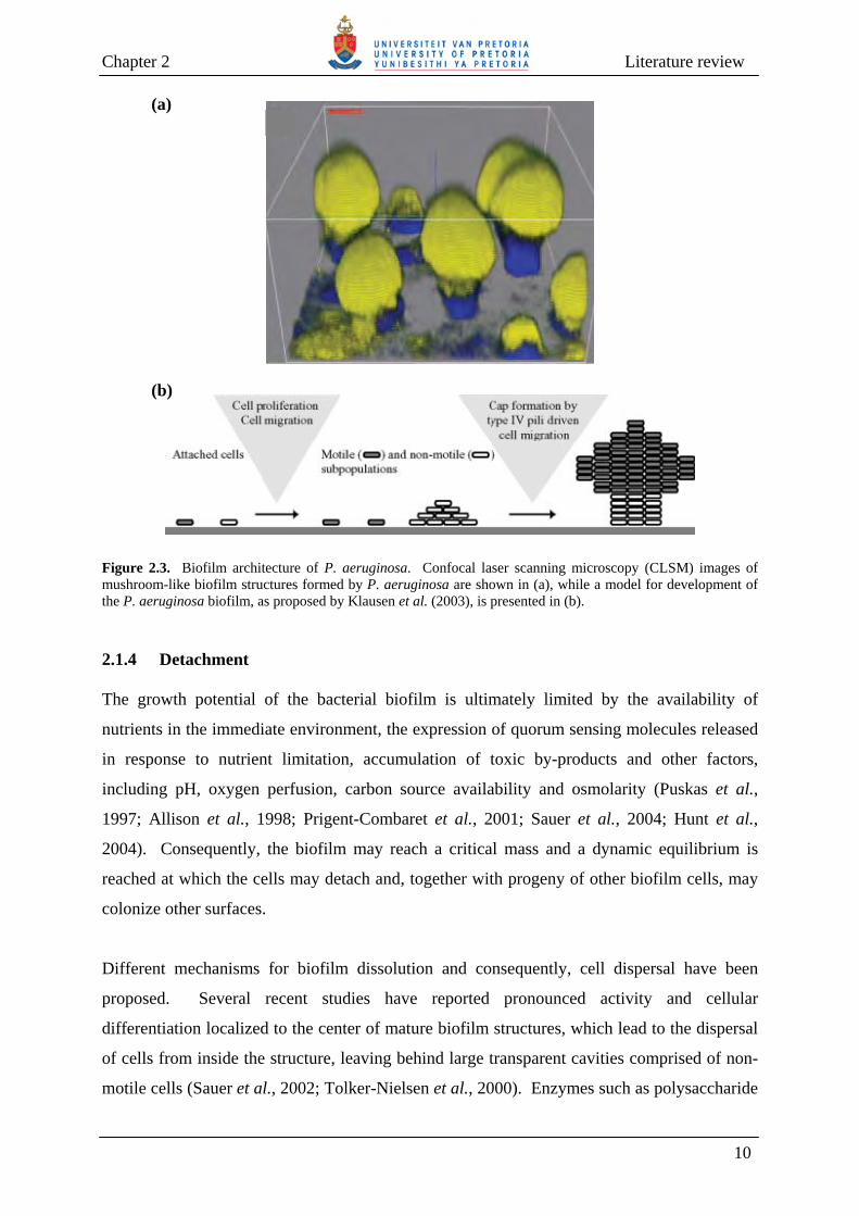
(Stoodley et al., 2002). The well-developed biofilm (Fig. 2.3a) is characterized by
mushroom- or pillar-like structures that are interspersed with fluid-filled channels (Costerton
et al., 1995; Tolker-Nielsen et al., 2000), and once fully developed, a biofilm generates
altered patterns of bacterial growth, physiological cooperation and metabolic efficiency
(Costerton et al., 1995; Geesey, 2001; Werner et al., 2004). Recently, a model for the
formation of mushroom-like biofilm structures by P. aeruginosa has been proposed by
Klausen et al. (2003). This model proposes that the bacteria are capable of constructing
mushroom-shaped structures. The propagation of non-motile bacteria results in the formation
of the mushroom stalk, while motile bacteria use their type IV pili to move up the stalk and
form the mushroom cap (Fig. 2.3b).
9
Chapter 2 Literature review
(a)
(b)
Figure 2.3. Biofilm architecture of P. aeruginosa. Confocal laser scanning microscopy (CLSM) images of mushroom-like biofilm structures formed by P. aeruginosa are shown in (a), while a model for development of the P. aeruginosa biofilm, as proposed by Klausen et al. (2003), is presented in (b). 2.1.4 Detachment The growth potential of the bacterial biofilm is ultimately limited by the availability of
nutrients in the immediate environment, the expression of quorum sensing molecules released
in response to nutrient limitation, accumulation of toxic by-products and other factors,
including pH, oxygen perfusion, carbon source availability and osmolarity (Puskas et al.,
1997; Allison et al., 1998; Prigent-Combaret et al., 2001; Sauer et al., 2004; Hunt et al.,
2004). Consequently, the biofilm may reach a critical mass and a dynamic equilibrium is
reached at which the cells may detach and, together with progeny of other biofilm cells, may
colonize other surfaces.
Different mechanisms for biofilm dissolution and consequently, cell dispersal have been
proposed. Several recent studies have reported pronounced activity and cellular
differentiation localized to the center of mature biofilm structures, which lead to the dispersal
of cells from inside the structure, leaving behind large transparent cavities comprised of non-
motile cells (Sauer et al., 2002; Tolker-Nielsen et al., 2000). Enzymes such as polysaccharide
10

Chapter 2 Literature review
lyases that degrade the exopolysaccharide matrix have been reported to play a role in biofilm
dissolution of P. fluorescens (Allison et al., 1998) and P. aeruginosa (Boyd and Chakrabarty,
1994), thereby aiding in the dispersal of the bacteria. Moreover, host-specific bacteriophages,
which are capable of synthesizing polysaccharide depolymerases that are also responsible for
degrading exopolysaccharides, have been reported to aid in the release of bacterial cells from
the outer layer of the biofilm matrix (Hughes et al., 1998a; 1998b). In the case of P.
aeruginosa, death of a subpopulation of cells has been observed to be a normal feature of
biofilm development (Webb et al., 2003). Cell death occurred inside microcolony structures,
and killed only a subpopulation of the biofilm cells. The cell death was linked to the
expression of a Pf1-like filamentous prophage of P. aeruginosa and it was suggested that
prophage-mediated cell death can assist in the spreading of a subpopulation of surviving cells
(Webb et al., 2003).
Biofilms may also be detached from surfaces by physical forces, i.e. shearing (continuous
removal of small portions of the biofilm), sloughing (rapid removal of large portions of the
biofilm) and abrasion (detachment due to collision of particles in the bulk fluid with the
biofilm) (Characklis and Marshall, 1990; Donlan, 2002). Sloughing is thought to result from
oxygen or nutrient depletion within the biofilm structure and is commonly observed with thick
biofilms that have developed in nutrient-rich environments (Characklis and Marshall, 1990;
Rice et al., 2005a). In contrast, biofilms growing in fluidized beds, filters and particle-laden
environments (surface waters) may be more prone to abrasion (Characklis and Marshall,
1990). Shearing and abrasion cause biofilm removal over the entire biofilm surface, but are
not responsible for removal up to the attachment surface. Sloughing, on the other hand,
allows for removal of biofilms up to the attachment surface, but does not occur over the entire
biofilm surface (Morgenroth and Wilderer, 2000).
2.2 FACTORS INFLUENCING BIOFILM FORMATION Several diverse factors contribute to bacterial attachment and the subsequent development of
mature biofilms. The attachment of bacterial cells to a surface is influenced by the properties
of the surface itself, environmental conditions, bacterial cell surface hydrophobicity, presence
of surface appendages (e.g., flagella and pili), outer membrane proteins and production of
extracellular polymeric substances (EPS) (Allison et al., 2000; Prakash, 2003; Ghannoum and
O’Toole, 2004). In addition, evidence suggests that the primary development of a biofilm
11

Chapter 2 Literature review
might be regulated at the level of population density-dependent gene expression controlled by
cell-to-cell signaling molecules (Allison et al., 1998; Davies et al., 1998; Parsek and
Greenberg, 2005).
2.2.1 Attachment surface As indicated previously, the formation of a conditioning film can lead to a change in the
chemical properties of the surface, thus affecting the number of cells capable of attaching to
the surface (Beveridge et al., 1997; Siboni et al., 2007). In addition, the nature of a surface,
especially its roughness and physicochemical properties, can also influence the number of
cells that may attach to the surface (Characklis and Marshall, 1990; Keevil et al., 1999;
Pasmore et al., 2002). Rough surfaces are preferred over smooth surfaces for biofilm
formation since they not only present a greater surface area to which bacterial cells can
adhere, but the attached bacteria also experience less friction with the bulk fluid (Percival et
al., 1999; Carlén et al., 2001; Pasmore et al., 2001; Morgan and Wilson, 2001). It has,
however, been noted that a rough surface might only assist in the attachment of the first cells
to the surface and that it does not have any further effect on later attachment events (Keevil et
al., 1999). Moreover, bacteria are capable of colonizing hydrophobic surfaces (e.g., Teflon
and other plastics) more readily than hydrophilic surfaces (e.g., glass and metals) (Fletcher
and Loeb, 1979; Pringle and Fletcher, 1983; Bendinger et al., 1993). The removal of biofilm
bacteria from surfaces also appears to be affected by the surface charge. Pasmore et al.
(2002) demonstrated that surfaces with neutral or low negative charges allowed for easy
removal of biofilms, while surfaces with high positive or negative charges harboured biofilms
that were difficult to remove.
2.2.2 Environmental conditions Biofilms can form under diverse nutrient conditions, ranging from high to almost non-
detectable nutrient levels (Prakash et al., 2003). Although it has been reported that biofilms
are more abundant, densely packed and thicker in environments with high nutrient levels
(Cowan et al., 1991), the precise role of nutrient availability in biofilm development has yet to
be determined. It has been reported that high nutrient levels promote the transition of
bacterial cells from the planktonic to biofilm mode of growth (O’Toole et al., 2000; Murga et
al., 2001), while depletion of nutrients causes detachment of biofilm cells from surfaces
(Allison et al., 1998; Hunt et al., 2004; Rice et al., 2005a). Other studies, however, have
12

Chapter 2 Literature review
shown that high nutrient levels can lead to the detachment of P. aeruginosa (Sauer et al.,
2004) and P. putida (Rochex and Lebeault, 2007) biofilms from surfaces. Moreover, Oh et al.
(2007) reported that Escherichia coli O157:H7 biofilms formed faster and a higher number of
cells attached to a glass surface under low nutrient conditions than under high nutrient
conditions.
Hydrodynamic conditions can influence the formation and structure of biofilms, as well as the
metabolic activity of biofilm cells (Stoodley et al., 1999; Simões et al., 2007). Biofilms
formed under turbulent flow are described as “streamers” and are typically formed by
filamentous bacteria. The microcolonies formed under these conditions are stretched out in
the direction of flow. In contrast, biofilms formed under low shear conditions (laminar flow
conditions) are characterized by spherical microcolonies that are divided by water channels
(Stoodley et al., 1999; Purevdorj et al., 2002). Simões et al. (2007) investigated the
physiological differences between biofilms formed by P. fluorescens grown under turbulent
and laminar flow conditions. Compared to biofilms grown under laminar flow conditions,
biofilms grown under turbulent flow were not only denser, had a higher biomass and were
metabolically more active, but they also produced higher amounts of exopolysaccharides
(Simões et al., 2007). Similarly, Trinet et al. (1991) and Chen et al. (1998) also reported that
biofilms formed under high flow velocities produced an increased amount of
exopolysaccharides that was shown to be required for stabilization of the biofilm structure.
Properties of an aqueous medium such as pH, temperature, oxygen and ionic strength may
also play a role in the rate at which microorganisms attach to a surface (Donlan, 2002). The
concentrations of cations such as calcium, sodium and ferric iron have been reported to
increase the attachment of P. fluorescens to glass surfaces, probably by reducing the repulsive
forces that exist between the cell surface and attachment surface (Fletcher, 1988).
2.2.3 Microbial cell surface Cell surface hydrophobicity and charge facilitates the attachment of bacterial cells to surfaces
and to each other. During adhesion the hydrophobic nature of the cell surface becomes
crucial since hydrophobic interactions are likely to increase when the non-polar properties of
the microbial cell surface and/or attachment surface increase (Donlan, 2002). Although most
bacterial cell surfaces are negatively charged, bacterial surface structures such as fimbriae, pili
13

Chapter 2 Literature review
and flagella contribute to the hydrophobicity of the cell surface due to their high content of
hydrophobic amino acid residues (Rosenberg and Kjelleberg, 1986). In addition, flagella are
also involved in the attachment of bacterial cells to the surface by assisting the cell to
overcome the initial electrostatic repulsive boundary that exists between the cell and the
substratum (Pratt and Kolter, 1998; O’Toole and Kolter, 1998a; Austin et al., 1998), while pili
have been shown to effect the formation of microcolonies during biofilm development
(O’Toole and Kolter, 1998a). Similarly, the csgBA operon of E. coli, which is responsible for
the production of curli, has been reported to assist in the adhesion of E. coli cells to surfaces
(Prigent-Combaret et al., 1999).
In addition to bacterial cell surface appendages, lipopolysaccharides (LPS) have also been
reported to affect attachment. P. aeruginosa produces A- and B-band LPS and studies have
shown that mutation of B-band LPS resulted in cells having a higher affinity for hydrophobic
than hydrophilic surfaces, while mutation of A-band LPS did not drastically influence
attachment to either surfaces (Makin and Beveridge, 1996). Al-Tahhan et al. (2000) reported
that P. aeruginosa, which had been treated with a biosurfactant (rhamnolipid) to remove the
LPS, gave rise to a hydrophobic cell surface. These cells were subsequently shown to attach
readily to hydrophobic substrates. Williams and Fletcher (1995) constructed mutations
resulting in the attenuation or loss of the P. fluorescens O antigen (polysaccharide side-chain).
Although adhesion of the mutant P. fluorescens cells to hydrophobic surfaces was improved,
adhesion to hydrophilic surfaces was decreased. It was concluded that the O antigen is partly
responsible for the hydrophilic nature of the bacterial cell and that removal of LPS from the
bacterial surface lead to a more hydrophobic cell surface (Williams and Fletcher, 1995).
Moreover, E. coli strains with mutations in core LPS biosynthesis genes (rfaG, rfaP and
galU) have also been reported to display reduced attachment to surfaces (Genevaux et al.,
1999).
Bacterial exopolysaccharides may also influence attachment and initial biofilm development
since they contribute to cell surface charge, which affects electrostatic interactions between
bacteria and the attachment surface (van Loosdrecht et al., 1989). In P. aeruginosa,
expression of the algC and algD genes has been reported to be up-regulated following
bacterial adhesion (Davies et al., 1993; Hoyle et al., 1993; Davies and Geesey, 1995). Both
these genes form part of the alginate biosynthetic operon, which controls alginate synthesis,
while the algC gene is also required for LPS core biosynthesis (Gacesa, 1998; Ramsay and
14
Chapter 2 Literature review
Wozniak, 2005). The expression of algC was shown to be activated as early as 15 min after
the bacterial cell attaches to a surface. Cells that did not undergo algC up-regulation were less
able to remain attached to the surface relative to cells in which expression was activated,
suggesting that algC may also play a role in maintenance of the biofilm (Davies and Geesey,
1995). In addition to its role in facilitating irreversible attachment, the production of
exopolysaccharides also appears to play a role in determining the biofilm structure (Stoodley
et al., 2002; Stapper et al., 2004). More detailed information regarding bacterial extracellular
polymeric substances and their function in bacterial biofilms is provided in Section 2.4.
Membrane proteins have been reported to have a substantial influence on attachment of
bacteria to surfaces and may also play a role in early biofilm development. Mutations in
surface and membrane proteins caused defects in the attachment of P. putida to corn seeds
(Espinosa-Urgel et al., 2000), while the outer membrane protein OprF (Yoon et al., 2002) and
the outer porin proteins OpdF (Finelli et al., 2003) and E1 (Sauer et al., 2002) have been
reported to play a role in P. aeruginosa biofilm development. Similarly, mutagenesis studies
have also shown that the large adhesion proteins (Lap) of P. fluorescens WCS365 are required
for attachment to several abiotic surfaces (Hinsa et al., 2003) and that the large outer
membrane protein A (LapA) is involved in the irreversible attachment of the bacterium to
surfaces (Hinsa and O’Toole, 2006).
2.2.4 Quorum sensing Several reports have linked quorum sensing, a process whereby bacteria can communicate
with each other via chemical signaling, and all stages of biofilm formation. Acyl-homoserine
lactones (Acyl-HSLs), which are quorum sensing signal molecules used by most Gram-
negative bacteria (Miller and Bassler, 2001), have been shown to affect the attachment,
maturation and detachment of biofilms (Parsek and Greenberg, 2005), as well as their spatial
arrangement (Schaber et al., 2007) and maintenance (Kjelleberg and Molin, 2002).
Certain bacteria can produce quorum sensing molecules that repress or enhance their
attachment to surfaces. The cyclic peptide-dependant accessory gene regulator agr and luxS
quorum sensing gene of Staphylococcus aureus and Helicobacter pylori, respectively,
suppress the attachment of these bacteria to surfaces (Yarwood and Schlievert, 2003; Cole et
al., 2004). In contrast, luxS of Salmonella enterica serovar Typhimurium has been reported to
15

Chapter 2 Literature review
be necessary for its attachment to human gallstones (Prouty et al., 2002). Quorum sensing
systems of P. aeruginosa (Davies et al., 1998; De Kievit et al., 2001a; Shih and Huang,
2002), Serratia liquefaciens (Labbate et al., 2004), Burkholderia cepacia (Huber et al., 2001)
and Aeromonas hydrophila (Lynch et al., 2002) have all been shown to affect biofilm
maturation. Mutations in these systems typically result in a change in the biofilm structure,
resulting in biofilms that are thinner and incapable of forming aggregates. Quorum sensing
systems of Rhodobacter sphaeroides (Puskas et al., 1997) and Yersinia pseudotuberculosis
(Atkinson et al., 1999) have been shown to be involved in cellular aggregation. More
recently, it has been reported that Vibrio cholerae makes use of quorum sensing to control the
amount of exopolysaccharides in the biofilm matrix by either producing enzymes that can
degrade the polysaccharides or by inhibiting their synthesis (Hammer and Bassler, 2003).
2.3 BIOFILM RESISTANCE Despite the availability of a wide range of antimicrobial agents, most of these compounds are,
however, not as effective against biofilms as they are against planktonic cells. Several studies
have shown resistance of biofilm cells against an array of antibiotics (Suci et al., 1994; Ishida
et al., 1998; Anderl et al., 2000; Stewart and Costerton, 2001; Kostenko et al., 2007) and
biocides (de Beer et al., 1994; Norwood and Gilmour, 2000; Grobe et al., 2002). Indeed, it
has been reported that bacteria existing in a biofilm can become up to 1000-times more
resistant to antimicrobial agents than their planktonic counterparts (Costerton et al., 1999;
Mah and O’Toole, 2001). Biofilm resistance does not appear to be dependant on a single
survival mechanism, but rather multiple factors have been proposed to play a role.
2.3.1 Biofilm resistance mechanisms The introduction of a biofilm phenotype has been proposed to lead to the activation of
mechanisms that are critical for the development of resistance to antimicrobials (Mah and
O’Toole, 2001; Drenkard, 2003). Using DNA microarrays, Whiteley et al. (2001) reported a
set of 20 genes that were differentially expressed in P. aeruginosa biofilms exposed to high
concentrations of the antibiotic tobramycin compared to untreated biofilms. Among them,
expression of dnaK and groES, both of which are involved in stress responses, and two
putative efflux systems were up-regulated. However, further studies are required to identify
the resistance mechanisms associated with the induction of specific genes in P. aeruginosa
biofilms. Several other mechanisms have also been suggested to account for biofilm
16

Chapter 2 Literature review
resistance to antimicrobial agents. These include slow growth (Gilbert et al., 1990; Evans et
al., 1991) owing to nutrient and oxygen limitations (Xu et al., 1998; Xu et al., 2000; Walters
et al., 2003) or owing to activation of the general stress response initiated by growth in a
biofilm (Brown and Barker, 1999; Cochran et al., 2000), the presence of multidrug efflux
pumps (Poole and Srikumar, 2001), and the presence of an exopolysaccharide matrix that can
react with antimicrobial agents and thus neutralize their effects and/or slow the diffusion of
these agents into the biofilm (Chen and Stewart, 1996; Stewart, 1996; Anderl et al., 2000; de
Beer et al., 1994; Kostenko et al., 2007). Several bacteria, including P. aeruginosa
(Giwercman et al., 1991) and Klebsiella pneumoniae (Anderl et al., 2000), produce β-
lactamases that are responsible for the inactivation of β-lactam antibiotics. These enzymes
have been reported to be immobilized in the exopolysaccharide matrix of the biofilm bacteria,
thus restricting penetration of the antibiotics through the biofilm. It has been proposed that
the penetration of antimicrobials through the biofilm can furthermore be influenced by (i) the
charge of both the exopolysaccharide matrix and the antimicrobial agent, (ii) the dose
concentration of the agent used, (iii) the viscosity of the exopolysaccharide matrix, (iv) the
thickness and distribution of the biofilm colonies, and (v) the hydrodynamic conditions (de
Beer et al., 1994; Stewart et al., 1998; Grobe et al., 2002; Cloete, 2003).
How each of the above-mentioned mechanisms contributes to the overall resistance displayed
by bacterial biofilms is not yet clear. For example, with the exception of aminoglycosides, the
exopolysaccharide matrix has not been found to notably retard diffusion of fluoroquinones
(Ishida et al., 1998; Walters et al., 2003). In addition, since most antibiotics target primarily
metabolically active cells it may therefore not be surprising that slow-growing and non-
growing bacteria could contribute significantly to a decrease in biofilm susceptibility to
antimicrobial agents (Lewis, 2001; Gilbert et al., 2002). Moreover, it has also been reported
that different multidrug resistance efflux pumps (MexAB-OprM, MexCD-OprJ, MexEF-OprN
and MexXY) do not play a role in P. aeruginosa biofilm resistance to antimicrobial agents
(Brooun et al., 2000; De Kieviet et al., 2001b). Similarly, the ArcAB efflux pump of E. coli
was not stimulated during biofilm growth and did not play a significant role in the resistance
of E. coli to ciprofloxacin (Maira-Literán et al., 2000).
17

Chapter 2 Literature review
2.3.2 Persistent cells (Persisters) In a study of P. aeruginosa biofilms, Brooun et al. (2000) reported that the majority of cells
were killed by low concentrations of antibiotics and despite further increases in antibiotic
concentration, a small fraction of biofilm cells (1% or less) remained that were invulnerable to
killing. It was subsequently shown that these cells, termed persister cells, were largely
responsible for the high tolerance of P. aeruginosa biofilms to antimicrobial agents (Spoering
and Lewis, 2001). According to Keren et al. (2004b) persister cells are “phenotypic variants
of the wild-type cells that upon re-inoculation produce a culture with a similar amount of
persister cells”. It has been proposed that the function of persister cells is thus to enable the
survival of the population in the presence of lethal factors (Lewis, 2001). Based on the
premise that persister cells are not directly responsible for cell death but that they cause cell
damage that indirectly triggers programmed cell death, Lewis (2001) proposed that persister
cells ensure survival of the biofilm by having a defective programmed cell death program.
More recently, the gene expression profile of persister cells of an E. coli culture was
determined and it was suggested that the formation of persister cells is dependent on
chromosomally encoded toxin-antitoxin modules (Keren et al., 2004b). Whilst
overexpression of both RelE and HipA toxins caused an increase in multidrug tolerant
persister cells, deletion of the hipBA module caused a 10- to 100-fold decrease in persister
cells in stationary and biofilm cultures. Based on these results, a revised model of persister
cell production and antibiotic tolerance was proposed. Due to random fluctuations in the ratio
of toxin-antitoxin in a population, it was proposed that some bacterial cells (1% of the
population) would express high levels of a toxin, thus giving rise to persister cells. Since
bactericidal antibiotics function by binding to a target protein and corrupting its functions,
thereby generating a lethal product that results in cell death, it may be that in persister cells
the target protein is blocked by binding of a toxin protein and its function is thus inhibited.
Although the antibiotic can bind to the blocked target protein, it can no longer corrupt its
function and the result is antibiotic tolerance, allowing cells to survive (Keren et al., 2004a;
Correia et al., 2006).
In addition to the persister cells reported above, Mah et al. (2003) reported the identification
of a mutant of P. aeruginosa PA14 that was capable of forming mature biofilms but did not
develop high level biofilm-specific resistance to antibiotics. The locus identified, designated
as ndvB, encodes for a glucosyltransferase that is necessary for cyclic glucan synthesis. Based
18

Chapter 2 Literature review
on the physical interaction of these glucans with the antibiotic tobramycin, it was suggested
that they may be responsible for entrapping antimicrobial agents in the periplasm and
therefore obstruct them from getting to their sites of action in the cytoplasm. Alternatively, it
was also proposed that the periplasmic glucans may contribute to antibiotic resistance of
biofilm cells by slowing diffusion of antibiotics into the cell, thereby allowing the bacteria
additional time to adapt to the antibiotic influx (Mah et al., 2003).
2.4 METHODS USED TO REMOVE BIOFILMS According to the National Institute of Health (NIH), biofilms are responsible for more than
60% of microbial infections, the most prominent being periodontitis, chronic lung infections
(as encountered in cystic fibrosis patients), native valve endocarditis and a range of infections
linked to biofilm-invaded medical devices (Schierholz and Beuth, 2001; Donlan and
Costerton, 2002; Hall-Stoodley et al., 2004). More than half of nosocomial infections
reported are predicted to be due to biofilms (Mah and O’Toole, 2001) and the treatment of
these biofilm-associated infections costs more than $1 billion annually in the United States
(Mah and O’Toole, 2001).
In addition to their medical consequences, biofilms also have a major impact on industrial
practices like water systems (water distribution systems, pipelines and cooling towers) (Melo
and Bott, 1997; Cloete et al., 1998; Ludensky, 2003), paper industries (Blanco et al., 1996;
Ludensky, 2003) and food industries (Kumar and Anand, 1998; Chmielewski and Frank,
2003). Financial losses are experienced due to microbial-induced corrosion of water systems,
as the equipment need to be replaced. In some cases, cooling water systems need to be
stopped in order to clean colonized surfaces and reduce or eliminate health risks posed by
biofilm microorganisms (Ludensky, 2003). Biofouling of heat exchangers has been reported
to amount in losses of £200 – 400 million per year in the United Kingdom, while chemical
treatment of biofilms in a moderate cooling water system could range from £20 – 30 000 per
year (Keevil et al., 1999). Biofilms in paper machines can cause paper defects such as stains
and holes, and lead to bad odours both in the mill and on the paper. Biofilms are also
responsible for the corrosion of surfaces in the paper mill system and for producing explosive
gases such as methane (Blanco et al., 1996). Food processing environments are also
susceptible to biofilm formation and biofilms can result in losses due to corrosion of
equipment, reduction in heat transfer, obstruction of pipelines and spoilage of food products
19
Chapter 2 Literature review
(Kumar and Anand, 1998; Chmielewski and Frank, 2003; Trachoo, 2003). Moreover,
pathogenic microorganisms present in biofilms are difficult to control due to the increased
resistance of biofilm bacteria to disinfectants and can therefore represent a potential health
risk (Chmielewski and Frank, 2003).
As a consequence of the economic losses associated with the deleterious effects of biofilms on
medical and industrial practices, different methods have been used for minimizing the
accumulation of biofilms on surfaces. These have included treatment of water to reduce the
number of bacteria entering the system, modification of surfaces to prevent their colonization
by biofilms, and chemical and mechanical treatments that result in removal of biofilms from
surfaces (Characklis and Marshall, 1990).
2.4.1 Chemical treatment Chemical treatment entails the use of biocides, dispersants and surfactants to decrease the
number of microorganisms entering a water system and/or aid in the removal of biofilms from
surfaces. Chemicals that are to be used for reducing the number of bacteria should be chosen
with care. Such chemicals must not pose health or environmental risks, should not affect the
activity of any other chemicals used or cause corrosion of the treated systems, and it must be
as cost-effective as possible (Charaklis and Marshall, 1990; Keevil et al., 1999). Also, several
factors need to be taken into account when selecting an appropriate chemical for the removal
of bacterial biofilms. Not only must the number and diversity of bacteria present at the site be
taken into account, but also the minimum inhibitory concentration (MIC) and contact time of
the chemical for each of the bacteria. Moreover, to lessen the occurrence of chemical
resistance, it is important to alternate the usage of different chemicals, apply the correct MIC
and combine the chemicals with surface-active compounds that can detach the bacterial
biofilms (Charaklis and Marshall, 1990; Cloete et al., 1998). In order for a biocide to be
effective in removing biofilms, it must be able to penetrate through the exopolysaccharide
matrix of biofilms. Since the composition of the matrix may differ for different biofilms, the
use of biocides that have a non-specific action is favoured (Meyer, 2003; Cloete, 2003).
2.4.1.1 Oxidizing biocides Oxidizing biocides target the thiol group of cysteine amino acid residues and consequently,
result in the oxidation of proteins. Since cysteines play an important role in determining
20
Chapter 2 Literature review
protein structure and function, oxidizing biocides can therefore lead to cellular metabolic
inhibition due to the oxidation of structural proteins present in cellular walls, membranes and
ribosomes (Cloete et al., 1998; Russel et al., 2003). Several different oxidizing biocides, as
discussed below, have been applied to the removal of bacterial biofilms in industrial practices.
Chlorine Amongst all of the oxidizing and non-oxidizing biocides in use, chlorine is the most
frequently used to control biofilms in water distribution systems (Freese and Nozaic, 2004).
This is due to the ability of chlorine to destroy the integrity of the biofilm matrix by oxidizing
and consequently, depolymerizing the exopolysaccharides present in the matrix (Characklis
and Dydek, 1976). The attachment of new bacterial cells to surfaces is therefore more
difficult in the presence of chlorine (Meyer, 2003). Chlorine also reacts with organic and
inorganic matter present in the bulk fluid (de Beer et al., 1994; Ryu and Beuchat, 2005),
therefore necessitating that higher chlorine concentrations be used in order for it to penetrate
biofilms (Characklis and Marshall, 1990). A concentration of as high as 1.5 mg/l residual
chlorine is sometimes unable to penetrate biofilms (Cloete et al., 1998). Norwood and
Gilmour (2000) indicated that sodium hypochlorite at a concentration of 1000 ppm free
chlorine was required to cause a substantial reduction in the number of biofilm bacteria, while
only 10 ppm free chlorine was enough to totally remove planktonic bacteria. Chlorine can
have a detrimental effect on humans and the environment, since its reaction with organic
matter in the water may lead to the formation of trihalomethanes (THMs) (Zhao et al., 2006),
which have carcinogenic properties (Tokmak et al., 2004). When used at high concentrations,
chlorine can also cause corrosion of metal surfaces (Pisigan and Singley, 1987) and it is very
sensitive to pH changes, making treatment of biofilms in cooling water systems difficult since
an alkaline pH predominates (Keevil et al., 1999). Despite these drawbacks, chlorine is
nevertheless an efficient and versatile biocide, its handling and dosing is reasonably effortless
and the costs involved in chlorine disinfection are low (Freese and Nozaic, 2004).
Chlorine dioxide Many of the above-mentioned problems associated with the use of chlorine can be overcome
by making use of chlorine dioxide, albeit not as cost-effective as chlorine and its production
being more complex (Anderson et al., 1982). Moreover, harmful by-products such as THMs
and haloacetic acids are formed at a much lower level when chlorine dioxide is used
21

Chapter 2 Literature review
compared to the use of chlorine (Volk et al., 2002; Zhao et al., 2006). Nevertheless, chlorine
dioxide and its by-products (chlorite and chlorate) (Korn et al., 2002) has been reported to
cause hemolytic anemia in rats (Abdel-Rahman et al., 1984). However, it has been reported
that there appears to be no link between the intake of chlorine dioxide-treated water and
human illness (Lubbers et al., 1981). Furthermore, chlorite has also been shown to lessen in
quantity as the water is transported through water distribution systems (Baribeau et al., 2002).
Chlorine dioxide is applied to water in a gaseous form and has been shown to be successful in
penetrating bacterial biofilms. When a chlorine dioxide system (ACTIV-OX®) was applied to
hot and cold water systems, Legionella populations were eliminated without requiring
additional chlorine dioxide (Keevil et al., 1999). Gagnon et al. (2004) compared the
effectiveness of chlorine dioxide, free chlorine and chloramines with regards to their ability to
remove bacteria in a laboratory-scale water distribution system and reported that chlorine
dioxide was the most effective on biofilm, as well as planktonic bacteria.
Hydrogen peroxide Hydrogen peroxide is not as cost-effective as chlorine, but it is less harmful to the
environment since it is reduced to oxygen and water (Carlsson, 2003; Mara and Horan, 2003).
Although hydrogen peroxide has been used to reduce microbial numbers in the cooling water
of nuclear reactors, studies have shown that hydrogen peroxide was not very successful in
removing biofilms from a laboratory-scale reactor (Characklis and Marshall, 1990). The
ineffectiveness of hydrogen peroxide to remove biofilms may be due to the production of
catalase, an enzyme responsible for the degradation of hydrogen peroxide. Catalase
production has been linked to the increased resistance of bacterial cells to hydrogen peroxide
(Mah and O’Toole, 2001). In this regard, Stewart et al. (2000) and Elkins et al. (1999)
reported that, in contrast to biofilms formed by wild-type P. aeruginosa, the biofilms formed
by a catalase-deficient mutant strain was not protected against hydrogen peroxide penetration
and killing.
Ozone Ozone is a much stronger oxidizing agent than chlorine (Dosti et al., 2005), but its use has
been limited due to its low water solubility, especially at high temperatures (Guzel-Seydim et
al., 2004). Ozone also readily reacts with organic matter present in water, thus reducing its
concentration and making its application in waters with high organic loads uneconomical
(Mara and Horan, 2003). Moreover, ozone requires production at the application site since it
22

Chapter 2 Literature review
possesses a high reactivity and unstable nature (Peleg, 1976; Staehelln and Holgné, 1982).
Nevertheless, ozone has been shown to be effective in controlling biofilm formation at
concentrations of lower than 0.5 g/m3 (Sugam, 1980). Since ozone does not have a lasting
effect, other disinfectants must be used in conjunction with ozone in order to keep water free
from microorganisms (Rennecker et al., 2000). Dosti et al. (2005) compared the efficacy of
ozone and chlorine with regards to killing of food-spoilage bacteria present in biofilms. The
number of biofilm bacteria was considerably reduced by treatment with ozone and chlorine,
and only a slight difference in their efficacy was noted. Ozone, however, was more effective
than chlorine in reducing bacterial numbers when tested against P. putida biofilms (Dosti et
al., 2005).
2.4.1.2 Non-oxidizing biocides Non-oxidizing biocides comprise a range of bactericidal organic compounds such as iso-
thiazolones, quaternary ammonium compounds (QACs), quaternary phosphonium compounds
(QPCs), amines and triazines (Cloete et al., 1998). Since these compounds are not
manufactured specifically with the aim of removing biofilm bacteria, major problems are
associated with their use as a means to control biofilms (Keevil et al., 1999). Notably, they do
not penetrate the biofilm readily (Stewart et al., 1998) and, in some cases, can decrease in
efficacy due to repeated application (Sundheim et al., 1998; Langsrud et al., 2003). Non-
oxidizing biocides are typically used at high initial concentrations in order to ensure bacterial
killing. As soon as the biocide concentration decreases and microbial counts increase, the
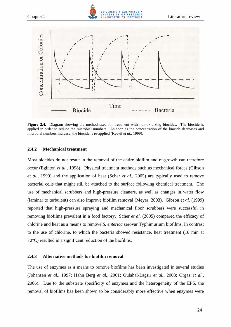
biocide is re-applied (Keevil et al., 1999) (Fig. 2.4). Of the different non-oxidizing biocides,
QACs have been used in a wide range of applications, e.g., as a disinfectant in food industries
(Langsrud et al., 2003) and in the medical field (Friedman et al., 1968; Murtough et al., 2000;
Ioannou et al., 2007). The positively-charged QACs form an electrostatic bond with
negatively-charged bacterial cell walls, which causes stress in the cell wall, denaturation of
proteins, disturbance of cell wall-permeability and decreased uptake of nutrients into the cell,
thereby resulting in cell lysis (Cloete et al., 1998; Ioannou et al., 2007).
23
Chapter 2 Literature review
Figure 2.4. Diagram showing the method used for treatment with non-oxidizing biocides. The biocide is applied in order to reduce the microbial numbers. As soon as the concentration of the biocide decreases and microbial numbers increase, the biocide is re-applied (Keevil et al., 1999). 2.4.2 Mechanical treatment Most biocides do not result in the removal of the entire biofilm and re-growth can therefore
occur (Eginton et al., 1998). Physical treatment methods such as mechanical forces (Gibson
et al., 1999) and the application of heat (Scher et al., 2005) are typically used to remove
bacterial cells that might still be attached to the surface following chemical treatment. The
use of mechanical scrubbers and high-pressure cleaners, as well as changes in water flow
(laminar to turbulent) can also improve biofilm removal (Meyer, 2003). Gibson et al. (1999)
reported that high-pressure spraying and mechanical floor scrubbers were successful in
removing biofilms prevalent in a food factory. Scher et al. (2005) compared the efficacy of
chlorine and heat as a means to remove S. enterica serovar Typhimurium biofilms. In contrast
to the use of chlorine, to which the bacteria showed resistance, heat treatment (10 min at
70°C) resulted in a significant reduction of the biofilms.
2.4.3 Alternative methods for biofilm removal The use of enzymes as a means to remove biofilms has been investigated in several studies
(Johansen et al., 1997; Hahn Berg et al., 2001; Oulahal-Lagsir et al., 2003; Orgaz et al.,
2006). Due to the substrate specificity of enzymes and the heterogeneity of the EPS, the
removal of biofilms has been shown to be considerably more effective when enzymes were
24

Chapter 2 Literature review
used in combination with detergents (Johansen et al., 1997), sonication (Oulahal-Lagsir et al.,
2003) and antibiotics (Selan et al., 1993). Moreover, it has been suggested that the use of
different types of enzymes, especially combinations of proteases and polysaccharases, may be
effective in removing bacterial biofilms (Meyer, 2003). The enzymatic removal of biofilms
will be discussed in greater detail in Section 2.6.
Some research has been carried out regarding the use of bacteriophages to remove bacterial
biofilms (Hibma et al., 1997; Hughes et al., 1998a; 1998b; Hanlon et al., 2001; Curtin and
Donlan, 2006). Host-specific bacteriophages can infect biofilm cells by first degrading the
EPS and then ultimately lysing the bacterial cells (Hughes et al., 1998a). However, since
bacteriophages and their bacterial hosts exist together in nature, they are not considered a
viable tool for biofilm removal (Tait et al., 2002). Another problem to take into consideration
is the ability of bacterial strains to become resistant against bacteriophage attack (Hill et al.,
1990; Foley et al., 1998). One way of circumventing these problems is through the use of
bacteriophages in combination with sanitizers. In this regard, Tait et al. (2002) reported that
bacteriophage-treated biofilms displayed a reduction in the amount of exopolysaccharides,
making the biofilms more sensitive to subsequent treatment with sanitizers.
Quorum sensing is proposed to affect various different cellular processes, amongst other,
biofilm formation (Rice et al., 2005b; Parsek and Greenberg, 2005; Harraghy et al., 2007).
Quorum sensing may therefore be a potentially important system to target in order to prevent
biofilm formation. Notably, it has been reported that an RNAIII-inhibiting peptide inhibited
quorum sensing and subsequently the attachment of Staphylococcus epidermidis (Balaban et
al., 2003) and S. aureus (Yarwood and Schlievert, 2003) to surfaces. Enzymatic degradation
of quorum sensing molecules has also been investigated as means to prevent biofilm
formation. Acyl-homoserine lactones can be degraded by N-Acyl homoserine lactonases from
several Bacillus species (B. cereus, B. thuringiensis and B. mycoides) (Dong et al., 2002),
Arthrobacter sp. IBN110 and K. pneumoniae (Park et al., 2003), as well as by acylases from
P. aeruginosa PAO1 (Sio et al., 2006) and Ralstonia sp. (Lin et al., 2003). Similarly, Xu et
al. (2003) reported that formation of biofilms grown from aquarium water decreased by 10-
40%, following addition of acylase I from porcine kidney cells. Delisea pulchra, an
Australian red macroalga, produces furanones that have been reported to interfere with the
quorum sensing signaling processes of S. liquefaciens (Rasmussen et al., 2000), V. fischeri
(Manefield et al., 2002) and P. aeruginosa (Hentzer et al., 2002). It was subsequently
25

Chapter 2 Literature review
proposed that their use, in combination with antimicrobial agents, may result in biofilm
removal at lower doses of the antimicrobial agent (Hentzer et al., 2003).
The removal of nutrients from the bulk fluid has been reported to be successful in reducing
biofilm formation (Allison et al., 1998), and also to increase the half-life of disinfectants such
as chlorine (Chandy and Angles, 2001). By making use of side-stream biofilters to reduce the
amount of nutrients available in cooling water systems, Daamen et al. (2000) demonstrated a
reduction in the number of bacteria and reported that the need for addition of biocides to
remove biofilms was reduced by at least 70%. Hunt et al. (2004) investigated the effect of
glucose starvation on the detachment of P. aeruginosa biofilms. Experimental work, as well
as a biofilm model, confirmed that biofilm detachment can occur due to nutrient starvation
(Hunt et al., 2004).
Biofilm formation can furthermore be limited or prevented through modification of the
attachment surface (Carlén et al., 2001; Pasmore et al., 2002). This approach may not be very
successful since many biofilms consist of a range of different bacterial species, each with their
own attachment preferences (Palmer and White, 1997). Alternatively, the surface can be
made attractive for the attachment of a specific microorganism in order to inhibit attachment
of other unwanted bacteria. These methods can, however, only be applied in simple,
controlled environments such as medical and dental equipment (Palmer and White, 1997).
The covering of medical devices, particularly urinary and vascular catheters, with antibiotics
or antimicrobial compounds has showed some success in reducing or completely preventing
the attachment of biofilms (Sheng et al., 2000; Donelli et al., 2002). However, covering of
these devices with antibiotics has raised concerns regarding the emergence of antibiotic-
resistant bacterial strains (Francolini et al., 2003).
2.5 MICROBIAL EXTRACELLULAR POLYMERIC SUBSTANCES (EPS) AND
THEIR ROLE IN BIOFILMS Biofilms are characterized by high levels of EPS and as much as 50-90% of biofilms’ total
organic matter consists of EPS (Allison, 2000). Geesey (1982) described EPS as
“extracellular polymeric substances of biological origin that participate in the formation of
microbial aggregates”. Although polysaccharides are believed to dominate the EPS,
exceptions do occur. For example, the EPS from wastewater biofilms (activated sludge) have
26

Chapter 2 Literature review
been reported to have a higher proportion of proteins than polysaccharides (Nielsen et al.,
1997; Bura et al., 1998).
2.5.1 Composition of EPS In several bacterial species studied, polysaccharides and proteins have been reported to
account for 75-89% of the EPS composition (Tsuneda et al., 2003) (Table 2.1). The main
configuration of the polysaccharides present in bacterial EPS is β and rarely α, with a high
prevalence of 1,3 or 1,4 linkages in the backbone of the polymer. The polysaccharide
compositions in EPS can range from very basic polysaccharides, containing only one type of
monomer (homopolysaccharides), to more complex polysaccharides composed of more than
two different monosaccharides (heteropolysaccharides). Neutral polysaccharides are scarce,
while positively-charged (due to the presence of amino sugars) and negatively-charged (due to
the presence of carboxyl, sulfate or phosphate groups) polymers are more abundant (Allison,
2000). The structure and physicochemical properties of polysaccharides can, however, be
altered by the presence of non-polymeric organic (acetyl, pyruvyl and succinyl) and inorganic
(sulphate and phosphate) compounds (Wingender et al., 1999).
In addition to polysaccharides, proteins also form part of the EPS. The proteins can be
glycosylated with oligosaccharides to form glycoproteins, while the oligosaccharides can also
be replaced with fatty acids to form lipoproteins (Wingender et al., 1999). EPS assimilate
proteins from living and lysed bacterial cells, and these proteins can be absorbed due to the
polyanionic properties of the EPS carbohydrates (Ghannoum and O’Toole, 2004). Proteins
are typically responsible for the hydrophobic nature of EPS (Dignac et al., 1998; Jorand et al.,
1998) and may assist in the attachment of bacteria to negatively-charged surfaces (Characklis
and Marshall, 1990). The proteins in EPS do not only play a structural role, but some of the
proteins might act as enzymes (Characklis and Marshall, 1990). Polysaccharases are
prominent in EPS and are responsible for degradation of EPS and other organic components
present in the bulk fluid, thereby supplying the biofilm cells with nutrients (Wingender et al.,
1999).
Other substances present in EPS, albeit in smaller amounts, include nucleic acids (DNA and
RNA), lipids and other products of cell lysis (Sutherland, 2001a; Whitchurch et al., 2002;
Allison, 2003; Tsuneda et al., 2003; Moscoso et al., 2006; Jurcisek and Bakaletz, 2007).
27

Chapter 2 Literature review
28

Chapter 2 Literature review
Cellular material comprises 2-15% of the biofilm matrix, but the amounts of these
components can differ (Sutherland, 2001b). Treatment of P. aeruginosa biofilms with DNase
I was reported to result in the removal of the biofilms from a surface, suggesting a role for
DNA in biofilm formation (Whitchurch et al., 2002).
2.5.2 Formation of EPS EPS are situated on or in close proximity to the outer surface of biofilm cells. The
exopolymeric substances can be released into the extracellular environment by active
secretion, during lysis of bacterial cells or by the detachment of membrane components from
the cell surface (Wingender et al., 1999). Several cellular systems that actively secrete
proteins and polysaccharides have been reported (Rehm and Valla 1997; Jonas and Farah,
1998), while the presence of nucleic acids in the EPS may be due to horizontal gene transfer
that occurs between the tightly packed biofilm cells (Christensen et al., 1998; Ghigo, 2001) or
as consequence of programmed cell lysis (Webb et al., 2003). In addition, cell lysis also
results in the release of the intracellular bacterial contents that can subsequently be captured
within the biofilm matrix (Wingender et al., 1999). Moreover, some intracellular components
of Gram-negative bacteria have been shown to be released into the extracellular environment
by the formation of membrane-derived vesicles (blebs) (Kadurugamuwa and Beveridge,
1995). Cellular components released in this manner include nucleic acids (Dorward and
Garon, 1990; Renelli et al., 2004) and enzymes (Kadurugamuwa and Beveridge, 1995), as
well as membrane components such as phospholipids (Schooling and Beveridge, 2006).
2.5.3 Roles of EPS in biofilms Due to its high heterogeneity among different bacterial strains, the function of EPS is only
partially understood. EPS, however, are involved in the attachment of bacterial cells to a
surface and in holding the biofilm cells together (Mack et al., 1996; Yildiz and Schoolnik,
1999; Danese et al., 2000; Hentzer et al., 2001; Lewandowski and Beyenal, 2007). In
addition, EPS can change the physicochemical properties of the attachment surface, thereby
making it easier for bacterial cells to attach. Tsuneda et al. (2003) reported that electrostatic
interactions reduced bacterial attachment when low amounts of EPS were produced, while
attachment was enhanced by polymeric interactions when the EPS amounts were high. In
contrast, Hanna et al. (2003) reported that colonic acid, a capsular polysaccharide produced
29

Chapter 2 Literature review
by E. coli, obstructed both specific binding and time-dependant interactions between the
bacteria and their attachment surfaces.
EPS not only act as a protective layer against antibiotics (Anderl et al., 2000; Kostenko et al.,
2007) and biocides (de Beer et al., 1994; Chen and Stewart, 1996; Wai et al., 1998; Stewart
and Costerton, 2001; Grobe et al., 2002), but also offer protection against heavy metals
(Teitzel and Parsek, 2003) and UV irradiation (Elasri and Miller, 1999), thus reducing their
penetration into the bacterial cells. Moreover, EPS also protects biofilm cells against
dehydration under water-limited conditions (Ophir and Gutnick, 1994; Schnider-Keel et al.,
2001), and other environmental conditions such as temperature fluctuations (Mao et al., 2001)
and osmotic shock (Schnider-Keel et al., 2001; Chen et al., 2004). The outer layer of EPS can
dry out under water-limited conditions and forms a hard, protective layer that prevents
dehydration of the inner cells (Sutherland, 2001a).
In addition to the above, EPS is also considered to be a virulence factor in several different
bacterial infections. Bereswill and Geider (1997) reported that production of the acidic EPS,
amylovoran, by Erwinia amylovora is linked to its virulence, since a strain that was incapable
of producing amylovoran was avirulent on its host plants. Several studies concluded that
mucoid P. aeruginosa initiates chronic lung infection due to the high production of EPS by
this strain (Pedersen et al., 1990; Gilligan, 1991; Hentzer et al., 2001). This “mucoid
exopolysaccharide” (MEP) is therefore considered a virulence factor in cystic fibrosis
infections (Gilligan, 1991). Furthermore, lectins bound to certain carbohydrates present in
EPS have been reported to play a role in the pathogenesis of some bacteria, and assist in the
attachment of the cell to its host and to other bacterial cells (Sharon and Lis, 2004).
2.6 ENZYMATIC REMOVAL OF BIOFILMS
As highlighted in the previous sections, most biocides and antibiotics are ineffective in
biofilm eradication and control. In addition, some of these disinfectants produce by-products
that are toxic or have irritant properties (Augustin and Ali-Vehmas, 2004). Therefore, there
exists a need to identify agents to which microorganisms cannot acquire resistance and that do
not produce harmful by-products. A significant target for detaching and eradicating biofilms
may perhaps be the biofilm matrix, since EPS play an important role in the structural integrity
and attachment properties of biofilms (Hentzer et al., 2001; Stoodley et al., 2002).
30
Chapter 2 Literature review
Consequently, various different agents have been investigated for their ability to promote the
detachment of bacterial biofilms (Table 2.2). Chemicals (e.g., NaCl2, MgCl2 and CaCl2) have
been used to change the ionic strength and composition of the liquid medium which, in turn,
influence the electrostatic interactions responsible for the attachment properties of the biofilm
matrix (Chen and Stewart, 2000). Chelating agents (e.g., EDTA and EGTA) have been used
to cross-link multivalent cations involved in biofilm attachment, while surfactants can
interrupt hydrophobic interactions implicated in the maintenance of the matrix structure
(Turakhia et al., 1983; Chen and Stewart, 2000). In addition to the above, enzymes capable of
degrading extracellular DNA (Whitchurch et al., 2002), as well as polysaccharides and
proteins (Johansen et al., 1997; Hughes et al., 1998a; Skillman et al., 1999; Kaplan et al.,
2004a; Itoh et al., 2005), the two major components of EPS responsible for its structural
stability (Wingender et al., 1999), have also been used to promote the detachment of biofilms.
Combinations of different enzymes, especially proteases and polysaccharases, in the presence
or absence of antimicrobial agents and detergents, is generally thought to be useful for
degradation of the biofilm EPS. Therefore, the following sections will focus mainly on these
two groups of enzymes and their application to the removal of biofilms.
2.6.1 Polysaccharases Polysaccharide hydrolases and polysaccharide lyases are capable of degrading microbial
exopolysaccharides. These enzymes are responsible for controlling the length of
polysaccharides during synthesis and can degrade the polymers in order to supply the
microorganism with sources of carbon and energy (Sutherland, 1999; Wingender et al., 1999).
Polysaccharases capable of degrading EPS have thus attracted considerable attention as a
means to remove bacterial biofilms (Bayer et al., 1992; Johansen et al., 1997; Allison et al.,
1998; Merritt et al., 2000; Orgaz et al., 2006; Walker et al., 2007). However, in mixed
biofilm populations, where a range of different exopolysaccharides is present, EPS
degradation may be more challenging (Wingender et al., 1999; Tait et al., 2002). In such
instances, a mixture of polysaccharases is typically used. Since the degradation products of
one enzyme might serve as a substrate for a different enzyme, it can thus ultimately lead to the
degradation of EPS and the removal of biofilms (Wingender et al., 1999).
31
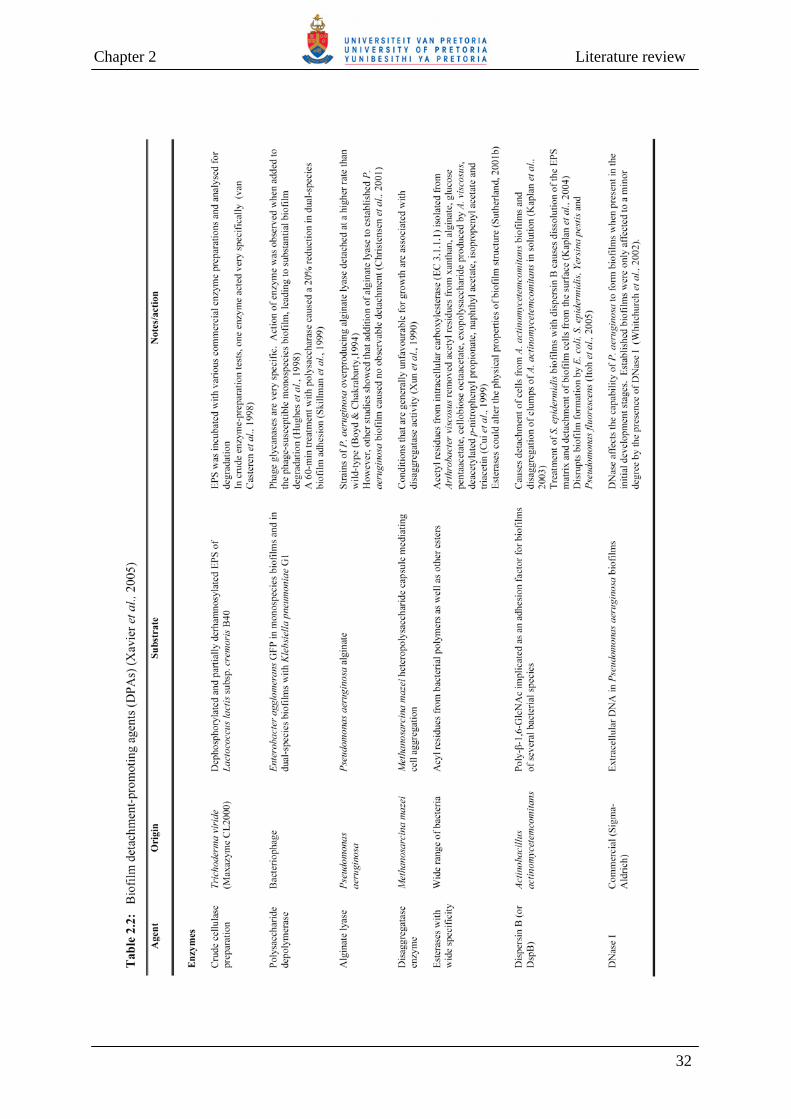
Chapter 2 Literature review
32

Chapter 2 Literature review
33

Chapter 2 Literature review
2.6.1.1 Enzymatic action Polysaccharide hydrolases and polysaccharide lyases are both capable of degrading EPS
(Sutherland, 1999; Wingender et al., 1999). The substrate specificity of the respective
enzymes is determined by the structure of the carbohydrates and, in particular, by the presence
of uronic acids. The presence of acyl substituents in the carbohydrate structure has been
reported to decrease the activity of certain polysaccharide lyases such as alginate and gellan
lyases (Sutherland, 1999). Enzymatic degradation of EPS can also be influenced by the
presence of biosurfactants. The biosurfactants associate with the polysaccharides and can
either enhance (Eriksson et al., 2002; Helle et al., 2004) or hinder the degradation process
(Helle et al., 2004). EPS-degrading enzymes can be classified as either endo- or exo-acting
(Sutherland, 1999; Wingender et al., 1999). Whereas endo-acting enzymes can reduce the
polysaccharide chain length rapidly by cleaving internal bonds of the polysaccharide, the exo-
acting enzymes are responsible for cleaving bonds at the ends of the polysaccharide and
therefore its breakdown is slower. The degradation of homopolysaccharides can be facilitated
by a range of endo- and exo-acting enzymes (Gilbert and Hazlewood, 1993; Mishra and
Robbins, 1995), while heteropolysaccharides are usually degraded by a single, very specific
endo-acting polysaccharase (Sutherland, 1999).
2.6.1.2 Production sources Polysaccharases are produced by polysaccharide-synthesizing microorganisms (endogenous
production), other eukaryotic and prokaryotic microorganisms (exogenous production) and
bacteriophages (Sutherland, 1999; Wingender et al., 1999).
Endogenous production Some polysaccharide-producing microorganisms synthesize polysaccharases that are capable
of degrading their own polysaccharides to use as a source of carbon and energy (Sutherland,
1999). More commonly, however, the endogenously-produced enzymes do not allow the
microorganism to utilize its own EPS as a carbon source, but they can be released into the
extracellular environment during cell division and cell lysis (due to autolysis, lytic phages and
protozoa) (Conti et al., 1994) (Table 2.3). Endogenously-produced polysaccharases such as
glycanases or lyases may often form part of the same gene cassettes that are responsible for
exopolysaccharide synthesis, polymerization and excretion (Matthysse et al., 1995;
Sutherland and Kennedy, 1996). As yet, it is not clear whether these enzymes are constantly
34
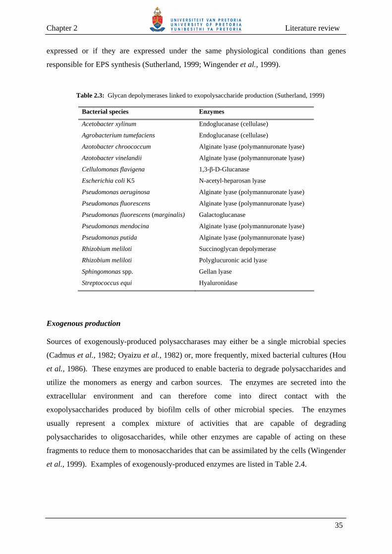
Chapter 2 Literature review
expressed or if they are expressed under the same physiological conditions than genes
responsible for EPS synthesis (Sutherland, 1999; Wingender et al., 1999).
Table 2.3: Glycan depolymerases linked to exopolysaccharide production (Sutherland, 1999)
Bacterial species Enzymes
Acetobacter xylinum Endoglucanase (cellulase)
Agrobacterium tumefaciens Endoglucanase (cellulase)
Azotobacter chroococcum Alginate lyase (polymannuronate lyase)
Azotobacter vinelandii Alginate lyase (polymannuronate lyase)
Cellulomonas flavigena 1,3-β-D-Glucanase
Escherichia coli K5 N-acetyl-heparosan lyase
Pseudomonas aeruginosa Alginate lyase (polymannuronate lyase)
Pseudomonas fluorescens Alginate lyase (polymannuronate lyase)
Pseudomonas fluorescens (marginalis) Galactoglucanase
Pseudomonas mendocina Alginate lyase (polymannuronate lyase)
Pseudomonas putida Alginate lyase (polymannuronate lyase)
Rhizobium meliloti Succinoglycan depolymerase
Rhizobium meliloti Polyglucuronic acid lyase
Sphingomonas spp. Gellan lyase
Streptococcus equi Hyaluronidase
Exogenous production Sources of exogenously-produced polysaccharases may either be a single microbial species
(Cadmus et al., 1982; Oyaizu et al., 1982) or, more frequently, mixed bacterial cultures (Hou
et al., 1986). These enzymes are produced to enable bacteria to degrade polysaccharides and
utilize the monomers as energy and carbon sources. The enzymes are secreted into the
extracellular environment and can therefore come into direct contact with the
exopolysaccharides produced by biofilm cells of other microbial species. The enzymes
usually represent a complex mixture of activities that are capable of degrading
polysaccharides to oligosaccharides, while other enzymes are capable of acting on these
fragments to reduce them to monosaccharides that can be assimilated by the cells (Wingender
et al., 1999). Examples of exogenously-produced enzymes are listed in Table 2.4.
35

Chapter 2 Literature review Table 2.4: Polysaccharide depolymerases produced by heterologous microorganisms (Sutherland, 1999)
Polysaccharide Enzyme action Linkage cleaved Enzyme source
Endoglycanases
Bradyrhizobium sp. Endorhamnosidase 1,4-β-L-Rha Bacillus sp.
Dextran Endoglucosidase 1,3- β -D-Glc Bacillus sp.
Dextran Endoglucosidase 1,2-α-D-Glc Flavobacterium sp.
Dextran Endoglucosidase 1,2-α-D-Glc Streptococcus
mutans
Streptococcus pneumoniae Endoglucosidase 1,3-β-D-Glc-1,4-β-D-GlcA Bacillus palustris
type 3
Lyases
Streptococcus pneumoniae 1,4-α-D-Gal-β-D-GlcA- Bacillus palustris
type 8
Sphingomonas sp. (Gellan) 1,4-β-D-Glc-β-D-GlcA- Pseudomonad
Sphingomonas sp. (Gellan) 1,4-β-D-Glc-β-D-GlcA- Bacillus sp.
Bacteriophages A diverse range of virulent bacteriophages is associated with EPS-producing bacteria
(Sutherland, 1999). Some of these bacteriophages produce polysaccharases that occur as
spikes on the base plate of the phage (Hughes et al., 1998a). Polysaccharase activity has also
been detected in the bacterial cell lysates, following release of mature phage particles from
infected host cells (Sutherland, 1999). The bacteriophage-produced polysaccharases are very
specific and they typically have a single target, except if the polysaccharides are similar in
structure. There are, however, a few exceptions where bacteriophage polysaccharases can
degrade polysaccharides from different bacterial species (Sutherland, 1999). Removal of
biofilms by means of bacteriophages relies greatly on the number of phages present and the
activity of the enzyme that they produce. In mixed biofilms, different EPS types are present
and these are almost impossible to degrade by a single phage. Phages are thus not very
successful when it comes to removing mixed biofilms, but can be used where biofilms of
known bacterial strains must be removed (Hughes et al., 1998a).
Degradation of bacterial EPS by bacteriophage-produced polysaccharases occurs in a specific
sequence of events (Hughes et al., 1998a) (Fig. 2.5). The bacteriophage first binds to a
secondary receptor present in the EPS, whereafter the EPS polymers are degraded
enzymatically until the cell surface is exposed. The bacteriophage can then bind to primary
36
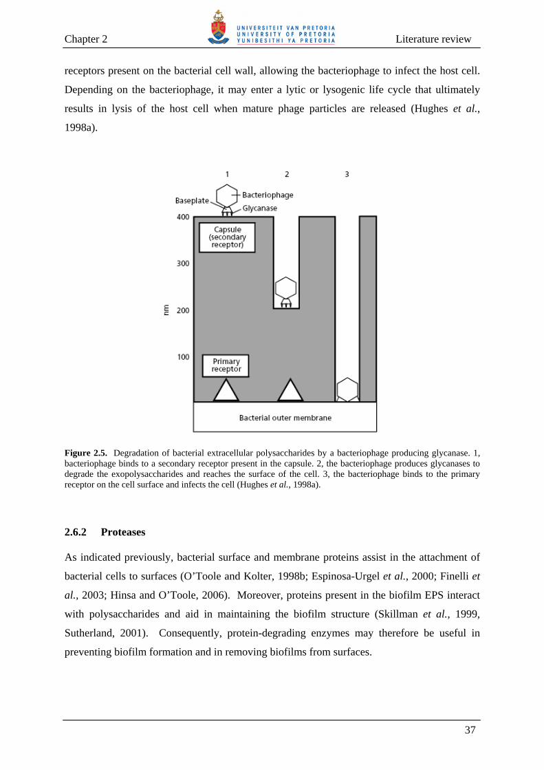
Chapter 2 Literature review
receptors present on the bacterial cell wall, allowing the bacteriophage to infect the host cell.
Depending on the bacteriophage, it may enter a lytic or lysogenic life cycle that ultimately
results in lysis of the host cell when mature phage particles are released (Hughes et al.,
1998a).
Figure 2.5. Degradation of bacterial extracellular polysaccharides by a bacteriophage producing glycanase. 1, bacteriophage binds to a secondary receptor present in the capsule. 2, the bacteriophage produces glycanases to degrade the exopolysaccharides and reaches the surface of the cell. 3, the bacteriophage binds to the primary receptor on the cell surface and infects the cell (Hughes et al., 1998a). 2.6.2 Proteases As indicated previously, bacterial surface and membrane proteins assist in the attachment of
bacterial cells to surfaces (O’Toole and Kolter, 1998b; Espinosa-Urgel et al., 2000; Finelli et
al., 2003; Hinsa and O’Toole, 2006). Moreover, proteins present in the biofilm EPS interact
with polysaccharides and aid in maintaining the biofilm structure (Skillman et al., 1999,
Sutherland, 2001). Consequently, protein-degrading enzymes may therefore be useful in
preventing biofilm formation and in removing biofilms from surfaces.
37

Chapter 2 Literature review
2.6.2.1 Enzymatic action Proteases can be placed into two major groups based on the position they cleave in the protein
substrate. Exopeptidases cleave peptide bonds close to the termini of the polypeptide, while
endopeptidases are responsible for cleaving peptide bonds in the inner region of the
polypeptide chain (Table 2.5). Exopeptidases are further classified into amino (N)-peptidases
and carboxy (C)-peptidases, depending on whether the enzyme removes amino acids at the N-
or C-terminus of the polypeptide. The endopeptidases are divided into four subgroups, i.e.
serine proteases, cysteine proteases, aspartic proteases and metalloproteases, based on their
catalytic mechanism. Moreover, proteases can also be classified as alkaline, acidic or neutral
proteases, depending on the pH where they are optimally active (Rao et al., 1998).
Table 2.5: Protease classification, based on mode of action (Rao et al., 1998)
Protease Mode of action Exopeptidases
Aminopeptidases ●-○-○-○-○---
Dipeptidyl peptidase ●-●-○-○-○---
Tripeptidyl peptidase ●-●-●-○-○---
Carboxypeptidase ---○-○-○-○-○-●
Serine-type protease
Metalloprotease
Cysteine-type protease
Peptidyl dipeptidase ---○-○-○-○-●-●
Dipeptidases ●-●
Omega peptidases *-●-○-○---
---○-○-○-●-*
Endopeptidases ---○-○-○-○-○-○---
Serine protease
Cysteine protease
Aspartic protease
Metalloprotease
Endopeptidases of unknown catalytic mechanism
○ = amino acid residues in polypeptide chain, ● = terminal amino acids, * = blocked termini, = enzyme action site
38

Chapter 2 Literature review
2.6.2.2 Production sources Proteases are produced by an extensive array of plants (Konno et al., 2004), animals (Furihata
et al., 1980; Fujinaga et al., 1987) and microorganisms (Leger et al., 1992; Ghorbel et al.,
2003). Fungi produce many different proteases that display activities over broad pH ranges
and show activity against a wider range of substrates than bacterial proteases. In contrast,
bacterial proteases display tolerance over a broader range of temperatures and the reaction rate
of these proteases are higher than those of fungal proteases. Indeed, the genus Bacillus is
responsible for producing the majority of commercial proteases, especially the neutral and
alkaline proteases. Viruses are noted for their production of aspartic, cysteine and serine
peptidases (Rao et al., 1998).
2.6.3 Application of enzymes to biofilm removal 2.6.3.1 Polysaccharide-degrading enzymes Loiselle and Anderson (2003) investigated the effect of a fungal cellulase on P. aeruginosa
biofilms. The cellulase caused a decrease in the biofilm biomass and colony forming units
(CFU). The enzyme was also tested against the purified biofilm EPS and it was shown by
size exclusion chromatography that the enzyme caused a reduction in the molecular weight of
the polymers. The enzyme did, however, not lead to the complete inhibition of biofilm
formation and it was therefore suggested that it might be more useful when used in
combination with other treatments or enzymes.
Kaplan et al. (2004b) reported that Actinobacillus actinomycetemcomitans produces an
enzyme, known as Dispersin B, which targets polymers that contain N-acetylglucosamine
(Kaplan et al., 2004a), and is capable of detaching biofilms of A. actinomycetemcomitans
(Kaplan et al., 2004b) and Staphylococcus epidermidis (Kaplan et al., 2004a). More recently,
Itoh et al. (2005) reported that Dispersin B likewise hydrolyzes the glycosidic linkages in the
capsular polysaccharide β-1,6-N-acetyl-D-glucosamine (poly-β-1,6-GlcNAc) required for
biofilm formation by E. coli and staphylococci. The synthesis of this polysaccharide is
dependent on the presence of pgaABCD and icaABCD loci. Dispersin B was capable of
detaching biofilms from E. coli, S. epidermidis, Y. pestis and P. fluorescens, all of which
posses either a pgaABCD or icaABCD locus (Itoh et al., 2005).
39
Chapter 2 Literature review
2.6.3.2 Proteases Hahn Berg et al. (2001) tested the activity of several proteases obtained from Antarctic krill
shrimp against oral biofilms, which can cause dental caries and periodontal diseases if they
are not controlled. The enzyme mixture, known as Krillase, is comprised of different endo-
and exopeptidases. Krillase not only prevented the attachment of oral bacteria by degrading
saliva proteins present on the surface, but it also removed existing biofilms by destroying
adhesion receptor interactions involved in the aggregation and attachment of bacterial cells to
each other and to the surface. Moreover, Krillase did not exhibit microbicidal activity and
therefore is unlikely to disrupt the balance of the oral bacteria.
2.6.3.3 Mixed enzymes Johansen et al. (1997) tested different commercial enzymes for their bactericidal and biofilm
removal properties. Staphylococcus aureus, S. epidermidis, P. fluorescens and P. aeruginosa
biofilms were grown on steel and polypropylene surfaces and subsequently treated with
glucose oxidase, lactoperoxidase or Pectinex Ultra SP, an enzyme mixture containing protease
activity and several polysaccharide-hydrolyzing enzymes. It was reported that although
glucose oxidase and lactoperoxidase each had a bactericidal effect on the biofilm bacteria,
they were incapable of removing the biofilms. In contrast, Pectinex Ultra SP was capable of
detaching the biofilms, but it did not show bactericidal activity against the biofilm bacteria.
However, when both the oxidoreductase enzymes were used together with Pectinex Ultra SP
the respective biofilms was not only removed from both types of surfaces but the viable cells
were also killed.
As a means to detach bacteria from soil particles, Böckelmann et al. (2003) compared a
conventional method (dispersal with sodium pyrophosphate buffer) with a method whereby
enzymes were used in combination with sonication. An enzyme mixture comprising two
polysaccharide-degrading enzymes (β-galactosidase and α-glucosidase) and a lipase were
used to assist in weakening of the EPS. The combined use of these enzymes and sonication
detached a 22-fold higher number of bacterial cells compared to the conventional method.
When the enzymes were used individually, β-galactosidase, α-glucosidase and lipase detached
a 2- to 10-fold, 1- to 7-fold and 1- to 3-fold higher amount of bacterial cells, respectively,
compared to the conventional method (Böckelmann et al., 2003).
40
Chapter 2 Literature review
Walker et al. (2007) tested a range of commercially available enzymes of various types for
their ability to destroy microbial biofilms. Biofilms of Lactobacillus brevis, Flavimonas
oryzihabitans, Leuconostoc mesenteroides and Saccharomyces cerevisiae were incubated with
an enzyme mixture containing α-amylase, β-glucuronidase, glucose oxidase, dextranase,
protease and pectinase. Enzyme mixtures comprising of either single strength (10 U/ml of
each enzyme) or double strength enzymes were used. The double strength enzyme mixture
was shown to be more effective in removing biofilms of L. brevis, L. mesenteroides and S.
cerevisiae compared to the use of a single strength enzyme mixture. The number of F.
oryzihabitans cells, however, increased when treated with the single strength enzyme mixture,
while the double strength enzyme mixture was less effective in removing biofilms of this
bacterium compared to the other microorganisms. Based on the results obtained, Walker et al.
(2007) noted that a range of factors such as biofilm composition, attachment surface,
application time and temperature may have a major effect on the efficacy of enzymes to
remove biofilms.
Orgaz et al. (2006) tested various different enzymes from fungal origin for their ability to
remove P. fluorescens biofilms from glass coupons. Although each of the enzymes, i.e.
cellulase, pectin lyase, polygalacturonase, arabinase, alginate lyase and proteinase, showed
some degree of biofilm removal, a mixture of these enzymes was less efficient in removing
the biofilms. The latter was ascribed to the proteinase degrading the other enzymes present in
the mixture. In a subsequent study, Orgaz et al. (2007) investigated the usefulness of three
polysaccharide-degrading enzymes (cellulase, pectin esterase and pectin lyase) and Pronase®
(a mixture of proteinases consisting of an alkaline protease, neutral protease, and amino- and
carboxy-peptidases) in the removal of P. fluorescens biofilms. These enzymes were tested
individually or in a sequential (two-step) application. Of the single polysaccharide-degrading
enzymes, pectin esterase was the most efficient in removing P. fluorescens biofilms, showing
removal of approximately 75% of the biofilm cells. Soluble Pronase® removed 80% of the
biofilm cells and 30% of the biofilm biomass. Sequential (two-step) treatment with cellulase,
pectin esterase or pectin lyase, followed by soluble Pronase®, obtained a 2-3 log10 count
reduction and up to 94% biomass removal. To allow enzyme treatment in a single step, a
newly developed form of delayed release encapsulated Pronase (DRE Pro), which only
releases proteases after the soluble enzymes have worked for 20-60 min, was also used.
Treatment of a P. fluorescens biofilm for 2 h with a combination of the DRE Pro and
41
Chapter 2 Literature review
polysaccharide-degrading enzymes resulted in a 4 log10 reduction in biofilm cells and
removed 96% of the biofilm from the surface.
Chaignon et al. (2007) assessed the susceptibility of biofilms of clinical staphylococcal strains
to a range of enzyme preparations, i.e. Dispersin B, pancreatin, proteinase K, Pectinex Ultra
SP, periodate and trypsin. It was demonstrated that Dispersin B was the most effective
against strains that contained N-acetylglucosamine as the major component in the extracellular
biofilm matrix, whereas the biofilms were not affected by protease treatment. Conversely,
strains that lacked N-acetylglucosamine were removed efficiently by treatment with proteases.
The components making up the matrix of staphylococcal biofilms therefore play an important
role in the enzymatic removal of these biofilms, especially since the composition may differ
among the clinical strains. Chaignon et al. (2007) proposed that by using both Dispersin B
and a protease (proteinase K or trypsin), biofilms formed by a range of staphylococcal strains
can be removed from inert surfaces.
2.6.3.4 Combined use of enzymes and chemicals Merritt et al. (2000) tested several cleaning agents (tap water, commercial detergents with
surfactants, detergents containing enzymes, enzymatic and non-enzymatic contact lens
cleaning solutions, commercial mouthwash, peroxide-based bleach, bleach containing 5.25%
sodium hypochlorite, Triton X-100) against biofilms of Staphylococcus epidermidis and
Candida albicans, as well as biofilms formed by oral microorganisms grown in 96-well
polystyrene plates. The results indicated that biofilms formed by the respective
microorganisms were the most successfully removed by detergents containing enzymes and
sodium hypochlorite (NaOCl), while the other detergents and surfactants were less effective.
Vickery et al. (2004) also tested the effectiveness of several non-enzymatic and enzymatic
detergents on the removal of E. coli biofilms from medical tubing used in endoscope and
infusion set construction. Of the different detergents tested, a non-enzymatic detergent,
Matrix, and a high-activity enzymatic detergent, Epizyme Rapid, were the most effective and
were capable of detaching 75% and 60% of the biofilm, respectively. Epizyme Rapid, which
consists of high amylase and protease activity, removed 30% of the exopolysaccharides, but it
did not show more than a 2 log10 reduction in the viable bacterial count. In contrast, the non-
42
Chapter 2 Literature review
enzymatic detergent Matrix reduced the viable bacterial count by more than 4 log10 and the
exopolysaccharides were reduced by 80%.
Oulahal et al. (2007) investigated the removal of biofilms formed by two meat spoilage
microorganisms (E. coli and S. aureus) by making use of combined ultrasonic and enzymatic
and/or chelating agent treatment. The application of ultrasound alone was not effective in
removing all biofilms from a stainless steel surface. When ultrasound was used in
combination with EDTA and/or enzymes, approximately 75% of E. coli and 100% of S.
aureus biofilms were removed. Similarly, Oulahal-Lagsir et al. (2003) reported that
ultrasonication combined with enzymatic treatment (proteolytic or glycolytic enzymes) were
more effective in removing E. coli biofilms from a stainless steel surface than application of
ultrasound alone.
2.7 REFERENCES
Abdel-Rahman, M.S., Couri, D. and Bull, R.J. 1984. Toxicity of chlorine dioxide in drinking
water. International Journal of Toxicology, 3: 277-284.
Allison, D.G. 2003. The biofilm matrix. Biofouling, 19: 139-150.
Allison, D.G., Gilbert, P., Lappin-Scott, H.M. and Wilson, M. 2000. Community structure
and co-operation in biofilms. Cambridge University Press, Cambridge.
Allison, D.G., Ruiz, B., SanJose, C., Jaspe, A. and Gilbert, P. 1998. Extracellular products as
mediators of the formation and detachment of Pseudomonas fluorescens biofilms. FEMS
Microbiology Letters, 167: 179-184.
Al-Tahhan, R.A., Sandrin, T.R., Bodour, A.A. and Maier, R.M. 2000. Rhamnolipid-induced
removal of lipopolysaccharide from Pseudomonas aeruginosa: Effect on cell surface
properties and interaction with hydrophobic substrates. Applied and Environmental
Microbiology, 66: 3262-3268.
Anderl, J.N., Franklin, M.J. and Stewart, P.S. 2000. Role of antibiotic penetration limitation
in Klebsiella pneumoniae biofilm resistance to ampicillin and ciprofloxacin. Antimicrobial
Agents and Chemotherapy, 44: 1818-1824.
Anderson, A.C., Reimers, R.S. and DeKernion, P. 1982. A brief review of the current status
of alternatives to chlorine disinfection of water. American Journal of Public Health, 72:
1290-1293.
Atkinson, S., Throup, J.P., Stewart, G.S.A.B. and Williams, P. 1999. A hierarchical quorum-
sensing system in Yersinia pseudotuberculosis is involved in the regulation of motility and
clumping. Molecular Microbiology, 33: 1267-1277.
43

Chapter 2 Literature review
Augustin, M. and Ali-Vehmas, T. 2004. Assessment of enzymatic cleaning agents and
disinfectants against bacterial biofilms. Journal of Pharmacy and Pharmaceutical Sciences, 7:
55-64.
Austin, J.W., Sanders, G., Kay, W.W. and Collinson, S.K. 1998. Thin aggregative fimbriae
enhance Salmonella enteritidis biofilm formation. FEMS Microbiology Letters, 162: 295-
301.
Balaban, N., Giacometti, A., Cirioni, O., Gov, Y., Ghiselli, R., Mocchegiani, F., Viticchi, C.,
Prete, M.S.D., Saba, V., Scalise, G. and Dell’Acqua, G. 2003. Use of the quorum-sensing
inhibitor RNAIII-inhibiting peptide to prevent biofilm formation in vivo by drug-resistant
Staphylococcus epidermidis. Journal of Infectious Diseases, 187: 625-630.
Baribeau, H., Prévost, M., Desjardins, R., Lafrence, P. and Gates, D.J. 2002. Chlorite and
chlorate ion variability in distribution systems. Journal of the American Water Works
Association, 94: 96-105.
Bayer, A.S., Park, S., Ramos, M.C., Nast, C.C., Eftekhar, F. and Schiller, N.L. 1992. Effects
of alginase on the natural history and antibiotic therapy of experimental endocarditis caused by
mucoid Pseudomonas aeruginosa. Infection and Immunity, 60: 3979-3985.
Bendinger, B., Rijnaarts, H.H.M., Altendorf, K. and Zehnder, A.B.J. 1993. Physicochemical
cell surface and adhesive properties of Coryneform bacteria related to the presence and chain
length of mycolic acids. Applied and Environmental Microbiology, 59: 3973-3977.
Bereswill, S. and Geider, K. 1997. Characterization of the rcsB gene from Erwinia
amylovora and its influence on exopolysaccharide synthesis and virulence of the fire blight
pathogen. Journal of Bacteriology, 179: 1354-1361.
Beveridge, T.J., Makin, S.A., Kadurugamuwa, J.L. and Li, Z. 1997. Interactions between
biofilms and the environment. FEMS Microbiology Reviews, 20: 291-303.
Blanco, M.A., Negro, C., Gaspar, I. and Tijero, J. 1996. Slime problems in the paper and
board industry. Applied Microbiology and Biotechnology, 45: 132-136.
Böckelmann, U., Szewzyk, U. and Grohmann, E. 2003. A new enzymatic method for the
detachment of particle associated soil bacteria. Journal of Microbiological Methods, 55: 201-
211.
Boland, T., Dufrêne, Y., Barger, B. and Lee, G. 2000. Molecular basis of cell adhesion to
polymers characterized by AFM. Critical Reviews in Biomedical Engineering, 28: 195-196.
Boyd, A. and Chakrabarty, A.M. 1994. Role of alginate lyase in cell detachment of
Pseudomonas aeruginosa. Applied and Environmental Microbiology, 60: 2355-2359.
Brooun, A., Liu, S. and Lewis, K. 2000. A dose-response study of antibiotic resistance in
Pseudomonas aeruginosa biofilms. Antimicrobial Agents and Chemotherapy, 44: 640-646.
Brown, M.R.W. and Barker, J. 1999. Unexplored reservoirs of pathogenic bacteria: Protozoa
and biofilms. Trends in Microbiology, 7: 46-50.
44
Chapter 2 Literature review
Bura, R., Cheung, M., Liao, B., Finlayson, J., Lee, B.C., Droppo, I.G., Leppard, G.G. and
Liss, S.N. 1998. Composition of extracellular polymeric substances in the activated sludge
floc matrix. Water Science and Technology, 37: 325-333.
Cadmus, M.C., Jackson, L.K., Burton, K.A., Plattner, R.D. and Slodki, M.E. 1982.
Biodegradation of xanthan gum by Bacillus sp. Applied and Environmental Microbiology, 44:
5-11.
Carlén, A., Nikdel, K., Wennerberg, A., Holmberg, K. and Olsson, J. 2001. Surface
characteristics and in vitro biofilm formation on glass ionomer and composite resin.
Biomaterials, 22: 481-487.
Carlsson, F.H.H. 2003. Elementary handbook of water disinfection. Water Research
Commission Report No. TT 205/03, South Africa.
Chaignon, P., Sadovskaya, I., Ragunah, C., Ramasubbu, N., Kaplan, J.B. and Jabbouri, S.
2007. Susceptibility of staphylococcal biofilms to enzymatic treatments depends on their
chemical composition. Applied Microbiology and Biotechnology, 75: 125-132.
Chandy, J.P. and Angles, M.L. 2001. Determination of nutrients limiting biofilm formation
and the subsequent impact on disinfectant decay. Water Research, 35: 2677-2682.
Characklis, W.G. and Dydek, S.T. 1976. Influence of carbon-nitrogen ratio on the
chlorination of microbial aggregates. Water Research, 10: 515-522.
Characklis, W.G. and Marshall, K.C. 1990. Biofilms. Wiley Interscience Publishers,
Canada.
Chen, J., Lee, S.M. and Mao, Y. 2004. Protective effect of exopolysaccharide colonic acid of
Escherichia coli O157:H7 to osmotic and oxidative stress. International Journal of Food
Microbiology, 93: 281-286.
Chen, M.J., Zhang, Z. and Bott, T.R. 1998. Direct measurement of the adhesive strength of
biofilms in pipes by micromanipulation. Biotechnology Techniques, 12: 875-880.
Chen, X. and Stewart, P.S. 1996. Chlorine penetration into artificial biofilm is limited by a
reaction-diffusion interaction. Environmental Science and Technology, 30: 2078-2083.
Chen, X. and Stewart, P.S. 2000. Biofilm removal caused by chemical treatments. Water
Research, 34: 4229-4233.
Chen, X. and Stewart, P.S. 2002. Role of electrostatic interactions in cohesion of bacterial
biofilms. Applied Microbiology and Biotechnology, 59: 718-720.
Chmielewski, R.A.N. and Frank, J.F. 2003. Biofilm formation and control in food processing
facilities. Comprehensive Reviews in Food Science and Food Safety, 2: 22-32.
Christensen, B.E., Ertesvag, H., Beyenal, H. and Lewandowski, Z. 2001. Resistance of
biofilms containing alginate-producing bacteria to disintegration by an alginate degrading
enzyme (AlgL). Biofouling, 17: 203-210.
45
Chapter 2 Literature review
Christensen, B.B., Sternberg, C., Andersen, J.B., Eberl, L., Møller, S. and Givskov, M. 1998.
Establishment of new genetic traits in a microbial biofilm community. Applied and
Environmental Microbiology, 64: 2247-2255.
Cloete, T.E. 2003. Resistance mechanisms of bacteria to antimicrobial compounds.
International Biodeterioration and Biodegradation, 51: 277-282.
Cloete, T.E., Jacobs, L. and Brözel, V.S. 1998. The chemical control of biofouling in
industrial water systems. Biodegradation, 9: 23-37.
Cochran, W.L., Suh, S-J., McFeters, G.A. and Stewart, P.S. 2000. Role of RpoS and AlgT in
Pseudomonas aeruginosa biofilm resistance to hydrogen peroxide and monochloramine.
Journal of Applied Microbiology, 88: 546-553.
Cole, S.P., Harwood, J., Lee, R., She, R. and Guiney, D.G. 2004. Characterization of
monospecies biofilm formation by Helicobacter pylori. Journal of Bacteriology, 186: 3124-
3132.
Conti, E., Flaibani, A., O’Regan, M. and Sutherland, I.W. 1994. Alginate from Pseudomonas
fluorescens and P. putida: Production and properties. Microbiology, 140: 1125-1132.
Correia, F.F., D’Onofrio, A., Rejtar, T., Li, L., Karger, B.L., Makarova, K., Koonin, E.V. and
Lewis, K. 2006. Kinase activity of overexpressed HipA is required for growth arrest and
multidrug tolerance in Escherichia coli. Journal of Bacteriology, 188: 8360-8367.
Costerton, J.W., Lewandowski, Z., Caldwell, D.E., Korber, D.R. and Lappin-Scott, H.M.
1995. Microbial biofilms. Annual Reviews in Microbiology, 49: 711-745.
Costerton, J.W., Stewart, P.S. and Greenberg, E.P. 1999. Bacterial biofilms: A common
cause of persistent infections. Science, 284: 1318-1322.
Cowan, M.M., Warren, T.M. and Fletcher, M. 1991. Mixed-species colonization of solid
surfaces in laboratory biofilms. Biofouling, 3: 23-34.
Cui, W., Winter, W.T., Tanenbaum, S.W. and Nakas, J.P. 1999. Purification and
characterization of an intracellular carboxylesterase from Arthrobacter viscosus NRRL B-
1973. Enzyme and Microbial Technology, 24: 200-208.
Curtin, J.J. and Donlan, R.M. 2006. Using bacteriophages to reduce formation of catheter-
associated biofilms by Staphylococcus epidermidis. Antimicrobial Agents and Chemotherapy,
50: 1268-1275.
Daamen, E.J., Wouters, J.W. and Savelkoul, J.T.G. 2000. Side stream biofiltration for
improved biofouling control in cooling water systems. Water Science and Technology, 41:
445-451.
Danese, P.N., Pratt, L.A. and Kolter, R. 2000. Exopolysaccharide production is required for
development of Escherichia coli K-12 biofilm architecture. Journal of Bacteriology, 182:
3593-3596.
46

Chapter 2 Literature review
Davey, M.E. and O’Toole, G.A. 2000. Microbial biofilms: From ecology to molecular
genetics. Microbiology and Molecular Biology Reviews, 64: 847-867.
Davies, D.G. and Geesey, G.G. 1995. Regulation of the alginate biosynthesis gene algC in
Pseudomonas aeruginosa during biofilm development in continuous culture. Applied and
Environmental Microbiology, 61: 860-867.
Davies, D.G., Chakrabarty, A.M. and Geesey, G.G. 1993. Exopolysaccharide production in
biofilms: Substratum activation of alginate gene expression by Pseudomonas aeruginosa.
Applied and Environmental Microbiology, 59: 1181-1186.
Davies, D.G., Parsek, M.R., Pearson, J.P., Iglewski, B.H., Costerton, J.W. and Greenberg, E.P.
1998. The involvement of cell-to-cell signals in the development of a bacterial biofilm.
Science, 280: 295-298.
de Beer, D., Srinivasan, R. and Stewart, P.S. 1994. Direct measurement of chlorine
penetration into biofilms during disinfection. Applied and Environmental Microbiology, 60:
4339-4344.
De Kievit, T.R., Gillis, R., Marx, S., Brown, C. and Iglewski, B. 2001a. Quorum-sensing
genes in Pseudomonas aeruginosa biofilms: Their role and expression patterns. Applied and
Environmental Microbiology, 67: 1865-1873.
De Kievit, T.R., Parkins, M.D., Gillis, R.J., Srikumar, R., Ceri, H., Poole, K., Iglewski, B.H.
and Storey, D.G. 2001b. Multidrug efflux pumps: Expression patterns and contribution to
antibiotic resistance in Pseudomonas aeruginosa biofilms. Antimicrobial Agents and
Chemotherapy, 45: 1761-1770.
Dignac, M-F., Urbain, V., Rybacki, D., Bruchet, A., Snidaro, D. and Scribe, P. 1998.
Chemical description of extracellular polymers: Implication on activated sludge floc structure.
Water Science and Technology, 38: 45-53.
Donelli, G., Francolini, I., Piozzi, A., Di Rosa, R. and Marconi, W. 2002. New polymer-
antibiotic systems to inhibit bacterial biofilm formation: A suitable approach to prevent
central venous catheter-associated infections. Journal of Chemotherapy, 14: 501-507.
Dong, Y-H., Gusti, A.R., Zhang, Q., Xu, J-L. and Zhang, L-H. 2002. Identification of
quorum-quenching N-Acyl Homoserine Lactonases from Bacillus species. Applied and
Environmental Microbiology, 68: 1754-1759.
Donlan, R. 2002. Biofilm: Microbial life on surfaces. Emerging Infectious Diseases, 8:
881-890.
Donlan, R.M. and Costerton, J.W. 2002. Biofilms: Survival mechanisms of clinically
relevant microorganisms. Clinical Microbiology Reviews, 15: 167-193.
Dorward, D.W. and Garon, C.F. 1990. DNA is packaged within membrane-derived vesicles
of Gram-negative but not Gram-positive bacteria. Applied and Environmental Microbiology,
56: 1960-1962.
47

Chapter 2 Literature review
Dosti, B., Guzel-Seydim, Z. and Greene, A.K. 2005. Effectiveness of ozone, heat and
chlorine for destroying common food spoilage bacteria in synthetic media and biofilms.
International Journal of Dairy Technology, 58: 19-24.
Drenkard, E. 2003. Antimicrobial resistance of Pseudomonas aeruginosa biofilms. Microbes
and Infection, 5: 1213-1219.
Eginton, P.J., Holah, J., Allison, D.G., Handley, P.S. and Gilbert, P. 1998. Changes in the
strength of attachment of microorganisms to surfaces following treatment with disinfectants
and cleaning agents. Letters in Applied Microbiology, 27: 101-105.
Elasri, M.O. and Miller, R.V. 1999. Study of the response of a biofilm bacterial community
to UV radiation. Applied and Environmental Microbiology, 65: 2025-2031.
Elkins, J.G., Hassett, D.J., Stewart, P.S., Schweizer, H.P. and McDermott, T.R. 1999.
Protective role of catalase in Pseudomonas aeruginosa biofilm resistance to hydrogen
peroxide. Applied and Environmental Microbiology, 65: 4594-4600.
Eriksson, T., Börjesson, J. and Tjerneld, F. 2002. Mechanism of surfactant effect in
enzymatic hydrolysis of lignocellulose. Enzyme and Microbial Technology, 31: 353-364.
Espinosa-Urgel, M., Salido, A. and Ramos, J-L. 2000. Genetic analysis of functions involved
in adhesion of Pseudomonas putida to seeds. Journal of Bacteriology, 182: 2363-2369.
Evans, D.J., Allison, D.G., Brown, M.R.W. and Gilbert, P. 1991. Susceptibility of
Pseudomonas aeruginosa and Escherichia coli biofilms towards ciprofloxacin: Effect of
specific growth rate. Journal of Antimicrobial Chemotherapy, 27: 177-184.
Finelli, A., Gallant, C.V., Jarvi, K. and Burrows, L.L. 2003. Use of in-biofilm expression
technology to identify genes involved in Pseudomonas aeruginosa biofilm development.
Journal of Bacteriology, 185: 2700-2710.
Fletcher, M. 1988. Attachment of Pseudomonas fluorescens to glass and influence of
electrolytes on bacterium-substratum separation distance. Journal of Bacteriology, 170:
2027-2030.
Fletcher, M. and Loeb, G.I. 1979. Influence of substratum characteristics on the attachment
of a marine Pseudomonas to solid surfaces. Applied and Environmental Microbiology, 37:
67-72.
Foley, S., Lucchini, S., Zwahlen, M-C. and Brüssow, H. 1998. A short non-coding viral
DNA element showing characteristics of a replication origin confers bacteriophage resistance
to Streptococcus thermophilus. Virology, 250: 377-387.
Francolini, I., Donelli, G. and Stoodley, P. 2004. Polymer designs to control biofilm growth
on medical devices. Reviews in Environmental Science and Bio/Technology, 2: 307-319.
Freese, S.D. and Nozaic, D.J. 2004. Chlorine: Is it really so bad and what are the
alternatives? Water SA, 30: 18-24.
48

Chapter 2 Literature review
Friedman, H., Volin, E. and Laumann, D. 1968. Terminal disinfection in hospitals with
quaternary ammonium compounds by use of a spray-fog technique. Applied Microbiology,
16: 223-227.
Fujinaga, M., Sielecki, A.R., Read, R.J., Ardelt, W., Laskowski, M. and James, M.N.G. 1987.
Crystal and molecular structures of the complex of α-chymotrypsin with its inhibitor turkey
ovomucoid third domain at 1.8 Å resolution. Journal of Molecular Biology, 195: 397-418.
Gacesa, P. 1998. Bacterial alginate biosynthesis – recent progress and future prospects.
Microbiology, 144: 1133-1143.
Gagnon, G.A., O’Leary, K.C., Volk, C.J., Stover, L. and Andrews, R.C. 2004. Comparative
analysis of chlorine dioxide, free chlorine and chloramines on bacterial water quality in model
distribution systems. Journal of Environmental Engineering, 130: 1269-1279.
Gavín, R., Merino, S., Altarriba, M., Canals, R., Shaw, J.G. and Tomás, J.M. 2003. Lateral
flagella are required for increased cell adherence, invasion and biofilm formation by
Aeromonas spp. FEMS Microbiology Letters, 224: 77-83.
Geesey, G.G. 1982. Microbial exopolymers: Ecological and economic considerations.
American Society for Microbiology News, 48: 9-14.
Geesey, G.G. 2001. Bacterial behavior at surfaces. Current Opinion in Microbiology, 4:
296-300.
Genevaux, P., Bauda, P., DuBow, M.S. and Oudega, B. 1999. Identification of Tn10
insertions in the rfaG, rfaP, and galU genes involved in lipopolysaccharide core biosynthesis
that affect Escherichia coli adhesion. Archives of Microbiology, 172: 1-8.
Ghannoum, M. and O’Toole, G.A. 2004. Microbial biofilms. ASM Press, USA.
Ghigo, J-M. 2001. Natural conjugative plasmids induce bacterial biofilm development.
Nature, 412: 442-445.
Ghorbel, B., Sellami-Kamoun, A. and Nasri, M. 2003. Stability studies of protease from
Bacillus cereus BG1. Enzyme and Microbial Technology, 32: 513-518.
Gibson, H., Taylor, J.H., Hall, K.E. and Holah, J.T. 1999. Effectiveness of cleaning
techniques used in the food industry in terms of the removal of bacterial biofilms. Journal of
Applied Microbiology, 87: 41-48.
Gilbert, H.J. and Hazlewood, G.P. 1993. Bacterial cellulases and xylanases. Microbiology,
139: 187-194.
Gilbert, P., Allison, D.G. and McBain, A.J. 2002. Biofilms in vitro and in vivo: Do singular
mechanisms imply cross-resistance? Journal of Applied Microbiology Symposium Supplement,
92: 98S-110S.
Gilbert, P., Collier, P.J. and Brown, M.R.W. 1990. Influence of growth rate on susceptibility
to antimicrobial agents: Biofilms, cell cycle, dormancy, and stringent response. Antimicrobial
Agents and Chemotherapy 34: 1865-1868.
49

Chapter 2 Literature review
Gilligan, P.H. 1991. Microbiology of airway disease in patients with cystic fibrosis. Clinical
Microbiology Reviews, 4: 35-51.
Giwercman, B., Jensen, E.T., Høiby, N., Kharazmi, A. and Costerton, J.W. 1991. Induction of
β-lactamase production in Pseudomonas aeruginosa biofilm. Antimicrobial Agents and
Chemotherapy, 35: 1008-1010.
Gordon, C.A., Hodges, N.A. and Marriott, C. 1991. Use of slime dispersants to promote
antibiotic penetration through the extracellular polysaccharide of mucoid Pseudomonas
aeruginosa. Antimicrobial Agents and Chemotherapy, 35: 1258-1260.
Grobe, K., Zahler, J. and Stewart, P. 2002. Role of dose concentration in biocide efficacy
against Pseudomonas aeruginosa biofilms. Journal of Industrial Microbiology and
Biotechnology, 29: 10-15.
Guzel-Seydim, Z., Greene, A.K. and Seydim, A.C. 2004. Use of ozone in the food industry.
Swiss Society of Food Science and Technology, 37: 453-460.
Habash, M. and Reid, G. 1999. Microbial biofilms: Their development and significance for
medical device-related infections. Journal of Clinical Pharmacology, 39: 887-898.
Hahn Berg, I.C., Kalfas, S., Malmsten, M. and Arnebrant, T. 2001. Proteolytic degradation of
oral biofilms in vitro and in vivo: Potential of proteases originating from Euphausia superba
for plaque control. European Journal of Oral Science, 109: 316-324.
Hall-Stoodley, L., Costerton, J.W. and Stoodley, P. 2004. Bacterial biofilms: From the
natural environment to infectious diseases. Nature Reviews Microbiology, 2: 95-108.
Hammer, B.K. and Bassler, B.L. 2003. Quorum sensing controls biofilm formation in Vibrio
cholerae. Molecular Microbiology, 50: 101-114.
Hanlon, G.W., Denyer, S.P., Olliff, C.J. and Ibrahim, L.J. 2001. Reduction in
exopolysaccharide viscosity as an aid to bacteriophage penetration through Pseudomonas
aeruginosa. Applied and Environmental Microbiology, 67: 2746-2753.
Hanna, A., Berg, M., Stout, V. and Razatos, A. 2003. Role of capsular colanic acid in
adhesion of uropathogenic Escherichia coli. Applied and Environmental Microbiology, 69:
4474-4481.
Harraghy, N., Kerdudou, S. and Herrmann, M. 2007. Quorum-sensing systems in
staphylococci as therapeutic targets. Analytical and Bioanalytical Chemistry, 387: 437-444.
Helle, S.S., Duff, S.J.B. and Cooper, D.G. 2004. Effect of surfactants on cellulose hydrolysis.
Biotechnology and Bioengineering, 42: 611-617.
Hentzer, M., Riedel, K., Rasmussen, T.B., Heydorn, A., Andersen, J.B., Parsek, M.R., Rice,
S.A., Eberl, L., Molin, S., Høiby, N., Kjelleberg, S. and Givskov, M. 2002. Inhibition of
quorum sensing in Pseudomonas aeruginosa biofilm bacteria by a halogenated furanone
compound. Microbiology, 148: 87-102.
50

Chapter 2 Literature review
Hentzer, M., Teitzel, G.M., Balzer, G.J., Heydorn, A., Molin, S., Givskov, M. and Parsek,
M.R. 2001. Alginate overproduction affects Pseudomonas aeruginosa biofilm structure and
function. Journal of Bacteriology, 183: 5395-5401.
Hentzer, M., Wu, H., Andersen, J.B., Riedel, K., Rasmussen, T.B., Bagge, N., Kumar, N.,
Schembri, M.A., Song, Z., Kristoffersen, P., Manefield, M., Costerton, J.W., Molin, S., Eberl,
L., Steinberg, P., Kjelleberg, S., Høiby, N. and Givskov, M. 2003. Attenuation of
Pseudomonas aeruginosa virulence by quorum sensing inhibitors. European Molecular
Biology Organization Journal, 22: 3803-3815.
Hibma, A.M., Jassim, S.A.A. and Griffiths, M.W. 1997. Infection and removal of L-forms of
Listeria monocytogenes with bred bacteriophage. International Journal of Food
Microbiology, 34: 197-207.
Hill, C., Miller, L.A. and Klaenhammer, T.R. 1990. Cloning, expression, and sequence
determination of a bacteriophage fragment encoding bacteriophage resistance in Lactococcus
lactis. Journal of Bacteriology, 172: 6419-6426.
Hinsa, S.M. and O’Toole, G.A. 2006. Biofilm formation by Pseudomonas fluorescens
WCS365: A role for LapD. Microbiology, 152: 1375-1383.
Hinsa, S.M., Espinosa-Urgel, M., Ramos, J.L. and O’Toole, G.A. 2003. Transition from
reversible to irreversible attachment during biofilm formation by Pseudomonas fluorescens
WCS365 requires an ABC transporter and a large secreted protein. Molecular Microbiology,
49: 905-918.
Hou, C.T., Barnabe, N. and Greaney, K. 1986. Biodegradation of xanthan by salt-tolerant
microorganisms. Journal of Industrial Microbiology and Biotechnology, 1: 31-37.
Hoyle, B.D., Williams, L.J. and Costerton, J.W. 1993. Production of mucoid
exopolysaccharide during development of Pseudomonas aeruginosa biofilms. Infection and
Immunity, 61: 777-780.
Huber, B., Riedel, K., Hentzer, M., Heydorn, A., Gotschlich, A., Givskov, M., Molin, S. and
Eberl, L. 2001. The cep quorum-sensing system of Burkholderia cepacia H111 controls
biofilm formation and swarming motility. Microbiology, 147: 2517-2528.
Hughes, K.A., Sutherland, I.W. and Jones, M.V. 1998a. Biofilm susceptibility to
bacteriophage attack: The role of phage-borne polysaccharide depolymerase. Microbiology,
144: 3039-3047.
Hughes, K.A., Sutherland, I.W., Clark, J. and Jones, M.V. 1998b. Bacteriophage and
associated polysaccharide depolymerases - novel tools for study of bacterial biofilms. Journal
of Applied Microbiology, 85: 583-590.
Hunt, S.M., Werner, E.M., Huang, B., Hamilton, M.A. and Stewart, P.S. 2004. Hypothesis
for the role of nutrient starvation in biofilm detachment. Applied and Environmental
Microbiology, 70: 7418-7425.
51

Chapter 2 Literature review
Inoue, T., Shingaki, R., Sogawa, N., Sogawa, C.A., Asaumi, J-I., Kokeguchi, S. and Fukui, K.
2003. Biofilm formation by a fimbriae-deficient mutant of Actinobacillus
actinomycetemcomitans. Journal of Microbiology and Immunology, 47: 877-881.
Ioannou, C.J., Hanlon, G.W. and Denyer, S.P. 2007. Action of disinfectant quaternary
ammonium compounds against Staphylococcus aureus. Antimicrobial Agents and
Chemotherapy, 51: 296-306.
Ishida, H., Ishida, Y., Kurosaka, Y., Otani, T., Sato, K. and Kobayashi, H. 1998. In vitro and
in vivo activities of levofloxacin against biofilm-producing Pseudomonas aeruginosa.
Antimicrobial Agents and Chemotherapy, 42: 1641-1645.
Itoh, Y., Wang, X., Hinnebusch, B.J., Preston, J.F. and Romeo, T. 2005. Depolymerization of
β-1,6-N-acetyl-D-glucosamine disrupts the integrity of diverse bacterial biofilms. Journal of
Bacteriology, 187: 382-387.
Jahn, A. and Nielsen, P.H. 1998. Cell biomass and exopolymers composition in sewer
biofilms. Water Science and Technology, 37: 17-24.
Johansen, C., Falholt, P. and Gram, L. 1997. Enzymatic removal and disinfection of bacterial
biofilms. Applied and Environmental Microbiology, 63: 3724-3728.
Jonas, R. and Farah, L.F. 1998. Production and application of microbial cellulase. Polymer
Degradation and Stability, 59: 101-106.
Jorand, F., Bouge-Bigne, F., Block, J.C. and Urbain, V. 1998. Hydrophobic/hydrophilic
properties of activated sludge exopolymeric substances. Water Science and Technology, 37:
307-316.
Jurcisek, J.A. and Bakaletz, L.O. 2007. Biofilms formed by non-typeable Haemophilus
influenzae in vivo contain both double-stranded DNA and type IV pilin protein. Journal of
Bacteriology, 189: 3868-3875.
Kadurugamuwa, J.L. and Beveridge, T.J. 1995. Virulence factors are released from
Pseudomonas aeruginosa in association with membrane vesicles during normal growth and
exposure to gentamicin: A novel mechanism of enzyme secretion. Journal of Bacteriology,
177: 3998-4008.
Kaplan, J.B., Ragunath, C., Ramasubbu, N. and Fine, D.H. 2003. Detachment of
Actinobacillus actinomycetemcomitans biofilm cells by an endogenous β-hexosaminidase
activity. Journal of Bacteriology, 185: 4693-4698.
Kaplan, J.B., Ragunath, C., Velliyagounder, K., Fine, D.H. and Ramasubbu, N. 2004a.
Enzymatic detachment of Staphylococcus epidermidis biofilms. Antimicrobial Agents and
Chemotherapy, 48: 2633-2636.
Kaplan, J.B., Velliyagounder, K., Ragunath, C., Rohde, H., Mack, D., Knobloch, J.K-M. and
Ramasubbu, N. 2004b. Genes involved in the synthesis and degradation of matrix
52

Chapter 2 Literature review
polysaccharide in Actinobacillus actinomycetemcomitans and Actinobacillus
pleuropneumoniae biofilms. Journal of Bacteriology, 186: 8213-8220.
Keevil, C.W., Godfree, A., Holt, D. and Dow, C. 1999. Biofilms in the aquatic environment.
The Royal Society of Chemistry, UK.
Keren, I., Kaldalu, N., Spoering, A., Wang, Y. and Lewis, K. 2004a. Persister cells and
tolerance to antimicrobials. FEMS Microbiology Letters, 230: 13-18.
Keren, I., Shah, D., Spoering, A., Kaldalu, N. and Lewis, K. 2004b. Specialized persister
cells and the mechanism of multidrug tolerance in Escherichia coli. Journal of Bacteriology,
186: 8172-8180.
Kjelleberg, S. and Molin, S. 2002. Is there a role for quorum sensing signals in bacterial
biofilms? Current Opinion in Microbiology, 5: 254-258.
Klausen, M., Heydorn, A., Ragas, P.C., Lambertsen, L., Aaes-Jørgensen, A., Molin, S. and
Tolker-Nielsen, T. 2003. Biofilm formation by Pseudomonas aeruginosa wild-type, flagella
and type IV pili mutants. Molecular Microbiology, 48: 1511-1524.
Konno, K., Hirayama, C., Nakamura, M., Tateishi, K., Tamura, Y., Hattori, M. and Kohno, K.
2004. Papain protects papaya trees from herbivorous insects: Role of cysteine proteases in
latex. The Plant Journal, 37: 370-378.
Korn, C., Andrews, R.C. and Escobar, M.D. 2002. Development of chlorine dioxide-related
by-product models for drinking water treatement. Water Research, 36: 330-342.
Kostenko, V., Ceri, H. and Martinuzzi, R.J. 2007. Increased tolerance of Staphylococcus
aureus to vancomycin in viscous media. FEMS Immunology and Medical Microbiology, 51:
277-288.
Kreft, J-U. 2004. Biofilms promote altruism. Microbiology, 150: 2751-2760.
Kumar, C.G. and Anand, S.K. 1998. Significance of microbial biofilms in food industry: A
review. International Journal of Food Microbiology, 42: 9-27.
Labbate, M., Queck, S.Y., Koh, K.S., Rice, S.A., Givskov, M. and Kjelleberg, S. 2004.
Quorum sensing-controlled biofilm development in Serratia liquefaciens MG1. Journal of
Bacteriology, 186: 692-698.
Langsrud, S., Sundheim, G. and Borgmann-Strahsen, R. 2003. Intrinsic and acquired
resistance to quaternary ammonium compounds in food-related Pseudomonas spp. Journal of
Applied Microbiology, 95: 874-882.
Leger, R.J.S.T., Frank, D.C., Roberts, D.W. and Staples, R.C. 1992. Molecular cloning and
regulatory analysis of the cuticle-degrading-protease structural gene from the
entomopathogenic fungus Metarhizium anisopliae. European Journal of Biochemistry, 204:
991-1001.
Lemon, K.P., Higgins, D.E. and Kolter, R. 2007. Flagellar motility is critical for Listeria
monocytogenes biofilm formation. Journal of Bacteriology, 189: 4418-4424.
53

Chapter 2 Literature review
Lewandowski, Z. and Beyenal, H. 2007. Fundamentals of biofilm research. CRC Press,
Boca Raton.
Lewis, K. 2001. Riddle of biofilm resistance. Antimicrobial Agents and Chemotherapy, 45:
999-1007.
Lin, Y-H., Xu, J-L., Hu, J., Wang, L-H., Ong, S.L., Leadbetter, J.R. and Zhang, L-H. 2003.
Acyl-homoserine lactone acylase from Ralstonia strain XJ12B represents a novel and potent
class of quorum-quenching enzymes. Molecular Microbiology, 47: 849-860.
Loiselle, M. and Anderson, K.W. 2003. The use of cellulase in inhibiting biofilm formation
from organisms commonly found on medical implants. Biofouling, 19: 77-85.
Lubbers, J.R., Chauhan, S. and Bianchine, J.R. 1981. Controlled clinical evaluations of
chlorine dioxide, chlorite and chlorate in man. Toxicological Sciences, 1: 334-338.
Ludensky, M. 2003. Control and monitoring of biofilms in industrial applications.
International Biodeterioration and Biodegradation, 51: 255-263.
Lynch, M.J., Swift, S., Kirke, D.F., Keevil, C.W., Dodd, C.E.R. and Williams, P. 2002. The
regulation of biofilm development by quorum sensing in Aeromonas hydrophila.
Environmental Microbiology, 4: 18-28.
Mack, D., Fischer, W., Krokotsch, A., Leopold, K., Hartmann, R., Egge, H. and Laufs, R.
1996. The intercellular adhesin involved in biofilm accumulation of Staphylococcus
epidermidis is a linear β-1,6-linked glucosaminoglycan: Purification and structural analysis.
Journal of Bacteriology, 178: 175-183.
Mah, T-F. and O’Toole, G.A. 2001. Mechanisms of biofilm resistance to antimicrobial
agents. Trends in Microbiology, 9: 34-39.
Mah, T-F., Pitts, B., Pellock, B. and Walker, G.C. 2003. A genetic basis for Pseudomonas
aeruginosa biofilm antibiotic resistance. Nature, 426: 306-310.
Maira-Litrán, T., Allison, D.G. and Gilbert, P. 2000. An evaluation of the potential of the
multiple antibiotic resistance operon (mar) and the multidrug effux pump arcAB to moderate
resistance towards ciprofloxacin in Escherichia coli biofilms. Journal of Antimicrobial
Chemotherapy, 45: 789-795.
Makin, S.A. and Beveridge, T.J. 1996. The influence of A-band and B-band
lipopolysaccharides on the surface characteristics and adhesion of Pseudomonas aeruginosa to
surfaces. Microbiology, 142: 299-307.
Manefield, M., Rasmussen, T.B., Hentzer, M., Andersen, J.B., Steinberg, P., Kjelleberg, S.
and Givskov, M. 2002. Halogenated furanones inhibit quorum sensing through accelerated
LuxR turnover. Microbiology, 148: 1119-1127.
Mao, Y., Doyle, M.P. and Chen, J. 2001. Insertion mutagenesis of wca reduces acid and heat
tolerance of enterohemorrhagic Escherichia coli O157:H7. Journal of Bacteriology, 183:
3811-3815.
54
Chapter 2 Literature review
Mara, D. and Horan, N. 2003. The Handbook of Water and Wastewater Microbiology.
Academic Press, UK.
Matthysse, A.G., White, S. and Lightfoot, R. 1995. Genes required for cellulose synthesis in
Agrobacterium tumefaciens. Journal of Bacteriology, 177: 1069-1075.
Melo, L.F. and Bott, T.R. 1997. Biofouling in water systems. Experimental Thermal and
Fluid Science, 14: 375-381.
Merritt, K., Hitchins, V.M. and Brown, S.A. 2000. Safety and cleaning of medical materials
and devices. Journal of Biomedical Materials Research, 53: 131-136.
Meyer, B. 2003. Approaches to prevention, removal and killing of biofilms. International
Biodeterioration and Biodegradation, 51: 249-253.
Miller, M.B. and Bassler, B.L. 2001. Quorum sensing in bacteria. Annual Reviews in
Microbiology, 55: 165-199.
Mishra, C. and Robbins, P.W. 1995. Specific β-glucanases as tools for polysaccharide
structure determination. Glycobiology, 5: 645-654.
Morgan, T.D. and Wilson, M. 2001. The effects of surface roughness and type of denture
acrylic on biofilm formation by Streptococcus oralis in a constant depth film fermentor.
Journal of Applied Microbiology, 91: 47-53.
Morgenroth, E. and Wilderer, P.A. 2000. Influence of detachment mechanisms on
competition in biofilm. Water Research, 34: 417-426.
Moscoso, M., García, E. and López, R. 2006. Biofilm formation by Streptococcus
pneumoniae: Role of choline, extracellular DNA, and capsular polysaccharide in microbial
accretion. Journal of Bacteriology, 188: 7785-7795.
Murga, R., Miller, J.M. and Donlan, R.M. 2001. Biofilm formation by Gram-negative
bacteria on central venous catheter connectors: Effect of conditioning films in a laboratory
model. Journal of Clinical Microbiology, 39: 2294-2297.
Murtough, S.M., Hiom, S.J., Palmer, M. and Russell, A.D. 2000. A survey of disinfectant use
in hospital pharmacy aseptic preparation areas. The Pharmaceutical Journal, 264: 446-448.
Nielsen, P.H., Jahn, A. and Palmgren, R. 1997. Conceptual model for production and
composition of exopolymers in biofilms. Water Science and Technology, 36: 11-19.
Norwood, D.E. and Gilmour, A. 2000. The growth and resistance to sodium hypochlorite of
Listeria monocytogenes in a steady-state multispecies biofilm. Journal of Applied
Microbiology, 88: 512-520.
O’Toole, G., Kaplan, H.B. and Kolter, R. 2000. Biofilm formation as microbial development.
Annual Reviews in Microbiology, 54: 49-79.
O’Toole, G.A. and Kolter, R. 1998a. Flagellar and twitching motility are necessary for
Pseudomonas aeruginosa biofilm development. Molecular Microbiology, 30: 295-304.
55
Chapter 2 Literature review
O’Toole, G.A. and Kolter, R. 1998b. Initiation of biofilm formation in Pseudomonas
fluorescens WCS365 proceeds via multiple, convergent signaling pathways: A genetic
analysis. Molecular Microbiology, 28: 449-461.
Oh, Y.J., Jo, W., Yang, Y. and Park, S. 2007. Influence of culture conditions on Escherichia
coli O157:H7 biofilm formation by atomic force microscopy. Ultramicroscopy, 107: 869-
874.
Ophir, T. and Gutnick, D.L. 1994. A role for exopolysaccharides in the protection of
microorganisms from desiccation. Applied and Environmental Microbiology, 60: 740-745.
Orgaz, B., Kives, J., Pedregosa, A.M., Monistrol, I.F., Laborda, F. and SanJose, C. 2006.
Bacterial biofilm removal using fungal enzymes. Enzyme and Microbial Technology, 40: 51-
56.
Orgaz, B., Neufeld, R.J. and SanJose, C. 2007. Single-step biofilm removal with delayed
release encapsulated Pronase mixed with soluble enzymes. Enzyme and Microbial
Technology, 40: 1045-1051.
Oulahal, N., Martial-Gros, A., Bonneau, M. and Blum, L.J. 2007. Removal of meat biofilms
from surfaces by ultrasound combined with enzymes and/or a chelating agent. Innovative
Food Science and Emerging Technologies, 8: 192-196.
Oulahal-Lagsir, N., Martial-Gros, A., Bonneau, M., d’Blum, L.J. 2003. ‘‘Escherichia coli-
milk’’ biofilm removal from stainless steel surfaces: Synergism between ultrasonic waves and
enzymes. Biofouling, 19: 159-168.
Oyaizu, H., Komagata, K., Amemura, A. and Harada, T. 1982. A succinoglycan-
decomposing bacterium, Cytophaga arvensicola sp. nov. Journal of General and Applied
Microbiology, 28: 369-388.
Palmer, R.J. and White, D.C. 1997. Developmental biology of biofilms: Implications for
treatment and control. Trends in Microbiology, 5: 435-440.
Park, S-Y., Lee, S.J., Oh, T-K., Oh, J-W., Koo, B-T., Yum, D-Y. and Lee, J-K. 2003. AhlD,
an N-acylhomoserine lactonase in Arthrobacter sp., and predicted homologues in other
bacteria. Microbiology, 149: 1541-1550.
Parsek, M.R. and Greenberg, E.P. 2005. Sociomicrobiology: The connections between
quorum sensing and biofilms. Trends in Microbiology, 13: 27-33.
Pasmore, M., Todd, P., Pfiefer, B., Rhodes, M. and Bowman, C. 2002. Effect of polymer
surface properties on the reversibility of attachment of Pseudomonas aeruginosa in the early
stages of biofilm development. Biofouling, 18: 65-71.
Pasmore, M., Todd, P., Smith, S., Baker, D., Silverstein, J., Coons, D. and Bowman, C.N.
2001. Effects of ultrafiltration membrane surface properties on Pseudomonas aeruginosa
biofilm initiation for the purpose of reducing biofouling. Journal of Membrane Science, 194:
15-32.
56

Chapter 2 Literature review
Pedersen, S.S., Kharazmi, A., Espersen, F. and Hoiby, N. 1990. Pseudomonas aeruginosa
alginate in cystic fibrosis sputum and the inflammatory response. Infection and Immunity, 58:
3363-3368.
Peleg, M. 1976. The chemistry of ozone in the treatment of water. Water Research, 10: 361-
365.
Percival, S.L. and Bowler, P.G. 2004. Biofilms and their potential role in wound healing.
Wounds, 16: 234-240.
Percival, S.L., Knapp, J.S., Wales, D.S. and Edyvean, R.G.J. 1999. The effect of turbulent
flow and surface roughness on biofilm formation in drinking water. Journal of Industrial
Microbiology and Biotechnology, 22: 152-159.
Pisigan, R.A. and Singley, J.E. 1987. Influence of buffer capacity, chlorine residual, and flow
rate on corrosion of mild steel and copper. Journal of American Water Works Association, 79:
62-70.
Poole, K. and Srikumar, R. 2001. Multidrug efflux in Pseudomonas aeruginosa:
Components, mechanisms and clinical significance. Current Topics in Medicinal Chemistry,
1: 59-71.
Prakash, B., Veeregowda, B.M. and Krishnappa, G. 2003. Biofilms: A survival strategy of
bacteria. Current Science, 85: 1299-1307.
Pratt, L.A. and Kolter, R. 1998. Genetic analysis of Escherichia coli biofilm formation:
Roles of flagella, motility, chemotaxis and type I pili. Molecular Microbiology, 30: 285-293.
Prigent-Combaret, C., Brombacher, E., Vidal, O., Ambert, A., Lejeune, P., Landini, P. and
Dorel, C. 2001. Complex regulatory network controls initial adhesion and biofilm formation
in Escherichia coli via regulation of the csgD gene. Journal of Bacteriology, 183: 7213-7223.
Prigent-Combaret, C., Vidal, O., Dorel, C. and Lejeune, P. 1999. Abiotic surface sensing and
biofilm-dependent regulation of gene expression in Escherichia coli. Journal of Bacteriology,
181: 5993-6002.
Pringle, J.H. and Fletcher, M. 1983. Influence of substratum wettability on attachment of
freshwater bacteria to solid surfaces. Applied and Environmental Microbiology, 45: 811-817.
Prouty, A.M., Schwesinger, W.H. and Gunn, J.S. 2002. Biofilm formation and interaction
with the surfaces of gallstones by Salmonella spp. Infection and Immunity, 70: 2640-2649.
Purevdorj, B., Costerton, J.W. and Stoodley, P. 2002. Influence of hydrodynamics and cell
signaling on the structure and behavior of Pseudomonas aeruginosa biofilms. Applied and
Environmental Microbiology, 68: 4457-4464.
Puskas, A., Greenberg, E.P., Kaplan, S. and Schaefer, A.L. 1997. A quorum- sensing system
in the free-living photosynthetic bacterium Rhodobacter sphaeroides. Journal of
Bacteriology, 179: 7530-7537.
57

Chapter 2 Literature review
Ramsay, D.M. and Wozniak, D.J. 2005. Understanding the control of Pseudomonas
aeruginosa alginate synthesis and the prospects for management of chronic infections in cystic
fibrosis. Molecular Microbiology, 56: 309-322.
Rao, M.B., Tanksale, A.M., Ghatge, M.S. and Deshpande, V.V. 1998. Molecular and
biotechnological aspects of microbial proteases. Microbiology and Molecular Biology
Reviews, 62: 597-635.
Rasmussen, T.B., Manefield, M., Andersen, J.B., Eberl, L., Anthoni, U., Christophersen, C.,
Steinberg, P., Kjelleberg, S. and Givskov, M. 2000. How Delisea pulchra furanones affect
quorum sensing and swarming motility in Serratia liquefaciens MG1. Microbiology, 146:
3237-3244.
Rehm, B.H. and Valla, S. 1997. Bacterial alginates: Biosynthesis and applications. Applied
Microbiology and Biotechnology, 48: 281-288.
Renelli, M., Matias, V., Lo, R.Y. and Beveridge, T.J. 2004. DNA-containing membrane
vesicles of Pseudomonas aeruginosa PAO1 and their genetic transformation potential.
Microbiology, 150: 2161-2169.
Rennecker, J.L., Driedger, A.M., Rubin, S.A. and Mariñas, B.J. 2000. Synergy in sequential
inactivation of Cryptosporidium parvum with ozone/free chlorine and ozone/monochloramine.
Water Research, 34: 4121-4130.
Rice, S.A., Koh, K.S., Queck, S.Y., Labbate, M., Lam, K.W. and Kjellerberg, S. 2005a.
Biofilm formation and sloughing in Serratia marcescens are controlled by quorum sensing and
nutrient cues. Journal of Bacteriology, 187: 3477-3485.
Rice, S.A., McDougald, D., Kumar, N. and Kjelleberg, S. 2005b. The use of quorum-sensing
blockers as therapeutic agents for the control of biofilm-associated infections. Current
Opinion in Investigative Drugs, 6: 178-184.
Rochex, A. and Lebeault, J-M. 2007. Effects of nutrients on biofilm formation and
detachment of a Pseudomonas putida strain isolated from a paper machine. Water Research,
41: 2885-2892.
Rosenberg, M. and Kjellerberg, S. 1986. Hydrophobic interactions: Role in bacterial
adhesion. Advances in Microbial Ecology, 9: 353-393.
Russell, A.D. 2003. Similarities and differences in the responses of microorganisms to
biocides. Journal of Antimicrobial Chemotherapy, 52: 750-763.
Ryu, J.-H. and Beuchat, L.R. 2005. Biofilm formation by Escherichia coli O157:H7 on
stainless steel: Effect of exopolysaccharide and curli production on its resistance to chlorine.
Applied and Environmental Microbiology, 71: 247-254.
Sauer, K. and Camper, A.K. 2001. Characterization of phenotypic changes in Pseudomonas
putida in response to surface-associated growth. Journal of Bacteriology, 183: 6579-6589.
58

Chapter 2 Literature review
Sauer, K., Camper, A.K., Ehrlich, G.D., Costerton, J.W. and Davies, D.G. 2002.
Pseudomonas aeruginosa displays multiple phenotypes during development as a biofilm.
Journal of Bacteriology, 184: 1140-1154.
Sauer, K., Cullen, M.C., Rickard, A.H., Zeef, L.A.H., Davies, D.G. and Gilbert, P. 2004.
Characterization of nutrient-induced dispersion in Pseudomonas aeruginosa PAO1 biofilm.
Journal of Bacteriology, 186: 7312-7326.
Schaber, J.A., Hammond, A., Carty, N.L., Williams, S.C., Colmer-Hamood, J.A., Burrowes,
B.H., Dhevan, V., Griswold, J.A. and Hamood, A.N. 2007. Diversity of biofilms produced by
quorum-sensing-deficient clinical isolates of Pseudomonas aeruginosa. Journal of Medical
Microbiology, 56: 738-748.
Scher, K., Romling, U. and Yaron, S. 2005. Effect of heat, acidification and chlorination on
Salmonella enterica serovar Typhimurium cells in a biofilm formed at the air-liquid interface.
Applied and Environmental Microbiology, 71: 1163-1168.
Schierholz, J.M. and Beuth, J. 2001. Implant infections: A haven for opportunistic bacteria.
Journal of Hospital Infection, 49: 87-93.
Schnider-Keel, U., Lejbølle, K.B., Baehler, E., Haas, D. and Keel, C. 2001. The sigma factor
AlgU (AlgT) controls exopolysaccharide production and tolerance towards desiccation and
osmotic stress in the biocontrol agent Pseudomonas fluorescens CHA0. Applied and
Environmental Microbiology, 67: 5683-5693.
Schooling, S.R. and Beveridge, T.J. 2006. Membrane vesicles: An overlooked component of
the matrices of biofilms. Journal of Bacteriology, 188: 5945-5957.
Selan, L., Berlutti, F., Passariello, C., Comodi-Ballanti, M.R. and Thaller, M.C. 1993.
Proteolytic enzymes: A new treatment strategy for prosthetic infections? Antimicrobial
Agents and Chemotherapy, 37: 2618-2621.
Semmler, A.B.T., Whitchurch, C.B. and Mattick, J.S. 1999. A re-examination of twitching
motility in Pseudomonas aeruginosa. Microbiology, 145: 2863-2873.
Sharon, N. and Lis, H. 2004. History of lectins: From hemagglutinins to biological
recognition molecules. Glycobiology, 14: 53R-62R.
Sheng, W-H., Ko, W-J., Wang, J-T., Chang, S-C., Hsueh, P-R. and Luh, K-T. 2000.
Evaluation of antiseptic-impregnated central venous catheters for prevention of catheter-
related infection in intensive care unit patients. Diagnostic Microbiology and Infectious
Disease, 38: 1-5.
Shih, P-C. and Huang, C-T. 2002. Effects of quorum-sensing deficiency on Pseudomonas
aeruginosa biofilm formation and antibiotic resistance. Journal of Antimicrobial
Chemotherapy, 49: 309-314.
59
Chapter 2 Literature review
Siboni, N., Lidor, M., Kramarsky-Winter, E. and Kushmaro, A. 2007. Conditioning film and
initial biofilm formation on ceramics tiles in the marine environment. FEMS Microbiology
Letters, 274: 24-29.
Simões, M., Pereira, M.O., Silankorva, S., Azeredo, J. and Vieira, M.J. 2007. The effect of
hydrodynamic conditions on the phenotype of Pseudomonas fluorescens biofilms. Biofouling,
23: 249-258.
Sio, C.F., Otten, L.G., Cool, R.H., Diggle, S.P., Braun, P.G., Bos, R., Daykin, M., Cámara,
M., Williams, P. and Quax, W.J. 2006. Quorum quenching by an N-acyl-homoserine lactone
acylase from Pseudomonas aeruginosa PAO1. Infection and Immunity, 74: 1673-1682.
Skillman, L.C., Sutherland, I.W. and Jones, M.V. 1999. The role of exopolysaccharides in
dual species biofilm development. Journal of Applied Microbiology, 85: 13S-18S.
Spoering, A.L. and Lewis, K. 2001. Biofilms and planktonic cells of Pseudomonas
aeruginosa have similar resistance to killing by antimicrobials. Journal of Bacteriology, 183:
6746-6751.
Staehelln, J. and Holgné, J. 1982. Decomposition of ozone in water: Rate of initiation by
hydroxide ions and hydrogen peroxide. Environmental Science and Technology, 16: 676-681.
Stapper, A.P., Narasimhan, G., Ohman, D.E., Barakat, J., Hentzer, M., Molin, S., Kharazmi,
A., Høiby, N. and Mathee, K. 2004. Alginate production affects Pseudomonas aeruginosa
biofilm development and architecture, but is not essential for biofilm formation. Journal of
Medical Microbiology, 53: 679-690.
Stewart, P.S. 1996. Theoretical aspects of antibiotic diffusion into microbial biofilms.
Antimicrobial Agents and Chemotherapy, 40: 2517-2522.
Stewart, P.S. and Costerton, J.W. 2001. Antibiotic resistance of bacteria in biofilms. The
Lancet, 358: 135-138.
Stewart, P.S., Grab, L. and Diemer, J.A. 1998. Analysis of biocide transport limitation in an
artificial biofilm system. Journal of Applied Microbiology, 85: 495-500.
Stewart, P.S., Roe, F., Rayner, J., Elkins, J.G., Lewandowski, Z., Ochsner, U.A. and Hassett,
D.J. 2000. Effect of catalase on hydrogen peroxide penetration into Pseudomonas aeruginosa
biofilms. Applied and Environmental Microbiology, 66: 836-838.
Stoodley, P., Lewandowski, Z., Boyle, J.D. and Lappin-Scott, H.M. 1999. Structural
deformation of bacterial biofilms caused by short-term fluctuations in fluid shear: An in situ
investigation of biofilm rheology. Biotechnology and Bioengineering, 65: 83-92.
Stoodley, P., Sauer, K., Davies, D.G. and Costerton, J.W. 2002. Biofilms as complex
differentiated communities. Annual Reviews in Microbiology, 56: 187-209.
Suci, P.A., Mittelman, M.W., Yu, F.P. and Geesey, G.G. 1994. Investigation of ciprofloxacin
penetration into Pseudomonas aeruginosa biofilms. Antimicrobial Agents and Chemotherapy,
38: 2125-2133.
60
Chapter 2 Literature review
Sugam, R. 1980. Biofouling control with ozone at the Bergen generation station. Report no.
CS-1629, EPRI project no. 733-1, Electric Power Research Institute, Palo Alto, CA.
Sundheim, G., Langsrud, S., Heir, E. and Holck, A.L. 1998. Bacterial resistance to
disinfectants containing quaternary ammonium compounds. International Biodeterioration
and Biodegradation, 41: 235-239.
Sutherland, I.W. 1999. Polysaccharases for microbial exopolysaccharides. Carbohydrate
Polymers, 38: 319-328.
Sutherland, I.W. 2001a. Biofilm exopolysaccharides: A strong and sticky framework.
Microbiology, 147: 3-9.
Sutherland, I.W. 2001b. The biofilm matrix – an immobilized but dynamic microbial
environment. Trends in Microbiology, 9: 222-227.
Sutherland, I.W. and Kennedy, L. 1996. Polysaccharide lyases from gellan-producing
Sphingomonas spp. Microbiology, 142: 867-872.
Tait, K., Skillman, L.C. and Sutherland, I.W. 2002. The efficacy of bacteriophage as a
method of biofilm eradication. Biofouling, 18: 305-311.
Teitzel, G.M. and Parsek, M.R. 2003. Heavy metal resistance of biofilm and planktonic
Pseudomonas aeruginosa. Applied and Environmental Microbiology, 69: 2313-2320.
Tokmak, B., Capar, G., Dilek, F.B. and Yetis, U. 2004. Trihalomethanes and associated
potential cancer risks in the water supply in Ankara, Turkey. Environmental Research, 96:
345-352.
Tolker-Nielsen, T., Brinch, U.C., Ragas, P.C., Andersen, J.B., Jacobsen, C.S. and Molin, S.
2000. Development and dynamics of Pseudomonas sp. biofilms. Journal of Bacteriology,
182: 6482-6489.
Trachoo, N. 2003. Biofilms and the food industry. Songklanakarin Journal of Science and
Technology, 25: 807-815.
Trinet, F., Heim, R., Amar, D., Chang, H.T. and Rittmann, B.E. 1991. Study of biofilm and
fluidization of bioparticles in a three-phase liquid-fluidized bed reactor. Water Science and
Technology, 23: 1347-1354.
Tsuneda, S., Aikawa, H., Hayashi, H., Yuasa, A. and Hirata, A. 2003. Extracellular
polymeric substances responsible for bacterial adhesion onto solid surface. FEMS
Microbiology Letters, 223: 287-292.
Turakhia, M.H., Cooksey, K.E. and Characklis, W.G. 1983. Influence of a calcium-specific
chelant on biofilm removal. Applied and Environmental Microbiology, 46: 1236-1238.
van Casteren, W.H.M., Kabel, M.A., Dijkema, C., Schols, H.A., Beldman, G. and Voragen,
A.G.J. 1998. Endoglucanase V and a phosphatase from Trichoderma viride are able to act on
modified exopolysaccharide from Lactococcus lactis subsp. cremoris B40. Carbohydrate
Research, 317: 131-144.
61
Chapter 2 Literature review
van Loosdrecht, M.C.M., Lyklema, J., Norde, W. and Zehnder, A.J.B. 1990. Influence of
interfaces on microbial activity. Microbiological Reviews, 54: 75-87.
van Loosdrecht, M.C.M., Lyklema, J., Norde, W. and Zehnder, A.J.B. 1989. Bacterial
adhesion: A physicochemical approach. Microbial Ecology, 17: 1-15.
Vickery, K., Pajkos, A. and Cossart, Y. 2004. Removal of biofilm from endoscopes:
Evaluation of detergent efficiency. American Journal of Infection Control, 32: 170-176.
Vidal, O., Longin, R., Prigent-Combaret, C., Dorel, C., Hooreman, M. and Lejeune, P. 1998.
Isolation of an Escherichia coli K-12 mutant strain able to form biofilms on inert surfaces:
Involvement of a new ompR allele that increases curli expression. Journal of Bacteriology,
180: 2442-2449.
Volk, C.J., Hofmann, R., Chauret, C., Gagnon, G.A., Ranger, G. and Andrews, R.C. 2002.
Implementation of chlorine dioxide disinfection: Effects of the treatment change on drinking
water quality in a full-scale distribution system. Journal of Environmental Engineering and
Science, 1: 323-330.
Wai, S.N., Mizunoe, Y., Takade, A., Kawabata, S-I. and Yoshida, S-I. 1998. Vibrio cholerae
O1 strain TSI-4 produces the exopolysaccharide materials that determine colony morphology,
stress resistance and biofilm formation. Applied and Environmental Microbiology, 64: 3648-
3655.
Walker, S.L., Fourgialakis, M., Cerezo, B. and Livens, S. 2007. Removal of microbial
biofilms from dispense equipment: The effect of enzymatic pre-digestion and detergent
treatment. Journal of the Institute of Brewing, 113: 61-66.
Walters, M.C., Roe, F., Bugnicourt, A., Franklin, M.J. and Stewart, P.S. 2003. Contributions
of antibiotic penetration, oxygen limitation and low metabolic activity to tolerance of
Pseudomonas aeruginosa biofilms to ciprofloxacin and tobramycin. Antimicrobial Agents and
Chemotherapy, 47: 317-323.
Webb, J.S., Thompson, L.S., James, S., Charlton, T., Tolker-Nielsen, T., Koch, B., Givskov,
M. and Kjelleberg, S. 2003. Cell death in Pseudomonas aeruginosa biofilm development.
Journal of Bacteriology, 185: 4585-4592.
Werner, E., Roe, F., Bugnicourt, A., Franklin, M.J., Heydorn, A., Molin, S., Pitts, B. and
Stewart, P.S. 2004. Stratified growth in Pseudomonas aeruginosa biofilms. Applied and
Environmental Microbiology, 70: 6188-6196.
Whitchurch, C.B., Tolker-Nielsen, T., Ragas, P.C. and Mattick, J.S. 2002. Extracellular DNA
required for bacterial biofilm formation. Nature, 295: 1487-1487.
Whiteley, M., Bangera, M.G., Bumgarner, R.E., Parsek, M.R., Teitzel, G.M., Lory, S. and
Greenberg, E.P. 2001. Gene expression in Pseudomonas aeruginosa biofilms. Nature, 413:
860-864.
62

Chapter 2 Literature review
Williams, V. and Fletcher, M. 1995. Pseudomonas fluorescens adhesion and transport
through porous media are affected by lipopolysaccharide composition. Applied and
Environmental Microbiology, 62: 100-104.
Wingender, J., Nue, T.R. and Flemming, H-C. 1999. Microbial extracellular polymeric
substances: Characterization, structure and function. Springer, Gemany.
Xavier, J.B., Picioreanu, C., Rani, S.A., van Loosdrecht, M.C.M. and Stewart, P.S. 2005.
Biofilm-control strategies based on enzymatic disruption of the extracellular polymeric
substance matrix – a modelling study. Microbiology, 151: 3817-3832.
Xu, K.D., McFeters, G.A. and Stewart, P.S. 2000. Biofilm resistance to antimicrobial agents.
Microbiology, 146: 547-549.
Xu, K.D., Stewart, P.S., Xia, F., Huang, C-T. and McFeters, G.A. 1998. Spatial physiological
heterogeneity in Pseudomonas aeruginosa biofilm is determined by oxygen availability.
Applied and Environmental Microbiology, 64: 4035-4039.
Xun, L., Mah, R.A. and Boone, D.R. 1990. Isolation and characterization of disaggregatase
from Methanosarcina mazei LYC. Applied and Environmental Microbiology, 56: 3693-3698.
Yarwood, J.M. and Schlievert, P.M. 2003. Quorum sensing in Staphylococcus infections.
Journal of Clinical Investigation, 112: 1620-1625.
Yildiz, F.H. and Schoolnik, G.K. 1999. Vibrio cholerae O1 El Tor: Identification of a gene
cluster required for the rugose colony type, exopolysaccharide production, chlorine resistance
and biofilm formation. Proceedings of the National Acadamy of Sciences USA, 96: 4028-
4033.
Yoon, S.S., Hennigan, R.F., Hilliard, G.M., Ochsner, U.A., Parvatiyar, K., Kamani, M.C.,
Allen, H.L., DeKievit, T.R., Gardner, P.R., Schwab, U., Rowe, J.J., Iglewski, B.H.,
McDermott, T.R., Mason, R.P., Wozniak, D.J., Hancock, R.E.W., Parsek, M.R., Noah, T.L.,
Boucher, R.C. and Hassett, D.J. 2002. Pseudomonas aeruginosa anaerobic respiration in
biofilms: Relationships to cystic fibrosis pathogenesis. Developmental Cell, 3: 593-603.
Zhao, Z-Y., Gu, J-D., Fan, X-J. and Li, H-B. 2006. Molecular size distribution of dissolved
organic matter in water of the Pearl river and trihalomethane formation characteristics with
chlorine and chlorine dioxide treatments. Journal of Hazardous Materials, B134: 60-66.
63
CHAPTER 3
CRUDE ENZYMES EXTRACTED AFTER THE AUTO-HYDROLYSIS
OF WASTEWATER BIOFILMS AND THEIR ABILITY TO REMOVE
BIOFILMS Abstract The auto-hydrolysis of wastewater biofilms supplemented with or without a mixture of
peptone and polysaccharides was investigated over a period of 24 days at room temperature.
Measurements of the biofilm viscosity, chemical oxygen demand (COD) and total suspended
solids (TSS) indicated a reduction in these parameters over the incubation period, which, in
turn, resulted in improved settling. Moreover, a decrease in the protein and carbohydrate
contents of the wastewater biofilm samples over the incubation period coincided with
detectable protein- and polysaccharide-degrading enzyme activities. Subsequent incubation
of Pseudomonas aeruginosa PAO1 biofilms with a crude extracellular enzyme extract
prepared from the auto-hydrolyzed wastewater biofilm resulted in efficient removal of the P.
aeruginosa biofilms from a glass surface. This could be ascribed to enzymatic hydrolysis of
both the protein and polysaccharide components present in the biofilm matrix of P.
aeruginosa PAO1. These results suggested that the wastewater biofilm contained enzymes
that could be used to remove detrimental biofilms.
3.1 INTRODUCTION Biological wastewater treatment entails the degradation of organic compounds present in the
incoming wastewater by microorganisms (Droste, 1997; Vesilind, 2003). Although the
organic composition of wastewater is site-specific, wastewaters typically contain a complex
mixture of single carbohydrates, amino acids, alcohols and volatile fatty acids mixed with
polymers and heteropolymers, including proteins (ca. 30% of the chemical oxygen demand,
COD), polysaccharides (ca. 20% of COD) and lipids (ca. 30% of COD) (Raunkjaer et al.,
1994; Confer and Logan, 1998; Goel et al., 1998). A large fraction of the wastewater organic
matter (60%) consists of particles of which the size exceeds 1000 atomic mass units (amu)
(Logan and Jiang, 1990; Confer and Logan, 1991; 1998). The large size of these particles
precludes them from being directly assimilated by microorganisms (Cadoret et al., 2002).
Consequently, these high-molecular-weight compounds need to be hydrolyzed into smaller
64

Chapter 3 Crude enzymes extracted after the auto-hydrolysis of wastewater biofilms
polymers or monomers by extracellular enzymes before they can be transported across the
bacterial cell wall. Confer and Logan (1998) suggested a general model for macromolecule
degradation in wastewater, which was applicable for both polysaccharide and protein
biodegradation (Confer and Logan, 1997a; 1997b). According to this model, macromolecules
make contact with bacterial cells by diffusion and are subsequently hydrolyzed into smaller
polymers that are returned to the surrounding environment. This process occurs repeatedly
until the macromolecules are degraded into small enough units (<1000 amu) that can be taken
up directly into the bacterial cells.
The high-molecular-weight organic matter is hydrolyzed by extracellular enzymes produced
in sewage biofilms (Confer and Logan, 1998) and enzymes localized in activated sludge flocs
(Confer and Logan, 1998; Sanders et al., 2000; Gessesse et al., 2003), while complex organic
matter can be degraded by several enzymes expressed by mixed-species microbial biofilms
(Boczar et al., 1992). The extracellular enzymes involved in biodegradation of the
macromolecules are either associated with the cell surface (ecto-enzymes) or are released into
the surrounding environment (exo-enzymes) (Vetter and Deming, 1999). Several studies
have, however, demonstrated that these extracellular enzymes are mostly cell surface-
associated and/or associated to the extracellular polymeric matrix in activated sludge flocs,
while very low enzyme activity is found in the bulk fluid (Confer and Logan, 1998; Goel et
al., 1998; Boczar et al., 2001; Cadoret et al., 2002; Xia et al., 2007). According to Confer and
Logan (1998), there are two possible reasons for why most of the enzyme activity is located
on the bacterial cell surface or within the EPS of wastewater biofilms. Firstly, the enzymes
produced are soluble proteins and can be easily degraded by other bacteria when released into
the surrounding environment. Secondly, if enzymes are released they can easily be lost from
the reactor therefore requiring the bacteria to produce more, a process which is energetically
costly (Confer and Logan, 1998).
Considering that the organic composition of wastewater typically comprises of 40-60%
proteins and 25-50% carbohydrates (Raunkjaer et al., 1994), it therefore follows that
wastewater microorganisms and biofilms may constitute a potentially useful source of
enzymes capable of degrading these organic macromolecules. Indeed, several recent reports
have demonstrated that biofilm EPS, which is comprised mostly of polysaccharides, proteins,
lipids and nucleic acids (Wingender et al., 1999), is also biodegradable and that mixed
bacterial cultures can degrade the EPS (Zhang and Bishop, 2003; Wang et al., 2007; Ceyhan
65

Chapter 3 Crude enzymes extracted after the auto-hydrolysis of wastewater biofilms
and Ozdemir, 2008). Consequently, in this part of the study, a wastewater biofilm was
investigated for its ability to produce protein- and polysaccharide-degrading enzymes that
may find application in the enzymatic removal of biofilms. For these studies, Pseudomonas
aeruginosa PAO1, a well characterized biofilm-forming organism, was used to assess the
ability of a crude enzyme extract prepared from an auto-hydrolyzed wastewater biofilm to
remove P. aeruginosa biofilms from a surface and to degrade EPS produced by the bacterium.
3.2 MATERIALS AND METHODS 3.2.1 Sampling site Wastewater biofilm was obtained from an aeration basin at the Leeuwkuil water purification
plant in Vereeniging, Gauteng, South Africa (Fig. 3.1). The water purification plant receives
water from approximately 18 000 domestic residences, 500 industrial sites (mainly iron and
other heavy metal processing and casting industries), a number of hospitals, an abattoir,
several butcheries and restaurants, a pharmaceutical industry and a tannery. The water
purification plant makes use of a biological nutrient removal (BNR) process, also known as
the modified Bardenpho process. Since no microorganisms are added to the process, nutrient
removal is achieved by making use of activated sludge return.
Figure 3.1. Aeration basin at the Leeuwkuil water purification plant. Biofilms were removed from the side of the aeration basin, 60 cm below the water level.
66

Chapter 3 Crude enzymes extracted after the auto-hydrolysis of wastewater biofilms
3.2.2 Sampling and storage of wastewater biofilm Wastewater biofilms were collected in acid-washed flasks from the sides of an aeration basin,
approximately 60 cm below the water surface to exclude the sampling of algae. Acid-washed
flasks were used to ensure that metals and other contaminants would not inhibit the enzymes
produced. The temperature of the water in the aeration tank varied between 20 and 26°C and
therefore samples were stored at ambient temperatures. The same biofilm sample was used
throughout this investigation.
3.2.3 Viscosity measurements A biofilm sample was placed in a flask and incubated at ambient temperature. The viscosity
of the sample was measured daily for a period of seven days with an Epprecht viscometer
(Spindle 2).
3.2.4 Biofilm auto-hydrolysis studies 3.2.4.1 Experimental setup For biofilm auto-hydrolysis studies, 500-ml biofilm samples were placed into 1 l acid-washed
flasks. The biofilm biomass in one flask, designated supplemented, was supplemented with
0.1% (w/v) peptone and a mixture of polysaccharides (0.1% [w/v] of each hydroxyethyl-
cellulose, xylan and amylose) in order to induce enzyme production (Parmar et al., 2001; Ten
et al., 2004). These compounds were added during the first day of incubation only. As a
control, the biofilm sample in the second flask was not supplemented with nutrients. The
flasks were left at room temperature and auto-hydrolysis of the biofilms was monitored over a
period of 24 days. Samples were removed from each flask every fourth day and used for total
suspended solids (TSS) and chemical oxygen demand (COD) measurements, as well as
settleability tests.
3.2.4.2 Analytical methods To determine total suspended solids (TSS), a well-mixed biofilm sample (20 ml) was
centrifuged at 7 000 × g for 10 min. After discarding the supernatant, the pellet was washed
with 20 ml water and centrifuged again at 7 000 × g for 10 min. The solids were transferred
to a pre-weighed glass dish and dried overnight at 100°C. The TSS was calculated as follows
67

Chapter 3 Crude enzymes extracted after the auto-hydrolysis of wastewater biofilms
(Clesceri et al., 1998): TSS (mg/l) = [(A-B) × 1000] / C, where A = weight of dish + solids
after drying (mg), B = weight of dish (mg), and C = volume of sample centrifuged (ml).
Settleability was determined, according to the method of Parmar et al. (2001), by placing 25
ml of a well-mixed biofilm sample in a measuring cylinder to settle at room temperature for 2
h. The volume of the settled solids was recorded and is presented as ml settled solids per 25
ml total sample. Chemical oxygen demand (COD) was determined with a spectrophotometric
test kit (Spectroquant test 14541, Merck) according to the manufacturer’s instructions. The
biofilm sample was sonicated for 30 s, centrifuged at 10 000 × g for 15 min and the COD of
the supernatant was subsequently determined.
3.2.5 Enzyme activity assays Based on reports indicating that proteases and polysaccharases can degrade biofilm EPS
(Johansen et al., 1997; Loiselle and Anderson, 2003; Itoh et al., 2005; Orgaz et al., 2006;
2007), the presence of these enzyme activities within the wastewater biofilm biomass was
specifically investigated.
3.2.5.1 Preparation of crude enzyme extracts from wastewater biofilm A crude enzyme extract was prepared from the sampled wastewater biofilm, following
incubation at room temperature for seven days. For this purpose, the method of Gessesse et
al. (2003) with the following minor modifications, was used. The pH of the biofilm sample
was adjusted to 7 by adding 10 mM Tris buffer (pH 7) to the biofilm sample in a ratio of 1:2.
Following addition of Triton X-100 to yield a final concentration of 0.1% (v/v), the biofilm-
buffer mixture was transferred to an acid-washed beaker and stirred for 1 h on ice. After
stirring, the sample was centrifuged at 10 000 × g for 15 min (4°C) and the supernatant was
used as the crude enzyme source.
3.2.5.2 Hydrolysis of proteins Protease activity was detected using azocasein as substrate according to the method of
Olajuyigbe et al. (2005) with a few modifications. The azocasein (Sigma-Aldrich) was
dissolved in 5 mM Tris-HCl buffer (pH 7) to yield a final concentration of 0.5% (w/v). The
protease assay was performed by incubating 100 µl of the crude enzyme preparation, prepared
as described above, with 500 µl of the azocasein substrate. Following incubation for 1 h at
30ºC, the reaction was terminated by addition of 500 µl of an ice-cold 10% (w/v)
68

Chapter 3 Crude enzymes extracted after the auto-hydrolysis of wastewater biofilms
trichloroacetic acid (TCA) solution and incubating on ice for 15 min. After centrifugation at
500 × g for 15 min (4ºC), the supernatant (1 ml) was added to an equal volume of 1 M NaOH
and the absorbance was measured at 440 nm against a reagent blank. One unit (U) of enzyme
activity was defined as the amount of enzyme which results in an absorbance increase of 0.01
at 440 nm in 60 min at 30ºC (Secades and Guijarro, 1999). All enzyme assays were performed
in triplicate on three independent samples.
3.2.5.3 Hydrolysis of polysaccharides Hydrolysis of polysaccharides was determined by measuring reducing sugars by the
dinitrosalicyclic (DNS) colorimetric method. The assay was performed as described by
Ghose (1987) with minor modifications. The biofilm sample (3 ml) was sonicated for 30 s,
followed by centrifugation at 10 000 × g for 15 min. The supernatant (1 ml) was added to 1
ml of a 1% DNS reagent (dinitrosalicylic acid, 10 g; sodium sulfite, 0.5 g; sodium hydroxide,
10 g; dH2O, 1 l) and the mixture was heated at 90°C for 15 min. Following addition of 335 µl
of potassium sodium tartrate (40% [w/v]) to allow colour stabilization, the solution was
cooled to room temperature and the absorbance was measured at 575 nm. Glucose was used
to generate a standard curve. The absorbance values were subsequently converted to mg/ml
reducing sugars released by making use of the glucose standard curve. One unit (U) of
enzyme activity was defined as the amount of enzyme which released 1 µmol of glucose
equivalents under the experimental setting (Ten et al., 2004). All enzyme assays were
performed in triplicate on three independent samples.
3.2.6 Protein and carbohydrate concentration determination Prior to these determinations, biofilm samples were sonicated for 30 s, followed by
centrifugation at 10 000 × g for 15 min and the supernatant was retained. The protein
concentration was determined by the method of Bradford (Bradford, 1976) and with bovine
serum albumin (BSA) as standard, while the carbohydrate concentration was determined
using anthrone reagent with glucose as standard (Roe, 1954). All assays were performed in
triplicate.
69
Chapter 3 Crude enzymes extracted after the auto-hydrolysis of wastewater biofilms
3.2.7 Enzymatic removal of Pseudomonas aeruginosa PAO1 biofilms 3.2.7.1 Bacterial strain and culturing conditions Pseudomonas aeruginosa PAO1 was obtained from the culture collection of the University of
Pretoria, Pretoria, South Africa. The culture was grown in Tryptone Soy Broth (Merck) or on
Tryptone Soy Agar (Merck) at 37°C, and maintained at -70°C as a glycerol culture. The
identity of the bacterial culture was verified with the API 20E test kit (bioMérieux, France)
prior to its use in experiments.
3.2.7.2 Growth of P. aeruginosa PAO1 biofilms on glass slides A culture of P. aeruginosa PAO1, grown overnight at 37°C on Tryptone Soy Agar, was
flooded with 10 ml of sterile Ringer’s solution to dislodge the cells and the bacterial
suspension was then inoculated into 200 ml of Tryptone Soy Broth in a 500-ml glass beaker.
To obtain biofilms, sterile glass slides were partially submerged in the broth and held in a
vertical position by clamps fixed to the side of the glass beaker. The culture was incubated at
37°C for 96 h with gentle agitation.
3.2.7.3 Enzyme treatment of P. aeruginosa PAO1 biofilms Following incubation, glass slides containing P. aeruginosa PAO1 biofilms were removed
and immersed in a solution containing 10 ml of 0.067 M phosphate buffer (pH 7) and 20 ml of
crude enzyme extract, prepared as described in Section 3.2.5.1. Following incubation at room
temperature for 1, 5, 10 and 22 h the glass slides were processed for scanning electron
microscopy, as described below. As a control, one of the P. aeruginosa biofilm-containing
glass slides was incubated for 22 h in 30 ml of the phosphate buffer only.
3.2.7.4 Scanning electron microscopy (SEM) The P. aeruginosa PAO1 cells were fixed by placing the glass slides for at least 1 h in
separate Petri dishes containing 20 ml of fixing solution (2.5% gluteraldehyde in 0.075 M
phosphate buffer). The cells were washed three times for 15 min each with 0.0375 M
phosphate buffer, before being dehydrated by sequential treatment for 15 min each in 50%,
70%, 90% and 100% ethanol. The treatment with 100% ethanol was repeated twice more to
ensure complete dehydration of the samples. The glass slides were critical point-dried,
scatter-coated with gold and observed in a JEOL 5800LV scanning electron microscope at 5
kV.
70

Chapter 3 Crude enzymes extracted after the auto-hydrolysis of wastewater biofilms
3.2.8 Extraction of EPS from P. aeruginosa PAO1 P. aeruginosa PAO1 was inoculated into Tryptone Soy Broth and incubated at 37ºC for 72 h
with agitation (200 rpm). The culture was homogenized by stirring thoroughly, after which
the EPS was extracted according to Zhang et al. (1999). Briefly, 10-ml aliquots of the culture
were centrifuged at 6 000 × g for 5 min. The supernatant was recovered and stored for later
use. The pellet was suspended in 10 ml of dH2O and sonicated for 15 s on ice at an output of
50%. The supernatant was added to the sonicated pellet and the suspension was centrifuged at
11 227 × g for 30 min (4ºC). The recovered supernatant was filtered through a 0.2-µm
syringe filter (Acrodisc® PF syringe filters, low protein binding) and the filtrate was used as
the crude EPS source.
3.2.9 Hydrolysis of P. aeruginosa PAO1 EPS proteins
3.2.9.1 Enzyme reactions A crude enzyme extract (10 ml), prepared from the auto-hydrolyzed wastewater biofilm, was
mixed with 10 ml of the EPS extract from P. aeruginosa PAO1. The reaction mixture was
incubated at 30ºC for 22 h with slow shaking (100 rpm). As a control, an inactivated crude
enzyme extract (boiled for 15 min, followed by rapid cooling) was also added to the EPS
extract. Following incubation, proteins were precipitated from the reaction mixture and
analyzed by SDS-polyacrylamide gel electrophoresis (SDS-PAGE).
3.2.9.2 Protein precipitation Prior to SDS-PAGE, the proteins were concentrated from the reaction mixtures using the
method of Wessel and Flugge (1984), which was adapted as follows for larger sample
volumes. The samples (3 ml) were mixed with 12 ml of methanol in 50-ml Greiner tubes and
then centrifuged at 9 000 × g for 2 min (4°C). After centrifugation, 3 ml of chloroform was
added to each tube and briefly vortexed before centrifugation at 9 000 × g for 2 min (4°C).
The pellets were rinsed with 9 ml of dH2O by vigorous vortexing of the tubes, followed by
brief centrifugation (9 000 × g, 30 s). The supernatants were carefully aspirated and the tubes
were incubated at room temperature for 3 min to allow all traces of the chloroform to
evaporate. Following incubation, 9 ml of methanol was added to the tubes and the samples
were centrifuged at 9 000 × g for 4 min to collect the white protein pellet. The supernatants
were discarded and the pellets were left to dry in a flow cabinet before being solubilized in 50
71

Chapter 3 Crude enzymes extracted after the auto-hydrolysis of wastewater biofilms
µl of 2 × sample buffer (0.125 M Tris-HCl, 4% [w/v] SDS, 20% [v/v] glycerol, 0.2 M DTT,
0.02% [w/v] bromophenol blue; pH 6.8).
3.2.9.3 SDS-PAGE Protein samples were analyzed by electrophoresis in a discontinuous gel system, as described
by Laemmli (1970), using a 4% stacking gel and 8% separating gel. Electrophoresis was
performed in a BioRad Protean II electrophoresis unit for 30 min at 60 V and then for 12.5 h
at 100 V in 1 × TGS buffer (25 mM Tris, 192 mM glycine, 0.1% [w/v] SDS; pH 8.3).
Following electrophoresis, the gels were stained for 30 min with 0.1% (w/v) Coomassie
brilliant blue (prepared in 45% [v/v] methanol; 10% [w/v] acetic acid), and then destained in a
solution containing 10% (v/v) methanol and 10% (v/v) glacial acetic acid until the proteins
were visible. An unstained protein molecular weight marker (Precision Plus; BioRad),
covering the 10-250 kDa range, was included in the SDS-polyacrylamide gel.
3.2.10 Hydrolysis of P. aeruginosa PAO1 EPS polysaccharides The ability of the crude enzyme extract prepared from the auto-hydrolyzed wastewater
biofilm to hydrolyze polysaccharides present in the EPS of P. aeruginosa PAO1 was assessed
according to a slightly modified method of Loiselle and Anderson (2003). For this purpose,
0.1 g of lyophilized EPS was dissolved in 5 ml of 0.05 M citrate-phosphate buffer (pH 6) and
incubated with 10 ml of the crude enzyme extract. Following incubation at 30°C for 22 h, the
reducing sugars was measured by the dinitrosalicyclic (DNS) colorimetric method, as
described previously (Section 3.2.5.3). The assays were performed in triplicate.
3.3 RESULTS AND DISCUSSION 3.3.1 Biofilm auto-hydrolysis studies In a preliminary experiment, the auto-hydrolysis of wastewater biofilms was evaluated by
measuring the viscosity of the biofilm biomass over a period of seven days at room
temperature. The results indicated that the viscosity of the wastewater biofilm biomass was
reduced by 60% after seven days (Fig. 3.2), suggesting that hydrolysis of the biofilm had
occurred.
72

Chapter 3 Crude enzymes extracted after the auto-hydrolysis of wastewater biofilms
0
20
40
60
80
100
% V
isco
sity
1 2 3 4 5 6 7
Day
Figure 3.2. Percentage reduction in viscosity of a wastewater biofilm, following incubation at room temperature for seven days. The reduction in viscosity of the wastewater biofilm was subsequently investigated in greater
detail. At regular time intervals, chemical oxygen demand (COD) and total suspended solids
(TSS) measurements, as well as settleability tests of an untreated biofilm sample and of a
biofilm sample supplemented with peptone and a mixture of different polysaccharides were
carried out. The addition of peptone and polysaccharides has previously been shown to
induce production of protease- and polysaccharide-degrading enzymes in different bacterial
cultures (Parmar et al., 2001; Ten et al., 2004).
Chemical oxygen demand (COD) can be viewed as an indirect measurement of the total
organic compounds present in a sample (Clesceri et al., 1998), while total suspended solids
(TSS) represents the total biomass (Zhang and Bishop, 2003). COD measurements indicated
an initial increase in the COD concentration over the first four days, but were followed by a
gradual decrease in the COD concentration up to day 24 (Fig. 3.3a). This was also observed
by Roman et al. (2006) and the initial increase was explained to be due to the solubilization of
organic matter present in sewage sludge due to enzymatic treatment. The COD was reduced
by 51.69% and 55.06% in the control and supplemented biofilm samples, respectively, at the
end of the 24-day incubation period. Likewise, a reduction in TSS was observed over the
incubation period, indicating that solids were biodegraded (Fig. 3.3b). The TSS was reduced
by 16.18% in the control biofilm sample and by 21.3% in the supplemented biofilm sample at
the end of the 24-day incubation period. Despite an apparent difference in the rate of
biodegradation, the concentrations of TSS after 24 days were similar in both biofilm samples
(26.79-26.94 mg/ml). In accordance with the reduction in COD and TSS, the reducing
volume of settled solids indicated that the settleability of the respective biofilm samples was
73

Chapter 3 Crude enzymes extracted after the auto-hydrolysis of wastewater biofilms
slightly improved over the incubation period (Fig. 3.3c). Although the initial reduction in
settled matter was notably faster in the supplemented biofilm sample, the amount of settled
matter remaining after 24 days were similar in both biofilm samples (18.5-17.5 ml solids/25
ml total volume). The pH of the biofilm samples remained fairly constant throughout the 24-
day incubation period, increasing from pH 7 on day one to pH 7.76 (control biofilm sample)
and 7.99 (supplemented biofilm sample) at the end of the incubation period (data not shown).
It is tempting to speculate that the reduction in viscosity of the wastewater biofilm may have
been due to enzymatic hydrolysis of the biofilm EPS (proteins and polysaccharides) and
possibly other organic compounds entrapped in the EPS (originating from the wastewater). In
this regard, Zhang and Bishop (2003) reported that biofilm bacteria can degrade their own
EPS when they were in a starved state. Similarly, Ceyhan and Ozdemir (2008) reported that
several bacterial strains from biofilms found in cooling water towers were able to degrade
their own EPS and that of other bacteria. Based on these reports, it therefore appears likely
that microorganisms present in the wastewater biofilm produced proteases and
polysaccharases that were capable of degrading the protein and polysaccharide components of
the biofilm EPS.
3.3.2 Hydrolysis of proteins and polysaccharides present in the wastewater biofilm Since the hydrolysis of macromolecules present in wastewater was reported to be mostly cell-
associated (Confer and Logan, 1998), it was subsequently determined whether extracellular
enzymes expressed by the wastewater biofilm may have contributed to the hydrolysis of the
wastewater biofilm. Consequently, crude enzyme extracts were prepared from the auto-
hydrolyzed wastewater biofilm according to the method of Gessesse et al. (2003). This
method was specifically chosen as it allows for the gentle and efficient extraction of enzymes
immobilized on the cell surface and in the EPS matrix. Use of the non-ionic detergent Triton
X-100 was proposed to detach the enzymes bound on the surface of cells or adsorbed in the
EPS matrix with little or no cell lysis. Moreover, the inclusion of a low shear strength stirring
step also assists in dispersion of the biofilm biomass and the release of enzymes.
Determination of the protein concentration in the control and supplemented biofilm samples
indicated that the concentration remained constant for the first eight days and then gradually
decreased. The protein content in the control and supplemented biofilm samples was reduced
74

Chapter 3 Crude enzymes extracted after the auto-hydrolysis of wastewater biofilms
(a)
25
26
27
28
29
30
31
32
33
34
35
1 4 8 12 16 20 24
Day
TSS
[mg/
l]
control supplemented
0
500
1000
1500
2000
2500
3000
1 4 8 12 16 20 24
Day
CO
D [m
g/l]
control supplemented
(b)
10
12
14
16
18
20
22
24
26
1 4 8 12 16 20 24
Day
Sett
led
solid
s [m
l/25
ml t
otal
vol
ume]
control supplemented
(c) Figure 3.3. Chemical oxygen demand (COD) (a), total suspended solids (TSS) (b), and volume of settled matter (c) measured during a 24-day incubation period of wastewater biofilms. The biofilm samples were either supplemented with peptone and polysaccharides (supplemented) or left unsupplemented (control).
75

Chapter 3 Crude enzymes extracted after the auto-hydrolysis of wastewater biofilms
by 50% and 41.03%, respectively, at the end of the 24-day incubation period (Fig. 3.4). To
determine whether the observed reduction in protein concentration was due to proteolytic
activity, protease enzyme activity assays were subsequently performed using a crude
extracellular enzyme extract prepared from the wastewater biofilm and azocasein as substrate.
The results indicated that there was an increase in protease activity in both the control and
supplemented biofilm samples over the first eight days, which decreased rapidly after 16 days
of incubation (Fig. 3.4).
In contrast to the protein concentration, the carbohydrate concentration in the control and
supplemented biofilm samples decreased over the first 12 days. This was followed by a slight
increase in the concentration between days 12 and 16, after which the carbohydrate
concentration decreased and remained at the same concentration until the end of the
incubation period. Nevertheless, the carbohydrate content in the control and supplemented
biofilm samples was reduced by 30.19% and 56.93%, respectively, at the end of the 24-day
incubation period (Fig. 3.5). Measurement of reducing sugars, resulting from the degradation
of carbohydrates, can be considered to be indicative of the presence of polysaccharide-
degrading enzymes (Dashek, 1997). In accordance with the low concentration of
carbohydrates present in the wastewater biofilm samples, very low amounts of released
reducing sugars were measured, therefore suggesting a low level of polysaccharide-degrading
enzyme activity in the biofilm samples (Fig. 3.5).
The results obtained suggested that a reduction in the wastewater biofilm organic matter
previously observed was accompanied by degradation of the protein and carbohydrate
contents (Fig. 3.3). Notably, the carbohydrate content of the wastewater biofilm was much
less than the protein content, as evidenced from the results obtained for the control,
unsupplemented wastewater biofilm sample. The higher initial protein and carbohydrate
concentrations observed in the supplemented biofilm sample compared to the control biofilm
sample can be ascribed to the addition of peptone and different polysaccharides
(carboxymethyl cellulose, xylan and amylose) to the wastewater biofilm sample. The
predominance of proteins in the wastewater biofilm samples is in agreement with studies
performed by Jahn and Nielsen (1995), Nielsen et al. (1997) and Sponza (2003). The high
level of proteins and proteolytic activity observed during the course of this study may be
indicative of the degradation of readily degradable organic compounds by microorganisms
76
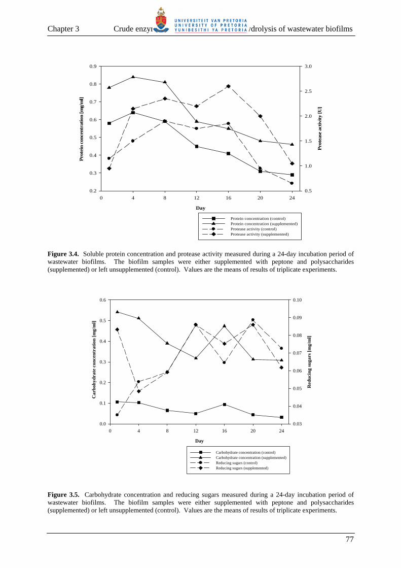
Chapter 3 Crude enzymes extracted after the auto-hydrolysis of wastewater biofilms
Day
0 4 8 12 16 20 24
Prot
ein
conc
entr
atio
n [m
g/m
l]
0.2
0.3
0.4
0.5
0.6
0.7
0.8
0.9
Prot
ease
act
ivity
[U]
0.5
1.0
1.5
2.0
2.5
3.0
Protein concentration (control)Protein concentration (supplemented)Protease activity (control)Protease activity (supplemented)
Figure 3.4. Soluble protein concentration and protease activity measured during a 24-day incubation period of wastewater biofilms. The biofilm samples were either supplemented with peptone and polysaccharides (supplemented) or left unsupplemented (control). Values are the means of results of triplicate experiments.
Day
0 4 8 12 16 20 24
Car
bohy
drat
e co
ncen
trat
ion
[mg/
ml]
0.0
0.1
0.2
0.3
0.4
0.5
0.6
Red
ucin
g su
gars
[mg/
ml]
0.03
0.04
0.05
0.06
0.07
0.08
0.09
0.10
Carbohydrate concentration (control)Carbohydrate concentration (supplemented)Reducing sugars (control)Reducing sugars (supplemented)
Figure 3.5. Carbohydrate concentration and reducing sugars measured during a 24-day incubation period of wastewater biofilms. The biofilm samples were either supplemented with peptone and polysaccharides (supplemented) or left unsupplemented (control). Values are the means of results of triplicate experiments.
77

Chapter 3 Crude enzymes extracted after the auto-hydrolysis of wastewater biofilms
present in the wastewater biofilm. Nevertheless, the addition of polysaccharides to the
wastewater biofilm clearly indicated the presence of polysaccharide-degrading enzymes in the
wastewater biofilm samples, albeit low. The low enzymatic activity may be due to the
extracellular matrix of the biofilm that assisted in the retention, the buildup or stabilization of
the enzymes, as suggested by Wingender et al. (1999), therefore making their extraction
inefficient with the method adopted in this study. Alternatively, since Gram-positive bacteria,
e.g., Micrococcus spp. and Bacillus spp., contain a high amount of polysaccharides compared
to Gram-negative bacteria (Cloete and Atlas, 2006), the low amount of polysaccharides
measured in the biofilm samples may indicate that the biofilm is composed mostly of Gram-
negative bacteria.
3.3.3 Enzymatic treatment of P. aeruginosa PAO1 biofilms Having established that the crude enzyme extract prepared from the wastewater biofilm
contained both protein- and polysaccharide-degrading enzyme activities, it was subsequently
investigated whether these enzymatic activities could facilitate biofilm removal. For this
purpose, P. aeruginsa PAO1 was chosen as model organism since it is amongst the best-
studied biofilm formers (O’Toole et al., 2000). To investigate, P. aeruginosa PAO1 biofilms
grown on glass slides for 96 h were treated with the crude enzyme extract for different times
and the glass slides were then examined by scanning electron microscopy (SEM).
The results obtained indicated that P. aeruginosa PAO1 was capable of attaching to the glass
surface and formed copious amounts of biofilm. This was evidenced by the glass slide being
densely covered by biofilm and the cells were embedded in an exopolymeric matrix (Fig.
3.6a). For P. aeruginosa PAO1 biofilm samples incubated with the crude enzyme extract, a
reduction in biofilm and EPS could be observed (Fig. 3.6c-f). Notably, after enzyme
treatment for 22 h, only a few individual P. aeruginosa PAO1 cells remained attached to the
surface and no EPS was visualized (Fig. 3.6f). In contrast, the P. aeruginosa PAO1 biofilm
was not removed from the glass surface when the biofilm was incubated for 22 h in the
presence of phosphate buffer lacking the crude enzyme extract (Fig. 3.6b). The results
therefore suggested that the crude enzyme extract prepared from the wastewater biofilm
contained different enzymes that were capable of degrading different components present in
the EPS of P. aeruginosa PAO1 biofilms, thus resulting in their removal from the glass
surface.
78

Chapter 3 Crude enzymes extracted after the auto-hydrolysis of wastewater biofilms
(e)
(c)
(b)
(f)
(d)
(a)
Figure 3.6. Scanning electron microscopy (SEM) micrographs of P. aeruginosa PAO1 biofilms treated with a crude enzyme extract prepared from a wastewater biofilm. P. aeruginosa PAO1 biofilms were cultured for 96 h in the presence of glass slides as attachment surface. Following incubation, the biofilms were treated with a crude enzyme extract prepared from an auto-hydrolyzed wastewater biofilm for 1 h (c), 5 h (d), 10 h (e) and 22 h (f). As controls, the P. aeruginosa PAO1 biofilm was left untreated (a) or incubated in phosphate buffer only for 22 h (b).
79

Chapter 3 Crude enzymes extracted after the auto-hydrolysis of wastewater biofilms
3.3.4 Enzymatic hydrolysis of P. aeruginosa PAO1 EPS components To determine whether removal of P. aeruginosa PAO1 biofilms from the glass surface was
indeed due to degradation of the protein and polysaccharide components present in the biofilm
EPS, the EPS of P. aeruginosa PAO1 was extracted and incubated for 22 h with a crude
enzyme extract prepared from the auto-hydrolyzed wastewater biofilm.
The effect of the crude enzyme extract on the amount of reducing sugar released from P.
aeruginosa PAO1 EPS is shown in Fig. 3.7. An untreated sample showed no increase in the
amount of reducing sugar. However, the amount of reducing sugar increased by 37.79%
when the EPS was treated with the crude enzyme extract. The increase in reducing sugar
concentration is in agreement with the previous results (Fig. 3.5), indicating that the
wastewater biofilm displayed a low level of polysaccharide-degrading enzyme activity.
0 220
0.2
0.4
0.6
0.8
1
1.2
Red
ucin
g su
gars
rel
ease
d [m
g/m
l]
22Incubation time (hours)
Untreated EPS Enzyme-treated EPS
Figure 3.7. Reducing sugars released from the extracted EPS of P. aeruginosa PAO1, following incubation for 22 h with a crude enzyme extract prepared from an auto-hydrolyzed wastewater biofilm. The extent of protein degradation, following incubation of P. aeruginosa PAO1 EPS with the
crude enzyme extract, was assessed by SDS-PAGE analysis of the samples following protein
extraction. The results are shown in Fig. 3.8. The results indicated a marked difference in the
protein profiles of samples treated with the crude enzyme extract (lanes 3 and 6) compared to
untreated samples (lanes 2 and 5) and samples treated with an inactivated enzyme extract
(lanes 4 and 7). Whereas the latter two control samples contained multiple protein bands that
were similar in size, the enzyme-treated P. aeruginosa EPS extracts contained very few
80
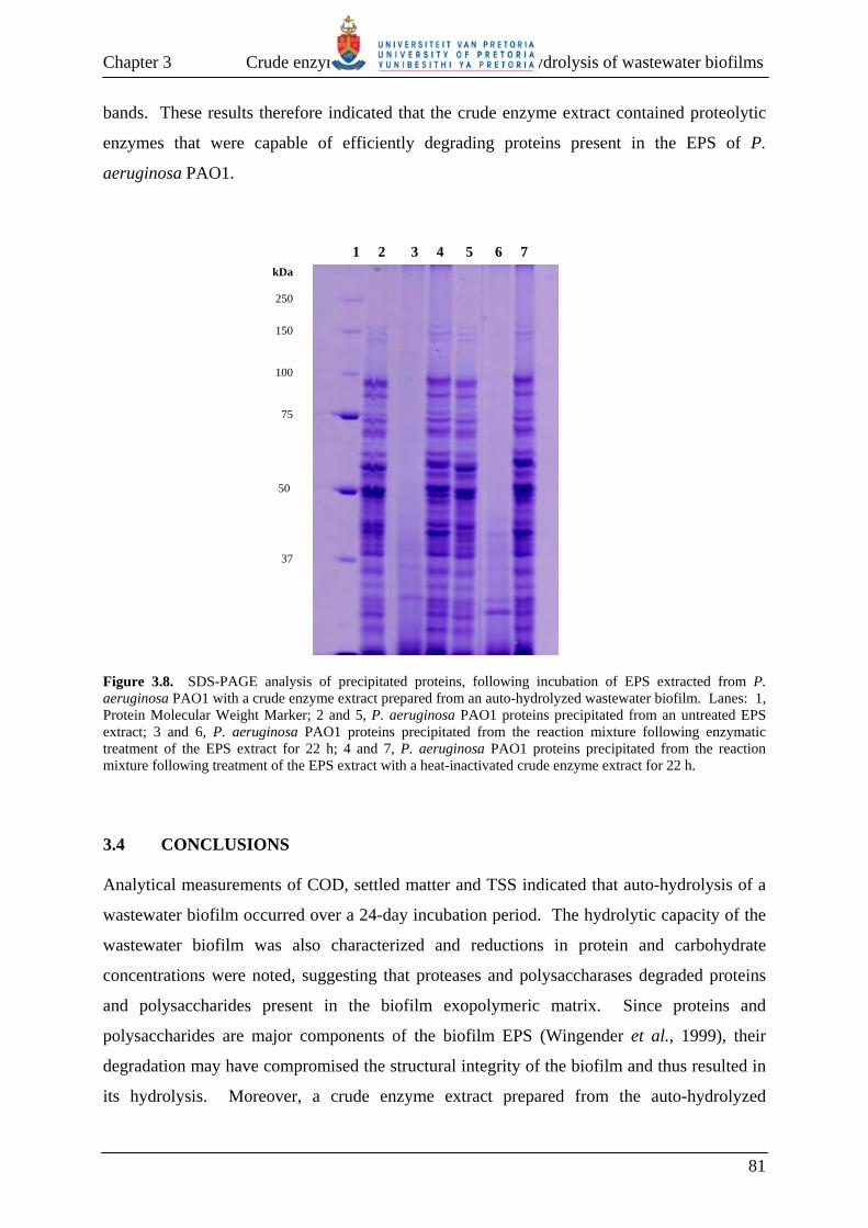
Chapter 3 Crude enzymes extracted after the auto-hydrolysis of wastewater biofilms
bands. These results therefore indicated that the crude enzyme extract contained proteolytic
enzymes that were capable of efficiently degrading proteins present in the EPS of P.
aeruginosa PAO1.
1 2 3 4 5 6 7
kDa
250
150 100 75
50 37
Figure 3.8. SDS-PAGE analysis of precipitated proteins, following incubation of EPS extracted from P. aeruginosa PAO1 with a crude enzyme extract prepared from an auto-hydrolyzed wastewater biofilm. Lanes: 1, Protein Molecular Weight Marker; 2 and 5, P. aeruginosa PAO1 proteins precipitated from an untreated EPS extract; 3 and 6, P. aeruginosa PAO1 proteins precipitated from the reaction mixture following enzymatic treatment of the EPS extract for 22 h; 4 and 7, P. aeruginosa PAO1 proteins precipitated from the reaction mixture following treatment of the EPS extract with a heat-inactivated crude enzyme extract for 22 h. 3.4 CONCLUSIONS Analytical measurements of COD, settled matter and TSS indicated that auto-hydrolysis of a
wastewater biofilm occurred over a 24-day incubation period. The hydrolytic capacity of the
wastewater biofilm was also characterized and reductions in protein and carbohydrate
concentrations were noted, suggesting that proteases and polysaccharases degraded proteins
and polysaccharides present in the biofilm exopolymeric matrix. Since proteins and
polysaccharides are major components of the biofilm EPS (Wingender et al., 1999), their
degradation may have compromised the structural integrity of the biofilm and thus resulted in
its hydrolysis. Moreover, a crude enzyme extract prepared from the auto-hydrolyzed
81
Chapter 3 Crude enzymes extracted after the auto-hydrolysis of wastewater biofilms
wastewater biofilm was shown to be effective in detaching P. aeruginosa PAO1 biofilms
from a glass surface and this could be ascribed to the degradation of EPS proteins and
polysaccharides. Further characterization of the wastewater biofilm bacteria and the enzymes
that they produce may thus lead to the identification of enzymes that could potentially be used
to remove detrimental biofilms.
3.5 REFERENCES
Boczar, B.A., Begley, W.M. and Larson, R.J. 1992. Characterization of enzyme activity in
activated sludge using rapid analyses for specific hydrolases. Water Environment Research,
64: 792-797.
Boczar, B.A., Rorney, L.J., Begley, W.M., Larson, R.J. and Federle, T.W. 2001.
Characterization and distribution of esterase activity in activated sludge. Water Research, 35:
4208-4216.
Bradford, M.M. 1976. A rapid and sensitive method for the quantitation of microgram
quantities of protein utilizing the principle of protein-dye binding. Analytical Biochemistry,
72: 248-254.
Cadoret, A., Conrad, A. and Block, J-C. 2002. Availability of low and high molecular weight
substrates to extracellular enzymes in whole and dispersed activated sludges. Enzymes and
Microbial Technology, 31: 179-186.
Ceyhan, N. and Ozdemir, G. 2008. Extracellular polysaccharides produced by cooling water
tower biofilm bacteria and their possible degradation. Biofouling, 24: 129-135.
Clesceri, L.S., Greenberg, A.E. and Eaton, A.D. 1998. Standard methods for the examination
of water and wastewater. 20th Edition. American Public Health Association, American Water
Works Association, Water Environment Federation, USA.
Cloete, T.E. and Atlas, R. 2006. Basic and Applied Microbiology. Van Schaik Publishers,
South Africa.
Confer, D.R. and Logan, B.E. 1991. Increased bacterial uptake of macromolecular substrates
with fluid shear. Applied and Environmental Microbiology, 57: 3093-3100.
Confer, D.R. and Logan, B.E. 1997a. Molecular weight distribution of hydrolysis products
during the biodegradation of model macromolecules in suspended culture and wastewater
biofilms. I. Bovine serum albumin. Water Research, 31: 2127-2136.
Confer, D.R. and Logan, B.E. 1997b. Molecular weight distribution of hydrolysis products
during the biodegradation of model macromolecules in suspended culture and wastewater
biofilms. II. Dextran and dextrin. Water Research 31: 2137-2145.
82

Chapter 3 Crude enzymes extracted after the auto-hydrolysis of wastewater biofilms
Confer, D.R. and Logan, B.E. 1998. Location of protein and polysaccharide hydrolytic
activity in suspended and biofilm wastewater cultures. Water Research, 32: 31-38.
Dashek, W.V. 1997. Methods in plant biochemistry and molecular biology. CRC Press,
USA.
Droste, R.L. 1997. Theory and practice of water and wastewater treatment. John Wiley and
Sons, Inc. Canada.
Gessesse, A., Dueholm, T., Petersen, S.B. and Nielsen, P.H. 2003. Lipase and protease
extraction from activated sludge. Water Research, 37: 3652-3657.
Ghose, T.K. 1987. Measurement of cellulase activities. Pure and Applied Chemistry, 59:
257-268.
Goel, R., Mino, T., Satoh, H. and Matsuo, T. 1998. Enzyme activities under anaerobic and
aerobic conditions in activated sludge sequencing batch reactor. Water Research, 32: 2081-
2088.
Itoh, Y., Wang, X., Hinnebusch, B.J., Preston, J.F. and Romeo, T. 2005. Depolymerization of
β-1,6-N-acetyl-D-glucosamine disrupts the integrity of diverse bacterial biofilms. Journal of
Bacteriology, 187: 382-387.
Jahn, A. and Nielsen, P.H. 1995. Extraction of extracellular polymeric substances (EPS) from
biofilms using a cation exchange resin. Water Science and Technology, 32:157–64.
Johansen, C., Falholt, P. and Gram, L. 1997. Enzymatic removal and disinfection of bacterial
biofilms. Applied and Environmental Microbiology, 63: 3724-3728.
Laemmli, U.K. 1970. Cleavage of structural proteins during the assembly of the head of
bacteriophage T4. Nature, 227: 680-685.
Logan, B.E. and Jiang, Q. 1990. A model for determining molecular size distributions of
DOM. Journal of Environmental Engineering, 116: 1046-1062.
Loiselle, M. and Anderson, K.W. 2003. The use of cellulase in inhibiting biofilm formation
from organisms commonly found on medical implants. Biofouling, 19: 77-85.
Nielsen, P.H., Jahn, A. and Palmgren, R. 1997. Conceptual model for production and
composition of exopolymers in biofilms. Water Science and Technology, 36: 11-19.
O’Toole, G., Kaplan, H.B. and Kolter, R. 2000. Biofilm formation as microbial development.
Annual Reviews in Microbiology, 54: 49-79.
Olajuyigbe, F.M. and Ajele, J.O. 2005. Production dynamics of extracellular protease from
Bacillus species. African Journal of Biotechnology, 4: 776-779.
Orgaz, B., Kives, J., Pedregosa, A.M., Monistrol, I.F., Laborda, F. and SanJose, C. 2006.
Bacterial biofilm removal using fungal enzymes. Enzyme and Microbial Technology, 40: 51-
56.
83

Chapter 3 Crude enzymes extracted after the auto-hydrolysis of wastewater biofilms
Orgaz, B., Neufeld, R.J. and SanJose, C. 2007. Single-step biofilm removal with delayed
release encapsulated Pronase mixed with soluble enzymes. Enzyme and Microbial
Technology, 40: 1045-1051.
Parmar, N., Singh, A. and Ward, O.P. 2001. Enzyme treatment to reduce solids and improve
settling of sewage sludge. Journal of Industrial Microbiology and Biotechnology, 26: 383-
386.
Raunkjær, K., Hvitved-Jacobsen, T. and Nielsen, R.H. 1994. Measurement of pools of
protein, carbohydrate and lipid in domestic wastewater. Water Research, 28: 251-262.
Roe, J.H. 1954. The determination of sugar in blood and spinal fluid with anthrone reagent.
Journal of Biological Chemistry, 208: 889-896.
Sanders, W.T.M., Geering, M., Zeeman, G. and Lettinga, G. 2000. Anaerobic hydrolysis
kinetics of particulates substrates. Water Science and Technology, 41: 17-24.
Secades, P. and Guijarro, J.A. 1999. Purification and characterization of an extracellular
protease from the fish pathogen Yersinia ruckeri and effect of culture conditions on
production. Applied and Environmental Microbiology, 65: 3969-3975.
Sponza, D.T. 2003. Investigation of extracellular polymer substances (EPS) and
physicochemical properties of different activated sludge flocs under steady-state conditions.
Enzyme and Microbial Technology, 32: 375-385.
Ten, L.N., Im, W-T., Kim, M-K., Kang, M.S. and Lee, S-T. 2004. Development of a plate
technique for screening of polysaccharide-degrading microorganisms by using a mixture of
insoluble chromogenic substrates. Journal of Microbiological Methods, 56: 375-382.
Vesilind, P.A. 2003. Wastewater treatment plant design. Water Environment Federation,
Alexandria, USA.
Vetter, Y.A. and Deming, J.W. 1999. Growth rates of marine bacterial isolates on particulate
organic substrates solubilized by freely released extracellular enzymes. Microbial Ecology,
37: 86-94.
Wang, Z-W., Liu, Y. and Tay, J-H. 2007. Biodegradability of extracellular polymeric
substances produced by aerobic granules. Applied Microbiology and Biotechnology, 74: 462-
466.
Wessel, D. and Flugge, U.I. 1984. A method for the quantitative recovery of protein in dilute
solution in the presence of detergents and lipids. Analytical Biochemistry, 138: 141-143
Wingender, J., Nue, T.R. and Flemming, H-C. 1999. Microbial extracellular polymeric
substances: Characterization, structure and function. Springer, Germany.
Xia, Y., Kong, Y. and Nielsen, P.H. 2007. In situ detection of protein-hydrolysing
microorganisms in activated sludge. FEMS Microbial Ecology, 60: 156-165.
Zhang, X. and Bishop, P.L. 2003. Biodegradability of biofilm extracellular polymeric
substances. Chemosphere, 50: 63-69.
84

Chapter 3 Crude enzymes extracted after the auto-hydrolysis of wastewater biofilms
Zhang, X., Bishop, P.L. and Kinkle, B.K. 1999. Comparison of extraction methods for
quantifying extracellular polymers in biofilms. Water Science and Technology, 39: 211-218.
85
CHAPTER 4
IDENTIFICATION OF WASTEWATER BIOFILM BACTERIA
CAPABLE OF DEGRADING PROTEINS AND POLYSACCHARIDES Abstract Bacteria were isolated from a wastewater biofilm previously shown to produce protein- and
polysaccharide-degrading enzymes. The bacteria were isolated on nutrient agar medium and
21 representative isolates, based on colony morphology, were purified and identified. The
identity of the cultured bacterial isolates was determined by extracting their chromosomal
DNA, followed by PCR amplification and sequencing of the 16S rRNA genes. The isolates
represented four major phylogenetic divisions of bacteria, i.e. Proteobacteria, Actinobacteria,
Firmicutes and Bacteroidetes. Screening of the bacterial isolates for their ability to degrade
proteins indicated that nine isolates produced protein-degrading enzymes, while ten isolates
produced polysaccharide-degrading enzymes. Based on the use of culture media
supplemented with different polysaccharide substrates, bacterial isolates capable of producing
amylase, xylanase, cellulase, α-glucosidase and β-glucosidase were identified.
4.1 INTRODUCTION Microbial biofilms are populations of microorganisms that are concentrated at an interface
and typically surrounded by an extracellular polymeric matrix (Costerton et al., 1995;
Watnick and Kolter, 2000; Donlan, 2002). Biofilms are responsible for causing problems
such as increased frictional resistance to fluids in technical water systems (Characklis and
Marshall, 1990; Flemming, 2002), biofouling in engineered systems (White et al., 1996;
Flemming, 2002), decreased heat transfer from heat exchangers (Ludensky, 2003), corrosion
of metallic substrata (LeChevallier et al., 1993), and contamination in food processing and
drinking water distribution systems (Kumar and Anand, 1998; Flemming et al., 2002).
Biofilms are also a severe problem in medical science and industry, causing prosthetic device
colonization and biofilm formation on medical implants (Sihorkar and Vyas, 2001; Hall-
Stoodley et al., 2004). Most treatment systems for controlling biofilms include the use of
antibiotics, biocides and disinfectants (Stewart and Costerton, 2001; Ludensky, 2003; Meyer,
2003). However, their efficacy may be limited due to the reduced susceptibility of
microorganisms in biofilms to such antimicrobial agents (Ishida et al., 1998; Anderl et al.,
86

Chapter 4 Identification of wastewater biofilm bacteria
2000; Norwood and Gilmour, 2000; Stewart and Costerton, 2001; Kostenko et al., 2007),
while some disinfectants have been reported to produce by-products that are toxic or have
irritant properties (Augustin and Ali-Vehmas, 2004). This therefore necessitates that
alternative approaches to control unwanted biofilms be investigated.
The main structural and physical component of microbial biofilms is the extracellular matrix
(Allison et al., 1998; Allison, 2003; Danese et al., 2000; Hentzer et al., 2001; Sutherland,
2001), which is comprised mostly of proteins and polysaccharides (Wingender et al., 1999;
Tsuneda et al., 2003). The exopolysaccharides present in the matrix are partly responsible for
bacterial adhesion and biofilm accumulation on surfaces (Allison et al., 1998; Cammarota and
Sant’Anna, 1998; Hentzer et al., 2001), and thus represent a potentially significant target for
biofilm removal and control. Several reports have indicated that enzymes can be used for
degradation of biofilms and for their removal from surfaces. Enzymes reported to be capable
of removing bacterial biofilms from different surfaces include: cellulase, α-amylase, protease,
esterase, glucose oxidase, arabinase, β-glucuronidase, α-glucosidase, lipase, xylanase, β-
glucanase, lactoperoxidase, polygalacturonase, lyases and Dispersin B (Selan et al., 1993;
Boyd and Chakrabarty, 1994; Johansen et al., 1997; Allison et al., 1998; Hahn Berg et al.,
2001; Loiselle and Anderson, 2003; Kaplan et al., 2004; Itoh et al., 2005 Orgaz et al., 2006;
Walker et al., 2007). However, due to the heterogeneity of the polysaccharides and proteins
present in the biofilm EPS, it has been suggested that a mixture of enzymes with different
specificities should be used in order to degrade bacterial biofilms effectively (Meyer, 2003).
In the previous Chapter it was shown that a biofilm from an aeration basin at an industrial
wastewater treatment plant produced enzymes that were capable of degrading the protein and
polysaccharide components of the EPS from Pseudomonas aeruginosa PAO1, thus resulting
in detachment of its biofilms from a glass surface. These findings suggested that the
wastewater biofilm-expressed enzymes might prove useful disinfectants or cleaning agents for
detrimental biofilms. Therefore, this study aimed to isolate and characterize the biofilm
bacteria. Although the microbial populations in wastewater treatment bioreactors have
received little attention, studies have shown that wastewater bioreactors are similar to many
natural environments, in that the number of culturable bacteria is 5-15% of the total number of
bacteria (Wagner et al., 1993; Kämpfer et al., 1996; Amann et al., 1998). Microbes in
wastewater treatment reactors may be identified using either traditional or molecular methods.
Using traditional methods, bacterial cultures are isolated and then identified by their
87
Chapter 4 Identification of wastewater biofilm bacteria
morphological, physiological and biochemical properties. However, these traditional
identification methods have a number of intrinsic limitations (Amann et al., 1995; Theron and
Cloete, 2000). Not only may the growth medium selectively favour certain populations, but
many bacteria may share similar morphological, physiological and biochemical properties
making it difficult to differentiate them. However, 16S rRNA-based molecular methods with
high degrees of precision and specificity have been widely used for environmental microbial
studies (Wagner et al., 1994; Bond et al., 1995; Theron and Cloete, 2000; Bramucci et al.,
2003; Liu et al., 2005). Using these methods, the microbial identities may be revealed from
their 16S rRNA sequences, following amplification of the 16S rRNA gene using polymerase
chain reaction (PCR).
Since the aim of this research project was to identify enzymes that may prove useful in
biofilm removal, pure cultures of the wastewater biofilm bacteria were required. Therefore,
the objectives of this part of the study were to isolate bacteria from the wastewater biofilm
using a culture-based approach and to identify representative isolates based on their 16S
rRNA sequences. Furthermore, the bacterial isolates were screened for production of
enzymes capable of degrading proteins and polysaccharides.
4.2 MATERIALS AND METHODS 4.2.1 Isolation of bacteria from wastewater biofilm
A thoroughly-mixed wastewater biofilm sample, previously obtained from an aeration basin at
the Leeuwkuil water purification plant in Vereeniging, Gauteng, South Africa (Chapter 3,
Section 3.2.2), was serially diluted (10-fold) in quarter strength Ringer’s solution (Merck).
Aliquots of 1 ml of each dilution were aseptically plated onto nutrient agar medium (Merck).
The agar plates were incubated at 25°C for 48 h. Following incubation, single colonies were
characterized with regard to their colony morphology, pigmentation and Gram-staining
reaction. The bacterial isolates were also screened for catalase and oxidase production, as
previously described by Harrigan and McCance (1966). Representative colonies were
purified by repeated sub-culturing onto nutrient agar and the pure cultures were then
maintained at -70°C as glycerol cultures.
88
Chapter 4 Identification of wastewater biofilm bacteria
4.2.2 PCR amplification and sequencing of the 16S rRNA genes from bacterial isolates 4.2.2.1 Genomic DNA extraction Genomic DNA was extracted from the bacterial isolates with cetyltrimethylammonium
bromide (CTAB), as described by Ausubel et al. (2002). The cells from 1.5 ml of an overnight
culture were pelleted by centrifugation at 11 000 × g for 5 min and suspended in 567 µl of 1 ×
TE buffer (10 mM Tris-HCl, 1 mM EDTA; pH 8). The cells were lysed by addition of SDS
to a final concentration of 0.5% (v/v) and proteins were digested by addition of Proteinase K
to a final concentration of 100 µg/ml in a total volume of 600 µl. Following incubation at
37°C for 1 h, 100 µl of 5 M NaCl and 80 µl of a CTAB/NaCl solution (10% [w/v] CTAB in
0.7 M NaCl) was added, mixed thoroughly and incubated at 65°C for 10 min. The suspension
was extracted with an equal volume of chloroform:isoamyl alcohol (24:1), followed by
centrifugation at 6 000 × g for 5 min. The supernatant, containing the genomic DNA, was
transferred to a clean microfuge tube. The residual CTAB was removed by addition of an
equal volume of phenol:choroform:isoamyl alcohol (25:24:1), followed by centrifugation at 6
000 × g for 5 min. The genomic DNA was precipitated from the recovered aqueous
supernatant by addition of 0.6 volume isopropanol. The precipitated genomic DNA was
pelleted by centrifugation at 11 000 × g for 2 min, washed with 70% ethanol, dried under
vacuum and suspended in 30 µl of 1 × TE buffer. An aliquot of the extracted genomic DNA
was analyzed by agarose electrophoresis, as described by Sambrook et al. (1989).
4.2.2.2 Polymerase chain reaction (PCR) amplification of 16S rRNA genes The 16S rRNA genes were PCR-amplified using the oligonucleotides PRUN518r (5’-
ATTACCGCGGCTGCTGG-3’) (Øvreås et al., 1997) and pA8f-GC (5’-
CGCCCGCCGCGCGCGGCGGGCGGGGCGGGGGCACGGGGGGAGAGTTTGATCCTG
GCTCAG-3’) (Fjellbirkeland et al., 2001). Each PCR reaction mixture (20 µl) contained
approximately 60 ng of genomic DNA as template, 50 µM of each oligonucleotide, 1 × PCR
buffer, 1.5 mM MgCl2, 200 µM of each dNTP and 0.25 U Supertherm Taq DNA polymerase
(Southern Cross Biotechnology). The tubes were placed in a Perkin-Elmer GeneAmp® 2700
thermal cycler. Following incubation at 95°C for 10 min, the reaction mixtures were
subjected to 35 cycles of denaturation at 94ºC for 30 s, annealing at 53ºC for 30 s and
elongation at 72ºC for 1 min. After the last cycle, a final elongation step was performed at
72°C for 10 min to complete synthesis of all DNA strands. For control purposes, a reaction
mixture containing all reagents except template DNA was included. Aliquots of the PCR
89

Chapter 4 Identification of wastewater biofilm bacteria
reaction mixtures were analyzed by electrophoresis on a 1.5% (w/v) agarose gel in the
presence of an appropriate DNA molecular weight marker (Gene Ruler™ 100-bp DNA Ladder
Plus; Fermentas), and amplicons were purified with the Wizard® SV Gel and PCR Clean-Up
System (Promega) according to the manufacturer’s instructions.
4.2.2.3 Nucleotide sequencing and sequence analyses The nucleotide sequence of the amplicons was determined with the ABI-PRISM® BigDye™
Terminator Cycle Sequencing Ready Reaction kit v.3.1 (Perkin-Elmer Applied Biosystems)
according to the manufacturer’s instructions. The oligonucleotides used in the PCR reaction
(PRUN518r and pA8f-GC), were used as the sequencing primers. The sequencing reactions
comprised approximately 70 ng of DNA, 2 µl of Terminator Ready Reaction Mix, 10 pmol of
the sequencing primer and UHQ water to a final volume of 5 µl. Cycle sequencing reactions
were performed in a Perkin-Elmer GeneAmp® 2700 thermal cycler with 25 of the following
cycles: denaturation at 96°C for 10 s, annealing at 50°C for 5 s and extension at 60°C for 4
min. The purified, denatured extension products were loaded onto a Hitachi 3100 capillary
array DNA sequencer (Sequencing facility of the University of Pretoria). Nucleotide
sequences were analyzed with BioEdit v.5.0.9.1 (Hall, 1999) and their identity were verified
by BLASTN searches (Altschul et al., 1997) against the GenBank database (available at
http://www.ncbi.nlm.nih.gov/BLAST).
4.2.3 Detection of protein-degrading bacterial isolates Bacterial isolates were screened for protease activity according to the method of Harrigan and
McCance (1966). The isolates were streaked onto 10% skim-milk agar medium and the agar
plates were incubated at 25°C for 48 h. Protein degradation is visualized as clear zones
surrounding the bacterial growth.
4.2.4 Detection of polysaccharide-degrading bacterial isolates 4.2.4.1 Screening of bacterial isolates for amylase and xylanase activity All chemicals used in the preparation of dye-labeled polysaccharide substrates were obtained
from Sigma-Aldrich. The chromogenic substrates were prepared, as described by Ten et al.
(2004), with the following minor modifications. Xylan was cross-linked to Cibacron Brilliant
Red 3B-A by adding 5 ml of 2 M NaOH, 0.6 ml of 1,4-butanediol diglycidyl ether and 0.95 g
90
Chapter 4 Identification of wastewater biofilm bacteria
of Cibacron Brilliant Red 3B-A to a xylan suspension, which had been prepared by dissolving
1 g of xylan in 15 ml of dH2O. The suspension was stirred for 5 min and incubated at room
temperature for 48 h to solidify in a gel. Subsequently, 50 ml of dH2O was added to the gel
and it was ground with a mortar and pestle until fine particles were produced. To remove the
unbound dye, the particles were washed with boiling water and filtered through Whatman
Type 1 filter paper until the filtrate was colourless. The xylan-red substrate was scraped from
the surface of the filter paper and used as a supplement to nutrient agar medium. Amylose-
blue was similarly prepared, except that amylose was cross-linked to Cibacron Blue 3GA
using 1.5 ml of 1,4-butanediol diglycidyl ether in the reaction mixture. Nutrient agar medium
was subsequently supplemented with 25 g of xylan-red and 75 g of amylose-blue per 1 l of
nutrient agar medium. To keep the particles dispersed, the autoclaved (121°C; 20 min)
nutrient agar was agitated gently while being poured into plates. Bacterial isolates were
streaked onto the supplemented nutrient agar media and the agar plates were incubated at
25°C for 2-5 days. Following incubation, plates were screened for solubilization of the
chromogenic substrates and halo formation around the bacterial growth.
4.2.4.2 Screening of bacterial isolates for cellulase activity Bacterial isolates were screened for cellulase activity according to a slightly modified method
of Teather and Wood (1982). Nutrient agar medium, supplemented with 8 g/l hydroxyethyl-
cellulose (Sigma-Aldrich), was autoclaved (121°C; 20 min) and thoroughly mixed before
pouring of plates. The agar medium was inoculated with the bacterial isolates and the agar
plates were incubated at 25°C for 72 h. The surface of the agar medium was rinsed with
0.85% (w/v) saline and then flooded with an aqueous solution of Congo red (1 mg/ml
prepared in dH2O). Following incubation at room temperature for 30 min, the Congo red
solution was poured off and the plates were flooded sequentially with 1 M NaCl and 1 M HCl
for 15 min each. Cellulose degradation is visualized as halo formation around the bacterial
growth.
4.2.4.3 Screening of bacterial isolates for lyase activity Bacterial isolates were screened for gellan lyase activity, as described by Kennedy and
Sutherland (1994), by supplementing nutrient agar medium with 15 g/l Phytagel (Sigma-
Aldrich). The isolates were streaked onto the medium and the agar plates were incubated at
25°C for 5 days. The agar plates were examined for the formation of depressions in the gel
91

Chapter 4 Identification of wastewater biofilm bacteria
due to enzymatic degradation of the gellan. The bacterial isolates were also screened for
alginate lyase activity, as described by Chavagnat et al. (1996). The isolates were streaked
onto “black medium” containing 0.5% (w/v) bacteriological agar, 0.5% (w/v) yeast extract,
1% (w/v) sodium alginate and 0.5% (w/v) activated charcoal. Following incubation of the
agar plates at 25°C for 5 days, the agar plates were flooded with 10% cetylpyridinium
chloride and incubated at room temperature for 20 min. Alginate degradation is visualized as
clear zones surrounding the bacterial growth.
4.2.4.4 Screening of bacterial isolates for glucosidase activity Bacterial isolates were screened for α- and β-glucosidase activity according to the method of
Arellano-Carbajal and Olmos-Soto (2002). The isolates were inoculated into nutrient broth
and incubated at 25°C for 48 h. To screen for α-glucosidase activity, 1 ml of the liquid
culture was mixed with 3 ml of 0.01 M sodium phosphate buffer (pH 7) and 1 ml of 0.1%
(w/v) p-nitrophenyl α-D-glucopyranoside (pNPG; Calbiochem, Merck). Following incubation
at 30°C for 30 min, the reaction was terminated by addition of 1 ml of 1 M Na2CO3. A
change in the colour of the reaction mixture to yellow indicated the presence of α-glucosidase
activity. The same methodology was used to screen the bacterial isolates for β-glucosidase
activity, except that p-nitrophenyl β-D-glucopyranoside (Calbiochem, Merck) was used as the
substrate in the enzyme assays.
4.3 RESULTS AND DISCUSSION 4.3.1 Isolation of biofilm bacteria To isolate biofilm bacteria, serial dilutions of a wastewater biofilm were plated onto nutrient
agar medium and representative colonies, based on colony morphology, were selected for
further characterization. The bacterial isolates were purified by repeated streaking onto
nutrient agar and designated isolates 1 through 21. Of the 21 bacterial isolates selected,
eleven were Gram-negative, eight were Gram-positive and two were visualized as Gram-
variable. The majority of the bacterial isolates displayed a rod-shaped morphology and only
six of the isolates produced coloured pigments following growth on nutrient agar. All of the
bacterial isolates produced catalase, and only seven of the isolates did not produce oxidase.
The morphological and physiological characteristics of the 21 cultured bacterial isolates are
summarized in Table 4.1.
92

Chapter 4 Identification of wastewater biofilm bacteria
Table 4.1: Morphological and physiological properties of bacterial isolates from wastewater biofilms
Isolate nr.
Gram straining
Cell morphology
Pigmentation Colony morphology Oxidase* Catalase*
1 + Rods None Beige, spreading, margin is
undulate, frosted appearance,
slimy center
++ ++
2 + Rods None Beige, irregular, undulate margin,
frosted appearance
++ ++
3 - Rods None Dark beige, smooth, circular,
mucoid
+ +
4 + Rods None Beige, undulate and translucent
margin, impregnated into media
+ ++
5 + Rods None Beige, large, irregular, undulate
margin, frosted appearance
++ ++
6 + Rods None Beige, flat, border around center + ++
7 + Rods Yellow Circular, iridescent, smooth,
entire margin
- +
8 - Rods None Beige, smooth, mucoid, entire
margin, quickly becoming
confluent
+ ++
9 - Rods None Beige, smooth, translucent, entire
margin
++ +
10 Gram
variable
Pleomorphic None Beige, translucent, smooth, entire
margin
- ++
11 - Rods None Beige, spreading margin,
translucent
++ ++
12 - Rods None Beige, small, circular, smooth to
slightly granular, translucent,
entire margin
+ +
13 - Rods None Beige, smooth to slightly
granular, entire margin,
translucent
++ +
14 - Rods Bright red Circular, iridescent with entire
margin, slightly convex, mucoid
- ++
15 - Rods Bright yellow Mucoid, entire margin, translucent - ++
16 - Rods Pale yellow Mucoid, translucent + +
17 Gram
variable
Pleomorphic Pale yellow Circular, entire margin, opaque,
elevated center
- +
18 + Rods None Beige, circular, entire margin,
iridescent
- ++
19 - Rods None Beige, very small, circular, entire
margin, translucent
+ +
20 - Rods None Beige, circular, translucent ++ ++
21 + Cocci Bright orange Circular, entire margin, iridescent,
convex
- ++
*: ++, strong positive; +, weakly positive; -, negative
93

Chapter 4 Identification of wastewater biofilm bacteria
4.3.2 Identification of wastewater biofilm bacteria based on 16S rRNA gene sequence analysis Genomic DNA was extracted from the individual bacterial isolates and the 16S rRNA gene
was amplified by PCR using a set of oligonucleotides specific for eubacteria. The amplicons
were approximately 500 bp in length and both DNA strands of the amplicons were sequenced.
The 16S rRNA sequences were subsequently compared with the sequences in GenBank by
BLAST. Table 4.2 summarizes the isolate number, the closest sequence found in GenBank
and their similarity. The results presented in Table 4.2 indicated that the 21 cultured isolates
represented four major phylogenetic divisions of bacteria, i.e. Proteobacteria, Actinobacteria,
Firmicutes and Bacteroidetes.
Table 4.2: Identity of cultured bacterial isolates from a wastewater biofilm based on their 16S rRNA sequences
Isolate no.
GenBank accession number/Organism Percentage similarity
Phylogenetic group
1 DQ520955 Bacillus subtilis 99% Firmicutes 2 AB284820 Bacillus cereus 99% Firmicutes 3 DQ298025 Rheinheimera chironomi 97% Proteobacteria 4 EF523475 Bacillus pumilus 97% Firmicutes 5 AY795568 Bacillus cereus 99% Firmicutes 6 EF433406 Bacillus amyloliquefaciens 99% Firmicutes 7 DQ350825 Microbacterium oxydans 98% Actinobacteria 8 AJ242584 Ochrobactrum tritici 98% Proteobacteria 9 AM490636 Ochrobactrum tritici 87% Proteobacteria 10 AJ243423 Arthrobacter oxydans 99% Actinobacteria 11 DQ229315 Pseudomonas putida 99% Proteobacteria 12 AF078764 Acidovorax delafieldii 96% Proteobacteria 13 DQ857897 Brevundimonas diminuta 96% Proteobacteria 14 EF114344 Serratia marcescens 99% Proteobacteria 15 AY275475 Chryseobacterium indotheticum 96% Bacteroidetes 16 AB166885 Stenotrophomonas koreensis 99% Proteobacteria 17 X80745 Arthrobacter protophormiae 99% Actinobacteria 18 AJ781046 Leucobacter chromiireducens 96% Actinobacteria 19 AF078764 Acidovorax delafieldii 96% Proteobacteria 20 AJ518818 Acidovorax delafieldii 97% Proteobacteria 21 AY211171 Kocuria rosea 97% Actinobacteria
Proteobacteria
Ten of the bacterial isolates, representing seven different genera, were closely affiliated with
Proteobacteria. Isolate 3 is related to Rheinheimera chironomi of little known characteristics,
but has been isolated from the egg mass of a chironomid (non-biting midge) that lays its eggs
in freshwater (Halpern et al., 2007). Both isolates 8 and 9 are related to Ochrobactrum tritici,
which has been isolated frequently from wastewater (Goris et al., 2003; Branco et al., 2004;
You et al., 2006). However, the isolates also displayed 97% and 87% similarity, respectively,
94

Chapter 4 Identification of wastewater biofilm bacteria
with O. anthropi, an opportunistic human pathogen (Ozdemir et al., 2006; Aly et al., 2007),
that has also been isolated from sewage (Swings et al., 2006) and activated sludge (Ozdemir
et al., 2003). Isolate 11 is most closely related to Pseudomonas putida. Members of the
Pseudomonas genus, including P. putida, have been isolated from activated sludge and
municipal wastewater (Leung et al., 2000; Lim et al., 2002). The three isolates 12, 19 and 20
are related to Acidovorax delafieldii, which has a flexible metabolism and is commonly found
in wastewater treatment plants (Schulze et al., 1999; Zhao and Ward, 1999; Khan et al.,
2002). Isolate 13 is closely related to Brevundimonas diminuta, which has been isolated
previously from activated sludge in a sequence batch reactor (Liu et al., 2005). Isolate 14 is
most closely related to Serratia marcescens, an opportunistic human pathogen (van Ogtrop et
al., 1997; Johnson et al., 1998), that occurs in soil, water and food (Vignolo et al., 1993;
Montpas et al., 1997). S. marcescens has also been isolated from wastewater (Filipkowska,
2003; Ajithkumar et al., 2003; Li et al., 2008) and tannery effluents (Campos et al., 2005).
Isolate 16 is most closely related to Stenotrophomonas koreensis, a newly described bacterial
species that was recently isolated from compost (Yang et al., 2006). However, the isolate also
displayed 96% similarity with S. maltophila, an opportunistic human pathogen (Denton and
Kerr, 1998). This bacterial species occurs in various natural environments and has been
isolated from several water sources (rivers, wells, hypereutrophic lake, bottled water and
wastewater), soil environments, plant rhizosphere environments and food sources (Denton and
Kerr, 1998; Brenner et al., 2005).
Actinobacteria
Five of the bacterial isolates, representing four different genera, were closely affiliated with
Actinobacteria. Isolate 7 is closely related to Microbacterium oxydans. Members of this
genus have been isolated from variety of environmental sources, including dairy products,
plants, soil and sewage (Funke et al., 1995), while M. oxydans has been isolated from
petroleum-contaminated soil (Supaphol et al., 2006) and from hydrocarbon-polluted soil
(Menendez-Vega et al., 2007), as well as from activated sludge (Chen et al., 2007). Isolates
10 and 17 are most closely related to Arthrobacter oxydans and A. protophormiae,
respectively. Members of this genus mostly inhabit soil environments (Jones and Keddie,
2006), but A. oxydans has been isolated from livestock farming wastewater (Chung et al.,
1997) and activated sludge (Song et al., 2005). Isolate 18 is related to Leucobacter
chromiireducens, which has been isolated from activated sludge (Morais et al., 2004), while
isolate 21 is related to Kocuria rosea, an opportunistic human pathogen (Altuntas et al.,
95

Chapter 4 Identification of wastewater biofilm bacteria
2004). Kocuria spp. have been isolated from wastewater and activated sludge (Leung et al.,
2000; Yasuhira et al., 2007).
Firmicutes
All five bacterial isolates affiliated with the Firmicutis group comprised species of the genus
Bacillus. Members of the genus Bacillus are ubiquitous and can be isolated from nearly all
natural environments, soil, freshwater, foods, milk and pharmaceuticals (Sneath et al., 1986).
Isolates 1 and 4 are closely related to B. subtilis and B. pumilus, respectively. Both these
bacterial species have been isolated from activated sludge and municipal wastewater (Leung
et al., 2000). Isolates 2 and 5 are most closely related to B. cereus, which has been isolated
from activated sludge (Sidat et al., 1999) and fish industry wastewater (Ghorbel et al., 2003).
Isolate 6 is most closely related to B. amyloliquefaciens, which has been previously isolated
from soil and water samples (Breccia et al., 1998).
Bacteroidetes
Only one of the cultured bacterial isolates was affiliated with the Bacteroidetes group. Isolate
15 is related to Chryseobacterium indotheticum, a member of the family Flavobacteriaceae,
which are found widespread in aquatic environments where they are responsible for the
degradation of complex organic material (Bernardet et al., 2002). Species within the genus
Chryseobacterium, including C. indotheticum, have also frequently been isolated from
wastewater and activated sludge (Kämpfer et al., 2003; Quan et al., 2007; Yoon et al., 2007).
The wastewater treatment plant from which the biofilm was sampled receives its main
effluents from domestic residences, several metal processing and casting industries, an
abattoir and hospitals. The latter may explain the isolation of several opportunistic human
pathogens during the course of this investigation. Many of the cultured bacterial isolates
identified in this study have been reported to occur in wastewater and activated sludge, but the
presence of Rheinheimera chironomi and Stenotrophomonas koreensis in this ecological niche
is reported here for the first time. Although not an objective of this study, investigations
regarding the microbial composition and functionality would be of importance to gain a better
understanding of the wastewater treatment process and to optimize the process. However,
culture-based methods cannot be directly used to analyze the microbial diversity because it is
widely recognized that only a small portion of the total microbiota in environmental samples
can be cultured (Wagner et al., 1993; Kämpfer et al., 1996; Amann et al., 1998).
96

Chapter 4 Identification of wastewater biofilm bacteria
Nevertheless, the results that were obtained in this study are in agreement with several other
studies indicating that culture-dependant methods typically show that Proteobacteria dominate
the microbial populations in activated sludge from municipal wastewater bioreactors
(Kämpfer et al., 1996; Amann et al., 1998; Bramucci and Nagarjan, 2000; Bramucci et al.,
2003). Since it was previously shown that a crude enzyme extract containing the wastewater
biofilm-expressed enzymes removed P. aeruginosa PAO1 biofilms from a glass surface
(Chapter 3), the cultured bacterial isolates were subsequently investigated for their ability to
produce enzymes that have previously been reported to aid biofilm removal.
4.3.3 Bacterial isolates with protein-degrading capabilities The bacterial isolates were screened for proteolytic activity on skim-milk agar, and hydrolysis
zones were visualized as clear zones against an opaque background (Fig. 4.1). The diameter
of the hydrolysis zones served as a semi-quantitative measure of the protease activity. Of the
21 bacterial isolates, nine were identified as being able to produce proteases. Isolates 3 and
14 displayed the most potent protease activity with hydrolysis zones of 7 mm each, followed
by isolate 6 (5 mm) and isolate 1 (4 mm). Less potent protease activity was observed for
isolates 2, 4 and 15 (3 mm), while isolates 16 and 5 displayed the weakest proteolytic activity
with hydrolysis zones of 2 and 1 mm, respectively (Fig. 4.2).
Figure 4.1. Detection of protein-degrading bacteria on skim-milk agar medium. Hydrolysis zones surrounding the bacterial growth are indicative of protease production.
97

Chapter 4 Identification of wastewater biofilm bacteria
0
1
2
3
4
5
6
7
Dia
met
er o
f hyd
roly
sis
zone
(mm
)
1 2 3 4 5 6 14 15 16
Proteolytic strains
Figure 4.2. Diameter of hydrolysis zones formed on skim-milk agar by bacterial isolates capable of producing proteases. Values are the means of results of triplicate experiments. 4.3.4 Bacterial isolates with polysaccharide-degrading capabilities 4.3.4.1 Xylan- and amylose-degrading bacteria To identify bacterial isolates with xylanase and/or amylase activity, nutrient agar medium was
amended with a mixture of two chromogenic substrates, i.e. amylose-blue and xylan-red.
Several bacterial isolates were capable of degrading xylan and amylose, as evidenced by
solubilization of the substrate particles and halo formation around the bacterial growth. By
using this approach, three isolates were identified that produced both xylan- and amylose-
degrading enzymes (isolates 1, 6 and 21), while three isolates exhibited amylose-degrading
activity (isolates 2, 3 and 5) and one isolate displayed xylan-degrading activity (isolate 4).
Representative photographs of the agar plates are presented in Fig. 4.3. Bacterial isolates
capable of degrading both xylan and amylose are shown in Figs. 4.3a and 4.3b, while isolates
capable of degrading only amylose or xylan is shown in Figs. 4.3c and 4.3d, respectively.
4.3.4.2 Cellulose-degrading bacteria The bacterial isolates were screened for cellulase activity by streaking the isolates onto
nutrient agar medium that was supplemented with hydroxyethyl-cellulose. Following
incubation, the plates were flooded with Congo red and the solution was then replaced with
NaCl to maximize binding of the dye to the polysaccharide. The plates were then flooded
with HCl, resulting in a colour change from red to blue, and thereby increasing the contrast
between degradation zones and the blue background. Using this approach, three bacterial
isolates (1, 4 and 5) were identified that were capable of degrading hydroxyethyl-cellulose
(Fig. 4.4).
98

Chapter 4 Identification of wastewater biofilm bacteria
(b) (c) (a)
(d) (e)
Figure 4.3. Detection of xylan- and amylose-degrading bacteria on nutrient agar supplemented with xylan-red and amylose-blue. (a) and (b) Bacterial isolates were capable of degrading both xylan and amylose, as evidenced by the solubilization of both red and blue chromogenic substances around the bacterial growth. (c) A visible blue halo was observed surrounding the bacterial growth, indicating that the bacterial isolate is capable of degrading amylose only. (d) A visible red halo was observed surrounding the bacterial growth, indicating that the bacterial isolate is capable of degrading xylan only. (e) No visible halo was observed surrounding the bacterial growth, indicating that the bacterial isolate is not capable of degrading amylose and/or xylan. (a) (b) (c)
Figure 4.4. Detection of cellulose-degrading bacteria on nutrient agar supplemented with hydroxyethyl-cellulose. Haloes surrounding the bacterial growth are indicative of cellulose degradation. Results are shown for isolates 1 (a), 4 (b), and 5 (c).
99

Chapter 4 Identification of wastewater biofilm bacteria
4.3.4.3 Alginate- and gellan-degrading bacteria The bacterial isolates were screened for both alginate and gellan lyase activity. However, the
bacterial isolates were unable to degrade the alginate present in the agar medium, as was
evidenced by a lack of haloes surrounding the bacterial growth. In addition, the bacterial
isolates also did not cause depressions in the nutrient agar medium supplemented with gellan.
These results therefore indicated that none of the bacterial isolates were capable of producing
alginate or gellan lyase (results not shown).
4.3.4.4 Glucosidase-producing bacteria The bacterial isolates were screened for α- and β-glucosidase using the synthetic substrates p-
nitrophenyl α-D-glucopyranoside and p-nitrophenyl β-D-glucopyranoside, respectively (Fig.
4.5). The assays are based on the determination of the hydrolytic release of p-nitrophenol
from the respective pNPG substrates. The p-nitrophenol is a yellow-coloured compound, the
colouration of which deepens in Na2CO3, thus providing a semi-quantitative measure of
enzyme activity (Chapdelaine et al., 1978). Of the 21 bacterial isolates screened, eight
isolates were capable of producing α-glucosidase (isolates 1, 2, 4-7, 10 and 15), while three
isolates produced β-glucosidase (isolates 4, 6 and 7).
1 2 3 4 65 7
Figure 4.5. Detection of α-glucosidase activity using the chromogenic substrate p-nitrophenyl α-D-glucopyranoside (tubes 2-4), and detection of β-glucosidase activity using the chromogenic substrate p-nitrophenyl β-D-glucopyranoside (tubes 5-7). The control, uninoculated tube showed no colour reaction (tube 1). The enzymatic activities displayed by the cultured wastewater biofilm bacteria are
summarized in Table 4.3. Of the 21 bacterial isolates, 12 isolates displayed protease- and/or
polysaccharide-degrading enzyme activities. Among bacteria, Bacillus spp. produces a
number of extracellular proteases and polysaccharide-degrading enzymes (Priest, 1977;
100

Chapter 4 Identification of wastewater biofilm bacteria
Schallmey et al., 2004; Saeki et al., 2007). Indeed, most of the bacterial isolates identified in
this study that revealed both proteolytic and polysaccharide-degrading activities belonged to
this genus. In particular, B. subtilis (isolate 1), B. pumilis (isolate 4) and B. amyloliquefaciens
(isolate 6) displayed a wide range of enzyme activities, followed by B. cereus (isolates 2 and
5), albeit that these two strains differed in their ability to produce cellulase (Table 4.3). In
addition, M. oxydans (isolate 7), A. oxydans (isolate 10) and K. rosea (isolate 21) produced
polysaccharide-degrading enzymes, while S. marcescens (isolate 14) and S. koreensis (isolate
16) displayed proteolytic activity. Moreover, R. chironomoni (isolate 3) diplayed both
amylase and protease activities, while C. indotheticum (isolate 15) displayed both α-
glucosidase and protease activities. These results are in agreement with previous reports
regarding enzymes produced by bacterial strains isolated during the course of this study
(Table 4.4).
Table 4.3: Enzyme activities displayed by 21 cultured bacterial isolates from a wastewater biofilm
Isolate Protease Amylase Xylanase Cellulase α-glucosidase
β-glucosidase
Alginate lyase
Gellan lyase
1 + + + + + - - -
2 + + - - + - - -
3 + + - - - - - -
4 + - + + + + - -
5 + + - + + - - -
6 + + + - + + - -
7 - - - - + + - -
8 - - - - - - - -
9 - - - - - - - -
10 - - - - + - - -
11 - - - - - - - -
12 - - - - - - - -
13 - - - - - - - -
14 + - - - - - - -
15 + - - - + - - -
16 + - - - - - - -
17 - - - - - - - -
18 - - - - - - - -
19 - - - - - - - -
20 - - - - - - - -
21 - + + - - - - -
101

Chapter 4 Identification of wastewater biofilm bacteria Table 4.4: Enzymes produced by several of the isolated bacteria, as indicated in previous studies
Bacterial strain Enzymes produced References Bacillus subtilis - Protease
- Amylase - Xylanase - Cellulase - α-glucosidase
- Tran et al., 1991; Sloma et al., 1991; Cheggour et al., 2000
- Yang et al., 1984 - Sá-Pereira et al., 2002 - Park et al., 1991 - Schönert et al., 1998
Bacillus cereus - Protease - Amylase - Cellulase - α-glucosidase
- Stark et al., 1992 - Bogas et al., 2007 - Thayer, 1978 - Suzuki et al., 1982
Rheinheimera chironomi - Protease
- Halpern et al., 2007
Bacillus pumilus - Protease - Xylanase - Cellulase - α-glucosidase - β-glucosidase
- Huang et al., 2003 - El Ahwany and Youssef, 2007 - Kotchoni and Shonukan, 2002 - Ouoba et al., 2004 - Ouoba et al., 2004
Bacillus amyloliquefaciens - Protease - Amylase - Xylanase - α-glucosidase
- Vasantha et al., 1984; Balaban et al., 2007
- Takkinen et al., 1983 - Breccia et al., 1998 - Fogarty et al., 1985
Microbacterium oxydans - β-glucosidase
- Laffineur et al., 2003
Arthrobacter oxydans - α-glucosidase
- Wauters et al., 2000
Serratia marcescens - Protease -Lyerly and Kreger, 1979; Matsumoto et al., 1984; Molla et al., 1987
4.4 CONCLUSIONS Bacteria from a wastewater biofilm were isolated, identified and subsequently screened using
plate screening methods to detect bacterial strains with protein- and polysaccharide-degrading
enzyme activities. The enzyme activities screened for in this study were selected based on
previous reports indicating their efficacy with regards to bacterial biofilm removal (Selan et
al., 1993; Loiselle and Anderson, 2003; Orgaz et al., 2006; Walker et al., 2007). With the
exception of lyase enzymes, bacterial strains were identified that produced protease, amylase,
xylanase, cellulase, α-glucosidase and β-glucosidase enzymes. Several bacterial strains
belonging to the genus Bacillus were identified that produced both proteases and different
types of polysaccharide-degrading enzymes. Since the EPS of bacterial biofilms is
heterogenous (Sutherland, 2001; Allison, 2003), enzyme mixtures comprising different
102
Chapter 4 Identification of wastewater biofilm bacteria
activities have been favoured for the efficient removal of biofilms (Meyer, 2003). The
identification of bacterial strains producing different types of enzymes may thus hold promise
as anti-biofilm agents.
4.5 REFERENCES
El Ahwany, A.M.D. and Youssef, A.S. 2007. Xylanase production by Bacillus pumilis:
Optimization by statistical and immobilization methods. Research Journal of Agriculture and
Biological Sciences, 3: 727-732.
Ajithkumar, B., Ajithkumar, V.P., Iriye, R., Doi, Y. and Sakai, T. 2003. Spore-forming
Serratia marcescens subsp. sakuensis subsp. nov., isolated from a domestic wastewater
treatment tank. International Journal of Systematic and Evolutionary Microbiology, 53: 253-
258.
Allison, D.G. 2003. The biofilm matrix. Biofouling, 19: 139-150.
Allison, D.G., Ruiz, B., SanJose, C., Jaspe, A. and Gilbert, P. 1998. Extracellular products
as mediators of the formation and detachment of Pseudomonas fluorescens biofilms. FEMS
Microbiology Letters, 167: 179-184.
Altschul, S.F., Madden, T.L., Schaffer, A.A., Zhang, J., Zhang, Z., Miller, W. and Lipman,
D.J. 1997. Gapped BLAST and PSI-BLAST: A new generation of protein database search
programs. Nucleic Acid Research, 25: 3389-3402.
Altuntas, F., Yildiz, O., Eser, B., Gündogan, K., Sumerkan, B. and Çetin, M. 2004. Catheter-
related bacteremia due to Kocuria rosea in a patient undergoing peripheral blood stem cell
transplantation. BMC Infectious Diseases, 4: 1-3.
Aly, N.Y.A., Salmeen, H.N. and Joshi, R.M. 2007. Ochrobactrum anthropi bacteremia in a
child with inborn error of mitochondrial fatty acid oxidation. Medical Principles and
Practices, 16: 436-465.
Amann, R., Lemmer, H. and Wagner, M. 1998. Monitoring the community structure of
wastewater treatment plants: A comparison of old and new techniques. FEMS Microbiology
Ecology, 25: 205-215.
Amann, R.I., Ludwig, W. and Schleifer, K.H., 1995. Phylogenetic identification and in situ
detection of individual microbial cells without cultivation. Microbiology Reviews, 59, 143-
169.
Anderl, J.N., Franklin, M.J. and Stewart, P.S. 2000. Role of antibiotic penetration limitation
in Klebsiella pneumoniae biofilm resistance to ampicillin and ciprofloxacin. Antimicrobial
Agents and Chemotherapy, 44: 1818-1824.
103

Chapter 4 Identification of wastewater biofilm bacteria
Arellano-Carbajal, F. and Olmos-Soto, J. 2002. Thermostable α-1,4- and α-1,6-glucosidase
enzymes from Bacillus sp. isolated from a marine environment. World Journal of
Microbiology and Biotechnology, 18: 791-795.
Augustin, M. and Ali-Vehmas, T. 2004. Assessment of enzymatic cleaning agents and
disinfectants against bacterial biofilms. Journal of Pharmacy and Pharmaceutical Sciences,
7: 55-64.
Ausubel, F.M., Brent, R., Kingston, R.E., Moore, D.D., Seidman, J.G., Smith, J.A. and Struhl,
K. 2002. Short Protocols in Molecular Biology. Fifth Edition, Volume One. Wiley-
Interscience, Canada.
Balaban, N.P., Malikova, L.A., Mardanova, A.M., Rudenskaya, G.N. and Sharipova, M.R.
2007. Purification and characterization of a subtilisin-like proteinases secreted in the
stationary growth phase of Bacillus amyloliquefaciens H2. Biochemistry, 72: 459-465.
Bernardet, J-F., Nakagawa, Y. and Holmes, B. 2002. Proposed minimal standards for
describing new taxa of the family Flavobacteriaceae and amended description of the family.
International Journal of Systematic and Evolutionary Microbiology, 52: 1049-1070.
Bogas, A.C., Watanabe, M.A.E., Barbosa, A.V-B., Laurival, A., Bonatto, A.C., Dekker, R.,
Souza, E.M. and Fungaro, M.H.P. 2007. Structural characterization of the bglH gene
encoding a β-glucosidase-like enzyme in an endophytic Bacillus pumilis strain. Genetics and
Molecular Biology, 30: 100-104.
Bond, P.L., Hugenholtz, P., Keller, J. and Blackall, L.L. 1995. Bacterial community
structures of phosphate-removing and non-phosphate-removing activated sludges from
sequencing batch reactors. Applied and Environmental Microbiology, 61: 1910-1916.
Boyd, A. and Chakrabarty, A.M. 1995. Pseudomonas aeruginosa biofilms: Role of the
alginate exopolysaccharide. Journal of Industrial Microbiology, 15: 162-168.
Bramucci, M., Kane, H., Chen, M. and Nagarajan, V. 2003. Bacterial diversity in an
industrial wastewater bioreactor. Applied Microbiology and Biotechnology, 65: 594-600.
Bramucci, M.G. and Nagarajan, V. 2000. Industrial wastewater bioreactors: Sources of
novel microorganisms for biotechnology. TIBTECH, 18: 501-505.
Branco, R., Alpoim, M.C. and Morais, P.V. 2004. Ochrobactrum tritici strain 5bvl1 –
characterization of a Cr(VI)-resistant and Cr(VI)-reducing strain. Canadian Journal of
Microbiology, 50: 679-703.
Breccia, J.D., Siñeriz, F., Baigori, M.D., Castro, G.R. and Hatti-Kaul, R. 1998. Purification
and characterization of a thermostable xylanase from Bacillus amyloliquefaciens. Enzyme and
Microbial Technology, 22: 42-49.
Brenner, D.J., Krieg, N.R. and Staley, J.T. 2005. Bergey’s Manual of Systematic
Bacteriology. Second Edition, Volume Two, Part B. Springer, USA.
104
Chapter 4 Identification of wastewater biofilm bacteria
Cammarota, M.C. and Sant’Anna, G.L. 1998. Metabolic blocking of exopolysaccharides
synthesis effects on microbial adhesion and biofilm accumulation. Biotechnology Letters, 20:
1-4.
Campos, V.L., Moraga, R., Yánez, J., Zaror, C.A. and Mondaca, M.A. 2005. Chromate
reduction by Serratia marcescens isolated from tannery effluent. Bulletin of Environmental
Contamination and Toxicology, 75: 400-406.
Chapdelaine, P., Tremblay, R.R. and Dube, J.Y. 1978. p-nitrophenol-alpha-D-
glucopyranoside as substrate for measurement of maltase activity in human semen. Clinical
Chemistry, 24: 208-211.
Characklis, W.G. and Marshall, K.C. 1990. Biofilms. Wiley-Interscience, Canada.
Chavagnat, F., Duez, C., Guinand, M., Potin, P., Barbeyron, T., Henrissat, B., Wallach, J. and
Ghuysen, J-M. 1996. Cloning, sequencing and overexpression in Escherichia coli of the
alginate lyase-encoding aly gene of Pseudomonas alginovora: Identification of three classes
of alginate lyases. Biochemical Journal, 319: 575-583.
Cheggour, A., Fanuel, L., Duez, C., Joris, B., Bouillenne, F., Devreese, B., Van Driessche, G.,
Van Beeumen, J., Frère, J-M. and Goffin, C. 2000. The dppA gene of Bacillus subtilis
encodes a new D-aminopeptidase. Molecular Microbiology, 38: 504-513.
Chen, J-A., Li, X., Li, J., Cao, J., Qiu, Z., Zhao, Q., Xu, C. and Shu, W. 2007. Degradation of
environmental endocrine disruptor di-2-ethylhexyl phthalate by a newly discovered bacterium,
Microbacterium sp. strain CQ0110Y. Applied Microbiology and Biotechnology, 74: 676-682
Chung, Y.-C., Huang, C. and Tseng, C-P. 1997. Biotreatment of ammonia from air by an
immobilized Arthrobacter oxydans CH8 biofilter. Biotechnology Progress, 13: 794-798.
Costerton, J.W., Lewandowski, Z., Caldwell, D.E., Korber, D.R. and Lappin-Scott, H.M.
1995. Microbial biofilms. Annual Reviews in Microbiology, 49: 711-745.
Danese, P.N., Pratt, L.A. and Kolter, R. 2000. Exopolysaccharide production is required for
development of Escherichia coli K-12 biofilm architechture. Journal of Bacteriology, 182:
3593-3596.
Denton, M. and Kerr, K.G. 1998. Microbiological and clinical aspects of infection associated
with Stenotrophomonas maltophilia. Clinical Microbiology Reviews, 11: 57-80.
Donlan, R. 2002. Biofilms: Microbial life on surfaces. Emerging Infectious Diseases, 8:
881-890.
Filipkowska, Z. 2003. Sanitary and bacteriological aspects of sewage treatment. Acta
Microbiologica Polonica, 52: 57-66.
Fjellbirkeland, A., Torsvik, V. and Øvreås, L. 2001. Methanotrophic diversity in an
agricultural soil as evaluated by denaturing gradient gel electrophoresis profiles of pmoA,
mxaF and 16S rDNA sequences. Antonie van Leeuwenhoek, 79: 209-217.
105

Chapter 4 Identification of wastewater biofilm bacteria
Flemming, H-C. 2002. Biofouling in water systems – cases, causes and countermeasures.
Applied Microbiology and Biotechnology, 59: 629-640.
Flemming H-C., Percival, S.I. and Walker, J.T. 2002. Contamination potential of biofilms in
water distribution systems. Water Science and Technology: Water Supply, 2: 271-280.
Fogarty, W.M., Kelly, C.T. and Kadam, S.K. 1985. Separation and characterization of an α-
glucosidase and maltase from Bacillus amyloliquefaciens. Canadian Journal of Microbiology,
31: 670-674.
Funke, G., Falsen, E. and Barreau, C. 1995. Primary identification of Microbacterium spp.
encountered in clinical specimens as CDC Coryneform group A-4 and A-5 bacteria. Journal
of Clinical Microbiology, 33: 188-192.
Ghorbel, B., Sellami-Kamoun, A. and Nasri, M. 2003. Stability studies of protease from
Bacillus cereus BG1. Enzyme and Microbial Technology, 32: 513-518.
Goris, J., Boon, N., Lebbe, L., Verstraete, W. and De Vos, P. 2003. Diversity of activated
sludge bacteria receiving the 3-chloroaniline-degradative plasmid pC1gfp. FEMS
Microbiology Ecology, 46: 221-230.
Hahn Berg, I.C., Kalfas, S., Malmsten, M. and Arnebrant, T. 2001. Proteolytic degradation
of oral biofilms in vitro and in vivo: Potential of proteases originating from Euphausia
superba for plaque control. European Journal of Oral Science, 109: 316-324.
Hall, T.A. 1999. BioEdit: A user-friendly biological sequence alignment editor and analysis
program for Windows 95/98/NT. Nucleic Acids Symposium Series, 41, 95-98.
Hall-Stoodley, L., Costerton, J.W. and Stoodley, P. 2004. Bacterial biofilms: From the
natural environment to infectious diseases. Nature Reviews Microbiology, 2: 95-108.
Halpern, M., Senderovich, Y. and Snir, S. 2007. Rheinheimera chironomi sp. nov., isolated
from a chironomid (Diptera; Chironomidae) egg mass. International Journal of Systematic
and Evolutionary Microbiology, 57: 1872-1875.
Harrigan, W.F. and McCance, M.E. 1966. Laboratory Methods in Microbiology. Academic
Press, London.
Hentzer, M., Teitzel, G.M., Balzer, G.J., Heydorn, A., Molin, S., Givskov, M. and Parsek,
M.R. 2001. Alginate overproduction affects Pseudomonas aeruginosa biofilm structure and
function. Journal of Bacteriology, 183: 5395-5401.
Huang, Q., Peng, Y., Li, X., Wang, H. and Zhang, Y. 2003. Purification and characterization
of an extracellular alkaline serine protease with dehairing function from Bacillus pumilus.
Current Microbiology, 46: 169-173.
Ishida, H., Ishida, Y., Kurosaka, Y., Otani, T., Sato, K. and Kobayashi, H. 1998. In vitro and
in vivo activities of levofloxacin against biofilm-producing Pseudomonas aeruginosa.
Antimicrobial Agents and Chemotherapy, 42: 1641-1645.
106

Chapter 4 Identification of wastewater biofilm bacteria
Itoh, Y., Wang, X., Hinnebusch, B.J., Preston, J.F. and Romeo, T. 2005. Depolymerization
of β-1,6-N-acetyl-D-glucosamine disrupts the integrity of diverse bacterial biofilms. Journal
of Bacteriology, 187: 382-387.
Johansen, C., Falholt, P. and Gram, L. 1997. Enzymatic removal and disinfection of bacterial
biofilms. Applied and Environmental Microbiology, 63: 3724-3728.
Johnson, S., Croall, J., Power, J.S.F. and Armstrong, G.R. 1998. Fatal Serratia marcescens
meningitis and myocarditis in a patient with an indwelling urinary catheter. Journal of
Clinical Pathology, 51: 789-790.
Jones, D. and Keddie, R.M. 2006. The genus Arthrobacter. Prokaryotes, 3: 945-960.
Kämpfer, P., Dreyer, U., Neef, A., Dott, W. and Busse, H-J. 2003. Chryseobacterium defluvii
sp. nov., isolated from wastewater. International Journal of Systematic and Evolutionary
Microbiology, 53: 93-97.
Kämpfer, P., Erhart, R., Beimfohr, C., Böhringer, J., Wagner, M. and Amann, R. 1996.
Characterization of bacterial communities from activated sludge: Culture-dependent
numerical identification versus in situ identification using group- and genus-specific rRNA-
targeted oligonucleotide probes. Microbial Ecology, 32: 101-121.
Kaplan, J.B., Ragunath, C., Velliyagounder, K., Fine, D.H. and Ramasubbu, N. 2004.
Enzymatic detachment of Staphylococcus epidermidis biofilms. Antimicrobial Agents and
Chemotherapy, 48: 2633-2636.
Kennedy, L. and Sutherland, I.W. 1994. Gellan lyases – novel polysaccharide lyases.
Microbiology, 140: 3007-3013.
Khan, S.T., Horiba, Y., Yamamoto, M. and Hiraishi, A. 2002. Members of the family
Comamonadaceae as primary poly(3-hydroxybutyrate-co-3-hydroxyvalerate)-degrading
denitrifiers in activated sludge as revealed by a polyphasic approach. Applied and
Environmental Microbiology, 68: 3206-3214.
Kostenko, V., Ceri, H. and Martinuzzi, R.J. 2007. Increased tolerance of Staphylococcus
aureus to vancomycin in viscous media. FEMS Immunology and Medical Microbiology, 51:
277-288.
Kotchoni, S.O. and Shonukan, O.O. 2002. Regulatory mutations affecting the synthesis of
cellulase in Bacillus pumilus. World Journal of Microbiology and Biotechnology, 18: 487-
491.
Kumar, C.G. and Anand, S.K. 1998. Significance of microbial biofilms in food industry: A
review. International Journal of Food Microbiology, 42: 9-27.
Laffineur, K., Avesani, V., Cornu, G., Charlier, J., Janssens, M., Wauters, G. and Delmée, M.
2003. Bacteremia due to a novel Microbacterium species in a patient with leukemia and
description of Microbacterium paraoxydans sp. nov. Journal of Clinical Microbiology, 41:
2242-2246.
107

Chapter 4 Identification of wastewater biofilm bacteria
LeChevalier, M.W., Lowry, C.D., Lee, R.G. and Gibbon, D.L. 1993. Examining the
relationship between iron corrosion and the disinfection of biofilm bacteria. Journal of
American Water Works Association, 85:111-123.
Leung, W.C., Wong, M-F., Chua, H., Lo, W., Yu, P.H.F. and Leung, C.K. 2000. Removal
and recovery of heavy metals by bacteria isolated from activated sludge treating industrial
effluents and municipal wastewater. Water Science and Technology, 41: 233-240.
Li, M.T., Hao, L.L., Sheng, L.X. and Xu, J.B. 2008. Identification and degradation
characterization of hexachlorobutadiene-degrading strain Serratia marcescens HL1.
BioResource Technology, 99: 6878-6884.
Lim, B-R., Hu, H-Y. and Fujie, K. 2002. Biological degradation and chemical oxidation
characteristics of coke-oven wastewater. Water, Air and Soil Pollution, 146: 23-33.
Liu, Y., Zhang, T. and Fang, H.H.P. 2005. Microbial community analysis and performance
of a phosphate-removing activated sludge. BioResource Technology, 96: 1205-1214.
Loiselle, M. and Anderson, K.W. 2003. The use of cellulase in inhibiting biofilm formation
from organisms commonly found on medical implants. Biofouling, 19: 77-85.
Ludensky, M. 2003. Control and monitoring of biofilms in industrial applications.
International Biodeterioration and Biodegradation, 51: 255-263.
Lyerly, D. and Kreger, A. 1979. Purification and characterization of a Serratia marcescens
metalloprotease. Infection and Immunity, 24: 411-421.
Matsumoto, K., Maeda, H., Takata, K., Kamata, R. and Okamura, R. 1984. Purification and
characterization of four proteases from a clinical isolate of Serratia marcescens kums 3958.
Journal of Bacteriology, 157: 225-232.
Menendez-Vega, D., Gallego, J.L.R., Pelaez, A.I., de Dordoba, G.F., Moreno, J., Muñoz, D.
and Sanchez, J. 2007. Engineered in situ bioremediation of soil and groundwater polluted
with weathered hydrocarbons. European Journal of Soil Biology, 43: 310-321.
Meyer, B. 2003. Approaches to prevention, removal and killing of biofilms. International
Biodeterioration and Biodegradation, 51: 249-253.
Molla, A., Matsumura, Y., Yamamoto, T., Okamura, R. and Maeda, H. 1987. Pathogenic
capacity of proteases from Serratia marcescens and Pseudomonas aeruginosa and their
suppression by chicken egg white ovomacroglobulin. Infection and Immunity, 55: 2509-
2517.
Montpas, S., Samson, J., Langlois, E., Lei, J., Piché, Y. and Chênevert, R. 1997. Degradation
of 2,4,6-trinitrotoluene by Serratia marcescens. Biotechnology Letters, 19: 291-294.
Morais, P.V., Francisco, R., Branco, R., Chung, A.P. and da Costa, M.S. 2004. Leucobacter
chromiireducens sp. nov, and Leucobacter aridicollis sp. nov., two new species isolated from
a chromium contaminated environment. Systematic and Applied Microbiology, 27: 646-652.
108

Chapter 4 Identification of wastewater biofilm bacteria
Norwood, D.E. and Gilmour, A. 2000. The growth and resistance to sodium hypochlorite of
Listeria monocytogenes in a steady-state multispecies biofilm. Journal of Applied
Microbiology, 88: 512-520.
Orgaz, B., Kives, J., Pedregosa, A.M., Monistrol, I.F., Laborda, F. and SanJose, C. 2006.
Bacterial biofilm removal using fungal enzymes. Enzyme and Microbial Technology, 40: 51-
56.
Ouoba, L.I.I., Diawara, B., Amoa-Awua, W.K., Traoré, A.S. and Møller, P.L. 2004.
Genotyping of starter cultures of Bacillus subtilis and Bacillus pumilus for fermentation of
African locust bean (Parkia biglobosa) to produce Soumbala. International Journal of Food
Microbiology, 90: 197-205.
Øvreås, L., Forney, L., Daae, F.L. and Torsvik, V. 1997. Distribution of bacterioplankton in
meromictic lake Sælenvannet, as determined by denaturing gradient gel electrophoresis of
PCR-amplified gene fragments coding for 16S rRNA. Applied and Environmental
Microbiology, 63: 3367-3373.
Ozdemir, D., Soypacaci, Z., Sahin, I., Bicik, Z. and Sencan, I. 2006. Ochrobactrum anthropi
endocarditis and septic shock in a patient with no prosthetic valve or rheumatic heart disease:
Case report and review of the literature. Japanese Journal of Infectious Diseases, 59: 264-
265.
Ozdemir, G., Ozturk, T., Ceyhan, N., Isler, R. and Cosar, T. 2003. Heavy metal biosorption
by biomass of Ochrobactrum anthropi producing exopolysaccharide in activated sludge.
BioResource Tachnology, 90: 71-74.
Park, S.H., Kim, H.K. and Pack, M.Y. 1991. Characterization and structure of the cellulase
gene of Bacillus subtilis BSE616. Agricultural and Biological Chemistry, 55: 441-448.
Priest, F.G. 1977. Extracellular enzyme synthesis in the genus Bacillus. Bacteriological
Reviews, 41: 711-753.
Saeki, K., Ozaki, K., Kobayashi, T. and Ito, S. 2007. Detergent alkaline proteases: Enzymatic
properties, genes, and crystal structures. Journal of Biosciences and Bioengineering, 103:
501-508.
Sambrook, J., Fritsch, E.F. and Maniates, T. 1989. Molecular Cloning: A Laboratory
Manual. Cold Spring Harbor Laboratory Press, Cold Spring Harbor, USA.
Schallmey, M., Singh, A. and Ward, O.P. 2004. Developments in the use of Bacillus species
for industrial production. Canadian Journal of Microbiology, 50:1-17.
Quan, Z-X., Kim, K.K., Kim, M-K., Jin, L. and Lee, S.-T. 2007. Chryseobacterium caeni sp.
nov., isolated from bioreactor sludge. International Journal of Systematic and Evolutionary
Microbiology, 57: 141-145.
109

Chapter 4 Identification of wastewater biofilm bacteria
Sá-Pereira, P., Costa-Ferreira, M. and Aires-Barros, M.R. 2002. Enzymatic proterties of a
neutral endo-1,3(4)-β-xylanase Xyl II from Bacillus subtilis. Journal of Biotechnology, 94:
265-275.
Schönert, S., Buder, T. and Dahl, M.K. 1998. Identification and enzymatic characterization
of the maltose-inducible α-glucosidase MalL (sucrase-isomaltase-maltase) of Bacillus subtilis.
Journal of Bacteriology, 180: 2574-2578.
Schulze, R., Spring, S., Amann, R., Huber, I., Ludwig, W., Schleifer, K.H. and Kämpfer, P.
1999. Genotypic diversity of Acidovorax strains isolated from activated sludge and
description of Acidovorax defluvii sp. nov. Systematics and Applied Microbiology, 22: 205-
214.
Selan, L., Berlutti, F., Passariello, C., Comodi-Ballanti, M.R. and Thaller, M.C. 1993.
Proteolytic enzymes: A new treatment strategy for prosthetic infections? Antimicrobial
Agents and Chemotherapy, 37: 2618-2621.
Sidat, M., Bux, F. and Kasan, H.C. 1999. Polyphosphate accumulation by bacteria isolated
from activated sludge. Water SA, 25: 175-180.
Sihorkar, V. and Vyas, S.P. 2001. Biofilm consortia on biomedical and biological surfaces:
Delivery and targeting strategies. Pharmaceutical Research, 18: 1247-1254
Sloma, A., Rufo, G.A., Theriault, K.A., Dwyer, M., Wilson, S.W. and Pero, J. 1991. Cloning
and characterization of the gene for an additional extracellular serine protease of Bacillus
subtilis. Journal of Bacteriology, 173: 6889-6895.
Sneath, P.H.A., Mair, N.S., Sharpe, M.E. and Holt, J.G. 1986. Bergey’s Manual of
Systematic Bacteriology. Volume 2. Williams and Wilkins, USA.
Song, Z., Edwards, S.R. and Burns, R.G. 2005. Biodegradation of naphthalene-2-sulfonic
acid present in tannery wastewater by bacterial isolates Arthrobacter sp. 2AC and
Comamonas sp. 4BC. Biodegradation, 16: 237-252.
Stark, W., Pauptit, R.A., Wilson, K.S. and Jansonius, J.N. 1992. The structure of neutral
protease from Bacillus cereus at 0.2-nm resolution. European Journal of Biochemistry, 207:
781-791.
Stewart, P.S. and Costerton, J.W. 2001. Antibiotic resistance of bacteria in biofilms. The
Lancet, 358: 135-138.
Supaphol, S., Panichsakpatana, S., Trakulnaleamsai, S., Tungkananuruk, N., Roughjanajirapa,
P. and O’Donnell, A.G. 2006. The selection of mixed microbial inocula in environmental
biotechnology: Example using petroleum-contaminated tropical soils. Journal of
Microbiological Methods, 65: 432-441.
Sutherland, I.W. 2001. Biofilm exopolysaccharides: A strong and sticky framework.
Microbiology, 147: 3-9.
110
Chapter 4 Identification of wastewater biofilm bacteria
Suzuki, Y., Aoki, R. and Hayashi, H. 1982. Assignment of a p-nitrophenyl-α-D-
glucopyranoside-hydrolyzing α-glucosidase of Bacillus cereus ATCC 7064 to an exo-oligo-
1,6-glucosidase. Biochimica et Biophysica Acta, 704: 476-483.
Swings, J., Lambert, B., Kersters, K. and Holmes, B. 2006. The genera Phyllobacterium and
Ochrobactrum. Prokaryotes, 5: 747-750.
Takkinen, K., Pettersson, R.F., Kalkkinen, N., Palve, I., Soderlund, H. and Kaariainen, L.
1983. Amino acid sequence of α-amylase from Bacillus amyloliquefaciens deduced from the
nucleotide sequence of the cloned gene. Journal of Biological Chemistry, 258: 1007-1013.
Teather, R.M. and Wood, P.J. 1982. Use of Congo red-polysaccharide interactions in
enumeration and characterization of cellulolytic bacteria from the bovine rumen. Applied and
Environmental Microbiology, 43: 777-780.
Ten, L.N., Im, W-T., Kim, M-K., Kang, M.S. and Lee, S-T. 2004. Development of a plate
technique for screening of polysaccharide-degrading microorganisms by using a mixture of
insoluble chromogenic substrates. Journal of Microbiological Methods, 56: 375-382.
Thayer, D.W. 1978. Carboxymethylcellulase produced by facultative bacteria from the hind-
gut of the termite Reticulitermes Hesperus. Journal of General Microbiology, 106: 13-18.
Theron, J. and Cloete, T.E. 2000. Molecular techniques for determining microbial diversity
and community structure in natural environments. Critical Reviews in Microbiology, 26: 37-
57.
Tran, L., Wu, X.C. and Wong, S.L. 1991. Cloning and expression of a novel protease gene
encoding an extracellular neutral protease from Bacillus subtilis. Journal of Bacteriology,
173: 6364-6372.
Tsuneda, S., Aikawa, H., Hayashi, H., Yuasa, A. and Hirata, A. 2003. Extracellular
polymeric substances responsible for bacterial adhesion onto solid surface. FEMS
Microbiology Letters, 223: 287-292.
Van Ogtrop, M.L., van Zoeren-Grobben, D., Verbakel-Salomons, E.M.A. and van Boven,
C.P.A. 1997. Serratia marcescens infections in neonatal departments: Description of an
outbreak and review of the literature. Journal of Hospital Infections, 36: 95-103.
Vasantha, N., Thompson, L.D., Rhodes, C., Banner, C., Nagle, J. and Filpula, D. 1984.
Genes for alkaline protease and neutral protease from Bacillus amyloliquefaciens contain a
large open reading frame between the regions coding for signal sequence and mature protein.
Journal of Bacteriology, 159: 811-819.
Vignolo, G.M., Suriani, F., de Ruiz Holgado, A.P. and Oliver, G. 1993. Antibacterial activity
of Lactobacillus strains isolated from dry fermented sausages. Journal of Applied
Bacteriology, 75: 344-349.
Wagner, M., Amann, R., Lemmer, H. and Schleifer, K-H. 1993. Probing activated sludge
with oligonucleotides specific for Proteobacteria: Inadequacy of culture-dependent methods
111
Chapter 4 Identification of wastewater biofilm bacteria
for describing microbial community structure. Applied and Environmental Microbiology, 59:
1520-1525.
Wagner, M., Erhart, R., Manz, W., Amann, R., Lemmer, H., Wedi, D. and Schleifer, K-H.
1994. Development of an rRNA-targeted oligonucleotide probe specific for the genus
Acinetobacter and its application for in situ monitoring in activated sludge. Applied and
Environmental Microbiology, 60, 792-800.
Walker, S.L., Fourgialakis, M., Cerezo, B. and Livens, S. 2007. Removal of microbial
biofilms from dispense equipment: The effect of enzymatic pre-digestion and detergent
treatment. Journal of the Institute of Brewing, 113: 61-66.
Wauters, G., Charlier, J., Janssens, M. and Delmée, M. 2000. Identification of Arthrobacter
oxydans, Arthrobacter luteolus sp. nov., and Arthrobacter albus sp. nov., isolated from human
clinical specimens. Journal of Clinical Microbiology, 38: 2412-2415.
Watnick, P. and Kolter, R. 2000. Biofilm, city of microbes. Journal of Bacteriology, 182:
2675-2679.
White, D.C., Arrage, A.A., Nivens, D.E., Palmer, R.J., Rice, J.F. and Sayler, G.S. 1996.
Biofilm ecology: On-line methods bring new insights into MIC and microbial biofouling.
Biofouling, 10: 1-3
Wingender, J., Nue, T.R. and Flemming, H-C. 1999. Microbial extracellular polymeric
substances: Characterization, structure and function. Springer, Germany.
Yang, M.Y., Ferrari, E. and Henner, D.J. 1984. Cloning of the neutral protease gene of
Bacillus subtilis and the use of the cloned gene to create an in vitro-derived deletion mutation.
Journal of Bacteriology, 160: 15-21.
Yang, H-C., Im, W-T., Kang, M.S., Shin, D-Y. and Lee, S-T. 2006. Stenotrophomonas
koreensis sp. nov., isolated from compost in South Korea. International Journal of Systematic
and Evolutionary Microbiology, 56: 81-84.
Yasuhira, K., Tanaka, Y., Shibata, H., Kawashima, Y., Ohara, A., Kato, D., Takeo, M. and
Negoro, S. 2007. 6-Aminohexanoate oligomer hydrolases from the alkalophilic bacteria
Agromyces sp. strain KY5R and Kocuria sp. strain KY2. Applied and Environmental
Microbiology, 73: 7099-7102.
Yoon, J-H., Kang, S-J. and Oh, T-K. 2007. Chryseobacterium daeguense sp. nov., isolated
from wastewater of a textile dye works. International Journal of Systematic and Evolutionary
Microbiology, 57: 1355-1359.
You, S.J., Tseng, D.H., Liu, C.C., Ou, S.H. and Chien, H.M. 2006. The performance and
microbial diversity of a membrane bioreactor treating real textile dyeing wastewater. Water
Practice and Technology, 1: 1-8.
112

Chapter 4 Identification of wastewater biofilm bacteria
Zhao, J-S. and Ward, O.P. 1999. Microbial degradation of nitrobenzene and mono-
nitrophenol by bacteria enriched from municipal activated sludge. Canadian Journal of
Microbiology, 45: 427-432.
113

CHAPTER 5
ENZYMATIC REMOVAL OF PSEUDOMONAD AND
MULTI-SPECIES BIOFILMS Abstract The biofilm removal potential of proteases and different polysaccharide-degrading enzymes
that are produced by bacterial strains previously isolated from a wastewater biofilm was
evaluated. Incubation of crude extracellular enzyme extracts prepared from the respective
bacterial strains with the EPS extracted from Pseudomonas aeruginosa PAO1, P. putida and
P. fluorescens indicated that proteases produced by four of the strains were effective in
degrading the EPS proteins of all three Pseudomonas spp., while all bacterial strains that
produced polysaccharide-degrading enzymes were capable of degrading the EPS
polysaccharides, albeit with different efficiencies. The experiments were subsequently
extended to screen single and combined crude enzyme extracts for their ability to remove
biofilms of the Pseudomonas spp. grown in 96-well microtiter plates. Efficient removal of P.
aeruginosa PAO1 biofilms was only achieved when mixtures of enzyme extracts, containing
protease and different types of polysaccharase activities, were used. Biofilms of P. putida and
P. fluorescens were readily removed with single enzyme extracts prepared from B. subtilis
and B. pumilus. Treatment of a multi-species biofilm, grown from dam water, with mixtures
of enzyme extracts yielded lower biofilm removal efficiencies than those obtained for mono-
species pseudomonad biofilms, possibly due to the heterogenous nature of the EPS.
5.1 INTRODUCTION In natural and artificial habitats, most bacteria have a strong tendency to adhere to surfaces
within microbial consortia called biofilms (Costerton et al., 1995). When bacterial cells
approach inert surfaces, they first bind to the substratum by relatively weak forces involving
their surface appendages (flagella, fimbriae and/or capsular components) (O’Toole and
Kolter, 1998; Vallet et al., 2001; Finelli et al., 2003). However, irreversible attachment of the
cells to the substratum coincides with the production of extracellular polymeric substances
(EPS), which forms a matrix embedding several layers of the cells, as the biofilm matures.
The EPS, which can make up between 50-90% of the total organic matter of biofilms,
comprises mainly of polysaccharides and proteins, but nucleic acids, lipids and humic
114
Chapter 5 Enzymatic removal of pseudomonad and multi-species biofilms
substances have also been identified (Sutherland, 2001; Allison, 2003; Tsuneda et al., 2003).
The EPS not only determine the structural and functional integrity of microbial biofilms, but
also contribute significantly to the organization of the biofilm community (Allison, 2003;
Branda et al., 2005). Furthermore, the EPS matrix acts as an anchor, securing the cells to a
surface and preventing their detachment under flow conditions (Donlan and Costerton, 2002).
In industrial settings, unwanted biofilms are responsible for, amongst other, the fouling of
water cooling towers, water pipes and membranes, while biofilms forming on indwelling
medical devices are a leading cause of human infections (Melo and Bott, 1997; Ludensky,
2003; Hall-Stoodley et al., 2004). There exist several methods to remove such unwanted
biofilms, which include: mechanical cleaning, the use of antimicrobial agents, stopping
biofilm growth by removal of nutrients, inhibition of microbial attachment to a surface and
promotion of biofilm detachment (Xavier et al., 2005). Of these, mechanical cleaning and
antimicrobial agents are the most frequently used methods. However, mechanical cleaning
can be expensive since systems need to be stopped and additional labour is required.
Moreover, a fouled surface may be inaccessible for cleaning (Xavier et al., 2005). In addition
to the reduced susceptibility of biofilm bacteria to antimicrobial agents, many of these agents
do not remove biofilms from surfaces and a clean surface may therefore not be obtained
(Gibson et al., 1999; Anderl et al., 2000; Norwood and Gilmour, 2000; Grobe et al., 2002;
Kostenko et al., 2007). In many industrial and medical settings, it may also not be possible to
control the nutrients and therefore application of this method as a means to stop biofilm
growth is limited (Xavier et al., 2005). However, promoting detachment of unwanted
biofilms by the use of substances that destroy the integrity of the biofilm matrix could
represent a viable alternative strategy whereby the complete removal of biofilms from
surfaces can be achieved.
Methods for promoting biofilm detachment have included the use of enzymes and chemical
agents such as chelating agents, surfactants and urea (Turakhia et al., 1983; Gordon et al.,
1991; Johansen et al., 1997; Chen and Stewart, 2000; Kaplan et al., 2004). Of these, enzymes
have attracted considerable attention as good anti-biofilm agents due to their biodegradability,
their weak toxicity, and they are readily available and can easily be produced at an industrial
scale (Karam and Nicell, 1997; Schmid et al., 2001). Special consideration has been given to
proteases and polysaccharases since they can degrade the primary constituents of the biofilm
EPS matrix, although nucleases have also been reported to prevent biofilm formation through
115
Chapter 5 Enzymatic removal of pseudomonad and multi-species biofilms
the degradation of extracellular DNA (Whitchurch et al., 2002). Due to the heterogeneity of
the biofilm matrix and the complex composition of multi-species biofilm EPS (Sutherland,
2001; Allison, 2003), different combinations of enzymes are usually required to degrade these
biofilms effectively (Meyer, 2003). In this regard, Pectinex Ultra SP, a multicomponent
enzyme preparation containing protease activity and a wide range of polysaccharases, has
been reported to remove biofilms from both Gram-positive and Gram-negative bacteria
(Johansen et al., 1997). More recently, Orgaz et al. (2007) similarly reported that a mixture of
different proteases and polysaccharases removed biofilms from Pseudomonas fluorescens
efficiently.
The objectives of this part of the study were to assess the effect of different protein- and
polysaccharide-degrading enzyme preparations on the extracted EPS of three Pseudomonas
species (P. aeruginosa PAO1, P. putida and P. fluorescens), and to assess the susceptibility of
mono-species and multi-species biofilms to these enzyme preparations. The three
pseudomonads were specifically chosen for this study as they are of clinical and ecological
importance (Fett et al., 1989). These three bacterial strains have also frequently been used in
previous studies to determine the efficacy of enzymes to remove biofilms (Johansen et al.,
1997; Böckelmann et al., 2003; Orgaz et al., 2006).
5.2 MATERIALS AND METHODS 5.2.1 Bacterial strains and culturing conditions Pseudomonas aeruginosa PAO1, P. fluorescens and P. putida were obtained from the culture
collection of the University of Pretoria, Pretoria, South Africa. P. aeruginosa PAO1 was
grown in Tryptone Soy Broth (Merck) at 37°C with shaking (200 rpm), while P. fluorescens
and P. putida were grown in nutrient broth at 30°C with shaking (150 rpm). The cultures
were maintained on Tryptone Soy Agar (Merck) and nutrient agar (Merck) at 4°C, or as
glycerol cultures at -70°C. Bacterial strains, previously isolated from wastewater biofilm and
shown to produce protease- and/or polysaccharide-degrading enzymes, are listed in Table 5.1,
together with their enzymatic activities. Bacterial strains capable of producing proteases were
grown for up to five days at 30°C with shaking (150 rpm) in 150 ml of nutrient broth
supplemented with 0.25% (w/v) peptone (Parmar et al., 2001). Bacterial strains capable of
producing amylase, xylanase or cellulase were grown for up to five days at 30ºC with shaking
(150 rpm) in 150 ml of nutrient broth supplemented with amylose (0.06% [w/v]), xylan from
116

Chapter 5 Enzymatic removal of pseudomonad and multi-species biofilms
oats spelt (0.03% [w/v]) or hydroxyethyl-cellulose (0.07% [w/v]) (Ten et al., 2004). Bacterial
strains capable of producing α- and β-glucosidase were grown for up to five days at 30°C with
shaking (150 rpm) in 150 ml of nutrient broth supplemented with 0.1% (w/v) starch (Ten et
al., 2005). All liquid cultures prepared in this study were standardized by making use of a
McFarland standard 1 solution (corresponding to approximately 3 × 10⁸ bacterial cells).
5.2.2 Preparation of crude enzyme extracts Crude extracellular enzyme extracts were prepared from bacterial cultures by centrifugation of
the cultures at 10 000 × g for 15 min at 4ºC. The cell-free culture supernatant was used as the
crude enzyme source.
5.2.3 In vitro enzyme activity assays 5.2.3.1 Protease activity assay Protease assays were performed using azocasein as substrate according to the method of
Olajuyigbe and Ajele (2005) with a few modifications. The azocasein (Sigma-Aldrich) was
dissolved in 5 mM Tris-HCl buffer (pH 7) to yield a final concentration of 0.5% (w/v). The
protease activity assays were performed by incubating 100 µl of crude enzyme extract with
500 µl of the azocasein substrate. Following incubation at 30ºC for 1 h, the reaction was
terminated by addition of 500 µl of an ice-cold 10% trichloroacetic acid (TCA) solution and
incubating on ice for 15 min. After centrifugation at 500 × g for 15 min (4ºC), the supernatant
(1 ml) was added to an equal volume of 1 M NaOH and the absorbance was measured at 440
nm against a reagent blank. One unit (U) of enzyme activity was defined as the amount of
enzyme which results in an absorbance increase of 0.01 at 440 nm in 60 min at 30ºC (Secades
and Guijarro, 1999). The assays were performed in triplicate.
5.2.3.2 Amylase, cellulase and xylanase activity assays Amylase, xylanase and cellulase activities were determined by measuring the release of
reducing sugars from amylose, oat spelt xylan and hydroxyethyl-cellulose, respectively. The
respective enzyme activity assays were performed by incubating 500 µl the crude enzyme
extract with 500 µl of amylose, xylan or hydroxyethyl-cellulose (1% [w/v] solution prepared
in 0.05 M citrate-phosphate buffer; pH 6). Following incubation at 30ºC for 30 min with slow
shaking (100 rpm), the release of reducing sugars from the substrates was measured by the
117

Chapter 5 Enzymatic removal of pseudomonad and multi-species biofilms
DNS method, as described below (Section 5.2.6). One unit (U) of amylase, xylanase and
cellulase activity was defined as the amount of enzyme that produced reducing sugars
corresponding to 1 µmol of glucose equivalents from amylase, xylan or hydroxyethyl-
cellulose, respectively, under the assay conditions (Ten et al., 2004). The assays were
performed in triplicate.
5.2.3.3 α- and β-glucosidase activity assays α- and β-glucosidase activity assays were performed according to the method of Arellano-
Carbajal and Olmos-Soto (2002). One (1) ml of the crude enzyme extract was mixed with 5
ml of 0.01 M sodium phosphate buffer (pH 7) and 1 ml of a 1% (w/v) solution of p-
nitrophenyl-α-D-glucopyranoside or p-nitrophenyl-β-D-glucopyranoside. Following
incubation at 30°C for 30 min, the reaction was terminated by addition of 1 ml of 1 M
Na2CO3 and then centrifuged at 1 300 × g for 5 min (4°C). The absorbance of the supernatant
was measured at 405 nm against a reagent blank. One unit of α- and β-glucosidase activity
(U) was defined as the amount of enzyme required to release 1 µmol p-nitrophenol under the
assay condition (Alcalde et al., 1999). The assays were performed in triplicate.
5.2.4 Extraction of EPS from Pseudomonas spp. EPS were extracted from cultures of P. aeruginosa PAO1, P. putida and P. fluorescens
according to the method of Zhang et al. (1999). The cultures were homogenized by stirring
thoroughly, after which a 10-ml aliquot of each culture was centrifuged at 6 000 × g for 5 min.
The supernatants were recovered and stored for later use. The pellets were suspended in 10
ml of dH2O and sonicated for 15 s on ice at an output of 50%. The supernatants were added
to the sonicated pellets and the suspensions were centrifuged at 11 227 × g for 30 min (4ºC).
The recovered supernatants were filtered through 0.2-µm syringe filters (Acrodisc® PF syringe
filters, low protein binding) and the filtrates were used as the crude EPS sources. A 10-ml
volume of each EPS extract was also freeze-dried and the dry weight of the EPS was
determined. The protein concentration of each EPS extract was determined by the method of
Bradford (1976) and with bovine serum albumin (BSA) as standard, while the carbohydrate
concentration was determined using anthrone reagent with glucose as standard (Roe, 1954).
All assays were performed in triplicate.
118

Chapter 5 Enzymatic removal of pseudomonad and multi-species biofilms
5.2.5 Enzymatic hydrolysis of proteins in the EPS of Pseudomonas spp. 5.2.5.1 Enzyme reactions The crude enzyme extracts (1.5 ml), prepared from bacterial strains with protease activity,
was mixed with 1.5 ml of the EPS extract from P. aeruginosa PAO1, P. putida and P.
fluorescens. The reaction mixtures were incubated at 30ºC for 22 h with slow shaking (100
rpm). As a control, inactivated crude enzyme extracts (boiled for 15 min, followed by rapid
cooling) were added to the EPS extracts. Following incubation, proteins were precipitated
from the reaction mixture and analyzed by SDS-polyacrylamide gel electrophoresis (SDS-
PAGE).
5.2.5.2 Protein precipitation Prior to SDS-PAGE, the proteins were concentrated from the reaction mixtures using the
method of Wessel and Flugge (1984), which was adapted as follows for larger sample
volumes. The samples (3 ml) were mixed with 12 ml of methanol in 50-ml Greiner tubes and
then centrifuged at 9 000 × g for 2 min (4°C). After centrifugation, 3 ml of chloroform was
added to each tube and briefly vortexed before centrifugation at 9 000 × g for 2 min (4°C).
The pellets were rinsed with 9 ml of dH2O by vigorous vortexing of the tubes, followed by
brief centrifugation (9 000 × g, 30 s). The supernatants were carefully aspirated and the tubes
were incubated at room temperature for 3 min to allow all traces of the chloroform to
evaporate. Following incubation, 9 ml of methanol was added to the tubes and the samples
were centrifuged at 9 000 × g for 4 min to collect the white protein pellet. The supernatants
were discarded and the pellets were left to dry in a flow cabinet before being solubilized in 50
µl of 2 × sample buffer (0.125 M Tris-HCl, 4% [w/v] SDS, 20% [v/v] glycerol, 0.2 M DTT,
0.02% [w/v] bromophenol blue; pH 6.8).
5.2.5.3 SDS-PAGE Protein samples were analyzed by electrophoresis in a discontinuous gel system, as described
by Laemmli (1970), using a 4% stacking gel and 8% separating gel. Electrophoresis was
performed in a BioRad Protean II electrophoresis unit for 30 min at 60 V and then for 12.5 h
at 100 V in 1 × TGS buffer (25 mM Tris, 192 mM glycine, 0.1% [w/v] SDS; pH 8.3).
Following electrophoresis, the gels were stained for 30 min with 0.125% (w/v) Coomassie
brilliant blue (prepared in 50% [v/v] methanol; 10% [w/v] acetic acid), and then destained in a
119

Chapter 5 Enzymatic removal of pseudomonad and multi-species biofilms
solution containing 25% (v/v) methanol and 10% (v/v) glacial acetic acid until the proteins
were visible. An unstained protein molecular weight marker (Fermentas, SM0431), covering
the 14.4-116 kDa range, was included in each SDS-polyacrylamide gel.
5.2.6 Enzymatic hydrolysis of polysaccharides in the EPS of Pseudomonas spp. EPS were extracted from P. aeruginosa PAO1, P. putida and P. fluorescens, as described
above (Section 5.2.4), and incubated with crude enzyme extracts prepared from bacterial
strains producing polysaccharide-degrading enzymes. For this purpose, 0.1 g of lyophilized
EPS from the respective Pseudomonas spp. was dissolved in 5 ml of 0.05 M citrate-phosphate
buffer (pH 6) and incubated with 10 ml of the crude enzyme extract. Following incubation at
30°C for 22 h, the reducing sugars was measured by the dinitrosalicyclic (DNS) colorimetric
method, as described by Ghose (1987). The supernatant (1 ml) was added to 1 ml of the DNS
reagent and the mixture was heated at 90°C for 15 min. Following addition of 335 µl of
potassium sodium tartrate (40% [w/v]), the solution was cooled to room temperature and the
absorbance was measured at 575 nm. Glucose was used to generate a standard curve. The
absorbance values were subsequently converted to mg/ml reducing sugars released by making
use of the glucose standard curve. The assays were performed in triplicate.
5.2.7 Enzymatic removal of biofilms of P. aeruginosa PAO1, P. putida and P. fluorescens
Biofilm removal, following treatment with different crude enzyme extracts, was determined
using a quantitative microtiter plate assay, as described by Stepanović et al. (2000) and Pitts et
al. (2003). The wells of a 96-well polystyrene microtiter plate (Nunc) were filled with 200 µl
of standardized cultures of the respective Pseudomonas spp. and the plates were incubated for
24 h at 37ºC (P. aeruginosa PAO1) or 30ºC (P. putida and P. fluorescens) with slow shaking
(100 rpm). Following incubation, the cultures were aspirated and crude enzyme extracts were
applied to the wells. When a single enzyme was tested, 300 µl of the enzyme extract was
added to the well, when combinations of two enzymes were tested, 150 µl of each enzyme
extract was added to the well, and when combinations of three enzymes were tested, 100 µl of
each enzyme extract was added to the well. The plates were incubated at 30ºC for 30 min.
Following incubation, the enzyme extract was aspirated and the wells were rinsed three times
with sterile Ringer’s solution. The plates were left to dry and the wells were stained with 300
µl of a 0.4% (w/v) solution of crystal violet (Sigma-Aldrich). Following incubation at room
120

Chapter 5 Enzymatic removal of pseudomonad and multi-species biofilms
temperature for 15 min, the wells were rinsed three times with Ringer’s solution to remove
excess stain and then filled with 250 µl of acetic acid (33% [v/v]) prior to measuring the
absorbance at 620 nm. Two controls were included on each plate. These comprised wells
containing sterile broth only (to ensure that the crystal violet did not non-specifically adsorb
to the microtiter surface) and wells containing bacterial biofilm treated with sterile broth (to
verify that biofilm removal was due to added enzyme extracts). The assays were performed in
triplicate. The percentage biofilm removal was calculated as follows: Percentage biofilm
removal = [(A-B) – (C-B)] / (A-B) × 100, where A represents the average absorbance in the
control well (untreated biofilm), B represents the average absorbance in the blank wells (no
biofilm) and C represents the average of the test samples (treated biofilm) (Pitts et al., 2003).
5.2.8 Enzymatic removal of a multi-species biofilm To determine the efficacy of enzyme extracts on the removal of a multi-species biofilm, water
was obtained from the Paul van Zyl dam, located on the sport grounds of the University of
Pretoria. The water was supplemented with nutrient broth (16 g/l) in a ratio of 7:1
(water:broth) in order to supply the bacteria with nutrients. The wells of six-well plates
(Nunc) were filled with 7 ml of the nutrient-supplemented water and the plates were incubated
at 30°C for 48 h with slow shaking (50 rpm). Following incubation, the water was aspirated
and the crude enzyme extracts were applied to the wells. When single enzymes were tested,
10 ml of the enzyme extract was added to the well, while for combinations of two and three
enzymes, 5 ml and 3.3 ml, respectively, of each crude enzyme extract was added to the well.
As controls, wells filled with sterile distilled water, and wells filled with nutrient-
supplemented water and treated with sterile nutrient broth, were included in each plate. The
plates were incubated at 30°C for 1 h. Following incubation, the enzyme extract was
aspirated and the plates were processed as described above to determine the percentage
biofilm removal. For these experiments, the wells were stained with 10 ml of the crystal
violet solution and 10 ml of acetic acid was added to the wells prior to measuring the
absorbency at 620 nm. The assays were performed in triplicate.
121

Chapter 5 Enzymatic removal of pseudomonad and multi-species biofilms
5.3 RESULTS AND DISCUSSION 5.3.1 Enzyme production by bacterial strains isolated from a wastewater biofilm It was previously established with semi-quantitative plate screen assays that the bacterial
strains isolated from a wastewater biofilm produced extracellular enzymes that may find
application in biofilm removal. To quantify the production levels of the different enzymes,
crude enzyme extracts were prepared daily over five days from cell-free culture supernatants,
and assayed by in vitro enzyme activity assays with enzyme-specific substrates. The results
of these assays are summarized in Table 5.1.
Of the different bacterial strains capable of producing proteases, the highest enzyme activity
(1 324 U/ml) was produced by S. marcescens after two days of culture, followed by activities
obtained for B. subtilis (984 U/ml) and B. pumilus (975 U/ml) after three and five days of
culture, respectively. High enzyme activity (573 U/ml) was also measured for B.
amyloliquefaciens after three days of culture. Maximal levels of protease activity was
measured for S. koreensis (20 U/ml) and C. indotheticum (26 U/ml) after two days of culture,
but was much lower when compared to the activity obtained for R. chironomi (123 U/ml) after
five days of culture. Although both B. cereus strains were capable of producing proteases,
their production levels were also low. Maximal enzyme activities of 52 U/ml for B. cereus
strain 2 and 17 U/ml for B. cereus strain 5 was measured after three and one day of culture.
Compared to the protease activities, much lower levels of polysaccharide-degrading enzyme
activities were measured. Of the six bacterial strains capable of producing amylase, B. subtilis
produced the highest enzyme activity (3.894 U/ml) after two days of culture, followed by B.
amyloliquefaciens (3.322 U/ml) and B. cereus strain 2 (3.199 U/ml) after three and two days
of culture, respectively. Lower levels of amylase activity was measured for R. chironomi
(0.223 U/ml), followed by Bacillus cereus strain 5 (0.136 U/ml) and K. rosea (0.063 U/ml)
after culturing for two and three days, respectively. With regards to xylanase activity, the
highest enzyme activity was obtained for B. subtilis (2.675 U/ml) after two days of culture,
followed by activities measured for B. pumilus (1.356 U/ml), B. amyloliquefaciens (1.161
U/ml) and K. rosea (0.373 U/ml) after three days of culture. Only three Bacillus spp. were
able to produce cellulase. Of these, B. subtilis produced the highest cellulase activity (0.314
U/ml) after two days of culture, while much lower cellulase activities was measured for B.
cereus strain 5 (0.096 U/ml) and B. pumilus (0.029 U/ml) after four days of culture.
122

Chapter 5 Enzymatic removal of pseudomonad and multi-species biofilms
*Cru
de e
nzym
es w
ere
extra
cted
fro
m e
ach
stra
in w
hen
the
com
bina
tion
of t
he e
nzym
es p
rodu
ced
by e
ach
stra
in w
as a
t its
hig
hest
act
ivity
. E
nzym
es w
ere
extra
cted
fro
m s
train
s 1,
2, 1
4, 1
5 an
d 16
afte
r 2
days
of
cultu
ring,
stra
ins
3, 5
, 6 a
nd 2
1 af
ter
3 da
ys o
f cu
lturin
g an
d fr
om s
train
s 4,
7 a
nd 1
0 af
ter
4 da
ys o
f cu
lturin
g
Tab
le 5
.1:
Enzy
me
activ
ities
of b
acte
rial s
train
s iso
late
d fr
om a
was
tew
ater
bio
film
123

Chapter 5 Enzymatic removal of pseudomonad and multi-species biofilms
Several of the bacterial strains were capable of producing α- and/or β-glucosidase. M.
oxydans (7.655 U/ml) and B. pumilus (6.072 U/ml) produced high levels of β-glucosidase
activity after culturing for four and three days, respectively, while 1.828 U/ml enzyme activity
was measured for B. amyloliquefaciens after five days of culture. The highest α-glucosidase
activity (34.475 U/ml) was obtained for B. cereus strain 2 after five days of culture, followed
by activities obtained for M. oxydans (10.003 U/ml), B. cereus strain 5 (7.823 U/ml) and B.
pumilus (5.407 U/ml) after culturing for three to four days. Lower levels of α-glucosidase
activity were measured for B. subtilis (2.921 U/ml) and C. indotheticum (2.478 U/ml) after
three days of culture, as well as for A. oxydans (3.273 U/ml) and B. amyloliquefaciens (1.973
U/ml) after four days of culture.
5.3.2 Characterization of the EPS extracted from different Pseudomonas spp. Prior to assessing the ability of the proteases and polysaccharases to degrade EPS of different
Pseudomonas spp., the EPS composition of the respective Pseudomonas spp. was
characterized. The EPS were extracted from liquid bacterial cultures, since sufficient amounts
of EPS for analyses could not be obtained from biofilms. It has previously been reported that
extracellular polymers present in biofilms EPS show high similarity to those present in the
EPS of planktonic cultures, although they are sometimes present in higher amounts
(Sutherland, 2001). Consequently, P. aeruginosa PAO1 was cultured in Tryptone Soy Broth,
while P. putida and P. fluorescens were both cultured in nutrient broth. The cultures were
grown for five days and the EPS was extracted daily from each bacterial culture, and then
characterized with regards to dry weight, as well as protein and carbohydrate contents.
Results obtained for P. aeruginosa PAO1 indicated that the EPS contained a higher
concentration of carbohydrates compared to proteins (Fig. 5.1a). The highest protein content
was observed in the EPS of cultures grown for two days, while similar high concentrations of
carbohydrates were present in the EPS of cultures grown for one to four days. In contrast, the
EPS of both P. putida (Fig. 5.1b) and P. fluorescens (Fig. 5.1c) contained low concentrations
of carbohydrates and high concentrations of proteins. For both these bacterial cultures, the
highest protein content was observed in the EPS of cultures grown for one day, while a similar
high concentration of carbohydrates was observed in the EPS of cultures grown for three and
four days. The results obtained in this part of the study are in agreement with several other
reports indicating that extracellular proteins dominate the biofilm EPS of both
124
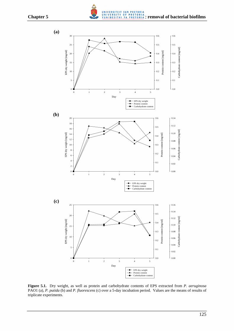
Chapter 5 Enzymatic removal of bacterial biofilms
Day
0 1 2 3 4 5
EPS
dry
wei
ght [
mg/
ml]
0
5
10
15
20
25
30
Prot
ein
cont
ent [
mg/
ml]
0.0
0.1
0.2
0.3
0.4
0.5
0.6
Car
bohy
drat
e co
nten
t [m
g/m
l]
0.0
0.1
0.2
0.3
0.4
0.5
0.6
EPS dry weight Protein contentCarbohydrate content
(a)
Day
0 1 2 3 4 5
EPS
dry
wei
ght [
mg/
ml]
0
2
4
6
8
10
12
14
16
18
20
Prot
ein
cont
ent [
mg/
ml]
0.0
0.1
0.2
0.3
0.4
0.5
0.6
Car
bohy
drat
e co
nten
t [m
g/m
l]
0.00
0.02
0.04
0.06
0.08
0.10
0.12
0.14
EPS dry weight Protein content Carbohydrate content
(b)
Day
0 1 2 3 4 5
EPS
dry
wei
ght [
mg/
ml]
0
5
10
15
20
25
Prot
ein
cont
ent [
mg/
ml]
0.0
0.1
0.2
0.3
0.4
0.5
0.6
Car
bohy
drat
e co
nten
t [m
g/m
l]
0.00
0.02
0.04
0.06
0.08
0.10
0.12
0.14
0.16
EPS dry weightProtein contentCarbohydrate content
(c)
Figure 5.1. Dry weight, as well as protein and carbohydrate contents of EPS extracted from P. aeruginosa PAO1 (a), P. putida (b) and P. fluorescens (c) over a 5-day incubation period. Values are the means of results of triplicate experiments.
125
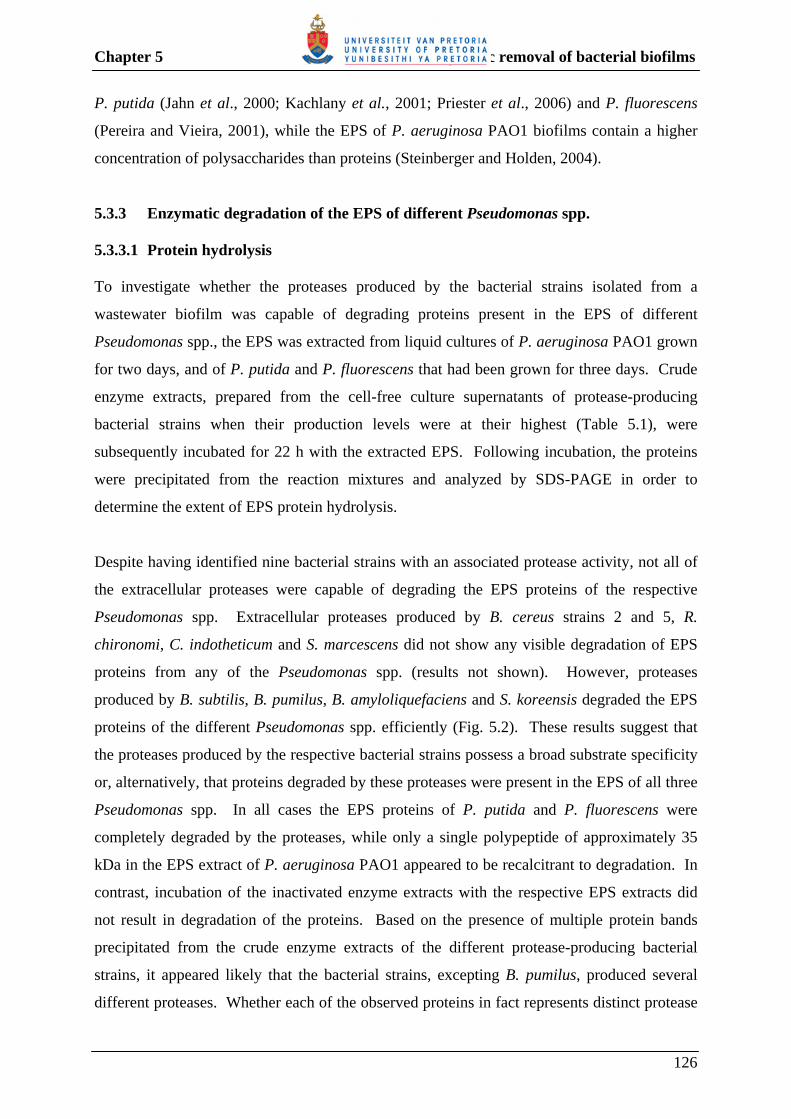
Chapter 5 Enzymatic removal of bacterial biofilms
P. putida (Jahn et al., 2000; Kachlany et al., 2001; Priester et al., 2006) and P. fluorescens
(Pereira and Vieira, 2001), while the EPS of P. aeruginosa PAO1 biofilms contain a higher
concentration of polysaccharides than proteins (Steinberger and Holden, 2004).
5.3.3 Enzymatic degradation of the EPS of different Pseudomonas spp. 5.3.3.1 Protein hydrolysis To investigate whether the proteases produced by the bacterial strains isolated from a
wastewater biofilm was capable of degrading proteins present in the EPS of different
Pseudomonas spp., the EPS was extracted from liquid cultures of P. aeruginosa PAO1 grown
for two days, and of P. putida and P. fluorescens that had been grown for three days. Crude
enzyme extracts, prepared from the cell-free culture supernatants of protease-producing
bacterial strains when their production levels were at their highest (Table 5.1), were
subsequently incubated for 22 h with the extracted EPS. Following incubation, the proteins
were precipitated from the reaction mixtures and analyzed by SDS-PAGE in order to
determine the extent of EPS protein hydrolysis.
Despite having identified nine bacterial strains with an associated protease activity, not all of
the extracellular proteases were capable of degrading the EPS proteins of the respective
Pseudomonas spp. Extracellular proteases produced by B. cereus strains 2 and 5, R.
chironomi, C. indotheticum and S. marcescens did not show any visible degradation of EPS
proteins from any of the Pseudomonas spp. (results not shown). However, proteases
produced by B. subtilis, B. pumilus, B. amyloliquefaciens and S. koreensis degraded the EPS
proteins of the different Pseudomonas spp. efficiently (Fig. 5.2). These results suggest that
the proteases produced by the respective bacterial strains possess a broad substrate specificity
or, alternatively, that proteins degraded by these proteases were present in the EPS of all three
Pseudomonas spp. In all cases the EPS proteins of P. putida and P. fluorescens were
completely degraded by the proteases, while only a single polypeptide of approximately 35
kDa in the EPS extract of P. aeruginosa PAO1 appeared to be recalcitrant to degradation. In
contrast, incubation of the inactivated enzyme extracts with the respective EPS extracts did
not result in degradation of the proteins. Based on the presence of multiple protein bands
precipitated from the crude enzyme extracts of the different protease-producing bacterial
strains, it appeared likely that the bacterial strains, excepting B. pumilus, produced several
different proteases. Whether each of the observed proteins in fact represents distinct protease
126
Chapter 5 Enzymatic removal of bacterial biofilms
Figu
re 5
.2.
SD
S-PA
GE
anal
yses
of
prec
ipita
ted
prot
eins
, fo
llow
ing
incu
batio
n of
EPS
ext
ract
ed f
rom
P.
aeru
gino
sa P
AO
1 (A
), P.
put
ida
(B)
and
P.
fluor
esce
ns (
C),
with
cru
de e
nzym
e ex
tract
s pr
epar
ed f
rom
the
bact
eria
l stra
ins
as in
dica
ted
in th
e fig
ure.
La
nes:
1,
Pro
tein
Mol
ecul
ar W
eigh
t Mar
ker;
2,
Unt
reat
ed E
PS e
xtra
ct; 3
, Pro
tein
s pr
ecip
itate
d fr
om c
ell-f
ree
cultu
re s
uper
nata
nts
of p
rote
ase-
prod
ucin
g ba
cter
ial s
train
s; 4
, Pro
tein
s pr
ecip
itate
d fr
om th
e re
actio
n m
ixtu
re fo
llow
ing
enzy
mat
ic tr
eatm
ent o
f the
EPS
ext
ract
for 2
2 h;
5, P
rote
ins
prec
ipita
ted
from
the
reac
tion
mix
ture
follo
win
g tre
atm
ent o
f the
EPS
ex
tract
with
a h
eat-i
nact
ivat
ed c
rude
enz
yme
extra
ct fo
r 22
h.
A
1 2
3
4
5
1
2
3
4 5
1 2
3
4
5
B
C
C
A
B
1
2
3
4
5
1
2
3
4
5
1
2
3
4
5
B. s
ubtil
is e
xtra
cellu
lar
prot
ease
s
B. p
umilu
s ext
race
llula
r pr
otea
ses
B. a
myl
oliq
uefa
cien
s ext
race
llula
r pr
otea
ses
S. k
oree
nsis
ext
race
llula
r pr
otea
ses
A
1
2
3
4 5
1
2
3
4
5
1
2
3
4
5
B
C
1
2
3
4
5
1
2
3
4
5
1
2
3
4
5
B
C
A
kDa
116.
0 66
.2
45.0
35
.0
25.0
18
.4
14.4
kD
a 11
6.0
66.2
45
.0
35.0
25.0
kDa
116.
0 66
.2
45.0
35
.0
25.0
18
.4
14.4
kD
a 11
6.0
66.2
45.0
35.0
25
.0
127
Chapter 5 Enzymatic removal of bacterial biofilms
species or whether some of these may represent unprocessed forms of other proteases must,
however, await further analysis and primary amino acid sequence comparisons.
5.3.3.2 Polysaccharide hydrolysis Using an approach similar to that described above, the effect of crude enzyme extracts,
prepared from polysaccharase-producing bacterial strains, on the amount of reducing sugar
released from the EPS of P. aeruginosa PAO1, P. putida and P. fluorescens was subsequently
examined. All of the polysaccharase-containing enzyme extracts were capable of degrading
the polysaccharides present in the EPS extract of the respective Pseudomonas spp., albeit with
different efficiencies (Fig. 5.3).
The results indicated that the amount of reducing sugar increased by more than 50% and 45%,
respectively, when the EPS extracts of all three Pseudomonas spp. were treated with crude
enzyme extracts prepared from B. subtilis (amylase, xylanase, cellulase and α-glucosidase
activities) and B. pumilus (xylanase, cellulase, α- and β-glucosidase activities). Indeed,
treatment of the EPS extracts of P. aeruginosa PAO1 and P. putida with an enzyme extract
prepared from B. subtilis resulted in the highest increase in the amount of reducing sugars
(53.12 and 62.31%, respectively) compared to the use of other enzyme extracts. For P.
fluorescens, the highest increase in the amount of reducing sugar (74.12%) was obtained
when the EPS extract was treated with an enzyme extract prepared from B. cereus strain 5
(amylase, cellulase and α-glucosidase activities). Notably, the amount of reducing sugar
increased by 59.78% when the EPS extract of P. putida was also treated with this crude
enzyme extract, albeit much more inefficient against the EPS extract of P. aeruginosa PAO1
(24.14%). The presence of cellulase activity in this enzyme extract, as also observed for
enzyme extracts derived from B. subtilis and B. pumilus, suggests that it may play an
important role in degrading the EPS of especially P. putida and P. fluorescens (Fig. 5.3).
This is also in accordance with the presence of cellulose in the EPS of these two
Pseudomonas spp. (Table 5.2).
Other differences in the efficacy of crude enzyme extracts to degrade the EPS polysaccharides
of the respective Pseudomonas spp. were also noted. Crude enzyme extracts prepared from R.
chironomi (amylase activity), B. amyloliquefaciens (amylase, xylanase, α- and β-glucosidase
activities), M. oxydans (α- and β-glucosidase activities) and A. oxydans (α-glucosidase
128
Chapter 5 Enzymatic removal of bacterial biofilms
activity) were able to degrade the EPS polysaccharides of both P. putida and P. fluorescens
more efficiently compared to that of P. aeruginosa PAO1. In contrast, a crude enzyme
extract from K. rosea (amylase and xylanase activities) showed a higher release of reducing
sugars from the EPS of P. aeruginosa PAO1 than from the other two Pseudomonas spp.
Crude enzyme extracts prepared from B. cereus strain 2 (amylase and α-glucosidase
activities) and C. indotheticum (α-glucosidase activity) were the most inefficient at degrading
the EPS polysaccharides of the respective Pseudomonas spp. (Fig. 5.3). The differences in
EPS polysaccharide hydrolysis, as highlighted above, may reflect on differences in the EPS
composition of the Pseudomonas spp. (Table 5.2).
5.3.4 Enzymatic removal of different Pseudomonas spp. biofilms The capability of selected crude enzyme extracts, singularly and in combination, to remove
biofilms of P. aeruginosa PAO1, P. putida and P. fluorescens from the surface of microtiter
plates was subsequently tested. In contrast to the use of viable plate count techniques, the
quantitative microtiter plate screening method adopted in this study is not only rapid and more
readily suited to the screening of a large number of anti-biofilm agents, but it also allows for
measurement of the removal of biofilms independently of the killing efficiency. Being able to
measure biofilm removal independently of killing is attractive, since it represents an
environmentally friendly approach that obviates the requirement for inherently toxic
disinfectants and biofilm removal is aimed directly at obtaining a clean (biofilm-free) surface,
which is the ultimate objective of a cleaning agent (Pitts et al., 2003).
Based on results obtained in the preceding sections, bacterial strains producing proteases,
which were capable of degrading the EPS proteins of the three Pseudomonas spp., were
included in the biofilm removal assays. Moreover, based on differences in the concentration
of polysaccharides compared to proteins in the EPS of the Pseudomonas spp., bacterial strains
that produced polysaccharide-degrading enzymes, resulting in increases of at least 20% (for
P. aeruginosa PAO1) and 50% (for both P. putida and P. fluorescens) in the amount of
reducing sugar released from the EPS, were also included in the biofilm removal assays.
129
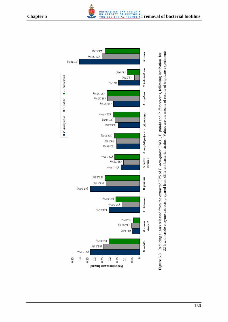
Chapter 5 Enzymatic removal of bacterial biofilms
Figu
re 5
.3.
Red
ucin
g su
gars
rele
ased
from
the
extra
cted
EPS
of P
. aer
ugin
osa
PAO
1, P
. put
ida
and
P. fl
uore
scen
s, fo
llow
ing
incu
batio
n fo
r 22
h w
ith c
rude
enz
yme
extra
cts p
repa
red
from
diff
eren
t bac
teria
l stra
ins.
Val
ues a
re th
e m
eans
of r
esul
ts o
f trip
licat
e ex
perim
ents
.
B. s
ubtil
is
(62.31%)
(53.12%)
(8.66%) B. c
ereu
s st
rain
2
(5.33%)
(31.45%)
R. c
hiro
nom
i(46.82%)
(45.60%)
B. p
umilu
s(50.65%)
(24.14%) B. c
ereu
sst
rain
5
(74.12%)
(22.66%)
B. a
myl
oliq
uefa
cien
s
(45.33%)
(13.63%)
M. o
xyda
ns(23.47%)
(19.57%)
A. o
xyda
ns
(32.37%)
(6.5%)
C. in
doth
etic
um(4.49%)
(27.36%) K. ro
sea
(22.41%)
0
0.050.
1
0.150.
2
0.250.
3
0.350.
4
0.45
Reducing sugars [mg/ml]
P. p
utid
a
P. a
erug
inos
aP.
fluo
resc
ens
(14.97%)
(50.86%)
(35.55%)
(48.41%)
(59.78%)
(39.74%)
(17.66%)
(38.20%)
(1.67%)
(22.34%)
130

Chapter 5 Enzymatic removal of bacterial biofilms
Table 5.2. Exopolysaccharides produced by P. aeruginosa PAO1, P. fluorescens and P. putida
Pseudomonas species
Exopolysaccharides produced
Composition Reference
P. aeruginosa PAO1
Alginate (acidic) -
Guluronate, mannuronate Glucose, rhamnose, mannose,
xylose, ketodeoxyoctulosonate (KDO), N-acetyl galactosamine, N-acetyl fucosamine, N-acetyl glucosamine
Schürks et al., 2002
Wozniak et al., 2003
P. fluorescens Alginate (acidic)a Marginalan (acidic)a
Levan (neutral)b
Novel EPS (acidic)a
- Cellulose
-
Guluronate, mannuronate Pyruvate, succinate, glucose,
galactose -
Pyruvate, acetate, rhamnose,
mannose, glucose D-glucose, 2-acetamido-2-
deoxy-D-gluocose, 4-O-acetyl-2-acetamido-2-deoxy-D-mannuronic acid
- Neutral monosaccharides:
Rhamnose, fucoes, ribose, arabinose, xylose, mannose, galactose, glucose Acidic groups: carboxylic acid
Fett et al., 1989 Fett et al., 1989
Fett et al., 1989
Fett et al., 1989
Osman et al., 1997
Spiers and Rainey,
2005 Hung et al., 2005
P. putida Alginate (acidic)a Marginalan (acidic)a
- Cellulose
Guluronate, mannuronate Pyruvate, succinate/ glucose,
galactose Glucose, N-acetylgalactosamine,
rhamnose, glucuronic acid, ribose
-
Fett et al., 1989 Fett et al., 1989
Kachlany et al.,
2001 Camesano and
Abu-Lail, 2002; Chang and Halverson, 2003
a Only one acidic exopolysaccharide can be produced by a single P. fluorescens and P. putida strain b Levan was only produced in strains that produced either alginate or the novel acidic EPS
131

Chapter 5 Enzymatic removal of bacterial biofilms
5.3.4.1 P. aeruginosa PAO1 biofilm removal Treatment of P. aeruginosa PAO1 biofilms with crude enzyme extracts prepared from
bacterial strains that produced proteases capable of degrading EPS proteins (B. subtilis, B.
pumilus, B. amyloliquefaciens and S. koreensis) resulted in a biofilm removal efficiency of
63-70% (Fig. 5.4). In contrast to the Bacillus spp., S. koreensis did not produce any of the
polysaccharide-degrading enzymes that were tested for in this study. Nevertheless, treatment
of P. aeruginosa PAO1 biofilms with a crude enzyme extract from S. koreensis resulted in
67% biofilm removal efficiency, suggesting that the proteases produced by this strain may be
particularly useful.
Results obtained for crude enzyme extracts, prepared from bacterial strains unable to degrade
P. aeruginosa PAO1 EPS proteins but producing enzymes capable of degrading the EPS
polysaccharides, indicated that enzyme extracts from R. chironomi and B. cereus strain 5 were
less efficient in removing the P. aeruginosa PAO1 biofilms (42% in both instances) compared
to that from K. rosea (82%). When crude enzyme extracts of either R. chironomi, B. cereus
strain 5 or K. rosea were combined with that of S. koreensis, the biofilm removal efficiencies
were much lower compared to treatment with the single enzyme extracts only. This may be
ascribed to the degradation of enzymes present in the extracts by the proteases produced by S.
koreensis. Similar observations have been noted by Johansen et al. (1997), who demonstrated
that some enzyme combinations do not work optimally together due to the inhibition of one
enzyme by another. Moreover, Orgaz et al. (2006) reported that the relative inefficiency of a
combination of proteases and polysaccharases to remove P. fluorescens biofilms was due to
the degradation of the other enzymes by the proteinases.
Analysis of the results obtained when mixtures of different crude enzyme extracts were used
to remove the P. aeruginosa PAO1 biofilms, indicated that maximal biofilm removal
efficiency was obtained when a crude enzyme extract of B. subtilis was combined with
enzyme extracts derived from either B. pumilus (89%) or B. cereus strain 5 (90%). When
mixtures of three different enzyme extracts were tested, the most efficient biofilm removal
was obtained with a mixture comprising enzyme extracts of B. pumilus, B. amyloliquefaciens
and K. rosea (98%). Significant biofilm removal was also obtained with a mixture of enzyme
extracts of B. pumilus, S. koreensis and K. rosea (90%), and of B. cereus strain 5, B.
amyloliquefaciens and S. koreensis (89%).
132

Chapter 5 Enzymatic removal of bacterial biofilms
0
20
40
60
80
100
1 3 4 5 6 16 21 1,3
1,4
1,5
1,6
1,16
1,21 3,4
3,5
3,6
3,16
3,21 4,5
4,6
4,16
4,21 5,6
5,16
5,21
6,16
6,21
16,2
1
1,3,
4
1,3,
5
1,3,
6
1,3,
16
Enzyme combinations
% B
iofil
m r
emov
al
0
20
40
60
80
100
1,3,
21
1,4,
5
1,4,
6
1,4,
16
1,4,
21
1,5,
6
1,5,
16
1,5,
21
1,6,
16
1,6,
21
1,16
,21
3,4,
5
3,4,
6
3,4,
16
3,4,
21
3,5,
6
3,5,
16
3,5,
21
3,6,
16
3,6,
21
3,16
,21
4,5,
6
4,5,
16
4,5,
21
4,6,
16
4,6,
21
4,16
,21
5,6,
16
5,6,
21
5,16
,21
6,16
,21
Enzyme combinations
% B
iofil
m r
emov
al
Key: 1 – B. subtilis 6 – B. amyloliquefaciens 3 – R. chironomi 16 – S. koreensis 4 – B. pumilus 21 – K. rosea 5 – B. cereus
Figure 5.4. Removal of P. aeruginosa PAO1 biofilm by treatment with enzyme extracts containing protease and/or polysaccharase enzyme activity. Biofilm removal was assayed by crystal violet staining of biofilm remaining in the wells of microtiter plates, following treatment with individual or combinations of crude enzyme extracts. In the figure, bars represent the mean values and error bars represent the standard deviations of three replicates of each treatment. 5.3.4.2 P. putida biofilm removal Treatment of P. putida biofilms with crude enzyme extracts prepared from bacterial strains
producing proteases capable of degrading the EPS proteins, indicated that proteases produced
by B. subtilis and B. pumilus were more efficient (95-98%) than enzyme extracts from B.
amyloliquefaciens and S. koreensis (55-56%) in removing the attached biofilms. In contrast
to the Bacillus spp., S. koreensis displayed none of the polysaccharide-degrading enzyme
activities tested for in this study. All three Bacillus spp. produced polysaccharide-degrading
enzymes, but B. amyloliquefaciens, in contrast to B. subtilis and B. pumilus, did not show
detectable cellulase activity. These results therefore suggest that both proteases and
polysaccharases, particularly cellulases, are required for efficient removal of P. putida
133

Chapter 5 Enzymatic removal of bacterial biofilms
biofilms. In this regard, not only does the genomic sequence of P. putida KT2440 contain a
putative operon for cellulose biosynthesis (Chang and Halverson, 2003), but
exopolysaccharides produced by P. putida KT2442 have been reported to comprise of
cellulose-like polymers (Camesano and Abu-Lail, 2002). Based on the high biofilm removal
efficiency of crude enzyme extracts prepared from B. subtilis and B. pumilus, it is therefore
not surprising that mixtures of enzyme extracts that included either of the two afore-
mentioned enzyme extracts were very efficient in removing P. putida biofilms. In all of these
cases the biofilm removal efficiency was in excess of 88%.
Treatment of P. putida biofilms with a crude enzyme extract prepared from B. cereus strain 5,
which displayed appreciable polysaccharase activity but did not produce proteases capable of
degrading the P. putida EPS proteins, yielded a biofilm removal efficiency of 33% (Fig. 5.5).
In accordance with their low biofilm removal efficiency, mixtures of crude enzyme extracts
derived from B. cereus strain 5 and B. amyloliquefaciens and/or S. koreensis were
comparatively inefficient in removing P. putida biofilms (38-59%).
0
20
40
60
80
100
120
1 4 5 6 16 1,4
1,5
1,6
1,16 4,5
4,6
4,16 5,6
5,16
6,16
1,4,
5
1,4,
6
1,4,
16
1,5,
6
1,5,
16
1,6,
16
4,5,
6
4,5,
16
4,6,
16
5,6,
16
Enzyme combinations
% B
iofil
m r
emov
al
Key: 1 – B. subtilis 6 – B. amyloliquefaciens 4 – B. pumilus 16 – S. koreensis 5 – B. cereus
Figure 5.5. Removal of P. putida biofilm by treatment with enzyme extracts containing protease and/or polysaccharase enzyme activity. Biofilm removal was assayed as described in the legend to Fig. 5.4. In the figure, bars represent the mean values and error bars represent the standard deviations of three replicates of each treatment. 5.3.4.3 P. fluorescens biofilm removal Similar to the results obtained for P. putida, crude enzyme extracts prepared from B. subtilis
and B. pumilus were the most efficient in removing P. fluorescens biofilms, yielding biofilm
134

Chapter 5 Enzymatic removal of bacterial biofilms
removal efficiencies of 100%. Crude enzyme extracts prepared from B. amyloliquefaciens
and S. koreensis was less efficient and yielded biofilm removal efficiencies of 68 and 60%,
respectively (Fig. 5.6). Since all of these bacterial strains produced proteases that were
capable of degrading P. fluorescens EPS proteins, these results support the use of a protease
to remove biofilms of this bacterium. A notable difference in the polysaccharide-degrading
enzyme activities of these bacterial strains is, however, the lack of detectable cellulase activity
in the crude enzyme extracts of B. amyloliquefaciens and S. koreensis. These results suggest
that cellulase activity, in addition to protease activity, is also important for removal of P.
fluorescens biofilms. In this regard, it has previously been reported that cellulose in the EPS
of P. fluorescens wrinkly spreader biofilms is involved in the development, strengthening and
integrity of the P. fluorescens biofilms (Spiers et al., 2003; Spiers and Rianey, 2005) The
importance of using both a protease and cellulase is furthermore supported by the observation
that B. cereus strain 5, which does not produce proteases capable of degrading the EPS
proteins of P. fluorescens but was the most proficient at degrading the EPS polysaccharides,
was comparatively inefficient in removing the biofilm (25%), despite the presence of cellulase
activity (Fig. 5.6). Excepting for a mixture of crude enzyme extracts derived from B. cereus
strain 5 and B. amyloliquefaciens, as well as combinations of these respective enzyme extracts
with that of S. koreensis, all other mixtures of the enzyme extracts yielded near 100% biofilm
removal efficiency.
0
20
40
60
80
100
120
1 4 5 6 16 1,4
1,5
1,6
1,16 4,5
4,6
4,16 5,6
5,16
6,16
1,4,
5
1,4,
6
1,4,
16
1,5,
6
1,5,
16
1,6,
16
4,5,
6
4,5,
16
4,6,
16
5,6,
16
Enzyme combinations
% B
iofil
m r
emov
al
Key: 1 – B. subtilis 6 – B. amyloliquefaciens 4 – B. pumilus 16 – S. koreensis 5 – B. cereus
Figure 5.6. Removal of P. fluorescens biofilm by treatment with enzyme extracts containing protease and/or polysaccharase enzyme activity. Biofilm removal was assayed as described in the legend to Fig. 5.4. In the figure, bars represent the mean values and error bars represent the standard deviations of three replicates of each treatment.
135

Chapter 5 Enzymatic removal of bacterial biofilms
5.3.5 Enzymatic removal of a multi-species biofilm Having established that crude enzyme extracts of selected wastewater biofilm bacteria were
capable of removing the biofilms of different Pseudomonas spp., it was next investigated
whether these enzyme extracts could also be used to remove multi-species biofilms. For this
purpose, multi-species biofilms were grown in six-well plates from dam water. Based on the
heterogeneity and complexity of the EPS of multi-species biofilms (Sutherland, 2001;
Allison, 2003), it has been recommended that mixtures of enzymes with different activities be
used in order to degrade a broader range of substrates (Meyer, 2003). Consequently, in this
part of the study, only mixtures of the enzyme extracts previously shown to remove mono-
species pseudomonad biofilms were used.
Of the 29 different mixtures of enzyme extracts tested, only 11 yielded biofilm removal
efficiencies in excess of 50%. The highest biofilm removal efficiencies were obtained with
enzyme extracts of B. subtilis and B. amyloliquefaciens combined with that of either K. rosea
(66%) or S. koreensis (62%), as well as with enzyme extracts of B. cereus strain 5 and S.
koreensis combined with that of either B. pumilus (61%) or B. subtilis (60%). Although the
EPS composition of the multi-species biofilm was not determined, the results nevertheless
suggest that the EPS comprised of constituents that served as substrates for one or more of the
different polysaccharases produced by the Bacillus spp. and K. rosea, in addition to the
proteases produced by the Bacillus spp. and S. koreensis (Table 5.1).
0
20
40
60
80
100
1,3
1,4
1,5
1,6
4,5
4,6
4,16
1,3,
4
1,3,
5
1,4,
6
1,4,
16
1,5,
6
1,5,
16
1,6,
16
1,6,
21
1,16
,21
3,4,
5
3,4,
6
3,4,
16
3,4,
21
4,5,
6
4,5,
16
4,5,
21
4,6,
16
4,6,
21
4,15
,16
4,16
,21
5,6,
16
5,6,
21
Enzyme combinations
% B
iofil
m r
emov
al
Key: 1 – B. subtilis 6 – B. amyloliquefaciens 3 – R. chironomi 15 – C. indotheticum 4 – B. pumilus 16 – S. koreensis 5 – B. cereus 21 – K. rosea
Figure 5.7. Removal of multi-species biofilm grown from dam water by treatment with combinations of enzyme extracts containing protease and/or polysaccharase enzyme activity. In the figure, bars represent the mean values and error bars represent the standard deviations of three replicates of each treatment.
136
Chapter 5 Enzymatic removal of bacterial biofilms
5.4 CONCLUSIONS Interest in the removal or degradation of bacterial biofilms has attracted great interest due to
the problems caused by biofilm formation in industrial and medical settings (Melo and Bott,
1997; Hall-Stoodley et al., 2004). One method that has been proposed for control of biofilms
is using enzymes to degrade bacterial EPS, which promote interaction with the surface
(Donlan and Costerton, 2002; Branda et al., 2005). Results obtained in this study indicated
that crude enzyme extracts containing protease and/or polysaccharase activity could remove
adhered biofilms of P. aeruginosa PAO1, P. putida and P. fluorescens. The polysaccharide-
degrading enzymes that showed biofilm removal potential, included xylanase, amylase,
cellulase, as well α- and β-glucosidases. These enzymes were capable of hydrolyzing the EPS
of the respective Pseudomonas spp., as was evidenced by an increase in production of
reducing sugar when the EPS was exposed to the crude enzyme extracts. The effectiveness of
the polysaccharases may be due to the presence of various different sugar molecules, e.g.,
glucose and xylose, in the EPS of the different Pseudomonas spp. (Frett et al., 1989; Wozniak
et al., 2003; Hung et al., 2005). Moreover, cellulase (produced by B. subtilis, B. pumilus and
B. cereus strain 5), in combination with proteases, may be particularly useful as a biofilm
detachment-promoting agent since the EPS of P. putida (Chang and Halverson, 2003) and P.
fluorescens (Spiers and Rainey, 2005) contains cellulose, while cellulase has also been
reported to inhibit biofilm formation by P. aeruginosa (Loiselle and Anderson, 2003).
The greater heterogeneity of multi-species biofilm matrix has been reported to limit the
potential of monocompound enzymes and consequently, the use of enzyme mixtures
containing different enzyme activities has been recommended for their elimination (Johansen
et al., 1997; Hahn Berg et al., 2001; Walker et al., 2007). In this study, lower biofilm
removal efficiency was obtained for the multi-species biofilms compared to the mono-species
pseudomonad biofilms, despite having used enzyme extracts effective in removing the
different Pseudomonas spp. biofilms. Nevertheless, it may be possible that the enzymes
identified in this study could be used in combination with other treatments to increase the
biofilm removal effectiveness or in combination with other enzymes to degrade the mixture of
proteins and polysaccharides present in the EPS of multi-species biofilms. Based on the
presence of nucleic acids and lipids in the EPS of biofilm bacteria (Sutherland, 2001; Tsuneda
et al., 2003), other enzymes such as nucleases and lipases can also be considered for use since
137

Chapter 5 Enzymatic removal of bacterial biofilms
previous studies have shown that DNaseI (Whitchurch et al., 2002) and lipase (Böckelmann et
al., 2003) can assist in biofilm removal.
Note should, however, be taken that crude enzyme extracts were used in this study to remove
the mono- and multi-species biofilms. Therefore, the possibility of biofilm removal not being
actually (or at least not singularly) caused by the enzyme activities assayed for in the study
can not be excluded. Consequently, future studies should aim to purify the individual
proteases and polysaccharide-degrading enzymes, and the purified enzymes should then be
tested individually or in combination for their biofilm removal capability, using a strategy
similar to that described in this study. In addition to measuring their biofilm removal
efficacy, viable cell counts could also be determined since it would be desirable if these
enzymes have bactericidal activity in order to prevent re-growth of the biofilms and/or
colonization of the surface by planktonic bacteria (Johansen et al., 1997).
5.5 REFERENCES
Alcalde, M., Plou, F.J., Andersen, C., Martin, M.T., Pedersen, S. and Ballesteros, A. 1999.
Chemical modification of lysine side chains of cyclodextrin glycosyltransferase from
Thermoanaerobacter causes a shift from cyclodextrin glycosyltransferase to α-amylase
specificity. FEBS Letters, 445: 333-337.
Allison, D.G. 2003. The biofilm matrix. Biofouling, 19: 139-150.
Anderl, J.N., Franklin, M.J. and Stewart, P.S. 2000. Role of antibiotic penetration limitation
in Klebsiella pneumoniae biofilm resistance to ampicillin and ciprofloxacin. Antimicrobial
Agents and Chemotherapy, 44: 1818-1824.
Arellano-Carbajal, F. and Olmos-Soto, J. 2002. Thermostable α-1,4- and α-1,6-glucosidase
enzymes from Bacillus sp. isolated from a marine environment. World Journal of
Microbiology and Biotechnology, 18: 791-795.
Böckelmann, U., Szewzyk, U. and Grohmann, E. 2003. A new enzymatic method for the
detachment of particle-associated soil bacteria. Journal of Microbiological Methods, 55: 201-
211.
Bradford, M.M. 1976. A rapid and sensitive method for the quantitation of microgram
quantities of protein utilizing the principle of protein-dye binding. Analytical Biochemistry,
72: 248-254.
Branda, S.S., Vik, A., Friedman, L. and Kolter, R. 2005. Biofilms: the matrix revisited.
Trends in Microbiology, 13: 20-26.
138

Chapter 5 Enzymatic removal of bacterial biofilms
Camesano, T.A. and Abu-Lail, N.I. 2002. Heterogeneity in bacterial surface polysaccharides,
probed on a singe-molecule basis. Biomacromolecules, 3: 661-667.
Chang, W-S. and Halverson, L.J. 2003. Reduced water availability influences the dynamics,
development, and ultrastructural properties of Pseudomonas putida biofilms. Journal of
Bacteriology, 185: 6199-6204.
Chen, X. and Stewart, P.S. 2000. Biofilm removal caused by chemical treatments. Water
Research, 34: 4229-4233.
Costerton, J.W., Lewandowski, Z., Caldwell, D.E., Korber, D.R. and Lappin-Scott, H.M.
1995. Microbial biofilms. Annual Reviews in Microbiology, 49: 711-745.
Donlan, R.M. and Costerton, J.W. 2002. Biofilms: Survival mechanisms of clinically
relevant microorganisms. Clinical Microbiology Reviews, 15: 167-193.
Fett, W.F., Osman, S.F. and Dunn, M.F. 1989. Characterization of exopolysaccharides
produced by plant-associated fluorescent Pseudomonads. Applied and Environmental
Microbiology, 55: 579-583.
Finelli, A., Gallant, C.V., Jarvi, K. and Burrows, L.L. 2003. Use of in-biofilm expression
technology to identify genes involved in Pseudomonas aeruginosa biofilm development.
Journal of Bacteriology, 185: 2700-2710.
Ghose, T.K. 1987. Measurement of cellulase activities. Pure and Applied Chemistry, 59:
257-268.
Gibson, H., Taylor, J.H., Hall, K.E. and Holah, J.T. 1999. Effectiveness of cleaning
techniques used in the food industry in terms of the removal of bacterial biofilms. Journal of
Applied Microbiology, 87: 41-48.
Gordon, C.A., Hodges, N.A. and Marriott, C. 1991. Use of slime dispersants to promote
antibiotic penetration through the extracellular polysaccharide of mucoid Pseudomonas
aeruginosa. Antimicrobial Agents and Chemotherapy, 35: 1258-1260.
Grobe, K., Zahler, J. and Stewart, P. 2002. Role of dose concentration in biocide efficacy
against Pseudomonas aeruginosa biofilms. Journal of Industrial Microbiology and
Biotechnology, 29: 10-15.
Hahn Berg, I.C., Kalfas, S., Malmsten, M. and Arnebrant, T. 2001. Proteolytic degradation
of oral biofilms in vitro and in vivo: Potential of proteases originating from Euphausia
superba for plaque control. European Journal of Oral Science, 109: 316-324.
Hall-Stoodley, L., Costerton, J.W. and Stoodley, P. 2004. Bacterial biofilms: From the
natural environment to infectious diseases. Nature Reviews Microbiology, 2: 95-108.
Hames, B.D. and Rickwood, D. 1990. Gel electrophoresis of proteins: A practical approach.
Second Edition. Oxford University Press, New York. pp. 245.
139

Chapter 5 Enzymatic removal of bacterial biofilms
Hung, C-C., Santschi, P.H. and Gillow, J.B. 2005. Isolation and characterization of
extracellular polysaccharides produced by Pseudomonas fluorescens biovar II. Carbohydrate
Polymers, 61: 141-147.
Jahn, A., Griebe, T. and Nielsen, P.H. 2000. Composition of Pseudomonas putida biofilms:
Accumulation of protein in the biofilm matrix. Biofouling 14: 49–57.
Johansen, C., Falholt, P. and Gram, L. 1997. Enzymatic removal and disinfection of bacterial
biofilms. Applied and Environmental Microbiology, 63: 3724-3728.
Kachlany, S.C., Levery, S.B., Kim, J.S., Reuhs, B.L., Lion, L.W. and Ghiorse, W.C. 2001.
Structure and carbohydrate analysis of the exopolysaccharide capsule of Pseudomonas putida
G7. Environmental Microbiology, 3: 774-784.
Kaplan, J.B., Ragunath, C., Velliyagounder, K., Fine, D.H. and Ramasubbu, N. 2004.
Enzymatic detachment of Staphylococcus epidermidis biofilms. Antimicrobial Agents and
Chemotherapy, 48: 2633-2636.
Karam, J. and Nicell, J.A. 1997. Potential application of enzymes in waste treatment.
Journal of Chemical Technology and Biotechnology, 69: 141-153.
Kostenko, V., Ceri, H. and Martinuzzi, R.J. 2007. Increased tolerance of Staphylococcus
aureus to vancomycin in viscous media. FEMS Immunology and Medical Microbiology, 51:
277-288.
Laemmli, U.K. 1970. Cleavage of structural proteins during the assembly of the head of
bacteriophage T4. Nature, 227: 680-685.
Loiselle, M. and Anderson, K.W. 2003. The use of cellulase in inhibiting biofilm formation
from organisms commonly found on medical implants. Biofouling, 19: 77-85.
Ludensky, M. 2003. Control and monitoring of biofilms in industrial applications.
International Biodeterioration and Biodegradation, 51: 255-263.
Melo, L.F. and Bott, T.R. 1997. Biofouling in water systems. Experimental Thermal and
Fluid Science, 14: 375-381.
Meyer, B. 2003. Approaches to prevention, removal and killing of biofilms. International
Biodeterioration and Biodegradation, 51: 249-253.
Norwood, D.E. and Gilmour, A. 2000. The growth and resistance to sodium hypochlorite of
Listeria monocytogenes in a steady-state multi-species biofilm. Journal of Applied
Microbiology, 88: 512-520.
O’Toole, G.A. and Kolter, R. 1998. Flagellar and twitching motility are necessary for
Pseudomonas aeruginosa biofilm development. Molecular Microbiology, 30: 295-304.
Olajuyigbe, F.M. and Ajele, J.O. 2005. Production dynamics of extracellular protease from
Bacillus species. African Journal of Biotechnology, 4: 776-779.
140

Chapter 5 Enzymatic removal of bacterial biofilms
Orgaz, B., Kives, J., Pedregosa, A.M., Monistrol, I.F., Laborda, F. and SanJose, C. 2006.
Bacterial biofilm removal using fungal enzymes. Enzyme and Microbial Technology, 40: 51-
56.
Orgaz, B., Neufeld, R.J. and SanJose, C. 2007. Single-step biofilm removal with delayed
release encapsulated Pronase mixed with soluble enzymes. Enzyme and Microbial
Technology, 40: 1045-1051.
Osman, S.F., Fett, W.F., Irwin, P., Cescutti, P., Brouillette, J.N. and Connor, J.V. 1997. The
structure of the exopolysaccharide of Pseudomonas fluorescens strain H13. Carbohydrate
Research, 300: 323-327.
Parmar, N., Singh, A. and Ward, O.P. 2001. Enzyme treatment to reduce solids and improve
settling of sewage sludge. Journal of Industrial Microbiology and Biotechnology, 26: 383-
386.
Pereira, M.O. and Vieira, M.J. 2001. Effects of the interactions between glutaraldehyde and
the polymeric matrix on the efficacy of the biocide against Pseudomonas fluorescens biofilms.
Biofouling, 17: 93-101.
Pitts, B., Hamilton, M.A., Zelver, N. and Stewart, P.S. 2003. A microtiter-plate screening
method for biofilm disinfection and removal. Journal of Microbiological Methods, 54: 269-
276.
Priester, J.H., Olson, S.G., Webb, S.M., Neu, M.P., Hersman, L.E. and Holden, P.A. 2006.
Enhanced exopolymer production and chromium stabilization in Pseudomonas putida
unsaturated biofilms. Applied and Environmental Microbiology, 72: 1988-1996.
Roe, J.H. 1954. The determination of sugar in blood and spinal fluid with anthrone reagent.
Journal of Biological Chemistry, 208: 889-896.
Schmid, A., Dordick, J.S., Hauer, B., Kiener, A., Wubbolts, M. and Witholt, B. 2001.
Industrial biocatalysis today and tomorrow. Nature, 409: 258-268.
Schürks, N., Wingender, J., Flemming, H-C. and Mayer, C. 2002. Monomer composition and
sequence of alginates from Pseudomonas aeruginosa. International Journal of Biological
Macromolecules, 30: 105-111.
Secades, P. and Guijarro, J.A. 1999. Purification and characterization of an extracellular
protease from the fish pathogen Yersinia ruckeri and effect of culture conditions on
production. Applied and Environmental Microbiology, 65: 3969-3975.
Spiers, A.J. and Rainey, P.B. 2005. The Pseudomonas fluorescens SBW25 wrinkly spreader
biofilm required attachment factor, cellulose fibre and LPS interactions to maintain strength
and integrity. Microbiology, 151: 2829-2839.
Spiers, A.J., Bohannon, J., Ghrig, S.M. and Rainey, P.B. 2003. Biofilm formation at the air-
liquid interface by the Pseudomonas fluorescens SBW25 wrinkly spreader requires an
acetylated form of cellulose. Molecular Microbiology, 50: 15-27.
141

Chapter 5 Enzymatic removal of bacterial biofilms
Steinberger, R.E. and Holden, P.A. 2004. Macromolecular composition of unsaturated
Pseudomonas aeruginosa biofilms with time and carbon source. Biofilms, 1: 37-47.
Stepanović, S., Vuković, D., Dakić, I., Svić, B. and Švabić-Vlahovi, M. 2000. A modified
microtiter-plate test for quantification of staphylococcal biofilm formation. Journal of
Microbiological Methods, 40: 175-179.
Sutherland, I.W. 2001. The biofilm matrix – an immobilized but dynamic microbial
environment. Trends in Microbiology, 9: 222-227.
Ten, L.N., Im, W-T., Kim, M-K. and Lee, S-T. 2005. A plate assay for simultaneous
screening of polysaccharide- and protein-degrading microorganisms. Letters in Applied
Microbiology, 40: 92-98.
Ten, L.N., Im, W-T., Kim, M-K., Kang, M.S. and Lee, S-T. 2004. Development of a plate
technique for screening of polysaccharide-degrading microorganisms by using a mixture of
insoluble chromogenic substrates. Journal of Microbiological Methods, 56: 375-382.
Tsuneda, S., Aikawa, H., Hayashi, H., Yuasa, A. and Hirata, A. 2003. Extracellular
polymeric substances responsible for bacterial adhesion onto solid surface. FEMS
Microbiology Letters, 223: 287-292.
Turakhia, M.H., Cooksey, K.E. and Characklis, W.G. 1983. Influence of a calcium-specific
chelant on biofilm removal. Applied and Environmental Microbiology, 46: 1236-1238.
Vallet, I., Olson, J.W., Lory, S., Lazdunski, A. and Filloux, A. 2001. The chaperone/usher
pathways of Pseudomonas aeruginosa: Identification of fimbrial gene clusters (cup) and their
involvement in biofilm formation. Proceedings of the National Academy of Sciences USA, 98:
6911-6916.
Walker, S.L., Fourgialakis, M., Cerezo, B. and Livens, S. 2007. Removal of microbial
biofilms from dispense equipment: The effect of enzymatic pre-digestion and detergent
treatment. Journal of the Institute of Brewing, 113: 61-66.
Wessel, D. and Flugge, U.I. 1984. A method for the quantitative recovery of protein in dilute
solution in the presence of detergents and lipids. Analytical Biochemistry, 138: 141-143.
Whitchurch, C.B., Tolker-Nielsen, T., Ragas, P.C. and Mattick, J.S. 2002. Extracellular
DNA required for bacterial biofilm formation. Science, 295: 1487-1487.
Wozniak, D.J., Wyckoff, T.J.O., Starkey, M., Keyser, R., Azadi, P., O’Toole, G.A. and
Parsek, M.R. 2003. Alginate is not a significant component of the extracellular
polysaccharide matrix of PA14 and PAO1 Pseudomonas aeruginosa biofilms. Proceedings of
the National Academy of Sciences USA, 100: 7907-7912.
Xavier, J.B., Picioreanu, C., Rani, S.A., van Loosdrecht, M.C.M. and Stewart, P.S. 2005.
Biofilm-control strategies based on enzymatic disruption of the extracellular polymeric
substance matrix – a modelling study. Microbiology, 151: 3817-3832.
142

Chapter 5 Enzymatic removal of bacterial biofilms
Zhang, X., Bishop, P.L. and Kinkle, B.K. 1999. Comparison of extraction methods for
quantifying extracellular polymers in biofilms. Water Science and Technology, 39: 211-218.
143
CHAPTER 6
GENERAL CONCLUSIONS
Wastewater biofilms serve as a habitat for diverse bacteria that encompass different
enzyme activities.
Crude enzyme extracts from wastewater biofilms were capable of degrading proteins and
polysaccharides present in the EPS of P. aeruginosa PAO1, and were capable of
removing biofilms of this bacterium from a glass surface. This could be ascribed to
presence of a wide variety of enzymes produced by the wastewater biofilms.
Proteases and polysaccharases produced by several bacterial strains isolated from the
wastewater biofilms showed degradation of proteins and polysaccharides present in the
EPS of P. aeruginosa PAO1, P. putida and P. fluorescens.
Crude enzyme extracts from individual bacterial strains and mixtures of enzyme extracts
from up to three different bacterial strains were capable of removing biofilms of P.
aeruginosa PAO1, P. putida and P. fluorescens. The presence of protein- and
polysaccharide-degrading enzymes in the crude enzyme extracts indicates that both
groups of enzymes are required for detachment of the biofilms.
Multi-species biofilms are more difficult to remove than single-species biofilms, possibly
due to the complex nature of their EPS.
144

![Capsular Polysaccharides Produced by the Bacterial ... · formation of biofilms [13]. Cell-surface polysaccharides have been shown to mediate the attachment of bacterial cells to](https://img.dokumen.tips/doc/110x75/5f20093a25e108007167d54a/capsular-polysaccharides-produced-by-the-bacterial-formation-of-biofilms-13.jpg)
![The composition of bacterial communities associated with … · [28,29]. Bacterial communities in marine biofilms are impaired by solar radiation [25,29]. Par-tial plastic photo-oxidation](https://img.dokumen.tips/doc/110x75/604603968ad23e38d63dc109/the-composition-of-bacterial-communities-associated-with-2829-bacterial-communities.jpg)